Embed Size (px)
Citation preview
FUNCTIONALLY GRADED PLGA-NANO APATITE-LAURIC ACID
BIOCOMPOSITE MEMBRANE FOR POTENTIAL
CLINICAL APPLICATIONS
JAMUNA THEVI A/P KALITHEERTHA THEVAR
A thesis submitted in fulfilment of the
requirements for the award of the degree of
Doctor of Philosophy (Biomedical Engineering)
Faculty of Biosciences and Medical Engineering
Universiti Teknologi Malaysia
MAY 2017
brought to you by COREView metadata, citation and similar papers at core.ac.uk
provided by Universiti Teknologi Malaysia Institutional Repository

iii
To my dearest mother………..
Mrs. Jannanayagam
For being a mentor, friend and pillar of strength
iv
ACKNOWLEDGEMENTS
I would like to express my sincere gratitude to my main supervisor, Prof.
Dato’ Ir. Dr. Mohammed Rafiq Abdul Kadir for his precious time, patience,
encouragement and expertise rendered throughout this project. His exquisite
attention to detail and demand for excellence has always encouraged me to widen my
research perspectives and writing of this thesis.
Besides my main supervisor, I sincerely appreciate and thank the efforts of
my co-supervisor, Dr. Nik Ahmad Nizam Nik Malek for his insightful comments and
encouragement for the writing of this thesis.
My sincere appreciation is also extended to Dr. Hendra Hermawan (currently
at Université Laval, Canada) who provided the opportunity to join his research team
at Universiti Teknologi Malaysia. Without his precious support and encouragement,
it would not be possible to conduct this research or to publish the related works.
Being the employee of SIRIM Berhad, I deeply appreciate the supports
provided by my employer to pursue an advanced degree, particularly for sponsoring
my studies continuously throughout the years. Also, an opportunity to broaden my
knowledge was made possible through the supports provided by Dr. Mohd Radzi
Mohd Toff (General Manager) and Dr. Kartini Noorsal (Head of Biomedical
Materials Section) who allowed me to use the research and laboratory facilities at
Advanced Materials Research Centre (AMREC), SIRIM Berhad for the purpose of
fulfilling PhD degree requirements.
I am also grateful to Prof. Dr. T.S Sampath Kumar (Indian Institute of
Technology Madras, India) for sharing his knowledge on fundamental science and
v
technical aspects of research instruments utilised in this project, during his visit to
AMREC, SIRIM Berhad.
I am greatly indebted to Ministry of Science, Technology and Innovation
(MOSTI), Malaysia for funding this research project through the Science Fund (03-
03-02-SF0280).
Last but not the least, my appreciation from the bottom of my heart is to my
family members, friends and colleagues who have invariably supported me all the
time despite it was a down fall or joyous moments while going through this
important stage of my life.
vi
ABSTRACT
Bone healing is a challenge in orthopaedics and dentistry. An occlusive membrane is used for the reconstruction of bone defects in guided bone regeneration (GBR) technique. Infection is the major cause for GBR membrane failure in which multiple antibiotics have been used to prevent bacterial colonisation in regenerative clinical practice. An anti-infective membrane with alternative antimicrobial agent to substitute antibiotics is paramount to overcome the incidence of bacterial resistance and side-effects. In this study, a composite membrane was developed by incorporating lauric acid (LA), a naturally derived antimicrobial substance. Poly(lactic-co-glycolic acid) (PLGA) based composite membrane was successfully fabricated using a combination of solvent casting-thermally induced phase separation (TIPS)-solvent leaching technique. The triple-layered membrane structure was attained via solvent casting of the composite solutions which then immediately phase separated by freezing at -18±1°C for 24 h. Then, the solvent in phase separated membrane was removed by immersing in precooled water at 3±1°C for 26 h, after which the membrane was air dried at 25°C for 3 days. The triple-layered construct of the PLGA composite membrane was developed with a gradient structure of LA and non-stoichiometric nanoapatite (NAp), to deliver the antimicrobial and osteconductive properties, respectively. The surface morphology and phase composition of the membrane were examined using scanning electron microscopy (SEM) and X-ray diffraction (XRD), respectively. The resulting graded membrane consisted of small pore size layer-1 containing 10wt% NAp + 1-3wt% LA, an intermediate labyrinth layer-2 with 20-50wt% NAp + 1wt% LA, and a large pore size layer-3 containing 30-100wt% NAp without LA. The existence of chemical interaction between PLGA, NAp and LA was identified using Fourier transform infrared spectrophotometry (FTIR) analysis. The synergistic effects of 10-30wt% NAp and 1wt% LA in dry membranes demonstrated higher tensile strength (0.61±0.17 MPa) and elastic modulus (23.15± 6.19 MPa). However, a more pliable behavior with a decrease in elastic modulus (12.50± 4.32MPa) was observed in 3wt% LA added membrane compared to the pure PLGA (20.17±2.21 MPa). The addition of LA resulted in a plasticizing effect at 3wt% due to weak intermolecular interactions in PLGA chains, caused by LA (-OH) and PLGA (C-O) bondings. These results were corroborated by the FTIR peak shift (1-3 cm-1) and glass transition temperature (Tg) reduction as detected using differential scanning calorimeter (DSC). The composite membrane retained its structural integrity with only 22% weight loss after incubation for 24 weeks in phosphate buffered saline (PBS), which indicates its potential use as a physical barrier. The 1-3wt% LA loaded composite membranes had good cell viability toward mouse fibroblasts and showed increased bacterial reduction with increased LA loadings against S. aureus. These results demonstrate the potential of LA loaded biocomposite membrane to provide anti-infective surfaces, useful in clinical applications.
vii
ABSTRAK
Penyembuhan tulang adalah satu cabaran dalam bidang ortopedik dan pergigian. Pertumbuhan semula tulang berpandukan (GBR) telah digunakan untuk pembinaan semula kecacatan tulang dengan menggunakan membran penghalang. Jangkitan adalah punca utama kegagalan membran tersebut di mana beberapa antibiotic telah digunakan untuk menghalang pertumbuhan bacteria dalam amalan klinikal. Agen antibakteria alternatif adalah perlu untuk mengatasi kesan sampingan dan rintangan bakteria yang dihasilkan oleh antibiotik. Dalam kajian ini, membran komposit telah dibangunkan melalui penggabungan asid laurik (LA) yang mempunyai sifat antibakteria. Membran komposit berasaskan asid poli(laktik-co-glycolic) (PLGA) telah berjaya direka dengan menggunakan gabungan teknik-teknik pelarut tuangan-pemisahan fasa haba teraruh-larut lesap pelarut. Struktur membran tiga-lapis telah dihasilkan melalui pelarut tuangan komposit yang telah melalui pemisahan fasa haba teraruh pada suhu -18±1°C selama 24 jam. Kemudian, pelarut membran telah dibuang dengan merendamkannya dalam air sejuk pada suhu 3±1°C selama 26 jam. Setelah itu, membran telah dikeringkan di udara pada 25°C selama 3 hari. Membran komposit PLGA tiga-lapis ini telah difabrikasi dengan struktur kecerunan melalui penambahan LA dan apatitnano bukan stoikiometrik (NAp) yang memainkan peranan sebagai antimikrob dan penggalak pertumbuhan tulang. Morfologi permukaan dan fasa komposisi membran telah diperiksa dengan menggunakan mikroskopi elektron imbasan (SEM) dan pembelauan sinar-X (XRD). Membran ini terdiri daripada lapisan-1 dengan saiz liang kecil yang mengandungi 10% berat NAp + 1-3% berat LA, lapisan-2 sebagai lapisan perantaraan dengan 20-50% berat NAp + 1% berat LA dan akhirnya lapisan-3 dengan saiz liang besar yang mengandungi 30-100% berat NAp tanpa LA. Kewujudan interaksi kimia antara PLGA, NAp dan LA telah dikenalpasti dengan menggunakan analisis spektrometer inframerah (FTIR). Kesan sinergi diantara 10-30% berat NAp dan 1% berat LA dalam membran komposit kering menunjukkan kekuatan tegangan (0.61± 0.17 MPa) dan modulus elastik (23.15±6.19 MPa) yang tinggi manakala membran mudah bentuk diperolehi dengan penurunan dalam modulus elastik (12.50±4.32 MPa) selepas penambahan 3% berat LA berbanding membran PLGA tulen (20.17±2.21 MPa). Penambahan 3% berat LA mengakibatkan kesan liat disebabkan interaksi lemah dalam rantaian PLGA melalui ikatan LA (-OH) dan PLGA (-CO). Ini telah dibuktikan melalui perubahan puncak FTIR (1-3 cm-1), dan juga penurunan suhu peralihan kaca (Tg) yang dikesan melalui kalorimeter pengimbas kebedaan (DSC). Membran komposit mengekalkan struktur integriti dengan penurunan berat sebanyak 22% selepas rendaman selama 24 minggu di dalam PBS dimana ianya mempunyai potensi sebagai penghalang fizikal. Membran komposit yang mengandungi 1-3% berat LA menunjukkan pertumbuhan sel-sel fibroblas tikus dan juga pengurangan bacteria S. aureus dengan peningkatan kandungan LA. Keputusan ini menunjukkan potensi membran komposit yang mengandungi LA sebagai membran anti-jangkitan untuk kegunaan dalam aplikasi klinikal.
viii
TABLE OF CONTENTS
CHAPTER TITLE PAGE
DECLARATION ii
DEDICATION iii
ACKNOWLEDGEMENTS iv
ABSTRACT vi
ABSTRAK vii
TABLE OF CONTENTS viii
LIST OF TABLES xvii
LIST OF FIGURES xx
LIST OF SYMBOLS xxxiii
LIST OF ABBREVIATIONS xxxiv
LIST OF APPENDICES xxxvii
1 INTRODUCTION 1
1.1 Background 1
1.2 Problem statements 4
1.3 Objectives of the study 5
1.4 Research hypothesis 6
1.5 Scope of the study 7
1.6 Significance of the study 8
1.7 Thesis outline 8
2 LITERATURE REVIEW 12
2.1 Introduction 12
2.2 Bone damage and tissue reconstruction 13
ix
2.3 Alveolar bone loss and treatment modalities 14
2.4 Principles of guided bone regeneration 15
2.5 Design criteria for GBR membrane 16
2.5.1 Space-making properties 17
2.5.2 Cell-occlusiveness 17
2.5.3 Biocompatibility 18
2.5.4 Tissue integration 18
2.5.5 Clinical manageability 18
2.6 Commercial GBR barrier membranes for clinical
applications 18
2.6.1 Non-resorbable membranes 19
2.6.2 Natural bioresorbable membranes 21
2.6.3 Synthetic bioresorbable membranes 22
2.7 Interface tissue specific functional surface
layers in barrier membranes 24
2.7.1 Bioresorbable polymer-calcium phosphate
composites as GBR membranes 24
2.7.1.1 Short chain saturated aliphatic
polyesters 27
2.7.1.2 Multiple ions substituted
non-stoichiometric nanoapatite 29
2.7.2 Antibiotics loaded GBR barrier
membranes 32
2.7.2.1 Systemic versus local antibiotic
treatment 34
2.7.2.2 Lauric acid as a potential antimicrobial
agent for clinical use 36
2.8 Functionally graded and layered membranes
for guided bone regeneration 38
2.8.1 Techniques for polymeric composite
barrier membrane fabrication 40
2.8.1.1 Solvent casting 41
2.8.1.2 Thermally induced phase separation 42
2.8.1.3 Solvent leaching 43
x
2.9 In vitro degradation characteristics of PLGA
based membranes 43
2.10 The drug release mechanism in PLGA based
membranes 45
2.11 Antimicrobial efficacy studies on antibiotic
loaded GBR membranes 48
2.12 Biocompatibility assessment 48
2.13 Challenges in guided bone
regeneration using barrier membrane 49
3 RESEARCH METHODOLOGY 51
3.1 Introduction 51
3.2 Synthesis and characterisation of multiple ions
substituted non-stoichiometric nanoapatite (NAp) 53
3.2.1 Materials for synthesis of NAp powder 54
3.2.2 Synthesis of nanoapatite powder: Effects of
temperature, concentration and multiple
ions substitution 54
3.2.3 Physico-chemical characterisation
of the synthesised powders 59
3.2.3.1 Qualitative and quantitative analysis
using X-ray diffraction (XRD) 59
3.2.3.2 Functional group characterisation
using Fourier transform
infrared spectrophotometry (FTIR) 61
3.2.3.3 Elemental analysis using
Inductively Coupled Plasma – Atomic
Emission Spectroscopy (ICP-AES) 62
3.2.3.4 Carbon, Hydrogen, Nitrogen elemental
analysis 61
3.2.3.5 Image analysis by Field Emission
Scanning Electron Microscopy (FESEM) 62
3.2.3.6 Image analysis by Transmission Electron
Microscopy (TEM) 62
xi
3.2.3.7 Thermal analysis using
thermogravimetric and differential thermal
analyser (TGA-DTA) 63
3.2.3.8 Measurement of specific surface area
by Brunauner-Emmett-Teller (BET)
gas adsorption method 63
3.2.3.9 Particle size analysis 63
3.2.4 In vitro cytotoxicity assay on synthesized
NHA and NAp powders 64
3.2.4.1 Materials for in vitro cytotoxicity
evaluation 64
3.2.4.2 Preparation of complete medium 65
3.2.4.3 Initiating cryopreserved cells 65
3.2.4.4 Subculturing adherent monolayer cells
from 25 cm2 to 75 cm2 flask 66
3.2.4.5 Split suspension 67
3.2.4.6 Determining cell number with
a hemocytometer and trypan blue
staining 67
3.2.4.7 Preparation of powder sample extracts 68
3.2.4.8 Alamar Blue assay 68
3.3 Development of triple layered composite membrane
graded with LA and NAp particles in PLGA matrix 70
3.3.1 Materials for fabrication of composite
membrane 70
3.3.2 A preliminary study on fabrication and
characterisation of triple-layered and graded
composite membranes using solvent casting – thermally
induced phase separation (TIPS) – solvent
leaching techniques 71
3.3.2.1 Identification of LA and NAp
presence on fabricated composite
membranes 74
xii
3.3.2.2 Reproducibility of membrane fabrication
using solvent casting – TIPS – solvent
leaching techniques 75
3.3.2.3 Morphological and chemical characterisation
of fabricated membranes using SEM and
Energy Dispersive X-ray Spectroscopy
(EDS) 76
3.3.2.4 Characterisation of LA in
composite membranes using FTIR 76
3.3.3 Fabrication of optimised composite membranes
with various PLGA, NAp and LA contents
for physical, mechanical and biological
assessments 77
3.3.4 Physico-chemical and mechanical
evaluation of the fabricated composite
membranes 83
3.3.4.1 Morphological characterisation using
VPSEM 83
3.3.4.2 Phase analysis using XRD 83
3.3.4.3 Interpretation of functional groups in
composite membranes using FTIR 84
3.3.4.4 Differential scanning calorimetric (DSC)
studies 84
3.3.4.5 Dry and wet mechanical strength
evaluation 85
3.4 In vitro hydrolytic degradation and lauric
acid release profiles of composite PLGA membranes 87
3.4.1 Materials for in vitro degradation and LA release
studies 87
3.4.2 In vitro degradation of composite PLGA
and pure PLGA membranes 87
3.4.3 In vitro lauric acid release studies of
composite PLGA membranes 90
3.4.3.1 Extraction of LA from degradation
xiii
medium 91
3.4.3.2 Loading efficiency of LA in composite
PLGA membranes 92
3.4.3.3 Derivatization of extracted lauric acid 93
3.4.3.4 Quantification of LA using Reversed
Phase High-Performance Liquid
Chromatography (HPLC) 94
3.4.4 Mathematical modeling to determine LA
release mechanism 95
3.4.4.1 Higuchi model 96
3.4.4.2 Ritger-Peppas model 96
3.4.4.3 First order kinetic model 97
3.4.4.4 Zero order kinetic model 97
3.5 Quantitative in vitro antimicrobial efficacy assay
on LA loaded composite membranes 98
3.5.1 Materials for in vitro antimicrobial efficacy assay 98
3.5.2 Bacterial culture and maintenance 98
3.5.3 Determination of mid-log exponential growth
phase of bacteria 99
3.5.2 Antimicrobial efficacy assay 99
3.6 In vitro cytotoxicity assay on fabricated membranes 101
3.6.1 Materials for in vitro cytotoxicity assay 101
3.6.2 Preparation of complete medium 102
3.6.3 Cell culture and maintenance 102
3.6.4 Preparation of membrane sample extracts 103
3.6.5 Alamar Blue Assay 103
4 RESULTS AND DISCUSSION 105
4.1 Introduction 105
4.2 Phase evaluation, physical and chemical characteristics,
elemental analysis, morphology and biological
evaluation of the synthesised as-prepared NHA
and NAp powders 106
4.2.1 Phase composition, crystallite size, crystallinity
xiv
and lattice parameters evaluation using XRD 107
4.2.2 Morphological evaluation using TEM 112
4.2.3 Physical and chemical characterisation
using TGA-DTA analysis 114
4.2.4 Elemental analysis 117
4.2.5 Chemical and structural characterisation 121
4.2.6 Thermal stability 124
4.2.7 Cytotoxicity assay on as-prepared apatite
powders 130
4.2.8 Summary of findings on synthesis and
characterization of nanoapatite powders 132
4.3 Development and characterisation of triple-layered
and graded composite PLGA membranes 133
4.3.1 A preliminary study on the design, processing
conditions and effects of NAp and LA in
PLGA membranes 134
4.3.1.1 The design and processing conditions
of triple-layered and graded composite
PLGA membranes using solvent
casting-TIPS-solvent leaching
technique 134
4.3.1.2 Morphological characterisation on the
effects of NAp and LA addition on
the formation of composite PLGA
membranes 137
4.3.1.3 Identification of LA and NAp on
PLGA matrices 147
4.3.1.4 Reproducible fabrication and
characterisation of triple layered
PLGA composite membranes with graded
composition 151
4.3.2 Fabrication and, morphological,
chemical and structural characterisation
of the optimised composite membrane 157
xv
4.3.2.1 Phase composition of membrane
Structure 169
4.3.2.2 Chemical characteristics of
PLGA-NAp-LA components in
composite membrane 173
4.3.2.3 Thermal transition of phases present
in composite membrane 180
4.4. Mechanical evaluation of composite membranes
in dry and wet state 184
4.4.1 Mechanical properties of composite
membranes in dry state 184
4.4.2 Mechanical properties of composite
membranes in wet state 190
4.5. In vitro degradation of triple-layered and graded
composite membranes containing NAp and LA in
PLGA matrices 194
4.5.1 Weight loss in composite membranes 195
4.5.2 Water uptake in composite membranes 210
4.6. In vitro quantification of loading yield, delivery
profile and release mechanism of LA 216
4.6.1 Quantification of LA loading yield in
composite membranes 216
4.6.2 The in vitro release profile of LA in
simulated physiological solution 219
4.6.3 The in vitro release mechanism of LA in
simulated physiological solution 221
4.7 In vitro antimicrobial efficacy assay on LA
loaded composite membranes 224
4.7.1 Growth curves to determine mid-log
exponential phase of bacteria 225
4.7.2 In vitro antimicrobial efficacy assay 226
4.8 Cytotoxicity assay on NAp and LA added PLGA
composite membranes 233
4.9 Summary of overall findings 236
xvi
5 CONCLUSION AND FUTURE RECOMMENDATIONS 238
5.1 Conclusion 238
5.2 Limitations and Future recommendations 239
REFERENCES 242
Appendices A-I 265-310
xvii
LIST OF TABLES
TABLE NO. TITLE PAGE 2.1 List of commercially available non-resorbable GBR
barrier membranes. 20
2.2 List of commercially available synthetic bioresorbable
membranes. 23
2.3 Drawbacks of antibiotics in systemic administration and
localised release systems. 35
3.1 Molar concentration of precursors used in the synthesis. 55
3.2 Freezing time for the formation of layered membranes.
Composition of PLGA, NAp and LA varied in layer 1 (L1),
layer 2 (L2) and layer 3 (L3) of the membranes. 72
3.3 Composition of NAp and LA added PLGA membranes used in
reproducibility studies. 75
3.4 Varied composition of PLGA, NAp and LA in layer 1 (L1),
layer 2 (L2) and layer 3 (L3) of the optimised membranes. 78
3.5 The composition of membrane samples tested for
mechanical strength in dry and wet conditions. 86
3.6 Composition of membranes used for in vitro degradation
test and lauric acid release studies. 88
3.7 Types of membranes used for extraction of LA and as
control. 91
3.8 Membranes with various LA contents for loading
efficiency studies. 92
3.9 The HPLC gradient elution profile using acetonitrile
and water based mobile phase to quantify LA release in
buffer medium. 95
xviii
4.1 Effect of various synthesis parameters on crystallite size,
crystallinity, specific surface area and lattice parameters
of as-prepared stoichiometric HA (NHA) and ions
substituted apatite (NApF1 & NApF2) powders. 108
4.2 Molar concentrations of the precursors used in the
synthesis of nanoapatite compared to actual molar
concentrations obtained in the as-prepared powders
by varying the (Ca & P) equimolar reactant
concentrations and synthesis temperature. Reaction I
involves synthesis at 37±2 °C whereas in reaction II, NAp
initially synthesised at 37±2 °C which then increased to 85±2 °C
until completion of synthesis reaction. 118
4.3 Molar concentrations of the precursors used in the
synthesis of nanoapatite compared to actual molar
concentrations obtained in the as-prepared powders by
reducing the substituents concentration (less than Table 4.2)
and using 1.5M (Ca & P) reactant concentrations. 119
4.4 Repeated batches of NApF2 powders synthesized using
1.5 M (Ca & P) reactant concentrations indicating
comparable reproducibility. 121
4.5 Average temperature changes during layering, TIPS
and solvent leaching steps within 24 h of each
membrane fabrication. 136
4.6 The photograph images of L1 and L2 surfaces of fabricated
membranes containing various amounts of PLGA, NAp
and LA. 139
4.7 The photograph images of fabricated membranes
containing various amounts of NAp and LA in PLGA.
PLGA was increased in L1 of membranes to produce less
porous structure. 145
4.8 SEM micrographs and processing conditions of reproducible
pure PLGA membranes. 153
xix
4.9 SEM micrographs and processing conditions of reproducible
10 – 30 wt% of NAp + 1 wt% of LA added PLGA composite
membranes. 154
4.10 SEM micrographs and processing conditions of reproducible
10 – 30 wt% of NAp + 2 wt% of LA added PLGA composite
membranes. 155
4.11 SEM micrographs and processing conditions of reproducible
10 – 30 wt% of NAp + 3 wt% of LA added PLGA composite
membranes. 156
4.12 Glass transition temperature of pure PLGA and composite
membranes fabricated using various NAp and LA contents in
PLGA matrices. 181
4.13 Mechanical properties of triple-layered membranes in dry
and wet state. Data are presented as mean ± SD, n=6. 186
4.14 Kinetic parameters of LA release from composite membrane
using various mathematical modeling. 223
xx
LIST OF FIGURES
FIGURE NO. TITLE PAGE
1.1 LA incorporation into barrier membrane as an antimicrobial
agent for adjunct treatment in GBR procedures to inhibit
bacterial infection. 7
1.2 Representation of thesis outline. 11
2.1 (a) An adequate bone volume (height and width) is
a prerequisite for successful implant treatment.
(b) Barrier membrane and bone graft as bone substitute
materials are placed to accelerate bone formation.
(c) Development of final prosthesis after the
formation of new bone. 15
2.2 The principle of guided bone regeneration using barrier
membranes to mechanically occlude soft tissue invasion
and to retain blood clot in a secluded space. Bone growth
occurs through bone cells migration from the surrounding
original bone. 16
2.3 Structure of lactic acid, glycolic acid and
poly (lactic-co-glycolic acid) (PLGA). 28
2.4 Structure of lauric acid. 37
2.5 Functionally graded and layered GBR barrier membrane. 40
3.1 The schematic illustration of LA and NAp incorporation
into PLGA, forming composite membranes by combined
techniques of solvent casting-TIPS-solvent leaching. 52
3.2 Process flow of experimental methods involved in the
fabrication and evaluation of composite membranes. 53
3.3 Differences between synthesis reaction method I and II.
In reaction method I (a), the synthesis process was carried
xxi
out and maintained at a lower temperature until the reaction
is completed. In reaction method II (b), initially the complete
ions addition was conducted at a lower temperature and
subsequently maintained at an elevated temperature until
the synthesis process is completed. 56
3.4 Process flow for the synthesis of multiple ions substituted
non-stoichiometric nanoapatite (NAp). (a)-(c) The PO43-, Na+
and CO32- ionic solutions were added slowly into the basic
suspension containing Ca2+, Mg2+ and K+ precursors while
vigorously stirring and heating it, (d) the precipitation product
was filtered and washed using DDI water, (e) the dried cake
was crushed, (f) crushed cake was ground to fine powders
and (g) sieved through 500 µm and 20-50 µm mesh size. 58
3.5 The refrigerator thermometer placed in the freezing
compartment to measure the temperature changes while
placing composite solutions to induce phase separation. 73
3.6 Membrane fabrication steps via solvent casting and TIPS.
(a-d) Weighed PLGA, LA and NAp separately for L1, L2
and L3, (e) mixed PLGA, LA and NAp in a vial,
(f-g) sonicated composite solution, (h) solvent casting,
and (i) cast solvent subjected to TIPS. 81
3.7 Washing steps for phase separated composite membranes.
(a) Frozen and phase separated solution placed in cool water,
(b-c) zoomed view of step (a) showing placement of petri dish
(containing frozen composite solution) in a glass beaker filled
with cool water, (d) membrane separated from petri dish and
placed in a fresh cool water, (e) zoomed view of step (d) and
finally room air dried composite membrane showing (f) L3
and (g) L1. 82
4.1 Triple layered composite PLGA membrane containing LA
and NAp promoting bone cells growth while preventing
bacteria and fibroblast cells. 106
4.2 XRD patterns of the as-prepared ions substituted apatite powder
(NApF2-1.5M) synthesised at 37±2 °C (reaction method I) and
xxii
37 and 85±2 °C (reaction method II). 107
4.3 XRD patterns of as-prepared ions substituted apatite (NApF2)
synthesised at 37 and 85±2 °C (reaction method II) using
1.0M, 1.5M and 2.0M equimolar precursors. 110
4.4 XRD patterns of as-prepared stoichiometric HA (NHA)
and ions substituted apatite (NApF1 & NApF2) powders
synthesised at 37 and 85±2 °C (reaction method II) using
1.5M equimolar precursors. 111
4.5 TEM micrographs of as prepared HA synthesised at 37±2 °C
using reactant concentration (a) 1.0M, (b) 1.5M and (c-d) 2.0M
at low magnification (a,b,c) and high magnification (d),
respectively. 112
4.6 TEM micrographs of as-prepared NApF2 synthesised at
37 & 85±2 °C using reactant concentration 1.5 M at low
magnification on three different spots (a)-(c) and
high magnification (d), respectively. 113
4.7 TGA and DTA curves of the stoichiometric HA (NHA)
powders synthesised at 37±2 °C using 1.0 M, 1.5 M and
2.0 M reactants and dried at 80 °C. 115
4.8 TGA and DTA curves of NHA, NApF1 and NApF2 powders
initially synthesised at 37 ±2 °C and then increased to
85 ±2 °C (reaction II) using 1.5M reactants and dried
at 80 °C. The samples denoted as 37 and 85 ± 2 °C. 117
4.9 FTIR spectra of as prepared stoichiometric HA (NHA)
and ionic substituted apatite (NApF1 & NApF2) powders
synthesised at 37 & 85±2 °C (reaction method II) using 1.5 M
equimolar precursors. 122
4.10 SEM micrograph for the morphology of (a) stoichiometric
HA (NHA); ions substituted nanoapatite (b) NApF1 and
(c) NApF2 powders synthesised at 37 and 85±2 °C using
1.5 M reactant concentration (30,000× magnification). 124
4.11 XRD patterns of stoichiometric HA (NHA) and ionic
substituted apatite (NApF1 & NApF2) powders synthesised
at 37 & 85±2 °C (reaction method II) using 1.5M equimolar
xxiii
precursors after heat-treatment at 900 °C in CO2 controlled
atmosphere. 125
4.12 XRD patterns of stoichiometric HA (NHA) and ionic
substituted apatite (NApF1 & NApF2) powders synthesised
at 37 & 85±2 °C (reaction method II) using 1.5M equimolar
precursors after sintering at 1250 °C in air. 126
4.13 FTIR spectra of stoichiometric HA (NHA) and ionic
substituted apatite (NApF1 & NApF2) powders synthesised
at 37 & 85± 2 °C (reaction method II) using 1.5 M equimolar
precursors after heat-treatment at 900 °C in CO2 controlled
atmosphere. 128
4.14 FTIR spectra of stoichiometric HA (NHA) and ionic
substituted apatite (NApF2) powders synthesised at
37 & 85±2 °C (reaction method II) using 1.5M equimolar
precursors after sintering at 1250 °C in air. 130
4.15 Representative 24-well plates for qualitative AB colour
changes. Colour change from blue to red indicates
presence of live cells whereas unchanged blue colour
indicates presence of dead cells after treatment with extracts.
(a) NHA and (b) NApF2 powder extracts, and
(c) positive control (phenol solution extracts). 131
4.16 Cytotoxicity assay results on L929 mouse fibroblast
cells viability in response to different extract concentrations
of NHA and NApF2 powder extracts. The data are presented
as means ± SEM values of two independent
experiments (n = 2). 132
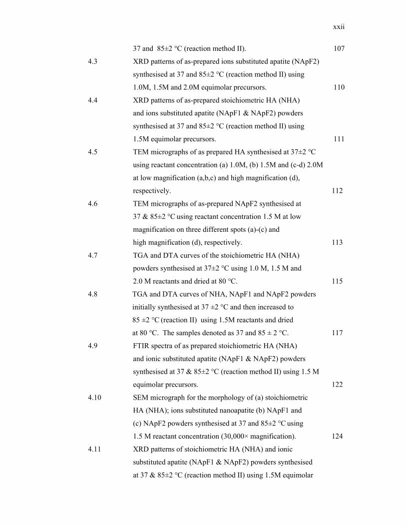
4.17 SEM micrographs of L1 of composite membranes containing
10-30wt% NAp + 1wt% LA and 10-100wt% NAp + 1wt% LA
graded in (a) 7, (b) 9, (c) 11 and (d) 13wt% of PLGA matrices.
L1 of pure PLGA membranes were compared as control. (e) The
representative EDS spectrum of the composite membranes
taken on L1. 141
xxiv
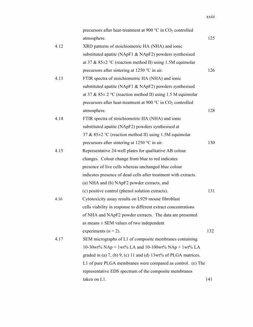
4.18 SEM micrographs of L3 of 10-30wt% NAp + 1wt% LA
and 10-100wt% NAp + 1wt% LA graded membranes in
(a) 7, (b) 9, (c) 11 and (d) 13wt% of PLGA matrices. L3
of pure PLGA membranes were compared as control. (e) The
representative EDS spectrum of the composite membranes
taken on L3. 142
4.19 SEM micrographs of L1 of (a) 15, (b) 17, (c) 20 and
(d) 23wt% PLGA membranes. 144
4.20 Photographs of (a) L1 and (b) L3 of PLGA membranes
with graded composition of 10-30 wt% NAp + 3 wt%
LA and the representative SEM micrographs for
(c) L1, (d) L3 and (e) cross section of membranes. 147
4.21 (A) Representative SEM micrographs of PLGA membranes
(a)-(c) and 3 wt% of LA added composite PLGA membranes
(d)-(f) on L1 surface. The representative micrographs
of (a) & (d) as-prepared membranes, and membranes
after immersion in ethanol for (b) & (e) 30 s and (c) & (f) 5min,
respectively with magnification 100 x. Insets show
high magnification (1000 x) view of the respective
membranes. Crossed marks (x) indicate peeled-off spots.
(B) FTIR spectra of 3 wt% of LA added (a) as-prepared
membrane; and membranes after immersion in ethanol
for (b) 30s and (c) 5 min; and (d) as-received LA and
(e) pure PLGA membrane. 149
4.22 SEM micrographs of 3 wt% LA + 10-30 wt% NAp added
PLGA composite membranes taken on (a) L1 and (b) L3
of as-prepared membranes, (c) 5 min immersed L3
surface of the membrane; and their respective EDS spectra.
Crossed (x) marks indicate EDS points. 151
4.23 Representative EDS spectra of the L3 of composite membranes
(Table 4.9 – 4.11 (c, f, i)) containing (a) 1 wt%, (b) 2 wt% and
(c) 3 wt% LA and 10-30 wt% of NAp, confirming the
presence of NAp particles within the membrane. 157
xxv
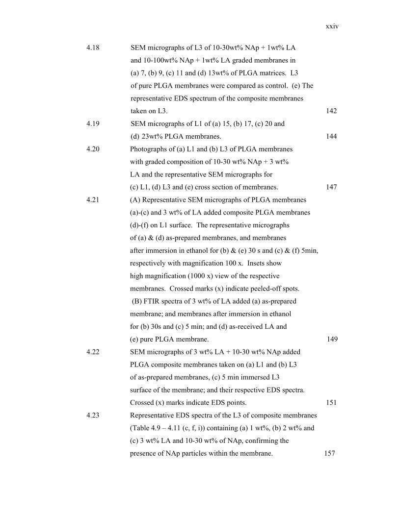
4.24 SEM micrographs of L1, L3 and cross-section of
(a) pure PLGA membrane (S105) and (b) 1wt% (S98),
(c) 2wt% (S99), (d) 3wt% (S100) of LA incorporated
triple layered membranes containing 10-30 wt% of NAp
in 9-20 wt% of PLGA matrices. (e) The representative
EDS spectrum of the composite membrane. 159
4.25 SEM micrographs of L1, L3 and cross-section of
(a) pure PLGA membrane (S105) and (b) 1wt% (S106),
(c) 2 wt% (S107), (d) 3 wt% (S108) of LA incorporated
triple layered membranes containing 10-100 wt% of NAp
in 9-20 wt% of PLGA matrices. (e) The representative
EDS spectrum of the composite membranes. 161
4.26 SEM micrographs of L1, L3 and cross-section of
(a) pure PLGA membrane (S181) and (b) 1 wt% (S183),
(c) 2 wt% (S185), (d) 3 wt% (S187) of LA incorporated
triple layered membranes containing 10-30 wt% of NAp in
9-17 wt% of PLGA matrices. (e) The representative EDS
spectrum of composite membranes. 163
4.27 SEM micrographs of L1, L3 and cross-section of
(a) pure PLGA membrane (S181) and (b)1 wt% (S193),
(c) 2 wt% (S191), (d) 3 wt% (S189) of LA incorporated
triple layered membranes containing 10-100 wt% of NAp
in 9-17 wt% of PLGA matrices. (e) The representative EDS
spectrum of composite membranes. 164
4.28 High magnification SEM micrographs of L1 (left) and L3
(right) of (a,b) 9-20 wt% of pure PLGA matrices (S105),
(c,d) 3 wt% LA + 10-30 wt% NAp added in 9-20 wt% of
PLGA matrices (S100), (e,f) 3 wt% LA + 10-100 wt% of NAp
added in 9-20 wt% of PLGA matrices (S108). (g) The representative
EDS spectrum of NAp particles on composite membranes
as indicated by the black arrows in (c) and (e). 166
xxvi
4.29 High magnification SEM micrographs of L1 (left) and L3
(right) of (a,b) 9-17wt% of pure PLGA matrices (S181),
(c,d) 3 wt% LA + 10-30wt% NAp added in 9-17wt% of PLGA
matrices (S187), (e,f) 3wt% LA + 10-100wt% of NAp added
in 9-17wt% of PLGA matrices (S189). (g) The representative EDS
spectrum of NAp particles as indicated by the white arrows
in (d) and (f). 168
4.30 XRD patterns for (a) pure PLGA membrane (S105);
L1 of 10-30wt% of NAp added in 9-20wt% of PLGA
composite membrane containing (b) 1wt% (S98),
(c) 2wt% (S99) and (d) 3wt% (S100) of LA. The opposite
layers of L1, i.e., L3 were not introduced with LA. The XRD
patterns of L3 were the opposite layers of L1 added with
(e) 1wt% (S98), (f) 2wt% (S99) and (g) 3wt% (S100) of
LA in triple layered membranes containing (h) NAp;
(i) magnified region for NAp showing apatite ( ) peaks,
(j) LA. L1 and L3 were incorporated with 10 and 30wt% of
NAp, respectively. 171
4.31 XRD patterns for (a) pure PLGA membrane (S105); L1 of
10-100wt% of NAp added in 9-20wt% of PLGA composite
membrane containing (b) 1wt% (S106), (c) 2wt% (S107) and
(d) 3wt% (S108) of LA. The opposite layers of L1, i.e., L3
were not introduced with LA. The XRD patterns of L3 were
the opposite layers of L1 added with (e) 1wt% (S106) ,
(f) 2wt% (S107) and (g) 3wt% (S108) of LA in triple
layered membranes containing (h) NAp; (i) magnified
region for NAp showing apatite ( ) peaks , (j) LA.
L1 and L3 were incorporated with 10 and 100wt% of NAp,
respectively. 172
4.32 FTIR spectra of (a) as-received PLGA, (b) LA particles,
(c) NAp powder and L1 surface of (d) pure PLGA (S112),
(e) 1 wt% (S110), (f) 2 wt% (S102) and (g) 3 wt% (S115)
of LA added composite membranes containing
xxvii
10-30 wt% of NAp in 9-20 wt% of PLGA matrices. 175
4.33 FTIR spectra of (a) NAp powder and L3 surface of
(b) pure PLGA (S112) membrane (c) 1 wt% of LA (S117),
(d) 2 wt% of LA (S118) and (e) 3 wt% of LA (S119) added
composite membranes containing 10-100 wt% of NAp in
9-20 wt% of PLGA matrices. 177
4.34 The illustration of possible interaction mechanisms between
PLGA-NAp-LA composite systems presenting (a) hydrogen
bonding between PLGA-NAp-LA and (b) ionic bonding
between NAp-LA. 179
4.35 DSC thermogram of LA. 180
4.36 DSC thermograms of pure PLGA membrane (S195),
1 wt% LA (S197), 2 wt% LA (S199) and
3 wt% LA (S201) added composite membranes
containing 10-30 wt% of NAp in 9-20 wt% of PLGA
matrices. 181
4.37 DSC thermograms of pure PLGA membrane (S195),
1 wt% LA (S203), 2 wt% LA (S205) and 3 wt% LA (S207)
added composite membranes containing 10-100 wt% of NAp
in 9-20 wt% of PLGA matrices. 182
4.38 Representative stress-strain curves of the 10-30 wt% of
NAp added 9-20 wt% of PLGA composite membranes
with different LA contents. Only one typical plot for each
membrane is shown. 185
4.39 Tensile strength of 1-3 wt% of LA added composite
membranes containing 10-30 wt% and 10-100 wt% NAp
in 9-20 wt% of PLGA matrices. Data are presented as
mean ± SD, n=6. 187
4.40 Representative stress-strain curves of the 10-100 wt% of
NAp containing 9-20 wt% of PLGA composite
membranes with different LA contents. Only one typical
plot for each membrane is shown. 189
4.41 Tensile strength of 1-3 wt% of LA added composite
membranes containing 10-30 wt% and 10-100 wt% NAp
xxviii
in 9-17 wt% of PLGA matrices. Data are presented
as mean ± SD, n=6. 192
4.42 The weight loss of 9-20 wt% of pure PLGA and composite
triple layered membranes. The composites loaded with
1 wt% of LA and NAp varied at 10-30 wt% and 10-100 wt%.
Data are presented as mean ± SD, n =3. 196
4.43 The weight loss of 9-20 wt% of pure PLGA and composite
triple layered membranes. The composites loaded with
2 wt% of LA and NAp varied at 10-30 wt% and 10-100 wt.
Data are presented as mean ± SD, n =3. 197
4.44 The weight loss of 9-20 wt% of pure PLGA and composite
triple layered membranes. The composites loaded with
3 wt% of LA with NAp varied at 10-30 wt% and 10-100 wt%.
Data are presented as mean ± SD, n =3. 198
4.45 SEM micrographs of L3 surfaces of pure PLGA and
composite membranes (a) before immersion and after
immersion for (b) 4 weeks, (c) 12 weeks and
(d) 24 weeks in PBS. The PLGA content is 9-20 wt%. The
PLGA content is 9-20 wt%. (e) The EDS spectrum of the
region marked by rectangle taken on 10-100 wt% of NAp
added composite membrane as in (d). 199
4.46 SEM micrographs of L3 surface morphology of pure
PLGA (9-20 wt%) membrane, (a) before immersion
and after immersion for (b) 4 weeks and (c) 24 weeks in
PBS. 200
4.47 SEM micrographs of L1 surfaces of pure PLGA and
composite membranes (a) before immersion and after
immersion for (b) 4 weeks, (c) 12 weeks and (d) 24 weeks
in PBS. The PLGA content is 9-20 wt%. (e) The EDS
spectrum of the region marked by rectangle taken on
10-100 wt% of NAp added composite membrane as in (c). 201
4.48 The weight loss of 9-20 wt% of pure PLGA and
composite triple layered membranes. The composites
loaded with 10-30 wt% of NAp and LA varied at 1-3 wt%.
xxix
Data are presented as mean ± SD, n=3. 202
4.49 The weight loss of 9-20 wt% of pure PLGA and composite
triple layered membranes. The composites loaded with
10-100 wt% of NAp and LA varied at 1-3 wt%.
Data are presented as mean ± SD, n=3. 203
4.50 The weight loss of 9-17 wt% of pure PLGA and composite
triple layered membranes. The composites loaded with
3 wt% of LA and NAp varied at 10-30 wt% and
10-100 wt%. Data are presented as mean ± SD, n=3. 204
4.51 SEM micrographs of L1 surface morphology of composite
PLGA (9-17 wt%) membranes with 10-100 wt% of NAp
and 3wt% LA, (a) before immersion and (b) after immersion
for 24 weeks in PBS. 204
4.52 The pH change of 9-20 wt% of pure PLGA and composite
triple layered membranes. The composites loaded with
10-30 wt% of NAp and LA varied at 1-3 wt%.
Data are presented as mean ± SD, n=3. 207
4.53 The pH change of 9-20 wt% of pure PLGA and composite
triple layered membranes. The composites loaded with
10-100 wt% of NAp and LA varied at 1-3 wt%.
Data are presented as mean ± SD, n=3. 207
4.54 The pH change of 9-17 wt% of pure PLGA and composite
triple layered membranes. The composites loaded with
3 wt% of LA and NAp varied at 10-30 wt% and 10-100 wt.
Data are presented as mean ± SD, n=3. 209
4.55 The pH change of 9-20 wt% of pure PLGA and composite
triple layered membranes. The composites loaded with
3 wt% of LA and NAp varied at 10-30 wt% and 10-100 wt%.
Data are presented as mean ± SD, n=3. 209
4.56 The water uptake of 9-20 wt% of pure PLGA and composite
triple layered membranes. The composites loaded with
10-30 wt% of NAp and LA varied at 1-3 wt%.
Data are presented as mean ± SD, n=3. 210
xxx
4.57 The water uptake of 9-20wt% of pure PLGA and composite
triple layered membranes. The composites loaded with
10-100wt% of NAp and LA varied at 1-3wt%.
Data are presented as mean ± SD, n=3. 211
4.58 The water uptake of 9-20 wt% of pure PLGA and composite
triple layered membranes. The composites loaded with
1 wt% of LA and NAp varied at 10-30 wt% and
10-100 wt%. Data are presented as mean ± SD, n=3. 212
4.59 The water uptake of 9-20 wt% of pure PLGA and
composite triple layered membranes. The composites
loaded with 2 wt% of LA and varied at 10-30 wt% and
10-100 wt% of NAp. Data are presented as mean ± SD, n=3. 213
4.60 The water uptake of 9-20 wt% of pure PLGA and
composite triple layered membranes. The composites
loaded with 3 wt% of LA and varied at 10-30 wt% and
10-100 wt% of NAp. Data are presented as mean ± SD, n=3. 213
4.61 The water uptake of 9-17 wt% of pure PLGA and
composite triple layered membranes. The composites
loaded with 3 wt% of LA and varied at 10-30 wt% and
10-100 wt% of NAp. Data are presented as mean ± SD, n=3. 214
4.62 The highest UV absorption intensity of derivatized
LA at the concentration of 2000 µg/mL and
(b) the corresponding linear calibration standard curve.
Data represents mean±SD of three replicates. 218
4.63 The cumulative release percentage of LA from 3 wt%
of LA loaded composite membranes containing 10-30 wt%
of NAp in 9-20 wt% of PLGA matrices. Data represents
mean±SD of two replicates. 220
4.64 The cumulative amount of LA released from the
formulated membrane fitted to Ritger-Peppas model. 221
4.65 The cumulative amount of LA released from the
formulated membrane fitted to Higuchi model. 222
4.66 The cumulative amount of LA released from the
formulated membrane fitted to First order kinetic
xxxi
model. 222
4.67 The cumulative amount of LA released from the
formulated membrane fitted to Zero order kinetic
model. 223
4.68 Growth curve of S. aureus (ATCC 6538) grown at
37 °C in TSB. Data represents means ± SEM
of three independent experiments (n = 3). 225
4.69 Growth curve of P. aeruginosa (ATCC 9027) grown
at 37 °C in TSB. Data represents means ± SEM of
three independent experiments (n = 3). 226
4.70 Antimicrobial activity of 1, 2, and 3 wt% of LA
incorporated composite membranes compared to pure
PLGA control group against S. aureus. The number of
viable microbes on the membranes after 24 h was
obtained using colony counting formation method.
Data represents means ± SEM of two
independent experiments (n = 2). 227
4.71 Antimicrobial activity. The recovery of S. aureus
on LA incorporated membranes after 24 h of incubation
at 37 °C. The number of viable microbes on (a) PLGA
membrane, and (b) 1 wt%, (c) 2 wt% and (d) 3 wt% of
LA incorporated membranes, was obtained using colony
counting formation method. The representative TSA
plates for all three replicates show S.aureus colony
formation after incubation. The TSA plates (e) represent
colony formation of recovered inoculums after serially
diluted to 10-1 (left plate) and 10-2 (right plate).
At higher dilution (10-2), fewer colonies were formed
on plates which indicate that serial dilutions of the
recovered inoculums were performed appropriately. 229
4.72 Antimicrobial activity of 1, 2, and 3 wt% of LA incorporated
composite membranes compared to pure PLGA control
group against P. aeruginosa. The number of viable
xxxii
microbes on the membranes after 24 h was obtained using
colony counting formation method. Data represents
means ± SEM of two independent experiments (n = 2).
After 24 h of incubation, the bacteria colonies had
increased by 102 in all samples compared to initial
loading of P. aeruginosa which demonstrated
growth of bacteria after incubation period. 230
4.73 The recovery of P. aeruginosa on LA incorporated
membranes after 24 h of incubation at 37 °C. The number
of viable microbes on (a) PLGA membrane, and (b) 1 wt%,
(c) 2 wt% and (d) 3 wt% of LA incorporated membranes,
was obtained using colony counting formation method.
The representative TSA plates for all three replicates
show formation of P. aeruginosa colonies after incubation.
The TSA plates (e) represent colony formation of recovered
inoculums after serially diluted to 10-3 (left plate) and
10-4 (right plate). At higher dilution (10-4), fewer colonies
were formed on plates which indicate that serial dilutions
of the recovered inoculums were performed appropriately.
The bacteria growth on LA added composite membranes
was prominently higher than pure PLGA membranes as
more dilutions were performed beyond 10-1 and 10-2. 231
4.74 Representative 24-well plates for AB colour changes
assessment. Cells exposed to extracts of (a) PLGA,
(b) 1 wt% LA, (c) 2 wt% LA, (d) 3 wt% LA and
(e) phenol solution. 234
4.75 Cytotoxicity assay results of the pure PLGA and
LA added composite membranes. Data are
means ± SEM of two independent experiments (n=2). 235
xxxiii
LIST OF SYMBOLS
cfu - Colony forming unit
d - Interplanar spacing
Exo - Exothermic
k - drug release kinetic constant
m0 - Initial weight
m1 - Wet weight
m2 - Dry weight
M - Molar
Mt - Amount of drug released at time t
M∞ - Total amount of drug released
n - Diffusional exponent
R2 - Correlation coefficient
rad - Radian
t - time
Tg - Transition temperature
Xc - Crystallinity
Xs - Crystallite size
θ - Diffraction Angle
λ - Wavelength of X-ray beam
xxxiv
LIST OF ABBREVIATIONS
AMP - Antimicrobial peptide
ATCC - American Type Culture Collection
ASTM - American Society for Testing and Materials
ATR - Attenuated total reflectance
BET - Brunauer – Emmet – Teller
CaP - Calcium phosphate
CHN - Carbon, Hydrogen, Nitrogen elemental analysis
DSC - Differential scanning calorimetry
DDI - Double distilled de-ionised
DMSO - Dimethyl sulfoxide
DTA - Differential thermal analysis
d-PTFE - Dense polytetrafluoroethylene
ECACC - European Collection of Cell Cultures
e-PTFE - expanded PTFE
et al. - and others
FDA - US Food and Drug Administration
FESEM - Field Emission Scanning Electron Microscope
FGM - Functionally graded membrane
FTIR - Fourier Transform Infrared spectrophotometry
F. nucleatum - Fusobacterium nucleatum
FWHM - Full width at half maximum
GBR - Guided bone regeneration
HA - Hydroxyapatite
HPLC - High Performance Liquid Chromatography
HSF - Human Skin Fibroblast cells
ICP-AES - Inductively Coupled Plasma-Atomic Emission
Spectroscopy
i.e. - that is
xxxv
IC80 - Inhibition concentration at 80% killing
ICDD - International Centre for Diffraction Data
ISO - International Organisation for Standardisation
LA - Lauric acid
L1 - Layer 1
L2 - Layer 2
L3 - Layer 3
MEM - Minimum Essential Medium
MePEG - Methoxypoly(ethyleneglycol)
MIC - Minimum inhibition concentration
MTT - 3-(4,5-Dimethylthiazol-2-yl)-2,5-
diphenyltetrazolium bromide
NAp - Non-stoichiometric nanoapatite
NApF1 - Non-stoichiometric nanoapatite Formulation 1
NApF2 - Non-stoichiometric nanoapatite Formulation 2
NHA - Stoichiometric nanohydroxyapatite
OFP - Open Flap Debridement
OD - Optical density
PBS - Phosphate buffered saline
PCL - Polycaprolactone
PDL - Periodontal ligament
PDLLA - poly(DL-lactic) acid
P. gingivalis - Porphyromonas gingivalis
PGA - Polyglycolic acid
P. intermedia - Prevotella intermedia
PLA - Polylactic acid
PLLA - poly(L-lactic) acid
PLGC - poly (L-lactide-co-glycolide-ε-caprolactone)
PLCL - poly (L-lactide-co-ε-caprolactone)
PU - Polyetherurethane
PTFE - polytetrafluoroethylene
rpm - revolution per minute
SEM - Scanning Electron Microscopy
SD - Standard Deviation
xxxvi
TEM - Transmission Electron Microscopy
TGA - Thermogravimetric analysis
TIPS - Thermally induced phase separation
UV - Ultraviolet
UV-Vis - Ultraviolet-Visible
wt% - Weight percentage
XRD - X-ray Diffraction
β-TCP - β-tricalcium phosphate
3D - 3 dimensional
xxxvii
LIST OF APPENDICES
APPENDICES TITLE PAGE
A List of publications 265
A1 Published article 1 266
A2 Published article 2 268
A3 Published article 3 270
A4 Published article 4 272
B1 Calculation for the preparation of 1.0 M Ca(OH)2
and H3PO4 reactants for the synthesis of NHA-1.0M. 274
B2 Calculation for the preparation of 1.5 M Ca(OH)2
and H3PO4 reactants for the synthesis of NHA-1.5M. 275
B3 Calculation for the preparation of 2.0 M Ca(OH)2
and H3PO4 reactants for the synthesis of NHA-2.0M. 276
B4 Calculation for the preparation of 1.0 M Ca(OH)2,
H3PO4 reactants and ionic solutions for the synthesis
of NApF1 and NApF2-1.0M. 277
B5 Calculation for the preparation of 1.5 M Ca(OH)2,
H3PO4 reactants and ionic solutions for the synthesis
of NApF1 and NApF2-1.5M. 278
B6 Calculation for the preparation of 2.0 M Ca(OH)2,
H3PO4 reactants and ionic solutions for the synthesis
of NApF1 and NApF2-2.0M. 279
C1 Lattice parameters calculation for as prepared
stoichiometric nanohydroxyapatite (NHA) powder
synthesized using reaction method I (37±2°C). 280
C2 Lattice parameters calculation for as prepared
stoichiometric nanohydroxyapatite (NHA) powder
xxxviii
synthesized using reaction method II (37&85±2°C). 281
C3 Lattice parameters calculation for as prepared
non-stoichiometric nanoapatite (NApF1) powder
synthesized using reaction method I (37±2°C). 282
C4 Lattice parameters calculation for as prepared
non-stoichiometric nanoapatite (NApF1) powder
synthesized using reaction method II (37&85±2°C). 283
C5 Lattice parameters calculation for as prepared
non-stoichiometric nanoapatite (NApF2) powder
synthesized using reaction method I (37±2°C). 284
C6 Lattice parameters calculation for as prepared
non-stoichiometric nanoapatite (NApF2) powder
synthesized using reaction method II (37&85±2°C). 285
C7 Lattice parameters calculation for NHA, NApF1 and
NApF2 powders sintered at 900°C in CO2. 286
C8 Lattice parameters calculation for NHA powders
sintered at 900°C in CO2. 287
C9 Lattice parameters calculation for NApF1 powders
sintered at 900°C in CO2. 288
C10 Lattice parameters calculation for NApF2 powders
sintered at 900°C in CO2. 289
C11 Lattice parameters calculation for NHA, NApF1
and NApF2 powders sintered at 1250°C in air. 290
C12 Crystallite size (Xs in nm) of as prepared
nanohydroxyapatite powders determined using
Scherrer equation. 291
C13 Fraction of crystalline phase (Xc) of the as prepared
NHA, NApF1 and NApF2 powders. 292
D1 Dry and wet tensile strength of 10-30 wt% of NAp
containing 1-3 wt% of LA added PLGA (9-20wt%)
composite membranes. 293
D2 Dry and wet tensile strength of 10-100 wt% of NAp
containing 1-3 wt% of LA added PLGA (9-20wt%)
composite membranes. 294
xxxix
D3 Dry and wet tensile strength of 10-30 wt% of NAp
containing 1-3 wt% of LA added PLGA (9-17wt%)
composite membranes. 295
D4 Dry and wet tensile strength of 10-100 wt% of NAp
containing 1-3 wt% of LA added PLGA (9-17wt%)
composite membranes. 296
E1 Weight loss measurements and weight loss difference
in post-immersed membranes added with 1-2wt% of
LA and varied with 10-30 wt% and 10-100wt% NAp. 297
E2 Weight loss measurements and weight loss difference
in post-immersed membranes added with 3 wt% of LA
and varied with 10-30 wt% and 10-100wt% NAp. 298
E3 Weight loss measurements of 9-20 wt% PLGA membranes
loaded with 10-30wt% and 10-100wt% of NAp and LA
varied at 1-3 wt%. 299
E4 pH measurements of 9-20 wt% membranes containing
10-30 wt% and 10-100 wt% NAp and LA varied at 1-3wt%. 300
E5 pH measurements of 9-17 wt% and 9-20 wt% membranes
containing 10-30 wt% and 10-100 wt% NAp and LA loaded
at 3wt%. 301
F1 Comparison of water uptake in membranes containing
10-30wt% and 10-100wt% of NAp and LA varied at 1-3wt%. 302
F2 Comparison of water uptake in membranes containing
LA loaded at 1 & 2wt% and NAp varied at 10-30wt%
and 10-100wt%. 303
F3 Comparison of water uptake in membranes containing
LA loaded at 3wt% and NAp varied at 10-30wt% and
10-100wt% in 9-20 wt% and 9-17wt% of PLGA matrices. 304
G1 Data for standard calibration curve and loading
efficiency studies. 305
G2 Calculations for LA release from 3wt% of LA
loaded composite membrane containing 10-30wt% of
NAp in 9-20wt% of PLGA matrices (Sample A). 306
xl
G3 Calculations for LA release from 3wt% of LA
loaded composite membrane containing 10-30wt% of
NAp in 9-20wt% of PLGA matrices (Sample B). 307
G4 Calculations for average LA release from 3wt% of LA
loaded composite membrane containing 10-30wt% of
NAp in 9-20wt% of PLGA matrices (Sample A+B). 308
H Data for S. aureus and P. aeruginosa growth inhibition against
LA concentration. 309
I Data for cell viability of membrane samples. 310
CHAPTER 1
INTRODUCTION
1.1 Background
Rapid bone defect filling with normal bone is a challenge in the fields of
orthopaedic and dentistry [1]. The bone has limited regeneration capability due to
insufficient blood supply, large defects and invasion of highly proliferative
nonosteogenic tissues that can impair bone repair [2,3]. Bone grafting is an
established treatment to restore bone tissue. However, problems such as redundant
fibrous connective tissue growth surrounding implanted bone graft and the
movement of bone graft particles are still remain to be solved [1]. GBR has become
an area of increasing interest in bone restorative procedures for guiding bone healing
and regeneration [2,3] due to its success in curing cranial, maxillofacial and alveolar
bone defects [4,5]. The concept of GBR is to cover the bone defect using a barrier
membrane that enhances new bone ingrowth while preventing the ingrowth of
fibrous tissue into the grafted site [6]. Hence, the bone regenerative approaches
using GBR membranes have been extensively investigated to reveal their clinical
potential [7,8,9].
GBR membranes have been widely studied as they are useful for bone repair
in oral and maxillofacial surgery where limited mechanical loading exists [5,10].
The commercially available GBR membranes are made of non-resorbable and
resorbable polymers. The non-resorbable polytetrafluoroethylene (PTFE)
membranes have exhibited significant disadvantages such as requirement for second
surgery and increased risk of infection leading to early removal of the membrane [9].
2
Collagen based resorbable membranes are widely used in clinical therapies. Since
majority collagen membranes are animal derived, these membranes carry the risk of
potential transmission of infectious agents, including the inappropriate immune
responses in patients [7]. The synthetic resorbable membranes have found
widespread use in clinical medicine as they are totally degradable, thus not requiring
second surgery [8,9]. Poly(lactic-co-glycolic acid) (PLGA) is a FDA approved
synthetic resorbable material and widely used in GBR applications [11,12].
Nonetheless, an inflammatory reaction by the accumulation of acidic degradation
products in resorbable membranes has been reported [4,13]. The combination of
calcium phosphate (CaP) with resorbable polymeric membranes is expected to
neutralize the acidic degradation products from the membranes; which is intended to
overcome inflammatory reaction in vivo [13,14,12,15,16]. Moreover, CaP particles
in polymeric membranes has been also reported to improve structural integrity,
flexibility and bone regeneration in vivo [17,15,18,14]. The aforementioned studies
emphasises the need for incorporation of CaP particles to improve physical and
mechanical properties of the resorbable polymeric membrane.
Currently, biomaterial-associated infection is regarded as a devastating
complication in clinical surgery. Therefore, anti-infective biomaterials need to be
developed as the main strategy to prevent infection in clinical applications [19]. A
bacteria-free environment is highly important to regenerate bone tissues in GBR
strategies [20]. Recently, the antibiotics incorporated GBR membranes have been
developed for local delivery of antimicrobial agents [21]. Nonetheless, the increasing
bacterial resistance prompted the development of alternative antimicrobial agent
incorporated GBR membranes [22,23,20,24]. In light of this, a naturally derived
antimicrobial agent to substitute the use of antibiotics is sought after to develop a
new antimicrobial membrane for clinical applications.
The antimicrobial properties of naturally found fatty acids have been
recognized for many years. Lauric acid (LA) is naturally found in coconut oil [25]
and has been recognized to possess broad-spectrum with effective antimicrobial
activity against gram-positive bacteria [26,27]. Unlike antibiotics, fatty acids and
their derivatives have diverse modes of action that appear to be non-specific and
3
development of resistance to these compounds has not been reported [28]. It is
suggested that LA kills Gram-positive bacteria by separating their inner and outer
membranes, resulting in cytoplasmic disorganization of the bacteria [25]. Thus, it is
envisaged that incorporating LA in composite membranes for anti-infective bone
regeneration purposes could possibly overcome clinical complications caused by the
administration of antibiotics.
The development of functionally graded and multiple layered membrane is to
enhance the features required for GBR, namely a combination of physical,
mechanical, biological and antimicrobial properties [13,23]. Also, the incorporation
of functional gradients in a multilayered membrane structure offers the possibilities
to overall usefulness to the membrane. Solvent casting technique offers the
formation of layered membrane structure [16] whereas porous network formation is
attainable through thermally induced phase separation (TIPS) [29] of the polymeric
materials. The presence of residual toxic organic solvent is a major concern in
solvent based fabrication technique. Thus, it is vital to include solvent removal step
to reduce possible toxicity by solvent residues in fabricated membranes [30]. In this
study, a new modified solvent casting-TIPS-solvent leaching technique is proposed
to fabricate triple layered and graded composite PLGA membrane. Collectively, it is
suggested that a new combination of CaP nanoparticles and LA as an antimicrobial
agent being graded and layered in PLGA matrices can potentially function as an
antimicrobial barrier membrane. This thesis will advance the knowledge in the area
of antimicrobial composite membrane development for potential use in cranial,
maxillofacial and dental applications. A new technique to establish the fabrication of
multilayered and graded composite membrane utilizing solvent casting-TIPS-solvent
leaching technique will be developed in this study. The fabrication and structural
properties of the triple-layered PLGA membrane, graded with various amounts of
LA and CaP nanoapatite will be studied. The effects of LA and CaP addition on the
physical, chemical, mechanical, biological and antimicrobial properties of the PLGA
composite membrane will also be explored. This membrane will deliver
antimicrobial and osteoconductive properties by the incorporation of LA and CaP
nanoapatite, respectively.
4
1.2 Problem statements
The major concerns in GBR surgical intervention are the problems related to
the increasing bacterial resistance and side effects caused by antibiotics [31,32].
Multiple antibiotics are currently used to protect the bone defect from bacterial
invasion, increasing the risks of bacterial resistance and side effects [33,22,31].
Hence, an alternative antimicrobial agent to substitute antibiotics is sought after. LA
has been exhibiting effective antimicrobial activity against gram-positive bacteria
that eliminates the need for multiple antibiotics to prevent bacteria colonization
[26,27]. Therefore, the incorporation of antimicrobial LA in the composite
membrane and its controlled release is proposed to circumvent the above mentioned
drawbacks.
Apart from antimicrobial property, other important membrane characteristics
such as surface morphology, pore size, membrane degradability, mechanical
properties and cytocompatibility should be equally evaluated. Hence, appropriate
materials selection and membrane design for GBR applications are highly
indispensable for a successful bone defect treatment [7]. Poly(lactic-co-glycolic
acid) (PLGA) is a FDA approved synthetic resorbable material which is widely used
in GBR applications [11,12]. However, the accumulations of acidic degradation
products from the synthetic bioresorbable membranes have been reported to cause
inflammatory reaction in vivo [8,9]. Hence, the combination of synthetic polymers
with CaP has been reported to neutralize the acidic degradation products from the
polymers using ionic interactions [13,14,12,15,16]. Moreover, CaP incorporation
improves structural integrity, flexibility and bone regeneration of the resorbable
membranes [17,15,18,14]. Therefore, the current clinical disadvantage of using pure
synthetic polymeric material as a GBR membrane could be overcome by
incorporating CaP particles to reduce the potential inflammatory reactions. Thus, in
this study, multiple ions substituted nanoapatite (NAp) powder which has close
resemblance to natural bone mineral composition will be synthesized and
incorporated into the PLGA matrices to form composite membranes.
5
Incorporating multiple additives in a composite membrane is a challenge as it
requires the development of multilayered and graded membrane structure [13,16,34].
In order to address GBR applications, two functional surface layers are required.
One of the surfaces with porous morphology allows bone ingrowth whereas the other
dense surface prevents fibrous tissue penetration [16,13]. Therefore, in this study a
triple-layered composite membrane with new combination of porous/dense layers
will be developed. The NAp particles and LA will be graded in each layer to deliver
osteoconductive and antimicrobial properties, respectively.
In order to develop a multilayered and graded composite membrane, an
appropriate technique is indispensable to achieve the desired membrane structure.
Currently, solvent casting [16] and TIPS [29] techniques have been employed to
fabricate composite membranes. However, there are two disadvantages of using
solvent casting method: i) toxic organic solvents application [15,18] that requires
critical attention especially on its exposure in biomedical applications, ii) CaP
particles can spontaneously precipitate from the polymer solution due to poor affinity
and can cause non-uniform dispersion of CaP in polymer matrix [18]. Hence, these
drawbacks could be overcome by freezing the CaP dispersed polymer matrix
structure through TIPS technique. Moreover, solvent removal from the fabricated
membrane is another important step to reduce toxic solvent residues [30,35]. Hence,
in this study, composite membranes will be fabricated utilizing a new combination of
solvent casting-TIPS-solvent leaching technique to address the formation of layered
and graded membrane, dispersed with CaP particles and removal of toxic solvent
from the membrane. The new modified technique is envisaged to form a composite
membrane with graded porous/dense structure that has functional gradients, i.e., NAp
and LA.
1.3 Objectives of the study
This work explores a novel fabrication technique, structure and design of a
polymer-ceramic composite membrane incorporating LA as an antimicrobial agent.
The goal is to design a functionally graded triple layered barrier membrane with
6
antimicrobial property using solvent casting-TIPS-solvent leaching techniques. In
order to achieve the main objective, the following specific objectives were executed.
a) To synthesise multiple ions substituted non-stoichiometric nanoapatite (NAp)
powder.
b) To establish a combined solvent casting-TIPS-solvent leaching techniques for the
formation of triple-layered PLGA composite membranes graded with LA and
NAp powder.
c) To determine the physical, chemical, mechanical and in vitro degradation
properties of the membrane.
d) To evaluate the cytocompatibility and antimicrobial efficacy of the membrane.
1.4 Research hypothesis
It is possible to achieve an antimicrobial composite membrane by
incorporating antimicrobial agents, in order to prevent biomaterial-associated
infection in GBR applications. Therefore, it is envisaged that incorporating LA in
the composite membrane could impart antimicrobial property which could prevent
bacterial infection associated to the membrane. Furthermore, a resorbable composite
membrane is desired to achieve less in vivo inflammation by reducing acidic
degradation products through the addition of CaP particles [8,9]. Moreover, the
combination of synthetic resorbable membranes with CaP is expected to deliver
improved mechanical strength to the composite membranes [17,15,18,14]. Hence, in
this study, it is hypothesised that varying the NAp and LA contents in PLGA
matrices can significantly alter the physico-chemical, mechanical and antimicrobial
properties of the membrane.
The GBR membrane is designed to have a smooth surface on one face to
inhibit soft tissue penetration while the opposite porous face is capable of
accommodating bone tissue ingrowth in vivo [16,36]. The dense/porous network
formation through TIPS [29] technique is easily attainable whereas a multilayered
membrane structure via solvent casting and the removal of solvent [30] could
translate a safer membrane fabrication technique for clinical practice. The solvent
7
casting-TIPS-solvent leaching technique will be used to test the hypothesis that one
can tailor the properties of the different layers to form a functionally graded
composite membrane to retain its structural, dimensional and mechanical properties
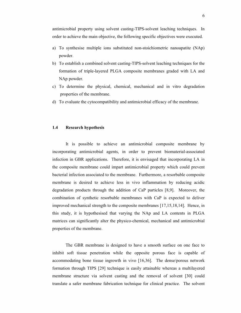
for bone regeneration. Figure 1.1 demonstrates the importance of incorporating LA
in composite membrane which may prevent bacterial infection on the membrane
surface. In addition, formation of dense membrane surface also excludes fibroblast
penetration into the barrier membrane.
Figure 1.1: LA incorporation into barrier membrane as an antimicrobial agent for
adjunct treatment in GBR procedures to inhibit bacterial infection.
1.5 Scope of the study
A new design of triple-layered and graded PLGA composite membrane has
been fabricated. The triple layered membrane is comprised of PLGA matrix, graded
with non-stoichiometric NAp and LA at each layer. PLGA with a lactic acid to
glycolic acid ratio of 85:15 degrade over 2–6 months [37] and have the ability to
deliver drugs locally in a controlled manner. These properties are making it suitable
for use as a GBR barrier membrane. Besides improving mechanical strength of the
membranes, the incorporation of CaP particles should be merely targeted for its
Barrier membrane
LA incorporated surface
Prevention of bacterial infection
Dense barrier surface excludes fibroblast penetration
8
osteoconductivity and hydrophilic nature to enhance bone growth into the polymer
surfaces [38]. NAp powder is synthesized by introducing substituents within
1.84wt% (Na), 1.46wt% (Mg), 0.06wt% (K) and 4.80wt% (CO32-) to closely mimic
natural bone apatite. The NAp powder is incorporated to enhance bioactivity and
osteoconductivity of the membrane. LA is added to introduce antimicrobial
properties to the composite membrane to prevent bacterial infection as it is known to
possess effective antimicrobial activity against gram-positive bacteria [26,27]. The
composite membrane is fabricated by employing a modified solvent casting-TIPS-
solvent leaching technique. The solvent casting facilitated lamination of multiple
layers of graded LA and NAp in PLGA matrices whereas TIPS used to form
porous/dense layers in the membrane structure. Solvent leaching is performed to
remove toxic solvent residues.
1.6 Significance of the study
LA, as a substitute for antibiotics is identified and incorporated in the
composite membrane which is to be used as a potential antimicrobial membrane for
clinical applications. Prevention of bacterial infection is a promising strategy
whereby LA imparts antimicrobial activity on the membrane surface. This would
render an antimicrobial barrier membrane appropriate for adjunctive treatment in
guiding bone regeneration. This work also reports the fabrication of PLGA-NAp-LA
composite membrane using solvent casting-TIPS-solvent leaching technique. This
new technique largely eliminates the solvent residue in the fabricated membrane
through solvent leaching step using water as the exchanging medium.
1.7 Thesis outline
Chapter 1 is the introduction to the study of this thesis. The entire outline of
the thesis is illustrated in Fig. 1.2.
9
Chapter 2 describes the review of literatures related to the development and
application of commercially available GBR membrane that has been related to its
profound improvement through current research to overcome clinically reported
shortcomings. Moreover, selection criteria for PLGA, LA and NAp are also
reviewed to ensure the fabricated composite membrane is more likely to possess
appropriate physical, structural, dimensional, mechanical, antimicrobial and
biological properties for potential use in bone regeneration procedures.
Chapter 3 deals with the materials and methods used to investigate the
appropriate parameters, experimental set-up, test conditions, characterization using
analytical equipment and material evaluation involved in the fabrication and
evaluation of the composite membranes. The synthesis of NAp powder is reported in
the first part of the chapter. Subsequently, the development of PLGA based NAp-LA
composite membrane through a new fabrication technique using solvent casting-
TIPS-solvent leaching is reported. This is followed by the development of methods
to test on the membrane’s properties such as physico-chemical, mechanical, in vitro
degradation profile over six months duration, quantification of LA release and
finally, LA release mechanism; since the effects of NAp and LA additions in the
PLGA membranes are highly imperative to meet the design criteria of membranes
for GBR applications.
Chapter 4 elaborates the outcome of NAp synthesis, fabrication of composite
membranes, degradation profiles for composite membranes, mechanical evaluation
of membranes in dry and wet condition, released LA concentration and its release
mechanism. Synthesis of NAp with the highest substitutent composition, the
morphology of triple layered membrane, phase composition, physical changes in
amorphous/crystalline state of LA, interaction mechanisms between PLGA-NAp-LA
in composite membranes, weight loss and water absorption of membranes, and
finally the quantification of LA release and its release mechanism from composite
membranes for sufficient antimicrobial effects while maintaining its
cytocompatibility are discussed. The cytocompatibility of synthesized NAp powder
and composite membranes along with antimicrobial evaluation on the effects of LA
addition in composite membranes were discussed.
10
Chapter 5 concludes structural, dimensional and mechanical integrity of the
layered and graded composite membrane. The effects of LA and NAp addition on
physico-chemical, mechanical and antimicrobial properties are also described.
Publications and presentations at conferences: This section forms part of the
thesis, which described the synthesis of NAp powder and the fabrication of
composite membranes published in peer reviewed impact factor journals and
presented at international conferences as listed in Appendix A.
11
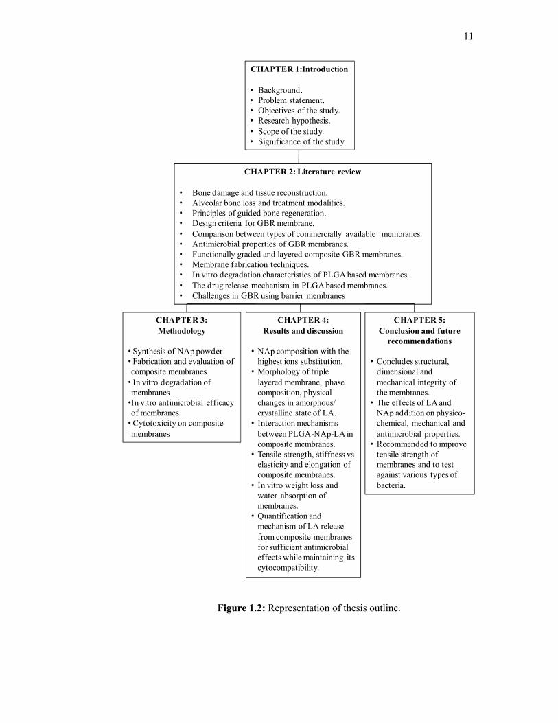
Figure 1.2: Representation of thesis outline.
CHAPTER 1:Introduction
• Background.• Problem statement.• Objectives of the study.• Research hypothesis.• Scope of the study.• Significance of the study.
CHAPTER 2: Literature review
• Bone damage and tissue reconstruction.• Alveolar bone loss and treatment modalities.• Principles of guided bone regeneration.• Design criteria for GBR membrane.• Comparison between types of commercially available membranes.• Antimicrobial properties of GBR membranes.• Functionally graded and layered composite GBR membranes.• Membrane fabrication techniques.• In vitro degradation characteristics of PLGA based membranes.• The drug release mechanism in PLGA based membranes.• Challenges in GBR using barrier membranes
CHAPTER 4: Results and discussion
• NAp composition with the highest ions substitution.
• Morphology of triple layered membrane, phase composition, physical changes in amorphous/ crystalline state of LA.
• Interaction mechanisms between PLGA-NAp-LA in composite membranes.
• Tensile strength, stiffness vselasticity and elongation of composite membranes.
• In vitro weight loss and water absorption of membranes.
• Quantification and mechanism of LA release from composite membranes for sufficient antimicrobial effects while maintaining its cytocompatibility.
CHAPTER 5: Conclusion and future
recommendations
• Concludes structural, dimensional and mechanical integrity of the membranes.
• The effects of LA and NAp addition on physico-chemical, mechanical and antimicrobial properties.
• Recommended to improve tensile strength of membranes and to test against various types of bacteria.
CHAPTER 3: Methodology
• Synthesis of NAp powder• Fabrication and evaluation of composite membranes• In vitro degradation of membranes•In vitro antimicrobial efficacy of membranes• Cytotoxicity on composite membranes
REFERENCES
1. Cai, Y., Guo, J., Chen, C., Yao, C., Chung, S.-m., Yao, J., Lee, I.-s. and
Kong, X. Silk fi broin membrane used for guided bone tissue regeneration.
Materials Science & Engineering C. 2017. 70: 148-154.
2. Pasetto, S., Herculano, R.D., Ereno, C., Silva, P., Graeff, C.F.O., Tavano, O.,
Baffa, O. and Kinoshita, A. Latex use as an occlusive membrane for guided
bone regeneration. Journal of Biomedical Materials Research A. 2010. 932-
939.
3. Moura, J.M.L., Ferreira, J.F., Marques, L., Holgado, L., Graeff, C.F.O. and
Kinoshita, A. Comparison of the performance of natural latex membranes
prepared with different procedures and PTFE membrane in guided bone
regeneration (GBR) in rabbits. Journal of Materials Science: Materials in
Medicine. 2014. 25(9): 2111-2120.
4. Fujihara, K., Kotaki, M. and Ramakrishna, S. Guided bone regeneration
membrane made of polycaprolactone/calcium carbonate composite nano-
fibers. Biomaterials. 2005. 26(19): 4139-4147.
5. Kellomäki, M., Niiranen, H., Puumanen, K., Ashammakhi, N., Waris, T. and
Törmälä, P. Bioabsorbable scaffolds for guided bone regeneration and
generation. Biomaterials. 2000. 21(24): 2495-2505.
6. Yang, F., Both, S.K., Yang, X., Walboomers, X.F. and Jansen, J.A.
Development of an electrospun nano-apatite/PCL composite membrane for
GTR/GBR application. Acta Biomaterialia. 2009. 5(9): 3295-3304.
7. Jung, R.E., Kokovic, V., Jurisic, M., Yaman, D., Subramani, K. and Weber,
F.E. Guided bone regeneration with a synthetic biodegradable membrane: a
comparative study in dogs. Clinical Oral Implants Research. 2011. 22(8):
802-807.
8. von Arx, T., Cochran, D.L., Schenk, R.K. and Buser, D. Evaluation of a
prototype trilayer membrane (PTLM) for lateral ridge augmentation: an
243
experimental study in the canine mandible. International Journal of Oral
and Maxillofacial Surgery. 2002. 31(2): 190-199.
9. van Leeuwen, A.C., Huddleston Slater, J.J.R., Gielkens, P.F.M., de Jong,
J.R., Grijpma, D.W. and Bos, R.R.M. Guided bone regeneration in rat
mandibular defects using resorbable poly(trimethylene carbonate) barrier
membranes. Acta Biomaterialia. 2012. 8(4): 1422-1429.
10. Peltola, O.L.J., Asikainen, A.J., Noponen, J. and Mesim, K.A. Tyrosine
derived polycarbonate membrane is useful for guided bone regeneration in
rabbit mandibular defects. Journal of Materials Science: Materials in
Medicine. 2005. 16: 753-758.
11. Jazayeri, H.E., Tahriri, M., Razavi, M., Khoshroo, K., Fahimipour, F.,
Dashtimoghadam, E., Almeida, L. and Tayebi, L. A current overview of
materials and strategies for potential use in maxillofacial tissue regeneration.
Materials Science & Engineering C. 2017. 70: 913-929.
12. Liao, S., Watari, F., Zhu, Y., Uo, M., Akasaka, T., Wang, W., Xu, G. and
Cui, F. The degradation of the three layered nano-carbonated
hydroxyapatite/collagen/PLGA composite membrane in vitro. Dental
Materials. 2007. 23(9): 1120-1128.
13. Bottino, M.C., Thomas, V. and Janowski, G.M. A novel spatially designed
and functionally graded electrospun membrane for periodontal regeneration.
Acta Biomaterialia. 2011. 7(1): 216-224.
14. Kikuchi, M., Koyama, Y., Yamada, T., Imamura, Y., Okada, T., Shirahama,
N., Akita, K., Takakuda, K. and Tanaka, J. Development of guided bone
regeneration membrane composed of β-tricalcium phosphate and poly (l-
lactide-co-glycolide-co-ε-caprolactone) composites. Biomaterials. 2004.
25(28): 5979-5986.
15. Puppi, D., Chiellini, F., Piras, A.M. and Chiellini, E. Polymeric materials for
bone and cartilage repair. Progress in Polymer Science. 2010. 35(4): 403-
440.
16. Liao, S., Wang, W., Uo, M., Ohkawa, S., Akasaka, T., Tamura, K., Cui, F.
and Watari, F. A three-layered nano-carbonated hydroxyapatite /collagen
/PLGA composite membrane for guided tissue regeneration. Biomaterials.
2005. 26(36): 7564-7571.
244
17. Song, X., Ling, F., Ma, L., Yang, C. and Chen, X. Electrospun
hydroxyapatite grafted poly(l-lactide)/poly(lactic-co-glycolic acid) nanofibers
for guided bone regeneration membrane. Composites Science and
Technology. 2013. 79: 8-14.
18. Zhou, H., Lawrence, J.G. and Bhaduri, S.B. Fabrication aspects of PLA-
CaP/PLGA-CaP composites for orthopedic applications: A review. Acta
Biomaterialia. 2012. 8(6): 1999-2016.
19. Campoccia, D., Montanaro, L. and Arciola, C.R. A review of the clinical
implications of anti-infective biomaterials and infection-resistant surfaces.
Biomaterials. 2013. 34(33): 8018-8029.
20. Münchow, E.A., Albuquerque, M.T.P., Zero, B., Kamocki, K., Piva, E.,
Gregory, R.L. and Bottino, M.C. Development and characterization of novel
ZnO-loaded electrospun membranes for periodontal regeneration. Dental
Materials. 2015. 31(9): 1038-1051.
21. Chen, D.W.C., Lee, F.Y., Liao, J.Y., Liu, S.J., Hsiao, C.Y. and Chen, J.K.
Preclinical Experiments on the Release Behavior of Biodegradable
Nanofibrous Multipharmaceutical Membranes in a Model of Four-Wall
Intrabony Defect. Antimicrobial Agents and Chemotherapy. 2012. 57(1): 9-
14.
22. Ji, W., Wang, H., van den Beucken, J.J.J.P., Yang, F., Walboomers, X.F.,
Leeuwenburgh, S. and Jansen, J.A. Local delivery of small and large
biomolecules in craniomaxillofacial bone. Advanced Drug Delivery Reviews.
2012. 64(12): 1152-1164.
23. Bottino, M.C., Thomas, V., Schmidt, G., Vohra, Y.K., Chu, T.-M.G.,
Kowolik, M.J. and Janowski, G.M. Recent advances in the development of
GTR/GBR membranes for periodontal regeneration—A materials
perspective. Dental Materials. 2012. 28(7): 703-721.
24. de Breij, A., Riool, M., Kwakman, P.H.S., de Boer, L., Cordfunke, R.A.,
Drijfhout, J.W., Cohen, O., Emanuel, N., Zaat, S.A.J., Nibbering, P.H. and
Moriarty, T.F. Prevention of Staphylococcus aureus biomaterial-associated
infections using a polymer-lipid coating containing the antimicrobial peptide
OP-145. Journal of Controlled Release. 2016. 222: 1-8.
25. Yang, D., Pornpattananangkul, D., Nakatsuji, T., Chan, M., Carson, D.,
Huang, C.-M. and Zhang, L. The antimicrobial activity of liposomal lauric
245
acids against Propionibacterium acnes. Biomaterials. 2009. 30(30): 6035-
6040.
26. Bergsson, G., Arnfinnsson, J., Steingrimsson, O. and Thormar, H. In Vitro
Killing of Candida albicans by Fatty Acids and Monoglycerides.
Antimicrobial Agents and Chemotherapy. 2001. 45(11): 3209-3212.
27. Rouse, M.S., Rotger, M., Piper, K.E., Steckelberg, J.M., Scholz, M.,
Andrews, J. and Patel, R. In Vitro and In Vivo Evaluations of the Activities
of Lauric Acid Monoester Formulations against Staphylococcus aureus.
Antimicrobial Agents and Chemotherapy. 2005. 49(8): 3187-3191.
28. Nobmann, P., Bourke, P., Dunne, J. and Henehan, G. In vitroantimicrobial
activity and mechanism of action of novel carbohydrate fatty acid derivatives
againstStaphylococcus aureusand MRSA. Journal of Applied Microbiology.
2010. 108: 2152-61.
29. Ho, M.-H., Hsieh, C.-C., Hsiao, S.-W. and Van Hong Thien, D. Fabrication
of asymmetric chitosan GTR membranes for the treatment of periodontal
disease. Carbohydrate Polymers. 2010. 79(4): 955-963.
30. Rowlands, A.S., Lim, S.A., Martin, D. and Cooper-White, J.J.
Polyurethane/poly(lactic-co-glycolic) acid composite scaffolds fabricated by
thermally induced phase separation. Biomaterials. 2007. 28(12): 2109-2121.
31. Schwach-abdellaoui, K., Vivien-castioni, N. and Gurny, R. Local delivery of
antimicrobial agents for the treatment of periodontal diseases. European
Journal of Pharmaceutics and Biopharmaceutics. 2000. 50: 83-99.
32. Sweeney, L.C. Antibiotic resistance in general dental practice--a cause for
concern? Journal of Antimicrobial Chemotherapy. 2004. 53(4): 567-576.
33. Herrera, D., Matesanz, P., Bascones-Martínez, A. and Sanz, M. Local and
Systemic Antimicrobial Therapy in Periodontics. Journal of Evidence Based
Dental Practice. 2012. 12(3): 50-60.
34. Sun, F., Zhou, H. and Lee, J. Various preparation methods of highly porous
hydroxyapatite/polymer nanoscale biocomposites for bone regeneration.
Acta Biomaterialia. 2011. 7(11): 3813-3828.
35. Vaquette, C. and Cooper-White, J. A simple method for fabricating 3-D
multilayered composite scaffolds. Acta Biomaterialia. 2013. 9(1): 4599-
4608.
246
36. Carlo Reis, E.C., Borges, A.P.B., Araújo, M.V.F., Mendes, V.C., Guan, L.
and Davies, J.E. Periodontal regeneration using a bilayered PLGA/calcium
phosphate construct. Biomaterials. 2011. 32(35): 9244-9253.
37. Owen, G.R., Jackson, J.K., Chehroudi, B., Brunette, D.M. and Burt, H.M.
An in vitro study of plasticized poly(lactic-co-glycolic acid) films as possible
guided tissue regeneration membranes: Material properties and drug release
kinetics. Journal of Biomedical Materials Research Part A. 2010. 95A(3):
857-869.
38. Lee, E.-J., Teng, S.-H., Jang, T.-S., Wang, P., Yook, S.-W., Kim, H.-E. and
Koh, Y.-H. Nanostructured poly(ε-caprolactone)–silica xerogel fibrous
membrane for guided bone regeneration. Acta Biomaterialia. 2010. 6(9):
3557-3565.
39. Yu, Z., Geng, J., Gao, H., Zhao, X. and Chen, J. Evaluations of guided bone
regeneration in canine radius segmental defects using autologous periosteum
combined with fascia lata under stable external fixation. Journal of
Orthopaedics and Traumatology. 2015. 16(2): 133-140.
40. Fu, S., Ni, P., Wang, B., Chu, B., Zheng, L., Luo, F., Luo, J. and Qian, Z.
Injectable and thermo-sensitive PEG-PCL-PEG copolymer/collagen/n-HA
hydrogel composite for guided bone regeneration. Biomaterials. 2012.
33(19): 4801-4809.
41. Lee, Y.J., Lee, J.-H., Cho, H.-J., Kim, H.K., Yoon, T.R. and Shin, H.
Electrospun fibers immobilized with bone forming peptide-1 derived from
BMP7 for guided bone regeneration. Biomaterials. 2013. 34(21): 5059-
5069.
42. Lee, E.-J., Shin, D.-S., Kim, H.-E., Kim, H.-W., Koh, Y.-H. and Jang, J.-H.
Membrane of hybrid chitosan–silica xerogel for guided bone regeneration.
Biomaterials. 2009. 30(5): 743-750.
43. Jung, R.E., Zwahlen, R., Weber, F.E., Molenberg, A., van Lenthe, G.H. and
Hammerle, C.H.F. Evaluation of an in situ formed synthetic hydrogel as a
biodegradable membrane for guided bone regeneration. Clinical Oral
Implants Research. 2006. 17(4): 426-433.
44. Nieminen, T., Kallela, I., Keränen, J., Hiidenheimo, I., Kainulainen, H.,
Wuolijoki, E. and Rantala, I. In vivo and in vitro degradation of a novel
247
bioactive guided tissue regeneration membrane. International Journal of
Oral and Maxillofacial Surgery. 2006. 35(8): 727-732.
45. Schliephake, H., Dard, M., Planck, H., Hierlemann, H. and Jakob, A. Guided
bone regeneration around endosseous implants using a resorbable membrane
vs a PTFE membrane. Clinical oral implants research. 2000. 11(3): 230-41.
46. Bottino, M.C., Kamocki, K., Yassen, G.H., Platt, J.A., Vail, M.M., Ehrlich,
Y., Spolnik, K.J. and Gregory, R.L. Bioactive Nanofibrous Scaffolds for
Regenerative Endodontics. Journal of Dental Research. 2013. 92(11): 963-
969.
47. Vasconcelos, M., Afonso, A., Branco, R. and Cavalheiro, J. Guided bone
regeneration using osteopatite granules and polytetrafluoroethylene
membranes. Journal of Materials Science: Materials in Medicine. 1997. 8:
815-818.
48. Hitti, R.A. and Kerns, D.G. Guided Bone Regeneration in the Oral Cavity :
A Review. Pathology. 2011. 5: 33-45.
49. Ten Heggeler, J.M.A.G., Slot, D.E. and Van der Weijden, G.A. Effect of
socket preservation therapies following tooth extraction in non-molar regions
in humans: a systematic review. Clinical Oral Implants Research. 2011.
22(8): 779-788.
50. Pilipchuk, S.P., Plonka, A.B., Monje, A., Taut, A.D., Lanis, A., Kang, B. and
Giannobile, W.V. Tissue engineering for bone regeneration and
osseointegration in the oral cavity. Dental Materials. 2015. 31(4): 317-338.
51. De Boever, A.L. and De Boever, J.A. Guided bone regeneration around non-
submerged implants in narrow alveolar ridges: a prospective long-term
clinical study. Clinical Oral Implants Research. 2005. 16(5): 549-556.
52. Rakhmatia, Y.D., Ayukawa, Y., Furuhashi, A. and Koyano, K. Current
barrier membranes: Titanium mesh and other membranes for guided bone
regeneration in dental applications. Journal of Prosthodontic Research.
2013. 57(1): 3-14.
53. Retzepi, M. and Donos, N. Guided Bone Regeneration: biological principle
and therapeutic applications. Clinical Oral Implants Research. 2010. 21(6):
567-576.
54. Hämmerle, C.H.F. and Jung, R.E. Bone augmentation by means of barrier
membranes. Periodontology 2000. 2003. 33: 36-53.
248
55. Dumitrescu, A.L. Guided Tissue Regeneration Barriers. 2011. 1-71.
56. Moses, O., Pitaru, S., Artzi, Z. and Nemcovsky, C.E. Healing of dehiscence-
type defects in implants placed together with different barrier membranes: a
comparative clinical study. Clinical Oral Implants Research. 2005. 16(2):
210-219.
57. Kim, Y.-K., Yun, P.-Y., Kim, S.-G. and Oh, D.S. In vitro scanning electron
microscopic comparison of inner surface of exposed and unexposed
nonresorbable membranes. Oral Surgery, Oral Medicine, Oral Pathology,
Oral Radiology, and Endodontology. 2009. 107(6): e5-e11.
58. Strietzel, F.P., Khongkhunthian, P., Khattiya, R., Patchanee, P. and Reichart,
P.A. Healing Pattern of Bone Defects Covered by Different Membrane
Types — A Histologic Study in the Porcine Mandible. Journal of Biomedical
Materials Research Part B: Applied Biomaterials. 2005. 35-46.
59. Donos, N., Kostopoulos, L. and Karring, T. Alveolar ridge augmentation
using a resorbable copolymer membrane and autogenous bone grafts. An
experimental study in the rat. Clinical oral implants research. 2002. 13(2):
203-213.
60. Belleggia, F. Treatment of an infected exposure of a dense
polytetrafluoroethylene membrane in a vertical guided bone regeneration
procedure : a protocol proposal. Clinical Oral Implants Research. 2014. 25
(Suppl. 10).
61. Harzeler, M.R., Kohal, R.J., Naghshbandi, J., Mota, L.F., Conradt, J., Mote,
L.F. and Conradt, J. Evaluation of a new bioresorbable barrier to facilitate
guided bone regeneration around exposed implant threads An experimental
study in the monkey. Int. J. Oral Maxillofacial Surgery. 1998. 27: 315-320.
62. Coelho, P.G., Giro, G., Kim, W., Granato, R., Marin, C., Bonfante, E.A.,
Bonfante, S., Lilin, T. and Suzuki, M. Evaluation of collagen-based
membranes for guided bone regeneration, by three-dimensional computerized
microtomography. Oral Surgery, Oral Medicine, Oral Pathology and Oral
Radiology. 2012. 114(4): 437-443.
63. Gentile, P., Chiono, V., Tonda-Turo, C., Ferreira, A.M. and Ciardelli, G.
Polymeric membranes for guided bone regeneration. Biotechnology Journal.
2011. 6(10): 1187-1197.
249
64. Song, J.-h., Kim, H.-e. and Kim, H.-w. Collagen-Apatite Nanocomposite
Membranes for Guided Bone Regeneration. Journal of Biomedical Materials
Research. 2007. 248-257.
65. Landi, E., Tampieri, A., Mattioli-Belmonte, M., Celotti, G., Sandri, M.,
Gigante, A., Fava, P. and Biagini, G. Biomimetic Mg- and Mg,CO3-
substituted hydroxyapatites: synthesis characterization and in vitro behaviour.
Journal of the European Ceramic Society. 2006. 26(13): 2593-2601.
66. Zhou, H. and Lee, J. Nanoscale hydroxyapatite particles for bone tissue
engineering. Acta Biomaterialia. 2011. 7(7): 2769-2781.
67. Park, J.K., Yeom, J., Oh, E.J., Reddy, M., Kim, J.Y., Cho, D.-W., Lim, H.P.,
Kim, N.S., Park, S.W., Shin, H.-I., Yang, D.J., Park, K.B. and Hahn, S.K.
Guided bone regeneration by poly(lactic-co-glycolic acid) grafted hyaluronic
acid bi-layer films for periodontal barrier applications. Acta Biomaterialia.
2009. 5(9): 3394-3403.
68. Parent, M., Nouvel, C., Koerber, M., Sapin, A., Maincent, P. and Boudier, A.
PLGA in situ implants formed by phase inversion: Critical physicochemical
parameters to modulate drug release. Journal of Controlled Release. 2013.
172(1): 292-304.
69. Rezwan, K., Chen, Q.Z., Blaker, J.J. and Boccaccini, A.R. Biodegradable
and bioactive porous polymer/inorganic composite scaffolds for bone tissue
engineering. Biomaterials. 2006. 27(18): 3413-3431.
70. Budyanto, L., Goh, Y.Q. and Ooi, C.P. Fabrication of porous poly(L-lactide)
(PLLA) scaffolds for tissue engineering using liquid–liquid phase separation
and freeze extraction. Journal of Materials Science: Materials in Medicine.
2008. 20(1): 105-111.
71. Zamani, M., Morshed, M., Varshosaz, J. and Jannesari, M. Controlled
release of metronidazole benzoate from poly ε-caprolactone electrospun
nanofibers for periodontal diseases. European Journal of Pharmaceutics and
Biopharmaceutics. 2010. 75(2): 179-185.
72. Chen, S., Hao, Y., Cui, W., Chang, J. and Zhou, Y. Biodegradable
electrospun PLLA/chitosan membrane as guided tissue regeneration
membrane for treating periodontitis. Journal of Materials Science. 2013.
48(19): 6567-6577.
250
73. Ranjbar-Mohammadi, M., Zamani, M., Prabhakaran, M.P., Bahrami, S.H.
and Ramakrishna, S. Electrospinning of PLGA/gum tragacanth nanofibers
containing tetracycline hydrochloride for periodontal regeneration. Materials
Science and Engineering: C. 2016. 58: 521-531.
74. Yüksel, E., Karakeçili, A., Demirtaş, T.T. and Gümüşderelioğlu, M.
Preparation of bioactive and antimicrobial PLGA membranes by magainin
II/EGF functionalization. International Journal of Biological
Macromolecules. 2016. 86: 162-168.
75. Dorozhkin, S.V. Nanosized and nanocrystalline calcium orthophosphates.
Acta Biomaterialia. 2010. 6(3): 715-734.
76. Laurencin, D., Almora-Barrios, N., de Leeuw, N.H., Gervais, C., Bonhomme,
C., Mauri, F., Chrzanowski, W., Knowles, J.C., Newport, R.J., Wong, A.,
Gan, Z. and Smith, M.E. Magnesium incorporation into hydroxyapatite.
Biomaterials. 2011. 32(7): 1826-1837.
77. Kannan, S., Ventura, J.M.G. and Ferreira, J.M.F. Synthesis and thermal
stability of potassium substituted hydroxyapatites and hydroxyapatite/β-
tricalciumphosphate mixtures. Ceramics International. 2007. 33(8): 1489-
1494.
78. Kothapalli, C., Wei, M., Vasiliev, A. and Shaw, M.T. Influence of
temperature and concentration on the sintering behavior and mechanical
properties of hydroxyapatite. Acta Materialia. 2004. 52(19): 5655-5663.
79. Pretto, M., Costa, A.L., Landi, E., Tampieri, A., Galassi, C. Dispersing
Behavior of Hydroxyapatite Powders Produced by Wet-Chemical Synthesis.
J. Am. Ceram. Soc. 2003. 86(9): 1534-39.
80. Kannan, S., Vieira, S.I., Olhero, S.M., Torres, P.M.C., Pina, S., da Cruz e
Silva, O.A.B. and Ferreira, J.M.F. Synthesis, mechanical and biological
characterization of ionic doped carbonated hydroxyapatite/β-tricalcium
phosphate mixtures. Acta Biomaterialia. 2011. 7(4): 1835-1843.
81. Kim, S.R., Lee, J.H., Kim, Y.T., Riu, D.H., Jung, S.J., Lee, Y.J., Chung, S.C.
and Kim, Y.H. Synthesis of Si, Mg substituted hydroxyapatites and their
sintering behaviors. Biomaterials. 2003. 24(8): 1389-98.
82. Sprio, S., Tampieri, A., Landi, E., Sandri, M., Martorana, S., Celotti, G. and
Logroscino, G. Physico-chemical properties and solubility behaviour of
251
multi-substituted hydroxyapatite powders containing silicon. Materials
Science and Engineering: C. 2008. 28(1): 179-187.
83. Landi, E., Tampieri, A., Celotti, G., Sprio, S., Sandri, M. and Logroscino, G.
Sr-substituted hydroxyapatites for osteoporotic bone replacement. Acta
Biomaterialia. 2007. 3(6): 961-969.
84. Ramesh, S., Tan, C.Y., Tolouei, R., Amiriyan, M., Purbolaksono, J., Sopyan,
I. and Teng, W.D. Sintering behavior of hydroxyapatite prepared from
different routes. Materials & Design. 2012. 34: 148-154.
85. Sopyan, I., Ramesh, S. and Hamdi, M. Synthesis of nano sized
hydroxyapatite powder using sol-gel technique and its conversion to dense
and porous bodies. Indian Journal of Chemistry. 2008. 47(November):
1626-1631.
86. Landi, E., Tampieri, A., Celotti, G. and Sprio, S. Densification behaviour
and mechanisms of synthetic hydroxyapatites. J. Euro. Ceram. Soc. 2000.
20: 2377-2387.
87. Uskoković, V. and Uskoković, D.P. Nanosized hydroxyapatite and other
calcium phosphates: Chemistry of formation and application as drug and gene
delivery agents. Journal of Biomedical Materials Research Part B: Applied
Biomaterials. 2011. 96B(1): 152-191.
88. Kuriakose, T.A., Kalkura, S.N., Palanichamy, M., Arivuoli, D., Dierks, K.,
Bocelli, G. and Betzel, C. Synthesis of stoichiometric nano crystalline
hydroxyapatite by ethanol-based sol–gel technique at low temperature.
Journal of Crystal Growth. 2004. 263(1-4): 517-523.
89. Pang, Y.X. and Bao, X. Influence of temperature, ripening time and
calcination on the morphology and crystallinity of hydroxyapatite
nanoparticles. Journal of the European Ceramic Society. 2003. 23(10):
1697-1704.
90. Cacciotti, I., Bianco, A., Lombardi, M. and Montanaro, L. Mg-substituted
hydroxyapatite nanopowders: Synthesis, thermal stability and sintering
behaviour. Journal of the European Ceramic Society. 2009. 29(14): 2969-
2978.
91. Boanini, E., Gazzano, M. and Bigi, A. Ionic substitutions in calcium
phosphates synthesized at low temperature. Acta Biomaterialia. 2010. 6(6):
1882-1894.
252
92. Lazic, S., Zec, S., Miljevic, N. and Milonjic, S. The effect of temperature on
the properties of hydroxyapatite precipitated from calcium hydroxide and
phosphoric acid. Thermochimica acta. 2001. 374: 13-22.
93. Reise, M., Wyrwa, R., Müller, U., Zylinski, M., Völpel, A., Schnabelrauch,
M., Berg, A., Jandt, K.D., Watts, D.C. and Sigusch, B.W. Release of
metronidazole from electrospun poly(l-lactide-co-d/l-lactide) fibers for local
periodontitis treatment. Dental Materials. 2012. 28(2): 179-188.
94. Xue, J., He, M., Niu, Y., Liu, H., Crawford, A., Coates, P., Chen, D., Shi, R.
and Zhang, L. Preparation and in vivo efficient anti-infection property of
GTR/GBR implant made by metronidazole loaded electrospun
polycaprolactone nanofiber membrane. International Journal of
Pharmaceutics. 2014. 475(1-2): 566-577.
95. Sundararaj, S.C., Thomas, M.V., Peyyala, R., Dziubla, T.D. and Puleo, D.A.
Design of a multiple drug delivery system directed at periodontitis.
Biomaterials. 2013. 34(34): 8835-8842.
96. Lan, S.-F., Kehinde, T., Zhang, X., Khajotia, S., Schmidtke, D.W. and Starly,
B. Controlled release of metronidazole from composite poly-ε-
caprolactone/alginate (PCL/alginate) rings for dental implants. Dental
Materials. 2013. 29(6): 656-665.
97. Kitahara, T., Aoyama, Y., Hirakata, Y., Kamihira, S., Kohno, S., Ichikawa,
N., Nakashima, M., Sasaki, H. and Higuchi, S. In vitro activity of lauric acid
or myristylamine in combination with six antimicrobial agents against
methicillin-resistant Staphylococcus aureus (MRSA). International Journal
of Antimicrobial Agents. 2006. 27(1): 51-57.
98. Pragati, S., Ashok, S. and Kuldeep, S. Recent advances in periodontal drug
delivery systems. International Journal of Drug Delivery. 2009. 1: 1-14.
99. Castillo, A., Liebana, J., Lopez, E., Baca, P., Liebana, M. and Castillo, F.
Interference of antibiotics in the growth curves of oral streptococci.
International Journal of Antimicrobial Agents. 2006. 27(3): 263-266.
100. Pornpattananangkul, D., Fu, V., Thamphiwatana, S., Zhang, L., Chen, M.,
Vecchio, J., Gao, W., Huang, C.-M. and Zhang, L. In Vivo Treatment
ofPropionibacterium acnesInfection with Liposomal Lauric Acids. Advanced
Healthcare Materials. 2013. 2(10): 1322-1328.
253
101. Huang, C.B., Alimova, Y., Myers, T.M. and Ebersole, J.L. Short- and
medium-chain fatty acids exhibit antimicrobial activity for oral
microorganisms. Archives of Oral Biology. 2011. 56(7): 650-654.
102. Kitahara, T., Koyama, N., Matsuda, J., Aoyama, Y., Hirakata, Y., Kamihira,
S., Kohno, S., Nakashima, M. and Sasaki, H. Antimicrobial activity of
saturated fatty acids and fatty amines against methicillin-resistant
Staphylococcus aureus. Biological & pharmaceutical bulletin. 2004. 27(9):
1321-1326.
103. Amet, Y., Adas, F. and Berthou, F. High performance liquid chromatography
of fatty acid metabolites Improvement of sensitivity by radiometric ,
fluorimetric and mass spectrometric methods. Analytica Chimica Acta.
2002. 465: 193-198.
104. Zhao, L., Hu, Y., Xu, D. and Cai, K. Surface functionalization of titanium
substrates with chitosan–lauric acid conjugate to enhance osteoblasts
functions and inhibit bacteria adhesion. Colloids and Surfaces B:
Biointerfaces. 2014. 119: 115-125.
105. Renouf-Glauser, A.C., Rose, J., Farrar, D. and Cameron, R.E. A degradation
study of PLLA containing lauric acid. Biomaterials. 2005. 26(15): 2415-
2422.
106. Teng, S.-H., Lee, E.-J., Wang, P., Shin, D.-S. and Kim, H.-E. Three-layered
membranes of collagen/hydroxyapatite and chitosan for guided bone
regeneration. Journal of Biomedical Materials Research Part B: Applied
Biomaterials. 2008. 87B(1): 132-138.
107. Vaquette, C., Frochot, C., Rahouadj, R. and Wang, X. An innovative method
to obtain porous PLLA scaffolds with highly spherical and interconnected
pores. Journal of Biomedical Materials Research Part B: Applied
Biomaterials. 2008. 86B(1): 9-17.
108. Vaquette, C. and Cooper-White, J.J. Increasing electrospun scaffold pore
size with tailored collectors for improved cell penetration. Acta
Biomaterialia. 2011. 7(6): 2544-2557.
109. Ji, C., Khademhosseini, A. and Dehghani, F. Enhancing cell penetration and
proliferation in chitosan hydrogels for tissue engineering applications.
Biomaterials. 2011. 32(36): 9719-9729.
254
110. Cho, W.J., Kim, J.H., Oh, S.H., Nam, H.H., Kim, J.M. and Lee, J.H.
Hydrophilized polycaprolactone nanofiber mesh-embedded poly(glycolic-co-
lactic acid) membrane for effective guided bone regeneration. Journal of
Biomedical Materials Research Part A. 2009. 91A(2): 400-407.
111. Teng, S.-H., Lee, E.-J., Yoon, B.-H., Shin, D.-S., Kim, H.-E. and Oh, J.-S.
Chitosan/nanohydroxyapatite composite membranes via dynamic filtration
for guided bone regeneration. Journal of Biomedical Materials Research
Part A. 2009. 88A(3): 569-580.
112. Sun, F., Lim, B.-K., Ryu, S.-C., Lee, D. and Lee, J. Preparation of multi-
layered film of hydroxyapatite and chitosan. Materials Science and
Engineering: C. 2010. 30(6): 789-794.
113. Yang, Y., Zhao, J., Zhao, Y., Wen, L., Yuan, X. and Fan, Y. Formation of
porous PLGA scaffolds by a combining method of thermally induced phase
separation and porogen leaching. Journal of Applied Polymer Science. 2008.
109(2): 1232-1241.
114. Ho, M.-H., Kuo, P.-Y., Hsieh, H.-J., Hsien, T.-Y., Hou, L.-T., Lai, J.-Y. and
Wang, D.-M. Preparation of porous scaffolds by using freeze-extraction and
freeze-gelation methods. Biomaterials. 2004. 25(1): 129-138.
115. Mu, C., Su, Y., Sun, M., Chen, W. and Jiang, Z. Fabrication of microporous
membranes by a feasible freeze method. Journal of Membrane Science.
2010. 361(1-2): 15-21.
116. Wu, L. and Ding, J. In vitro degradation of three-dimensional porous
poly(d,l-lactide-co-glycolide) scaffolds for tissue engineering. Biomaterials.
2004. 25(27): 5821-5830.
117. Orava, E., Korventausta, J., Rosenberg, M., Jokinen, M. and Rosling, A. In
vitro degradation of porous poly(dl-lactide-co-glycolide) (PLGA)/bioactive
glass composite foams with a polar structure. Polymer Degradation and
Stability. 2007. 92(1): 14-23.
118. Deb, S., Braden, M. and Bonfield, W. Water absorption characteristics of
modified hydroxyapatite bone cements. Biomaterials. 1995. 16: 1095-1100.
119. Tang, C.Y., Chen, D.Z., Yue, T.M., Chan, K.C., Tsui, C.P. and Yu, P.H.F.
Water absorption and solubility of PHBHV/HA nanocomposites. Composites
Science and Technology. 2008. 68(7-8): 1927-1934.
255
120. Yang, Y., Zhao, Y., Tang, G., Li, H., Yuan, X. and Fan, Y. In vitro
degradation of porous poly(l-lactide-co-glycolide)/β-tricalcium phosphate
(PLGA/β-TCP) scaffolds under dynamic and static conditions. Polymer
Degradation and Stability. 2008. 93(10): 1838-1845.
121. Dorati, R., Colonna, C., Genta, I., Modena, T. and Conti, B. Effect of
porogen on the physico-chemical properties and degradation performance of
PLGA scaffolds. Polymer Degradation and Stability. 2010. 95(4): 694-701.
122. Fredenberg, S., Wahlgren, M., Reslow, M. and Axelsson, A. The
mechanisms of drug release in poly(lactic-co-glycolic acid)-based drug
delivery systems—A review. International Journal of Pharmaceutics. 2011.
415(1-2): 34-52.
123. Stevanović, M., Bračko, I., Milenković, M., Filipović, N., Nunić, J., Filipič,
M. and Uskoković, D.P. Multifunctional PLGA particles containing poly(l-
glutamic acid)-capped silver nanoparticles and ascorbic acid with
simultaneous antioxidative and prolonged antimicrobial activity. Acta
Biomaterialia. 2014. 10(1): 151-162.
124. Said, S.S., Aloufy, A.K., El-Halfawy, O.M., Boraei, N.A. and El-Khordagui,
L.K. Antimicrobial PLGA ultrafine fibers: Interaction with wound bacteria.
European Journal of Pharmaceutics and Biopharmaceutics. 2011. 79(1):
108-118.
125. Woodruff, M.A. and Hutmacher, D.W. The return of a forgotten polymer—
Polycaprolactone in the 21st century. Progress in Polymer Science. 2010.
35(10): 1217-1256.
126. Ma, D. and McHugh, A.J. The interplay of membrane formation and drug
release in solution-cast films of polylactide polymers. International Journal
of Pharmaceutics. 2010. 388(1-2): 1-12.
127. Machín, R., Isasi, J.R. and Vélaz, I. Hydrogel matrices containing single and
mixed natural cyclodextrins. Mechanisms of drug release. European Polymer
Journal. 2013. 49(12): 3912-3920.
128. Wei, Z., Wang, C., Liu, H., Zou, S. and Tong, Z. Facile fabrication of
biocompatible PLGA drug-carrying microspheres by O/W pickering
emulsions. Colloids and Surfaces B: Biointerfaces. 2012. 91: 97-105.
256
129. Lao, L.L., Peppas, N.A., Boey, F.Y.C. and Venkatraman, S.S. Modeling of
drug release from bulk-degrading polymers. International Journal of
Pharmaceutics. 2011. 418(1): 28-41.
130. Peppas, N.A. and Narasimhan, B. Mathematical models in drug delivery:
How modeling has shaped the way we design new drug delivery systems.
Journal of Controlled Release. 2014. 190: 75-81.
131. Chou, A.H.K., LeGeros, R.Z., Chen, Z. and Li, Y. Antibacterial Effect of
Zinc Phosphate Mineralized Guided Bone Regeneration Membranes. Implant
Dentistry. 2007. 16(1): 89-100.
132. Nerem, R. Principles of tissue engineering. In: R.L. RP Lanza, J Vancanti. ed.
The Challenge of Imitating Nature. San Diego: Academic Press. 9-11; 2000.
133. Zhang, M. Biomaterials and Tissue engineering. In: D. Shi. ed.
Biocompatibility of materials. Heidelberg: Springer. 83-143; 2004.
134. Franks, K., Salih, V., Knowles, J. and Olsen, I. The effect of MgO on the
solubility behavior and cell proliferation in a quaternary soluble phosphate
based glass system. Journal of Materials Science: Materials in Medicine.
2002. 13: 549-556.
135. Marques, A., Reis, R. and Hunt, J. The biocompatibility of novel starch-
based polymers and composites: in vitro studies. Biomaterials. 2002. 23:
1471-1478.
136. Renouf-Glauser, A.C., Rose, J., Farrar, D.F. and Cameron, R.E. Comparison
of the hydrolytic degradation and deformation properties of a PLLA-lauric
acid based family of biomaterials. Biomacromolecules. 2006. 7(2): 612-617.
137. Li, Z.Y., Lam, W.M., Yang, C., Xu, B., Ni, G.X., Abbah, S.A., Cheung,
K.M.C., Luk, K.D.K. and Lu, W.W. Chemical composition, crystal size and
lattice structural changes after incorporation of strontium into biomimetic
apatite. Biomaterials. 2007. 28(7): 1452-1460.
138. Lima, I.R.D., Alves, G.G., Soriano, C.A., Campaneli, A.P., Gasparoto, T.H.,
Schnaider, E., Junior, R. and Sena, D. Understanding the impact of divalent
cation substitution on hydroxyapatite: An in vitro multiparametric study on
biocompatibility. Journal of Biomedical Materials Research Part A. 2011.
351-358.
139. Bottino, M.C., Coelho, P.G., Henriques, V.A.R., Higa, O.Z., Bressiani,
A.H.A. and Bressiani, J.C. Processing, characterization, and in vitro/in vivo
257
evaluations of powder metallurgy processed Ti-13Nb-13Zr alloys. Journal of
Biomedical Materials Research Part A. 2009. 88(3): 689-696.
140. Stevanović, M.M., Škapin, S.D., Bračko, I., Milenković, M., Petković, J.,
Filipič, M. and Uskoković, D.P. Poly(lactide-co-glycolide)/silver
nanoparticles: Synthesis, characterization, antimicrobial activity, cytotoxicity
assessment and ROS-inducing potential. Polymer. 2012. 53(14): 2818-2828.
141. Tripathi, G., Gough, J.E., Dinda, A. and Basu, B. In vitro cytotoxicity and in
vivo osseointergration properties of compression-molded HDPE-HA-Al2O3
hybrid biocomposites. J Biomed Mater Res Part A. 2013. 101A: 1539-1549.
142. Bonnier, F., Keating, M.E., Wróbel, T.P., Majzner, K., Baranska, M., Garcia-
munoz, A. and Blanco, A. Toxicology in Vitro Cell viability assessment
using the Alamar blue assay: A comparison of 2D and 3D cell culture
models. Toxicology Invitro. 2015. 29(1): 124-131.
143. Akiko Watabe, Y., Yamaguchi, T., Kawanishi, T. and Uchida, E. Target-cell
specificity of fusogenic liposomes: Membrane fusion-mediated
macromolecule delivery into human blood mononuclear cells. Biochimica et
Biophysica Acta. 1999. 1416: 339-348.
144. International Standard Organisation. Tests for cytotoxicity: in vitro methods.
Switzerland, ISO-10993-5. 2009.
145. Coecke, S., Balls, M., Bowe, G., Davis, J., Cstraunthaler, G., Hartung, T.,
Hay, R., Merten, O., Price, A., Schectman, L., Stacey, G. and Stokes, W.
Guidance on Good Cell Culture Practice, A Report of the Second ECVAM
Task Force on Good Cell Culture Practice, ATLA. 2005. 33: 261-287.
146. Fentem, J., Curren, R., Liebsch, M. Guidance Document on Using In vitro
Data to Estimate In vivo Starting Doses for Acute Toxicity. NIH Publication
No. 01-4500. August 2001.
147. American Type Culture Collection (ATCC). American Type Culture
Collection Animal Cell Culture Guide (tips and techniques for continuous
cell lines). U.S. 2014.
148. Cadena-herrera, D., Lara, J.E.E.-d., Ramírez-ibañez, N.D., López-morales,
C.A., Pérez, N.O., Flores-ortiz, L.F. and Medina-rivero, E. Validation of
three viable-cell counting methods: Manual, semi-automated, and automated.
Biotechnology Reports. 2015. 7: 9-16.
258
149. K.S. Louis, A.C.S., G.A. Levy. Comparison of manual versus automated
trypan blue dye exclusion method for cell counting. In: Stoddart, M.J. ed.
Mammalian cell viability: Methods and Protocols, Methods in Molecular
Biology, New York: Springer Protocols. 7-12; 2015.
150. International Standard Organisation. Biological Evaluation of
MedicalDevices—Part 12: Sample Preparation and Reference Materials.
Switzerland, ISO-10993-12. 2002.
151. Dygai, A.M., Ogorodova, L.M., Psakhie, S.G., Belsky, Y.P., Belska, N.V.,
Danilets, M.G., Ligatcheva, A.A. and Churin, A.A. A study of the
Cytotoxicity of a New Nonwoven Polymeric Fibrous Bandaging Material in
Vitro. Journal of Biomaterials and Nanobiotechnology. 2011. 2(July): 234-
238.
152. Tavares, D.D.S., Castro, L.D.O., Soares, G.D.D.A., Alves, G.G. and
Granjeiro, J.M. Synthesis and cytotoxicity evaluation of granular magnesium
substituted β-tricalcium phosphate. Journal of Applied Oral Science. 2013.
21(1): 37-42.
153. Tomida, M., Nakano, K., Matsuura, S. and Kawakami, T. Comparative
examination of subcutaneous tissue reaction to high molecular materials in
medical use. European Journal of Medical Research. 2011. 16: 249-252.
154. Dimitrievska, S., Petit, A., Ajji, A., Bureau, M.N., Yahia, L.H. and Montre,
P.D. Biocompatibility of novel polymer-apatite nanocomposite fibers.
Journal of Biomedical Materials Research Part A. 2008. 84A(1): 44-53.
155. Wang, B.L.-s., Chow, P.-y., Phan, T.-t., Lim, I.J. and Yang, Y.-y. Fabrication
and Characterization of Nanostructured and Thermosensitive Polymer
Membranes for Wound Healing and Cell Grafting. Adv. Funct. Mater. 2006.
16: 1171-1178.
156. Xue, M., Hu, H., Jiang, Y., Liu, J., He, H. and Ye, X. Biodegradable
polymer-coated, gelatin hydrogel/bioceramics ternary composites for
antitubercular drug delivery and tissue regeneration. Journal of
Nanomaterials. 2012. (Volume 2012): Article ID 530978.
157. Fisher, Material Safety Data Sheet of Dimethyl Sulfoxide. UK: 2012.
158. American Society for Testing and Materials. Standard Test Method for in
vitro Degradation Testing of Hydrolytically Degradable Polymer Resins and
Fabricated Forms for Surgical Implants. U.S., F1635-11. 2011.
259
159. Durect LACTEL Absorbable Polymers. Material Safety Data Sheet of
Poly(lactic-co-glycolic) acid. U.S.:2012.
160. Loo, S.C.J., Tan, Z.Y.S., Chow, Y.J. and Lin, S.L.I. Drug Release From
Irradiated PLGA and PLLA Multi-Layered Films. Journal of pharmaceutical
sciences. 2010. 99(7): 3060-3071.
161. Mehta, A., Oeser, a.M. and Carlson, M.G. Rapid quantitation of free fatty
acids in human plasma by high-performance liquid chromatography. Journal
of chromatography. B, Biomedical sciences and applications. 1998. 719(1-
2): 9-23.
162. Jaidev, L.R., Krishnan, U.M. and Sethuraman, S. Gemcitabine loaded
biodegradable PLGA nanospheres for in vitro pancreatic cancer therapy.
Materials Science and Engineering: C. 2015. 47: 40-47.
163. Simchi, A., Tamjid, E., Pishbin, F. and Boccaccini, A.R. Recent progress in
inorganic and composite coatings with bactericidal capability for orthopaedic
applications. Nanomedicine: Nanotechnology, Biology, and Medicine. 2011.
7(1): 22-39.
164. Raphel, J., Holodniy, M., Goodman, S.B. and Heilshorn, S.C.
Multifunctional Coatings to Simultaneously Promote Osseointegration and
Prevent Infection of Orthopaedic Implants. Biomaterials. 2016. 84: 301-314.
165. Hoben, H.J. and Somasegaran, P. Comparison of the Pour, Spread and Drop
plate methods for enumeration of Rhizobium spp. in inoculants made from
presterilized peat. Applied and Environmental Microbiology. 1982. 44(5):
1246-1247.
166. Miles. A. A. and Misra, S.S. The estimation of the bactericidal power of
blood. Journal of Hygiene. 1938. 38: 732-749.
167. American Society for Testing and Materials. Determining the activity of
Incorporated Antimicrobial Agent(s) In Polymeric or Hydrophobic Materials.
U.S., E2180-07. 2012.
168. Braghirolli, D.I., Steffens, D., Quintiliano, K., Acasigua, G.A.X., Gamba, D.,
Fleck, R.A., Petzhold, C.L. and Pranke, P. The effect of sterilization methods
on electronspun poly(lactide-co-glycolide) and subsequent adhesion
efficiency of mesenchymal stem cells. Journal of Biomedical Materials
Research - Part B Applied Biomaterials. 2014. 102(4).
260
169. Cañadas, C., Alvarado, H., Calpena, A.C., Silva, A.M., Souto, E.B., García,
M.L. and Abrego, G. In vitro , ex vivo and in vivo characterization of PLGA
nanoparticles loading pranoprofen for ocular administration. International
Journal of Pharmaceutics. 2016. 511: 719-727.
170. Rameshbabu, N., Sampath Kumar, T.S., Prabhakar, T.G., Sastry, V.S., Murty,
K.V.G.K. and Prasad Rao, K. Antibacterial nanosized silver substituted
hydroxyapatite: Synthesis and characterization. Journal of Biomedical
Materials Research Part A. 2007. 80A(3): 581-591.
171. Panda, R.N., Hsieh, M.F., Chung, R.J. and Chin, T.S. FTIR , XRD , SEM
and solid state NMR investigations of synthesized by hydroxide-gel
technique. Journal of Physics and Chemistry of Solids. 2003. 64: 193-199.
172. Bogdanoviciene, I., Beganskiene, A., Tõnsuaadu, K., Glaser, J., Meyer, H.J.
and Kareiva, A. Calcium hydroxyapatite, Ca10(PO4)6(OH)2 ceramics
prepared by aqueous sol–gel processing. Materials Research Bulletin. 2006.
41(9): 1754-1762.
173. Costescu, A., Pasuk, I., Ungureanu, F., Dinischiotu, A., Huneau, F., Galaup,
S., Coustumer, P.L.E., Predoi, D. and Ftir, C. Physico-chemical properties of
nano-sized hexagonal hydroxyapatite powder synthesised by sol-gel. Journal
of Nanomaterials. 2010. 5(4): 989-1000.
174. Landi, E., Tampieri, A., Celotti, G., Vichi, L. and Sandri, M. Influence of
synthesis and sintering parameters on the characteristics of carbonate apatite.
Biomaterials. 2004. 25(10): 1763-1770.
175. Greish, Y.E., Sturgeon, J.L., Singh, A., Krogman, N.R., Touny, A.H.,
Sethuraman, S., Nair, L.S., Laurencin, C.T., Allcock, H.R. and Brown, P.W.
Formation and properties of composites comprised of calcium-deficient
hydroxyapatites and ethyl alanate polyphosphazenes. Journal of Materials
Science: Materials in Medicine. 2008. 19(9): 3153-3160.
176. Gustavsson, J., Ginebra, M.P., Engel, E. and Planell, J. Ion reactivity of
calcium-deficient hydroxyapatite in standard cell culture media. Acta
Biomaterialia. 2011. 7(12): 4242-4252.
177. Koutsopoulos, S. Synthesis and characterization of hydroxyapatite crystals: a
review study on the analytical methods. Journal of Biomedical Materials
Research Part A. 2002. 62: 600-612.
261
178. Gibson, I.R., Ke, S., Best, S.M. and Bonfield, W. Effect of powder
characteristics on the sinterability of hydroxyapatite powders. Journal of
materials science. Materials in medicine. 2001. 12(2): 163-171.
179. Suetsugu, Y., Takahashi, Y., Okamura, F.P. and Tanaka, J. Structure analysis
of A-type carbonate apatite by a single-crystal x-ray diffraction method.
Journal of Solid State Chemistry. 2000. 155: 292-297.
180. El Feki, H., Savariault, J.M., Ben Salah, A. Structure refinements by the
Rietveld method of partially substituted hydroxyapatite:
Ca9Na0.5(PO4)4.5(CO3)1.5(OH)2. Journal of Alloys and Compounds. 1999.
287: 114-120.
181. Gibson, I.R. and Bonfield, W. Novel synthesis and characterization of an
AB-type carbonate-substituted hydroxyapatite. Journal of Biomedical
Materials Research. 2002. 59(4): 697-708.
182. Joschek, S., Nies, B., Krotz, R. and Göferich, A. Chemical and
physicochemical characterization of porous hydroxyapatite ceramics made of
natural bone. Biomaterials. 2000. 21(16): 1645-58.
183. Fu, X., Liu, Z., Xiao, Y., Wang, J. and Lei, J. Preparation and properties of
lauric acid/diatomite composites as novel form-stable phase change materials
for thermal energy storage. Energy and Buildings. 2015. 104: 244-249.
184. Hao, S., Wang, Y., Wang, B., Deng, J., Zhu, L. and Cao, Y. Formulation of
porous poly(lactic-co-glycolic acid) microparticles by electrospray deposition
method for controlled drug release. Materials science & engineering. C,
Materials for biological applications. 2014. 39: 113-119.
185. Wei, J. and Li, Y. Tissue engineering scaffold material of nano-apatite
crystals and polyamide composite. European Polymer Journal. 2004. 40(3):
509-515.
186. Jose, M., Thomas, V., Johnson, K., Dean, D. and Nyairo, E. Aligned
PLGA/HA nanofibrous nanocomposite scaffolds for bone tissue engineering.
Acta Biomaterialia. 2009. 5(1): 305-315.
187. Tanaka, H., Watanabe, T., Chikazawa, M., Kandori, K. and Ishikawa, T.
TPD , FTIR , and Molecular Adsorption Studies of Calcium Hydroxyapatite
Surface Modified with Hexanoic and Decanoic Acids. Journal of Colloid
and Interface Science. 1999. 214: 31-37.
262
188. Zhou, S., Zheng, X., Yu, X., Wang, J., Weng, J., Li, X., Feng, B. and Yin, M.
Hydrogen Bonding Interaction of Poly ( D , L -Lactide )/ hydroxyapatite
Nanocomposites. Chemical Materials. 2007. 19(20): 247-253.
189. Ma, S., Chen, Z., Qiao, F., Sun, Y., Yang, X., Deng, X., Cen, L., Cai, Q., Wu,
M., Zhang, X. and Gao, P. Guided bone regeneration with tripolyphosphate
cross-linked asymmetric chitosan membrane. Journal of Dentistry. 2014.
42(12): 1603-1612.
190. Cao, N., Yang, X. and Fu, Y. Effects of various plasticizers on mechanical
and water vapor barrier properties of gelatin films. Food Hydrocolloids.
2009. 23(3): 729-735.
191. Tarvainen, M., Sutinen, R., Peltonen, S., Mikkonen, H., Maunus, J., Vähä-
Heikkilä, K., Lehto, V.-P. and Paronen, P. Enhanced film-forming properties
for ethyl cellulose and starch acetate using n-alkenyl succinic anhydrides as
novel plasticizers. European Journal of Pharmaceutical Sciences. 2003.
19(5): 363-371.
192. Vieira, M.G.A., da Silva, M.A., dos Santos, L.O. and Beppu, M.M. Natural-
based plasticizers and biopolymer films: A review. European Polymer
Journal. 2011. 47(3): 254-263.
193. Duan, B., Wu, L., Yuan, X., Hu, Z., Li, X., Zhang, Y., Yao, K. and Wang, M.
Hybrid nanofibrous membranes of PLGA/chitosan fabricated via an
electrospinning array. Journal of Biomedical Materials Research Part A.
2007. 83A(3): 868-878.
194. Ebrahimian-Hosseinabadi, M., Ashrafizadeh, F., Etemadifar, M. and
Venkatraman, S.S. Preparation and mechanical behavior of PLGA/nano-BCP
composite scaffolds during in-vitro degradation for bone tissue engineering.
Polymer Degradation and Stability. 2011. 96(10): 1940-1946.
195. Lu, L., Peter, S.J., Lyman, M.D., Lai, H.-l., Leite, S.M., Tamada, J.A.,
Uyama, S., Vacanti, J.P., Langer, R. and Mikos, A.G. In vitro and in vivo
degradation of porous poly (DL-lactic-co-glycolic acid) foams. Biomaterials.
2000. 21: 1837-1845.
196. Ramchandani, M., Pankaskie, M. and Robinson, D. The influence of
manufacturing procedure on the degradation of poly(lactide-co-glycolide)
85:15 and 50:50 implants. Journal of Controlled Release. 1997. 43: 161-
173.
263
197. Tarola, A.M., Girelli, A.M. and Lorusso, S. High Performance Liquid
Chromatography Determination of Fatty Acids in Drying Oils Following
Lipase Action. Journal of Chromatographic Science. 2012. 50(4): 294-300.
198. Ding, A.G. and Schwendeman, S.P. Determination of water-soluble acid
distribution in poly(lactide-co-glycolide). Journal of pharmaceutical
sciences. 2004. 93(2): 322-331.
199. Peppas, N.A. and Khare, A.R. Preparation , structure and diffusional
behavior of hydrogels in controlled release. Advanced Drug Delivery
Reviews. 1993. 11: 1-35.
200. Pastorino, D., Canal, C. and Ginebra, M.-P. Drug delivery from injectable
calcium phosphate foams by tailoring the macroporosity–drug interaction.
Acta Biomaterialia. 2015. 12: 250-259.
201. Zuleger, S. and Lippold, B.C. Polymer particle erosion controlling drug
release . I . Factors influencing drug release and characterization of the
release mechanism. International Journal of Pharmaceutics. 2001. 217:
139-152.
202. Matsuo, M., Oogai, Y., Kato, F., Sugai, M. and Komatsuzawa, H. Growth-
phase dependence of susceptibility to antimicrobial peptides in
Staphylococcus aureus. Microbiology. 2011. 157(6): 1786-1797.
203. Farokhi, M., Mottaghitalab, F., Shokrgozar, M.A., Ou, K.-L., Mao, C. and
Hosseinkhani, H. Importance of dual delivery systems for bone tissue
engineering. Journal of Controlled Release. 2016. 225: 152-169.
204. Kirkham, L.-a.S., Corscadden, K.J., Wiertsema, S.P., Currie, A.J. and
Richmond, P.C. A practical method for preparation of pneumococcal and
nontypeable Haemophilus influenzae inocula that preserves viability and
immunostimulatory activity. BMC Research Notes. 2013. 6: 522.
205. Cells, H., Harris, L.G., Foster, S.J., Richards, R.G. and Harris, L. An
introduction to staphylococcus aureus, and techniques for identifying and
quantifying s.aureus adhesins in relation to adhesin to biomaterials: Review.
European Cells and Materials. 2002. 4: 39-60.
206. Udekwu, K.I., Parrish, N., Ankomah, P., Baquero, F. and Levin, B.R.
Functional relationship between bacterial cell density and the efficacy of
antibiotics. Journal of Antimicrobial Chemotherapy. 2009. 63(4): 745-757.
264
207. Dayrit, F.M. The Properties of Lauric Acid and Their Significance in
Coconut Oil. Journal of the American Oil Chemists' Society. 2014. 92(1): 1-
15.
208. Fischer, C.L., Drake, D.R., Dawson, D.V., Blanchette, D.R., Brogden, K.A.
and Wertz, P.W. Antibacterial Activity of Sphingoid Bases and Fatty Acids
against Gram-Positive and Gram-Negative Bacteria. Antimicrobial Agents
and Chemotherapy. 2011. 56(3): 1157-1161.
209. Umerska, A., Cassisa, V., Matougui, N., Joly-Guillou, M.-L., Eveillard, M.
and Saulnier, P. Antibacterial action of lipid nanocapsules containing fatty
acids or monoglycerides as co-surfactants. European Journal of
Pharmaceutics and Biopharmaceutics. 2016. 108: 100-110.