Embed Size (px)
Citation preview
Proc. Nat. Acad. Sci. USAVol. 70, No. 10, pp. 2853-2857, October 1973
Functions of a New Photoreceptor Membrane(halophilism/photosynthesis/chemiosmotic energy coupling/proton transport/rhodopsin)
DIETER OESTERHELT AND WALTHER STOECKENIUS
Institute fdr Biochemie, Chemisches Laboratorium der Universitat Mulnchen, Munchen; and Cardiovascular Research Institute andDepartment of Biochemistry and Biophysics, University of California, San Francisco, Calif. 94143
Communicated by Feodor Lynen, June 22, 1973
ABSTRACT The purple membrane of Halobacteriumhalobium contains only one protein, bacteriorhodopsin,which closely resembles the visual pigments of animals.Light flashes cause a rapid transient shift of its absorptionmaximum from 560 to 415 nm. This shift is accompaniedby release and uptake of protons. Respiring cells acidifythe medium in the dark; if they contain purple mem-brane their 02 consumption is reduced in the light.Starved or anaerobic cells containing purple membrane,in the absence of any apparent source of energy, generateand maintain a proton gradient across the cell membraneas long as they are exposed to light. We postulate that thelight-generated proton gradient arises from a vectorialrelease and uptake of protons by bacteriorhodopsin,which is suitably oriented in the cell membrane andunder continuous illumination oscillates rapidly betweenthe long- and short-wavelength form. Preliminary resultsindicate that the gradient in H. halobium plays the centralrole in energy coupling attributed to such electrochemicalgradients by Mitchell's chemiosmotic theory.
The extreme halophile Halobacterium halobium synthesizescell membrane that contains a rhodopsin-like protein, bac-teriorhodopsin. It forms distinct patches with a hexagonallattice structure in the plane of the membrane. The patcheshave been isolated and show a strong absorption band near560 nm. They have been termed the purple membrane (1, 2).
Photoreactions similar to those in rhodopsin have been ob-served in bacteriorhodopsin. Illumination causes a transientdecrease in the absorption maximum near 560 nm with a cor-responding increase in absorption near 415 nm. This findingsuggests the possibility that the purple membrane functionsas a photoreceptor. We, therefore, investigated light responsesin halobacteria and found: phototaxis; ATP synthesis; andchanges in 02 consumption, purple membrane biosynthesis,and proton translocation. We report here on the last threeeffects, which suggest that the purple membrane may functionas an energy-coupling membrane for light.A photocoupling* function of purple membrane appeared
more likely than a photosensing* function, because underoptimal conditions nearly half of the total surface area of thecells may be occupied by purple membrane. A photocouplingfunction does, of course, not exclude a simultaneous photo-sensing function (3).
* We use photoreceptor as the general term comprising bothphotocouplers and photosensors. A photosensing function impliesthat light absorption triggers a process that derives its requiredenergy from other sources. A photocoupling function impliesthat the light energy absorbed is used as the driving force forsuch a process, for instance ion translocation or ATP synthesis.
2853
MATERIALS AND METHODS
Growth of H. halobium and isolation of purple membrane havebeen described (4-6). To inhibit purple membrane synthesis,we added diphenylamine to cells growing in the dark (7).Purple membrane content of cells was measured as described(1).pH was measured on cells suspended in basal salt solution
(growth medium without nutrients; ref. 4). For anaerobicconditions a thermostated measuring cell (Rank Bros., Not-tingham, England) and a pH electrode (Ingold no. 401) wereused. For aerobic conditions a similar cell was constructedthat allowed gas exchange through a hydrophobic Milliporefilter (Millipore Corp., Bedford, Mass., RAHP02500) be-tween the cell suspension and an exterior gas chamber. Cellswere starved with constant aeration for 20-30 hr in basal saltsolution. When 02 consumption had dropped to 50% or less,aliquots were transferred to the measuring cell in which theycould be maintained for several days. 02 consumption was alsomeasured in a continuously recording Warburg apparatus (8).For illumination, quartz-iodine lamps of 250-1,000 W wereused in combination with cut-off filters.
Reversible bleaching of isolated purple membrane in ether-saturated solution has been described (9). In other bleachingexperiments a special single-beam spectrophotometer (10)and a flash spectrophotometer, designed and constructed byR. A. Cone, were used.
Freeze-fracturing techniques have been described (2, 5).To obtain an estimate of the proportion of purple membrane inelectron micrographs of freeze-fractured cells, the areas ofpurple membrane and other surface membrane ("red mem-brane"; ref. 5) were cut out and weighed separately for A andB faces.
RESULTS
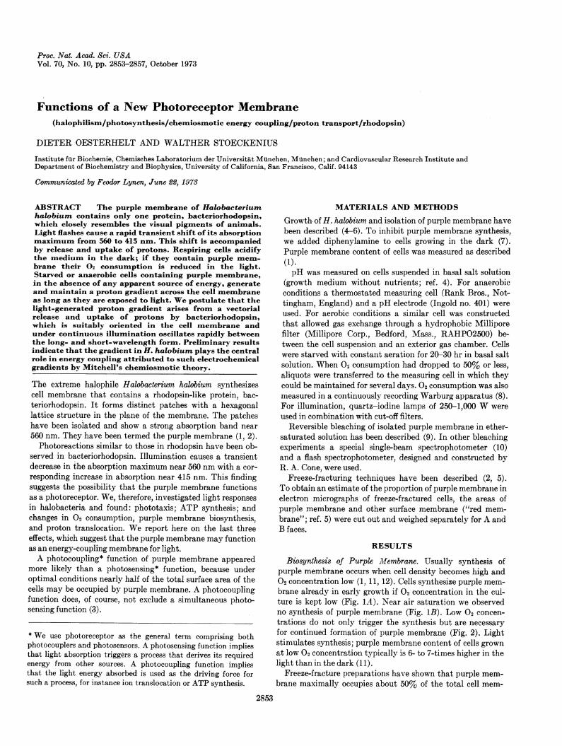
Biosynthesis of Purple Mfembrane. Usually synthesis ofpurple membrane occurs when cell density becomes high and02 concentration low (1, 11, 12). Cells synthesize purple mem-brane already in early growth if 02 concentration in the cul-ture is kept low (Fig. 1A). Near air saturation we observedno synthesis of purple membrane (Fig. 1B). Low 02 concen-trations do not only trigger the synthesis but are necessaryfor continued formation of purple membrane (Fig. 2). Lightstimulates synthesis; purple membrane content of cells grownat low 02 concentration typically is 6- to 7-times higher in thelight than in the dark (11).
Freeze-fracture preparations have shown that purple mem-brane maximally occupies about 50% of the total cell mem-
2854 Cell Biology: Oesterhelt and Stoeckenius
2.C
1.5
4 1.08o,.
.9
Co
Oa
30 60 90
B-6-.....
Ii
30 60 90
A
5 -
7
4 2d
1°
HoursFIG. 1. Synthesis of purple membrane. (A ) 120 liters of
air per hr, stirring at 300 rpm; (B) 180 liters of air per hr, 500rpm. Growth in suspension culture in peptone medium at 39°.For determination of purple membrane, see ref. 1.
brane area; this estimate entails the assumption that tan-gential fractures occur with equal probability in purple mem-brane and red membrane. It may actually be too low a value,because tangential fractures are seen far less frequently inisolated purple membrane than in red membrane. In any case,
it is clear that the cells can convert a substantial part of theirtotal surface membrane into purple membrane. Electronmicrographs further show purple-membrane patches relativelyhomogeneously distributed over all cells.
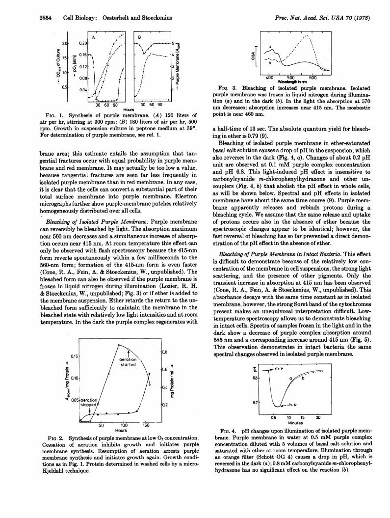
Bleaching of Isolated Purple Membrane. Purple membranecan reversibly be bleached by light. The absorption maximumnear 560 nm decreases and a simultaneous increase of absorp-tion occurs near 415 nm. At room temperature this effect can
only be observed with flash spectroscopy because the 415-nmform reverts spontaneously within a few milliseconds to the560-nm form; formation of the 415-nm form is even faster(Cone, R. A., Fein, A.- & Stoeckenius, W., unpublished). Thebleached form can also be observed if the purple membrane isfrozen in liquid nitrogen during illumination (Lozier, R. H.& Stoeckenius, W., unpublished; Fig. 3) or if ether is added tothe membrane suspension. Ether retards the return to the un-
bleached form sufficiently to maintain the membrane in thebleached state with relatively low light intensities and at roomtemperature. In the dark the purple complex regenerates with
0.15
.0
& 0.loE
"I nncs
150
400 500Wvegth i mn
600
FIG. 3. Bleaching of isolated purple membrane. Isolatedpurple membrane was frozen in liquid nitrogen during illumina-tion (a) and in the dark (b). In the light the absorption at 570nm decreases; absorption increases near 415 nm. The isosbesticpoint is near 460 nm.
a half-time of 13 sec. The absolute quantum yield for bleach-ing in ether is 0.79 (9).
Bleaching of isolated purple membrane in ether-saturatedbasal salt solution causes a drop of pH in the suspension, whichalso reverses in the dark (Fig. 4, a). Changes of about 0.2 pHunit are observed at 0.1 mM purple complex concentrationand pH 6.8. This light-induced pH effect is insensitive tocarbonylcyanide m-chlorophenylhydrazone and other un-
couplers (Fig. 4, b) that abolish the pH effect in whole cells,as will be shown below. Spectral and pH effects in isolatedmembrane have about the same time course (9). Purple mem-brane apparently releases and rebinds protons during a
bleaching cycle. We assume that the same release and uptakeof protons occurs also in the absence of ether because thespectroscopic changes appear to be identical; however, thefast reversal of bleaching has so far prevented a direct demon-stration of the pH effect in the absence of ether.
Bleaching of Purple Membrane in Intact Bacteria. This effectis difficult to demonstrate because of the relatively low con-
centration of the membrane in cell suspensions, the strong lightscattering, and the presence of other pigments. Only thetransient increase in absorption at 415 nm has been observed(Cone, R. A., Fein, A. & Stoeckenius, W., unpublished). Thisabsorbance decays with the same time constant as in isolatedmembrane, however, the strong Soret band of the cytochromespresent makes an unequivocal interpretation difficult. Low-temperature spectroscopy allows us to demonstrate bleachingin intact cells. Spectra of samples frozen in the light and in thedark show a decrease of purple complex absorption around585 nm and a corresponding increase around 415 nm (Fig. 5).This observation demonstrates in intact bacteria the same
spectral changes observed in isolated purple membrane.
0.6+.Ec
0.4 2
E
3.2
Hours
FIG. 2. Synthesis of purple membrane at low 02 concentration.Cessation of aeration inhibits growth and initiates purplemembrane synthesis. Resumption of aeration arrests purplemembrane synthesis and initiates growth again. Growth condi-tions as in Fig. 1. Protein determined in washed cells by a micro-Kjeldahl technique.
I
6S
6.7
0.5 1D 15Minutes
20
FIG. 4. pH changes upon illumination of isolated purple mem-brane. Purple membrane in water at 0.5 mM purple complexconcentration diluted with 5 volumes of basal salt solution andsaturated with ether at room temperature. Illumination throughan orange filter (Schott OG 4) causes a drop in pH, which isreversed in the dark (a); 0.8mM carbonylcyanide m-chlorophenyl-hydrazone has no significant effect on the reaction- (b).
,
I.I %~~~~~~~~~~
I I'a-h
Al-. -h v
Proc. Nat. Acad. Sci. USA 70 (1973)
Functions of a New Photoreceptor Membrane 2855
c=a -b
b 585
400 500 600Waveength in nm
FIG. 5. Bleaching of purple membrane in intact bacteria.3.5 X 109 cells per ml of H. halobium RL3, which contains re-
duced amounts of bacterioruberin; peaks at 480, 512, and 547 nmare due to this pigment. One aliquot is illuminated with orangelight for 1 min and then frozen in the light (a) or in the dark (b).Difference spectrum a - b (c). Prior illumination at room tem-perature, which shifts the absorption spectrum of the purplecomplex to slightly longer wavelength with a very slow return inthe dark, (1), was used in b to give comparable samples. The peakin the difference spectrum appears at 585 nm rather than 570nm because low temperature causes a small shift to longer wave-
length.
Light Effects on pH and Respiration in Cell Suspensions.Respiring cells of H. halobium, similar to other prokaryoticcells (13), acidify the medium. Cell suspensions in basal saltsolution have a pH between 6.2 and 7.2. Aliquots transferredto the closed measuring cell become rapidly anaerobic; simul-taneously the pH drops and rises again briefly. After about10 min it has reached a nearly stable value but continues toincrease very slowly. This quasistable state is used as the basisfor the study of light effects. Upon illumination with wave-
lengths absorbed by the purple membrane, the pH, after a
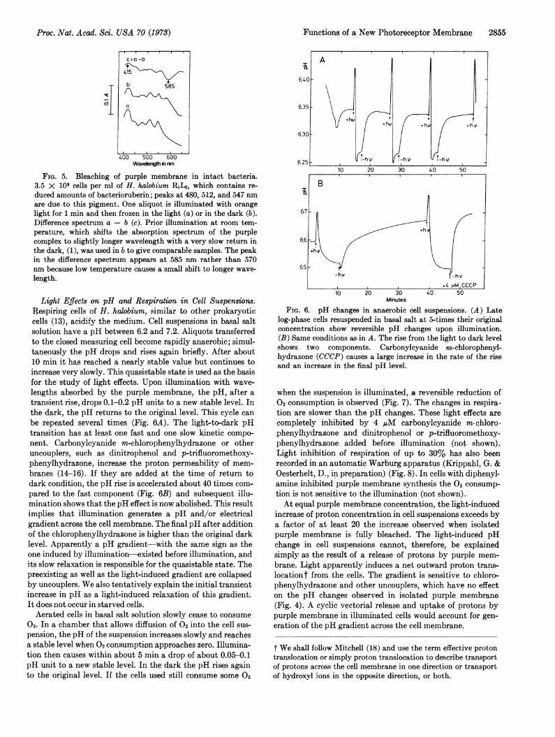
transient rise, drops 0.1-0.2 pH units to a new stable level. Inthe dark, the pH returns to the original level. This cycle can
be repeated several times (Fig. 6A). The light-to-dark pHtransition has at least one fast and one slow kinetic compo-nent. Carbonylcyanide m-chlorophenylhydrazone or otheruncouplers, such as dinitrophenol and p-trifluoromethoxy-phenylhydrazone, increase the proton permeability of mem-branes (14-16). If they are added at the time of return todark condition, the pH rise is accelerated about 40 times com-
pared to the fast component (Fig. 6B) and subsequent illu-mination shows that the pH effect is now abolished. This resultimplies that illumination generates a pH and/or electricalgradient across the cell membrane. The final pH after additionof the chlorophenylhydrazone is higher than the original darklevel. Apparently a pH gradient-with the same sign as theone induced by illumination-existed before illumination, andits slow relaxation is responsible for the quasistable state. Thepreexisting as well as the light-induced gradient are collapsedby uncouplers. We also tentatively explain the initial transientincrease in pH as a light-induced relaxation of this gradient.It does not occur in starved cells.Aerated cells in basal salt solution slowly cease to consume
02. In a chamber that allows diffusion of 02 into the cell sus-
pension, the pH of the suspension increases slowly and reachesa stable level when 02 consumption approaches zero. Illumina-tion then causes within about 5 min a drop of about 0.05-0.1pH unit to a new stable level. In the dark the pH rises againto the original level. If the cells used still consume some 02
10 20 30 40 50Minutes
FIG. 6. pH changes in anaerobic cell suspensions. (A) Latelog-phase cells resuspended in basal salt at 5-times their originalconcentration show reversible pH changes upon illumination.(B) Same conditions as in A. The rise from the light to dark levelshows two components. Carbonylcyanide m-chlorophenyl-hydrazone (CCCP) causes a large increase in the rate of the rise
and an increase in the final pH level.
when the suspension is illuminated, a reversible reduction of02 consumption is observed (Fig. 7). The changes in respira-tion are slower than the pH changes. These light effects are
completely inhibited by 4 /AM carbonylcyanide m- chloro-phenylhydrazone and dinitrophenol or p-trifluoromethoxy-phenylhydrazone added before illumination (not shown).Light inhibition of respiration of up to 30% has also beenrecorded in an automatic Warburg apparatus (Krippahl, G. &Oesterhelt, D., in preparation) (Fig. 8). In cells with diphenyl-amine inhibited purple membrane synthesis the 02 consump-tion is not sensitive to the illumination (not shown).At equal purple membrane concentration, the light-induced
increase of proton concentration in cell suspensions exceeds bya factor of at least 20 the increase observed when isolatedpurple membrane is fully bleached. The light-induced pHchange in cell suspensions cannot, therefore, be explainedsimply as the result of a release of protons by purple mem-
brane. Light apparently induces a net outward proton trans-locationt from the cells. The gradient is sensitive to chloro-phenylhydrazone and other uncouplers, which have no effecton the pH changes observed in isolated purple membrane(Fig. 4). A cyclic vectorial release and uptake of protons bypurple membrane in illuminated cells would account for gen-
eration of the pH gradient across the cell membrane.
t We shall follow Mitchell (18) and use the term effective protontranslocation or simply proton translocation to describe transportof protons across the cell membrane in one direction or transportof hydroxyl ions in the opposite direction, or both.
TI
Proc. Nat. Acad. Sci. USA 70 (1973)
2856 Cell Biology: Oesterhelt and Stoeckenius
0.12
0.10
0.08
o~oE
0
7.1
7.0
200 400Minutes
FIG. 7. pH changes and 02 consumption in aerobic cell sus-
pensions. Late log-phase cells were washed twice in basal saltsolution. Aeration was through a Millipore filter. Illuminationcauses a reversible drop in pH and rise in po0. The differencefrom Fig. 0, a lack in the transient rise in pit, is typical forstarved cells.
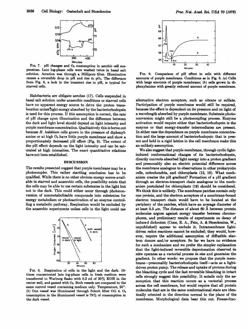
Halobacteria are obligate aerobes (17). Cells suspended inbasal salt solution under anaerobic conditions or. starved cellshave no apparent energy source to drive the proton trans-location unlesslight energy absorbed by the bacteriorhodopsinis used for this process. If this assumption is correct, the rateof pH change upon illumination and the difference betweenthe dark and light level should depend on light intensity andpurple membrane concentration. Qualitatively this is borne outbecause H. halobium cells grown in the presence of diphenyl-amine or at high 02 have little purple membrane and show a
proportionately decreased pH effect (Fig. 9). The extent ofthe pH effect depends on the light intensity and can be sat-
urated at high intensities. The exact quantitative relationshave-not been established.
DISCUSSION
The results presented suggest that purple membrane may be a
photocoupler. This rather startling conclusion has to bequalified. While there is no other obvious energy source avail-able in starved and anaerobic cells, the possibility exists thatthe cells may be able to use certain substrates in the light butnot in the dark. This could either occur through photocon-version of nonmetabolizable compounds into substrates forenergy metabolism or photoactivation of an enzyme control-ling a metabolic pathway, Respiration would be excluded bythe anaerobic experiments unless cells in the light could use
0
E 20E
40E
co) 60
0
x 800
100
I both vesselsin dark
vessel vesselin dark \ light
20 4 6020 40 60 80
Minutes
FIG. 8, Respiration of cells in the light and the dark. 10-
times concentrated late log-phase cells in fresh medium weretransferred to Warburg flasks with 0.2 ml of 20% KOH in thecenter well, and gassed with 02. Both vessels are compared to thesame control vessel containing medium only. Temperature, 250.(b) One vessel was illuminated through Schott filter OG 4. 0,
consumption in the illuminated vessel is 79% of consumption inthe dark vessel.
Minutes
FiP. 9. Comparison of pH effect in cells with differentamounts of purple membrane. Conditions as in Fig. 6. (a) Cellswith large amounts of purple membrane; (b) cells grown in di-phenylanine with greatly reduced amount of purple membrane.
alternative electron acceptors, such as nitrate or sulfate.Participation of purple membrane would still be required,because the effect is dependent on its presence and on light ofa wavelength absorbed by purple membrane. Substrate photo-conversion might still be a photocoupling process. Enzymeactivation would require either that bacteriorhodopsin is theenzyme or that energy-transfer intermediates are present.In either case the dependence on purple membrane concentra-tion and the large amount of bacteriorhodopsin that is pres-ent and held in a rigid lattice in the cell membrane make thisan unlikely assumption.We also suggest that purple membrane, through cyclic light-
induced conformational changes of its bacteriorhodopsin,directly converts absorbed light energy into a proton gradientand presumably also an. electric potential difference acrossthe membrane analogous to observations in other prokaryoticcells, mitochondria, and chloroplasts (13, 19). What mech-anism creates the pH gradient? Formation of a pH gradientthrough an electron-transport chain analogous to the mech-anism postulated for chloroplasts (19) should be considered.We think this is unlikely. The membrane patches contain onlyone protein, and the electron acceptors of a membrane-boundelectron transport chain would have to be located at theperiphery of the patches, which have an average diameter ofat least 0.5 pum. The distance of about 60 X between proteinmolecules argues against energy transfer between chromo-
phores, and preliminary results of experiments on decay ofinduced dichroism (Cone, R. A., Fein, A. & Stoeckenius, W.,unpublished) appear to exclude it. Intratnembrane light-driven redox reactions cannot be excluded; they would, how-ever, require the additional assumption of diffusible elec-tron donors and/or acceptors. So far we have no evidencefor such a mechanism and we prefer the simpler explanationthat the light-induced reversible deprotonation observed invitro operates as a vectorial process in vivo and generates thegradient. In other words: we propose that the purple mem-brane-presumably bacteriorhodopsin itself-acts as a light-driven proton pump. The release and uptake of protons duringthe bleaching cycle and the fast reversible bleaching in intactcells strongly suggest this possibility. It entails only the as-sumption that this reaction occurs as a vectorial processacross the cell membrane, but would require that all proteinmolecules that are in the same conformational state are iden-tically oriented in the direction normal to the plane of themembrane. Morphological data bear this out. Freeze-frac-
I
lV{-h-vp1
Proc. Nat. Acad, Sci. USA 70 (1973)
Functions of a New Photoreceptor Membrane 2857
turing of the dark-adapted membrane leaves all protein at-tached to the cytoplasmic side which, together with the ar-rangement of the protein in the planar hexagonal lattice,shows that the protein has the required orientation (2,20). A direct pumping action of bacteriorhodopsin in thepurple membrane therefore fits all observations and requiresthe fewest additional assumptions.The interpretation of our results necessarily leads to a con-
sideration of Mitchell's chemiosmotic theory (19), which statesthat in respiration and photosynthesis a series of redox reac-tions is used to generate the proton gradient and its associatedelectrical potential across the membrane. We propose that inH. halobium absorption of light quanta causes a transient con-formational change in one membrane protein which, throughthe accompanying vectorial release and uptake of protons,generates the gradient. The utilization of the energy storedin the gradient presumably is the same as proposed by thechemiosmotic theory. A membrane-bound ATPase has beenfound in H. halobium (21), which may function in ATP syn-thesis driven by the gradient. ATP synthesis in anaerobic cellsin the light but not in the dark has been demonstrated(Danon, A. & Stoeckenius, W., in preparation); we expect,however, that transport processes across the cell membranemay also be driven directly by the electrochemical gradientin analogy to observations in other prokaryotic cells (13).H. halobium apparently has two alternative possibilities tocreate the gradient: respiration and light absorption. If ourinterpretation of purple-membrane function is correct, thecentral role of the pH gradient for energy conversion becomesobvious.
If the purple membrane functions as indicated here, itwould be, beside the thylakoid membrane, the only otherknown photocoupling membrane. This hypothesis throws anew light on photoreceptor evolution. Apparently membranescontaining rhodopsin-like proteins as well as chlorophyll-con-taining membranes function both as photocouplers and photo-sensors, but the main evolutionary trend has apparently beentoward photosensing in the case of retinal-protein complexesand toward photocoupling in the case of chlorophylls. Whythis should be so is an intriguing question.
Purple membrane can easily be obtained in pure form andlarge quantities; detailed chemical and structural data areavailable. In addition to its significance in problems ofphotosynthesis, evolution, and the chemiosmotic theory, itmay be the best available model to study an ion pump.Because of its similarity to the visual pigments, the resultsobtained may also be relevant for the physiology of vision.
Note Added in Proof. Since this manuscript was submittedfor publication we have also demonstrated light-driven proton
translocation in a model system. It consists of vesicles formedfrom purified lipids into which purple membrane has beenincorporated (Racker, E. & Stoeckenius, W., submitted forpublication). These experiments confirm the conclusion thatthe purple membrane functions as a light-driven protonpump and further reduce the probability that other mech-anisms are responsible for light energy conversion in H.halobium.
We thank Profs. F. Lynen and W. Butler for permission to usetheir laboratory facilities; Dr. R. A. Cone, Arlette Danon, R.Lozier, and G. Krippahl for permission to publish results ofexperiments done in collaboration. The competent technical helpof M. Meentzen, L. Schuhmann, K. Ames, J. Woodard, and I.Marot is acknowledged. This work was supported by NIH GrantHL 06285, NASA Life Scientist Grant NGL 05-025-014, and theDeutsche Forschungsgemeinschaft.
1. Oesterhelt, D. & Stoeckenius, W. (1971) Nature New Biol.233, 149-152.
2. Blaurock, A. E. & Stoeckenius, W. (1971) Nature New Biol.233, 152-155.
3. Clayton, R. K. (1964) in Photophysiology, ed. Giese, A. C.(Academic Press, New York), Vol. 11, pp. 51-77.
4. Stoeckenius, W. & Rowen, R. (1967) J. Cell Biol. 34, 365-393.
5. Stoeckenius, W. & Kunau, W. H. (1968) J. Cell Biol. 38,337-357.
6. Oesterhelt, D. & Stoeckenius, W. (1973) in Methods inEnzymology. Biomembranes, eds. Fleischer, S., Packer, L. &Estabrook, R. W. (Academic Press, New York), in press.
7. Liaaen-Jensen, S., Cohen-Bazire, G., Nakayama, T. 0. M. &Stanier, R. Y. (1958) Biochim. Biophys. Acta 29, 477-498.
8. Warburg, O., Krippahl, G. & Birkicht, E. (1964) Biochem.Z. 340, 1-12.
9. Oesterhelt, D. & Hess, B. (1973) Eur. J. Biochem., in press.10. Butler, W. L. (1972) in Methods in Enzymology. Photo-
synthesis and Nitrogen Fixation Part B, ed. San Pietro,A. (Academic Press, New York), Vol. XXIV, pp. 3-25.
11. Danon, A. & Stoeckenius, W. (1972) NASA Symp. ExtremeEnvironments. Mechanisms of Microbial Adaption, AmesRes. Ctr., Moffett Field, Calif., June, p. 25.
12. Oesterhelt, D. (1972) Hoppe-Seyler's Z. Physiol. Chem. 353,1554-1555.
13. Harold, F. M. (1972) Bacteriol. Rev. 36, 172-230.14. Mitchell, P. & Moyle, J. (1967) Biochem. J. 105, 1147-1162.15. Hopfer, U., Lehninger, A. L. & Thompson, T. E. (1968)
Proc. Nat. Acad. Sci. USA 59, 484-490.16. Liberman, E. A. & Topaly, V. P. (1968) Biochim. Biophys.
Acta 163, 125-136.17. Larsen, H. (1967) Advan. Microbiol. Physiol. 1, 97-132.18. Mitchell, P. (1966) Biol. Rev. 41, 445-502.19. Mitchell, P. (1972) J. Bioenergetics 3, 5-24.20. Stoeckenius, W. (1973)1st Annu. Mtg. Amer. Soc. Photobiol.,
Sarasota, Fla.21. De, K., Passow, H., Stoeckenius, W. & White, M. (1966)
Pflugers Arch. ges. Physiol. 289, S. R 15.
Proc. Nat. Acad. Sci. USA 70 (1978)