Embed Size (px)
DESCRIPTION
Functions of the Urinary System. Elimination of waste products Nitrogenous wastes Toxins Drugs. Functions of the Urinary System. Regulate aspects of homeostasis Water balance Electrolytes Acid-base balance in the blood Blood pressure Red blood cell production Activation of vitamin D. - PowerPoint PPT Presentation
Citation preview

© 2012 Pearson Education, Inc.
Functions of the Urinary System
•Elimination of waste products
•Nitrogenous wastes
•Toxins
•Drugs

© 2012 Pearson Education, Inc.
Functions of the Urinary System
•Regulate aspects of homeostasis
•Water balance
•Electrolytes
•Acid-base balance in the blood
•Blood pressure
•Red blood cell production
•Activation of vitamin D
© 2012 Pearson Education, Inc.
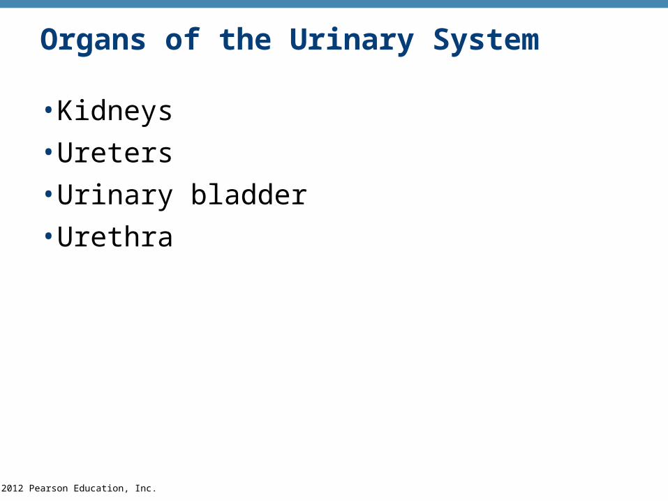
Organs of the Urinary System
•Kidneys
•Ureters
•Urinary bladder
•Urethra
© 2012 Pearson Education, Inc.
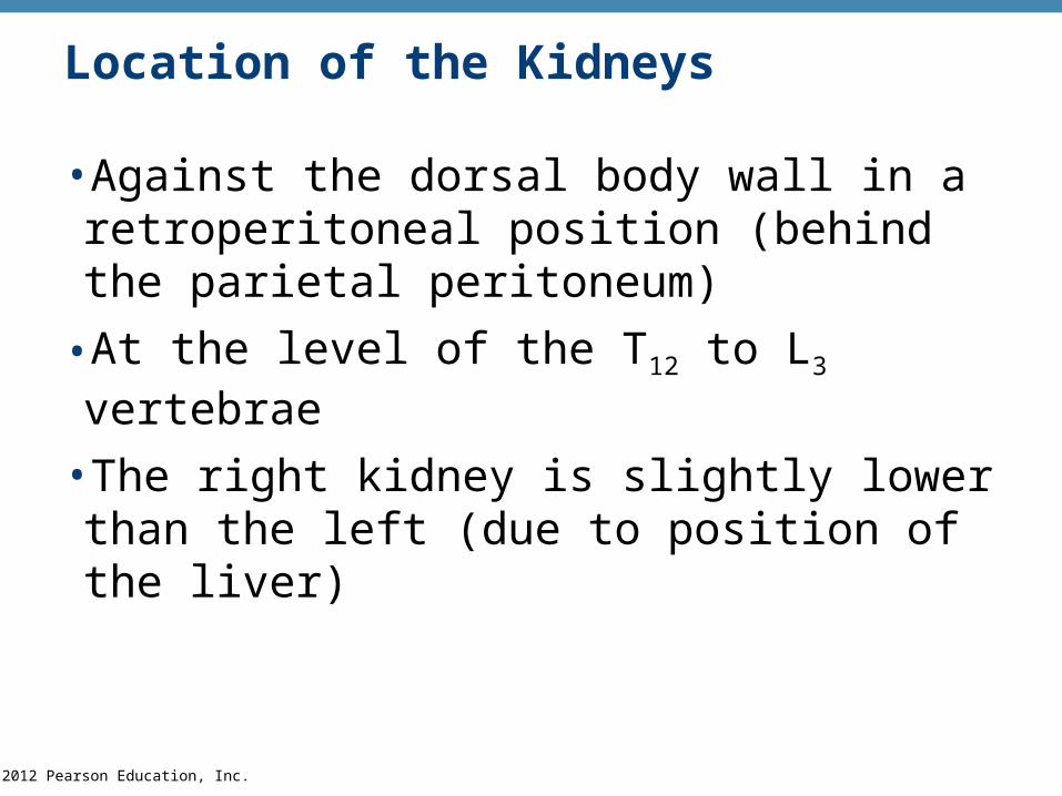
Location of the Kidneys
•Against the dorsal body wall in a retroperitoneal position (behind the parietal peritoneum)
•At the level of the T12 to L3 vertebrae
•The right kidney is slightly lower than the left (due to position of the liver)

© 2012 Pearson Education, Inc. Figure 15.1b
12th rib
(b)

© 2012 Pearson Education, Inc. Figure 15.1a
Hepatic veins (cut)
Inferior vena cava
Adrenal gland
Aorta
Iliac crest
Rectum (cut)
Uterus (partof femalereproductivesystem)
(a)
Renal artery
Renal hilum
Renal vein
Kidney
Ureter
Urinarybladder
Urethra

© 2012 Pearson Education, Inc.
Regions of the Kidney
•Renal cortex—outer region
•Renal medulla—inside the cortex
•Renal pelvis—inner collecting tube

© 2012 Pearson Education, Inc.
Kidney Structures
•Renal or medullary pyramids—triangular regions of tissue in the medulla
•Renal columns—extensions of cortex-like material inward that separate the pyramids
•Calyces—cup-shaped structures that funnel urine towards the renal pelvis

© 2012 Pearson Education, Inc. Figure 15.2a
Renal column
Major calyx
Renalcortex
Minor calyx
Renalpyramid
(a)

© 2012 Pearson Education, Inc. Figure 15.2b
Renal column
Renalcortex
Minor calyx
Renalpyramid
Fibrous capsule
Cortical radiatevein
Cortical radiateartery
Arcuate vein
Arcuate artery
Interlobar vein
Interlobar artery
Segmentalarteries
Renal vein
Renal artery
Renal pelvis
Major calyx
Ureter
(b)

© 2012 Pearson Education, Inc.
Blood Supply
•One-quarter of the total blood supply of the body passes through the kidneys each minute
•Renal artery provides each kidney with arterial blood supply
© 2012 Pearson Education, Inc.
Nephron Anatomy and Physiology
•The structural and functional units of the kidneys
•Responsible for forming urine
•Main structures of the nephrons
•Glomerulus
•Renal tubule
© 2012 Pearson Education, Inc.
Nephron Anatomy
•Glomerulus
•Knot of capillaries
•Glomerulus sits within a glomerular (Bowman’s) capsule (the first part of the renal tubule)
© 2012 Pearson Education, Inc.
Nephron Anatomy
•Renal tubule extends from glomerular capsule and ends at the collecting duct
•Glomerular (Bowman’s) capsule
•Proximal convoluted tubule (PCT)
•Loop of Henle
•Distal convoluted tubule (DCT)

© 2012 Pearson Education, Inc.
Nephron Anatomy
•Nephrons are associated with two capillary beds
•Glomerulus
•Peritubular capillary bed

© 2012 Pearson Education, Inc. Figure 15.3a
Renal cortex
Renal medulla
Renal pelvis
Renalcortex
Ureter
Renalmedulla
Corticalnephron Fibrous capsule
Collectingduct
Proximalconvoluted tubule
Glomerulus
Distalconvoluted tubuleLoopof Henle
Juxtamedullarynephron
(a)

© 2012 Pearson Education, Inc. Figure 15.3b
Proximalconvolutedtubule (PCT)
Glomerularcapillaries
Glomerular(Bowman’s) capsule
Efferent arteriole
Afferent arteriole
Cells of thejuxtaglomerularapparatus
Cortical radiate artery
Arcuate artery
Cortical radiatevein
Arcuatevein
Collecting duct
Loop of Henle
Distalconvolutedtubule(DCT)
Peritubularcapillaries
(b)

© 2012 Pearson Education, Inc.
Collecting Duct
•Receives urine from many nephrons
•Run through the medullary pyramids
•Deliver urine into the calyces and renal pelvis

© 2012 Pearson Education, Inc. Figure 15.3b
Proximalconvolutedtubule (PCT)
Glomerularcapillaries
Glomerular(Bowman’s) capsule
Efferent arteriole
Afferent arteriole
Cells of thejuxtaglomerularapparatus
Cortical radiate artery
Arcuate artery
Cortical radiatevein
Arcuatevein
Collecting duct
Loop of Henle
Distalconvolutedtubule(DCT)
Peritubularcapillaries
(b)

© 2012 Pearson Education, Inc.
Three steps to Urine Formation
•Glomerular filtration
•Tubular reabsorption
•Tubular secretion
© 2012 Pearson Education, Inc.
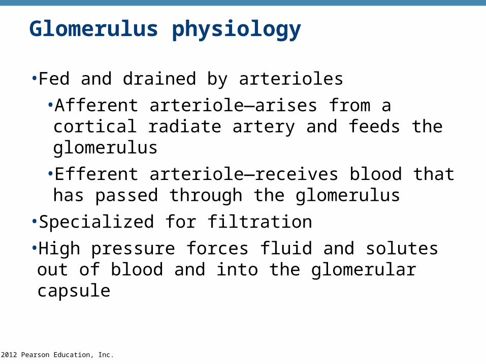
Glomerulus physiology
•Fed and drained by arterioles
•Afferent arteriole—arises from a cortical radiate artery and feeds the glomerulus
•Efferent arteriole—receives blood that has passed through the glomerulus
•Specialized for filtration
•High pressure forces fluid and solutes out of blood and into the glomerular capsule

© 2012 Pearson Education, Inc. Figure 15.3c
PCT Glomerularcapsularspace
Glomerularcapillarycovered bypodocytes
Efferentarteriole
Afferentarteriole
(c)

© 2012 Pearson Education, Inc.
Glomerular Filtration
•Nonselective passive process
•Water and solutes smaller than proteins are forced through capillary walls
•Proteins and blood cells are normally too large to pass through the filtration membrane
•Filtrate is collected in the glomerular capsule and leaves via the renal tubule

© 2012 Pearson Education, Inc.
Tubular Reabsorption
•The peritubular capillaries reabsorb useful substances
•Water
•Glucose
•Amino acids
• Ions
•Some reabsorption is passive, most is active
•Most reabsorption occurs in the proximal convoluted tubule

© 2012 Pearson Education, Inc.
Tubular Reabsorption
•What materials are not reabsorbed?
•Nitrogenous waste products
•Urea—protein breakdown
•Uric acid—nucleic acid breakdown
•Creatinine—associated with creatine metabolism in muscles

© 2012 Pearson Education, Inc.
Tubular Secretion: Reabsorption in Reverse•Some materials move from the blood of the peritubular capillaries into the renal tubules
•Hydrogen and potassium ions
•Creatinine
•Process is important for getting rid of substances not already in the filtrate
•Materials left in the renal tubule move toward the ureter

© 2012 Pearson Education, Inc. Figure 15.4
Afferent arterioleGlomerularcapillaries
Efferentarteriole
Glomerularcapsule
Rest ofrenal tubulecontainingfiltrate
Peritubularcapillary
To corticalradiate vein
UrineThree majorrenal processes:
Corticalradiateartery
Glomerular filtration: Water and solutes smaller than proteins are forced through thecapillary walls and pores of the glomerular capsule into the renal tubule.
Tubular reabsorption: Water, glucose, amino acids, and needed ions are transported out of the filtrate into the tubule cells and then enter the capillary blood.
Tubular secretion: H+, K+, creatinine, and drugs are removed from the peritubular blood and secreted by the tubule cells into the filtrate.
1
2
3
1
2
3

© 2012 Pearson Education, Inc. Figure 15.3b
Proximalconvolutedtubule (PCT)
Glomerularcapillaries
Glomerular(Bowman’s) capsule
Efferent arteriole
Afferent arteriole
Cells of thejuxtaglomerularapparatus
Cortical radiate artery
Arcuate artery
Cortical radiatevein
Arcuatevein
Collecting duct
Loop of Henle
Distalconvolutedtubule(DCT)
Peritubularcapillaries
(b)

© 2012 Pearson Education, Inc.
Characteristics of Urine
• In 24 hours, about 1.0 to 1.8 liters of urine are produced
•Urine and filtrate are different
•Filtrate contains everything that blood plasma does (except proteins)
•Urine is what remains after the filtrate has lost most of its water, nutrients, and necessary ions through reabsorption
•Urine contains nitrogenous wastes and substances that are not needed
© 2012 Pearson Education, Inc.
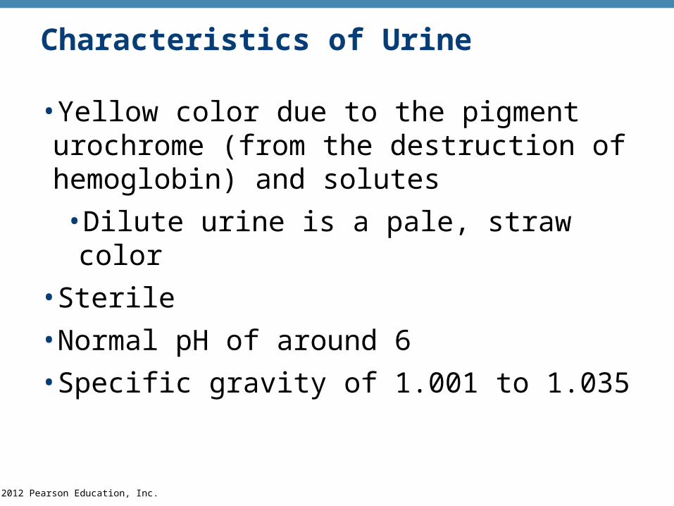
Characteristics of Urine
•Yellow color due to the pigment urochrome (from the destruction of hemoglobin) and solutes
•Dilute urine is a pale, straw color
•Sterile
•Normal pH of around 6
•Specific gravity of 1.001 to 1.035

© 2012 Pearson Education, Inc.
Characteristics of Urine
•Solutes normally found in urine
•Sodium and potassium ions
•Urea, uric acid, creatinine
•Ammonia
•Bicarbonate ions

© 2012 Pearson Education, Inc.
Characteristics of Urine
•Solutes NOT normally found in urine
•Glucose
•Blood proteins
•Red blood cells
•Hemoglobin
•White blood cells (pus)
•Bile
© 2012 Pearson Education, Inc.
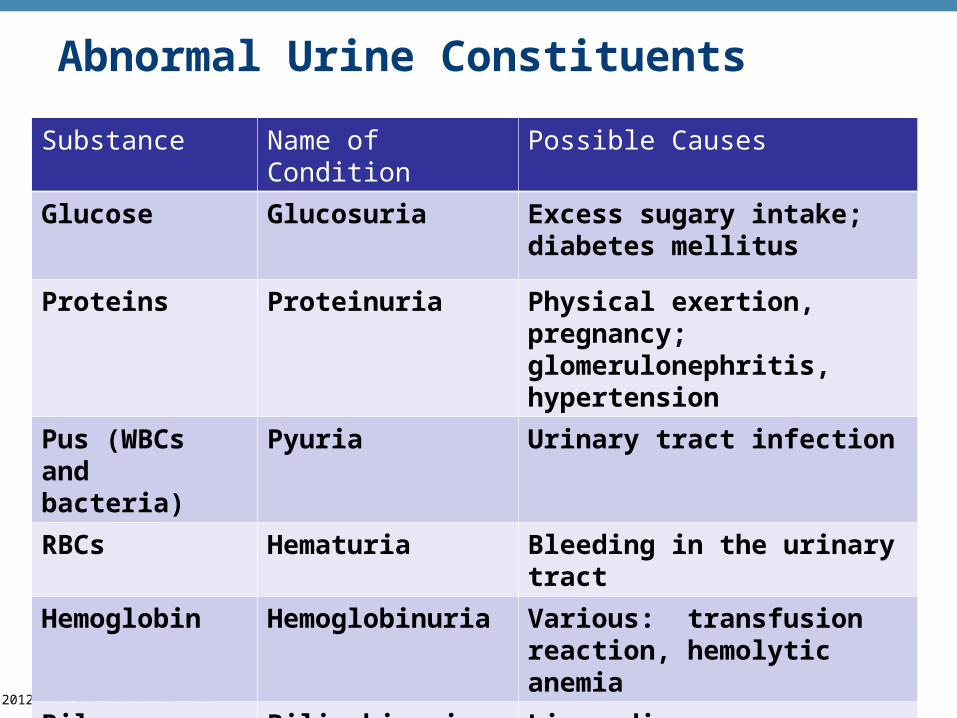
Abnormal Urine Constituents
Substance Name of Condition Possible Causes
Glucose Glucosuria Excess sugary intake; diabetes mellitus
Proteins Proteinuria Physical exertion, pregnancy; glomerulonephritis, hypertension
Pus (WBCs and bacteria)
Pyuria Urinary tract infection
RBCs Hematuria Bleeding in the urinary tract
Hemoglobin Hemoglobinuria Various: transfusion reaction, hemolytic anemia
Bile pigments Bilirubinuria Liver disease (hepatitis)

© 2012 Pearson Education, Inc.
Ureters
•Slender tubes attaching the kidney to the bladder
•Continuous with the renal pelvis
•Enter the posterior aspect of the bladder

© 2012 Pearson Education, Inc. Figure 15.1a
Hepatic veins (cut)
Inferior vena cava
Adrenal gland
Aorta
Iliac crest
Rectum (cut)
Uterus (partof femalereproductivesystem)
(a)
Renal artery
Renal hilum
Renal vein
Kidney
Ureter
Urinarybladder
Urethra

© 2012 Pearson Education, Inc.
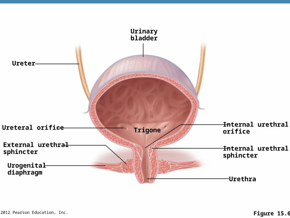
Urinary Bladder
•Smooth, collapsible, muscular sac
•Temporarily stores urine
•Trigone—triangular region of the bladder base
•Three openings
•Two from the ureters
•One to the urethra
• In males, the prostate gland surrounds the neck of the bladder
© 2012 Pearson Education, Inc. Figure 15.6
Urinarybladder
Internal urethralorifice
Internal urethralsphincter
Urethra
Urogenitaldiaphragm
External urethralsphincter
Ureteral orifice
Ureter
Trigone
© 2012 Pearson Education, Inc.
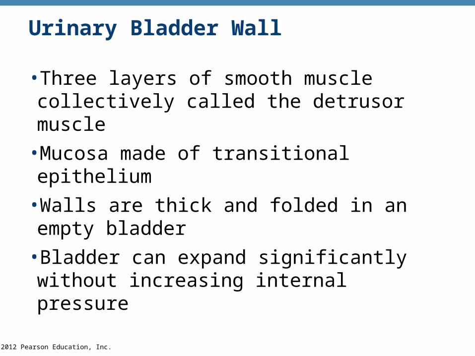
Urinary Bladder Wall
•Three layers of smooth muscle collectively called the detrusor muscle
•Mucosa made of transitional epithelium
•Walls are thick and folded in an empty bladder
•Bladder can expand significantly without increasing internal pressure

© 2012 Pearson Education, Inc.
Urinary Bladder Capacity
•A moderately full bladder is about 5 inches long and holds about 500 mL of urine
•Capable of holding twice that amount of urine
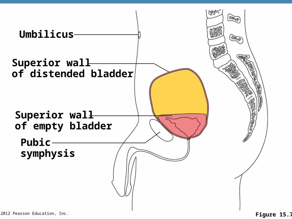
© 2012 Pearson Education, Inc. Figure 15.7
Umbilicus
Superior wallof distended bladder
Superior wallof empty bladder
Pubicsymphysis

© 2012 Pearson Education, Inc.
Urethra
•Thin-walled tube that carries urine from the bladder to the outside of the body by peristalsis
•Release of urine is controlled by two sphincters
• Internal urethral sphincter
• Involuntary and made of smooth muscle
•External urethral sphincter
•Voluntary and made of skeletal muscle

© 2012 Pearson Education, Inc. Figure 15.6
Urinarybladder
Internal urethralorifice
Internal urethralsphincter
Urethra
Urogenitaldiaphragm
External urethralsphincter
Ureteral orifice
Ureter
Trigone

© 2012 Pearson Education, Inc.
Urethra Gender Differences
•Length
•Females is 3 to 4 cm (1 inch)
•Males is 20 cm (8 inches)
•Location
•Females—anterior to the vaginal opening
•Males—travels through the prostate and penis

© 2012 Pearson Education, Inc.
Urethra Gender Differences
•Function
•Females—only carries urine
•Males—carries urine and is a passageway for sperm cells and semen

© 2012 Pearson Education, Inc.
Micturition (Voiding)
•Both sphincter muscles must open to allow voiding
•The internal urethral sphincter is relaxed after stretching of the bladder
•Pelvic splanchnic nerves initiate bladder to go into reflex contractions
•Urine is forced past the internal urethra sphincter and the person feels the urge to void
•The external urethral sphincter must be voluntarily relaxed to void
© 2012 Pearson Education, Inc.
Fluid, Electrolyte, and Acid-Base Balance
•Kidneys have four roles in maintaining blood composition
•Excretion of nitrogen-containing wastes (previously discussed)
•Maintaining water balance of the blood
•Maintaining electrolyte balance of the blood
•Ensuring proper blood pH

© 2012 Pearson Education, Inc.
Maintaining Water Balance
•Normal amount of water in the human body
•Young adult females = 50 percent
•Young adult males = 60 percent
•Babies = 75 percent
•The elderly = 45 percent
•Water is necessary for many body functions, and levels must be maintained
© 2012 Pearson Education, Inc.
Distribution of Body Fluid
• Intracellular fluid (ICF)
•Fluid inside cells
•About two-thirds of body fluid
•Extracellular fluid (ECF)
•Fluids outside cells that includes
• Interstitial fluid
•Blood plasma
© 2012 Pearson Education, Inc.
The Link Between Water and Salt
•Solutes in the body include electrolytes like sodium, potassium, and calcium ions
•Changes in electrolyte balance causes water to move from one compartment to another
•Alters blood volume and blood pressure
•Can impair the activity of cells

© 2012 Pearson Education, Inc.
Maintaining Water Balance
•Water intake must equal water output
•Sources for water intake
• Ingested foods and fluids
•Water produced from metabolic processes
•Thirst mechanism is the driving force for water intake

© 2012 Pearson Education, Inc.
Maintaining Water Balance
•Sources for water output
•Vaporization out of the lungs (insensible since we cannot sense the water leaving)
•Lost in perspiration
•Leaves the body in the feces
•Urine production

© 2012 Pearson Education, Inc. Figure 15.10
© 2012 Pearson Education, Inc.
Maintaining Water Balance
•Dilute urine is produced if water intake is excessive
•Less urine (concentrated) is produced if large amounts of water are lost
•Proper concentrations of various electrolytes must be present
© 2012 Pearson Education, Inc.
Developmental Aspects of the Urinary System•Functional kidneys are developed by the third month of fetal life
•Urinary system of a newborn
•Bladder is small
•Urine cannot be concentrated for first 2 months
•Void 5 to 40 times per day

© 2012 Pearson Education, Inc.
Developmental Aspects of the Urinary System•Control of the voluntary urethral sphincter does not start until age 18 months
•Complete nighttime control may not occur until the child is 4 years old
•Urinary infections are the only common problems before old age
•Escherichia coli (E. coli), a type of bacteria, accounts for 80 percent of UTI (urinary tract infections)

© 2012 Pearson Education, Inc.
Aging and the Urinary System
•There is a progressive decline in urinary function
•The bladder shrinks and loses bladder tone with aging

© 2012 Pearson Education, Inc.
Aging and the Urinary System
•Associated problems with aging
•Urgency—feeling that it is necessary to void
•Frequency—frequent voiding of small amounts of urine
•Nocturia—need to get up during the night to urinate
• Incontinence—loss of control
•Urinary retention—common in males, often the result of hypertrophy of the prostate gland