Embed Size (px)
Citation preview

Fungal Genetics and Biology 75 (2015) 64–71
Contents lists available at ScienceDirect
Tools and Techniques
Fungal Genetics and Biology
journal homepage: www.elsevier .com/ locate/yfgbi
Next generation multilocus sequence typing (NGMLST) and the
Mitchell b,
analytical software program MLSTEZ enable efficient, cost-effective,high-throughput, multilocus sequencing typing
Yuan Chen a,b,⇑, Aubrey E. Frazzitta a,1, Anastasia P. Litvintseva b,2, Charles Fang a, Thomas G.
http://dx.doi.org/10.1016/j.fgb.2015.01.0051087-1845/� 2015 The Authors. Published by Elsevier Inc.This is an open access article under the CC BY license (http://creativecommons.org/licenses/by/4.0/).
⇑ Corresponding authors at: Division of Infectious Diseases, Department ofMedicine, Duke University Medical Center, Durham, NC, USA (Y. Chen and J.R.Perfect). Fax: +1 919 6848902.
E-mail addresses: [email protected] (Y. Chen), [email protected](J.R. Perfect).
1 Present address: Oregon Health and Science University, School of Medicine,Portland, OR, USA.
2 Present address: Mycotic Diseases Branch, Centers for Disease Control andPrevention, Atlanta, GA, USA.
Deborah J. Springer b, Yun Ding c, George Yuan d, John R. Perfect a,⇑a Division of Infectious Diseases, Department of Medicine, Duke University Medical Center, Durham, NC, USAb Department of Molecular Genetics and Microbiology, Duke University Medical Center, Durham, NC, USAc Janelia Research Campus, HHMI, Ashburn, VA, USAd Pacific Sciences, Menlo Park, CA, USA
a r t i c l e i n f o a b s t r a c t
Article history:Received 28 October 2014Accepted 17 January 2015Available online 24 January 2015
Keywords:MLSTNext generation sequencing (NGS)Multiplex PCRPacBio CCS sequencingSoftwareCryptococcus neoformans
Multilocus sequence typing (MLST) has become the preferred method for genotyping many biologicalspecies, and it is especially useful for analyzing haploid eukaryotes. MLST is rigorous, reproducible,and informative, and MLST genotyping has been shown to identify major phylogenetic clades, moleculargroups, or subpopulations of a species, as well as individual strains or clones. MLST molecular types oftencorrelate with important phenotypes. Conventional MLST involves the extraction of genomic DNA andthe amplification by PCR of several conserved, unlinked gene sequences from a sample of isolates ofthe taxon under investigation. In some cases, as few as three loci are sufficient to yield definitive results.The amplicons are sequenced, aligned, and compared by phylogenetic methods to distinguish statisticallysignificant differences among individuals and clades. Although MLST is simpler, faster, and less expensivethan whole genome sequencing, it is more costly and time-consuming than less reliable genotypingmethods (e.g. amplified fragment length polymorphisms). Here, we describe a new MLST method thatuses next-generation sequencing, a multiplexing protocol, and appropriate analytical software to provideaccurate, rapid, and economical MLST genotyping of 96 or more isolates in single assay. We demonstratethis methodology by genotyping isolates of the well-characterized, human pathogenic yeast Cryptococcusneoformans.� 2015 The Authors. Published by Elsevier Inc. This is an open access article under the CC BY license (http://
creativecommons.org/licenses/by/4.0/).
1. Introduction pulsed-field gel electrophoresis (PFGE) (Schwartz and Cantor,1984) and amplified fragment length polymorphism (AFLP) (Vos
Efficient methods for estimating the genetic diversity amongmicroorganisms are essential for understanding their evolutionaryhistory, geographic distribution, and pathogenicity. In the past dec-ades, numerous methods have been developed for typing bacteriaand fungi (Li et al., 2009; Vanhee et al., 2010). Some of these meth-ods can characterize a large number of isolates at low cost, such as
et al., 1995). However, the results of these methods are laboratoryspecific and usually are not comparable among laboratories.Conversely, DNA sequencing results can be archived and sharedamong laboratories, and therefore, these methods are widely usedin microbial studies today (Janbon et al., 2014; Li et al., 2009;Litvintseva et al., 2006; Tavanti et al., 2005; Taylor and Fisher,2003; Vanhee et al., 2010). Multilocus sequence typing (MLST) tar-gets multiple genomic loci and is considered one of the most reli-able and informative methods for molecular genotyping (Maidenet al., 1998; Schwartz and Cantor, 1984). MLST has been appliedto many pathogenic microorganisms, and there is increasing inter-est in the variation among isolates and within microbial popula-tions, especially in studies of microbial evolution, pathogenesis,ecology, and microbiomes (Byrnes et al., 2009; Chen et al., 2013;Litvintseva and Mitchell, 2012; Meyer et al., 2009). Moreover,
online MLST databases have been constructed for several bacterial subpopulations of these recognized species and varieties were also
Y. Chen et al. / Fungal Genetics and Biology 75 (2015) 64–71 65
and fungal species to facilitate molecular epidemiological studiesand surveillance (Chan et al., 2001). MLST genotyping is a superbapproach to delineate species and strains, but the current method-ology is costly, time-consuming, and laborious.
To accelerate automation and expand the versatility of the cur-rent MLST method, we developed a high-throughput next-genera-tion sequencing approach, NGMLST, and an automated softwareprogram for data analyses, MLSTEZ. We adapted multiplex PCR,which may save more than 75% of the PCR work (calculated basedon using seven MLST loci). For next-generation sequencing, weemployed the Pacific Biosciences (PacBio) circular consensussequencing (CCS) technology, which is capable of generating rela-tively inexpensive, single-molecule consensus reads of 1–2 kbp inlength. Unlike the usual PacBio read, a CCS read is an error-corrected consensus read generated from the consensus alignmentof single-molecule circular sequencing (Eid et al., 2009). Therefore,the accuracy of a CCS read is correlated with the number ofsequencing passes of the template molecule (Travers et al.,2010). With the benefit of these higher quality reads, our software,MLSTEZ, can automatically identify the barcodes and primers usedin the PCR, correct sequencing errors, generate the MLST profile foreach isolate, and predict potentially heterozygous loci.
Cryptococcus neoformans is a well-characterized, opportunistichuman fungal pathogen, and it is responsible for approximately600,000 annual deaths worldwide (Park et al., 2009). In this study,we targeted the nine MLST loci that are commonly used to geno-type isolates of the C. neoformans/Cryptococcus gattii species com-plex. As controls, we selected 28 clinical and environmentalhaploid strains with known MLST genotypes that represented eachmajor subpopulation or molecular type of the species complex, aswell as six previously described diploid hybrid strains (Litvintsevaet al., 2006; Simwami et al., 2011; Stephen et al., 2002; Sun et al.,2012; Xu et al., 2009). We pooled the amplicons of these 34 isolateswith those of another 62 wild type C. neoformans isolates andsequenced them in one PacBio SMRT Cell. The NGMLST methodand MLSTEZ software produced high quality, unambiguous MLSTprofiles of all 96 isolates, and the sequences of the reference strainswere identical to their genotypes, which were previously deter-mined by the conventional MLST method. The MLSTEZ successfullydetected heterozygous loci in the hybrid strains and identified thesequences of each allele.
2. Materials and methods
2.1. Strains of C. neoformans
As reference controls, we selected conventionally MLST-geno-typed strains of C. neoformans var. grubii (Cng), C. neoformans var.neoformans (molecular type VNIV), and C. gattii. Distinct genetic
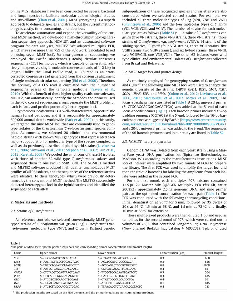
Table 1Nine pairs of MLST locus specific primer sequences and corresponding primer concentrati
Locus Upper primer Lower primer
SOD1 50-GGCACAACTCCACCGATCA 50-CTTACATGACACLAC1 50-AACATGTTCCCTGGACCTGTG 50-ACGTGGATCTCGMPD1 50-TGCCCTGGATCCTAATGCTCT 50-ACCCAGACTGCCTEF1 50-AATCGTCAAGGAGACCAACG 50-CGTCACCAGACTCAP59 50-CTCTACGTCGAGCAAGTCAAG 50-TCCGCTGCACAAPLB1 50-CTTCAGGCGGAGAGAGGTTT 50-GATTTGGCGTTGGPD1 50-ATGGTCGTCAAGGTTGGAAT 50-GTATTCGGCACCIGS1 50-GGGACCAGTGCATTGCATGA 50-ATCCTTTGCAGAURA5 50-ATGTCTTCCCAAGCCCTCGAC 50-TTAAGACCTCTG
a The production lengths are based on the H99 genome, and the primer lengths are n
considered when we selected control strains. For example, weincluded all three molecular types of Cng (VNI, VNB and VNII)(Litvintseva et al., 2006) and the four molecular types of C. gattii(VGI, VGII, VGIII, and VGIV). The number of strains for each molec-ular type are as follows (Table S1): 11 strains of C. neoformans var.grubii (five VNI strains, three VNB strains, three VNII strains); threestrains of C. neoformans var. neoformans (VNIV); 14 strains of thesibling species, C. gattii (four VGI strains, three VGII strains, fiveVGIII strains, two VGIV strains); and six hybrid strains (three VNIII,two VGII/VGIII, one VNB/VNII). The other 62 isolates were wildtype clinical and environmental isolates of C. neoformans collectedfrom Brazil and Botswana.
2.2. MLST target loci and primer design
As routinely employed for genotyping strains of C. neoformansand C. gattii, the following nine MLST loci were used to analyze thegenetic diversity of the strains: CAP59, GPD1, IGS1, LAC1, PLB1,SOD1, URA5, TEF1 and MPD1 (Colom et al., 2012; Litvintseva et al.,2006, 2011; MacDougall et al., 2007; Meyer et al., 2009). Thelocus-specific primers are listed in Table 1. A 20-bp universal primer(50-CTGGAGCACGAGGACACTGA) was added at the 50 end of eachlocus-specific primer (Fig. 1). Each barcode primer included a 5-bppadding sequence (GGTAG) at the 50 end, followed by the 16-bp bar-code sequence as suggested by PacBio (http://www.smrtcommunity.com/servlet/servlet.FileDownload?file=00P7000000W067VEAR),and a 20-bp universal primer was added to the 30 end. The sequencesof the 96 barcode primers used in our study are listed in Table S2.
2.3. NGMLST library preparation
Genomic DNA was isolated from each yeast strain using a Mas-terPure yeast DNA purification kit (Epicentre Biotechnologies,Madison, WI) according to the manufacturer’s instructions. MLSTloci of interest were amplified by two rounds of PCRs to preparethe library. The first PCR was used to amplify the target loci andthen the unique barcodes for labeling the amplicons from each iso-late were added in the second PCR.
For the first round, each multiplex PCR mixture contained12.5 lL 2� Master Mix (QIAGEN Multiplex PCR Plus Kit, cat #206152), approximately 2.5 ng genomic DNA, and nine primerpairs at the optimized concentration for each pair (Table 1). ThePCR was conducted with the following thermocycling conditions:initial denaturation at 95 �C for 5 min, followed by 35 cycles of30 s at 95 �C, 1.5 min at 58 �C, and 1.5 min at 72 �C, and finally,10 min at 68 �C for extension.
These multiplexed products were then diluted 1:50 and used astemplates for the second round of PCR, which were carried out involumes of 25 lL that contained LongAmp Taq DNA Polymerase(New England BioLabs Inc., catalog # M0323L), 1 lL of diluted
ons and product lengths.
Concentration (lM) Product lengtha
CGCAGGCA 0.3 668GGAGGA 0.3 816GCTGTCGTC 0.8 1008TGACGAAC 0.4 811GTGATACCC 0.3 564GTTTCAGT 0.3 635AGCCTCA 0.4 561CGACTTGA 0.1 845AACACCGTACTC 0.4 733
ot counted into products.
multiplex PCR product, and 2 lL 10 lM barcode primer. The PCR
sequencing (CCS) reads with less than four full passes were also fil-
3. Results
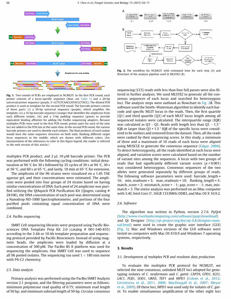
Fig. 1. Two rounds of PCRs are employed in NGMLST. In the first PCR round, eachprimer consists of a locus-specific sequence (blue, see Table 1) and a 20-bpuniversal primer sequence (purple, 50-GCTGTCAACGATACGCTACG). The diluted PCRproduct is used as template for the second PCR round. The barcode primers consistof three parts: (i) a 20-bp universal sequence (purple), which amplifies thetemplate; (ii) a 16-bp barcode sequence (orange) that identifies the amplicons fromeach different isolate; (iii) and a 5-bp padding sequence (green) to provideequivalent binding affinities for adding the PacBio sequencing adapters. Becausemultiplex PCRs were used in the first PCR round, primer pairs for each of the nineloci are added to the PCR mix at the same time. In the second PCR round, the variousbarcode primers are used to identify each isolates. The final products of each isolatewould have the same sequence structure on both ends, flanking different targetlocus sequences in the middle, which are shown with different colors. (Forinterpretation of the references to color in this figure legend, the reader is referredto the web version of this article.)
Fig. 2. The workflow for NGMLST with estimated time for each step (A) andflowchart of the analysis pipeline used in MLSTEZ (B).
66 Y. Chen et al. / Fungal Genetics and Biology 75 (2015) 64–71
was performed with the following cycling conditions: initial dena-turation at 94 �C for 30 s followed by 35 cycles of 30 s at 94 �C, 30 sat 50 �C, and 60 s at 65 �C, and lastly, 10 min at 65 �C for extension.
The amplicons of the 96 strains were visualized on a 1.4% TAEagarose gel, and their concentrations were estimated. The ampli-cons were pooled into four groups of 24 strains based on havingsimilar concentrations of DNA. Each pool of 24 amplicons was puri-fied utilizing the QIAquick PCR Purification Kit (Qiagen, catalog #28106), the DNA concentration of each pool was determined usinga Nanodrop ND-1000 Spectrophotometer, and portions of the fourpurified pools containing equal concentration of DNA werecombined.
2.4. PacBio sequencing
SMRT Cell sequencing libraries were prepared using Pacific Bio-sciences DNA Template Prep Kit 2.0 (catalog # 001-540-835)according to the 3-kb or 10-kb template preparation and sequenc-ing protocol provided by Pacific Biosciences. Instead of using mag-netic beads, the amplicons were loaded by diffusion at aconcentration of 300 pM. The PacBio RS II platform was used forsequencing the amplicons. One SMRT Cell was used to sequenceall 96 pooled isolates. The sequencing run used 1 � 180 min moviewith P4-C2 chemistry.
2.5. Data analysis
Primary analysis was performed using the PacBio SMRT Analysisversion 2.1 program, and the filtering parameters were as follows:minimum polymerase read quality of 0.75; minimum read lengthof 50 bp; and minimum subread length of 50 bp. Circular consensus
tered in further analysis. We used MLSTEZ to generate all the con-sensus sequences of each locus and searched for heterozygousloci. The analysis steps were outlined as flowchart in Fig. 2B. Thissoftware used the Smith–Waterman algorithm to identify each bar-code and specific MLST locus in the reads. Then, the first quartile(Q1) and third quartile (Q3) of each MLST locus length among allsequenced isolates were calculated. The interquartile range (IQR)was calculated as Q3 � Q1. Reads with length less than Q1 � 1.5 ⁄
IQR or larger than Q3 + 1.5 ⁄ IQR of the specific locus were consid-ered to be outliers and removed from the dataset. Then, all the readswere ranked by their sequencing scores. In this study, a minimumof three and a maximum of 10 reads of each locus were alignedusing MUSCLE to generate the consensus sequence (Edgar, 2004).To detect heterozygosity, all the reads identified at each locus werealigned, and variation scores were calculated based on the numberof variant sites among the sequences. A locus with two groups ofreads that had significantly different variant scores (p < 0.001)was considered heterozygous. Consensus sequences of the twoalleles were generated separately by different groups of reads.The following software parameters were used: barcode_length =16; min_readnum = 3; max_readnum = 10; flanking_length = 5;match_score = 2; mismatch_score = �1; gap_score = �1; max_mis-match = 3. The entire analysis was performed on an iMac computerwith 3.4G Intel Core i7, 16GB 1333MHz DDR3, and Mac OS X 10.9.2.
2.6. Software
The algorithm was written in Python, version 2.7.6. PyQt4(http://www.riverbankcomputing.com/software/pyqt/download)and Qt Designer (http://qt-project.org/doc/qt-4.8/designer-man-ual.html) were used to create the graphic user interface (GUI)(Fig. 3). Mac and Windows versions of the GUI software weretested on computers with Mac OS X10.9 and Windows 7 operatingsystems, respectively.
3.1. Development of multiplex PCR and resultant data production
To evaluate the multiplex PCR protocol for NGMLST, weselected the nine consensus, unlinked MLST loci adopted for geno-typing isolates of C. neoformans and C. gattii: CAP59, GPD1, IGS1,LAC1, PLB1, SOD1, URA5, TEF1 and MPD1 (Colom et al., 2012;Litvintseva et al., 2011, 2006; MacDougall et al., 2007; Meyeret al., 2009). Of these loci, MPD1 was used only for isolates of C. gat-tii. To enable simultaneous amplification of the other eight loci

from most isolates of C. neoformans and C. gattii, we designed new
amplicons of the first PCR round were amplified by the same uni-
3.2. Data processing
Fig. 3. Graphic user interface of MLSTEZ under Mac OS X system. The interfaceconsists of four parts: toolbox bar (top), list of analyses panel (mid-left), analysisresult panel (mid-right), and running status panel (bottom).
Y. Chen et al. / Fungal Genetics and Biology 75 (2015) 64–71 67
pairs of primers that were specific for five loci (IGS1, TEF1, LAC1,SOD1, and URA5), which targeted the same regions used in previousstudies, and we used previously designed primers for CAP59, GPD1,PLB1, and MPD1 (Table 1). In addition, all nine MLST locus-specificprimers were modified to include a universal primer sequence atthe 50 end (Fig. 1), which was needed to facilitate the addition ofbarcodes in the subsequent step (Fig. 2A). The nine pairs oflocus-specific primers were admixed with the optimized concen-trations (Table 1), and all the loci were amplified simultaneously.Although some strains and/or species differed in the efficiencywith which they were amplified (Table 3), all the loci were success-fully amplified in most tested isolates (Fig. 4).
The barcode primers for the second PCR round consisted ofthree parts (Fig. 1). The padding sequence was used to ensure thateach product had equal efficiency to ligate to the sequencing adap-ter. The barcode sequence was unique to each isolate and was usedto separate the amplicons from different isolates by MSLTEZ. The
Fig. 4. Two rounds of PCR products of isolates H99 (VNI molecular type), R265(VGII), and JEC21 (VNIV) are shown on 1.4% TAE agarose gel. R1 and R2 stand for thefirst and second PCR round, respectively. The expected PCR product sizes are shownin Table 1. The bands from top to bottom are PCR products of MPD1, IGS1, LAC1,TEF1, URA5, SOD1, PLB1, CAP59 and GPD1. Some bands are overlapped because ofsimilar product lengths. The gel image indicates that the MPD1 (top band) locus wasamplified with greater efficiency from R265 than H99 and JEC21. The primer pairsof other loci also reveal different amplification efficiencies among isolates fromdifferent molecular groups (Table 2).
versal primer that we had added into the barcode primer.To test the accuracy of the PacBio sequencing platform for
NGMLST, we selected 28 diverse reference strains that representedthe eight major haploid molecular types of C. neoformans andC. gattii and six hybrids, which are very difficult to genotype usingthe conventional MLST protocol (Table S1). In addition, DNA from62 wild type isolates of C. neoformans were also added to the testmixtures. We pooled all the barcoded amplicons of 96 isolatesand sequenced them in one PacBio SMRT Cell. Four full passesyielded 37,906 CCS reads with an average CCS read length of730 bp. As expected, more than 80% of the reads ranged between600 and 1100 bp (Fig. 5A).
The first step of the analysis pipeline to generate the MLST pro-file for each isolate is to identify the unique barcode sequenceadded during the second round of amplification. MLSTEZ success-fully identified barcode sequences on 32,932 of 37,906 (86.9%)CCS reads. The average number of reads obtained for each isolatewas 343.0 (Fig. 5B). Subsequently, the barcode-called ampliconswere separated by the locus-specific primer sequences. Due tothe low sequencing qualities of some reads, primer sequences
Fig. 5. Length distribution of CCS reads generated from 96 isolates (A). More than80% of the reads have sequence lengths between 600 and 1100 bp. Normaldistribution is shown for the read count of the 96 isolates (B). Distribution of readcounts for isolates. The average read count of each isolate is 343, and minimal andmaximal read counts are 34 and 829, respectively.

could not be identified on 1641 of 32,932 (5.0%) CCS reads. These sequencing data. A minimal of five reads were required for analysis
4. Discussion
68 Y. Chen et al. / Fungal Genetics and Biology 75 (2015) 64–71
reads were then removed from further analysis.We obtained CCS reads from 818 of 864 (94.7%) alleles. The fail-
ure to obtain the sequences of certain loci in some isolates couldprobably be explained by the sequence diversity between the iso-lates and the primer sequences, which resulted in low amplifica-tion efficiencies of some primer in certain molecular type isolates(Table 2). This result was verified by electrophoretic gel images(data not shown), and the data from missing loci were thensequenced manually using Sanger technology.
3.3. Verification of NGMLST data
Both conventional MLST and NGMLST genotyping requiresequence data of very high quality. Compared with other next gen-eration sequencing platforms, such as Roche 454, Illumina HiSeq,or Ion Torrent, PacBio has the advantage providing reads of longerlength, but the analysis of PacBio reads requires dealing with a rel-atively high error rate prior to consensus sequence determination(Koren et al., 2012; Quail et al., 2012). Therefore, we needed to ver-ify that PacBio was able to generate high quality NGMLST profilesthat were comparable to data obtained by conventional MLST. The34 reference strains tested here included a total of 306 alleles, and206 of these alleles were previously sequenced by Sanger method.Thus, the sequences of these alleles were compared with the corre-sponding sequences produced by NGMLST and MLSTEZ.
We obtained on average 37.8 CCS reads for each allele of the 34isolates. However, due to the low efficiency of several primers inisolates of certain molecular types, 22 of 206 alleles did not havemore than three reads, which was our minimal requirement togenerate a consensus. The newly generated NGMLST profiles werecompared with 184 MLST alleles previously obtained by Sangersequencing. The result demonstrated that 172 alleles were 100%identical between the two protocols, and the other 12 alleles onlyhad very limited mismatches (63 SNP per sequence). Thus, thesequencing accuracy has surpassed 99.98%. Using the phylogeneticanalysis, the other 62 isolates were identified as 1 VNI, 3 VNB, 18VGI, 15 VGII, 1 VGIII, 22 VGIV and 2 VN/VG hybrids. This resultclearly confirmed the high quality of MLST profiles generated byNGMLST, which is also a more rapid and less expensive alternativeto the conventional method.
3.4. Identification of hybrids and allelic sequences
We assessed utility of NGMLST for simultaneous sequencingand differentiating alleles in the diploid hybrid strains by includingsix hybrid C. neoformans strains: three VNIII (VNI + VNIV) hybrids,two VGII/VGIII hybrids, and one VNII/VNB hybrid. The heterozy-gous locus discovery function of MLSTEZ was used to analyze the
Table 2
Primer efficiencies in multiplex PCR of different molecular type isolates. Increasednumber of ‘‘+’’ stands for higher efficiency of the primers. Primers with ‘‘+++’’ havevery high efficiency in all test isolates. Primers ‘‘++’’ work well in most isolates, andenough read coverage (P3) for loci to be obtained. Primers with ‘‘+’’ workinconsistently among the isolates, and they may occasionally not be able to yieldsufficient reads. The primers labeled ‘‘�’’ rarely worked with the correspondingmolecular types among the isolates tested.IGS1 TEF1 GPD1 LAC1 PLB1 MPD1 CAP59 SOD1 URA5
VNI +++ +++ +++ +++ +++ + + +++ +++VNB +++ ++ +++ ++ +++ � + +++ +++VNII +++ +++ +++ ++ +++ + + +++ +++VNIV ++ +++ +++ � +++ + +++ +++ +++VGI ++ +++ +++ +++ ++ � +++ ++ +++VGII � +++ � +++ + +++ +++ +++ +++VGIII ++ +++ +++ +++ � +++ +++ +++ +++VGIV ++ +++ +++ +++ + + +++ +++ +++
for the heterozygous locus analysis. As expected, multiple hetero-zygous loci were reported by the software for each hybrid (Table 3).Phylogenetic analysis of the recovered alleles showed that thecompositions of most heterozygous loci of the hybrids were consis-tent with previous studies (Fig. 6). A few loci from some haploidisolates were erroneously reported as having a heterozygous locus.Additional analysis revealed that these false positive results werecaused by reads of low quality and quantity.
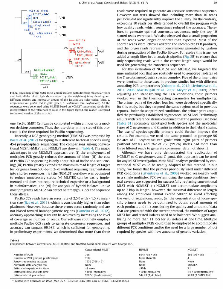
In studies of molecular epidemiology, pathogenicity, and phy-logenetics, MLST has become the standard method of genotypingmany fungi, including strains of the C. neoformans/C. gattii speciescomplex. Furthermore, it also widely used in genotyping other fun-gal species such as Candida (Jackson et al., 2009), Aspergillus (Bainet al., 2007), and Pseudallescheria (Bernhardt et al., 2013). Althoughwhole genome sequence typing (WGST) is becoming more accessi-ble, especially for organisms with small genomes, such as bacteriaand viruses, it is not yet practical for genotyping numerous isolatesof eukaryotic species. MLST will not soon become obsolete becauseit provides an economical and efficient method of screening wildtype isolates, assigning them to established clades, subpopulations,or phenotypic groups, and determining whether they warrantmore extensive analysis or WGST. However, compared to lessreproducible and more subjective methods of rapid genotyping,such as generating amplified fragment length polymorphisms,MLST has the disadvantages of being more time consuming as wellas costly due to the use of Sanger sequencing. To resolve theseissues, we have developed a new high-throughput method of MLSTgenotyping that generates CCS PacBio next-generation sequencingreads, NGMLST, and a novel multifunctional software program,MLSTEZ, which provides simplified and automated analysis ofNGMLST data. The average time required for processing DNA from96 isolates was 7 h. Previously, 2–4 weeks were required to obtainsequence data from the same number of strains using conventionalMLST.
To interface with NGMLST, we developed the multifaceted soft-ware program, MLSTEZ, which is available on the Internet at nocost (https://sourceforge.net/projects/mlstez/). The program isfully automated and requires a general sequencing format file(FASTQ and FASTA) as input, which means that NGMLST will sup-port all sequencing platforms that can generate full-length bar-coded amplicons. Because a sequence assembly feature is notincluded in the program, fragmented amplicons must first beassembled before analysis by MLSTEZ. MLSTEZ can perform bar-code and primer identification, recognize consensus sequences,and predict heterozygous loci. All the results that are generatedby the software can be easily exported as sequence files, graphs,or tables. In addition, the MLSTEZ output sequence files can beused directly for phylogenetic analyses, which significantlyreduces the time required for many follow-up studies. With themultiprocessing features of MLSTEZ, the analyses of data from
Table 3
Heterozygous loci of the hybrids predicted by MLSTEZ. ‘Yes’ indicates the identifi-cation of two alleles, ‘No’ indicates that only one allele was identified, and the lociwithout insufficient reads (<5) for analysis are labeled ‘NA’.Strain CAP59 GPD1 IGS1 LAC1 MPD1 PLB1 SOD1 TEF1 URA5
Cng9 NA Yes No No No No Yes No YesZG287 No Yes No Yes NA Yes Yes Yes YesCBS132 No Yes No NA No No Yes No YesNC34-21 No Yes No Yes NA Yes Yes Yes YesEJB34 No No NA Yes Yes NA Yes Yes YesEJB41 No No No Yes No NA Yes No No
one PacBio SMRT Cell can be completed within an hour on a mod-
reads were required to generate an accurate consensus sequence.
Fig. 6. Phylogeny of the SOD1 locus among isolates with different molecular typesand both alleles of six hybrids visualized by the neighbor-joining dendrogram.Different species and molecular groups of the isolates are color-coded (blue, C.neoformans var. grubii; red, C. gatii; green, C. neoformans var. neoformans). All thesequences were generated using MLSTEZ based on NGMLST sequencing result. (Forinterpretation of the references to color in this figure legend, the reader is referredto the web version of this article.)
Y. Chen et al. / Fungal Genetics and Biology 75 (2015) 64–71 69
ern desktop computer. Thus, the rate-determining step of this pro-tocol is the time required for PacBio sequencing.
Recently, a NGS genotyping method (HiMLST) was proposed byBoers et al. (2012) for typing four different bacterial species using454 pyrophosphate sequencing. The comparisons among conven-tional MLST, HiMLST and NGMLST are shown in Table 4. The majoradvantages in our MGMLST approach are: (i) the employment ofmultiplex PCR greatly reduces the amount of labor; (ii) the costof PacBio CCS sequencing is only about 20% of Roche 454 sequenc-ing; (iii) PacBio greatly extends the maximum read length of targetloci or genes from 500-bp to 2-kb without requiring fragmentationinto shorter sequences; (iv) the NGMLST workflow was optimizedto reduce unnecessary steps; (v) MLSTEZ can be easily imple-mented and does not require technical expertise or a backgroundin bioinformatics; and (vi) for analysis of hybrid isolates, unlikemost programs, MLSTEZ can detect heterozygous loci and sequencetheir alleles.
PacBio CCS reads have an error rate of 2.5% with �1.5 kb inser-tion size (Jiao et al., 2013), which is considerably higher than otherplatforms. However, because these errors occur randomly and arenot biased toward homopolymeric regions (Carneiro et al., 2012),accuracy approaching 100% can be achieved by increasing the levelof coverage or number of reads. Our software routinely employsmultiple PacBio CCS reads to generate consensus sequences, andaccuracy can surpass 99.98%, which is sufficient for genotyping.In preliminary experiments, we determined that more than three
Table 4
Comparisons between conventional MLST, HiMLST and NGMLST based on 96 isolates withConventional MLST
Number of PCRs 768Number of PCR product purifications 768Sanger sequencing reaction 1536 (768 � 2)Automate data analysis tool NoneEstimated experimental time >1 weekEstimated data analysis time >10 h (manually)Estimated cost per isolate $70.56 (bi-directional)
a Tested with 8 threads on iMac (Mac OS X 10.9.2) on 3.4G Intel Core i7, 16GB 1333M
However, our tests showed that including more than 10 readsper locus did not significantly improve the quality. On the contrary,exceeding 10 reads per allele tended to overfill the program withlow quality reads, which sometimes reduced the accuracy. There-fore, to generate optimal consensus sequences, only the top 10scored reads were used. We also observed that a small proportionof the reads were longer or shorter than expected. Most of theshorter reads were leftover adaptor and incomplete PCR products,and the longer reads represent concatemers generated by ligationduring preparation of the PacBio library. To resolve this issue, weadded a length filter in our analysis pipeline (Fig. 2B) to ensure thatonly sequencing reads within the correct length range would beused for generating the consensus sequences.
For this evaluation of NGMLST and MLSTEZ, we targeted thenine unlinked loci that are routinely used to genotype isolates ofthe C. neoformans/C. gattii species complex. Five of the primer pairswere identical to those used in previous studies but with differentannealing PCR temperatures (Colom et al., 2012; Litvintseva et al.,2011, 2006; MacDougall et al., 2007; Meyer et al., 2009). Afteradjusting and standardizing the PCR conditions, these primersworked well in the thermocycling parameters for multiplexing.The primer pairs of the other four loci were developed specificallyfor this study, but they targeted the same regions used in previousreports. Under these optimized conditions, the primer pairs ampli-fied the previously established cryptococcal MLST loci. Preliminaryresults with reference strains confirmed that the primers used here(Table S1) accurately genotyped both species and the moleculartypes of C. neoformans and C. gattii in addition to the hybrid strains.The use of species-specific primers could further improve theresults. For example, we used the same protocol to genotype 96isolates of C. neoformans var. grubii with eight pairs of primers(without MPD1), and 762 of 768 (99.2%) alleles had more thanthree filtered reads to generate consensus (data not shown).
Although we have only demonstrated the application ofNGMLST to C. neoformans and C. gattii, this approach can be usedfor any MLST investigation. Most MLST analyses performed by con-ventional MLST could be readily adapted to this method. In ourstudy, we found that the primers previously used under differentPCR conditions (Litvintseva et al., 2006) worked reasonably wellin a single multiplex PCR system using the same conditions. Sev-eral caveats are suggested for successfully replacing conventionalMLST with NGMLST: (i) NGMLST can accommodate amplicomsup to 2 kbp in length; however, the maximal difference in lengthamong the amplicons cannot exceed 500 bp to avoid affectingthe yield of sequencing reads; (ii) the concentration of locus-spe-cific primers needs to be optimized to obtain equal amounts ofeach product; and (iii) considering the quality and amount of datathat are generated with the current protocol, the numbers of targetMLST loci and tested isolates need to be balanced. We suggest ana-lyzing no more than 11 loci for 96 isolates at one time. Multiplegroups of multiplex PCRs could then be employed to accommodatedifferent PCR conditions and/or the need for a large number of locirequired by species with low amounts of genetic variation.
8 target loci.
HiMLST NGMLST
864 (768 + 96) 192 (96 + 96)More than 96 4None NoneNone MLSTEZ>30 h 7 h>10 h (manually) �1 h (automatically)a
$42.23 (1/4 plate) $8.83 (1 SMRT Cell)
Hz DDR3.

Our results show that NGMLST and MLSTEZ not only work well fumigatus. J. Clin. Microbiol. 45, 1469–1477. http://dx.doi.org/10.1128/JCM.00064-07.
70 Y. Chen et al. / Fungal Genetics and Biology 75 (2015) 64–71
on the haploid strains but also can be used to detect and analyzehybrid strains, which are difficult to MLST genotype using conven-tional Sanger sequencing. Among our six control hybrid strains,most were detected by more than three heterozygous loci. Unfor-tunately, some haploid strains were erroneously identified withheterozygous loci; these results were caused by low coverage orreads of poor quality. Therefore, we strongly recommend repeatingthe analysis on putative hybrids. In addition, MLST only targets alimited number of genomic loci, and aneuploid strains are verycommon in some fungal species (Kwon-Chung and Chang, 2012;Selmecki et al., 2009). It remains difficult to determine the ploidyof test strains even when multiple loci have been identified to beheterozygous. Other methods to determine aneuploidy, such asanalysis of the cells by fluorescent-activated cell sorting (FACS)could help to confirm MLST data and ploidy.
This investigation evaluated a novel NGMLST method of geno-typing, which has proven to be rapid and relatively inexpensive,as well as amenable to the high-throughput analyses of large sam-ples. Coupled with the automated multifunctional software, MLS-TEZ, high quality MLST profiles can be acquired with very simpleoperations in a short period of time. The approach demonstratedhere was evaluated with the heterobasidiomycetous human path-ogen, Cryptococcus, but it can be applied to many other fungal orother eukaryotic taxa, including haploid, diploid, and hybridorganisms.
Conflict of interest
None of the authors have a conflict of interest.
Data accessibility
The source code and pre-compiled GUI applications forMacintosh and Windows PC of MLSTEZ are freely available onhttps://sourceforge.net/projects/mlstez/files/. The GUI applicationis only available for Macintosh OS X 10.6+ and Windows XP andlater version. The manual of the application is available onhttps://sourceforge.net/p/mlstez/wiki/Manual/. MLSTEZ is underactive development. Bug reports and suggestions from users willbe helpful for the improvements of the software in future version.
Acknowledgments
The authors thank to Sun Sheng (Duke University MedicalCenter), Matthew Fisher (Imperial College London), Tom Harrison(St George’s, University of London), Vinicius Ponzio (FederalUniversity of São Paulo), Arnaldo L. Colombo (Federal University ofSão Paulo) and Annemarie Brouwer (Radboud University Nijmegen)for providing isolates for the study. The authors also thank to JoshGranek (Duke University Medical Center), Olivier Fedrigo andGraham Alexander (Duke Center for Genomic and ComputationalBiology) for discussion and sample sequencing. This work wassupported by Public Health Service Grants AI73896 and AI93257(JRP).
Appendix A. Supplementary material
Supplementary data associated with this article can be found, inthe online version, at http://dx.doi.org/10.1016/j.fgb.2015.01.005.
References
Bain, J.M., Tavanti, A., Davidson, A.D., Jacobsen, M.D., Shaw, D., Gow, N.A.R., Odds,F.C., 2007. Multilocus sequence typing of the pathogenic fungus Aspergillus
Bernhardt, A., Sedlacek, L., Wagner, S., Schwarz, C., Würstl, B., Tintelnot, K., 2013.Multilocus sequence typing of Scedosporium apiospermum and Pseudallescheriaboydii isolates from cystic fibrosis patients. J. Cyst. Fibros. 12, 592–598. http://dx.doi.org/10.1016/j.jcf.2013.05.007.
Boers, S.A., van der Reijden, W.A., Jansen, R., 2012. High-throughput multilocussequence typing: bringing molecular typing to the next level. PLoS ONE 7,e39630. http://dx.doi.org/10.1371/journal.pone.0039630.
Byrnes, E.J., Bildfell, R.J., Frank, S.A., Mitchell, T.G., Marr, K.A., Heitman, J., 2009.Molecular evidence that the range of the Vancouver Island outbreak ofCryptococcus gattii infection has expanded into the Pacific Northwest in theUnited States. J. Infect. Dis. 199, 1081–1086. http://dx.doi.org/10.1086/597306.
Carneiro, M.O., Russ, C., Ross, M.G., Gabriel, S.B., Nusbaum, C., DePristo, M.A., 2012.Pacific biosciences sequencing technology for genotyping and variationdiscovery in human data. BMC Genom. 13, 375. http://dx.doi.org/10.1038/nbt.1754.
Chan, M.S., Maiden, M.C., Spratt, B.G., 2001. Database-driven multi locus sequencetyping (MLST) of bacterial pathogens. Bioinformatics 17, 1077–1083.
Chen, Y., Toffaletti, D.L., Tenor, J.L., Litvintseva, A.P., Fang, C., Mitchell, T.G.,McDonald, T.R., Nielsen, K., Boulware, D.R., Bicanic, T., Perfect, J.R., 2013. TheCryptococcus neoformans transcriptome at the site of human meningitis. MBio 5,e01087-13. http://dx.doi.org/10.1128/mBio.01087-13.
Colom, M.F., Hagen, F., Gonzalez, A., Mellado, A., Morera, N., Linares, C., García, D.F.,Peñataro, J.S., Boekhout, T., Sánchez, M., 2012. Ceratonia siliqua (carob) trees asnatural habitat and source of infection by Cryptococcus gattii in theMediterranean environment. Med. Mycol. 50, 67–73. http://dx.doi.org/10.3109/13693786.2011.574239.
Edgar, R.C., 2004. MUSCLE: multiple sequence alignment with high accuracy andhigh throughput. Nucleic Acids Res. 32, 1792–1797. http://dx.doi.org/10.1093/nar/gkh340.
Eid, J., Fehr, A., Gray, J., Luong, K., Lyle, J., Otto, G., Peluso, P., Rank, D., Baybayan, P.,Bettman, B., Bibillo, A., Bjornson, K., Chaudhuri, B., Christians, F., Cicero, R., Clark,S., Dalal, R., Dewinter, A., Dixon, J., Foquet, M., Gaertner, A., Hardenbol, P.,Heiner, C., Hester, K., Holden, D., Kearns, G., Kong, X., Kuse, R., Lacroix, Y., Lin, S.,Lundquist, P., Ma, C., Marks, P., Maxham, M., Murphy, D., Park, I., Pham, T.,Phillips, M., Roy, J., Sebra, R., Shen, G., Sorenson, J., Tomaney, A., Travers, K.,Trulson, M., Vieceli, J., Wegener, J., Wu, D., Yang, A., Zaccarin, D., Zhao, P., Zhong,F., Korlach, J., Turner, S., 2009. Real-time DNA sequencing from singlepolymerase molecules. Science 323, 133–138. http://dx.doi.org/10.1126/science.1162986.
Jackson, A.P., Gamble, J.A., Yeomans, T., Moran, G.P., Saunders, D., Harris, D., Aslett,M., Barrell, J.F., Butler, G., Citiulo, F., Coleman, D.C., de Groot, P.W.J., Goodwin,T.J., Quail, M.A., McQuillan, J., Munro, C.A., Pain, A., Poulter, R.T., Rajandream, M.-A., Renauld, H., Spiering, M.J., Tivey, A., Gow, N.A.R., Barrell, B., Sullivan, D.J.,Berriman, M., 2009. Comparative genomics of the fungal pathogens Candidadubliniensis and Candida albicans. Genome Res. 19, 2231–2244. http://dx.doi.org/10.1101/gr.097501.109.
Janbon, G., Ormerod, K.L., Paulet, D., Byrnes, E.J., Yadav, V., Chatterjee, G., Mullapudi,N., Hon, C.-C., Billmyre, R.B., Brunel, F., Bahn, Y.-S., Chen, W., Chen, Y., Chow,E.W.L., Coppée, J.-Y., Floyd-Averette, A., Gaillardin, C., Gerik, K.J., Goldberg, J.,Gonzalez-Hilarion, S., Gujja, S., Hamlin, J.L., Hsueh, Y.-P., Ianiri, G., Jones, S.,Kodira, C.D., Kozubowski, L., Lam, W., Marra, M., Mesner, L.D., Mieczkowski, P.A.,Moyrand, F., Nielsen, K., Proux, C., Rossignol, T., Schein, J.E., Sun, S.,Wollschlaeger, C., Wood, I.A., Zeng, Q., Neuvéglise, C., Newlon, C.S., Perfect,J.R., Lodge, J.K., Idnurm, A., Stajich, J.E., Kronstad, J.W., Sanyal, K., Heitman, J.,Fraser, J.A., Cuomo, C.A., Dietrich, F.S., 2014. Analysis of the genome andtranscriptome of Cryptococcus neoformans var. grubii reveals complex RNAexpression and microevolution leading to virulence attenuation. PLoS Genet. 10,e1004261. http://dx.doi.org/10.1371/journal.pgen.1004261.
Jiao, X., Zheng, X., Ma, L., Kutty, G., Gogineni, E., Sun, Q., Sherman, B.T., Hu, X., Jones,K., Raley, C., Tran, B., Munroe, D.J., Stephens, R., Liang, D., Imamichi, T., Kovacs,J.A., Lempicki, R.A., Huang, D.W., 2013. A benchmark study on error assessmentand quality control of CCS reads derived from the PacBio RS. J. Data Min. Genom.Proteom. 4. http://dx.doi.org/10.4172/2153-0602.1000136.
Koren, S., Schatz, M.C., Walenz, B.P., Martin, J., Howard, J.T., Ganapathy, G., Wang, Z.,Rasko, D.A., McCombie, W.R., Jarvis, E.D., Phillippy, A.M., 2012. Hybrid errorcorrection and de novo assembly of single-molecule sequencing reads. Nat.Biotechnol. 30, 693–700. http://dx.doi.org/10.1038/nbt.2280.
Kwon-Chung, K.J., Chang, Y.C., 2012. Aneuploidy and drug resistance in pathogenicfungi. PLoS Pathog. 8, e1003022. http://dx.doi.org/10.1371/journal.ppat.1003022.
Li, W., Raoult, D., Fournier, P.-E., 2009. Bacterial strain typing in the genomic era.FEMS Microbiol. Rev. 33, 892–916. http://dx.doi.org/10.1111/j.1574-6976.2009.00182.x.
Litvintseva, A.P., Mitchell, T.G., 2012. Population genetic analyses reveal the Africanorigin and strain variation of Cryptococcus neoformans var. grubii. PLoS Pathog. 8,e1002495. http://dx.doi.org/10.1371/journal.ppat.1002495.
Litvintseva, A.P., Thakur, R., Vilgalys, R., Mitchell, T.G., 2006. Multilocus sequencetyping reveals three genetic subpopulations of Cryptococcus neoformans var.grubii (serotype A), including a unique population in Botswana. Genetics 172,2223–2238. http://dx.doi.org/10.1534/genetics.105.046672.
Litvintseva, A.P., Carbone, I., Rossouw, J., Thakur, R., Govender, N.P., Mitchell, T.G.,2011. Evidence that the human pathogenic fungus Cryptococcus neoformans var.grubii may have evolved in Africa. PLoS ONE 6, e19688. http://dx.doi.org/10.1371/journal.pone.0019688.

MacDougall, L., Kidd, S.E., Galanis, E., Mak, S., Leslie, M.J., Cieslak, P.R., Kronstad, J.W.,Morshed, M.G., Bartlett, K.H., 2007. Spread of Cryptococcus gattii in British
Simwami, S.P., Khayhan, K., Henk, D.A., Aanensen, D.M., Boekhout, T., Hagen, F.,Brouwer, A.E., Harrison, T.S., Donnelly, C.A., Fisher, M.C., 2011. Low diversity
Y. Chen et al. / Fungal Genetics and Biology 75 (2015) 64–71 71
Columbia, Canada, and detection in the Pacific Northwest, USA. Emerg. Infect.Dis. 13, 42–50. http://dx.doi.org/10.3201/eid1301.060827.
Maiden, M.C., Bygraves, J.A., Feil, E., Morelli, G., Russell, J.E., Urwin, R., Zhang, Q.,Zhou, J., Zurth, K., Caugant, D.A., Feavers, I.M., Achtman, M., Spratt, B.G., 1998.Multilocus sequence typing: a portable approach to the identification of cloneswithin populations of pathogenic microorganisms. Proc. Natl. Acad. Sci. U.S.A.95, 3140–3145.
Meyer, W., Aanensen, D.M., Boekhout, T., Cogliati, M., Diaz, M.R., Esposto, M.C.,Fisher, M., Gilgado, F., Hagen, F., Kaocharoen, S., Litvintseva, A.P., Mitchell, T.G.,Simwami, S.P., Trilles, L., Viviani, M.A., Kwon-Chung, J., 2009. Consensus multi-locus sequence typing scheme for Cryptococcus neoformans and Cryptococcusgattii. Med. Mycol. 47, 561–570. http://dx.doi.org/10.1080/13693780902953886.
Park, B.J., Wannemuehler, K.A., Marston, B.J., Govender, N., Pappas, P.G., Chiller, T.M.,2009. Estimation of the current global burden of cryptococcal meningitis amongpersons living with HIV/AIDS. AIDS 23, 525–530. http://dx.doi.org/10.1097/QAD.0b013e328322ffac.
Quail, M.A.M., Smith, M.M., Coupland, P.P., Otto, T.D.T., Harris, S.R.S., Connor, T.R.T.,Bertoni, A.A., Swerdlow, H.P.H., Gu, Y.Y., 2012. A tale of three next generationsequencing platforms: comparison of Ion Torrent, Pacific Biosciences andIllumina MiSeq sequencers. BMC Genom. 13, 341. http://dx.doi.org/10.1186/1471-2164-13-341.
Schwartz, D.C., Cantor, C.R., 1984. Separation of yeast chromosome-sized DNAs bypulsed field gradient gel electrophoresis. Cell 37, 67–75.
Selmecki, A.M., Dulmage, K., Cowen, L.E., Anderson, J.B., Berman, J., 2009.Acquisition of aneuploidy provides increased fitness during the evolution ofantifungal drug resistance. PLoS Genet. 5, e1000705. http://dx.doi.org/10.1371/journal.pgen.1000705.
Cryptococcus neoformans variety grubii multilocus sequence types from Thailandare consistent with an ancestral African origin. PLoS Pathog. 7, e1001343.http://dx.doi.org/10.1371/journal.ppat.1001343.
Stephen, C., Lester, S., Black, W., Fyfe, M., Raverty, S., 2002. Multispecies outbreak ofcryptococcosis on southern Vancouver Island, British Columbia. Can. Vet. J. 43,792–794.
Sun, S., Hsueh, Y.-P., Heitman, J., 2012. Gene conversion occurs within the mating-type locus of Cryptococcus neoformans during sexual reproduction. PLoS Genet.8, e1002810. http://dx.doi.org/10.1371/journal.pgen.1002810.
Tavanti, A., Davidson, A.D., Johnson, E.M., Maiden, M.C.J., Shaw, D.J., Gow, N.A.R.,Odds, F.C., 2005. Multilocus sequence typing for differentiation of strains ofCandida tropicalis. J. Clin. Microbiol. 43, 5593–5600. http://dx.doi.org/10.1128/JCM.43.11.5593-5600.2005.
Taylor, J.W., Fisher, M.C., 2003. Fungal multilocus sequence typing – it’s not just forbacteria. Curr. Opin. Microbiol. 6, 351–356. http://dx.doi.org/10.1016/S1369-5274(03)00088-2.
Travers, K.J., Chin, C.-S., Rank, D.R., Eid, J.S., Turner, S.W., 2010. A flexible andefficient template format for circular consensus sequencing and SNP detection.Nucleic Acids Res. 38, e159. http://dx.doi.org/10.1093/nar/gkq543.
Vanhee, L.M.E., Nelis, H.J., Coenye, T., 2010. What can be learned from genotyping offungi? Med. Mycol. 48 (Suppl. 1), S60–S69. http://dx.doi.org/10.3109/13693786.2010.484816.
Vos, P., Hogers, R., Bleeker, M., Reijans, M., van de Lee, T., Hornes, M., Frijters, A., Pot,J., Peleman, J., Kuiper, M., 1995. AFLP: a new technique for DNA fingerprinting.Nucleic Acids Res. 23, 4407–4414.
Xu, J.J., Yan, Z.Z., Guo, H.H., 2009. Divergence, hybridization, and recombination inthe mitochondrial genome of the human pathogenic yeast Cryptococcus gattii.Mol. Ecol. 18, 2628–2642. http://dx.doi.org/10.1111/j.1365-294X.2009.04227.x.