Embed Size (px)
Citation preview
J. Indian Fish. Assoc., 34:59- 73, 2007
FURUNCULOSIS IN SNOW TROUT (SCHIZOTHORACINAE) IN KASHMIR: FIRST REPORT
Salman Rauoof Chalkoo, A.M. Najar*, T. A. Qureshit and Aamira Shafit Department of Fisheries, Government of Jammu and Kashmir,
Srinagar- 190 001, India *Division of Fisheries, Sher-e-Kashmir University of Agricultural
Sciences and Technology, Shuhama, Alusteng, Srinagar- 190 006, India
1Department of Applied Aquaculture, Barkatullah University, Bhopal- 462 024, India
59
tDepartment of Fisheries, Degree College, Baramulla, Kashmir-193 101, India
ABSTRACT
Incidences of furunculosis were reported in Schizothorax spp. (Schizothorax niger, S. esocinus, S. curvifrons and S. labiatus) in Wular Lake, Kashmir, from 2003 to 2005. The disease was reported during summer and winter months, but the percentage of infection was maximum during winter. Mortality rate ranged from 8 to 15°/o. Artificial challenge of Schizothorax spp. with Aeromonas salmonicida produced symptoms pertinent to furunculosis. The incidence of disease was the highest (13.87o/o) in December, and lowest (0.40°/o) in May and October. S. esocinus exhibited the maximum ( 44.48°/o) percentage of infection, while asS. labiatus exhibited the minimum (14.28°/o) throughout the study period. Haematological investigations revealed devastating changes in various blood parameters. Chemotherapeutic tests revealed complete recovery of the disease using 20 ppm oxytetracycline and 30 ppm streptomycin.
Keywords: Furunculosis, Aero monas salmonicida, Schizothoracinae, treatment
INTRODUCTION
Furunculosis is the designation presently recognized (Ghittino, 1968) for an epidemic bacteriosis of fishes, the pathogen of which is Aermnonas salmonicida. Furunculosis has spread to almost all countries (Snieszko, 1973). Furunculosis and its pathogens were originally described by Emmerich and Weibel (1890, 1894) in a Bavarian fish farm. Thereafter, in a matter of few years,
it was encountered in several European countries. It was reported in West North America, England and Ireland. It seems more probable that it could have been diagnosed and bacteriologically ascertained in these countries subsequent to the first descriptions by Emmerich and Weibel (1890, 1894), although it had occurred earlier in these countries. In this respect, the exposition by HeuschmannBrunner (1974) may also be referred to. McCarthy (1975) assumes a possible lin1c
60 Salman Rauoof Chalkoo, A. M. Najar, T. A. Qureshi and Aamira Shaft
between the introduction of the shasta rainbow trout (Salmo gairdnerii) into Europe in 1880 and the first record of furunculosis in Germany in 1890. Andrew et al. (2003) reported the sporadic incidences of furunculosis in snow trout in the fish farms ofEurope.
The typical symptoms of furunculosis are boils and ulcers, which are found isolated or in groups, chiefly in the dorsal region. These ulcers are tinged . with blood. The bigger ones contain sticky, dark reddish pus. Ulcers may be absent, and in such an event, autopsy shows intestinal inflammation, principally in the pyloric and rectal regions. The swim bladder is hyperaemic; small spots and haemorrhages are found in the liver. Similar haemorrhages are encountered at times on the inner side of the opercula, in the eyes and on the fins. In the event of a bacteraemia, these symptoms would be usually lacking and the blood is filled with bacteria.
The causative agent of furunculosis is A. salmonicida (Emmerich and Weibel; 1890, 1894). It measures 0.8 x 0.5 J.tm, and is non-motile, non-flagellated and gramnegative. Agar colonies at 20°C become dark and even black in colour within 2-3 days due to the formation of melanin in the presence of oxygen. Gelatin slab cultures give a very characteristic liquefaction in the form of a funnel within 2-3 days, accompanied by abundant sediment formation. A. salmonicida grows at an optimum temperature of20-30°C and dies at 3 7°C. It is ubiquitous and may be found in water and mud, often in very large numbers.
Traditionally, A. salmonicida was thought to have a predilection for
salmonids. Over the years, however, the apparent host range of the pathogen has steadily expanded. Thus, infections are lmown to occur among representatives of several major families of Osteichthyes including Cyprinidae, Serranidae and Anoplopomatidae, in addition to Salmonidae and Agnatha. Non -salmonids which have been documented as suffering from the disease of A. salmonicida etiology include minnow and goldfish, carp (Bootsma et al., 1977), perch (Bucke, 1978) bream, roach, dace, chub, tench, pike bullheads (McCarthy, 1975), and snow trout (Andrew etal., 2003).
Therapy, prophylaxis and hygiene as in all other infectious diseases, and removal and destruction of dead and gravely infected fish are suggested. These should preferably be either burned or buried. Fish which are suspected of having furunculosis are isolated in special tanks, taking care to ensure that these are fed with an independent supply of water, which should be cold and clean. Sulfonamides (sulfamerazine, sulfaguanidine, sulfadiazine, sulfamethazine, sulfisoxazole, etc.) given orally with the food at the rate of 10 g for 45 kg of fish per day are useful for therapy (Snieszko and Hoffman, 1963). Among the antibiotics, chloramphenicol and oxytetracycline are reported to be the best with a dosage of2.5-3.5 gper45 kg offish per day (Snieszko and Hoffman, 1963). Leaman ( 1965) reported excellent results in preventing furunculosis in returning Atlantic salmon by giving 10 mg chloramphenicol per 0.45 kg fish intra peritonially.
FURUNCULOSIS IN SNOW TROUT (SCHIZOTHORACINAE) IN KASHMIR: FIRST REPORT 61
OBSERVATIONS
Occurrence
Furunculosis appeared in the winter season, i.e., at the end of October. Its severity increased with the decrease in temperature reaching the peak during intense cold in the month of January. It declined with the increase in temperature in March and by the end of April, it vanished completely. Maximum number of incidences of furunculosis was recorded during winter months. It appears that low temperature is conducive for the spread of furunculosis. Mohan and Shanker (1995) contended that lowering of water temperature may lower the defense competence of fish, thereby, predisposing it to diseases. Salman et al. (2004) also support this view.
Symptoms
Fishes show lethargic behaviour, slight exophthalmia, blood-shot fins, bloody discharge from vent and multiple hemorrhages. Necrosis in the skin and underlying musculature is prominent. The disease is characterized by open ulcers on the back and sides of the body; the bigger ulcers contain sticky, dark reddish pus. Punctiform bleeding and petechiae appear in the viscera, especially the liver, and also on the skin, gills, musculature and the bases of fins. The intestine is very red and its blood vessels are enlarged. In the kidneys, besides bleeding, degenerative changes and necrosis of the haematopoietic connective tissue are found. In some cases, haemorrhagic liver may be observed. Ascites is observed in the abdominal cavity. Anus is prolapsed with marked reddening.
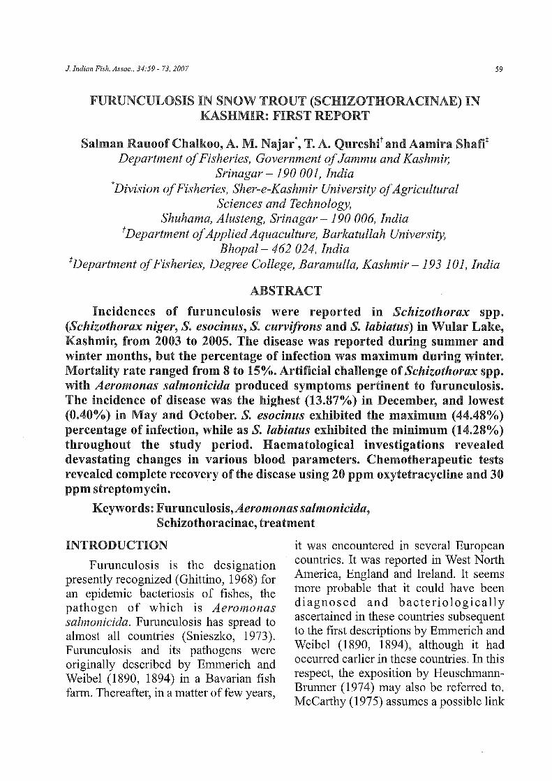
Percentage ofinfection
During the first year of study, the minimum percentage of infection (1.22) was recorded in the month of May 2004 and the maximum (13.87) in the month of December 2003. During the second year of study, the minimum percentage of
16
14 -
12
g 10
i ~ ~ 6
2-
Fig. 1: Furunculosis in Schizothorax spp. from 2003 to 2005
infection (0.40) was recorded in the months of October 2004 and May 2005, while the maximum (11.83) was in the month of January 2005 (Fig. 1 ). During the whole
30
~:~~~ 10
5
0 / --~-~---.-------,
Fig. 2: Furunculosis in four species of Schizothorax in Kashmir valley
study period, the minimum percentage of infection (14.28) was recorded in Schizo thorax labiatus, while the maximum (44.48) inS. esocinus (Fig. 2).
62 Salman RauoofChalkoo, A.M. Najar, T. A. QureshiandAamira Shafi
Isolation
A. salmonicida was found chiefly associated with open ulcers and viscera, especially the liver, of fishes suffering from furunculosis. Morphologically, the isolate was a gram-negative, non-motile, non- flagellated b acteri urn of approximately 0.8 x 0.5 llffi. The bacterium was isolated on trypticase soy agar (TSA) and a selective medium, Coomassie brilliant blue (CBB). On the non-selective medium, the bacterium developed colonies surrounded by a dark brown water-soluble pigment after incubation at 20-25°C for 3-4 days. On the selective medium, the bacteria developed as dark blue colonies. Agar colonies at 20°C became dark and even black in colour within 2-3 days, due to the formation. of melanin in the presence of oxygen.
Experimental infection trials with A. salmonicida
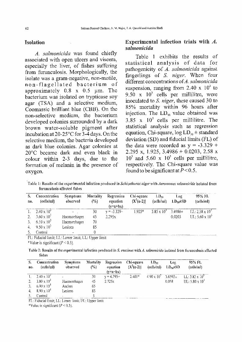
Table 1 exhibits the results of statistical analysis of data for pathogenicity of A. salmonicida against fingerlings of S. niger. When four different concentrations of A. salmonicida suspension, ranging from 2.40 x 10³ to 9.50 x 10³ cells per millilitre, were inoculated to S. niger, these caused 30 to 85% mortality within 96 hours after injection. The LD50 value obtained was 3.85 x 10³ cells per millilitre. The statistical analysis such as regression equation, Chi-square, log LD50 ±standard deviation (SD) and fiducial limits (FL) of the data were recorded as y = -3.329 + 2.295 X, 1.925, 3.4986 ± 0.0203, 2.58 X
103 and 5.60 x 10³ cells per millilitre, respectively. The Chi -square value was found to be significant at P < 0. 5.
Table 1: Results of the experimental infection produced in Schizotlwrax niger with Aeromo11as salmouicida isolated from furunculosis affected fishes
s. Concentration Symptoms Mortality Regression Chi-square LDso Log 95% FL no. (cells/ml) observed (%) equation {X2(n-2)} (cells/ml) LD5o±SD ( cells/ml)
1. 2.40 X I 03 (y=a+bx)
30 y- -3.329+ 1.925* 3.85 X 103 3.4986± LL: 2.58 X 10³
2. 3.60 X 10³ Haemorrhages 45 2.295x 0.0203 UL: 5.60 X I 03
' 6.10 X !03 J. Haemorrhages 70 4. 9.50 X 10³ Lesions 85 5. Control 0 FL: FiduciallLmit; LL: Lower limit; UL: Upper limit *Value is significant (P < 0.5).
Table 2: Results of the experimental infection produced inS. esocinus with A. sa/mouicida isolated from furunculosis affected fishes
S. Concentration Symptoms no. (cells/ml) observed
Mortality (%)
J. 2.40 X JO-' 30 2. 3.80 x I 03 Haemorrhages 45 3. 6.90 x 10³ Ascites 65 4. 8.90x 10³ Lesions 85 5. Control 5
Regression Chi-square equation {X2(n-2)) (y=a+bx)
y- 4.295+ 2.401 * 2.725x
FL: Fiducial limit; LL: Lower limit; UL: U:-pp-er--:-1!::-.m-=-it _____ _ *Value is signillcunt (P < 0.5 ).
LD~o Log 95% FL ( cells/ml) LDso±SD (cells/ml)
4.90 X 10³ 3.6592± LL: 3.82 X 10³
0.058 UL: 5.80 x 10³
FURUNCULOSIS IN SNOW TROUT (SCHIZOTHORACINAE) IN KASHMIR: FIRST REPORT 63
Table 2 exhibits the results of statistical analysis of data for pathogenicity of A. salmonicida against fingerlings of S. esocinus. When four different concentrations of A. salmonicida suspension, ranging from 2.40 x 1 03 to 8.90 x 10³ cells per millilitre, were inoculated to S. esocinus, these caused 30 to 85% mortality within 96 hours after injection. The LD50 value obtained was 4.90 x 10³ cells per millilitre. The statistical analysis such as regression equation, Chi-square, log LD50 ± SD and FL of the data were recorded as y = 4.295 + 2.725 X, 2.401, 3.6592 ± 0.058, 3.82 X
103 and 5.80 x 10³ cells per millilitre, respectively. The Chi-square value was found to be significant at P < 0. 5.
Haematology
In healthy specimens of S. niger, the blood parameters were: haemoglobin 8.32 ± 1.4 mg%, erythrocytes 1.325 ± 1.8 m, leukocytes 73 ± 3.9 x 10³ t, haematocrit value 27.33 ± 10.2 mg% and blood sugar
57.16 ± 1.79 mg%. Histochemical parameters showed the value of total albumin as 2.775 ± 0.14 mg%, albumin 0.89 ± 0.01 mg% and globulin 2.12 ± 0.05 mg%. Uric acid content was 1.68 ± 0.03 mg%, total cholesterol showed a value of 289.4 ± 1.37 mg%; bilirubin was 1.84 ± 0.03 mg%, while glucose was 87.0 ± 1.83 mg%. PCV and MCV showed values of 53.11 ± 1.34 mg% and224.83 ± 1.14!lm3
,
respectively. MCH and MCHC were observed to be 65.91 ± 1.89 Pg and 33 ± 2.5 g%, respectively.
In diseased specimens of S. niger suffering from furunculosis, haemoglobin showed a value of 3.15 ± 2.21 mg%. The total numbers of erythrocytes and leukocytes were 0.52 ± 1.8 m and 103.2 ± 2. 8 x 10³ t respectively. Haematocrit value was 18.22 ± 1.63 mg%, while blood sugar was 33.16 ± 2.97 mg%. Out of histochemical parameters, total albumin was 0.97 ± 0.13 mg% while albumin was 0.15 ± 0.01 mg%. Globulin was 1.0 ± 0.001 mg% and uric acid was 2.89 ± 0.02
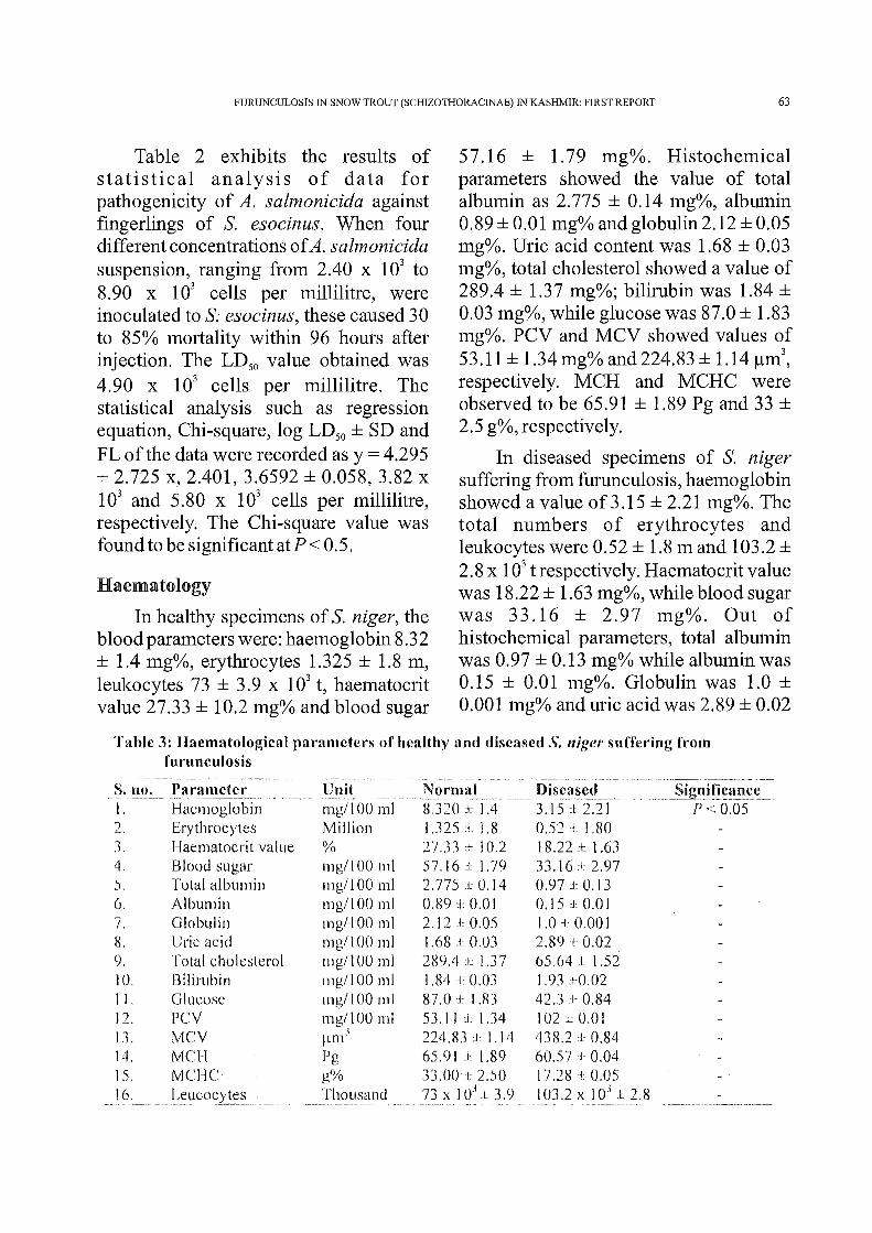
Table 3: Haematological parameters of healthy and diseased S. niger suffering from furunculosis
--~· _!!{)~-- Parametct· _________ Unit ______ ~_j'lorma!_ ~ _____ f?isc_~se~------------ Significan_cc __ I. 2. ., ,).
4. 5. 6. 7. 8. 9. 10. 11. 12. 13. 14. 15. 16.
Haemoglobin mgll 00 ml 8.320 J 1.4 3.15 J 2.21 P < 0.05 Erythrocytes Million 1 J25 L 1.8 0.52 I 1.80 1--laematocrit value % 27.33 :1:: I 0.2 18.22 :t 1.63 Blood sugar mg/100 ml 57.16 J 1.79 33.16 J. 2.97 Total albumin mg/100 ml 2.775 ± 0.14 0.97 + 0.13 Albumin mg/1 00 ml 0.89 + 0.0 I 0.15 :l: 0.01 Globulin mg/1 00 ml 2.!2 ± 0.05 1.0 ::l: 0.001 Uric acid mg/1 00 ml 1.68 + 0.03 2.89 t 0.02 Total cholesterol mg/1 00 ml 289.4 l: 1.37 65.64 1 1.52 Bilirubin mg/1 00 ml 1.84 + 0.03 l. 93 +0.02 Glucose mg/1 00 ml 87.0 -1: 1.83 42.3 :!: 0.84 PCV mg/100 ml 53.11 :l 1.34 102 L: 0.01 MCV ~m1J 224.83 :t 1.14 438.2 :l 0.84 MCH Pg 65.91 J 1.89 60.57 :!. 0.04 MCHC g% 33.00 .1 2.50 17.28 ± 0.05
____ L_e.!:_I~~Clcyte~-------- _ :}11_()_~~-~~l ____ _l~~-} 0~~1: _ _3. 9 _I 03.23 __ ! Q: L ~.8_ --------·

64 Salman Rauoof Chalkoo, A. M. Najar, T. A. Qureshi and Aamira Shafi
mg%. Total cholesterol was 65.64 ± 1.52 mg%. Bilirubin was 1.93 ± 0.02 mg%, while glucose was 42.3 ± 0.84 mg%. PCV had a value of102±0.01 mg%, MCVwas 438.2 ± 0.84 ~m3, MCH and MCHC were 60.57 ± 0.04 Pg and 17.28 ± 0.05 g%, respectively (Table 3).
Therapeutic Measures
The drug and chemical treatment as a therapeutic measure against furunculosis in Schizothorax spp. revealed promising results. Potassium permanganate was used in two concentrations, viz., 5 and 10 ppm, to observe the recovery of
place after eight days, while 30% was observed after 15 days, using 5 ppm concentration of potassium permanganate. By using 10 ppm concentration of potassium permanganate, it was observed that 20% mortality took place after eight days and 10% after 15 days. Complete healing was observed in fishes treated with 10 ppm concentration within five days while recovery started after ten days of treatment with 5 ppm concentration (Table4).
In the case of artificially induced fishes, it was observed that 20% mortality
Table 4: Effect of drugs/chemicals on S. niger suffering from furunculosis ···-----------·------------· ----------·-·------------·---- ---------------
s. Drug/chemical used Concentration Fish used _ _M_~!:talit)'__e6~) in .h!I!.sc~ dav~---no. (ppm) (no.) I to 8 ____ 2_t_<_>__!~ ___ __1_6_~25_ I. Potassium 5 09 20 30
2.
.1.
4.
5.
6.
7.
8.
9.
permanganate Malachite green
Methylene blue
Chloramphenicol
Oxytetracycline
Ciprotloxacin
Tetracycline
Streptomycin
Control
10 5 10 5 10 10 15 20 10 I 5 20 10 20 30 10 20 30 10 20 30
specimens naturally suffering from and artificially induced with furunculosis. In the case of naturally suffering fishes, it was observed that 40% mortality took
09 20 20 09 40 30 09 20 10 09 40 30 09 09 09 09 09 09 09 09 09 09 09 09 09 09 09 09 09 40
20 30 30 20 30 20 10 40 20
30 40 30 30 20 10 20
20 20
20 10
30 10
20 20 20 10 10
40
took place after eight days and 30% after 15 days using 5 ppm concentration of potassium permanganate, while 20% mortality was observed after eight days

FURUNCULOSIS IN SNOW TROUT (SCHIZOTHORACINAE) IN KASHMIR: FIRST REPORT 65
and 20% mortality after 15 days using 1 0 ppm concentration. The healing started after five days in fishes treated with 1 0 ppm and after 15 days in fishes treated with 5 ppm of potassium permanganate (Table 5).
Malachite green was used in two concentrations, viz., 5 and 10 ppm, for 25
10 ppm concentration of malachite green. Complete healing of fishes was observed after 1 0 days in fishes treated with 5 ppm concentration and five days in case of 10 ppm treated fishes (Table 4).
In the case of artificially induced furunculosis, it was observed that 40% mortality was after eight days and 30%
Table 5: Effect of drugs/chemicals on :u·tificially induced furunculosis inS. niger ···--------~-~~~~"'~- ----- -~-----
s. Drug/chemical used Concentration no. (ppfl1J .....
----"---~---------·--~-
I. Potassium 5 permanganate 10
2. Malachite green 5 10
3. Methylene blue 5 10
4. Chloramphenicol I 5 20
5. Oxytetracycline 15 20
6. Ciprofloxacin 20 30
7. Tetracycline 20 30
8. Streptomycin 20 30
9. Control
days. The water along with the drug was changed after every five days during the experiment. Healing started within five days in fishes treated with 10 ppm and 10 days in fishes treated with 5 ppm of malachite green. It was observed that in fishes naturally suffering from furunculosis, 40% mortality took place after eight days and 30% after 15 days using 5 ppm concentration of malachite green. A total of20% mortality took place after eight days of treatment at 10 ppm of malachite green. However, no mortality was observed after 15 days oftreatmentof
Fish used ·--~-()!!:llit)'. c>!t•l irJ_I_~~e_c.J__~~--(no.). l to 8 9 to 15 16 to 25
------_, ________
--------~-----
09 20 10 09 10 10 09 40 30 09 30 10 09 30 20 09 30 10 09 30 20 09 20 10 09 20 10 09 10 09 40 30 09 30 20 09 40 30 09 20 10 09 30 20 09 10 09 so 30 20
after 15 days in fishes treated with 5 ppm of malachite green. A total of 20% mortality after eight days and 10% mortality after 15 days was observed in fishes treated with 1 0 ppm concentration of malachite green. The healing started after ten days in fishes treated with 10 ppm and after 15 days in fishes treated with 15 ppm of malachite green (Table 5).
Methylene blue was used in two concentrations, viz., 5 and 10 ppm, for 25 days. The water along with the drug was changed after every five days during the experiment. Healing started within five
66 Salman Rauoof Chalkoo, A. M. Najar, T. A. Qureshi and Aamira Shafi
days in fishes treated with 10 ppm and 10 days in fishes treated with 5 ppm of methylene blue. It was observed that in fishes naturally suffering from furunculosis, 40% mortality took place after eight days and 30% after 15 days using 5 ppm concentration of methylene blue. A total of 20% mortality was observed after eight days of treatment of 10 ppm of methylene blue. However, no mortality was recorded after 15 days of treatment in 10 ppm concentration of methylene blue. Complete healing of fishes was observed after 15 days in fishes treated with 5 ppm concentration and eight days in case of 10 ppm treated fishes (Table4).
In the case of artificially induced furunculosis, it was observed that 30% mortality took place after eight days and 20% after 15 days in fishes treated with 5 ppm of methylene blue. A total of 30% mortality after eight days and 10% mortality after 15 days was observed in fishes treated with 10 ppm concentration of methylene blue. The healing started after five days in fishes treated with 1 0 ppm and after ten days in fishes treated with 5 ppm of methylene blue (Table 5).
Chloramphenicol was used in three concentrations, viz., 10, 15 and 20 ppm, for 25 days. The water along with the chemical was changed after every five days during the experiment. Healing started within five days in fishes treated with 20 ppm and 10 days in fishes treated with 15 ppm of chloramphenicol. Prolonged period ofhealing was observed in the group treated with 10 ppm concentration, in which healing took place after 15 days. It was observed that in fishes naturally suffering from
furunculosis, 30% mortality took place after eight days and 20% after 15 days using 10 ppm concentration of chloramphenicol. A total of30% mortality was observed after eight days and 20% after 15 days of treatment in chloramphenicol, using 15 ppm concentration. After using 20 ppm concentration of chloramphenicol, 20% mortality was observed after eight days, while no mortality took place after 15 days. Complete healing of fishes was observed after ten days in fishes treated with 10 ppm concentration and five days in the case of20 ppm treated fishes (Table 4).
In the case of artificially induced furunculosis, it was observed that 30% mortality took place after eight days and 20% after 15 days in fishes treated with 15 ppm of chloramphenicol. A total of 20% mortality after eight days and 10% mortality after 15 days was observed in fishes treated with 20 ppm concentration of chloramphenicol. The healing started after five days in fishes treated with 20 ppm and after ten days in fishes treated with 15 ppm of chloramphenicol (Table 5).
Oxytetracycline was used in three concentrations, viz., 10, 15 and 20 ppm, for 25 days. The water along with the chemical was changed after every five days during the experiment. Healing started within five days in fishes treated with 20 ppm and 10 days in fishes treated with 15 ppm of oxytetracycline. Recovery was observed after 15 days in fishes treated with 10 ppm of oxytetracycline. It was observed that in fishes naturally suffering from furunculosis, 30% mortality took place after eight days and

FURUNCULOSIS IN SNOW TROUT (SCHIZOTHORACINAE) IN KASHMIR: FIRST REPORT 67
20% after 15 days using 10 ppm concentration of oxytetracycline. A total of20% mortality was observed after eight days and 10% after 15 days oftreatmentof oxytetracycline, using 15 ppm concentration. After using 20 ppm concentration of oxytetracycline, 10% mortality was observed after eight days, while no mortality took place after 15 days. Complete healing of fishes was observed after 10 days in fishes treated with 10 ppm concentration and five days in case of20 ppm treated fishes (Table 4).
In the case of artificially induced furunculosis, it was observed that 20% mortality took place after eight days and 1 0% after 15 days in fishes treated with 15 ppm of oxytetracycline. A total of 10% mortality was recorded after eight days in fish treated with 20 ppm concentration. However, no mortality was observed after 15 days in fishes treated with 20 ppm of oxytetracycline. The healing started after five days in fishes treated with 20 ppm and after ten days in fishes treated with 15 ppm of oxytetracycline (Table 5).
Ciprofloxacin was used in three concentrations, viz., 10, 20 and 30 ppm, for 25 days. The water along with the chemical was changed after every five days during the experiment. Healing started within five days in fishes treated with 30 ppm and ten days in fishes treated with 20 ppm of ciprofloxacin. Prolonged period of healing was observed in the group treated with 10 ppm concentration, in which healing took place after 12 days. It was observed that in fishes naturally suffering from furunculosis, 40% mortality took place after eight days and 30% after 15 days using 10 ppm concentration of ciprofloxacin. A total of
20% mortality was observed after eight days and 10% after 15 days of treatment of ciprofloxacin, using 20 ppm concentration. After using 30 ppm concentration of ciprofloxacin, no mortality was observed after eight and 15 days. Complete healing was observed after ten days in fishes treated with 10 ppm concentration and five days in the case of 30 ppm treated fishes (Table 4).
In the case of artificially induced furunculosis, it was observed that 40% mortality took place after eight days and 30% after 15 days in fishes treated with 20 ppm of ciprofloxacin. A total of 30% mortality after eight days and 20% mortality after 15 days was observed in fishes treated with 30 ppm of ciprofloxacin. The healing started after five days in fishes treated with 30 ppm and after 12 days in fishes treated with 20 ppm ciprofloxacin (Table 5).
Tetracycline was used in three concentrations, viz., 10, 20 and 30 ppm, for 25 days. The water along with the chemical was changed after every five days during the experiment. Healing started within three days in fishes treated with 30 ppm and five days in fishes treated with 20 ppm of tetracycline. It was seen that in fishes naturally suffering from furunculosis, 30% mortality occurred after eight days and 20% after 15 days using 10 ppm concentration of tetracycline. A total of 40% mortality was observed after eight days and 20% after 15 days of treatment of tetracycline, using 20 ppm concentration. After using 30 ppm concentration of tetracycline, 30% mortality was observed after eight days and 20% after 15 days. Complete healing of fishes was observed after ten days in
68 Salman Rauoof Chalkoo, A. M. Najar, T. A. Qureshi and Aamira Shafi
fishes treated with 10 ppm concentration and five days in the case of30 ppm treated fishes (Table 4).
In the case of artificially induced furunculosis, it was observed that 40% mortality took place after eight days and 30% after 15 days in fishes treated with 20 ppm of tetracycline. A total of 20% mortality after eight days and 10% mortality after 15 days was observed in fishes treated with 30 ppm of tetracycline. The healing started after five days in fishes treated with 30 ppm and after 12 days in fishes treated with 20 ppm of tetracycline (Table 5).
Streptomycin was used in three concentrations, viz., 10, 20 and 30 ppm, for 25 days. The water along with the chemical was changed after every five days during the experiment. Healing started within five days in fishes treated with 30 ppm and ten days in fishes treated with 20 ppm of streptomycin. It was observed that in fishes naturally suffering from furunculosis, 30% mortality took place after eight days and 1 0% after 15 days using 1 0 ppm concentration of streptomycin. A total of 20% mortality was observed after eight days and 10% after 15 days oftreatmentofstreptomycin, using 20 ppm concentration. After using 30 ppm concentration of streptomycin, 10% mortality was observed after eight days, while no mortality was recorded after 15 days. Complete healing of fishes was observed after ten days in fishes treated with 1 0 ppm concentration and five days in the case of 30 ppm treated fishes (Table 4).
In the case of artificially induced futunculosis, it was observed that 30% mortality took place after eight days and
20% after 15 days in fishes treated with 20 ppm of streptomycin. A total of 1 0% mortality after eight days and no mortality after 15 days was observed in fishes treated with 30 ppm concentration of streptomycin. The healing started after five days in fishes treated with 30 ppm and after 12 days in fishes treated with 20 ppm ofstreptomycin(Table 5).
In the case of control, the condition of the fishes improved after ten days of transferring them into clean water. It was also observed that 20% mortality took place after ten days.
DISCUSSION
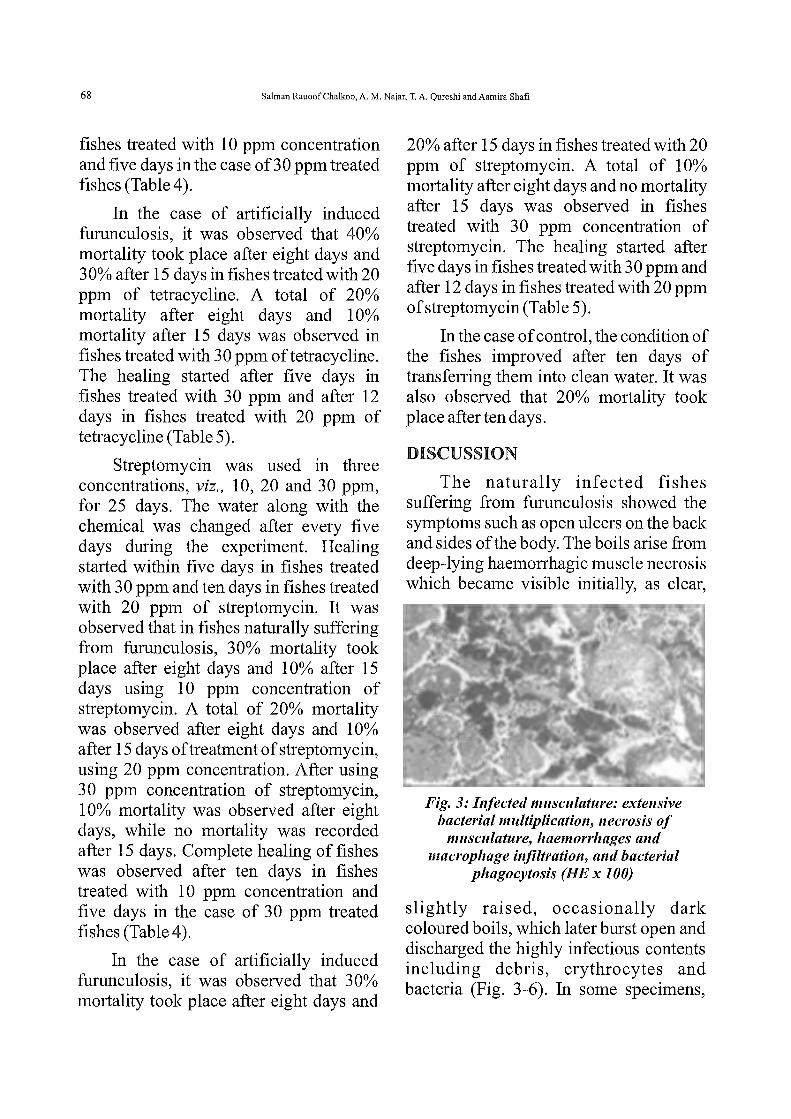
The naturally infected fishes suffering from furunculosis showed the symptoms such as open ulcers on the back and sides of the body. The boils arise from deep-lying haemorrhagic muscle necrosis which became visible initially, as clear,
Fig. 3: Infected musculature: extensive bacterial multiplication, necrosis of
musculature, haemorrhages and macrophage infiltration, and bacterial
phagocytosis (HEx 100)
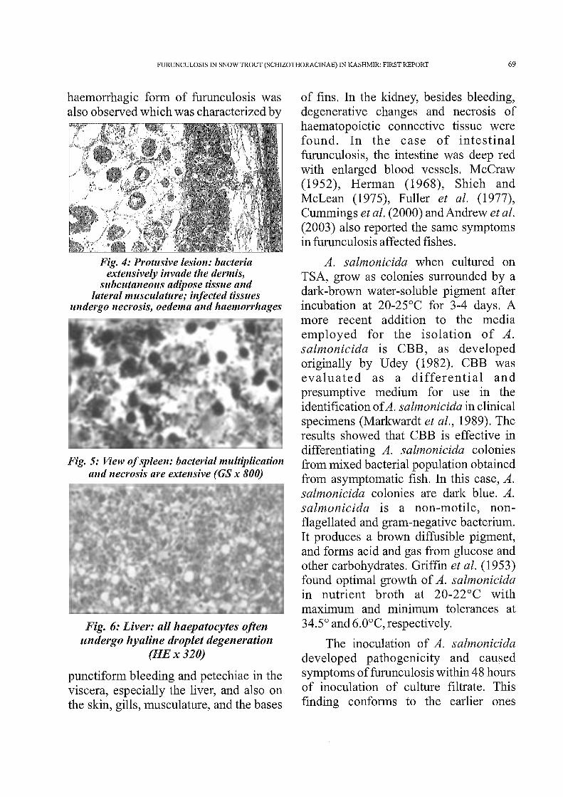
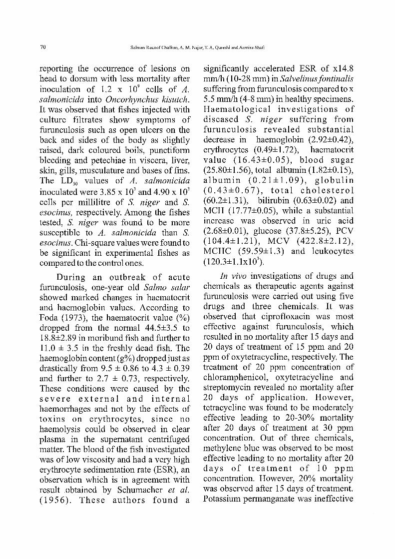
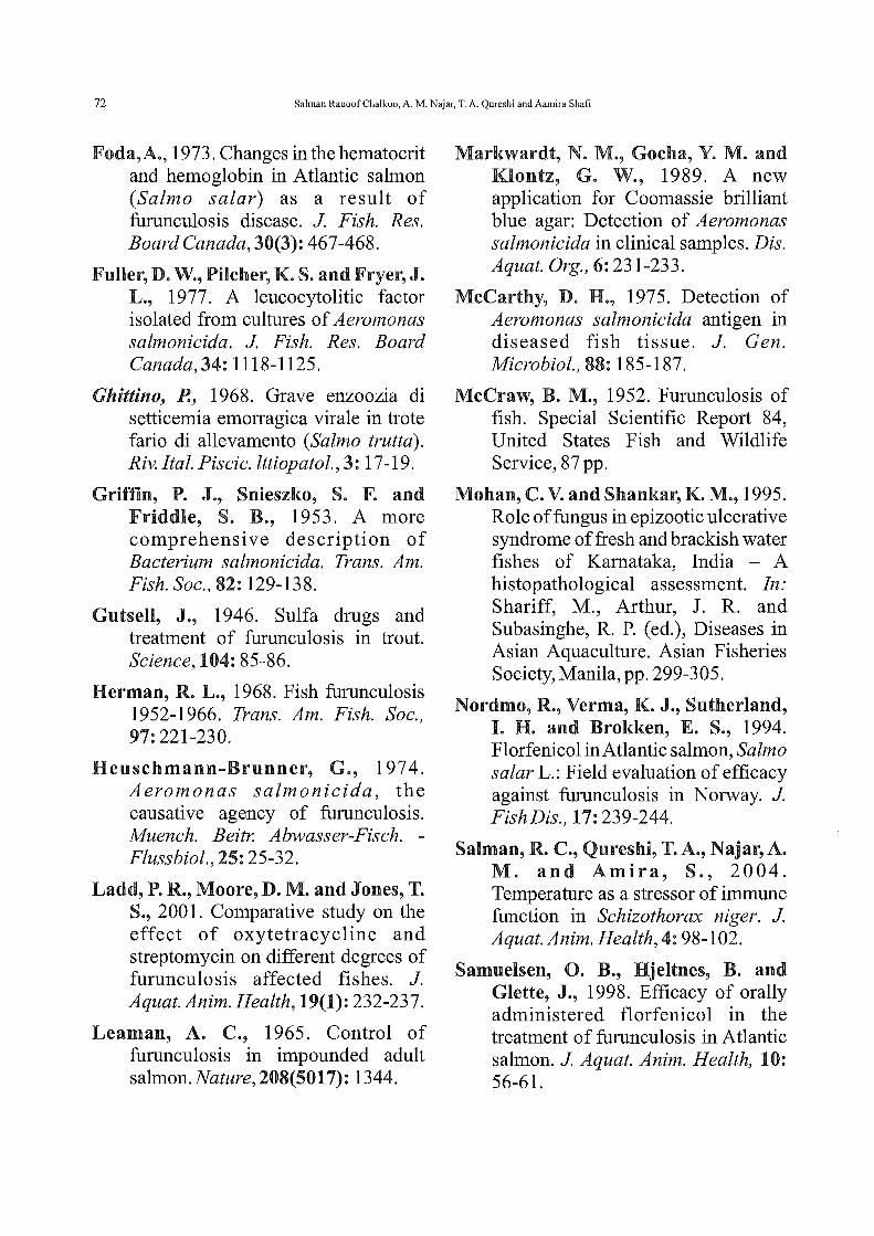
slightly raised, occasionally dark coloured boils, which later burst open and discharged the highly infectious contents including debris, erythrocytes and bacteria (Fig. 3-6). In some specimens,
FURUNCULOSIS IN SNOW TROUT (SCHIZOTHORACINAE) IN KASHMIR: FIRST REPORT 69
haemorrhagic form of furunculosis was also observed which was characterized by
Fig. 4: Protusive lesion: bacteria extensively invade the dermis,
subcutaneous adipose tissue and lateral musculature; infected tissues
undergo necrosis, oedema and haemorrhages
Fig. 5: View of spleen: bacterial multiplication and necrosis are extensive (GS x 800)
Fig. 6: Liver: all haepatocytes often undergo hyaline droplet degeneration
(HEx320)
punctiform bleeding and petechiae in the viscera, especially the liver, and also on the skin, gills, musculature, and the bases
of fins. In the kidney, besides bleeding, degenerative changes and necrosis of haematopoietic connective tissue were found. In the case of intestinal furunculosis, the intestine was deep red with enlarged blood vessels. McCraw (1952), Herman (1968), Shieh and McLean (1975), Fuller et al. (1977), Cummings et al. (2000) and Andrew et al. (2003) also reported the same symptoms in furunculosis affected fishes.
A. salmonicida when cultured on TSA, grow as colonies surrounded by a dark-brown water-soluble pigment after incubation at 20-25°C for 3-4 days. A more recent addition to the media employed for the isolation of A. salmonicida is CBB, as developed originally by Udey (1982). CBB was evaluated as a differential and presumptive medium for use in the identification of A. salmonicida in clinical specimens (Markwardt et al., 1989). The results showed that CBB is effective in differentiating A. salmonicida colonies from mixed bacterial population obtained from asymptomatic fish. In this case, A. salmonicida colonies are dark blue. A. salmonicida is a non-motile, nonflagellated and gram-negative bacterium. It produces a brown diffusible pigment, and forms acid and gas from glucose and other carbohydrates. Griffin et al. (1953) found optimal growth of A. salnwnicida in nutrient broth at 20-22°C with maximum and minimum tolerances at 34.5° and 6.0°C, respectively.
The inoculation of A. salmonicida developed pathogenicity and caused symptoms of furunculosis within 48 hours of inoculation of culture filtrate. This finding conforms to the earlier ones
70 Salman Rauoof Chalkoo, A. M. Najar, T. A. Qureshi and Aamira Shafi
reporting the occurrence of lesions on head to dorsum with less mortality after inoculation of 1.2 x 10⁹ cells of A. salmonicida into Oncorhynchus ldsutch. It was observed that fishes injected with culture filtrates show symptoms of furunculosis such as open ulcers on the back and sides of the body as slightly raised, dark coloured boils, punctiform bleeding and petechiae in viscera, liver, skin, gills, musculature and bases of fins. The LD50 values of A. salmonicida inoculated were 3.85 x 10³ and 4.90 x 10³
cells per millilitre of S. niger and S. esocinus, respectively. Among the fishes tested, S. niger was found to be more susceptible to A. salmonicida than S. esocinus. Chi-square values were found to be significant in experimental fishes as compared to the control ones.
During an outbreak of acute furunculosis, one-year old Salmo salar showed marked changes in haematocrit and haemoglobin values. According to Foda (1973), the haematocrit value (%) dropped from the normal 44.5±3.5 to 18.8±2.89 in moribund fish and further to 11.0 ± 3.5 in the freshly dead fish. The haemoglobin content (g%) dropped just as drastically from 9.5 ± 0.86 to 4.3 ± 0.39 and further to 2.7 ± 0.73, respectively. These conditions were caused by the severe external and internal haemorrhages and not by the effects of toxins on erythrocytes, since no haemolysis could be observed in clear plasma in the supernatant centrifuged matter. The blood of the fish investigated was of low viscosity and had a very high erythrocyte sedimentation rate (ESR), an observation which is in agreement with result obtained by Schumacher et al. ( 19 56). These authors found a
significantly accelerated ESR of x14.8 mm/h (10-28 mm) inSalvelinusfontinalis suffering from furunculosis compared to x 5.5 mrn!h (4-8 mm) in healthy specimens. Haematolo gical investigations of diseased S. niger suffering from furunculosis revealed substantial decrease in haemoglobin (2.92±0.42), erythrocytes (0.49±1.72), haematocrit value (16.43±0.05), blood sugar (25.80±1.56), total albumin (1.82±0.15), albumin (0.21±1.09), globulin (0.43±0.67), total cholesterol (60.2±1.31 ), bilirubin (0.63±0.02) and MCH (17.77±0.05), while a substantial increase was observed in uric acid (2.68±0.01), glucose (37.8±5.25), PCV (104.4±1.21), MCV (422.8±2.12), MCHC (59.59±1.3) and leukocytes (120.3±1.1x1 0\
In vivo investigations of drugs and chemicals as therapeutic agents against furunculosis were carried out using five drugs and three chemicals. It was observed that ciprofloxacin was most effective against furunculosis, which resulted in no mortality after 15 days and 20 days of treatment of 15 ppm and 20 ppm of oxytetracycline, respectively. The treatment of 20 ppm concentration of chloramphenicol, oxytetracycline and streptomycin revealed no mortality after 20 days of application. However, tetracycline was found to be moderately effective leading to 20-30% mortality after 20 days of treatment at 30 ppm concentration. Out of three chemicals, methylene blue was observed to be most effective leading to no mortality after 20 days of treatment of 10 ppm concentration. However, 20% mortality was observed after 15 days of treatment. Potassium permanganate was ineffective

FURUNCULOSIS IN SNOW TROUT (SCIDZOTHORACINAE) IN KASHMIR: FIRST REPORT 71
leading to 20% mortality after 15 and 20 days of treatment. However, malachite green was observed to lead to only 10% mortality after 20 days of treatment.
In the case of fishes artificially challenged with furunculosis, only oxytetracycline and streptomycin proved to be very effective leading to no mortality after 20 days of treatment at 30 ppm of the drugs. Chloramphenicol and tetracycline were moderately effective leading to 10% mortality after 20 days of treatment at a concentration of 30 ppm. Ciprofloxacin was observed to be least effective leading to 30 and 20% mortality after 15 and 20 days, respectively, of treatment at 30 ppm concentration. In the case of chemicals, more or less similar response was observed leading to only 10% mortality after 20 days of treatment at 10 ppm concentration of potassium permanganate, malachite green and methylene blue. However, out of the three chemicals, potassium permanganate was found to be most effective. The present result lends support to the work of Outsell (1946), Barnes et al. (1991), Nordmo et al. (1994), Cipriano et al. (1996), Samuelsen et al. (1998), Ladd et al. (2001) and Dunaway etal. (2002).
REFERENCES
Andrew, J., Barrymore, L., Millis, D. and Mix, T., 2003. On the sporadic incidence of furunculosis outbreak in snow trout in fish farms in Europe: Investigation into cause and relation with cr ion concentration. Bull. Jap. Soc. Sci. Fish., 22:716-720.
Barnes, A. C., Lewin, C. S., Hastings, T. S. andAmyes, S. G. B., 1991. In vitro susceptibility of the fish pathogen Aeromonas salmonicida to
flumequine. Antimicrob. Agents Chemother., 35:2634-2635.
Bootsma, R. Fijan, N. and Blommaert, J., 1977. Isolation and identification of the causative agent of carp erythrodermatitis. Vet. Arhiv., 47: 291-302.
Bucke, D., 1978. Bacterial Kidney Disease (BKD) of Fish. Fisheries Notice No. 60. Ministry of Agriculture, Forestry and Food, Lowestoft, 4 pp.
Cipriano, R. C., Frod, L. A., Starliper, C. E., Teska, J.D., Nelson, J. T. and Jensen, B. N., 1996. Control of external Aeromonas salmonicida: Topical disinfection of salmonids with Chloroamine T. J Aquat. Anim. Health, 8: 52-57.
Cummings, T., Pybus, V., Loutit, M. W. and Tagg, J. R., 2000. Aeromonas salmonicida in water and fish using a species specific DNA probe combined with membrane filtration. N Z. J Mm~ Freshwat. Res., 28: 309-315.
Dunaway, D. H., Grey, J. M. and Duff, H. P., 2002. Resistance of Aeromonas salmonicida and Aeromonas hydrophila to neomycin: Temperature versus concentration effect. J Vet. Microbial., 22: 128-135.
Emmerich, R. and Weibel, E., 1890. On an infectious disease of trout caused by bacteria. Arch. Hyg., 21 : 1-21.
Emmerich, R. and Weibel, E., 1894. Uber eine durch Bakterien erzengte Seuche unter den Forellen. Arch. HygieneBakteriol., 21: 1-21.
72 Salman Rauoof Chalkoo, A. M. Najar, T. A. Qureshi and Aamira Shafi
Foda,A., 1973. Changes in the hematocrit and hemoglobin in Atlantic salmon (Salmo salar) as a result of furunculosis disease. J Fish. Res. Board Canada, 30(3): 467-468.
Fuller, D. W., Pilcher, K. S. and Fryer, J. L., 1977. A leucocytolitic factor isolated from cultures of Aeromonas salmonicida. J Fish. Res. Board Canada,34: 1118-1125.
Ghittino, P., 1968. Grave enzoozia di setticemia emorragica virale in trote fario di allevamento (Salmo trutta). Riv. I tal. Piscic. lttiopatol., 3: 17-19.
Griffin, P. J., Snieszko, S. F. and Friddle, S. B., 1953. A more comprehensive description of Bacterium salmonicida. Trans. Am. Fish. Soc., 82: 129-138.
Gutsell, J., 1946. Sulfa drugs and treatment of furunculosis in trout. Science, 104: 85-86.
Herman, R. L., 1968. Fish furunculosis 1952-1966. Trans. Am. Fish. Soc., 97:221-230.
Heuschmann-Brunner, G., 1974. Aeromonas salmonicida, the causative agency of furunculosis. Muench. Beitr. Abwasser-Fisch. -Flussbiol., 25: 25-32.
Ladd, P.R., Moore, D. M. and Jones, T. S., 200 1. Comparative study on the effect of oxytetracycline and streptomycin on different degrees of furunculosis affected fishes. J. Aquat.Anim. Health, 19(1): 232-237.
Leaman, A. C., 1965. Control of furunculosis in impounded adult salmon. Nature, 208(5017): 1344.
Markwardt, N. M., Gocha, Y. M. and Klontz, G. W., 1989. A new application for Coomassie brilliant blue agar: Detection of Aeromonas salmonicida in clinical samples. Dis. Aquat. Org., 6:231-233.
McCarthy, D. H., 1975. Detection of Aeromonas salmonicida antigen in diseased fish tissue. J. Gen. Microbial., 88: 185-187.
McCraw, B. M., 1952. Furunculosis of fish. Special Scientific Report 84, United States Fish and Wildlife Service, 87 pp.
Mohan, C. V. and Shankar, K. M., 1995. Role of fungus in epizootic ulcerative syndrome of fresh and brackish water fishes of Kamataka, India - A histopathological assessment. In: Shariff, M., Arthur, J. R. and Subasinghe, R. P. ( ed. ), Diseases in Asian Aquaculture. Asian Fisheries Society, Manila,pp. 299-305.
Nordmo, R., Verma, K. J., Sutherland, I. H. and Brokken, E. S., 1994. Florfenicol in Atlantic salmon, Salmo salar L.: Field evaluation of efficacy against furunculosis in Norway. J Fish Dis., 17:239-244.
Salman, R. C., Qureshi, T. A., Najar, A. M. and Amira, S., 2004. Temperature as a stressor of immune function in Schizothorax niger. J A quat. Anim. Health, 4: 98-102.
Samuelsen, 0. B., Hjeltnes, B. and Glette, J., 1998. Efficacy of orally administered florfenicol in the treatment of furunculosis in Atlantic salmon. J Aquat. Anim. Health, 10: 56-61.

FURUNCULOSIS IN SNOW TROUT (SCHIZOTHORACINAE) IN KASHMIR: FIRST REPORT 73
Schumacher, R. E., Hamilton, C. H. and Longtin, E. J., 1956. Blood sedimentation rates of brook trout affected by furunculosis. Prog. Fish-Cult., 18: 147-148.
Shieh, H. S. and MacLean, J. R., 197 5. Purification and properties of an extra-cellular protease of Aeromonas salmonicida, the causative agent of furunculosis. Int. J Biochem., 6: 653-656.
Snieszko, S. F., 1973. Recent advances in scientific knowledge and development pertaining to diseases of fish. Adv. Vet. Sci. Camp. Med., 17:291-314 ..
Snieszko, S. F. and Hoffman, G. L., 1963. Control offish diseases. Lab. Anim. Care, 13: 197-206.
Udey, L. R., 1982. A differential medium for distinguishing Air+ from A/r- phenotypes in Aeromonas salmonicida. In: Proceedings of the 13th Annual Conference and Workshop and 71
h
Eastern Fish Health Workshop. International Association for Aquatic Animal Medicine, Baltimore, p. 41.