Embed Size (px)
Citation preview
Received 12/01/2016 Review began 12/29/2016 Review ended 01/14/2017 Published 01/20/2017
© Copyright 2017Granville et al. This is an openaccess article distributed under theterms of the Creative CommonsAttribution License CC-BY 3.0.,which permits unrestricted use,distribution, and reproduction in anymedium, provided the originalauthor and source are credited.
An Innovative Use of Cortoss Bone Cementto Stabilize a Nonunion after InterbodyFusionMichelle Granville , Robert E. Jacobson
1. Miami Neurosurgical Center, University of Miami Hospital
Corresponding author: Michelle Granville, [email protected] Disclosures can be found in Additional Information at the end of the article
AbstractA 65-year-old male originally had surgery for spondylolisthesis at L5-S1 in 2008 and then wenton to have an L4-5 transforaminal lumbar interbody fusion (TLIF) with pedicle screw fixationfrom L4 to S1 and interbody graft in 2010. Despite having two surgical procedures, hecontinued with intractable back pain and was told he had a failed lumbar fusion. When he wasevaluated with a computerized tomography (CT) scan from April 2015, it demonstrated anerosive nonunion of the L4-5 interbody fusion without incorporation of thepolyetheretherketone (PEEK) cage. In an attempt to perform a minimally invasive stabilizationof the L4-5 nonunion, he underwent a percutaneous lateral foraminal approach with aninjection of Cortoss® cement (Stryker®, Malvern, PA) into the L4-5 interspace and around thegraft. The objective was to stabilize the nonunion, resulting in intermediate relief of pain.
Categories: Pain Management, NeurosurgeryKeywords: failed fusion, failed back syndrome, minimally invasive
IntroductionA 65-year-old male was evaluated six months after refusing surgery at another clinic forincreased severe and persistent deep low lumbar pain after multiple previous lumbar surgeries.The pain actually worsened after an L4-5 interbody graft and pedicle screw fixation from L4 toS1. He was evaluated with plain X-rays and CT scan, including a fine-cut CT, whichdemonstrated nonunion and actual endplate subsidence and erosion both inferiorly at L4 andsuperiorly at L5, as shown in Figures 1-2. In order to ensure the patient did not haveosteomyelitis or an underlying infection, a sedimentation rate, white blood cell (WBC) count,and bone scan were ordered and found to be within normal limits.
1 1
Open Access TechnicalReport DOI: 10.7759/cureus.986
How to cite this articleGranville M, Jacobson R E (January 20, 2017) An Innovative Use of Cortoss Bone Cement to Stabilize aNonunion after Interbody Fusion. Cureus 9(1): e986. DOI 10.7759/cureus.986
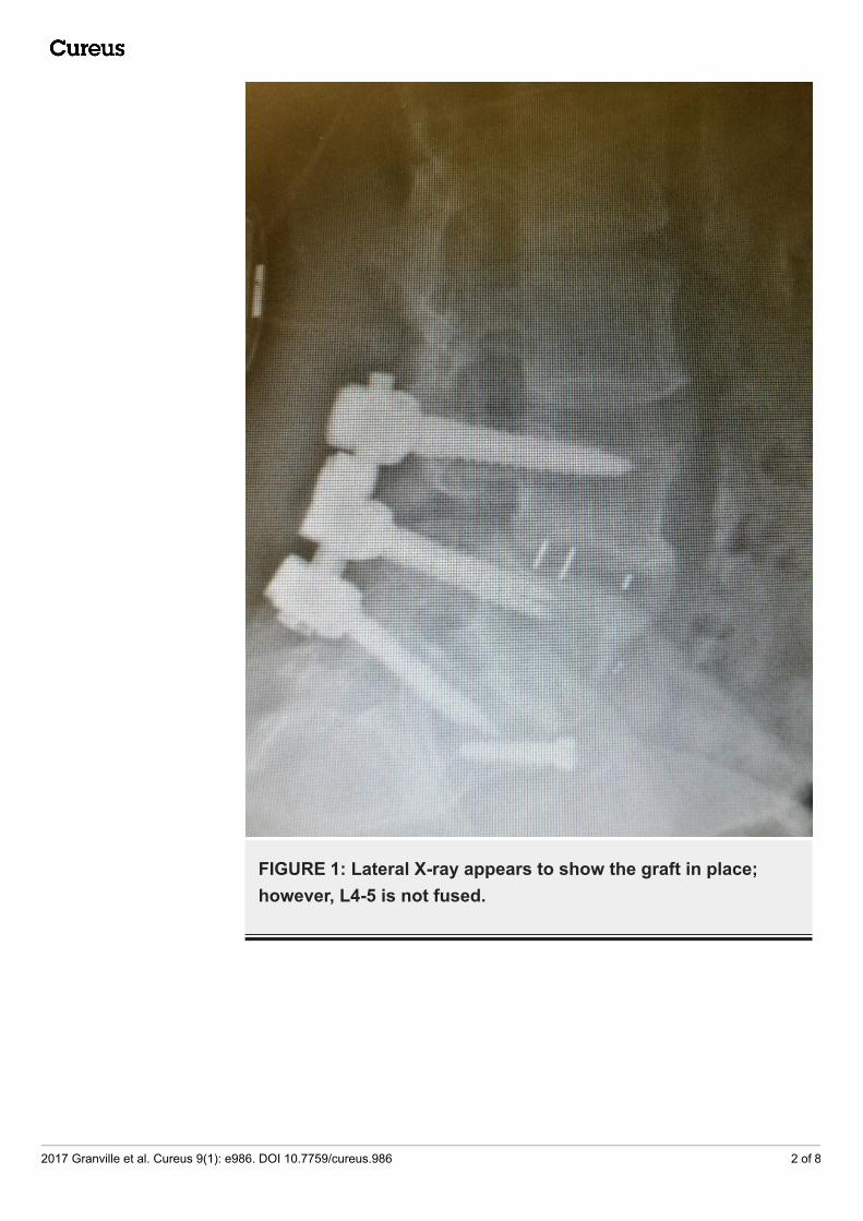
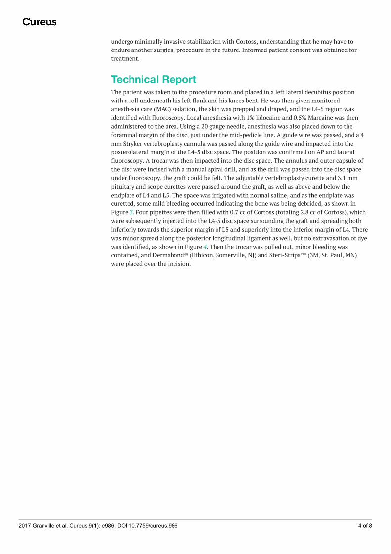
FIGURE 1: Lateral X-ray appears to show the graft in place;however, L4-5 is not fused.
2017 Granville et al. Cureus 9(1): e986. DOI 10.7759/cureus.986 2 of 8
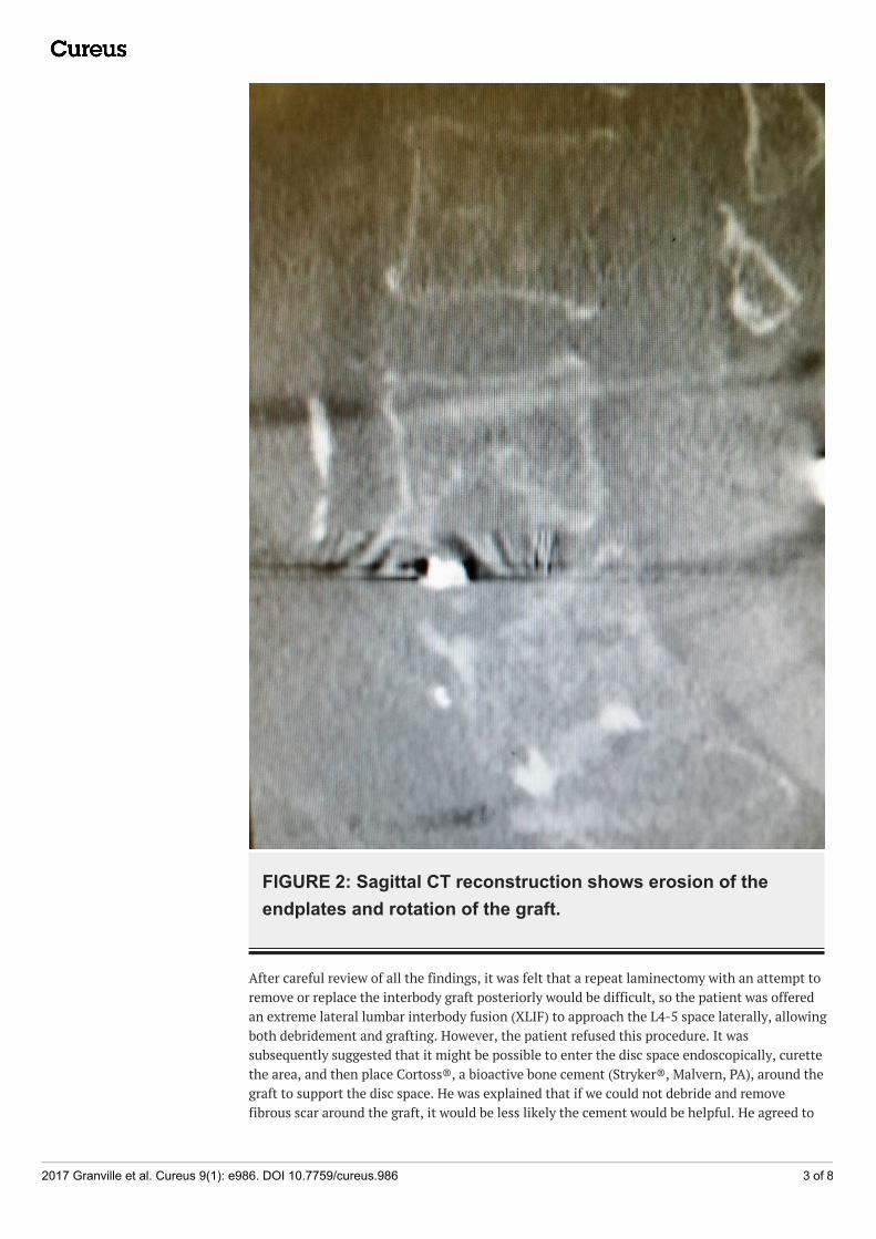
FIGURE 2: Sagittal CT reconstruction shows erosion of theendplates and rotation of the graft.
After careful review of all the findings, it was felt that a repeat laminectomy with an attempt toremove or replace the interbody graft posteriorly would be difficult, so the patient was offeredan extreme lateral lumbar interbody fusion (XLIF) to approach the L4-5 space laterally, allowingboth debridement and grafting. However, the patient refused this procedure. It wassubsequently suggested that it might be possible to enter the disc space endoscopically, curettethe area, and then place Cortoss®, a bioactive bone cement (Stryker®, Malvern, PA), around thegraft to support the disc space. He was explained that if we could not debride and removefibrous scar around the graft, it would be less likely the cement would be helpful. He agreed to
2017 Granville et al. Cureus 9(1): e986. DOI 10.7759/cureus.986 3 of 8
undergo minimally invasive stabilization with Cortoss, understanding that he may have toendure another surgical procedure in the future. Informed patient consent was obtained fortreatment.
Technical ReportThe patient was taken to the procedure room and placed in a left lateral decubitus positionwith a roll underneath his left flank and his knees bent. He was then given monitoredanesthesia care (MAC) sedation, the skin was prepped and draped, and the L4-5 region wasidentified with fluoroscopy. Local anesthesia with 1% lidocaine and 0.5% Marcaine was thenadministered to the area. Using a 20 gauge needle, anesthesia was also placed down to theforaminal margin of the disc, just under the mid-pedicle line. A guide wire was passed, and a 4mm Stryker vertebroplasty cannula was passed along the guide wire and impacted into theposterolateral margin of the L4-5 disc space. The position was confirmed on AP and lateralfluoroscopy. A trocar was then impacted into the disc space. The annulus and outer capsule ofthe disc were incised with a manual spiral drill, and as the drill was passed into the disc spaceunder fluoroscopy, the graft could be felt. The adjustable vertebroplasty curette and 3.1 mmpituitary and scope curettes were passed around the graft, as well as above and below theendplate of L4 and L5. The space was irrigated with normal saline, and as the endplate wascuretted, some mild bleeding occurred indicating the bone was being debrided, as shown inFigure 3. Four pipettes were then filled with 0.7 cc of Cortoss (totaling 2.8 cc of Cortoss), whichwere subsequently injected into the L4-5 disc space surrounding the graft and spreading bothinferiorly towards the superior margin of L5 and superiorly into the inferior margin of L4. Therewas minor spread along the posterior longitudinal ligament as well, but no extravasation of dyewas identified, as shown in Figure 4. Then the trocar was pulled out, minor bleeding wascontained, and Dermabond® (Ethicon, Somerville, NJ) and Steri-Strips™ (3M, St. Paul, MN)were placed over the incision.
2017 Granville et al. Cureus 9(1): e986. DOI 10.7759/cureus.986 4 of 8
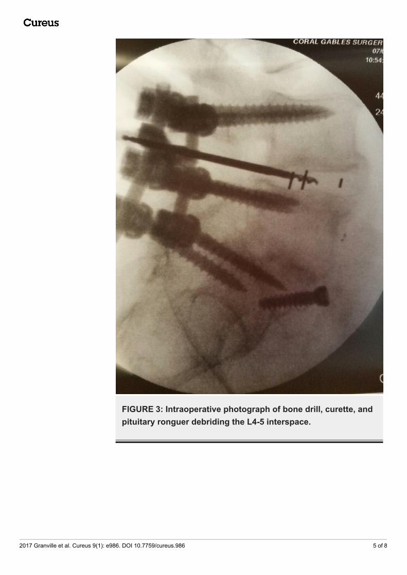
FIGURE 3: Intraoperative photograph of bone drill, curette, andpituitary ronguer debriding the L4-5 interspace.
2017 Granville et al. Cureus 9(1): e986. DOI 10.7759/cureus.986 5 of 8
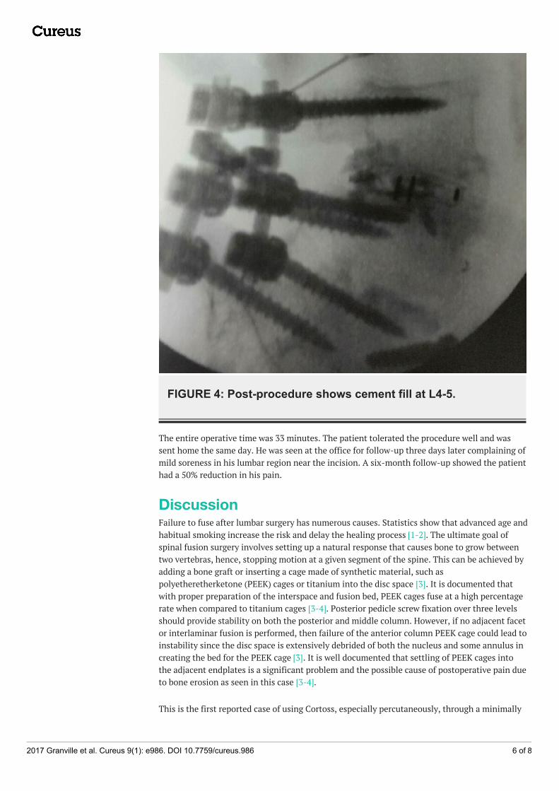
FIGURE 4: Post-procedure shows cement fill at L4-5.
The entire operative time was 33 minutes. The patient tolerated the procedure well and wassent home the same day. He was seen at the office for follow-up three days later complaining ofmild soreness in his lumbar region near the incision. A six-month follow-up showed the patienthad a 50% reduction in his pain.
DiscussionFailure to fuse after lumbar surgery has numerous causes. Statistics show that advanced age andhabitual smoking increase the risk and delay the healing process [1-2]. The ultimate goal ofspinal fusion surgery involves setting up a natural response that causes bone to grow betweentwo vertebras, hence, stopping motion at a given segment of the spine. This can be achieved byadding a bone graft or inserting a cage made of synthetic material, such aspolyetheretherketone (PEEK) cages or titanium into the disc space [3]. It is documented thatwith proper preparation of the interspace and fusion bed, PEEK cages fuse at a high percentagerate when compared to titanium cages [3-4]. Posterior pedicle screw fixation over three levelsshould provide stability on both the posterior and middle column. However, if no adjacent facetor interlaminar fusion is performed, then failure of the anterior column PEEK cage could lead toinstability since the disc space is extensively debrided of both the nucleus and some annulus increating the bed for the PEEK cage [3]. It is well documented that settling of PEEK cages intothe adjacent endplates is a significant problem and the possible cause of postoperative pain dueto bone erosion as seen in this case [3-4].
This is the first reported case of using Cortoss, especially percutaneously, through a minimally
2017 Granville et al. Cureus 9(1): e986. DOI 10.7759/cureus.986 6 of 8
invasive tubular approach under local anesthesia as a means to stabilize a failed interbody bonegraft. There are reports of using polymethylmethacrylate (PMMA), which has a higher viscositywhen compared to Cortoss, for stabilizing vertebral body metastatic tumors and as an adjunctduring open surgery to fill voids for vertebral body replacement [5-6]. Cortoss is the firstclinically sustained substitute to PMMA, which has both calcium and phosphate polymersmixed with micro glass that binds to bone, giving it similar properties to human bone [6-7].This also allows it to reach around 75% of the biomechanical strength of cortical bone within 15minutes of injection [8-9]. Thus, this allowed for significant support so there was a reasonablechance of not only stabilizing the graft but actually enhancing the ability of the graft to fuseand incorporate the interbody PEEK cage and lead with the L4 and L5 endplates.
ConclusionsIn the last several years, various methods have been developed to place PEEK cages and graftsin the disc space with the goal of increasing bony growth to allow adequate fusion. This casedemonstrates that it is possible to stabilize a nonunion of a lumbar interbody graft using avariation of the technique for transforaminal lumbar endoscopic discectomy and combiningthis approach with the use of bone cement. This made it possible to offer the patient aminimally invasive short and median term solution to a complex interbody fusion nonunionfailure.
Additional InformationDisclosuresHuman subjects: Consent was obtained by all participants in this study. Animal subjects: Allauthors have confirmed that this study did not involve animal subjects or tissue. Conflicts ofinterest: In compliance with the ICMJE uniform disclosure form, all authors declare thefollowing: Payment/services info: All authors have declared that no financial support wasreceived from any organization for the submitted work. Financial relationships: All authorshave declared that they have no financial relationships at present or within the previous threeyears with any organizations that might have an interest in the submitted work. Otherrelationships: All authors have declared that there are no other relationships or activities thatcould appear to have influenced the submitted work.
AcknowledgementsI would like to acknowledge Maria Freed, administrator of Coral Gables Surgery Center.
References1. Rohde V, Mielke D, Ryang Y, Gilsbach JM: The immediately failed lumbar disc surgery:
incidence, aetiologies, imaging and management. Neurosurg Rev. 2015, 38:191-95.10.1007/s10143-014-0573-3
2. Kerr EJ 3rd, Jawahar A, Wooten T, Kay S, Cavanaugh DA, Nunley PD: The use of osteo-conductive stem-cells allograft in lumbar interbody fusion procedures: an alternative torecombinant human bone morphogenetic protein. J Surg Orthop Adv. 2011, 20:193-97.
3. Chong E, Pelletier MH, Mobbs RJ, Walsh WR: The design evolution of interbody cages inanterior cervical discectomy and fusion: a systematic review. BMC Musculoskelet Disord.2015, 16:99. 10.1186/s12891-015-0546-x
4. Williams AL, Gornet MF, Burkus JK: CT evaluation of lumbar interbody fusion: currentconcepts. AJNR Am J Neuroradiol. 2005, 26:2057-66.
5. Hayeri MR, Tehranzadeh J: Diagnostic imaging of spinal fusion and complications . ApplRadiol. 2009, Accessed: 11/02/2016: http://appliedradiology.com/articles/diagnostic-imaging-of-spinal-fusion-and-complications.
6. Erbe EM, Clineff TK, Gualtieri G: Comparison of a new bisphenol-a-glycidyl dimethacrylate-
2017 Granville et al. Cureus 9(1): e986. DOI 10.7759/cureus.986 7 of 8
based cortical bone void filler with polymethyl methacrylate. Eur Spine J. 2001, 10:S147-52.10.1007/s005860100288
7. Bae H, Hatten HP Jr, Linovitz R, Tahernia AD, Schaufele MK, McCollom V, Gilula L, Maurer P,Benyamin R, Mathis JM, Persenaire M: A prospective randomized FDA-IDE trial comparingCortoss with PMMA for vertebroplasty: a comparative effectiveness research study with 24-month follow-up. Spine (Phila Pa 1976). 2012, 37:544-50. 10.1097/BRS.0b013e31822ba50b
8. Cook WD, Standish PM: Polymerization kinetics of resin-based restorative materials . J BiomedMater Res. 1983, 17:275–82. 10.1002/jbm.820170206
9. Fan S, Hu Z, Zhao F, Zhao X, Huang Y, Fang X: Multifidus muscle changes and clinical effectsof one-level posterior lumbar interbody fusion: minimally invasive procedure versusconventional open approach. Eur Spine J. 2010, 19:316–24. 10.1007/s00586-009-1191-6
2017 Granville et al. Cureus 9(1): e986. DOI 10.7759/cureus.986 8 of 8