Embed Size (px)
Citation preview
Downl
Fuzzy Hopfield neural network with fixed weightfor medical image segmentation
Chwen-Liang ChangYu-Tai Ching, MEMBER SPIENational Chiao Tung UniversityDepartment of Computer and Information
ScienceTaiwan
Abstract. Image segmentation is a process for dividing a given imageinto meaningful regions with homogeneous properties. A new two stepapproach is proposed for medical image segmentation using a fuzzyHopfield neural network based on both global and local gray-level infor-mation. The membership function simulated with neuron outputs is de-termined using a fuzzy set, and the synaptic connection weights be-tween the neurons are predetermined and fixed to improve the efficiencyof the neural network. The proposed method needs initial cluster centers.The initial centers can be obtained from the global information about thedistribution of the intensities in the image, or from prior knowledge of theintensity of the region of interest. It is shown by experiments that theproposed fuzzy Hopfield neural network approach is better than mostprevious approaches. We also show that the global information can beused by applying the hard c-means to estimate the initial cluster centers.© 2002 Society of Photo-Optical Instrumentation Engineers. [DOI: 10.1117/1.1428298]
Subject terms: medical image segmentation; fuzzy clustering; Hopfield neuralnetwork.
Paper 010071 received Feb. 27, 2001; revised manuscript received July 19,2001; accepted for publication July 20, 2001.
intoesm-ro-tio
sesseg
rtie
s inom
ataap-l ine,inethey-Aionthe
traifint
on-eldlnded
isn theng,
rkg-
tegyeg-
innceen-eth-calodel
cticaledalfednt
ationa-
ay-ni-ct
hipge.theter-heess
and
1 Introduction
Image segmentation, a process to divide a given imagemeaningful regions with homogeneous properties, is ansential step in image analysis and recognition. A large nuber of algorithms, for example Refs. 1–3, have been pposed in the past. Those conventional image segmentaalgorithms can be categorized generally into three clas~1! histogram-based schemes, where the pixels aremented into classes based on overall gray levels;~2!region-based schemes, by which homogeneous propearound a given pixel are enlarged; and~3! edge-basedschemes, which detect the pixels with abrupt changegray levels, and then connects selected pixels to form cpletely enclosed boundaries.
Recently, neural network-based architectures4–11 havebeen applied for image segmentation. Dhawan and Ar4
proposed a two-dimensional self-organizing feature mbased approach that incorporates both local and globaformation about the gray-level distribution of the imagand explores the useful features of the image to determand extract meaningful regions of interest. They usedconcept of competitive learning to find the overall gralevel distribution of the image as the global information.contrast measure defining the homogeneity of the regwas also used as the local information. In Refs. 5 and 6,image segmentation process was formulated as a conssatisfaction problem~CSP! by interpreting it as a process oassigning labels to pixels. A three-dimensional constrasatisfaction neural network was developed to form the cstraints in the CSP. A method using a competitive Hopfineural network~CHNN!, based on the global gray-levevalues distribution, was proposed by Cheng, Lin, aMao.7 The problem of image segmentation is regard
Opt. Eng. 41(2) 351–358 (February 2002) 0091-3286/2002/$15.00
oaded From: http://opticalengineering.spiedigitallibrary.org/ on 04/28/2014 T
-
n:-
s
-
-
nt
there as minimization of a cost function, which, in turn,defined as the mean value of distance measures betweegray-level values and the members of classes. Lin, Cheand Mao8 proposed a fuzzy Hopfield neural netwo~FHNN!, based on the pixel classification, for image sementation. This approach added a fuzzy reasoning strainto a neural network. In FHNN, the process of image smentation is also regarded as a minimization problemwhich the cost function is defined as the Euclidean distabetween the gray levels in a histogram and the cluster cters represented in the gray levels. In general, those mods for medical image segmentation make use of the loinformation, i.e., the gray-level values of the neighborhopixels, or the global information, i.e., the overall gray-levdistribution in the image.
In medical image segmentation, the half volume effein CT images and the noise in ultrasound images are typdifficulties encountered. The local information-basmethod cannot correctly find the boundary when the hvolume effect is significant. The global information-basmethod cannot work correctly when there is significanoise, as in ultrasound images. We propose a segmentalgorithm that incorporates both local and global informtion for medical image segmentation. First, the global grlevel information is used to segment the image for the itial partition, and then local information is used to construa Hopfield neural network, to simulate the membersfunction. Each neuron corresponds to a pixel in the imaA pair of neurons are connected if they are neighbors inimage, and the synaptic connection weights are predemined and fixed to improve the efficiency. Second, tfuzzy set approach is applied and a defuzzification procis triggered to determine the outputs of the neurons;
351© 2002 Society of Photo-Optical Instrumentation Engineers
erms of Use: http://spiedl.org/terms
ocia
hedhewe
hm
s-enton,ss
-
ute.
to
hehip
-
rts
sen-
e
eldhel in
ted
fol-
Chang and Ching: Fuzzy Hopfield neural network . . .
Downl
when the network converges into a stable state, the asstion of a pixel to a cluster is decided.
The remainder of this work is organized as follows. Thard c-means and fuzzyc-means algorithms are reviewein Sec. 2, and the fuzzy Hopfield neural network with tfixed-weight approach is described in Sec. 3. In Sec. 4,present the results obtained by the proposed algoritConclusions are given in Sec. 5.
2 Hard c-Means and Fuzzy c-Means Algorithms
The hardc-means and the fuzzyc-means algorithms arewell known for classification of points in space into cluters. When these algorithms are applied for image segmtation, the pixels with similar intensity are gathered inclusters to identify the region of interest. In this sectiothese algorithms are briefly described for completeneThe details can be found in Ref. 12.
2.1 Hard c-Means Algorithm
A set of n points is denoted asX5$x1 ,x2 , . . . ,xn% inspace. Suppose we divide the set of points intoc clusters. Itis said that the matrixU5@uik#PMnc is a hardc-partitionof X if it satisfies the following conditions:
(k51
c
uik51,
(i 51
n
uik,n, and
(i 51
n
(k51
c
uik5n,
whereuikP$0,1%.The procedure of the hardc-means algorithm is summa
rized in the following steps.
1. Choose a primary set ofc points, $vkuk51,2, . . . ,c% as the cluster centers.
2. Calculate the membership matrixU based on theminimum Euclidean distance as follows:
uik5H1, if ~ uuxi2vkuu!25minj51
c
$~uuxi2vjuu!2%, for all i ,k.
0, otherwise
3. Update the new cluster centersvk ,
vk5(i51
n ~uik!~xi!
(i51n uik
.
4. If U5U then stop; otherwise go to step 2.
The hardc-means algorithm is easy to implement. Bit is sensitive to the noise in the imag
352 Optical Engineering, Vol. 41 No. 2, February 2002
oaded From: http://opticalengineering.spiedigitallibrary.org/ on 04/28/2014 T
-
.
-
.
2.2 Fuzzy c-Means Algorithm
The hardc-means algorithm allows a point to belongonly one cluster. But in the fuzzyc-means algorithm, everypoint belongs to all clusters with different degrees of tmembership functions. The conditions of the membersmatrix U of the fuzzyc-means are modified as follows:
(k51
c
uik51, 1< i<n,
(i 51
n
uik,n, 1<k<c, and
(i 51
n
(k51
c
uik5n,
where 0<uik<1.The procedure of the fuzzyc-means algorithm is sum
marized in the following steps.
1. Choose a set of points,$vkuk51,2, . . . ,c%, as theinitial cluster centers.
2. Calculate membership matrixU for all points to allclusters using the following equation.
uik5@uuxi2vkuu22#1/q21
( j 51c @ uuxi2v j uu22#1/q21
.
3. Update the new cluster centersvk ,
vk5(i51
n ~uik!qxi
(i51n ~uik!
q.
4. If maxikuuik2uiku,e, wheree is a stop criterion, thenwe stop the iteration. Otherwise the iteration restaat step 2.
The fuzzification factor,q, a given real number which isgreater than 1, decides the convergence speed and thesitivity to noise. Ifq is set to 11, then it converges quicklyand is less sensitive to the noise. On the other hand, ifq isset to a large number, it will converge slowly and will bmore sensitive to the noise.
3 Proposed Method
In this section, we present the proposed fuzzy Hopfineural network with a fixed weight model to simulate tmembership matrix for image segmentation. Each pixethe image is a point in the plane. If there aren pixels to bedivided intoc clusters, then each pixel hasc neurons asso-ciated with it. There aren3c neurons in this network.Similar to that in FCM, the outputs of the neurons, denoO5oik,0< i<n,1<k<c, form a membership matrix. Amembership corresponds to a partition if it satisfies thelowing conditions:
erms of Use: http://spiedl.org/terms
ertoonitstic
edbe-nge
nn
se
teracthe
s oalecalter-romters
e
of
ing
the
the
ing
as
Chang and Ching: Fuzzy Hopfield neural network . . .
Downl
(k51
c
oik51, for all i ,
(i 51
n
oik,n for all k, and
(i 51
n
(k51
c
5n,
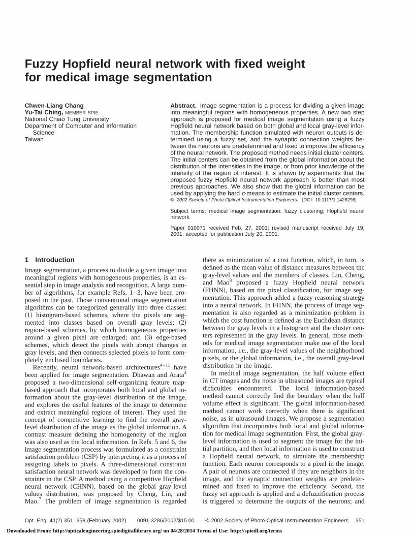
where 0<oik<1. Two neurons are neighbors to each othif their corresponding pixels in the image are neighborseach other. As shown in Fig. 1, each neuron receives ctributions from the neighboring neurons and itself asinput. These contributions are weighted by the synapweightsW. In our approach, the synaptic weights are fixand should be determined first. The synaptic weighttween two neuronsi and j is determined by the Euclideadistance, and the similarity of their intensities in the imais as shown in the following equation.
wi j 51
a1@DI ~ i , j !#21a2@D~ i , j !#2. ~1!
In Eq. ~1!, DI ( i , j ) is the difference in intensity betweepixel i and j, andD( i , j ) is the Euclidean distance betweepixel i and j. a1 and a2 are the weights to balance thetwo factors.
The proposed method requires a set of initial cluscenters. The initial cluster centers do not need to be exbut they should not be far away from the true centers. Tinitial cluster centers can be either given by user assistobtained from the global information about the gray-scdistribution of the image. In most of the cases for mediimage segmentation, the intensities of the regions of inest are known. Users can provide the cluster centers fsuch available knowledge. Otherwise, the cluster cencan be estimated using ac-means method.
Given a set ofc initial cluster centers, we perform thinitial partition as:
oik(0)5
$1/@ I ~ i !2vk#2%1/q21
( j 51c $1/@ I ~ i !2v j #
2%1/q21, 1< i<n,1<k<c,
whereI ( i ) andvk are, respectively, the intensity valuespixel i and thek’th class center.
Fig. 1 The structure of a neuron.
oaded From: http://opticalengineering.spiedigitallibrary.org/ on 04/28/2014 T
-
,
r
Recall that a neuron receives outputs from neighborneurons and itself. The net value of the neuroni is de-scribed as
Netik(t11)5 (
j PNi
wi j ojk(t)1u i , ~2!
where Netik(t11) is the net value of neuroni associated with
classk in iteration t11, ojk(t) is the output state of neuronj
associated with classk in iterationt, andu i is the offset biasfed to the neuroni. In our approach, theu of all neurons isset to zero. So Eq.~2! becomes
Netik(t11)5(
jwi j ojk
(t) . ~3!
Each pixel hasc neurons associated withc clusters torepresent the membership degree of each cluster. Whennet values of the neurons have been updated in Eq.~3!, theoutputs of all neurons will be updated depending onnew net values. Following the fuzzyc-means algorithm, thenew output values can be obtained using the followequation:
oik(t11)5
@Netik(t11)#1/q21
( j 51c @Neti j
(t11)#1/q21.
The proposed segmentation algorithm is summarizedfollows:
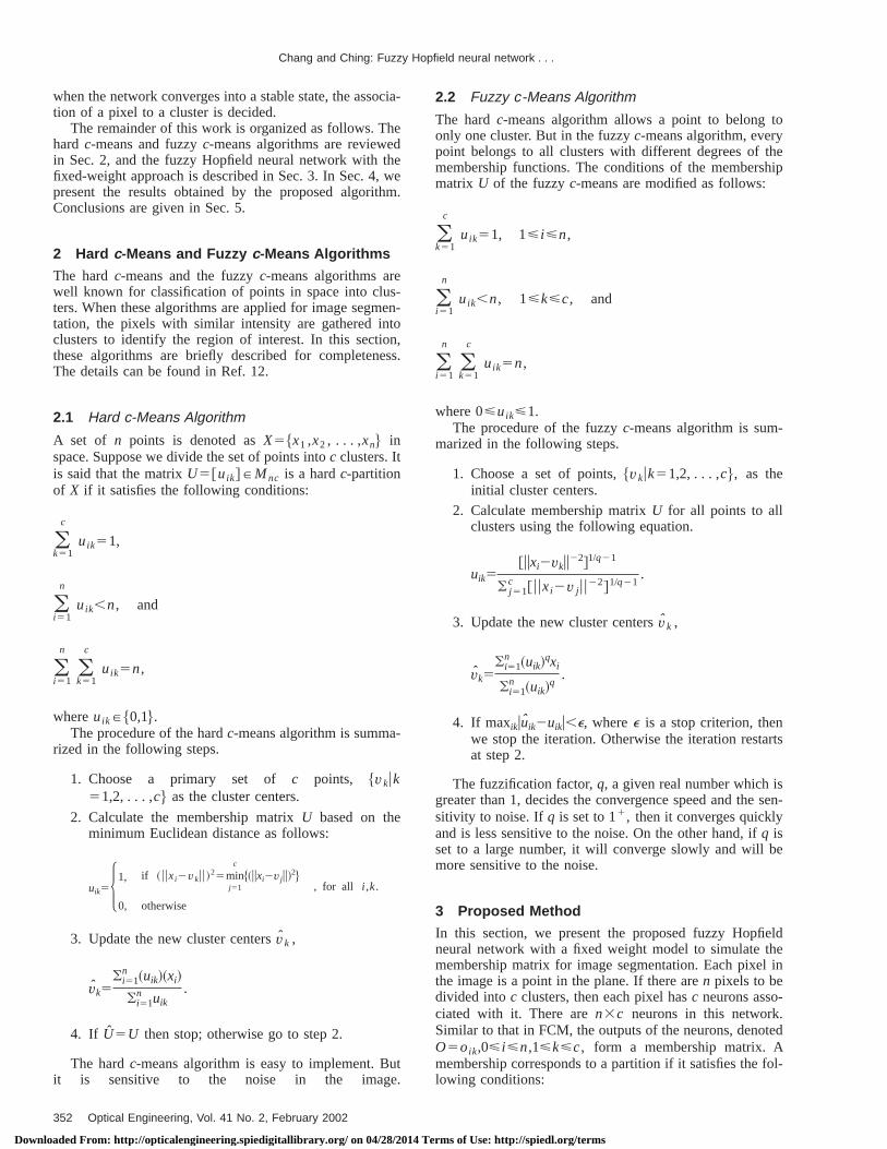
Fig. 2 The simulated image with (a) a constant gray level in back-ground and each disk; (b), (c), and (d) are the simulated images withadded noise levels K520, 23, and 25, respectively.
353Optical Engineering, Vol. 41 No. 2, February 2002
erms of Use: http://spiedl.org/terms
Chang and Ching: Fuzzy Hopfield neural network . . .
Downl
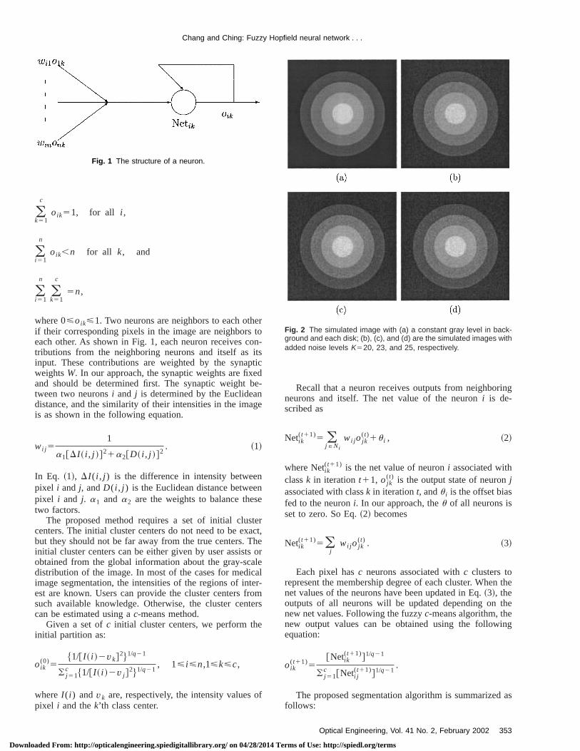
Fig. 3 The segmentation results with added noise level K520 using(a) HCM, (b) FCM, (c) CHNN, and (d) the proposed approach.
Fig. 4 The segmentation results with added noise level K523 using(a) HCM, (b) FCM, (c) CHNN, and (d) the proposed approach.
354 Optical Engineering, Vol. 41 No. 2, February 2002
oaded From: http://opticalengineering.spiedigitallibrary.org/ on 04/28/2014 T
Fig. 5 The segmentation results with added noise level K525 using(a) HCM, (b) FCM, (c) CHNN, and (d) the proposed approach.
Table 1 The number of misclassified pixels and error rate with noiselevel K520.
HCM FCM CHNN proposedapproach
Number ofmisclassified pixels
0 0 0 0
Error rate 0.00 0.00 0.00 0.00
Table 2 The number of misclassified pixels and error rate with noiselevel K523.
HCM FCM CHNNproposedapproach
Number ofmisclassified pixels
31614 5090 9434 59
Error rate 0.4823 0.0776 0.1439 0.0009
Table 3 The number of misclassified pixels and error rate with noiselevel K525.
HCM FCM CHNNproposedapproach
Number ofmisclassified pixels
31644 8898 14022 361
Error rate 0.4828 0.1357 0.2139 0.0055
erms of Use: http://spiedl.org/terms

Chang and Ching: Fuzzy Hopfield neural network . . .
Downloaded From: http://o
Fig. 6 The images in the first column are the original images. Images in the second column wereobtained by first applying a mean filter to the original images, then applying intensity thresholding. Theimages in the third column were obtained by the proposed method.
-
on
andro-
NN
gee of
to75,
up-ayhe
iserd
-are
N,thelu-
Step 1. Determine the neighborhood window,N, and cal-culate the weights,wi j , 1< i<n, for a neuronj PNi using Eq.~1!.
Step 2. Initial clustering.Step 3. Calculate the net value using Eq.~3!.Step 4. Update the output states using Eq.~3!.Step 5. If maxikuoik
(t11)2oik(t)u,e then go to Step 6; other
wise t5t11 and go to Step 3.Step 6. Output the final result using the defuzzificati
process asSi5k, if oik5max1<j<c$oij%,whereSi is the segmentation label of pixeli.
4 Simulation and Experiment Results
In our experiments, we used a set of phantom datamedical images to evaluate the performance of the pposed algorithm. The phantom data set based on CH7
was used, as shown in Fig. 2~a!, which was produced from
pticalengineering.spiedigitallibrary.org/ on 04/28/2014 T
four overlapping disks and the background. An averagray scale for each region was: the average gray valuthe background was 30, and from the outer most circlethe center, the average gray values of four disks were120, 165, and 210, respectively.
The gray scale in each region was not a constant. Spose thatm is the average gray scale in a region. The grscales in the region were uniformly distributed over trange @m2k,m1k#, where k is a constant. Figures 2~b!through 2~d! are the phantom data sets containing nowith K520, 23, and 25, respectively. We applied the hac-means ~HCM!, fuzzy c-means ~FCM!, competitiveHopfield neural network~CHNN!, and the proposed approach to process the phantom data sets. Figures 3–5the segmentation results with noise levelsK520, 23, and25, respectively. In these figures, parts~a! through~d! arethe results segmented by applying HCM, FCM, CHNand the proposed approach, respectively. We comparedamount of misclassified pixels and the error rates to eva
355Optical Engineering, Vol. 41 No. 2, February 2002
erms of Use: http://spiedl.org/terms
reethhe
eodns
reaCTCTitysetro-od.
thesityini-anaseent
hentsthethe
Chang and Ching: Fuzzy Hopfield neural network . . .
Downl
ate the performance. Tables 1–3 show the comparisonsults from these phantom data sets using these four mods. All of the four methods perfectly segmented tobjects when the noise level wasK520. If the noise levelswere higher whenK523 and 25, the performance of thproposed approach was better than other three methboth in perception quality and quantitative compariso~see Figs. 4, 5, and Tables 2 and 3!.
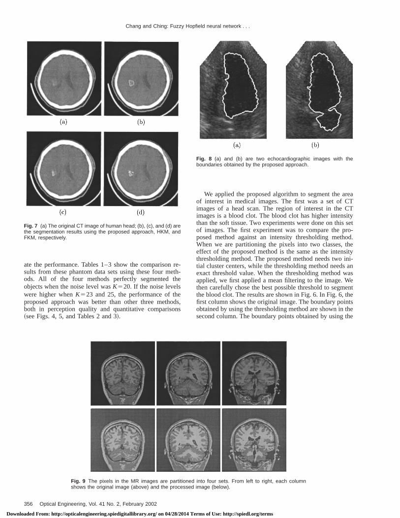
Fig. 7 (a) The original CT image of human head; (b), (c), and (d) arethe segmentation results using the proposed approach, HKM, andFKM, respectively.
356 Optical Engineering, Vol. 41 No. 2, February 2002
oaded From: http://opticalengineering.spiedigitallibrary.org/ on 04/28/2014 T
--
s,
We applied the proposed algorithm to segment the aof interest in medical images. The first was a set ofimages of a head scan. The region of interest in theimages is a blood clot. The blood clot has higher intensthan the soft tissue. Two experiments were done on thisof images. The first experiment was to compare the pposed method against an intensity thresholding methWhen we are partitioning the pixels into two classes,effect of the proposed method is the same as the intenthresholding method. The proposed method needs twotial cluster centers, while the thresholding method needsexact threshold value. When the thresholding method wapplied, we first applied a mean filtering to the image. Wthen carefully chose the best possible threshold to segmthe blood clot. The results are shown in Fig. 6. In Fig. 6, tfirst column shows the original image. The boundary poiobtained by using the thresholding method are shown insecond column. The boundary points obtained by using
Fig. 8 (a) and (b) are two echocardiographic images with theboundaries obtained by the proposed approach.
Fig. 9 The pixels in the MR images are partitioned into four sets. From left to right, each columnshows the original image (above) and the processed image (below).
erms of Use: http://spiedl.org/terms
Chang and Ching: Fuzzy Hopfield neural network . . .
Downloaded From: http://o
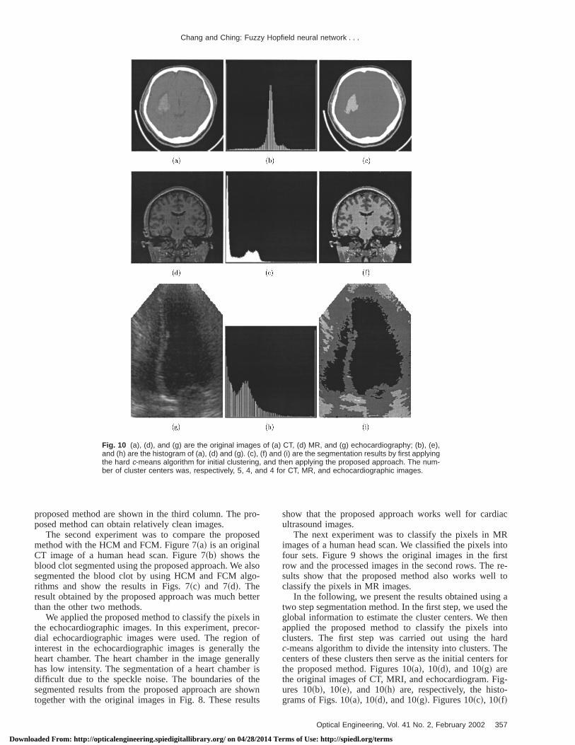
Fig. 10 (a), (d), and (g) are the original images of (a) CT, (d) MR, and (g) echocardiography; (b), (e),and (h) are the histogram of (a), (d) and (g). (c), (f) and (i) are the segmentation results by first applyingthe hard c-means algorithm for initial clustering, and then applying the proposed approach. The num-ber of cluster centers was, respectively, 5, 4, and 4 for CT, MR, and echocardiographic images.
ro-
se
also-
ette
incor
otherallr istheowlts
iac
Rintorst
re-l to
g atheen
ntoardhes for
ig--
proposed method are shown in the third column. The pposed method can obtain relatively clean images.
The second experiment was to compare the propomethod with the HCM and FCM. Figure 7~a! is an originalCT image of a human head scan. Figure 7~b! shows theblood clot segmented using the proposed approach. Wesegmented the blood clot by using HCM and FCM algrithms and show the results in Figs. 7~c! and 7~d!. Theresult obtained by the proposed approach was much bthan the other two methods.
We applied the proposed method to classify the pixelsthe echocardiographic images. In this experiment, predial echocardiographic images were used. The regioninterest in the echocardiographic images is generallyheart chamber. The heart chamber in the image genehas low intensity. The segmentation of a heart chambedifficult due to the speckle noise. The boundaries ofsegmented results from the proposed approach are shtogether with the original images in Fig. 8. These resu
pticalengineering.spiedigitallibrary.org/ on 04/28/2014 T
d
o
r
-f
y
n
show that the proposed approach works well for cardultrasound images.
The next experiment was to classify the pixels in Mimages of a human head scan. We classified the pixelsfour sets. Figure 9 shows the original images in the firow and the processed images in the second rows. Thesults show that the proposed method also works welclassify the pixels in MR images.
In the following, we present the results obtained usintwo step segmentation method. In the first step, we usedglobal information to estimate the cluster centers. We thapplied the proposed method to classify the pixels iclusters. The first step was carried out using the hc-means algorithm to divide the intensity into clusters. Tcenters of these clusters then serve as the initial centerthe proposed method. Figures 10~a!, 10~d!, and 10~g! arethe original images of CT, MRI, and echocardiogram. Fures 10~b!, 10~e!, and 10~h! are, respectively, the histograms of Figs. 10~a!, 10~d!, and 10~g!. Figures 10~c!, 10~f!
357Optical Engineering, Vol. 41 No. 2, February 2002
erms of Use: http://spiedl.org/terms
stery,es.ive
n o
re-tedy-ldton.the
tharoo
sti-rde-arderiter
-E-nce
ch-
gh
ral
ion
ti-
al
ale
he
g a
l-
Chang and Ching: Fuzzy Hopfield neural network . . .
Downl
and 10~i! are the segmented results. The number of clucenters for the hardc-means algorithm were, respectivel5, 4, and 4, for CT, MR, and echocardiographic imagNote that we used five cluster centers for CT images. Finitial cluster centers were needed because the regiointerest, the blood clot, is relatively small in the image.
5 Conclusion
A new medical image segmentation technique was psented. The global gray-level information was incorporato perform the initial clustering, and then the local gralevel information was used to construct a fuzzy Hopfieneural network. A fixed weights approach was utilizedreduce the computing time for neural network stabilizatioAccording to our experiments on the phantom data set,performance of the proposed approach is much betterthe hardc-means, fuzzyc-means, and CHNN methods. Ouexperiments on the real medical images, demonstrated gresults, which were either the initial cluster centers emated by the hardc-means algorithm or the initial clustecenters obtained from the prior knowledge. However,ciding the best number of cluster centers for the hc-means method becomes another problem. Our expments show that increasing the number for cluster cenfor the hardc-means method can achieve good result.
Acknowledgment
This work was supported under contracts NSC-88-2213009-019 and NSC-89-2213-E-009-098, National ScieCouncil, Taiwan, Republic of China.
References1. K. S. Fu and J. K. Mui, ‘‘A survey on image segmentation,’’Pattern
Recogn.13, 3–16~1981!.2. N. R. Pal and S. K. Pal, ‘‘A review on image segmentation te
niques,’’Pattern Recogn.26, 1277–1294~1993!.3. R. C. Gonzalez and R. E. Woods,Digital Image Processing, Addison-
Wesley, Reading, MA~1992!.
358 Optical Engineering, Vol. 41 No. 2, February 2002
oaded From: http://opticalengineering.spiedigitallibrary.org/ on 04/28/2014 T
f
n
d
-s
4. A. P. Dhawan and L. Arata, ‘‘Segmentation of medical images throucompetitive learnings,’’Comput. Methods Programs Biomed.40,203–215~1993!.
5. W. C. Lin, E. C. Tsao, and C. T. Chen, ‘‘Constrain satisfaction neunetworks for image segmentation,’’Pattern Recogn.25~7!, 679–693~1992!.
6. C. T. Chen, E. C. Tsao, and W. C. Lin, ‘‘Medical image segmentatby a constraint satisfaction neural network,’’IEEE Trans. Nucl. Sci.38~2!, 678–686~1991!.
7. K. S. Cheng, J. S. Lin, and C. W. Mao, ‘‘The application of competive Hopfield neural network to medical image segmentation,’’IEEETrans. Med. Imaging15~4!, 560–567~1996!.
8. J. S. Lin, K. S. Cheng, and C. W. Mao, ‘‘A fuzzy Hopfield neurnetwork for medical image segmentation,’’IEEE Trans. Nucl. Sci.43~4!, 2389–2398~1996!.
9. J. S. Lin, K. S. Cheng, and C. W. Mao, ‘‘A modified Hopfield neurnetwork with fuzzyc-means technique for multispectral MR imagsegmentation,’’Proc. IEEE Int. Conf. Image Processing, 327–330~1996!.
10. G. Coppini, R. Poli, and G. Valli, ‘‘Recovery of the 3-D shape of tleft ventricle from echocardiographic images,’’IEEE Trans. Med. Im-aging 14~2!, 301–317~1995!.
11. Y. Zhu and H. Yan, ‘‘Computerized tumor boundary detection usinHopfield neural network,’’IEEE Trans. Med. Imaging16~1!, 55–67~1997!.
12. J. C. Bezdek,Pattern Recognition with Fuzzy Objective Function Agorithm, Plenum, New York~1981!.
Chwen-Liang Chang received the BS de-gree in computer science from Chung-Cheng Institute of Technology, Taiwan, in1988. He is currently a PhD student in thecomputer and information science depart-ment at National Chiao-Tung University.His research interests are in the areas ofimage processing, computational geom-etry, medical imaging, deformable models,and neural networks.
Yu-Tai Ching received his BS degree in industrial engineering fromTsing Hua University, Taiwan, in 1980, and MS and PhD degrees incomputer science from Northwestern University, Evanston, Illinois,in 1983 and 1987. His research interests are medical image analy-sis, computer graphics, design, and analysis of algorithms.
erms of Use: http://spiedl.org/terms
![Chapter 3: Fuzzy Rules & Fuzzy Reasoning513].pdf · CH. 3: Fuzzy rules & fuzzy reasoning 1 Chapter 3: Fuzzy Rules & Fuzzy Reasoning ... Application of the extension principle to fuzzy](https://img.pdfslide.net/doc/110x75/5b3ed7b37f8b9a3a138b5aa0/chapter-3-fuzzy-rules-fuzzy-513pdf-ch-3-fuzzy-rules-fuzzy-reasoning.jpg)