Embed Size (px)
Citation preview
GABA\(_A\) Receptors Containing the \(\alpha\)2 Subunit Are Critical for Direction-Selective Inhibition in the Retina
CitationAuferkorte, Olivia Nicola, Tom Baden, Sanjeev Kumar Kaushalya, Nawal Zabouri, Uwe Rudolph, Silke Haverkamp, and Thomas Euler. 2012. GABA\(_A\) receptors containing the \(\alpha\)2 subunit are critical for direction-selective inhibition in the retina. PLoS ONE 7(4): e35109.
Published Versiondoi:10.1371/journal.pone.0035109
Permanent linkhttp://nrs.harvard.edu/urn-3:HUL.InstRepos:10347176
Terms of UseThis article was downloaded from Harvard University’s DASH repository, and is made available under the terms and conditions applicable to Other Posted Material, as set forth at http://nrs.harvard.edu/urn-3:HUL.InstRepos:dash.current.terms-of-use#LAA
Share Your StoryThe Harvard community has made this article openly available.Please share how this access benefits you. Submit a story .
Accessibility

GABAA Receptors Containing the a2 Subunit Are Criticalfor Direction-Selective Inhibition in the RetinaOlivia Nicola Auferkorte1*, Tom Baden2,3, Sanjeev Kumar Kaushalya4, Nawal Zabouri1, Uwe Rudolph5,6,
Silke Haverkamp1, Thomas Euler2,3,7
1 Neuroanatomy, Max-Planck-Institute for Brain Research, Frankfurt/M., Germany, 2 Bernstein Center for Computational Neuroscience (BCCN), University of Tubingen,
Tubingen, Germany, 3 Werner Reichardt Centre for Integrative Neuroscience (CIN), University of Tubingen, Tubingen, Germany, 4 Department of Biomedical Optics, Max-
Planck-Institute for Medical Research, Heidelberg, Germany, 5 Laboratory of Genetic Neuropharmacology, McLean Hospital, Belmont, Massachusetts, United States of
America, 6 Department of Psychiatry, Harvard Medical School, Boston, Massachusetts, United States of America, 7 Institute for Ophthalmic Research, University Hospital
Tubingen, Tubingen, Germany
Abstract
Far from being a simple sensor, the retina actively participates in processing visual signals. One of the best understoodaspects of this processing is the detection of motion direction. Direction-selective (DS) retinal circuits include severalsubtypes of ganglion cells (GCs) and inhibitory interneurons, such as starburst amacrine cells (SACs). Recent studiesdemonstrated a surprising complexity in the arrangement of synapses in the DS circuit, i.e. between SACs and DS ganglioncells. Thus, to fully understand retinal DS mechanisms, detailed knowledge of all synaptic elements involved, particularly thenature and localization of neurotransmitter receptors, is needed. Since inhibition from SACs onto DSGCs is crucial forgenerating retinal direction selectivity, we investigate here the nature of the GABA receptors mediating this interaction. Wefound that in the inner plexiform layer (IPL) of mouse and rabbit retina, GABAA receptor subunit a2 (GABAAR a2) aggregatedin synaptic clusters along two bands overlapping the dendritic plexuses of both ON and OFF SACs. On distal dendrites ofindividually labeled SACs in rabbit, GABAAR a2 was aligned with the majority of varicosities, the cell’s output structures, andfound postsynaptically on DSGC dendrites, both in the ON and OFF portion of the IPL. In GABAAR a2 knock-out (KO) mice,light responses of retinal GCs recorded with two-photon calcium imaging revealed a significant impairment of DS responsescompared to their wild-type littermates. We observed a dramatic drop in the proportion of cells exhibiting DS phenotype inboth the ON and ON-OFF populations, which strongly supports our anatomical findings that a2-containing GABAARs arecritical for mediating retinal DS inhibition. Our study reveals for the first time, to the best of our knowledge, the precisefunctional localization of a specific receptor subunit in the retinal DS circuit.
Citation: Auferkorte ON, Baden T, Kaushalya SK, Zabouri N, Rudolph U, et al. (2012) GABAA Receptors Containing the a2 Subunit Are Critical for Direction-Selective Inhibition in the Retina. PLoS ONE 7(4): e35109. doi:10.1371/journal.pone.0035109
Editor: Steven Barnes, Dalhousie University, Canada
Received December 5, 2011; Accepted March 13, 2012; Published April 10, 2012
Copyright: � 2012 Auferkorte et al. This is an open-access article distributed under the terms of the Creative Commons Attribution License, which permitsunrestricted use, distribution, and reproduction in any medium, provided the original author and source are credited.
Funding: This study was funded by Deutsche Forschungsgemeinschaft (DFG) (HA 5277/3-1, EXC307) and the Max Planck Society. This study is part of theresearch program of the Bernstein Center for Computational Neuroscience, Tubingen, funded by the German Federal Ministry of Education and Research (BMBF;FKZ: 01GQ1002). The funders had no role in study design, data collection and analysis, decision to publish, or preparation of the manuscript.
Competing Interests: The authors have declared that no competing interests exist.
* E-mail: [email protected]
Introduction
One of many neural computations performed by the retina is
direction selectivity (DS), with certain types of retinal ganglion
cells (RGCs) tuned to specific directions of image motion.
Direction-selective ganglion cells (DSGCs) were first systemati-
cally studied in the rabbit several decades ago [1]. They robustly
fire when presented with a light stimulus moving in a particular
(‘‘preferred’’) direction, but are silent when the stimulus moves in
the opposite (‘‘null’’) direction. Since then, three functionally
distinct types of DSGCs have been identified: the originally
described ON-OFF type in the rabbit [1,2] makes up the most
numerous population of DSGCs, has a bistratified morphology
[3,4] and detects brighter-than-background (‘‘ON’’) as well as
darker-than-background (‘‘OFF’’) moving stimuli [5]. The other
‘‘classical’’ DSGC, the ON type [2], has monostratified dendrites
[6,7] and only responds to brighter-than-background stimuli. The
ON and ON-OFF DSGCs can further be distinguished by their
velocity tuning – the ON type prefers slower stimuli – and the
fact that ON DSGCs detect global movement whereas ON-OFF
DSGCs are specialized for local motion [5,8]. Furthermore, ON
and ON-OFF DSGCs comprise different functional subtypes
[5,6,9,10,11,12,13]. Recently, in the mouse retina another GC
was described that exhibits a DS phenotype: an OFF cell with a
strongly asymmetrical wedge-shaped dendritic tree that is
pointing ventrally [14]. It responds preferentially to motion from
the soma to the dendritic tips, suggesting that the underlying DS
mechanism makes use of the cell’s highly asymmetric morphol-
ogy. In contrast, most classical DSGCs have rather symmetrical
dendritic arbors that do not necessarily correlate with the cell’s
preferred motion direction (but see [15]). These DGSCs rely
mainly on spatially asymmetric inhibition from GABAergic
interneurons, the starburst amacrine cells (SACs; for review see
[16,17,18]), which generate local DS signals within their
dendrites [19,20,21]. However, classical DSGCs may contain a
subpopulation with more pronounced asymmetrical dendritic
arbors, shown to contribute to DS responses, at least for lower
motion velocities [15].
PLoS ONE | www.plosone.org 1 April 2012 | Volume 7 | Issue 4 | e35109

Thanks to the detailed knowledge of retinal organization and
the possibility of easily testing even complex response paradigms in
an intact neural network, retinal DS has long served as a popular
model for neural computations (reviewed in [22]; see also
[23,24,25]). It is now understood that the mechanism of DS relies
on complex, genetically pre-programmed, highly selective spatial
synaptic arrangements between its various elements (e.g.
[26,27,28]). One important aspect in unraveling this complexity
is identifying the specific neurotransmitter receptors involved and
determining their precise localization in the circuit. This is
particularly important for GABA receptors, since GABAergic
connections play crucial roles at different levels in the DS circuitry.
Pharmacological blockade of GABAA receptors [29,30,31,32] or
ablation of the entire SAC population [33,34] abolishes the
directional tuning of classical DSGCs, while leaving their basic
light responsiveness intact. Together with the co-fasciculation of
SAC and DSGC dendrites within the inner plexiform layer (IPL),
where the SACs form output synapses onto DSGCs [35], this
indicates that the mechanism rendering DSGC responses
directional rely on GABAA receptor-mediated inhibition from
SACs. Two studies investigated GABAARs in ON-OFF DSGCs
[36,37], showing antibody labeling against the a1 subunit in these
cells. Here we identified GABAA receptors containing the a2
subunit as the specific substrate of direction-selective inhibition
from SACs onto DSGCs.
Using immunostaining and confocal microscopy, we show that
GABAAR a2 clusters in the IPL of the rabbit and mouse retinae
are concentrated in distinct strata, which correspond to the
dendritic plexuses of SACs and DSGCs. We quantified the
distribution of the GABAAR a2 subunit in both cell types,
following labeling by dye injection in the rabbit, and show that
these receptors are presynaptically aligned with SAC varicosities
(the cell’s output structures [35]) and postsynaptically with DSGC
dendrites. By population recordings of light-driven GC activity
[26] using two-photon calcium imaging [38] in GABAAR a2
knock-out mice [39], we functionally confirm that lack of the
GABAAR a2 subunit leads to severe impairment in the DS tuning
of RGCs. Taken together, our findings indicate that GABAA a2-
containing receptors are crucial mediators of directionally tuned
inhibition in the retina.
Results
Candidate GABA receptor subunits in the DS circuitImmunocytochemical studies of the retina have previously
identified several different GABAA receptor subunits in the IPL
[40,41]. Similar to what has been found for other receptor types
(e.g. [42,43]), synaptic clusters of GABAARs are not evenly
distributed across the IPL. Of all GABAA receptor subunits
reported in the retina, only a2 and d aggregate within the two IPL
bands that can be labeled with antibodies against choline acetyl
transferase (ChAT). These so-called cholinergic bands correspond
to the dendritic plexuses of SACs, which in addition to GABA also
use acetylcholine as a neurotransmitter [44,45,46]. It has been
previously shown that the delta subunit of the GABAAR is
exclusively expressed in SACs, where immunoreactivity was found
in the soma and throughout the dendritic arbor [40] (data not
shown). In contrast, GABAAR a2 immunoreactivity was found to
be restricted to synaptic clusters in the IPL, which have also been
attributed to the SACs [40]. However, since SAC processes tightly
co-fasciculate with dendrites of DSGCs [35,47], it is not clear
whether the receptor staining is confined to SAC dendrites, DSGC
dendrites, or present on both. Here we aimed at identifying the
GABAAR that mediates directionally tuned inhibition from SACs
onto DSGCs. Due to its selective enrichment in the cholinergic
bands and its potential postsynaptic localization, GABAAR a2
stands out as a prime candidate (Fig. 1A–B, see also Fig. S4A–B).
For comparison, we also examined the expression of GABAAR a1,
the only GABAAR subunit reported in DSGCs so far [36,37]. In
the following, we analyzed the distribution of these two subunits
on the presynaptic (SACs) and postsynaptic (DSGCs) circuit
elements.
Localization of GABAAR a1 and a2 at DS elements:presynaptic side (SACs)
Like many amacrine cells, SACs lack dedicated axons and
instead use their dendrites for both synaptic input and output.
SACs have radially symmetric dendritic arbors (Fig. S1), with 5–6
primary dendrites that ramify mostly in the distal third, where they
show morphological specializations known as ‘‘varicosities’’ (e.g.
[48]). Individual SAC dendrites receive synaptic inputs along their
whole length, but their synaptic outputs are restricted to the distal
third, the varicose zone [49]. Here, the presynaptic starburst
element ‘‘wraps’’ around the postsynaptic DSGC dendrite,
resulting in a characteristic hook-like formation (Fig. S1B–C)
[35,47].
Individually Neurobiotin-injected ON SACs in rabbit were co-
stained with GABAAR a2 or GABAAR a1 and analyzed for
immunolabeled receptor puncta tightly associated with varicosities
(Fig. 1C–D), quantified as percentage of varicosities with puncta.
Most varicosities (90%) contained at least one GABAAR a2
punctum and many (41%) contained multiple puncta (Fig. 1C and
3A). GABAAR a2 receptors could also be found on proximal SAC
dendrites (n = 2 cells, data not shown), but only at random
incidence (p.0.05, 1-way ANOVA with Tuckey’s multiple
comparison test, see also Methods). GABAAR a1-containing
receptors were more sparsely distributed: less than half of the
varicosities (38%) were associated with single GABAAR a1 puncta
and only 8% contained multiple puncta (Fig. 1D and 3A). In
comparison, GABAAR a2 receptors were significantly enriched in
the varicose zone of the SACs (p,0.001). To determine the
chance level for co-localization of GABAAR puncta and SAC
dendrites, we randomized the data by rotating one fluorescence
channel of the micrographs by 90u and re-analyzed the
distribution (Fig. 1c9–d9). We found only GABAAR a2 immuno-
labeling associated with varicosities at levels significantly higher
than chance, while for GABAAR a1 we found no significant
association (Fig. 3A–B).
Localization of GABAAR a1 and a2 at DS elements:postsynaptic side (DSGCs)
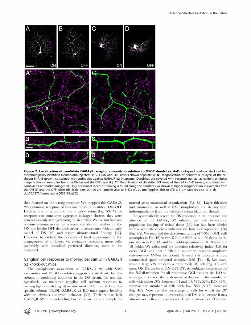
We next analyzed the distribution of GABAAR a2 and
GABAAR a1 along the dendrites of ON-OFF DSGCs, individ-
ually labeled by Neurobiotin injection (Fig. 2). Other than in the
case of SACs, for which we were able to focus our analysis of
receptor co-localization to synaptic structures (varicosities), the
DSGC side displays no morphological specializations that can be
recognized in light microscopy and reliably mark synaptic input
sites. Therefore, we analyzed all co-localizations along dendrites
and quantified receptor distribution as puncta per 100 mm of
dendritic length. The dendritic arbor of the ON-OFF DSGC type
occupies two distinct strata in the IPL: the ON dendrites co-stratify
in the inner part with ON SACs, whereas the OFF dendrites co-
stratify with OFF SACs in the outer IPL. We found similar
distributions of both receptor subunits in the ON and the OFF
layer. The a2 subunit was strongly co-localized with the ON and
OFF dendrites, at densities of ,25 puncta/100 mm dendritic
Direction-Selective Inhibition in the Retina
PLoS ONE | www.plosone.org 2 April 2012 | Volume 7 | Issue 4 | e35109

length (Fig. 2a–b and 3C–D). The a1 subunit was also occasionally
found on both DSGC arbors, however at much lower incidences:
,7 and ,4 puncta/100 mm dendritic length on ON and OFF
arbors, respectively (Fig. 2c–d and 3C–D). Similar to SAC
varicosities, the density of GABAAR a2 puncta on DSGC
dendrites in both layers was significantly higher than that of
GABAAR a1 (p,0.001, Fig. 3C–D). When compared to the
respective randomized controls, only GABAAR a2 receptor
density was found significantly higher than chance (Fig. 3C–D).
To determine the precise position of GABAAR a2 subunits at
the SAC-DSGC synapse, we also studied the localization of this
subunit by pre-embedding immuno-electron microscopy. While
the tissue preservation was compromised due to the short fixation
necessary for proper receptor staining, we were able to verify that
the GABAAR a2 subunit clusters at membranes opposite to
ChAT-immunolabeled SACs dendrites, at presumed SAC-DSGC
synapses (Fig. S2).
Two previous studies described the distribution of GABAAR a1
receptors on ON-OFF DSGCs, addressing the possibility that the
spatially asymmetric GABAergic inhibition impinging on ganglion
cells is reflected by local (anisotropies with neighboring excitatory
receptors, [36]) or global (overall receptor distribution across the
dendritic arbor, [37]) asymmetries in the receptor distribution.
Since our quantitative analysis shows a much higher density of
GABAAR a2 puncta on ON-OFF DSGCs, the question arises
whether previous attempts to find such asymmetries failed because
Figure 1. Localization of candidate GABAAR receptor subunits in relation to SAC dendrites and varicosities. A–B. Immunolabelingpattern of the GABAAR a2 subunit (magenta) in a vertical section of rabbit retina (single optical section; INL, inner nuclear layer; IPL, inner plexiformlayer; GCL, ganglion cell layer). Starburst amacrine cells (SACs) were labeled against choline acetyltransferase (ChAT, green). ON SACs have somata inthe GCL and their dendrites form the inner ChAT band; OFF SACs are located in the INL and their dendrites form the outer band. Synaptic receptorclusters (magenta) are unevenly distributed across the neuropile, with GABAAR a2 puncta concentrating in two bands along the SAC processes. C.Distal dendrites of a SAC injected with Neurobiotin (green) and co-stained with GABAAR a2 antibody (magenta) (single optical section): the majority ofSAC varicosities are associated with receptor staining. Examples for such association are illustrated at higher magnification: c1 and c2 show observedreceptor distribution, c19 and c29 show randomized control (magenta channel rotated 90u clockwise). Significantly fewer varicosities are associatedwith receptor clusters in the rotated control. D. Distal dendrites (green) of a SAC injected as in C but co-stained with GABAAR a1 (magenta) (singleoptical section): Some varicosities are associated with receptor clusters (see also magnifications in d1–d2). No obvious change in the degree of signaloverlap is seen for the randomized control (d19 and d29). Scale bars: A, 10 mm (applies also to B); C, 10 mm (applies also to D); c1, 5 mm (applies to allinsets).doi:10.1371/journal.pone.0035109.g001
Direction-Selective Inhibition in the Retina
PLoS ONE | www.plosone.org 3 April 2012 | Volume 7 | Issue 4 | e35109
they focused on the wrong receptor. We mapped the GABAAR
a2-containing receptors of two anatomically identified ON-OFF
DSGCs, one in mouse and one in rabbit retina (Fig. S3). While
receptors can sometimes aggregate in larger clusters, they were
generally evenly arranged along the dendrites. We did not find any
obvious asymmetries in the receptor distribution, neither for the
ON nor for the OFF dendritic arbor, in accordance with an early
model of DS [46] and recent ultrastructural findings [47].
However, to exclude the presence of local anisotropies in the
arrangement of inhibitory vs. excitatory receptors, more cells,
preferably with identified preferred direction, need to be
evaluated.
Ganglion cell responses to moving bar stimuli in GABAARa2 knock-out mice
The conspicuous association of GABAAR a2 with SAC
varicosities and DSGC dendrites suggests a critical role for this
subunit in mediating inhibition in the DS circuit. To test this
hypothesis, we measured ganglion cell calcium responses to
moving light stimuli (Fig. 4) in knock-out (KO) mice lacking this
specific subunit [39,50]. GABAAR a2 KO mice appear healthy,
with no obvious abnormal behavior [39]. Their retinae lack
GABAAR a2 immunolabeling but otherwise show a completely
normal gross anatomical organization (Fig. S4). Layer thickness
and lamination, as well as SAC morphology and density were
indistinguishable from the wild-type retina (data not shown).
To systematically screen for DS responses in the presence and
absence of the GABAA a2 subunit, we used two-photon
population imaging of retinal tissue [38] that had been labeled
with a synthetic calcium indicator via bulk electroporation [26]
(Fig. 4A). We recorded the directional tuning of .3500 GCL cells
(examples in Fig. 4B) in two KO (n = 2553 cells in 30 fields, as the
one shown in Fig. 4A) and four wild-type animals (n = 1002 cells in
18 fields). We calculated the direction selectivity index (DSi) for
every GCL cell that fulfilled a minimum response-amplitude
criterion (see Methods for details). A small DSi indicates a more
symmetrical spatio-temporal receptive field (Fig. 4B, blue traces),
while a large DSi indicates a (presumed) DS cell (Fig. 4B: green
traces, ON DS; red traces, ON-OFF DS). An unbiased comparison of
the DSi distribution for all responsive GCL cells in the KO vs.
wild-type mice revealed a dramatic reduction in the number of
cells with higher DSis (between 0.4 and 0.8; WT: 52%; KO: 33%),
whereas the number of cells with low DSis (,0.3) increased
(Fig. 4C). Note that the percentage of cells for which the DSi
changes may represent an overestimate of DS cells, because it may
also include cells with asymmetric dendritic arbors (see Discussion).
Figure 2. Localization of candidate GABAAR receptor subunits in relation to DSGC dendrites. A–D. Collapsed confocal stacks of twomorphologically identified Neurobiotin-injected DSGCs (ON and OFF arbors shown separately). A9. Magnification of dendrite (ON layer) of the cellshown in A–B (green), co-stained with antibodies against GABAAR a2 (magenta). Dendrites are covered with receptor puncta, as evident at highermagnification in examples from the ON (a) and the OFF layer (b). C9. Magnification of dendrite (ON layer) of the cell in C–D (green), co-stained withGABAAR a1 antibodies (magenta). Only occasional receptor staining is found along the dendrites, as shown at higher magnification in examples fromthe ON (c) and the OFF arbor (d). Scale bars: A, 100 mm (applies also to B–D); A9, 20 mm (applies also to C9); a, 5 mm (applies also to b–d).doi:10.1371/journal.pone.0035109.g002
Direction-Selective Inhibition in the Retina
PLoS ONE | www.plosone.org 4 April 2012 | Volume 7 | Issue 4 | e35109

The change in DSi distribution is highly significant (p,0.01, using
resampling and bootstrap percentile confidence intervals) and
consistent with a critical role of a2-containing GABAARs in
mediating DSCG directional tuning.
An advantage of such population recordings is that also
potential ‘‘side-effects’’ of the receptor elimination on other retinal
circuitries can be evaluated, for instance by comparing the relative
frequency of the main functional types of GCs. We quantified the
relative contribution of ON, OFF, ON-OFF and non-responding
(NR) somata and averaged the data over the imaged fields
(Fig. 4D). This revealed similar overall distributions of response
polarities in wild-type and KO animals, suggesting that the lack of
GABAAR a2 has no grave effects on other retinal circuits.
Nevertheless, the KO retinae comprised a significantly higher
fraction of ON-OFF cells and fewer NR cells compared to wild-
type retina. This increase of light responsiveness in the KO is likely
due to the overall reduction in GABAAR (a2) mediated inhibition
and reflects unmasking of existing excitatory inputs. In support,
similar results were obtained in the wild-type when applying
GABAA and/or GABAC receptor antagonists (see below). For
comparing the proportion of ON and ON-OFF DSGCs between
genotypes, we considered all recorded cells that fulfilled the
minimum response criterion, showed the correct response polarity
(ON or ON-OFF), and had a DSi$0.4. From this sample we also
discarded, after visual inspection of their responses, cells with
unstable baseline levels (e.g. change in baseline before light
stimulation) or inconsistent responses over different trials of the
same stimulus condition (e.g. cells that responded for less than 3
trials), but by definition also ‘‘non classical’’ types of DSGCs (see
Discussion). Therefore, the percentage of DS cells resulting from
this analysis (Fig. 4E) likely represents an underestimate.
Nevertheless, also by this conservative but more subjective
comparison, we found a highly significant drop in the number of
both ON and ON-OFF DS cells in the GABAAR a2 KO retina
(Fig. 4E).
We also analyzed our data with respect to the earlier described
OFF DSGCs [14], which are not expected to be affected in the
KO animals, since the dendrites of these cells stratify outside the
IPL strata with pronounced GABAAR a2 density. However, we
found only very few clear examples of OFF DSGCs in both the
control (15 of 1002 cells) and KO mice (7 of 2553 cells, data not
shown). It is possible that our stimulation parameters (i.e. positive
contrast, motion velocity, stimulus size) were not optimal to detect
these cells.
Analysis of the synaptic inputs to ON-OFF DS ganglion cells
showed that not only inhibitory but also excitatory inputs are
directionally tuned ([51,52,53,54], but see [55]). Presynaptic
GABAergic inhibition, i.e. at the terminals of bipolar cells, has
been proposed as one of the potential underlying pathways. To test
whether the remaining DS observed in the KO animals can be
attributed to such a mechanism, we recorded RGC responses in
the presence of GABAA and GABAC receptor blockers (50 mM
gabazine, 50 mM TPMPA; data not shown). Blocking GABA Rs in
the KO mouse strongly increased the overall light-responsiveness
of the cells (mean response amplitude with GABAR block: 240%
of control) and caused a shift in DSi distribution towards smaller
values (mean DSi from Gaussian fit of histogram, control:
0.3860.08, n = 470 cells; GABA R block: 0.3360.13, n = 514
cells; p,0.05, T-Test; data not shown), indicating that additional
GABAergic pathways that do not contain GABAAR a2 –possibly
Figure 3. Quantification of candidate GABAA receptor distribution on SACs and DSGCs. A. GABAAR a2 and a1 puncta distribution inrelation to SAC dendrites in percentages of varicosities associated with at least a single punctum ( = receptor cluster) and with multiple puncta (a2,n = 245 varicosities from 5 cells in 4 retinae; a1, n = 181 varicosities from 5 cells in 3 retinae). Percentages and standard deviation values are given forthe original data (correct distribution) and for data with one fluorescence channel rotated by 90u (random distribution). B. Percentages of varicositiesassociated with single or multiple puncta plotted for the correct distribution (open bars) and randomized controls (filled bars). C. GABAAR a2 and a1puncta distribution in relation to DSGC dendrites in number of receptor clusters per 100 mm of dendritic length for both ON and OFF dendritic arbors(a2, n = 3 cells from 3 retinae; a1, n = 3 cells from 2 retinae). D. Number of receptor clusters in ON and OFF sublayers plotted for the original data(correct distribution, open bars) and the randomized controls (random distribution, filled bars). Average and standard deviation values plotted in thisfigure were determined across cells.doi:10.1371/journal.pone.0035109.g003
Direction-Selective Inhibition in the Retina
PLoS ONE | www.plosone.org 5 April 2012 | Volume 7 | Issue 4 | e35109

presynaptic inhibition at bipolar cell terminals– may contribute to
the DS circuit. We still observed cells with high DSi values in the
KO mice under this additional GABA R blockade (data not
shown). This shows that GABA-independent response asymme-
tries (cf. [15]) contribute strongly to the DS as calculated by our
analysis.
Taken together, our data strongly suggests that DS is severely
impaired in the absence of GABAAR a2. This supports our
anatomical findings that this receptor is located at the SAC-DSGC
synapse (likely on the DSGC side), where it mediates direction-
selective inhibition.
Discussion
Many studies investigated the direction-selective circuit of the
retina, as a model of a relatively simple neural computation. Our
present understanding of the available data suggests highly
complex synaptic arrangements that go beyond simple connectiv-
ity between cell types (see [47]), towards very selective local
processing [19,20,21,56]. It is therefore essential to gain
knowledge about the underlying structural elements, particularly
which neurotransmitter receptors are expressed at specific
synapses on different cells in this circuitry. Here we provide the
Figure 4. Analysis of DS responses in GABAAR a2 knock-out mice and wild-type animals. A. Two-photon micrograph showing an opticalsection (1106110 mm2, at the level of the GCL) of mouse retina, with 56 ganglion cells (and displaced amacrine cells) stained by the calcium indicatordye OGB-1 via electroporation. B. Calcium responses (DF/F) evoked by a bar stimulus moving in 8 directions measured in four exemplary GCs (tracecolor matches color of ROIs [regions of interest] in A): an ON (blue), an ON-DS (green), an ON-OFF (yellow) and an ON-OFF-DS (red) ganglion cell. Polarplots of the response amplitudes, with the preferred direction (black line) indicated, are shown in the center of the traces and reflect the different DStuning strength of the cells (see also direction selectivity index, DSi; for definition see Methods). C. Histogram (top) showing the DSi distribution acrossall recorded GCL cells in GABAAR a2 knock-out mice (gray bars, n = 2553 cells in 2 mice) and wild-type controls (black bars, n = 1002 cells in 4 mice).Bottom: Difference between histograms (from top), illustrating the drop in the number of cells with higher DS-indices. D. Percentage of ON, OFF, ON-OFF, as well as non-responsive (NR) GCL cells in control (black bars) and knock-out animals (gray bars). E. ON-OFF and ON cells with a DSi.0.4 in thetwo groups of animals (cells were included or rejected after manual inspection of responses; see Results for complete criteria). (For E and D, relativecell type numbers were determined for each of the recorded GCL field –18 fields in wild-type, 30 in knock-out mice; with approx. 50–60 cells each–and then averaged; error bars indicate S.E.M.).doi:10.1371/journal.pone.0035109.g004
Direction-Selective Inhibition in the Retina
PLoS ONE | www.plosone.org 6 April 2012 | Volume 7 | Issue 4 | e35109

first compelling evidence for the precise localization of a specific
receptor subunit in the DS circuit.
Two lines of evidence suggest that GABAA receptors containing
a2 subunits specifically mediate direction-selective inhibition from
SACs onto DSGCs. First, our anatomical experiments show that
a2 is not only strongly expressed within the cholinergic bands in
the IPL ([40]; Fig. 1A–B, Fig. S4A–B), but is aggregated
particularly at SAC varicosities (Fig. 1C–D, 3A–B) –the cell’s
synaptic output structures– and also at the postsynaptic side,
significantly co-localized with DSGC dendrites (Fig. 2, 3C–D).
This is in contrast to GABAAR a1 (Fig. 3), a subunit that has been
previously reported to be present in ON-OFF DSGCs [36,37].
Second, our population recordings in GABAAR a2 KO mice
reveal a significant drop in directionally selective cells (Fig. 4C,E),
whereas the overall light responsiveness and the relative
frequencies of ON, OFF and ON-OFF retinal ganglion cells are
largely the same as in the wild-type animals (Fig. 4D).
The majority of anatomical data was obtained in the rabbit,
whereas for the recordings we used mice. Each model presents its
advantages: rabbits are the classic animals for studies of direction-
selectivity and facilitate selective targeting of DSGC somata,
thanks to the very characteristic semilunar-shaped nuclei they
exhibit in this species. On the other hand, mice are indispensable
for the deeper understanding of DS circuitry function through the
availability of genetically modified animals. Previous studies
showed that the physiology of DSGCs in mouse and rabbit is
very similar (e.g. mouse: [57,58]; rabbit: [52,53]), and our findings
support the view that retinal direction-selectivity is based on
analogous mechanisms in the two species.
Receptor subunit localization at light-microscopical levelThe 3D structure of the varicosities, obvious ultrastructurally
[47,49] and also apparent in our confocal micrographs (Fig. S1 B–
C), makes it difficult to resolve the exact localization of associated
receptors. Both the SACs’ own receptors as well as those located
postsynaptically, on the DSGC dendrites, will mostly appear co-
localized with the varicosities in confocal microscopy. While we
cannot exclude GABAAR a2 expression in SACs, we offer three
lines of evidence for their postsynaptic localization: first, GABAAR
a2 immunolabeling is significantly associated with SAC varicos-
ities, the sites where SACs make output synapses, but not with the
proximal dendrites, where mainly input is received (data not
shown). While SACs might receive inputs also from other
amacrine cells at the majority of varicosities, GABAAR a2 is also
highly co-localized with DSGC dendrites, which we know from
physiological studies to express GABAA receptors, thus arguing for
a postsynaptic localization. Second, our pre-embedding double
labeling EM experiments indicate the presence of GABAAR a2
postsynaptically to SAC dendrites, on membranes most likely
belonging to ganglion cells (Fig. S2). Third, should GABAAR a2
expression be restricted to SACs, one would not expect a clear
phenotype in the GABAAR a2 KO mice, since GABAAR blockers
do not interfere with SAC ability to generate DS signals
[19,20,21,58], and so direction selectivity downstream would
remain intact.
Remaining DS in GABAAR a2 knock-out miceDirection selective cells were defined in our analysis of the
physiological experiments as ON or ON-OFF cells with a
minimum DSi of 0.4. This threshold was used in accordance with
previous measurements of DS in the mouse retina [11,12,59,60].
We found ,5% of the cells in the GCL to be ON-OFF DS
ganglion cells (and ,2.5% ON DS), which is consistent with
earlier reports using the same recording method [26,47]. Since the
population imaging does not reveal the morphological identity of
the recorded cells, our analysis presumably also includes non-DS
cells that reach a high DSi as a result of an asymmetrical dendritic
arbor and/or active dendritic properties [15]. This is likely the
main explanation for why we also found cells with high DSis in the
KO mice (Fig. 4C). Also the aforementioned OFF DSGCs, which
have strongly asymmetrical dendrites and display light responses
tuned to upward motion [14], likely contribute to the cells that
remain DS in the KO animals, although we found only few
convincing examples of such RGCs (data not shown). Asymmetric
dendritic morphologies have also been described for several other
GC types of the mouse retina (e.g.: B3, C6, D1 [61], some cells in
clusters 6 [62], 2, 5, 6, 7, 8, 9 [63] or G3, G5, G12, G18 [64].)
Furthermore, a recent study identified a subset of classical ON-
OFF DSGCs, which are able to maintain directional tuning to
slow moving stimuli even in the presence of GABAR blockers, due
to active dendritic properties [15].
There is also a technical reason that may bias the responses to
the moving bar stimulus such that also non-DS cells display some
apparent directional tuning: The bar stimulus cannot be equally
well centered on all cells in a recorded field, therefore the spatial
offset between the cells location and the center of the stimulation
field (i.e. where the trajectories of the moving bar stimuli cross) can
in principle distort the resulting tuning curve. This effect would be
augmented by asymmetric dendritic arbors – in line with recent
observations [15]. Nevertheless, we did not see substantial
differences in the distribution of directionally-tuned GCs across
each recorded field (i.e. edge vs. center), likely because the
recorded fields were relatively small (,1006100 mm) compared to
the dendritic tree diameters of mouse RGCs (,100 to 250 mm,
[64]). Since such a potential bias is resulting from the recording
situation, it is expected to be the same for both KO and wild-type
animals and therefore should not affect our conclusions about the
response differences between the two genotypes. In addition,
indirect network mechanisms, other than direct GABA transmis-
sion from SACs to DSGCs, may contribute to the remaining DS in
the KO. Such mechanisms have been suggested to operate
presynaptically through DS inhibition at bipolar cell terminals
[51,52,53,54,65] or be mediated by asymmetric cholinergic
excitatory inputs from SACs [56]. Our experiments in which
GABAA and GABAC receptors were pharmacologically blocked in
the GABAAR a2 KO (data not shown) support a role for
presynaptic inhibition at bipolar cell terminals in retinal DS. The
same experiments showed that cells with high DSi values persisted
under these conditions, consistent with the finding that asymmetric
dendritic morphology and/or active dendritic properties [15,66]
provide sufficient directional tuning in at least some DSGCs.
Specific receptor subtypes for specific functional rolesGABAA receptors are pentamers generally composed of two a,
two b and one c subunit and mediate fast synaptic and tonic
extrasynaptic inhibition (for review see [67]). Much evidence has
accumulated from immunocytochemical, physiological and phar-
macological studies throughout the central nervous system
supporting that specific receptor subtypes are targeted to distinct
neural circuits [67], as well as to specific synaptic sites within one
neuron [68,69,70,71]. This pattern has been consistently demon-
strated in the retina, where subunit receptor expression has been
extensively studied for both excitatory and inhibitory receptors
[43,72,73,74,75]. Here we report selective expression of GABAAR
a2, but not a1, at a specific synapse in the DS circuit, supporting
the idea that these subunits belong to different receptors and,
likely, different synaptic complexes. Despite the fact that GABAA
a1 receptor density that we counted in ON-OFF DSGCs did not
Direction-Selective Inhibition in the Retina
PLoS ONE | www.plosone.org 7 April 2012 | Volume 7 | Issue 4 | e35109

reach levels significantly higher than chance, we cannot exclude
low expression of these receptors, possibly at synapses other than
from SACs (outside the DS circuit). Interestingly, the choice for
receptors in the DS circuit appears to be conserved between
pathways (ON and ON-OFF; Fig. 4) and among species, as
suggested by our findings in rabbit (Fig. 1) and mouse (Fig. S4,
Fig. 4), as well as by those reported previously in rat [40]. As for
the complete structure of the GABAA receptor expressed
postsynaptically in the DSGCs, judging from the composition of
most GABAARs found in the higher brain, a2b3c2 is the most
likely candidate [67].
So far no prominent functional characteristics have been
reported for GABAAR a2. Therefore, it is unclear whether and
how this specific subunit could support DS computation other
than ‘‘simply’’ relaying asymmetric inhibition from SACs. On the
other hand, it is surprising to find a specific subunit almost
exclusively at a very particular location in a neuronal circuit,
especially when the circuit relies on highly complex arrangements
at synaptic level – as retinal DS does. In view of the extensive
rewiring that appears to take place in the DS circuit within 1–2
days during development (e.g. [27,28]), it is possible that specific
subunits at distinct locations may facilitate the differential targeting
of receptors to different synapses. In this scenario, a particular
subunit may play less a functional but more an organizational role.
Notably, the d subunit, one of the less common GABAAR
subunits, was previously shown to be expressed in the retina
selectively by SACs [40] and the responsible Gabard gene was
recently identified as one of several specific markers of SACs in a
wide screening of adult retinal cell types’ transcriptomes [S.
Siegert, Friedrich Miescher Institute for Biomedical Research,
personal communication]. In other brain regions, e.g. dentate
gyrus and cerebellum, d-containing GABAA receptors mediate
tonic inhibition and are activated by low ambient GABA levels
[76]. It is tempting to speculate on possible interactions of d and
a2 subunits, should SACs be shown to express the latter as well.
However, to our knowledge, the presence of the a2 and d subunits
in the same receptor complex in vivo has not been reported. More
likely, d and a2 belong to different GABAARs located in SACs and
DSGCs, respectively (see discussion on receptor localization
above). Nonetheless, they could be localized at the same synaptic
site, for example with presynaptic GABAAR d serving as
modulators for the DS signals passing downstream.
Materials and Methods
Animals and tissue preparationAll animal procedures were carried out in accordance with
institutional guidelines of the Max-Planck-Institute for Brain
Research, Frankfurt, and the University of Tubingen, following
the standards described by the German animal protection law
(Tierschutzgesetz). Euthanasia of both mice and rabbits for organ
harvesting (retinae) used in this study has been approved by the
animal welfare officer of the respective facility (MPI for Brain
Research and University of Tubingen) and reported to the local
authorities (Regierungsprasidium Darmstadt and Regierungspra-
sidium Tubingen, respectively).
For single-cell dye injections, adult New Zealand White rabbits
(Charles River Laboratories) were deeply anesthetized by intra-
muscular injection of ketamine (50 mg/kg body weight; Curamed
Pharma/Pharmaselect) and xylazine (Rompun, 10 mg/kg body
weight; Bayer) and then sacrificed with intravenous pentobarbital
(Narcoren, 160 mg/kg body weight; Merial). The eyes were quickly
enucleated, and the retinae were dissected in carboxygenated (5%
CO2, 95% O2) Ames medium (Sigma-Aldrich, Munich, Germany).
For physiological recordings, adult (.4 weeks old) wild-type and
GABAAR a2 knock-out (mutant allele Gabra2tm2.2Uru on C57BL/
6J background) [39,50] mice were dark adapted for at least one hour
prior to experiments. All further tissue handling was carried out
under dim red illumination. Mice were anaesthetized with
Isoflurane (Baxter, Unterschleißheim, Germany) and killed by
cervical dislocation. Eyes were enucleated, transferred to carbox-
ygenated Ringer’s solution (Biometra, Gottingen, Germany) and the
retinae dissected into two halves. For LM immunocytochemistry,
the posterior eyecups of adult wild-type and GABAAR a2 knock-out
mice were fixed with 4% paraformaldehyde in 0.1M phosphate
buffer (PB) for 10 minutes at room temperature (RT). For electron
microscopy, eyecups were fixed in 4% paraformaldehyde, 0.05%
glutaraldehyde and 0.2% picric acid in 0.1M PB for 25 minutes at
RT.
Cell injections and immunolabelingA piece of flat mounted rabbit retina, with the ganglion cell
layer on top, was placed under the microscope and perfused with
warmed (32uC), carboxygenated medium. The tissue was
visualized using custom built infrared scattered light detection
system (designed by W. Denk, MPImF) on a custom built eyecup
scope ([77]; for details see also section Two-photon calcium imaging).
Cells in the ganglion cell layer were targeted for injection:
displaced (ON) SACs were identified by their small round somata
(10 mm in diameter); DSGCs were recognized by their medium-
sized cell bodies (16 to 20 mm) with semi-lunar nuclei. These
somata were impaled with microelectrodes (borosilicate glass with
filament, O.D. 1.0 mm, I.D. 0.58 mm; 100–200 MV) filled with
4% neurobiotin in 3M KCl. A current of 0.5 to 1.0 nA was
applied for 3 to 5 minutes using a multiclamp amplifier (Molecular
Devices GmbH, Ismaning, Germany). Small concentrations of
Alexa-488 were added to the pipette solution to visualize the
electrode tip during injections and to check the morphology of the
injected cells immediately after injections. The electrode was then
retracted carefully and the tissue was left to rest for about
30 minutes. Afterwards, the retina was fixed with 4% parafor-
maldehyde in PB for 10–20 minutes at RT. After rinsing in PB
(4615 min), the tissue was preincubated for 2 h in blocking
solution (10% normal donkey serum, 1% bovine serum albumin,
and 1% Triton X-100 in PB) at RT. Neurobiotin was revealed
with Streptavidin-Alexa Fluor 488 (Invitrogen, Darmstadt,
Germany, Cat.No. S-11223) diluted 1:1000 in 3% normal donkey
serum, 1% bovine serum albumin, and 1% Triton X-100 in PB,
and incubated overnight at RT or for 3 days at 4uC. Receptor
antibodies were usually incubated together with the Streptavidin.
Antisera that recognize specific amino acid sequences of GABAA
receptor subunits alpha 1 and 2 (GABAAR a1 and GABAAR a2)
were used: GABAAR a1 was stained with a monoclonal antibody
raised in mouse (Chemicon International, Temecula, CA, USA,
Cat. No. MAB339), working dilution 1:50; GABAAR a2 was
labeled with a polyclonal rabbit antibody (Synaptic Systems,
Gottingen, Germany, Cat. No. 224103), working dilution 1:2000.
Some retinae were also co-stained for choline acetyl transferase
(ChAT), with a polyclonal antibody raised in goat (Chemicon, Cat.
No. AB144P). Following incubation with the primary antibody–
Streptavidin mix, the tissue was washed several times in PB, then
incubated for 2 h at RT with the secondary antibody diluted 1:500
in the same incubation solution as above. The following secondary
antibodies were used: donkey-anti-mouse Cy3 (Jackson Immu-
noResearch, West Grove, PA, USA, Cat. No. 715-165-150),
donkey-anti-rabbit Cy3 (Jackson ImmunoResearch, Cat. No. 711-
165-152) and donkey-anti-goat Cy5 (Jackson ImmunoResearch,
Cat. No. 705-175-003). Retinae were finally washed in PB,
Direction-Selective Inhibition in the Retina
PLoS ONE | www.plosone.org 8 April 2012 | Volume 7 | Issue 4 | e35109

mounted in Aqua Polymount medium (Polysciences, Eppelheim,
Germany) and stored at 4uC until visualized by confocal
microscopy.
For immunolabeling on cryostat sections, the fixed mouse
retinae were cryoprotected in graded sucrose (10%, 20%, and
30%) and sectioned vertically (16 mm) with a cryostat. Sections
were labeled for calbindin, with a polyclonal antibody raised in
rabbit (Swant, Bellinzona, Switzerland, Cat. No. 300), working
dilution 1:2000, and for protein kinase C alpha, with a monoclonal
mouse antibody (Biodesign, Saco, ME,USA, clone MC5), working
dilution 1:100.
Pre-embedding immunoelectron microscopyThe localization of GABAAR a2 subunit was investigated at the
ultrastructural level using a pre-embedding double labeling method
in combination with ChAT [78]. A short fixation (25 min) in 4%
paraformaldehyde, 0.05% glutaraldehyde and 0.2% picric acid was
necessary to preserve the immunoreactivity adequately. After
fixation, and cryoprotection, the tissue was frozen and thawed,
and vertical vibratome sections (60 mm) were cut. Vibratome
sections were blocked for 2 hours in PBS containing 10% NDS
and 0.2% acetylated BSA (BSAc, Aurion, Wageningen, Nether-
lands), then incubated in anti-GABAAR a2 (rabbit, dilution 1:500,
Synaptic Systems) and anti-ChAT (goat, dilution 1:100, Chemicon)
with 3% NDS, 0.2% BSAc in PBS for 4 days at 4uC. Donkey anti-
goat biotin-conjugated (Vector, Burlingame, CA) and donkey anti-
rabbit conjugated to 0.8 nm nanogold particles (Aurion) secondary
antibodies were applied (1:100 and 1:50, respectively), followed by
an immunoperoxidase reaction (Vectastain Elite ABC kit; Vector) to
visualize the biotinylated antibody. The sections were then post-fixed
with 2.5% (v/v) glutaraldehyde in cacodylate buffer for 1 hour. The
product of the enzymatic reaction and the nanogold particles were
silver intensified using the silver enhancement system (R-Gent) from
Aurion, as described by the manufacturer. The sections were then
incubated in 0.5% (w/v) osmium tetroxide for 30 minutes at 4uC.
Dehydration was carried out by an ethanol series and propylene
oxide. Specimens were embedded in Epon, and a series of ultrathin
sections (60 nm) were collected on copper grids and stained with
uranyl acetate and lead citrate. Ultrathin sections were examined
with a Leo 912 AB Omega transmission electron microscope (Carl
Zeiss SMT AG, Oberkochen, Germany) and photographed with a
wide-angle Dual Speed 2K-CCD camera in combination with
ImageSP (TRS, Moorenweis, Germany) software.
Confocal microscopy and image analysisImage stacks were acquired with a confocal microscope
(Olympus Fluoview FV1000) equipped with Helium-Neon and
Argon lasers. Cell morphology was documented at low magnifi-
cation, using a UPlanSApo 206/0.75 objective. For quantitative
analysis, high-resolution image stacks (102461024 pixels at
0.5 mm Z-intervals) of individual dendrites were obtained using a
UPlanSApo 606/1.35 oil objective. Image resolution was within
the confidence range of the Z-axis resolution of the microscope
(0.46 mm for the 606 oil objective). Micrographs used for
illustration were cropped and adjusted for contrast and brightness
in Photoshop 11.0 (Adobe Systems GmbH, Munich).
Synaptic puncta that were co-localized with SAC varicosities or
DSGC dendrites were documented manually throughout the
stacks. Receptor clusters were included in the analysis only when
they showed at least 75% overlap with the dendritic profiles. In the
case of DSGCs, the total length of dendrites within a collapsed
image stack was measured and the number of co-localized synaptic
puncta was normalized per 100 mm dendritic length to allow
comparison among samples. To assess whether the density of
receptors on varicosities and dendrites reached higher-than-
chance levels, we generated random superimpositions of the
receptor and cell labeling (by rotating one of the signals by 90u)and repeated counting. The value obtained was defined as chance
level co-localization. Incidence levels of synaptic labeling were
tested against the randomized controls by one-way analysis of
variance (ANOVA) followed by Tuckey’s multiple comparison
test, using Prism 5 software (GraphPad Software, Inc., La Jolla,
CA, USA).
Bulk electroporation and two-photon calcium imagingBulk electroporation of fluorescent calcium indicator dye into
the ganglion cells was carried out as described previously [26]. In
brief, each retina half was mounted ganglion cell side up on a filter
membrane (Anodisc 13, 0.2 mm pore size, Whatman, Maidstone,
UK) and positioned between two 4 mm horizontal plate electrodes
(CUY700P4E/L, Nepagene/Xceltis, Meckesheim, Germany). A
10 ml drop of 5 mM Oregon Green-BAPTA-1 hexapotassium salt
(Invitrogen, Darmstadt, Germany) in Ringer was suspended from
the upper electrode and lowered onto the specimen. After the
application of 10 pulses (+11 V, 10 ms pulse width, at 1 Hz) from
a pulse generator/wide-band amplifier combination (TGP110 and
WA301, Thurlby Thandar/Farnell, Oberhaching, Germany), the
tissue was moved to the recording chamber of the microscope (see
below), where it was continuously perfused with carboxygenated
Ringer at ,37uC and left to recover for ,60 minutes before the
recordings started.
We used a custom built two-photon microscope [77] equipped
with through-the-objective light stimulation and two detection
channels for fluorescence imaging (red, HQ 610 BP 75, and green,
D 535 BP 75; Chroma, Tubingen, Germany). A mode-locked Ti/
sapphire laser (MaiTai, Spectra Physics, Newport, Santa Clara,
CA) tuned to 927 nm served as the excitation source. To visualize
the retinal structure in the red fluorescence channel, ,0.1 mM of
sulforhodamine 101 (Sigma-Aldrich, Munich, Germany) was
added to the bathing medium. Changes in fluorescence reflecting
Ca2+ activity were recorded from the ganglion cell somata in the
green channel. A light stimulator [77] based on a miniature liquid-
crystal-on-silicon (LCoS) display (i-glasses; EST, Kaiserslautern,
Germany) and driven by custom-written software was used to
project moving bar stimuli onto the retina. The stimulus consisted
of a bright bar (30061000 mm, at 3.87N103 R*/s/cone) on a dark
background (0.28N103 R*/s/cone) moving in eight angular
directions at 0.5 mm/s (n = 3 trials per direction). Although color
was supported, we restricted the stimuli used here to ‘‘gray’’
stimuli (with both blue and green stimulator components at the
same opsin photoisomerization rate). Typically, 1106110 mm
fields comprising 50–60 GCL cells at a time (for example see
Fig. 4A) were recorded at 64664 pixels (at 2 ms per line = 7.8 Hz
frame rate) using custom-written software (CfNT, M. Muller,
MPImF, Heidelberg). A synchronization signal was acquired (@
500 Hz) together with the image data to allow matching calcium
signals with the stimuli.
Analysis of Ca2+-imaging dataImages were analyzed offline using IgorPro 6.2 (Wavemetrics,
Lake Oswego, OR, USA). Regions of interest (ROIs) were
manually selected around each soma in a field of view and Ca2+
(DF/F0) signal traces were extracted using the freely available
image processing package SARFIA [79]. Responses to individual
stimulus presentations were extracted using the synchronization
signal from the light stimulator, embedded in the image data. Note
that we did not attempt to distinguish between RGCs and
displaced amacrine cells, of which the latter make up almost 60%
Direction-Selective Inhibition in the Retina
PLoS ONE | www.plosone.org 9 April 2012 | Volume 7 | Issue 4 | e35109

of the GCL cells in mouse retina [80]. It is conceivable that the
fraction of non-responsive (NR) cells mainly consists of displaced
amacrine cells; however, one cannot exclude also amacrine cells
exhibiting light-evoked somatic Ca2+ changes (for discussion see
also [26]). All further analysis was carried out using custom-written
IgorPro routines. To quantify Ca2+ responses (e.g. Fig. 4B), the
area (A) under the DF/F0 trace comprising both the ON and the
OFF response (to the bar’s leading and trailing edge, respectively),
was calculated for each stimulus direction, resulting in 8 response
vectors (v). To limit inclusion of cells with Ca2+ responses too close
to noise, we discarded the 35%-fraction of cells with the smallest
responses (‘‘minimum response criterion’’). For the remaining
cells, a directional selectivity index (DSi) was determined as follows:
DSi~P
vj j=P
vj j. It ranges from 0 and 1, with small values
indicating a more symmetrical spatio-temporal response profile
and high values indicating directional tuning.
To test whether or not the difference in DSi distribution of
knock-out vs. wild-type (Fig. 4C) is statistically significant the
distributions were resampled (n = 1000) and compared using the
bootstrap percentile confidence interval (build-in IgorPro func-
tions). To statistically compare the percentage of (manually
classified) ON, OFF, ON-OFF and NR cells (Fig. 4D) as well as
ON and ON-OFF DSGCs (Fig. 4E) in wild-type and knock-out we
determined the respective mean percentage (and the standard
error) by averaging across the recorded fields. Note that when
plotting these percentages over the time-course of the experiments
we found no detectable trend in the relative fractions for the
different functional cell types, indicating that recording quality was
constant (data not shown).
Supporting Information
Figure S1 Morphology of varicosities on the distaldendrites of starburst amacrine cells (SACs). A. Distal
dendrites of a dye-injected SAC (see inset) in a rabbit retina. B–C.
High-magnification examples of varicosities, which are hook-like
formations resulting from the presynaptic dendrite wrapping
around the postsynaptic element [35,47]. Scale bars: A, 15 mm
(inset, 50 mm); B–C, 2 mm.
(TIF)
Figure S2 Electron micrographs of ultrathin sections ofmouse retina, pre-embedded and double labeled forChAT and GABAAR a2. Single section shown in A, two serial
sections shown in B9–B0. The GABAAR a2 subunit is labeled with
gold particles (tiny black dots indicated by arrows), which are visible
on postsynaptic membranes apposed to ChAT-labeled SAC
profiles, stained with DAB (big black dots, entire profiles overlaid
in green). Note that the postsynaptic profiles do not contain vesicles,
and are thus putative ganglion cell processes (compare to profile
marked with asterisk in B9, which illustrates a typical presynaptic
element, full of synaptic vesicles.) In B0 also note the elongated
microtubule-like structures along the postsynaptic profile, typical
for ganglion cell dendrites. Scale bar 0.5 mm.
(TIF)
Figure S3 Spatial distribution of GABAA a2 receptorstaining across the dendritic arbors of direction-selec-tive ganglion cells (DSGCs). A–B. Reconstructions of putative
ON-OFF DSGCs in mouse (A) and rabbit (B). The cell in the
mouse retina was labeled in a transgenic GFP-O line, described in
detail in [81]. ON and OFF dendritic trees are shown separately,
with receptor puncta in red for the ON layer and blue for the OFF
layer. In A, each dot represents a single receptor cluster ( = an
immunoreactive punctum); in B, dot size encodes number of
receptor clusters (see scale beneath). Numbers indicate receptor
cluster density per 100 mm dendritic length. The higher density of
puncta found for the rabbit DSGC could reflect differences
between species or, more likely, fixation degree. No obvious
asymmetries can be seen across either cells or layers, as expected
from recent statistical EM data [47] and illustrated in C. (While
the values for the quadrants in B differ to some degree, regions
with higher and lower densities are not segregated to particular
sides of the dendritic field, e.g. null vs. preferred side.) C. SACs
synapse onto DSGCs with different preferred directions (PD) with
the constraint that each DSGC type is contacted by only those
SAC dendrites oriented approximately along the DSGC’s null
direction (ND) (left). Together with the intrinsic DS tuning of the
SAC output, this spatial arrangement warrants that SAC
inhibition selectively vetoes the response to motion in null
direction. Repeating this connectivity pattern for all SACs
overlapping one given DSGC (middle) results in a relatively even
distribution of receptors across the DSGC’s dendritic tree (right).
Scale bars: 100 mm.
(TIF)
Figure S4 Retinal gross organization in GABAAR a2knock-out mice. Vertical sections of wild-type (A–D) and
GABAAR a2 KO retinae (A9–D9) immunolabeled against
GABAAR a2 (magenta in A–A9 and B–B9), choline acetyl
transferase (ChAT; green in B–B9), calbindin (C–C9), and protein
kinase C alpha (D–D9). The retinae of the KO animals are void of
a2 receptor staining, but otherwise do not show any obvious
difference in gross retinal organization (e.g. thickness or lamina-
tion) when compared to the wild-type. Scale bars: 20 mm (B9
applies also to A, A9, B; C9 applies to C; D9 applies to D).
(TIF)
Acknowledgments
We thank H. Wassle for helpful discussions and critically reading the
manuscript and W. Denk for generous support. Further we thank G.-S.
Nam and G. Eske for excellent technical assistance.
Author Contributions
Conceived and designed the experiments: ONA SH TE. Performed the
experiments: ONA TB SKK NZ. Analyzed the data: ONA TB.
Contributed reagents/materials/analysis tools: UR SH TE. Wrote the
paper: ONA TB SH TE. Additions/corrections/approval of final
manuscript: SKK NZ UR.
References
1. Barlow HB, Hill RM (1963) Selective sensitivity to direction of movement in
ganglion cells of the rabbit retina. Science 139: 412–414.
2. Barlow HB, Hill RM, Levick WR (1964) Retinal Ganglion Cells Responding
Selectively to Direction and Speed of Image Motion in the Rabbit. J Physiol 173:
377–407.
3. Amthor FR, Oyster CW, Takahashi ES (1984) Morphology of on-off direction-
selective ganglion cells in the rabbit retina. Brain Res 298: 187–190.
4. Amthor FR, Takahashi ES, Oyster CW (1989) Morphologies of rabbit retinal
ganglion cells with complex receptive fields. J Comp Neurol 280: 97–121.
5. Oyster CW, Barlow HB (1967) Direction-selective units in rabbit retina:
distribution of preferred directions. Science 155: 841–842.
6. Oyster CW, Simpson JI, Takahashi ES, Soodak RE (1980) Retinal ganglion cells
projecting to the rabbit accessory optic system. J Comp Neurol 190: 49–61.
7. Buhl EH, Peichl L (1986) Morphology of rabbit retinal ganglion cells projecting
to the medial terminal nucleus of the accessory optic system. J Comp Neurol
253: 163–174.
8. Grzywacz NM, Amthor FR (2007) Robust directional computation in on-off
directionally selective ganglion cells of rabbit retina. Vis Neurosci 24: 647–661.
Direction-Selective Inhibition in the Retina
PLoS ONE | www.plosone.org 10 April 2012 | Volume 7 | Issue 4 | e35109

9. Kanjhan R, Sivyer B (2010) Two types of ON direction-selective ganglion cellsin rabbit retina. Neurosci Lett 483: 105–109.
10. Hoshi H, Tian LM, Massey SC, Mills SL (2011) Two distinct types of ONdirectionally-selective ganglion cells in the rabbit retina. J Comp Neurol 519:
2509–2521.
11. Rivlin-Etzion M, Zhou K, Wei W, Elstrott J, Nguyen PL, et al. (2011)
Transgenic mice reveal unexpected diversity of on-off direction-selective retinalganglion cell subtypes and brain structures involved in motion processing.
J Neurosci 31: 8760–8769.
12. Yonehara K, Ishikane H, Sakuta H, Shintani T, Nakamura-Yonehara K, et al.
(2009) Identification of retinal ganglion cells and their projections involved incentral transmission of information about upward and downward image motion.
PLoS One 4: e4320.
13. Kay JN, De la Huerta I, Kim IJ, Zhang Y, Yamagata M, et al. (2011) Retinal
ganglion cells with distinct directional preferences differ in molecular identity,
structure, and central projections. J Neurosci 31: 7753–7762.
14. Kim IJ, Zhang Y, Yamagata M, Meister M, Sanes JR (2008) Molecularidentification of a retinal cell type that responds to upward motion. Nature 452:
478–482.
15. Trenholm S, Johnson K, Li X, Smith RG, Awatramani GB (2011) Parallel
mechanisms encode direction in the retina. Neuron 71: 683–694.
16. Hausselt SE, Euler T (2008) Starburst Amacrine Cells and Direction Selectivity.
In: Binder MD, Hirokawa N, Windhorst U, eds. Encyclopedia of Neuroscience.
Berlin, Heidelberg: Springer. pp 3501–3507.
17. Masland RH (2005) The many roles of starburst amacrine cells. Trends
Neurosci 28: 395–396.
18. Demb JB (2007) Cellular mechanisms for direction selectivity in the retina.Neuron 55: 179–186.
19. Euler T, Detwiler PB, Denk W (2002) Directionally selective calcium signals indendrites of starburst amacrine cells. Nature 418: 845–852.
20. Lee S, Zhou ZJ (2006) The synaptic mechanism of direction selectivity in distalprocesses of starburst amacrine cells. Neuron 51: 787–799.
21. Hausselt SE, Euler T, Detwiler PB, Denk W (2007) A dendrite-autonomousmechanism for direction selectivity in retinal starburst amacrine cells. PLoS Biol
5: e185.
22. Euler T, Hausselt SE (2008) Direction Selective Cells. In: Masland RH,
Albright TD, eds. The Senses - A Comprehensive Reference. San Diego:Academic Press. pp 413–422.
23. Gollisch T, Meister M (2010) Eye smarter than scientists believed: neuralcomputations in circuits of the retina. Neuron 65: 150–164.
24. Borst A, Haag J, Reiff DF (2010) Fly motion vision. Annu Rev Neurosci 33:49–70.
25. Borst A, Euler T (2011) Seeing things in motion: models, circuits, andmechanisms. Neuron 71: 974–994.
26. Briggman KL, Euler T (2011) Bulk electroporation and population calciumimaging in the adult mammalian retina. J Neurophysiol 105: 2601–2609.
27. Wei W, Hamby AM, Zhou K, Feller MB (2011) Development of asymmetricinhibition underlying direction selectivity in the retina. Nature 469: 402–406.
28. Yonehara K, Balint K, Noda M, Nagel G, Bamberg E, et al. (2011) Spatiallyasymmetric reorganization of inhibition establishes a motion-sensitive circuit.
Nature 469: 407–410.
29. Wyatt HJ, Daw NW (1976) Specific effects of neurotransmitter antagonists on
ganglion cells in rabbit retina. Science 191: 204–205.
30. Caldwell JH, Daw NW, Wyatt HJ (1978) Effects of picrotoxin and strychnine on
rabbit retinal ganglion cells: lateral interactions for cells with more complexreceptive fields. J Physiol 276: 277–298.
31. Kittila CA, Massey SC (1997) Pharmacology of directionally selective ganglioncells in the rabbit retina. J Neurophysiol 77: 675–689.
32. Massey SC, Linn DM, Kittila CA, Mirza W (1997) Contributions of GABAAreceptors and GABAC receptors to acetylcholine release and directional
selectivity in the rabbit retina. Visual Neurosci 14: 939–948.
33. Yoshida K, Watanabe D, Ishikane H, Tachibana M, Pastan I, et al. (2001) A key
role of starburst amacrine cells in originating retinal directional selectivity andoptokinetic eye movement. Neuron 30: 771–780.
34. Amthor FR, Keyser KT, Dmitrieva NA (2002) Effects of the destruction ofstarburst-cholinergic amacrine cells by the toxin AF64A on rabbit retinal
directional selectivity. Visual Neurosci 19: 495–509.
35. Famiglietti EV (1992) Dendritic co-stratification of ON and ON-OFF
directionally selective ganglion cells with starburst amacrine cells in rabbitretina. J Comp Neurol 324: 322–335.
36. Jeon CJ, Kong JH, Strettoi E, Rockhill R, Stasheff SF, et al. (2002) Pattern ofsynaptic excitation and inhibition upon direction-selective retinal ganglion cells.
J Comp Neurol 449: 195–205.
37. Chen YC, Chiao CC (2008) Symmetric synaptic patterns between starburst
amacrine cells and direction selective ganglion cells in the rabbit retina. J CompNeurol 508: 175–183.
38. Denk W, Strickler JH, Webb WW (1990) Two-photon laser scanningfluorescence microscopy. Science 248: 73–76.
39. Vollenweider I, Smith KS, Keist R, Rudolph U (2011) Antidepressant-likeproperties of alpha2-containing GABA(A) receptors. Behav Brain Res 217:
77–80.
40. Brandstatter JH, Greferath U, Euler T, Wassle H (1995) Co-stratification of
GABAA receptors with the directionally selective circuitry of the rat retina. VisNeurosci 12: 345–358.
41. Greferath U, Grunert U, Fritschy JM, Stephenson A, Mohler H, et al. (1995)GABAA receptor subunits have differential distributions in the rat retina: in situ
hybridization and immunohistochemistry. J Comp Neurol 353: 553–571.
42. Brandstatter JH, Koulen P, Wassle H (1998) Diversity of glutamate receptors in
the mammalian retina. Vision Res 38: 1385–1397.
43. Wassle H, Koulen P, Brandstatter JH, Fletcher EL, Becker CM (1998) Glycine
and GABA receptors in the mammalian retina. Vision Res 38: 1411–1430.
44. Kosaka T, Tauchi M, Dahl JL (1988) Cholinergic neurons containing GABA-
like and/or glutamic acid decarboxylase-like immunoreactivities in various brainregions of the rat. Exp Brain Res 70: 605–617.
45. Brecha N, Johnson D, Peichl L, Wassle H (1988) Cholinergic amacrine cells ofthe rabbit retina contain glutamate decarboxylase and gamma-aminobutyrate
immunoreactivity. Proc Natl Acad Sci U S A 85: 6187–6191.
46. Vaney DI, Young HM (1988) GABA-like immunoreactivity in cholinergic
amacrine cells of the rabbit retina. Brain Res 438: 369–373.
47. Briggman KL, Helmstadter M, Denk W (2011) Wiring specificity in the
direction-selectivity circuit of the retina. Nature 471: 183–188.
48. Vaney DI (1990) The Mosaic of Amacrine Cells in the Mammalian Retina. Prog
Retinal Res. pp 49–100.
49. Famiglietti EV (1991) Synaptic organization of starburst amacrine cells in rabbit
retina: analysis of serial thin sections by electron microscopy and graphicreconstruction. J Comp Neurol 309: 40–70.
50. Witschi R, Punnakkal P, Paul J, Walczak JS, Cervero F, et al. (2011) Presynaptic{alpha}2-GABAA Receptors in Primary Afferent Depolarization and Spinal
Pain Control. J Neurosci 31: 8134–8142.
51. Borg-Graham LJ (2001) The computation of directional selectivity in the retina
occurs presynaptic to the ganglion cell. Nature Neurosci 4: 176–183.
52. Taylor WR, Vaney DI (2002) Diverse synaptic mechanisms generate direction
selectivity in the rabbit retina. J Neurosci 22: 7712–7720.
53. Fried SI, Munch TA, Werblin FS (2002) Mechanisms and circuitry underlying
directional selectivity in the retina. Nature 420: 411–414.
54. Fried SI, Munch TA, Werblin FS (2005) Directional selectivity is formed atmultiple levels by laterally offset inhibition in the rabbit retina. Neuron 46:
117–127.
55. Poleg-Polsky A, Diamond JS (2011) Imperfect space clamp permits electrotonic
interactions between inhibitory and excitatory synaptic conductances, distorting
voltage clamp recordings. PLoS One 6: e19463.
56. Lee S, Kim K, Zhou ZJ (2010) Role of ACh-GABA cotransmission in detecting
image motion and motion direction. Neuron 68: 1159–1172.
57. Chen M, Weng S, Deng Q, Xu Z, He S (2009) Physiological properties of
direction-selective ganglion cells in early postnatal and adult mouse retina.J Physiol 587: 819–828.
58. Oesch NW, Taylor WR (2010) Tetrodotoxin-resistant sodium channels
contribute to directional responses in starburst amacrine cells. PLoS One 5:
e12447.
59. Weng S, Sun W, He S (2005) Identification of ON-OFF direction-selective
ganglion cells in the mouse retina. J Physiol 562: 915–923.
60. Huberman AD, Wei W, Elstrott J, Stafford BK, Feller MB, et al. (2009) Genetic
identification of an On-Off direction-selective retinal ganglion cell subtypereveals a layer-specific subcortical map of posterior motion. Neuron 62:
327–334.
61. Sun W, Li N, He S (2002) Large-scale morphological survey of mouse retinal
ganglion cells. J Comp Neurol 451: 115–126.
62. Badea TC, Nathans J (2004) Quantitative analysis of neuronal morphologies in
the mouse retina visualized by using a genetically directed reporter. J CompNeurol 480: 331–351.
63. Kong JH, Fish DR, Rockhill RL, Masland RH (2005) Diversity of ganglion cellsin the mouse retina: unsupervised morphological classification and its limits.
J Comp Neurol 489: 293–310.
64. Volgyi B, Chheda S, Bloomfield SA (2009) Tracer coupling patterns of the
ganglion cell subtypes in the mouse retina. J Comp Neurol 512: 664–687.
65. Grzywacz NM, Sernagor E (2000) Spontaneous activity in developing turtle
retinal ganglion cells: statistical analysis. Visual Neurosci 17: 229–241.
66. Schachter MJ, Oesch N, Smith RG, Taylor WR (2010) Dendritic spikes amplify
the synaptic signal to enhance detection of motion in a simulation of thedirection-selective ganglion cell. PLoS Computational Biol 6.
67. Fritschy JM, Brunig I (2003) Formation and plasticity of GABAergic synapses:physiological mechanisms and pathophysiological implications. Pharmacol Ther
98: 299–323.
68. Nyıri G, Freund TF, Somogyi P (2001) Input-dependent synaptic targeting of
alpha(2)-subunit-containing GABA(A) receptors in synapses of hippocampalpyramidal cells of the rat. Eur J Neurosci 13: 428–442.
69. Brunig I, Scotti E, Sidler C, Fritschy JM (2002) Intact sorting, targeting, andclustering of gamma-aminobutyric acid A receptor subtypes in hippocampal
neurons in vitro. J Comp Neurol 443: 43–55.
70. Herd MB, Haythornthwaite AR, Rosahl TW, Wafford KA, Homanics GE, et al.
(2008) The expression of GABAA beta subunit isoforms in synaptic andextrasynaptic receptor populations of mouse dentate gyrus granule cells. J Physiol
586: 989–1004.
71. Houston CM, Hosie AM, Smart TG (2008) Distinct regulation of beta2 and
beta3 subunit-containing cerebellar synaptic GABAA receptors by calcium/calmodulin-dependent protein kinase II. J Neurosci 28: 7574–7584.
72. Koulen P (1999) Clustering of neurotransmitter receptors in the mammalianretina. J Membr Biol 171: 97–105.
Direction-Selective Inhibition in the Retina
PLoS ONE | www.plosone.org 11 April 2012 | Volume 7 | Issue 4 | e35109

73. Ghosh KK, Haverkamp S, Wassle H (2001) Glutamate receptors in the rod
pathway of the mammalian retina. J Neurosci 21: 8636–8647.74. Zhang J, Diamond JS (2009) Subunit- and pathway-specific localization of
NMDA receptors and scaffolding proteins at ganglion cell synapses in rat retina.
J Neurosci 29: 4274–4286.75. Wassle H, Heinze L, Ivanova E, Majumdar S, Weiss J, et al. (2009) Glycinergic
transmission in the Mammalian retina. Front Mol Neurosci 2: 6.76. Zheleznova NN, Sedelnikova A, Weiss DS (2009) Function and modulation of
delta-containing GABA(A) receptors. Psychoneuroendocrinology 34 Suppl 1:
S67–73.77. Euler T, Hausselt SE, Margolis DJ, Breuninger T, Castell X, et al. (2009)
Eyecup scope–optical recordings of light stimulus-evoked fluorescence signals inthe retina. Pflugers Arch 457: 1393–1414.
78. Panzanelli P, Bardy C, Nissant A, Pallotto M, Sassoe-Pognetto M, et al. (2009)
Early synapse formation in developing interneurons of the adult olfactory bulb.
J Neurosci 29: 15039–15052.
79. Dorostkar MM, Dreosti E, Odermatt B, Lagnado L (2010) Computational
processing of optical measurements of neuronal and synaptic activity in
networks. J Neurosci Meth 188: 141–150.
80. Jeon CJ, Strettoi E, Masland RH (1998) The major cell populations of the mouse
retina. J Neurosci 18: 8936–8946.
81. Majumdar S, Heinze L, Haverkamp S, Ivanova E, Wassle H (2007) Glycine
receptors of A-type ganglion cells of the mouse retina. Vis Neurosci 24: 471–487.
Direction-Selective Inhibition in the Retina
PLoS ONE | www.plosone.org 12 April 2012 | Volume 7 | Issue 4 | e35109


![A - Benzodiazepine-Chloride Receptor-Targeted Therapy for ......nisms through GABAA and GABAB receptors [12]. GABA is classified into two main categories: GABAA and GABAB. GABAA and](https://img.pdfslide.net/doc/110x75/60f82a0e0bab2d34196b5ccd/a-benzodiazepine-chloride-receptor-targeted-therapy-for-nisms-through.jpg)