Embed Size (px)
Citation preview

Research ArticleGaining Surgical Access for Repositioningthe Inferior Alveolar Neurovascular Bundle
Saif Yousif Abdullah Al-Siweedi,1 P. Nambiar,1
P. Shanmuhasuntharam,2 and W. C. Ngeow2
1 Department of Diagnostic and Integrated Dental Practice, Faculty of Dentistry, University of Malaya, 50603 Kuala Lumpur, Malaysia2 Department of Oro-Maxillofacial Surgical & Medical Sciences, Faculty of Dentistry, University of Malaya,50603 Kuala Lumpur, Malaysia
Correspondence should be addressed to P. Nambiar; [email protected]
Received 9 November 2013; Accepted 6 February 2014; Published 24 April 2014
Academic Editors: D. Cogulu and D. Flanagan
Copyright © 2014 Saif Yousif Abdullah Al-Siweedi et al. This is an open access article distributed under the Creative CommonsAttribution License, which permits unrestricted use, distribution, and reproduction in any medium, provided the original work isproperly cited.
This study is aimed at determining anatomical landmarks that can be used to gain access to the inferior alveolar neurovascular(IAN) bundle. Scanned CBCT (i-CAT machine) data of sixty patients and reconstructions performed using the SimPlant dentalimplant software were reviewed. Outcome variables were the linear distances of the mandibular canal to the inferior border andthe buccal cortex of the mandible, measured immediately at the mental foramen (D1) and at 10, 20, 30, and 40mm (D2–D5) distalto it. Predictor variables were age, ethnicity, and gender of subjects. Apicobasal assessment of the canal reveals that it is curvingdownward towards the inferior mandibular border until 20mm (D3) distal to the mental foramen where it then curves upwards,making an elliptic-arc curve.Themandibular canal also forms a buccolingually oriented elliptic arc in relation to the buccal cortex.Variations due to age, ethnicity, and gender were evident and this study provides an accurate anatomic zone for gaining surgicalaccess to the IAN bundle.The findings indicate that the buccal cortex-IAN distance was greatest at D3.Therefore, sites between D2and D5 can be used as favorable landmarks to access the IAN bundle with the least complications to the patient.
1. Introduction
The inferior alveolar nerve (IAN) is the branch of themandibular division of the trigeminal nerve that providesinnervation to themandible. Togetherwith the inferior alveo-lar artery and vein, it enters the mandible via the mandi-bular foramen and runs an intraosseous course within themandibular canal before dividing into two smaller branches,namely, the mental and the incisive nerves. It exits themandible as the mental nerve via the mental foramen [1].Having an in-depth knowledge of intraosseous position andcourse of the IAN is essential prior to commencing dentalprocedures in themandible.This is because injury to the IANhas been reported to occur in restorative dentistry, endodon-tology, orthodontics, and, of course, oral and maxillofacialsurgery [2, 3]. Oral and maxillofacial surgical proceduresthat are commonly associated with IAN injury range from
minor oral surgical procedures, such as surgical removal ofmandibular third molar [1], to more major surgeries, suchas sagittal split osteotomy and mandibular jaw resection [4].Lately, accidental encroachment of dental implants into themandibular canal has been highlighted as another source ofIAN injury [5].
The placement of endosseous dental implants in theposterior mandible to support fixed restorative prosthesesis now a common treatment option for replacing missingmolars. However, in cases where the alveolar bone hasseverely resorbed, sufficiently long fixtures cannot be placedwithout encroaching into the mandibular canal. Treatmentoptions in such a case involve either the placement of shortimplant fixtures away from themandibular canal, positioningof implants alongside the mandibular canal, onlay bonegrafting to increase ridge height, alveolar distraction, orrepositioning the IAN bundle to create adequate height for
Hindawi Publishing Corporatione Scientific World JournalVolume 2014, Article ID 719243, 11 pageshttp://dx.doi.org/10.1155/2014/719243

2 The Scientific World Journal
implant placement [6, 7]. The last approach, first describedby Jensen and Nock [8], has been favored by some authorswhen dealing with severely resorbed mandible [7, 9].
Repositioning the IAN bundle, either by lateralizationor fenestration, involves performing an osteotomy on thebuccal cortex of the body of the mandible to create awindow that enables the oral and maxillofacial surgeon togain access to the IAN bundle. The IAN bundle will thenbe retracted more laterally to facilitate the insertion of longfixtures into the body of mandible [9]. This procedure isnot without complication and, most often, short term (3–6months) neurosensory dysfunction is the well-acknowledgedrisk that every patient has to take. Other unusual complica-tions include pathological fracture to the body of mandible,presumably due toweakening of themandiblewhen excessivebone was removed [10]. IAN dysfunction happens becauseof the distension of the nerve during the surgical procedureor by compression/distension that follows [11]. Nevertheless,studies have shown that although this procedure resulted ina high percentage of minor IAN injuries (usually ischemia ofthe nerve), it provides a viable surgical procedure that allowsimplant placement in the resorbed posterior mandible withacceptable minor neurosensory alteration [11, 12].
Damage to the vital structures such as nerve and vesselsand excessive removal of cortical bone may arise if cliniciansmerely use panoramic radiographs for presurgical planning.Hence, it is essential to have an in-depth knowledge ofthe location and the course of the mandibular canal aspanoramic radiographs do not provide information on thehorizontal (buccolingual) position of the IAN [13]. Recently,Pyun et al. [13] tried to predict the horizontal course of themandibular canal by determining the location of the mentalforamen on panoramic radiographs. The use of computedradiographic technology such as CT or CBCT is greatlyadvisable. In addition to preventing iatrogenic injury to theIAN, the information gathered will also avoid unnecessarybone removal that may further weaken a severely resorbedmandible.
It is the main aim of the present study to work out asafe zone for gaining access to the IAN bundle in a groupof selected Asian populations. This is undertaken by firstlydetermining the presence and course of themandibular canalby means of cone-beam computed tomography (CBCT),followed by determining the points of access for IAN repo-sitioning. In essence, the specific objectives were
(i) to determine the presence (i.e., visualization) of themandibular canal using CBCT,
(ii) to measure the diameter of the mandibular canal atvarious predetermined points,
(iii) to measure the apicobasal distance of the mandibularcanal from the inferior border of the mandible atvarious predetermined points,
(iv) tomeasure the buccolingual distance of themandibu-lar canal from the buccal cortex of the mandible atvarious predetermined points,
(v) to determine if the points ofmeasurement can be usedas landmarks to gain access to the IAN bundle,
(vi) to determine if these anatomical landmarks areaffected by the age, ethnicity, and/or gender of thesubjects.
This study is undertaken using CBCT as it has proved tobe a useful tool in detecting the mandibular canal [14] andnegates the need to find cadaveric bodies of Malay samplesas their Muslim religion advocated burial within 24 hours ofdemise.
2. Materials and Methods
2.1. Imaging. This study received the local Ethical CommitteeBoard approval prior to commencement. Cone-beam com-puted tomography images were obtained with the i-CATimaging system (Imaging Sciences International Inc., Hat-field, USA). All images were taken by the same technologistfollowing a standardized protocol for patient positioning andexposure parameter setting (120 kVp, 3–7mA, 20 sec) andimage acquisition at 0.3mm voxel size. Images were obtainedfrom 100 consecutive patients referred to the Oral RadiologyDivision of the Faculty ofDentistry,University ofMalaya.Thefollowing were the inclusion and exclusion criteria used forselecting suitable images.
Inclusion Criteria.They are as follows:
(1) dentate adult patients between the ages 18 and 80years,
(2) healthy, medically compromised, or even previouslyradiated patients that did not involve the body of themandible.
Exclusion Criteria.They are as follows:
(1) patients with history of trauma or pathology to themandible,
(2) syndromic patients and patients with congenital dis-orders that affect the size of the jaw bones,
(3) patients aged below 18 or above 80 years,(4) patients with history of surgical intervention to the
body of the mandible, like orthognathic surgery orrepair of a fracture,
(5) patients of mixed racial origins,(6) patients with existing pathological disorder at
mandible, such as cysts, tumours, osteomyelitis, andfibrous dysplasia,
(7) the reformatted CBCT images, which appear dis-torted or blurred due to patients’ movements.
2.2. Processing the Images. The visibility rating and dimen-sional measurements were performed by only one researcher(first author) who is trained in the interpretation of oraland maxillofacial images. Using cross-sectional images asdescribed below, the visibility of the mandibular canal wascategorized as “clearly visible, probably visible, or invisible”[15].TheDICOMdata obtained were imported into SimPlant

The Scientific World Journal 3
(SimPlant 3-D Pro version 13.1; Materialise Inc., Leuven,Belgium) and additional axial and/or sagittal images in thevolume were analyzed if necessary. The SimPlant softwareallows viewing of axial, cross-sectional, panoramic, and 3Dvisualization of the jaw on the same screen.
All images were scored by the same observer, withrandomly selected samples repeated again 2 weeks later toensure reliability. Firstly, the mental foramen was identifiedand marked red. Then, the visible mandibular canal imagewas drawn onto the scan.
2.2.1. Landmarks and Base Lines. Measurements were madefor the direct linear distances between
(a) the buccal aspect of the mandibular canal and theouter buccal cortical margin of the mandible (Line 𝐵;Figure 1),
(b) the inferior aspect of the mandibular canal perpen-dicular to the inferior point of the lower border of themandible (Line 𝐼; Figure 1).
These measurements were done immediately at (D1) andat 10 (D2), 20 (D3), 30 (D4), and 40 (D5) mm distal to themental foramen (Figure 2). In addition, the diameter of themandibular canal was also measured at these points.
The widest diameter of the mandibular canal was mea-sured on a horizontal plane (𝐷), as shown in Figure 1.
Intrarater reliabilitywas done by derivingCronbach alphacoefficient for each of the 12 randomly selected recordsoutcomes done at 2-week interval. Measurements error, thatis, the difference between the corresponding measurementsexpressed in terms of 𝑆(𝑖) values, was also calculated.
2.3. Statistical Analysis. All data were gathered, entered, andanalyzed using SPSS 16.00 (SPSSFW, SPSS, Chicago, IL)software program. Descriptions of parameters were given asmean ± standard deviation (SD) and 95% confidence intervalof mean. Independent 𝑡-test and correlation or analysis ofvariance (ANOVA) were used to determine the influenceof site, age, gender, and ethnicity on the visibility of themandibular canal and the course of the mandibular canal,respectively. Multiple regression analysis was then performedto determine which combination of predictors affects theresults obtained. A 5% level of significance was chosen.
3. Results
The subjects for this study included images of 60 patients (30males and 30 females) retrieved from the Division of Oralradiology, with ages of patients ranging from 20 to 60 years(mean age, 47 years). Forty other patients were excluded asthey did not meet the inclusion criteria required.
The mandibular canal could be visualized clearly in all60 CBCT images (120 sites). Independent 𝑡-tests showed thatneither the side of image nor gender influences the visibilityof the mandibular canal in the body of the mandible (𝑃 >0.05). Furthermore, the age and ethnicity of the subjects alsodid not affect the visibility of the mandibular canal.
CL
B
I
D
Figure 1: Cross-sectional sagittal view of a CBCT image showingthe site for obtaining measurements. CL = crest of alveolar bone,𝐵 = distance between the mandibular canal and buccal cortex,𝐼 = distance between the mandibular canal and inferior body ofmandible, and𝐷 = diameter of the mandibular canal.
D1 D2 D3 D4 D5
Figure 2: Illustration shows the locations of the measurementsat every 10mm interval starting from the distal aspect of mentalforamen backwards (D1–D5). (D1) is the location of the mandibularcanal at the distal aspect of mental foramen. (D2) is the location ofthe mandibular canal at 10mm away from D1 distally. (D3) is thelocation of the mandibular canal at 10mm away from D2 distally.(D4) is the location of the mandibular canal at 10mm away fromD3distally. (D5) is the location of the mandibular canal at 10mm awayfrom D4 distally.
There was a good standardization and reproducibilityof the base lines and measurements. The data analyzed forreliability of the measurements showed a Cronbach alphacoefficient of 𝑟 = 0.91 and the value of the measurementerrors, 𝑆(𝑖), was 0.05mm or less for all the compareddata, which strongly suggested that the present method ofobtaining measurements is considered reliable and accurate.
The side of mandible did not influence the obtainedresults. The mean distance of the mandibular canal from thelower border of the mandible was 10.09 ± 3.69mm (95% CI= 9.79–10.39mm). The apicobasal measurements obtainedshowed that the mandibular canal formed an elliptic-arccurve, similar to that described by Liu et al. [16] The mean

4 The Scientific World Journal
Table 1: Mean measurement with standard deviation and thecorresponding 95% confidence interval for the distances of themandibular canal to the inferior border and buccal cortex of themandible.
PositionMean ± SD; 95% confidence interval of distancefrom the mandibular canal to two cortical borders
of mandible, namely,Inferior Buccal
D1 9.37 ± 1.69
(9.07–9.68) mm3.90 ± 1.01
(3.71–4.08) mm
D2 8.24 ± 1.69
(7.93–8.55) mm5.59 ± 1.20
(5.38–5.81) mm
D3 7.96 ± 1.93
(7.61–8.31) mm6.71 ± 1.34
(6.47–6.95) mm
D4 9.66 ± 2.54
(9.20–10.12) mm5.69 ± 1.63
(5.39–5.98) mm
D5 15.21 ± 4.18
(14.46–15.97) mm4.25 ± 1.60
(3.96–4.54) mm
Overall 10.09 ± 3.69
(9.79–10.39) mm5.23 ± 1.71
(5.09–5.36) mm
inferior measurement was 9.37 ± 1.69mm at D1, 8.24 ±1.69mm at D2, 7.96 ± 1.93mm at D3, 9.66 ± 2.54 at D4, and15.21 ± 4.18mm at D5 (Table 1). The measurement taken atD3 was the closest to the inferior mandibular border as thispoint formed the lowest point of the elliptic-arc curve. Thiscorresponds with the location around the secondmandibularmolar tooth.
The study showed that the gender of the subjects affectsthe distance of the mandibular canal to the inferior borderof the mandible at D1, D2, and D3. The measurements werelarger inmales in all instances, that is, 10.10± 1.66, 8.96± 1.64,and 8.47 ± 1.93mm in comparison to 8.64 ± 1.40, 7.92 ± 1.43,and 7.46 ± 1.82mm for females.
Further analysis comparing the distances obtained for thethree different ethnic groups shows all of them complyingto the same elliptic-arc curve pattern (Figure 3). However,the mandibular canal of Chinese subjects was, in general,located significantly further away from the inferior borderof the mandible when compared to the two other ethnicgroups at the molar region (D3, ANOVA, 𝑃 = 0.002 andD4, ANOVA, 𝑃 = 0.002). However, further analysis usingregression analysis suggests that race is not a predictor of thedistance of the MC to the inferior border of mandible (𝑃 =0.455). Themain predictor that influences this distance is theage of the subject and it affects measurements at 4 locations.At D2 and D3, age and gender of the subjects affected theresults obtained, while at D4 and D5, this was affected solelyby the age of patients.
Themean distance from buccal cortex was 5.23 ± 1.71mm(95% CI = 5.09–5.36mm). The average buccolingual posi-tions at D1, D2, D3, D4, and D5 were 3.90 ± 1.01mm,5.59 ± 1.20mm, 6.71 ± 1.34mm, 5.69 ± 1.63mm, and 4.25 ±1.60mm, respectively. This also gives the mandibular canalan appearance of an elliptic-arc curve that spanned buccol-ingually. Taken together with the apicocoronal measurement
18
16
14
12
10
8
6
4
2
0
D1 D2 D3 D4 D5
Dist
ance
to th
e inf
erio
r bor
der
Location
Malay (IBM)Indian (IBM)Chinese (IBM)
of m
andi
ble (
mm
)
Figure 3: Distances of the mandibular canal to the inferior borderof mandible immediately at (D1) and at 10 (D2), 20 (D3), 30 (D4),and 40 (D5)mm distal to the mental foramen of 3 different ethnicgroups. An elliptic-arc curve pattern is observed when the resultswere put together.
D1
D2
D3
D4
D5
9.37
3.90
8.245.59
7.866.71
9.66
5.09
15.21
4.25
Figure 4: Distances of the mandibular canal to the buccal andinferior cortices of mandible immediately at (D1) and at 10 (D2),20 (D3), 30 (D4), and 40 (D5)mm distal to the mental foramenof 3 different ethnic groups. A 3D elliptic-arc curve appearance isobserved when both results were put together.
described above, this gives themandibular canal a 3D elliptic-arc curve appearance, as shown in Figure 4.
Unlike for the distance between the MC and the inferiorborder of mandible (IBM), the gender influence was notedonly for one measurement on the buccal surface of themandible; the measurement for males at 6.09 ± 1.70mm wassignificantly higher than the females 5.28 ± 1.47mm at 3 cmdistal to the mental foramen (D4).

The Scientific World Journal 5
0
1
2
3
4
5
6
7
8
D1 D2 D3 D4 D5Location
Dist
ance
to th
e buc
cal c
orte
x
Malay (IBM)Indian (IBM)Chinese (IBM)
of m
andi
ble (
mm
)
Figure 5: Distances of the mandibular canal to the buccal cortexof mandible immediately at (D1) and at 10 (D2), 20 (D3), 30 (D4),and 40 (D5) mm distal to the mental foramen of 3 different ethnicgroups. A buccolingual elliptic-arc curve pattern is observed inrelation to the buccal cortex.
However, the ethnicity of the subjects seems to exerta strong influence on the mean distances of the MC fromthe buccal cortex, as analyzed using analysis of variance(ANOVA, 𝑃 < 0.001; Figure 5). Chinese subjects in generalpresented with measurements that were significantly furthestfrom buccal cortex of the mandible when compared to theother two ethnic groups. Their mandibular canal was locatedsignificantly further away than Indians at all 5 locationsand also Malays at D5. In contrast, Indians presented withthe shortest distance between the MC and buccal cortex(Figure 5).
Regression analysis shows that the ethnicity of the sub-jects affects all measurements from D1 to D5. In addition toit, the age of the subjects was influential at D1 and D3 and theage and gender of subjects were influential at D4.
Tables 2, 3, and 4 summarize various measurementsobtained for all three ethnic groups of both genders. Thisinformation can become a handy guide for dental sur-geon/oral surgeons needing to access the IAN in patients ofthese ethnic origins.
The overall mean size of the mandibular canal is 2.16 ±0.44mm (95% CI = 2.13–2.20mm). As there was no ethnicdifference in the size of the mandibular canal (ANOVA, 𝑃 >0.05), the data were pooled and are shown in Table 5. Thereis, however, gender difference, whereby the mean diameter ofthe mandibular canal of males was significantly larger thanthose of the females at all 5 points of measurement.
4. Discussion
The inferior alveolar neurovascular (IAN) bundle that ishoused within the mandibular canal is an important anatom-ical structure within the body of mandible. It may needto be repositioned to provide adequate bone height forimplant placement in cases of moderate to severely resorbed
posterior mandible. However, this procedure increases therisk of neurosensory dysfunction and/or haemorrhage andthus raises the demand for proper presurgical assessmentand planning beside the need to have an in-depth knowledgeof the anatomy concerned. It was therefore the aim of thisstudy to determine the position and course of the IAN bundlein Asian subjects as well as to determine possible surgicalentry points because the latter information is missing. Thefindings will provide a guide for accessing the IAN that canavoid causing iatrogenic nerve damage and, at the same time,provide the ability to gauge the amount of bone needed to beremoved without weakening the mandible.
This study employs CBCT images instead of using cadav-eric specimens as the visibility of the mandibular canal andthe marginal crest as well as the observer agreement of thelocation of these structures has been reported to be high [14].Studies have shown that measurements obtained fromCBCTimages are also comparable to direct cadavericmeasurements[17]. In addition, Maloney et al. [18] have shown that resultsobtained using SimPlant dental software were accurate andnot different from direct cadaveric measurement or mea-surement using the original i-CAT CBCT software. Thisapproach will negate the need for Malay cadavers as they areimpossible to obtain due to their Muslim religion affiliationthat requires deceased persons to be buried within 24 hours.Malay, Indian, and Chinese subjects were included in thisstudy as they represent a majority of the Asian population(Malay: Indonesia and Malaysia, 250 million; Indian: India,1.2 billion; Chinese: China, 1.33 billion).
In this study, all measurements were made on buccal andinferior surfaces of the basal bone where the mandibularcanal is located. This area undergoes less resorption ascompared to alveolar bone. A study has shown that thedistance of the mandibular canal to the external lingualand buccal cortical plates remained remarkably constantwith increasing atrophy [19]. Thus, the authors believe thatthe results obtained were consistent, regardless of whetherthe patients were dentate or edentulous. However, furtherstudies with bigger sample sizes are needed to confirm thesuggestion that the lack of teeth in the part of the jaw doesnot influence the results obtained. This is because Kilic etal. [20] reported different measurements in subjects whowere dentate, partially dentate, and edentulous. The inferiorborder of the mandible was used as a base for measurementas several studies have shown that it is a valid reference pointto determine the course of the mandibular canal [15, 21, 22].
Analysis comparing the left and right side of themandibleshows that the results were not different, confirming previ-ous findings that suggested the symmetrical appearance ofanatomical structures of the mandible [23]. All mandibularcanals follow an elliptic-arc curve as described by Liu et al.[16] or the catenary-like configuration described by Wor-thington [24] and Ozturk et al. [25]. The mean distance ofthe mandibular canal from the inferior border of mandible,as reported here (10.09 ± 3.69mm), is slightly shorter thanthe mean of 10.52mm reported by Kilic et al. [20] but issimilar to the distance of 10mm described by Gowgiel [22],Rajchel et al. [21], and Ozturk et al. [25]. Yu and Wong [26]have reported a shorter mean inferior distance of 7.6mm at
6 The Scientific World Journal
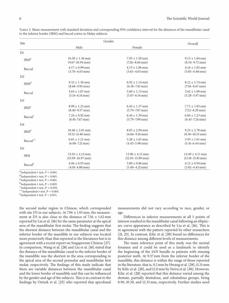
Table 2: Mean measurement with standard deviation and corresponding 95% confidence interval for the distances of the mandibular canalto the inferior border (IBM) and buccal cortex in Malay subjects.
Site GenderOverall
Male FemaleD1
IBM𝛼 10.30 ± 1.36mm(9.67–10.94mm)
7.95 ± 1.50mm(7.26–8.66mm)
9.13 ± 1.84mm(8.54–9.72mm)
Buccal 4.17 ± 0.99mm(3.70–4.63mm)
4.15 ± 1.06mm(3.65–4.65mm)
4.16 ± 1.02mm(3.83–4.48mm)
D2
IBM𝛽 9.32 ± 1.36mm(8.68–9.95mm)
6.92 ± 1.14mm(6.38–7.45mm)
8.12 ± 1.74mm(7.56–8.67mm)
Buccal 5.64 ± 1.07mm(5.14–6.14mm)
5.60 ± 1.13mm(5.07–6.14mm)
5.62 ± 1.09mm(5.28–5.97mm)
D3
IBM𝛾 8.99 ± 1.25mm(8.40–9.57mm)
6.43 ± 1.37mm(5.79–7.07mm)
7.71 ± 1.83mm(7.12–8.29mm)
Buccal∗ 7.24 ± 0.92mm(6.81–7.67mm)
6.44 ± 1.39mm(5.79–7.09mm)
6.84 ± 1.23mm(6.45–7.24mm)
D4
IBM𝛿 10.46 ± 2.01mm(9.52–11.40mm)
8.03 ± 2.94mm(6.66–9.41mm)
9.25 ± 2.78mm(8.36–10.13mm)
Buccal∗∗ 6.65 ± 1.21mm(6.08–7.21mm)
5.20 ± 1.65mm(4.43–5.98mm)
5.93 ± 1.61mm(5.41–6.44mm)
D5
IBM 15.03 ± 4.14mm(13.09–16.97mm)
13.96 ± 4.12mm(12.03–15.89mm)
14.49 ± 4.11mm(13.18–15.81mm)
Buccal§ 4.44 ± 0.93mm(4.01–4.88mm)
3.80 ± 0.86mm(3.40–4.21mm)
4.12 ± 0.94mm(3.82–4.43mm)
𝛼Independent 𝑡-test, 𝑃 < 0.001.𝛽Independent 𝑡-test, 𝑃 < 0.001.𝛾Independent 𝑡-test, 𝑃 < 0.001.𝛿Independent 𝑡-test, 𝑃 = 0.005.∗Independent 𝑡-test, 𝑃 = 0.039.∗∗Independent 𝑡-test, 𝑃 = 0.003.§Independent 𝑡-test, 𝑃 = 0.031.
the second molar region in Chinese, which correspondedwith site D3 in our subjects. At 7.96 ± 1.93mm, the measure-ment at D3 is also close to the distance of 7.56 ± 1.62mmreported by Liu et al. [16] for the same parameter at the apicalarea of the mandibular first molar. The finding suggests thatthe shortest distance between the mandibular canal and theinferior border of the mandible in our subjects was locatedmore posteriorly than that reported in the literatures but is inagreement with a recent report on Singaporean Chinese [27].In comparison, Wang et al. [28] and Liu et al. [16] stated thatthe distance of the mandibular canal to the inferior border ofthe mandible was the shortest in the area corresponding tothe apical area of the second premolar and mandibular firstmolar, respectively. The findings of this study indicate thatthere are variable distances between the mandibular canaland the lower border of mandible and this can be influencedby the gender and age of the subjects.This is in contrast to thefindings by Ozturk et al. [25] who reported that apicobasal
measurements did not vary according to race, gender, orage.
Differences in inferior measurements at all 5 points ofinterest resulted in themandibular canal following an elliptic-arc curve appearance as described by Liu et al. [16]. This isin agreement with the pattern reported by other researchers[21, 23]. In contrast, Kilic et al. [20] found no differences forthis distance among different levels of measurements.
The main reference point of this study was the mentalforamen and it could be used as a landmark to identifythe beginning of the IAN bundle in patients with missingposterior teeth. At 9.37mm from the inferior border of themandible, this distance is within the range of those reportedin the literature, that is, 9.2mmbyHwang et al. [29], 11.51mmby Kilic et al. [20], and 12.0mm by Neiva et al. [30]. However,Kilic et al. [20] reported that this distance varied among thedentate, partially dentulous, and edentulous groups, being8.90, 10.30, and 12.33mm, respectively. Further studies need
The Scientific World Journal 7
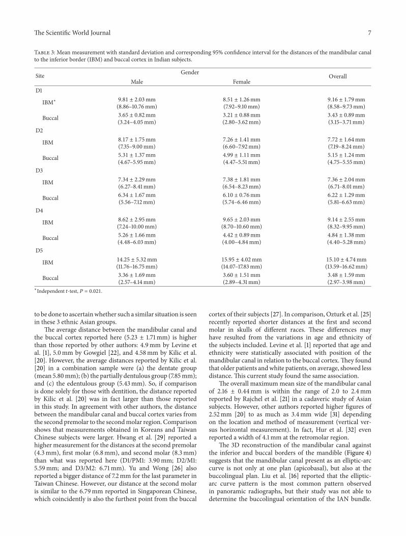
Table 3: Mean measurement with standard deviation and corresponding 95% confidence interval for the distances of the mandibular canalto the inferior border (IBM) and buccal cortex in Indian subjects.
Site Gender OverallMale Female
D1
IBM∗ 9.81 ± 2.03mm(8.86–10.76mm)
8.51 ± 1.26mm(7.92–9.10mm)
9.16 ± 1.79mm(8.58–9.73mm)
Buccal 3.65 ± 0.82mm(3.24–4.05mm)
3.21 ± 0.88mm(2.80–3.62mm)
3.43 ± 0.89mm(3.15–3.71mm)
D2
IBM 8.17 ± 1.75mm(7.35–9.00mm)
7.26 ± 1.41mm(6.60–7.92mm)
7.72 ± 1.64mm(7.19–8.24mm)
Buccal 5.31 ± 1.37mm(4.67–5.95mm)
4.99 ± 1.11mm(4.47–5.51mm)
5.15 ± 1.24mm(4.75–5.55mm)
D3
IBM 7.34 ± 2.29mm(6.27–8.41mm)
7.38 ± 1.81mm(6.54–8.23mm)
7.36 ± 2.04mm(6.71–8.01mm)
Buccal 6.34 ± 1.67mm(5.56–7.12mm)
6.10 ± 0.76mm(5.74–6.46mm)
6.22 ± 1.29mm(5.81–6.63mm)
D4
IBM 8.62 ± 2.95mm(7.24–10.00mm)
9.65 ± 2.03mm(8.70–10.60mm)
9.14 ± 2.55mm(8.32–9.95mm)
Buccal 5.26 ± 1.66mm(4.48–6.03mm)
4.42 ± 0.89mm(4.00–4.84mm)
4.84 ± 1.38mm(4.40–5.28mm)
D5
IBM 14.25 ± 5.32mm(11.76–16.75mm)
15.95 ± 4.02mm(14.07–17.83mm)
15.10 ± 4.74mm(13.59–16.62mm)
Buccal 3.36 ± 1.69mm(2.57–4.14mm)
3.60 ± 1.51mm(2.89–4.31mm)
3.48 ± 1.59mm(2.97–3.98mm)
∗Independent 𝑡-test, 𝑃 = 0.021.
to be done to ascertainwhether such a similar situation is seenin these 3 ethnic Asian groups.
The average distance between the mandibular canal andthe buccal cortex reported here (5.23 ± 1.71mm) is higherthan those reported by other authors: 4.9mm by Levine etal. [1], 5.0mm by Gowgiel [22], and 4.58mm by Kilic et al.[20]. However, the average distances reported by Kilic et al.[20] in a combination sample were (a) the dentate group(mean 5.80mm); (b) the partially dentulous group (7.85mm);and (c) the edentulous group (5.43mm). So, if comparisonis done solely for those with dentition, the distance reportedby Kilic et al. [20] was in fact larger than those reportedin this study. In agreement with other authors, the distancebetween the mandibular canal and buccal cortex varies fromthe second premolar to the secondmolar region. Comparisonshows that measurements obtained in Koreans and TaiwanChinese subjects were larger. Hwang et al. [29] reported ahigher measurement for the distances at the second premolar(4.3mm), first molar (6.8mm), and second molar (8.3mm)than what was reported here (D1/PM1: 3.90mm; D2/M1:5.59mm; and D3/M2: 6.71mm). Yu and Wong [26] alsoreported a bigger distance of 7.2mm for the last parameter inTaiwan Chinese. However, our distance at the second molaris similar to the 6.79mm reported in Singaporean Chinese,which coincidently is also the furthest point from the buccal
cortex of their subjects [27]. In comparison, Ozturk et al. [25]recently reported shorter distances at the first and secondmolar in skulls of different races. These differences mayhave resulted from the variations in age and ethnicity ofthe subjects included. Levine et al. [1] reported that age andethnicity were statistically associated with position of themandibular canal in relation to the buccal cortex.They foundthat older patients andwhite patients, on average, showed lessdistance. This current study found the same association.
The overall maximummean size of the mandibular canalof 2.16 ± 0.44mm is within the range of 2.0 to 2.4mmreported by Rajchel et al. [21] in a cadaveric study of Asiansubjects. However, other authors reported higher figures of2.52mm [20] to as much as 3.4mm wide [31] dependingon the location and method of measurement (vertical ver-sus horizontal measurement). In fact, Hur et al. [32] evenreported a width of 4.1mm at the retromolar region.
The 3D reconstruction of the mandibular canal againstthe inferior and buccal borders of the mandible (Figure 4)suggests that the mandibular canal present as an elliptic-arccurve is not only at one plan (apicobasal), but also at thebuccolingual plan. Liu et al. [16] reported that the elliptic-arc curve pattern is the most common pattern observedin panoramic radiographs, but their study was not able todetermine the buccolingual orientation of the IAN bundle.

8 The Scientific World Journal
Table 4: Mean measurement with standard deviation and corresponding 95% confidence interval for the distances of the mandibular canalto the inferior border (IBM) and buccal cortex in Chinese subjects.
Site Gender OverallMale Female
D1
IBM 10.18 ± 1.56mm(9.45–10.91mm)
9.47 ± 1.02mm(8.99–9.95mm)
9.83 ± 1.35mm(9.40–10.26mm)
Buccal 4.35 ± 1.11mm(3.83–4.87mm)
3.85 ± 0.74mm(3.50–4.19mm)
4.10 ± 0.97mm(3.79–4.41mm)
D2
IBM∗ 9.40 ± 1.54mm(8.68–10.13mm)
8.40 ± 1.34mm(7.77–9.03mm)
8.90 ± 1.52mm(8.42–9.39mm)
Buccal 6.06 ± 1.13mm(5.54–6.59mm)
5.96 ± 1.17mm(5.41–6.50mm)
6.01 ± 1.13mm(5.65–6.37mm)
D3
IBM 9.08 ± 1.67mm(8.30–9.86mm)
8.56 ± 1.64mm(7.79–9.33mm)
8.82 ± 1.65mm(8.29–9.35mm)
Buccal 7.09 ± 1.53mm(6.37–7.80mm)
7.06 ± 1.26mm(6.47–7.64mm)
7.07 ± 1.38mm(6.63–7.51mm)
D4
IBM 10.50 ± 1.94mm(9.59–11.41mm)
10.69 ± 2.20mm(9.66–11.72mm)
10.60 ± 2.05mm(9.94–11.25mm)
Buccal 6.37 ± 1.90mm(5.48–7.26mm)
6.23 ± 1.20
(5.67–6.80)6.30 ± 1.57mm(5.80–6.80mm)
D5
IBM 15.81 ± 3.10mm(14.35–17.26mm)
16.30 ± 4.04mm(14.41–18.20mm)
16.06 ± 3.57mm(14.92–17.20mm)
Buccal∗∗ 4.44 ± 1.51mm(3.73–5.15mm)
5.85 ± 1.64mm(5.08–6.61mm)
5.14 ± 1.71mm(4.60–5.69mm)
∗Independent 𝑡-test, 𝑃 = 0.035.∗∗Independent 𝑡-test, 𝑃 = 0.007.
Table 5: Mean measurement with standard deviation and 95%confidence interval for the diameter of the mandibular canal.
Site Gender OverallMale Female
D1∗ 2.38 ± 0.39mm(2.28–2.48mm)
2.14 ± 0.53mm(2.00–2.28mm)
2.26 ± 0.48mm(2.17–2.35mm)
D2∗∗ 2.20 ± 0.44mm(2.09 ± 2.32mm)
1.82 ± 0.30mm(1.74–1.90mm)
2.01 ± 0.42mm(1.93–2.09mm)
D3§ 2.25 ± 0.34mm(2.16–2.33mm)
1.96 ± 0.35mm(1.87–2.05mm)
2.10 ± 0.37mm(2.04–2.17mm)
D4¶ 2.36 ± 0.41mm(2.26–2.47mm)
2.00 ± 0.35mm(1.91–2.09mm)
2.18 ± 0.42mm(2.11–2.26mm)
D5𝛾 2.34 ± 0.50mm(2.22–2.47mm)
2.17 ± 0.33mm(2.08–2.25mm)
2.25 ± 0.43mm(2.18–2.33mm)
∗Independent 𝑡-test,𝑃 = 0.005.∗∗Independent 𝑡-test,𝑃 < 0.001.§Independent 𝑡-test,𝑃 < 0.001.¶Independent 𝑡-test,𝑃 < 0.001.𝛾 Independent 𝑡-test,𝑃 = 0.024.
Kim et al. [15] reported that the buccolingual orientationof the mandibular canal followed either of these 3 differentpatterns.
Type 1 (70%): the canal follows the lingual corticalplate at the mandibular ramus and body.Type 2 (15%): the canal follows the middle of theramus behind the second molar and the lingual platepassing through the second and first molars.Type 3 (15%): the canal follows the middle or thelingual one-third of the mandible from the ramus tothe body.
Hwang et al. [29] found themandibular canal to be nearerto the lingual side in the posterior two-third of the mandible,but nearer to the buccal side in the anterior one-third. Sucha finding may possibly be observed for the mental regionand anterior part of the body of the mandible in this study.However, caution must be expressed as the buccal cortex ofthe mandible is not a flat surface; hence, in reality, the longerdistance between the MC and the buccal cortex is a resultof an outward curvature over the body of the mandible, asshown in Figure 6.
Two inferior alveolar nerve repositioning techniques havebeen described, namely, lateralization and fenestration [7].In lateralization, drilling is done around the mental foramento obtain a ring of external cortical bone. An extensionof about 5mm is made anteriorly to the mental foramen

The Scientific World Journal 9
5.59mm6.71mm
5.69mm4.25mm
D1
D2
D3
D4
D5
3.39mm
Figure 6: Superimposition of the findings of this study over a curvedbody of mandible displays differences in distance between variouspoints of the MC to the buccal cortex.
to avoid damaging the anterior loop and another posteriorwindow is performed along the intrabony trajectory of thenerve. The incisive nerve that is located about 5mm fromthe mental foramen needs to be sectioned in order tosecure complete mobilization of the alveolar nerve [6]. Inthe case of fenestration, sectioning of the incisive nerve isnot performed. This approach only involves the preparationof a cortical bone window located posterior to the mentalforamen. While both techniques described the approach thatcan be used to reposition the IAN bundle, recent journalreports do not describe the anatomical landmarks that canbe used for guidance.
So, in order to perform IAN repositioning in Asians, it issuggested that the surgeons first locate the mental foramenbased on available radiographs. The most common locationfor the mental foramen in some Asian ethnic groups suchas Malay, Indians, and Chinese has been reported to be atapical to the second premolar [33–35]. An osteotomy shouldbe done with a margin of about 2mm around the mentalforamen to free the nerve. This can be done by using aLindemann or stainless steel surgical bur with a diameterof 1.5 to 2mm [1] to a depth that is similar to the entirediameter of the bur.There should not be any worry of causingiatrogenic injury to the IAN bundle beneath as the meandistance from the buccal margin to the canal is almost 4mm.Adopting results of this study, this inferior cut should be done7mm from the inferior border of the mandible.
Following this, position D3, which is 20mm distal to themental foramen, is identified using a metal ruler/caliper.Thisis the site where the mandibular canal presented with theshortest distance (7.96 ± 1.93mm) to the lower border ofmandible and concurs to the region of the second molar. Around surgical stainless steel bur can be used to make thissecond osteotomy mark, which should be about 6mm fromthe inferior border of the mandible. This distance will avoid
cutting into the lower cortical plate that has been reported torange between 3.2 and 3.5mm in thickness in Asian subjects[36]. Later on, position D2 is located and an osteotomymark is made about 6mm from the lower border of themandible. Subsequent markings at position D4 can be madealso at 9mm from the lower border of mandible, if necessary.Chinese subjects seemed to have a mandibular canal that islocated further from the inferior border of mandible thanMalay or Indian subjects, so this fact needs to be given dueconsideration during the making of osteotomy marks. Thiscan be done by confirming radiographic findings prior to IANbundle repositioning.
The depth of drilling for making osteotomymarks shouldbe between 2.5 and 3.0mm as studies have shown that theseare the thicknesses of buccal plates of Asians at the molarregions [36]. One is unlikely to injure the IAN at these depthsas the current results show that the mandibular canal islocated between 3.90 and 6.71mm from the buccal cortex.
Considering the fact that themaximummean diameter ofthe mandibular canal is 2.25mm, corresponding osteotomymarks on the superiormargin should bemade 6mm superiorto the inferior osteotomy marks. These osteotomy marks canthen be joined using an ultrasound bone surgical device asit has proven to spare injuring nerves, in case variationshappen with the location of the IAN bundle [37]. At thissize, this osteotomy window should be small enough to notweaken the mandible, but adequate to facilitate bone repair.This also ensures that there is adequate bone tissue abovethe canal so that the primary stability needed for implantinsertion is not compromised [38]. If lateralization of the IANbundle is necessary, further osteotomy ismade into the buccalcortex at 5mm mesial to the mental foramen, following therecommendation by Morrison et al. [6].
This study found that the IAN follows an elliptic-curve arccourse in relation to the curved body of the mandible, so oneneeds to ensure that more cancellous bone is removed at D3(i.e., 20mm or around the region of the mandibular secondmolar) in order to reach the IAN. The findings of this studysuggested that there are some ethnic features that influencethe horizontal location of the mandibular canal; hence, theyare of importance in clinical practice. For example, the IANof Chinese subjects is located further from the buccal cortexthan Indian subjects. Therefore, more bone needs to beremoved for Chinese subjects than Indian or Malay subjects.Further studies with larger sample size are needed to verifythese suggestions.
5. Conclusion
Themandibular canal was visible in all (100%) CBCTs, that is,120 sites. The ease of detection of the mandibular canal usingCBCT and SimPlant dental software indicates the potentiallyhigh preoperative value of CBCT scan for the purpose ofpreoperative planning. Apicobasal assessment of the canalreveals that it was curving downward towards the inferiormandibular border until 20mm distal to the mental foramen(D3) and then reverts upwards, making an elliptic-arc curve.The mandibular canal also forms an elliptic arc in relation

10 The Scientific World Journal
to the curved body of mandible, with the furthest buccalpoint located atD3.While acknowledging that there is humanvariability, this study provides an accurate anatomic locationof the mandibular canal, which in return helps to determinea safe zone to access the IAN bundle. This hopefully willbecome a useful guide in centers whereCBCT is not available.When such a facility is available, it is recommended thatclinicians make use of it to overcome the shortfalls observedin conventional radiography.
Ethical Approval
This project has been approved by the Medical Ethics Com-mittee of the Faculty of Dentistry, University of Malaya.(Ethical approval: DF DP1303/0014 [P].)
Conflict of Interests
The authors declare that there is no conflict of interestsregarding the publication of this paper.
Acknowledgments
The authors would like to express their gratitude to Mrs.Thamilarasi A/P Thanaperumal for preparing the excellentillustrations used in this paper.This project was supported bya High Impact Research Grant provided by the Ministry ofHigher EducationMalaysia (UM.C/HIR/MOHE/DENT/19).
References
[1] M. H. Levine, A. L. Goddard, and T. B. Dodson, “Inferioralveolar nerve canal position: a clinical and radiographic study,”Journal of Oral andMaxillofacial Surgery, vol. 65, no. 3, pp. 470–474, 2007.
[2] W. C. Ngeow, “Is there a “safety zone” in the mandibularpremolar region where damage to the mental nerve can beavoided if periapecal extrusion occurs?” Journal of the CanadianDental Association, vol. 76, no. 1, article a61, 2010.
[3] M. A. Pogrel and S. Thamby, “The etiology of altered sensationin the inferior alveolar, lingual, and mental nerves as a result ofdental treatment,” Journal of the California Dental Association,vol. 27, no. 7, pp. 531–538, 1999.
[4] C. C. Alling III, “Dysesthesia of the lingual and inferior alveolarnerves following third molar surgery,” Journal of Oral andMaxillofacial Surgery, vol. 44, no. 6, pp. 454–457, 1986.
[5] T. Renton, “Prevention of iatrogenic inferior alveolar nerveinjuries in relation to dental procedures,”Dental Update, vol. 37,no. 6, pp. 350–360, 2010.
[6] A. Morrison, M. Chiarot, and S. Kirby, “Mental nerve functionafter inferior alveolar nerve transposition for placement ofdental implants,” Journal (CanadianDental Association), vol. 68,no. 1, pp. 46–50, 2002.
[7] J. L. Del-Castillo-Pardo-de-Vera, M. Chamorro-Pons, and J.L. Cebrian-Carretero, “Repositioning of the inferior alveolarnerve in cases of severe mandibular atrophy. A clinical case,”Medicina Oral, Patologia Oral y Cirugia Bucal, vol. 13, no. 12,pp. E778–E782, 2008.
[8] O. Jensen andD. Nock, “Inferior alveolar nerve repositioning inconjunction with placement of osseointegrated implants: a case
report,”Oral Surgery, Oral Medicine, Oral Pathology, vol. 63, no.3, pp. 263–268, 1987.
[9] P. J. Louis, “Inferior alveolar nerve repositioning,” Atlas of theOral and Maxillofacial Surgery Clinics of North America, vol. 9,no. 2, pp. 93–128, 2001.
[10] J. Y. K. Kan, J. L. Lozada, P. J. Boyne, C. J. Goodacre, andK. Rungcharassaeng, “Mandibular fracture after endosseousimplant placement in conjunction with inferior alveolar nervetransposition: a patient treatment report,” International Journalof Oral and Maxillofacial Implants, vol. 12, no. 5, pp. 655–659,1997.
[11] P. F. Nocini, D. De Santis, E. Fracasso, and G. Zanette, “Clin-ical and electrophysiological assessment of inferior alveolarnerve function after lateral nerve transposition,” Clinical OralImplants Research, vol. 10, no. 2, pp. 120–130, 1999.
[12] H. M. Hashemi, “Neurosensory function following mandibularnerve lateralization for placement of implants,” InternationalJournal of Oral andMaxillofacial Surgery, vol. 39, no. 5, pp. 452–456, 2010.
[13] J.-H. Pyun, Y.-J. Lim, M.-J. Kim, S.-J. Ahn, and J. Kim,“Position of the mental foramen on panoramic radiographsand its relation to the horizontal course of the mandibularcanal: a computed tomographic analysis,”Clinical Oral ImplantsResearch, vol. 24, no. 8, pp. 890–895, 2013.
[14] S. Lofthag-Hansen, K. Grondahl, and A. Ekestubbe, “Cone-beam CT for preoperative implant planning in the posteriormandible: visibility of anatomic landmarks,” Clinical ImplantDentistry and Related Research, vol. 11, no. 3, pp. 246–255, 2009.
[15] S. T. Kim, K.-S. Hu, W.-C. Song, M.-K. Kang, H.-D. Park, andH.-J. Kim, “Location of the mandibular canal and the topog-raphy of its neurovascular structures,” Journal of CraniofacialSurgery, vol. 20, no. 3, pp. 936–939, 2009.
[16] T. Liu, B. Xia, and Z. Gu, “Inferior alveolar canal course: aradiographic study,” Clinical Oral Implants Research, vol. 20, no.11, pp. 1212–1218, 2009.
[17] K. Kamburoglu, E. Kolsuz, H. Kurt, C. Kılıc, T. Ozen, and C. S.Paksoy, “Accuracy of CBCT measurements of a human skull,”Journal of Digital Imaging, vol. 24, no. 5, pp. 787–793, 2011.
[18] K. Maloney, J. Bastidas, K. Freeman, T. R. Olson, and R.A. Kraut, “Cone beam computed tomography and simplantmaterialize dental software versus direct measurement of thewidth and height of the posterior mandible: an anatomic study,”Journal of Oral andMaxillofacial Surgery, vol. 69, no. 7, pp. 1923–1929, 2011.
[19] C. W. Ulm, P. Solar, R. Blahout, M. Matejka, G. Watzek, and H.Gruber, “Location of the mandibular canal within the atrophicmandible,” British Journal of Oral andMaxillofacial Surgery, vol.31, no. 6, pp. 370–375, 1993.
[20] C. Kilic, K. Kamburoglu, T. Ozen et al., “The position of themandibular canal and histologic feature of the inferior alveolarnerve,” Clinical Anatomy, vol. 23, no. 1, pp. 34–42, 2010.
[21] J. Rajchel, E. Ellis III, andR. J. Fonseca, “The anatomical locationof the mandibular canal: its relationship to the sagittal ramusosteotomy,”The International Journal of Adult Orthodontics andOrthognathic Surgery, vol. 1, no. 1, pp. 37–47, 1986.
[22] J. M. Gowgiel, “The position and course of the mandibularcanal,”The Journal of Oral Implantology, vol. 18, no. 4, pp. 383–385, 1992.
[23] L. C. Anderson, T. F. Kosinski, and P. J. Mentag, “A review of theintraosseous course of the nerves of the mandible,”The Journalof Oral Implantology, vol. 17, no. 4, pp. 394–403, 1991.

The Scientific World Journal 11
[24] P. Worthington, “Injury to the inferior alveolar nerve duringimplant placement: a formula for protection of the patientand clinician,” International Journal of Oral and MaxillofacialImplants, vol. 19, no. 5, pp. 731–734, 2004.
[25] A. Ozturk, A. Potluri, and A. R. Vieira, “Position and courseof the mandibular canal in skulls,” Oral Surgery, Oral Medicine,Oral Pathology, Oral Radiology and Endodontology, vol. 113, no.4, pp. 453–458, 2012.
[26] I. H. Yu and Y. K. Wong, “Evaluation of mandibular anatomyrelated to sagittal split ramus osteotomy using 3-dimensionalcomputed tomography scan images,” International Journal ofOral and Maxillofacial Surgery, vol. 37, no. 6, pp. 521–528, 2008.
[27] R. Nagadia, A. B. G. Tay, L. L. Chan, and E. S.-Y. Chan, “Thespatial location of themandibular canal in Chinese: a CT study,”International Journal of Oral and Maxillofacial Surgery, vol. 40,no. 12, pp. 1401–1405, 2011.
[28] J.-C. Wang, L. Gui, Z.-Y. Zhang, F. Niu, W. Liu, and X.-J. Tang,“The measurement of the mandibular canal’s location in themandibular body of the young women,” Zhonghua Zheng XingWai Ke Za Zhi, vol. 23, no. 3, pp. 212–214, 2007.
[29] K. Hwang,W. J. Lee, Y. B. Song, and I. H. Chung, “Vulnerabilityof the inferior alveolar nerve and mental nerve during genio-plasty: an anatomic study,” Journal of Craniofacial Surgery, vol.16, no. 1, pp. 10–14, 2005.
[30] R. F. Neiva, R. Gapski, andH.-L.Wang, “Morphometric analysisof implant-related anatomy in Caucasian skulls,” Journal ofPeriodontology, vol. 75, no. 8, pp. 1061–1067, 2004.
[31] K. Ikeda, K.-C. Ho, B. H. Nowicki, and V. M. Haughton,“Multiplanar MR and anatomic study of the mandibular canal,”The American Journal of Neuroradiology, vol. 17, no. 3, pp. 579–584, 1996.
[32] M. S. Hur, H. C. Kim, S. Y. Won et al., “Topography and spatialfascicular arrangement of the human inferior alveolar nerve,”Clinical Implant Dentistry and Related Research, vol. 15, no. 1,pp. 88–95.
[33] R. M. Green, “The position of the mental foramen: a compar-ison between the southern (Hong Kong) Chinese and otherethnic and racial groups,” Oral Surgery, Oral Medicine, OralPathology, vol. 63, no. 3, pp. 287–290, 1987.
[34] J. Neo, “The position of the mental foramen in SingaporeanMalays and Indians,”Anesthesia Progress, vol. 36, no. 6, pp. 276–278, 1989.
[35] W. C. Ngeow and Y. Yuzawati, “The location of the mentalforamen in a selectedMalay population,” Journal ofOral Science,vol. 45, no. 3, pp. 171–175, 2003.
[36] H.-J. Kim, H.-Y. Lee, I.-H. Chung, I.-H. Cha, and C.-K. Yi,“Mandibular anatomy related to sagittal split ramus osteotomyinKoreans,”YonseiMedical Journal, vol. 38, no. 1, pp. 19–25, 1997.
[37] M. Bovi, A. Manni, L. Mavriqi, G. Bianco, and R. Celletti,“The use of piezosurgery to mobilize the mandibular alveolarnerve followed immediately by implant insertion: a case seriesevaluating neurosensory disturbance,”The International Journalof Periodontics & Restorative Dentistry, vol. 30, no. 1, pp. 73–81,2010.
[38] B. Rosenquist, “Implant placement in combination with nervetranspositioning: experiences with the first 100 cases,” Interna-tional Journal of Oral & Maxillofacial Implants, vol. 9, pp. 522–531, 1994.

Submit your manuscripts athttp://www.hindawi.com
Hindawi Publishing Corporationhttp://www.hindawi.com Volume 2014
Oral OncologyJournal of
DentistryInternational Journal of
Hindawi Publishing Corporationhttp://www.hindawi.com Volume 2014
Hindawi Publishing Corporationhttp://www.hindawi.com Volume 2014
International Journal of
Biomaterials
Hindawi Publishing Corporationhttp://www.hindawi.com Volume 2014
BioMed Research International
Hindawi Publishing Corporationhttp://www.hindawi.com Volume 2014
Case Reports in Dentistry
Hindawi Publishing Corporationhttp://www.hindawi.com Volume 2014
Oral ImplantsJournal of
Hindawi Publishing Corporationhttp://www.hindawi.com Volume 2014
Anesthesiology Research and Practice
Hindawi Publishing Corporationhttp://www.hindawi.com Volume 2014
Radiology Research and Practice
Environmental and Public Health
Journal of
Hindawi Publishing Corporationhttp://www.hindawi.com Volume 2014
The Scientific World JournalHindawi Publishing Corporation http://www.hindawi.com Volume 2014
Hindawi Publishing Corporationhttp://www.hindawi.com Volume 2014
Dental SurgeryJournal of
Drug DeliveryJournal of
Hindawi Publishing Corporationhttp://www.hindawi.com Volume 2014
Hindawi Publishing Corporationhttp://www.hindawi.com Volume 2014
Oral DiseasesJournal of
Hindawi Publishing Corporationhttp://www.hindawi.com Volume 2014
Computational and Mathematical Methods in Medicine
ScientificaHindawi Publishing Corporationhttp://www.hindawi.com Volume 2014
PainResearch and TreatmentHindawi Publishing Corporationhttp://www.hindawi.com Volume 2014
Preventive MedicineAdvances in
Hindawi Publishing Corporationhttp://www.hindawi.com Volume 2014
EndocrinologyInternational Journal of
Hindawi Publishing Corporationhttp://www.hindawi.com Volume 2014
Hindawi Publishing Corporationhttp://www.hindawi.com Volume 2014
OrthopedicsAdvances in