Embed Size (px)
Citation preview

ECG Recognition of
Myocardial Ischemia &
Infarction
J. Lee Garvey, MD
Department of Emergency Medicine Carolinas Medical Center

Objectives
To detect myocardial ischemia & infarction
on an electrocardiogram
To define the areas of the heart to which the
twelve standard ECG leads correspond
To correlate coronary anatomy with areas of
ischemia & infarction

Acute Myocardial Injury

Acute Myocardial Injury
Acute Myocardial Injury

EKG Basics
The
electrocardiogram
(ECG): the
electrical activity of
the heart recorded
at the body surface

ECG Basics
Anatomy of the heart: positioning in chest

Coronary Anatomy

Coronary Anatomy
There are two
coronary arteries
which supply the
heart with blood

Coronary Anatomy
LCA

Coronary Anatomy
The LEFT coronary artery has 2 major
branches:
– Left Anterior Descending (LAD)- supplies
Anterior wall of the ventricles
& septum
– Circumflex branch- supplies
Lateral wall of the left ventricle
& atrium

Coronary Anatomy
RCA

Coronary Anatomy RIGHT coronary artery (RCA)
The RCA supplies:
Right atrium
SA & AV nodes
Posterior regions of ventricles

Coronary Anatomy
RCA

EKG Basics

EKG Basics
The EKG – essentially a voltmeter.
Measures voltage - electrical potential -between two points.
Records this voltage over time.

EKG Basics
The EKG – 12 voltmeters.
Upward deflections move towards the (+) electrode.
Downward deflections move toward the (-) electrode.

EKG Basics
Chest Leads
Exploring leads
(V1 – V6) are (+)
Reference lead (-)
is Wilson’s Central
Terminus

EKG Basics
The EKG:
electrical
activity of atria
and ventricles
Depolarization
and
repolarization
P
QRS
T

EKG Basics
The EKG:
Standardized grid
– small box 40 mSec
100 uV
– Large box
200 mSec
500 uV

EKG - Leads and
Electrode Positioning
The standard EKG is composed of 12 Leads
Six limb leads: I, II, III, aVR, aVL, aVF
Six chest leads: V1, V2, V3, V4, V5, V6

EKG - Leads and
Electrode Positioning
For a STANDARD RESTING 12 LEAD
Extremity leads placed:
Beyond the tip of the clavicles (arm leads)
Beyond the inguinal ligament (leg leads)
Monitoring lead placement – more centrally
on torso (Mason- Likar lead positions)

EKG - Leads and
Electrode Positioning
Chest Leads: V1 – V6
Palpate chest to locate
landmarks
Small lead position
changes can lead to
changes in interpretation.

EKG - Leads and
Electrode Positioning
Chest Leads: V1 – V6
V1 – 4th IC space, R of sternum
V2 – 4th IC space, L of sternum
V3 – between V2 and V4
V4 – 5th IC space, Mid clav line
V5- Lat to V4, Anterior Ax line
V6 – Lat to V4 and V5, Mid Ax

EKG Basics
On a standard EKG mounting, the six
chest leads and six limb leads are typically
arrayed in columns:
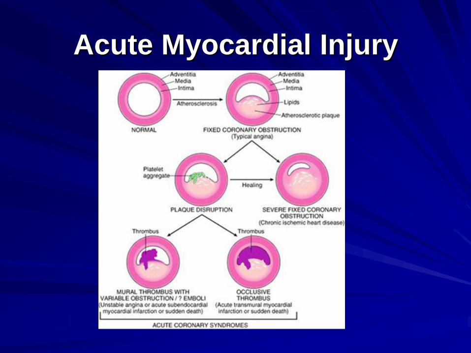
Localization of MI
Area of Infarction
Anterior wall
*Anteroseptal
Lateral wall
Inferior wall
Right ventricle
Posterior wall
Leads Involved
V1, V2, V3, V4
V1, V2
I, AVL, V5, V6
II, III, AVF
V4 R
V7, V8, V9 +
Tall R & ST V1, V2

Limb Leads
To obtain the 6 limb leads, electrodes are
placed on the right arm, the left arm & the
left leg forming a triangle

Bipolar Limb Leads
Leads I, II, III are formed by a pair of electrodes
Each records from a different perspective: going away from the (-), and lead toward (+) lead

Bipolar Limb Leads
Leads I, II, III are formed by a pair of
electrodes

Bipolar Limb Leads
Leads I, II, III are formed by a pair of
electrodes

“Augmented” limb leads
Are unipolar limb leads, stressing the
importance of the (+) electrode
AVR- Right arm positive
AVL- Left arm positive
AVF- Foot (left) positive

“Augmented Limb leads”

Frontal Plane leads

Limb Leads
Leads I and AVL view
the: high lateral wall
of the heart
Leads II, III & AVF view the: inferior wall of the heart

Limb Leads
Lead AVR looks “away” from the heart
Therefore the “P”, “QRS” & “T waves”
should be inverted
If they are upright in AVR, then the
electrodes are likely misplaced.

ECG - Chest Leads
Chest Leads: V1 – V6
Each lead gives a
different perspective
of the heart… sees
the electrical activity
from a slightly
different view.

Chest leads

Chest Leads
The ECG tracing from V1-V6 shows
gradual changes in all the waves as the
position of each lead changes

Right sided chest lead: V4 R
Looks at right ventricle
5th ICS, Rt. midclavicular line

Left posterior leads: V7, V8, V9
Look at the posterior wall
V7- 5th ICS, post axillary line
V8- 5th ICS, midscapular line
V9- 5th ICS, 2cm left of vert column

Myocardial Ischemia & Infarction

ECG: Ischemia / Injury
Identify the most SEVERE abnormality –
this is the ‘name’ injury:
eg: Anterior STEMI
Look for ‘RECIPROCAL’ findings –
typically ST depression or T wave
inversion in the setting of ST elevation.

Myocardial Infarction Problems with diagnosis
History: symptoms & signs often vague
Enzyme markers: take time to detect
EKG: non-diagnostic in up to 60%
0.4 - 3% of patients are sent home with MI
& up to 25% of these die!

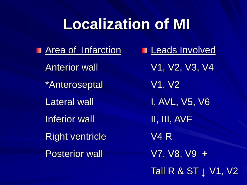
Myocardial Infarction
Acute Injury Phase
Isoelectric point: somewhere in T-P interval
Measure ST elevation : J point + 60 mSec

Myocardial Infarction Acute Injury Phase
Initially see tall peaked T waves and
ST segment elevation

Myocardial Infarction Acute Injury Phase

Myocardial Infarction Acute Injury Phase
Myocardial Infarction Acute Injury Phase

Localization of MI
Area of Infarction
Anterior wall
*Anteroseptal
Lateral wall
Inferior wall
Right ventricle
Posterior wall
Leads Involved
V1, V2, V3, V4
V1, V2
I, AVL, V5, V6
II, III, AVF
V4 R
V7, V8, V9 +
Tall R & ST V1, V2

Myocardial Infarction Acute Injury Phase

Myocardial Infarction Acute Injury Phase

Posterior wall infarction
If an Anterior wall MI is manifested by
Q waves & ST segment elevation
Then a Posterior wall MI will appear just
the opposite (R waves & ST depression)

Posterior wall infarction
In acute posterior infarctions, there is
a large R wave with ST depression in:
V1, V2 and / or V3

Myocardial Infarction Posterior wall MI
Note that the electrical activity of the
anterior and posterior wall of the LV
is in opposite directions

ST Segment Elevation
Not as easy as it sounds
– Inconsistent interpretation Interobserver and intraobserver
– Up to 14% inconsistently classified
– Many reasons for STE 29% of prehospital ECGs in CP pts have at least 100 uV of STE on 2 contiguous limb leads or 200 uV of STE on 2 contiguous precordial leads
But only 49% and 15% (limb/ precord) have AMI
Majority have LVH, LBBB, BER, or ventricular aneurysm

ST Segment Elevation
How often are we right/ wrong in initiating
reperfusion therapy?
– 11% of lytic patients did not have AMI
– 9 of 83 lytic treated pts – exposed to risk of Rx
If STE is minor, it is more difficult to
definitively call, and leads to delay in Rx
– D2Drug < 30 min: ST Segment Sum 21.5 mm
– D2Drug > 30 min: ST Segment Sum 11.5

The ST Segment
Myocardial Infarction/ Ischemia
Ventricular aneurysm
LVH
LBBB
Early repolarization/ normal variant
Acute pericarditis
Hyperkalemia
Hypothermia
Hypercalcemia
Post cardioversion

Early Repolarization

Early Repolarization
Early Repolarization
– Usually mid-precordial leads
– Elevated J point ( up to
~300 uV)
– ST usually concave
– Notching in downstroke of
QRS
– Large symmetric T waves
– Relatively fixed pattern
Early Repolarization
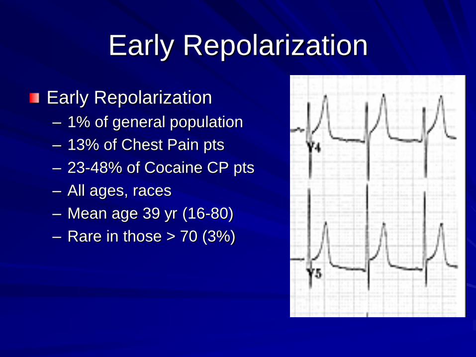
Early Repolarization
– 1% of general population
– 13% of Chest Pain pts
– 23-48% of Cocaine CP pts
– All ages, races
– Mean age 39 yr (16-80)
– Rare in those > 70 (3%)

Early Repolarization
Early Repolarization
– Limb leads involved ~ 45%
of cases
– “Isolated” BER in limb leads
is VERY RARE
Think of other causes for STE

Early Repolarization

LBBB

LBBB

LBBB + Injury

LBBB + Injury
ems12lead.blogspot.com

LBBB + Injury

LBBB + Injury

LVH

The ST Segment
Left Ventricular Hypertrophy
– A number of different ECG criteria
proposed
– Vary in sensitivity and specificity
– Easiest: Sokolow – Lyon
RaVL > 1.1 mV or
SV1 + (RV5 or RV6) > 3.5 mV
Sensitivity 10 – 35%; Specificity 85%
– Repolarization abnormalities
increase the assoc with anatomic
LVH

Standard LVH
Expected findings in LVH:
* STE discordant with QRS – panels A and B
* STD and T inversion discordant with QRS – panels C and D

LVH with STE - AMI

Practice EKG’s