Embed Size (px)
Citation preview
8
Gastric Ulcer Healing – Role of Serum Response Factor
Jianyuan Chai VA Long Beach Healthcare System, Long Beach and the
University of California, Irvine, USA
1. Introduction
Histologically, a gastric ulcer is viewed as a necrotic lesion penetrating through the entire mucosal thickness of the stomach. Because of its great similarities with ulcers in other parts of the digestive tract, gastric ulcer is often reviewed with esophageal and duodenal ulcers together as peptic ulcer disease (PUD). Although it is not as common as duodenal ulcers, gastric ulcers are more often to develop malignancy. PUD can be found in any part of the world and is probably the most common chronic infection in human population. It causes considerable loss of life year and creates a great economic burden (Figure 1). It had a tremendous effect on morbidity and mortality until the last few decades of the last century when epidemiological trends started to point to an impressive fall in its incidence, particularly in the Western countries. The reason why the rates of PUD decreased is thought to be the development of new effective medication, and of course, the discovery of the pathogen – Helicobacter pylori. It is now commonly accepted that the main cause of PUD is H. pylori, a helix-shaped Gram-negative bacterium, which infects more than 50% of world population and can be transmitted by contaminated food, groundwater, and even through human saliva (such as from kissing or sharing food utensils). For this reason, higher incidence of PUD is found in the third world countries and low socioeconomic groups. In the developed countries, on the other hand, although H. pylori infection is under controlled, thanks to the easy access to advanced treatment and better living condition, extensive use of non-steroidal anti-inflammatory drugs (NSAIDs) keeps the incidence of complicated gastric ulcer and hospitalization stable (Feinstein et al, 2010). Treatment of PUD usually involves a combination of antibiotics (e.g. metronidazole, clarithromycin, tetracycline, amoxicillin), acid suppressors (e.g. cimetidine, ranitidine, omeprazole, lansoprazole), and mucosa protectors (e.g. bismuth subsalicylate). Unfortunately, patients have to take as many as 20 pills a day and often end up with multiple side effects including nausea, vomiting, diarrhea, dizziness, and headache. Perforated ulcers require surgical repair, while bleeding ulcers have to be taken care by endoscopic cautery, injection or clipping. In any case, healing of an ulcer normally requires multiple molecular and cellular processes to achieve. This chapter will dissect molecular and cellular mechanisms of gastric ulcer healing and focus on an important molecule – Serum Response Factor (SRF) and its role in this event.
www.intechopen.com
Peptic Ulcer Disease
144
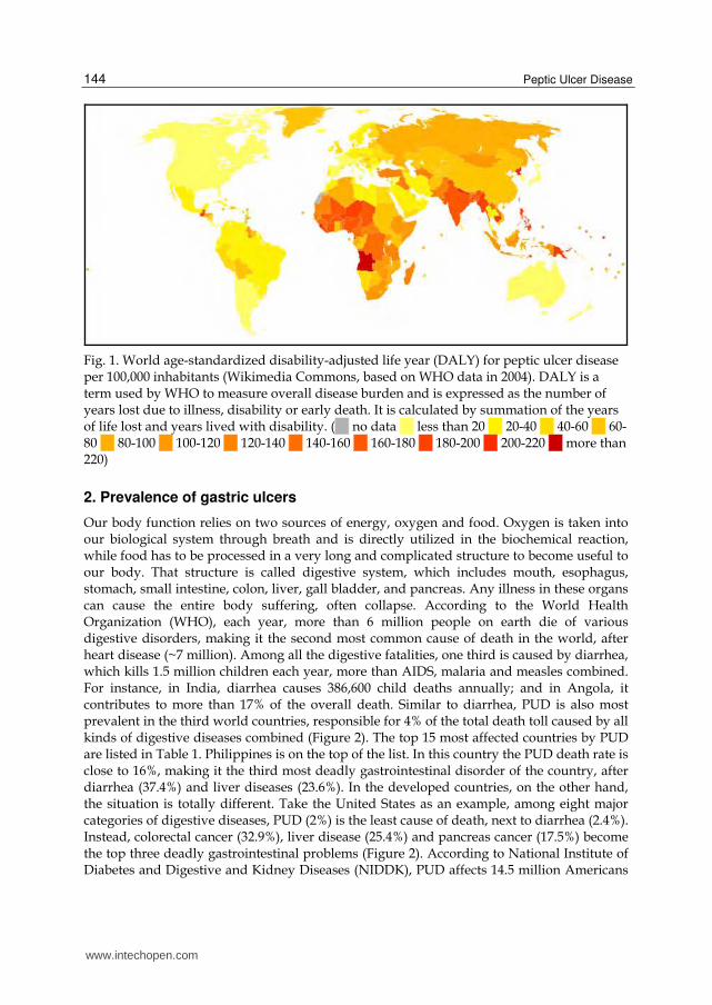
Fig. 1. World age-standardized disability-adjusted life year (DALY) for peptic ulcer disease per 100,000 inhabitants (Wikimedia Commons, based on WHO data in 2004). DALY is a term used by WHO to measure overall disease burden and is expressed as the number of years lost due to illness, disability or early death. It is calculated by summation of the years of life lost and years lived with disability. ( no data less than 20 20-40 40-60 60-80 80-100 100-120 120-140 140-160 160-180 180-200 200-220 more than 220)
2. Prevalence of gastric ulcers
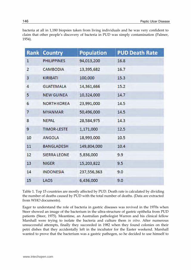
Our body function relies on two sources of energy, oxygen and food. Oxygen is taken into our biological system through breath and is directly utilized in the biochemical reaction, while food has to be processed in a very long and complicated structure to become useful to our body. That structure is called digestive system, which includes mouth, esophagus, stomach, small intestine, colon, liver, gall bladder, and pancreas. Any illness in these organs can cause the entire body suffering, often collapse. According to the World Health Organization (WHO), each year, more than 6 million people on earth die of various digestive disorders, making it the second most common cause of death in the world, after heart disease (~7 million). Among all the digestive fatalities, one third is caused by diarrhea, which kills 1.5 million children each year, more than AIDS, malaria and measles combined. For instance, in India, diarrhea causes 386,600 child deaths annually; and in Angola, it contributes to more than 17% of the overall death. Similar to diarrhea, PUD is also most prevalent in the third world countries, responsible for 4% of the total death toll caused by all kinds of digestive diseases combined (Figure 2). The top 15 most affected countries by PUD are listed in Table 1. Philippines is on the top of the list. In this country the PUD death rate is close to 16%, making it the third most deadly gastrointestinal disorder of the country, after diarrhea (37.4%) and liver diseases (23.6%). In the developed countries, on the other hand, the situation is totally different. Take the United States as an example, among eight major categories of digestive diseases, PUD (2%) is the least cause of death, next to diarrhea (2.4%). Instead, colorectal cancer (32.9%), liver disease (25.4%) and pancreas cancer (17.5%) become the top three deadly gastrointestinal problems (Figure 2). According to National Institute of Diabetes and Digestive and Kidney Diseases (NIDDK), PUD affects 14.5 million Americans
www.intechopen.com

Gastric Ulcer Healing – Role of Serum Response Factor
145
and about 350,000 new cases are diagnosed each year. Among them, duodenal ulcers are four times as many as gastric ulcers. The annual mortality is approximately 3,000.
Fig. 2. Death caused by major digestive diseases. Diarrhea is the No.1 cause of death worldwide; however, in the developed countries such as the U.S.A, it becomes the least concern. So is PUD. (Data are extracted from WHO documents).
3. Causes of gastric ulcers
For decades, the causes of gastric ulcers were believed to be spicy food, stress, and excessive acid secretion. As the German Protestant theologian Karl Schwarz said “Ohne saueren Magensaft kein peptisches Geschwür”, meaning no acid, no ulcer. Therefore, treatment options were confined to acid suppression medications and surgical operation. The successful rate of PUD treatment by acid suppressive operations was reported in the literature repeatedly. At the time, people did not believe that bacteria could survive in the human stomach, as the stomach produces extensive amounts of acid of strength similar to the acid found in a car battery. By 1875, German scientists Bottcher and Letulle had examined the base of ulcers and found bacteria growing on the floors as well as on the margins of ulcers (Kidd & Modlin, 1998). They postulated, but never proved, that bacteria play a role in the development of PUD. Further effort had been made to dig the issue. In 1886, a Polish clinical researcher Jaworski found the same bacteria in the sediments of stomach washings from human and published his work in the Handbook of Gastric diseases in 1899, but the work had little impact because it was written in Polish (Konturek, 2003). The same bacteria were also found in the stomachs of animals including dogs (Bizzozero, 1892), cats and mice (Salomon, 1896). In 1938, Doenges discovered that 43% of 242 stomachs that he examined contained spirochete-like bacteria (Doenges, 1938); and in 1947, Freedburg and Barron confirmed this discovery in 37% of 35 specimens that they examined and they also noticed these bacteria appearing more frequently near ulcers than ulcer inside (Freedburg & Barron, 1940). Based on their observations, they concluded that the bacteria were opportunistic infections rather than the cause of PUD. However, interest in the role of bacteria in gastric diseases faded when Palmer, a pathologist at Walter Reed Army Medical Center in Washington DC, found no
www.intechopen.com
Peptic Ulcer Disease
146
bacteria at all in 1,180 biopsies taken from living individuals and he was very confident to claim that other people’s discovery of bacteria in PUD was simply contamination (Palmer, 1954).
Table 1. Top 15 countries are mostly affected by PUD. Death rate is calculated by dividing the number of deaths caused by PUD with the total number of deaths. (Data are extracted from WHO documents).
Eager to understand the role of bacteria in gastric diseases was revived in the 1970s when Steer showed an image of the bacterium in the ultra-structure of gastric epithelia from PUD patients (Steer, 1975). Meantime, an Australian pathologist Warren and his clinical fellow Marshall were trying to isolate the bacteria and culture them in vitro. After numerous unsuccessful attempts, finally they succeeded in 1982 when they found colonies on their petri dishes that they accidentally left in the incubator for the Easter weekend. Marshall wanted to prove that the bacterium was a gastric pathogen, so he decided to use himself to
www.intechopen.com

Gastric Ulcer Healing – Role of Serum Response Factor
147
do an experiment. He swallowed the bacteria isolated from a 66-year-old man with known dyspepsia. Two weeks later, he found the bacteria colonized in his stomach in association with gastritis, proving his speculation (Marshall, 2002). The bacterium was later identified as a new species named Helicobacter pylori, which infects upper gastrointestinal tract of more than half of the world’s population, and in some regions of Africa and Asia, the prevalence can be as high as 80-90% of the local residents. In the developed countries, the rate is around 25% (Pounder & Ng, 1995). The ability of H. pylori surviving in the stomach comes from an enzyme – urease, which can break down urea into carbon dioxide and ammonia. The ammonia is converted into ammonium by taking a proton (H+), which leaves only hydroxyl ion. Hydroxyl ions then react with carbon dioxide, producing carbonate, which neutralizes gastric acid. Urease activity is low at neutral pH but can increase 10- to 20-fold as the external pH falls between 6.5 and 5.5, and remains high at pH 2.5 (Scott et al, 1998). H. pylori also expresses another protein – urel, which is a urea transporter that brings urea into the cytoplasm of the bacteria for urease to digest. About 50-70% of H. pylori strains in Western countries carry the cag pathogenicity island (cag PAI), a 40kb DNA segment containing more than 30 genes (Peek & Crabtree, 2006). Patients infected with this strain have a stronger inflammatory response in the stomach and are at a greater risk of developing peptic ulcers or stomach cancer than those infected with strains lacking the island (Kusters et al, 2006). The bacterium produces many different molecules that allow it to adhere to the mucosal surface. Following attachment of H. pylori to stomach epithelial cells, the type IV secretion system expressed by the cag PAI "injects" the inflammation-inducing agent, peptidoglycan, from their own cell wall into the epithelial cells. The injected peptidoglycan is recognized by the cytoplasmic pattern recognition receptor (immune sensor) Nod1, which then stimulates expression of cytokines that promote inflammatory response, such as gastritis, from the host (Viala et al, 2004). This inflammation leads to mucosal atrophy in the host, which predisposes to formation of ulcers. Therefore, eradication of the bacterium from the host has been proven to efficiently eliminate ulcer reoccurrence. However, gastric ulcers are also found in people without H. pylori infection. Studies have associated this group of patients with overly use of NSAIDs. Most NSAIDs are non-selective inhibitors of cyclooxygenases (Cox-1, Cox-2), which convert arachidonic acid to prostaglandins (Pai et al, 2001). Prostaglandins are mediators of inflammation. Inhibition of prostaglandin synthesis in the stomach causes increased gastric acid secretion and decreased mucus secretion, thereby weakening gastric mucosa protection and allowing the acid to come into close contact with the mucosal epithelium. It is currently believed that 70-90% of gastric ulcers are caused by Helicobacter pylori infection, and utilization of NSAIDs is responsible for the remainder. However, in both conditions, doctors have noticed that adding acid-suppressive drugs to the treatment regimen can greatly help ulcer healing and prevent ulcer reoccurrence. Some even argue that H. pylori itself cannot cause ulcers at all; even Dr. Robin Warren, the Noble laureate for the discovery of H. pylori as the pathogen of gastric ulcers, admitted that the bacteria cannot be responsible for so many ulcers without acid. Therefore, acid is still a factor. It is my belief that no matter H. pylori or NSAIDs, their actions lead to removal of mucosal protection, which allows the acid to come into a direct contact with the mucosal epithelium and that causes ulcer development. Zollinger-Ellison syndrome is an example,
www.intechopen.com
Peptic Ulcer Disease
148
in which gastric acid is over-secreted due to high level of hormone gastrin. Gastrin induces parietal cells to produce more acid and also stimulates parietal cell hyperplasia, which leads to severe gastric ulceration. One might conclude that the dictum “no acid, no ulcer” still holds true.
4. Molecular and cellular mechanisms of gastric ulcer healing
A gastric ulcer is a deep wound in the stomach wall that involves epithelium, endothelium, connective tissue, and smooth muscle. Therefore, healing of a gastric ulcer means a restoration of all these tissue components that have been damaged during ulceration. At the cellular level, this process requires participation of all the cell types that originally make these tissues, including epithelial cells, endothelial cells, fibroblasts, myofibroblasts, smooth muscle cells, and immune cells. All these cells are activated to move towards the ulcer to fill in the positions that had been vacant due to damage and loss. Some of these cells (e.g. epithelial cells) need to divide to make up the number, while others (e.g. immune cells) need to be differentiated from progenitor stem cells. In addition to cell proliferation and differentiation, there is a third source to get the cell supply needed to re-build the tissue, that is, cell transformation. Some of these cells, if not all, can transform from one cell type to another (Chai et al, 2010a). For example, epithelial cells can start to express mesenchymal molecules (e.g. vimentin, N-cadherin, smooth muscle ┙-actin) to become fibroblasts or even myofibroblasts, while fibroblasts or myofibroblasts can express epithelial markers (e.g. E-cadherin, ZO-1, ┛-catenin) to connect with each other and form cellular sheets like epithelium. The former event is called epithelial-mesenchymal transition (EMT), and the later, of course, is mesenchymal-epithelial transition (MET). In a normal individual, all these events take place in a well synchronized spatial and temporal manner so that the damaged tissue is eventually replaced by new tissue precisely like the old tissue before ulceration. This job is done at the molecular level. Like any other wounds, ulcer healing starts with a process of coagulation and hemostasis immediately after ulceration is initiated. The principal of this process is to prevent exsaguination and to provide a matrix for the cells coming into the ulcer in the later phase of healing. A dynamic balance between endothelial cells, platelets, coagulation, and fibrinolysis regulates hemostasis and determines the amount of fibrin deposited at the wound site, thereby influencing the progress of healing. Normally, endothelial cells produce heparin-like molecules and thrombomodulin to prevent blood coagulation and also nitric oxide and prostacyclin to inhibit platelet aggregation; however, when a vascular injury occurs during ulceration, these cells stop making these molecules, instead, start to secrete von Willebrand factor and thromboplastin to adhere platelets to the exposed collagen and to convert prothrombin to thrombin. Thrombin then converts fibrinogen to fibril to strengthen platelet plug. Once platelets come in contact with collagen, they become activated to release growth factors and cytokines, such as platelet derived growth factor (PDGF), transforming growth factor-┚ (TGF-┚), epidermal growth factor (EGF), insulin-like growth factor (IGF), basic fibroblast growth factor (bFGF), vascular endothelial growth factor (VEGF), Tumor necrosis factor-┙ (TNF-┙), interleukin-1 (IL-1), and interleukin-6 (IL-6). These molecules act as promoters in the ulcer healing cascade by activating and attracting neutrophils and later, macrophages, endothelial cells, fibroblasts, and myofibroblasts to the ulcer area, and move
www.intechopen.com
Gastric Ulcer Healing – Role of Serum Response Factor
149
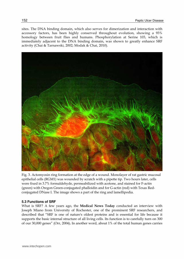
the healing process to the next phase – inflammation. The main function of neutrophils is to prevent infection. These cells can destroy and remove bacteria and damaged tissue by phagocytosis. Once this task is completed, neutrophils are eliminated by apoptosis. Then macrophages move in to clean up the cell remnants and apoptotic bodies of neutrophils. Macrophages are key regulatory cells during ulcer healing because they not only continue neutrophil’s job, but also produce an abundant reservoir of potent growth factors to activate additional endothelial cells and fibroblasts. The inflammatory phase is ended when lymphocytes attracted to the ulcer by IL-1, an important regulator of collagenase activity that is later needed for extracellular matrix (ECM) remodeling. Fibroblasts synthesize ECM to replace the provisional network of fibrin and fibronectin and form granulation tissue under the ulcer bed. Fibroblasts are attracted to the ulcer by TGF-┚ and PDGF that are produced by inflammatory cells and platelets. Once in the ulcer, fibroblasts proliferate rapidly and produce abundant ECM proteins, such as fibronectin, proteoglycans and procollagen, whose accumulation in the ulcer provides further support for cell migration and tissue repair. Thereafter, fibroblasts transform into myofibroblasts with thick actin bundles underneath the cell membrane which generate powerful forces to pull the wound edges together to close the ulcer. Granulation tissue is a reflection of active angiogenesis. A number of molecules released during hemostasis are angiogenic factors, such as VEGF, PDGF, bFGF, and TGF-┚, which can stimulate resident endothelial cells to proliferate. The activated endothelial cells produce proteases (matrix metalloproteinases or MMPs) to digest the basal lamina in the parental vessels in order to crawl through the ECM and to re-gather to form new blood vessels in the wound center, giving bumpy appearance to the ulcer bed. Angiogenesis is essential for ulcer healing, because it provides nutrients for the healing process to move forward. Meantime, mucosal epithelial cells at the ulcer margin are stimulated by ulceration to form a contractile actomyosin ring around the ulcer. Actomyosin ring is made of filamentous actin (F-actin) and myosin-II in association with radially organized microtubules (Mandato & Bement, 2003). F-actin cable in each epithelial cell at the ulcer margin links to neighboring cells through adherens junctions and is operated by the motor protein myosin-II, jointly like a purse string provides the force necessary to draw the wound edges together to achieve re-epithelialization (Figure 3). The whole process is regulated by the small GTPases including RhoA, Rac and Cdc42. RhoA activates the assembly of F-actin stress fibers by cortical flow, Rac is required for the rapid actin polymerization to form lamellipodia, and Cdc42 is essential for myosin-II organization and actin assembly/disassembly (Garcia-Fernandez et al, 2009; Darenfed & Mandato, 2005). The cells directly bordering the ulcer are connected by a continuous actomyosin cable, anchored at cell-cell junctions, and form lamellipodia at their leading edge (Figure 3). At the final stage of wound closure, opposing leading edge cells make contact through lamellipodia and seal the gap. Epithelial cell migration stops once the gap is sealed. However, healing process still continues into the next phase – tissue remodeling within the ulcer. A new basement membrane starts to build underneath the epithelium. Granulation tissue is gradually replaced by regenerated tissue that more closely resembles the original tissue before ulceration. The main players in this phase are MMPs and their antagonists TIMPs. They keep in a very delicate dynamic balance and work together in a coordinated fashion to allow tissue synthesis and breakdown to take place simultaneously.
www.intechopen.com
Peptic Ulcer Disease
150
5. Serum Response Factor in gastric ulcer healing
During ulcer healing, epithelial cells proliferate and migrate from nearby to close the wound; smooth muscle cells and myofibroblasts multiply to restore the musculature; endothelial cells are motivated to generate vessels to make sure the newly generated tissue has an adequate nutrient supply; and immune cells stand by to guard the wounded area and protect from invasions of pathogens. All these cellular activities are directed and regulated by dozens of molecules including growth factors, cytokines, chemokines, and more importantly, transcription factors, because every one of these molecules has to be transcribed from its gene fundamentally and transcription factors are the ones for this job. Among many transcription factors involved in ulcer healing, Serum response factor (SRF) is the master regulator. SRF is ubiquitously expressed in every type of tissue and its targeted genes take up nearly 1% of our entire genome (Sun et al, 2006; Miano, 2010). SRF can be activated by growth factors, cytokines and chemokines, and in return, activated SRF can direct expressions of these molecules to heal ulcers in a precisely organized manner. Moreover, CagA, one of the main products of H. pylori, can increase SRF binding capacity by 40 fold (Hirata et al, 2002). SRF is involved in every stage of the healing process including re-epithelialization, angiogenesis and granulation tissue remodeling.
5.1 Story of SRF SRF was first identified by a British scientist Richard Treisman in 1986 (Treisman, 1986), for which he was awarded the EMBO Medal in 1995. Treisman’s discovery was built on a prior observation by Michael Greenberg, a postdoctoral research fellow at the time in Edward Ziff’ lab at New York University. Greenberg’s work showed that resting fibroblasts responded to serum addition with a rapid activation of c-fos (Greenberg & Ziff, 1984). Since its activation does not require new protein synthesis, c-fos was classified as an immediate early gene. Later, it was found that in addition to serum, other mitogenic agents such as growth factors have the same effect on c-fos activation (Rollins & Stiles, 1989). During that time, Treisman was a struggling postdoctoral research fellow at Harvard University who was interested in c-myc regulation (Treisman, 1995). In the summer of 1984, he met Edward Ziff and heard about Greenberg’s discovery. Treisman immediately forsook c-myc and switched to c-fos. After he returned to England, Treisman rapidly proceeded with c-fos study by focusing on 5’ regulatory region. Several regulatory DNA elements were identified in the promoter region of c-fos gene, but a particular attention was given to a short sequence located about 300bp upstream of the transcription initiation site. For convenience, Treisman named this sequence Serum Response Element (SRE) and the protein that identifies this sequence Serum Response Factor (Treisman, 1986). SRE is an A/T rich core flanked by an inverted repeat, CC(A/T)6GG, and for this reason, SRE is also referred to as CArG box. Treisman demonstrated that c-fos activation by serum requires SRF binding to SRE. By that time, several other labs also identified the existence of SRF (Gilman et al, 1986; Prywes & Roeder, 1986; Greenberg et al, 1987). Since then, SRE has been identified in many genes across our entire genome (Sun et al, 2006). The list of SRE-containing genes is still growing. In 1986, Greenberg moved to Boston and became a faculty of Harvard Medical School with his own lab. His initial observation stimulated many researchers to look in that direction and led to a series of important discoveries in the area of gene transcriptional regulation. His colleagues wrote a song to portrait him and his work around c-fos:
www.intechopen.com
Gastric Ulcer Healing – Role of Serum Response Factor
151
“He was a bald headed man He was brutally handsome And they were terminally busy They held him up And he held them for ransom In a lab in a cold, cold city He had a nasty reputation As a cru-el dude They said he was ruthless, Said he was crude They had one thing in common They were always uptight He'd say "Faster, faster, Let's publish by tonight" Life in the fos lane Surely make you lose your mind Life in the fos lane Eager for action Hot for the game The Sephadex fraction The quest for the fame They read all the right journals They paid gigantic bills They threw outrageous parties They had infamous spills There were bands on the Northern But no counts could be traced He pretended not to notice He was caught up in the race In every evening, until it was light They were so tired, they faked it He was too tired to fight about it
Life in the fos lane Surely make you lose your mind Life in the fos lane Life in the fos lane Everything, all the time Life in the fos lane Rapid and transient Transcribed in a burst In all cell responses c-fos turns on first He said listen Bernie We need space to work in We've been up and down this hallway And never seen Ed Lin He said call Howard Hughes I think I'm gonna crash Six post-docs are coming And I'm almost out of cash He kept pushing them to publish "Go for Cell" he would shout They didn't care They were just dying to get out And it was Life in the fos lane Surely make you lose your mind Life in the fos lane Life in the fos lane Everything all the time Life in the fos lane”
5.2 Biology of SRF The human SRF gene is 10607bp long containing 7 exons and is mapped to the chromosome 6p21.1. The full length of SRF transcript is 4201bp including exon 1 (1-871), exon 2 (872-1138), exon 3 (1139-1400), exon 4 (1401-1520), exon 5 (1521-1712), exon 6 (1713-1789), and exon 7 (1790-4201). SRF can be expressed in different isoforms due to alternative splicing and some of them appear to display tissue specificity. For instance, SRF-S, which lacks both exon 4 and 5 (Δ4, 5), has only been detected in the aorta, while SRF-I, which is the shortest isoform (missing exon 3, 4 and 5), is specific to embryonic tissues. On the other hand, SRF-M, which lacks only exon 5, has been shown as a dominant negative mutant. SRF expression is self regulated, because SRF gene promoter contains four SRE sites. Full length SRF protein (~67 kDa) contains three distinct domains: a SRE DNA binding domain, a transactivation domain and multiple phosphorylation
www.intechopen.com
Peptic Ulcer Disease
152
sites. The DNA binding domain, which also serves for dimerization and interaction with accessory factors, has been highly conserved throughout evolution, showing a 93% homology between fruit flies and humans. Phosphorylation at Serine 103, which is immediately adjacent to the DNA binding domain, was shown to greatly enhance SRF activity (Chai & Tarnawski, 2002; Modak & Chai, 2010).
Fig. 3. Actomyosin ring formation at the edge of a wound. Monolayer of rat gastric mucosal epithelial cells (RGM1) was wounded by scratch with a pipette tip. Two hours later, cells were fixed in 3.7% formaldehyde, permeabilized with acetone, and stained for F-actin (green) with Oregon Green-conjugated phalloidin and for G-actin (red) with Texas Red-conjugated DNase I. The image shows a part of the ring and lamellipodia.
5.3 Functions of SRF What is SRF? A few years ago, the Medical News Today conducted an interview with Joseph Miano from University of Rochester, one of the prominent SRF researchers, and described that “SRF is one of nature's oldest proteins and is essential for life because it supports the basic internal structure of all living cells. Its function is to carefully turn on 300 of our 30,000 genes” (Orr, 2004). In another word, about 1% of the total human genes carries
www.intechopen.com
Gastric Ulcer Healing – Role of Serum Response Factor
153
SRF target – SRE. These genes fall into a broad spectrum and some of them have multiple SRE sites, for example, EGR1 has six and CCN1 has five, even SRF itself has four SRE sequences (Sun et al, 2006). In addition to the well-known immediate early genes (e.g. FOS and EGR1), SRF also controls a long list of muscle-related genes (ACTA2, MYH6, MYH11, SM22┙, TNNT1, ATP2A1, etc). In fact, most of the published SRF studies focus on its role in muscular structures including cardiac muscle, smooth muscle and skeletal muscle. Ten years ago, I was a postdoctoral research fellow at Harvard University, walking behind the giants like Greenberg and Treisman and trying to find new meanings for SRF. We created transgenic mice with cardiac-specific overexpression of SRF. The mice died within 6 months after birth due to heart failure. Histological examination revealed severe cardiomyocyte hypotrophy and interstitial fibrosis (Zhang et al, 2001a). The image made to the cover of the American Journal of Physiology. From this study, we have learned that too much SRF can drive overexpression of numerous cardiac genes (MYH7, ACTA1, NPPA, etc.) and end up with a bigger and heavier heart than in normal individuals. The heart-to-body weight ratio was almost 4 times greater in transgenic mice compared to non-transgenic littermates. To look at the other side of the coin, we also produced transgenic mice that express a dominant mutant SRF in heart. The mutated SRF gene generated a protein product that was incapable to bind to SRE, and therefore, cardiac genes never had a chance to fully express during embryogenesis. As a result, most embryos died before born, and a few survivors barely made to the second week of their age. Histological examination displayed serious cardiac ventricle dilation and myofiber degeneration (Zhang et al, 2001b). These studies demonstrate that properly functional SRF is essential for both embryonic development and post-natal development. This concept is also supported by the earlier transgenic study showing that complete knockout of SRF was lethal (Arsenian et al, 1998). Similar consequences have been also observed in transgenics of skeletal muscle (Li et al, 2005; Chavret et al, 2006; Lahoute et al, 2008) and smooth muscle (Miano et al, 2004; Werth et al, 2010). SRE has also been identified in cytoskeletal genes (ACTB, CFL1, DES, DSTN, TTN, KRT17, etc.), another major category with more than 1,000 members, whose protein products form an intracellular network connecting membranous subcellular structures to the cell membrane and the nucleus. Some of these genes are expressed in all types of cells, suggesting that SRF is essential for maintenance of cell shape and locomotion in everywhere of our body. SRF regulates cytoskeletal organization; on the other hand, SRF itself is regulated by the dynamics of actin cytoskeleton. For instance, every time G-actin polymerizes into F-actin, SRF gets activated. The remaining SRE-containing gene products fall into many diversified categories, such as growth factors (e.g. IGF2, FGF10, FGFR3, TGFB1i1, etc.), ECM proteins (e.g. CCN1, CTGF, etc.), cell adhesion molecules (e.g. ITGA1, ITGA5, ITGB1, etc.), intercellular junctional molecules (e.g. TJP1, CDH5, CDH11, etc.), neuronal receptors (e.g. NR4A1, NR4A2, etc.), and apoptosis regulators (BCL2). In addition to the hundreds of genes that SRF directly regulates, a growing number of genes that do not contain SRE have been found to respond to SRF activity (Khachigian & Collins, 1997; Miano et al, 2007). From this, one can imagine the influence of SRF on life.
5.4 SRF in ulcer healing Like any other human diseases, gastric ulcer and gastric ulcer healing have been studied both clinically as well as experimentally. Since the rules and regulations on clinical studies
www.intechopen.com
Peptic Ulcer Disease
154
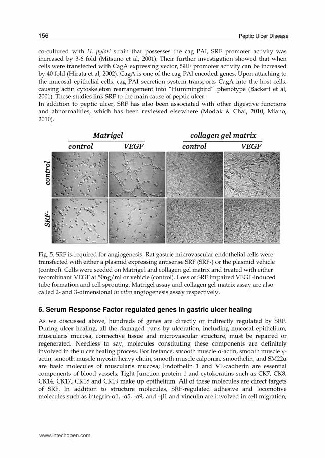
are extremely strict, most of the mechanistic studies have to be done in animal models complemented by in vitro cell culture. Researchers have developed several animal models for gastric ulcer study, which generally can be classified as chemical-induced and surgical-induced. Comparison of all these models reveals striking similarities in the morphological evolution as well as molecular dynamics involved in healing process. Therefore, it is generally accepted that ulcer undergoes common stages of healing, as discussed above, once it develops, regardless the cause (Tarnawski, 2005). Figure 4A shows a typical gastric ulcer developed in rat by topical application of acetic acid on the serosal side of the stomach. This model was initially developed by Japanese researchers and modified and validated by others (Okabe & Pfeiffer, 1972). Briefly, the animal needs to be fasted 12 hours before operation, otherwise, the food in the stomach would interfere ulcer induction. Laparotomy is performed under anesthesia to expose the stomach. Hold the stomach tightly with one hand and apply 50µl of acetic acid to the wall of the glandular stomach with the other hand, through a pipette tip (Ǿ 4.00mm). Hold for 90 seconds and then clean up the area with saline. In this way, a gastric ulcer can develop within 3-5 days after induction (Chai et al, 2004a; Nguyen et al, 2007). Immunohistological examination shows that SRF is highly activated in the ulcerated mucosa as well as in underneath connective tissue (Figure 4B). Figure 4C shows a higher magnification of regenerating gastric mucosal glands in the ulcer, and the bright red nuclear stain indicates SRF activation. The similar result can be seen in human gastric ulcer as well (Figure 4D). To determine what role SRF plays in gastric ulcer healing, we injected SRF expressing plasmid around the ulcer induction site to boost the local level of SRF. As a result, ulcer healing was significantly accelerated by the treatment (Chai et al, 2004a). In particular, re-epithelialization process was speeded up. In addition, a massive amount of smooth muscle ┙-actin expressing cells were found in the granulation tissue under the ulcer bed, indicating an increase of smooth muscle cells and/or myofibroblasts. In vitro overexpression of SRF in gastric mucosal epithelial cells (RGM1) and smooth muscle cells (A7R5) all proved promotions in cell migration and proliferation, as reflected by increased actin polymerization and activations of c-fos and egr-1, suggesting that the acceleration of ulcer healing by SRF gene therapy is due to SRF-driven cell migration and proliferation. As we discussed above, no matter re-epithelialization or smooth muscle structure restoration, they all require blood supply provided by angiogenesis, a process that makes sure the newly generated structures during ulcer healing will survive. In order to test what influence SRF has on angiogenesis during ulcer healing, we did same injection around the ulcer, but this time the SRF cDNA in the plasmid was flipped over to become an antisense generator. The idea was to interfere with local SRF expression and to create a local SRF deficiency. As a result, less number of micro-vessels was found in the granulation tissue, indicating that SRF deficiency impairs angiogenesis (Chai et al, 2004b). This conclusion was also supported by in vitro study, which showed that SRF deficiency impaired endothelial cells migration and proliferation capability so that even the most powerful angiogenic factor like VEGF could not stimulate tube formation in Matrigel or collagen gel matrix (Figure 5), a phenomenon called in vitro angiogenesis normally observed in the presence of an angiogenic factor (Jones et al, 2001). Further dissection of the mechanisms revealed that VEGF activates SRF through MEK-ERK and Rho signaling, and blocking these pathways interrupts SRF mediated endothelial cell migration and proliferation, and eventually causes failure of angiogenesis.
www.intechopen.com

Gastric Ulcer Healing – Role of Serum Response Factor
155
Fig. 4. SRF activation during gastric ulcer healing. A. A gastric ulcer induced in rat by acetic acid experimentally. B. Immunohistochemistry shows SRF activation in the ulcer region. C. SRF activation in the regenerating mucosal glands. D. SRF activation in the human gastric ulcer.
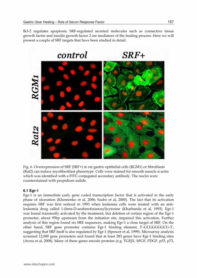
In addition to epithelial cells, endothelial cells, and smooth muscle cells, we have also examined another type of cell – myofibroblast. As we discussed above, myofibroblasts play an important role in ulcer healing by producing many growth factors and ECM molecules to mediate the healing process and also by providing contractive force to close the ulcer. We found that ulceration can trigger myofibroblast differentiation from the epithelial cells adjacent to the wound and from the fibroblasts within the ulcer bed (Chai et al, 2007). These cells can be distinguished from their ancestors by their expression of smooth muscle ┙-actin, and from smooth muscle cells by the absence of smoothelin. Many myofibroblasts were a transient phenotype, once the ulcer was healed, they disappeared. Local increase of SRF level by injecting SRF expressing plasmid into the ulcer greatly boosted the number of cells that express smooth muscle ┙-actin but not smoothelin, indicating that SRF promotes myofibroblast differentiation. This conclusion was also supported by in vitro experiments, which demonstrated that overexpression of SRF in both epithelial cells and fibroblasts induced expression of smooth muscle ┙-actin (Figure 6). The involvement of SRF in gastric ulcer healing was also strengthened by the finding of its association with H. pylori. In 2001, Japanese researchers found that when gastric cells were
www.intechopen.com
Peptic Ulcer Disease
156
co-cultured with H. pylori strain that possesses the cag PAI, SRE promoter activity was increased by 3-6 fold (Mitsuno et al, 2001). Their further investigation showed that when cells were transfected with CagA expressing vector, SRE promoter activity can be increased by 40 fold (Hirata et al, 2002). CagA is one of the cag PAI encoded genes. Upon attaching to the mucosal epithelial cells, cag PAI secretion system transports CagA into the host cells, causing actin cytoskeleton rearrangement into “Hummingbird” phenotype (Backert et al, 2001). These studies link SRF to the main cause of peptic ulcer. In addition to peptic ulcer, SRF has also been associated with other digestive functions and abnormalities, which has been reviewed elsewhere (Modak & Chai, 2010; Miano, 2010).
Fig. 5. SRF is required for angiogenesis. Rat gastric microvascular endothelial cells were transfected with either a plasmid expressing antisense SRF (SRF-) or the plasmid vehicle (control). Cells were seeded on Matrigel and collagen gel matrix and treated with either recombinant VEGF at 50ng/ml or vehicle (control). Loss of SRF impaired VEGF-induced tube formation and cell sprouting. Matrigel assay and collagen gel matrix assay are also called 2- and 3-dimensional in vitro angiogenesis assay respectively.
6. Serum Response Factor regulated genes in gastric ulcer healing
As we discussed above, hundreds of genes are directly or indirectly regulated by SRF. During ulcer healing, all the damaged parts by ulceration, including mucosal epithelium, muscularis mucosa, connective tissue and microvascular structure, must be repaired or regenerated. Needless to say, molecules constituting these components are definitely involved in the ulcer healing process. For instance, smooth muscle ┙-actin, smooth muscle ┛-actin, smooth muscle myosin heavy chain, smooth muscle calponin, smoothelin, and SM22┙ are basic molecules of muscularis mucosa; Endothelin 1 and VE-cadherin are essential components of blood vessels; Tight Junction protein 1 and cytokeratins such as CK7, CK8, CK14, CK17, CK18 and CK19 make up epithelium. All of these molecules are direct targets of SRF. In addition to structure molecules, SRF-regulated adhesive and locomotive molecules such as integrin-┙1, -┙5, -┙9, and –┚1 and vinculin are involved in cell migration;
www.intechopen.com
Gastric Ulcer Healing – Role of Serum Response Factor
157
Bcl-2 regulates apoptosis; SRF-regulated secreted molecules such as connective tissue growth factor and insulin growth factor 2 are mediators of the healing process. Here we will present a couple of SRF targets that have been studied in detail.
Fig. 6. Overexpression of SRF (SRF+) in rat gastric epithelial cells (RGM1) or fibroblasts (Rat2) can induce myofibroblast phenotype. Cells were stained for smooth muscle ┙-actin which was identified with a FITC-conjugated secondary antibody. The nuclei were counterstained with propidium iodide.
6.1 Egr-1 Egr-1 is an immediate early gene coded transcription factor that is activated in the early phase of ulceration (Khomenko et al, 2006; Szabo et al, 2000). The fact that its activation requires SRF was first noticed in 1993 when leukemia cells were treated with an anti-leukemia drug called 1-(beta-D-arabinofuranosyl)cytosine (Kharbanda et al, 1993). Egr-1 was found transiently activated by the treatment, but deletion of certain region of the Egr-1 promoter, about 95bp upstream from the initiation site, impaired this activation. Further analysis of this region found six SRE sequences, making Egr-1 a close target of SRF. On the other hand, SRF gene promoter contains Egr-1 binding element, 5’-GCGGGGGCG-3’, suggesting that SRF itself is also regulated by Egr-1 (Spencer et al, 1999). Microarray analysis screened 12,000 gene promoters and found that at least 283 genes have Egr-1 binding sites (Arora et al, 2008). Many of these genes encode proteins (e.g. TGF┚1, bFGF, PDGF, p53, p73,
www.intechopen.com

Peptic Ulcer Disease
158
PTEN, EGFR, BMP4, MMP9, ITGA5, CK16, Egr-2, etc.) that are known to contribute to ulcer or other wound healing. Through regulation of these genes, Egr-1 greatly extends SRF power.
6.2 CCN1 CCN1 (formerly known as Cyr61 or IGFBP10) is another important gene directly regulated by SRF and contains five SRE sites located about 3751bp upstream in the gene promoter. It encodes a matricellular protein that is best known for its angiogenic activity because it stimulates neovascularization in rat corneas and cyr61-null mice suffer embryonic death due to vascular defects (Mo et al, 2002). One study demonstrated that intramuscular injection of a CCN1-expression adenovirus in rabbits with ischemic hindlimb improves tissue perfusion even greater than injection of VEGF (Fataccioli et al, 2002). The involvement of CCN1 in wound healing was first known ten years ago in a cutaneous wound model (Chen et al, 2001; Lantinkic et al, 2001). During the experiment, CCN1 was found highly up-regulated in the granulation tissue five days after wounding and remained high for a week till the re-epithelialization was completed. It was shown that CCN1 promotes angiogenesis not only directly but also indirectly through induction of VEGF.
Fig. 7. Gastric ulceration induces CCN1 expression at the ulcer margin (A) and granulation tissue (B).
As a matricellular protein, CCN1 has features intermediate between conventional growth factors and structural ECM molecules; therefore, it can influence tissue remodeling without being an integral element of the structural ECM. In addition to being an angiogenic factor, CCN1 also supports cell adhesion, migration, proliferation, differentiation and survival. Recently, we have found that CCN1 is highly up-regulated in the gastric epithelial cells adjacent to the ulcer and remains high until the wound is healed (Figure 7; Chai et al, 2010b). This was demonstrated by epithelial injury both in vivo (gastric ulcer margin) and in vitro (gastric epithelial cell culture). Its elevation induces a transient phenotypic change in the mucosal epithelial cells at the ulcer margin and drives the wound closure. These cells lose their epithelial identities and become mesenchymal-like cells. At the molecular level, it shows down-regulation of epithelial markers such as E-cadherin, Occludin and cytokeratins, and up-regulation of mesenchymal markers such as vimentin, N-cadherin and
www.intechopen.com

Gastric Ulcer Healing – Role of Serum Response Factor
159
metalloproteinases. Once the wound is healed, these cells and their progeny can resume their original epithelial phenotype as evidenced both in vitro and in vivo. However, when CCN1 is knocked down in gastric mucosal epithelial cells, injury-induced EMT is disrupted and wound closure is delayed. We have further dissected the molecular mechanisms of this process and found that CCN1-induced E-cadherin loss is not due to transcriptional repression, which is the main mechanism of E-cadherin loss in many other systems (Zhou et al, 2004; Hayashida et al, 2006; Kang & Massague, 2004), but rather protein degradation caused by the collapse of adherens junctions, which is ignited by ┚-catenin nuclear translocation. CCN1-activated integrin-linked kinase mediates this event. In addition, our in vivo study demonstrated that local injection of recombinant CCN1 protein into gastric ulcers can induce expression of vimentin and smooth muscle ┙-actin in the mucosal epithelial cells and promote re-epithelialization during ulcer healing, and that local injection of CCN1 antibody neutralizes the effect and delays healing process. We have also found that TGF┚1 up-regulates CCN1 expression in gastric epithelial cells through SRF and it fails to do so when SRF is inhibited by shRNA.
7. Conclusions
SRF is a ubiquitously expressed transcription factor that targets genes containing SRE (Or CArG box). SRE has been found in nearly 1% of total number of human genes and the list is still growing. Some of these genes encode transcription factors (e.g. FOS, FOSB, EGR1, EGR2, EGR4, ELK1, etc.) which have their own specific gene targets. For example, transcription factor Egr-1 has six SRF binding sites in its gene promoter region, indicating a tight control by SRF. It has been shown that Egr-1 is capable to bind to 283 genes, which double the number of genes directly regulated by SRF and extend SRF power to 2% of the human genome. Some other members of SRF targets encode growth factors (e.g. IGF2, TGFB1I1, FGF10, etc.), integrins (e.g. ITGA1, ITGA5, ITGA9, ITGB1, etc.), and matricellular proteins (e.g. CTGF, CCN1, etc.) and all these molecules can transduce signals to influence many other genes. Taken together, SRF influence, including both direct and indirect, can probably reach a quarter of the entire human genome. By now, one can imagine how powerful SRF is. Ulcer healing is just one of the things SRF does. One can easily find SRF contributions in each phase of ulcer healing: it promotes the production of growth factors and cytokines to mediate inflammation; it regulates formation of actomyosin ring and lamellipodia to promote re-epithelialization; it regulates apoptosis to remove the dead tissue and unnecessary cells; it supports angiogenesis through regulating endothelial cell migration and proliferation; it coordinates tissue remodeling by synchronizing proteases with their antagonists; and much, much more...
8. Acknowledgement
This work is supported by the Department of Veterans Affairs of the United States.
9. References
Arora, S., Wang, Y., Jia, Z., Vardar-Sengul, S., Munawar, A., Doctor, K.S., Birrer, M., McClelland, M., Adamson, E., & Mercola, D. (2008). Egr1 regulates the coordinated
www.intechopen.com
Peptic Ulcer Disease
160
expression of numerous EGF receptor target genes as identified by ChIP-on-chip. Genome Biol, 9(11): R166.
Arsenian, S., Weinhold, B., Oelgeschläger, M., Rüther, U., & Nordheim, A. (1998). Serum response factor is essential for mesoderm formation during mouse embryogenesis. EMBO J, 17(21): 6289-99.
Backert, S., Moese, S., Selbach, M., Brinkmann, V., & Meyer, T.F. (2001). Phosphorylation of tyrosine 972 of the Helicobacter pylori CagA protein is essential for induction of a scattering phenotype in gastric epithelial cells. Mol Microbiol, 42(3): 631-44.
Bizzozero, G. (1893). Ueber die schlauchförmigen Drüsen des Magendarmkanals und die Beziehungen ihres Epitheles zu dem Oberflächenepithel der Schleimhaut. Archiv
für mikroskopische Anatomie, 42: 82–152. Chai, J., Baatar, D., & Tarnawski, A. (2004a). Serum response factor promotes re-
epithelialization and muscular structure restoration during gastric ulcer healing. Gastroenterology, 126(7): 1809-18.
Chai, J., Jones, M.K., & Tarnawski, A.S. (2004b). Serum response factor is a critical requirement for VEGF signaling in endothelial cells and VEGF-induced angiogenesis. FASEB J, 18(11): 1264-6.
Chai, J., Modak, C., Mouazzen, W., Narvaez, R., & Pham, J. (2010a). Epithelial or mesenchymal: Where to draw the line? Biosci Trends, 4(3): 130-42.
Chai, J., Norng, M., Modak, C., Reavis, K.M., Mouazzen, W., & Pham, J. (2010b). CCN1 induces a reversible epithelial-mesenchymal transition in gastric epithelial cells. Lab Invest, 90(8): 1140-51.
Chai, J., Norng, M., Tarnawski, A.S., & Chow, J. (2007). A critical role of serum response factor in myofibroblast differentiation during experimental oesophageal ulcer healing in rats. Gut, 56(5): 621-30.
Chai J, Tarnawski AS. (2002). Serum response factor: discovery, biochemistry, biological roles and implications for tissue injury healing. J Physiol Pharmacol, 53(2): 147-57.
Charvet, C., Houbron, C., Parlakian, A., Giordani, J., Lahoute, C., Bertrand, A., Sotiropoulos, A., Renou, L., Schmitt, A., Melki, J., Li, Z., Daegelen, D., & Tuil, D. (2006). New role for serum response factor in postnatal skeletal muscle growth and regeneration via the interleukin 4 and insulin-like growth factor 1 pathways. Mol Cell Biol, 26(17): 6664-74.
Darenfed, H. & Mandato, C.A. (2005). Wound-induced contractile ring: a model for cytokinesis. Biochem Cell Biol, 83(6): 711-20.
Doenges, J.L. (1938). Spirochaetei in gastric glands of macaccus rhesus and humans without definite history of related disease. Proc Soc Exp Biol Med, 38: 536-538.
Fataccioli, V., Abergel, V., Wingertsmann, L., Neuville, P., Spitz, E., Adnot, S., Calenda, V., & Teiger, E. (2002). Stimulation of angiogenesis by Cyr61 gene: a new therapeutic candidate. Hum Gene Ther, 13(12): 1461-70.
Feinstein LB, Holman RC, Yorita CKL, Steiner CA, Swerdlow DL. (2010). Trends in hospitalizations for peptic ulcer disease, United States, 1998-2005. Emerg Infect
Dis, 16(9): 1410-1418.
www.intechopen.com
Gastric Ulcer Healing – Role of Serum Response Factor
161
Freedberg, A.S & Barron, L.E. (1940).The presence of spirochaetes in human gastric mucosa. Am J Dig Dis, 38: 443-445.
Garcia-Fernandez, B., Campos, I., Geiger, J., Santos, A.C., & Jacinto, A. (2009). Epithelial resealing. Int J Dev Biol, 53(8-10): 1549-56.
Gilman, M.Z., Wilson, R.N., & Weinberg, R.A. (1986). Multiple protein-binding sites in the 5'-flanking region regulate c-fos expression. Mol Cell Biol, 6(12): 4305-16.
Greenberg, M.E, Siegfried, Z., & Ziff, E.B. (1987). Mutation of the c-fos gene dyad symmetry element inhibits serum inducibility of transcription in vivo and the nuclear regulatory factor binding in vitro. Mol Cell Biol, 7(3): 1217-25.
Greenberg, M.E & Ziff, E.B. (1984). Stimulation of 3T3 cells induces transcription of the c-fos proto-oncogene. Nature, 311(5985): 433-8.
Hayashida, Y., Urata, Y., Muroi, E., Kono, T., Miyata, Y., Nomata, K., Kanetake, H., Kondo, T., & Ihara, Y. (2006). Calreticulin represses E-cadherin gene expression in Madin-Darby canine kidney cells via Slug. J Biol Chem, 281: 32469-84.
Hirata, Y., Maeda, S., Mitsuno, Y., Tateishi, K., Yanai, A., Akanuma, M., Yoshida, H., Kawabe, T., Shiratori, Y., & Omata, M. (2002). Helicobacter pylori CagA protein activates serum response element-driven transcription independently of tyrosine phosphorylation. Gastroenterology, 123(6): 1962-71.
Jones, M.K., Kawanaka, H., Baatar, D., Szabó, I.L., Tsugawa, K., Pai, R., Koh, G.Y., Kim, I., Sarfeh, I.J., & Tarnawski, A.S. (2001). Gene therapy for gastric ulcers with single local injection of naked DNA encoding VEGF and angiopoietin-1. Gastroenterology, 121(5): 1040-7.
Kang, Y. & Massague, J. (2004). Epithelial-mesenchymal transitions: twist in development and metastasis. Cell, 118: 277-9.
Khachigian, L.M. & Collins, T. (1997). Inducible expression of Egr-1-dependent genes. A paradigm of transcriptional activation in vascular endothelium. Circ Res, 81(4): 457-61.
Kharbanda, S., Saleem, A., Rubin, E., Sukhatme, V., Blenis, J., & Kufe, D. (1993). Activation of the early growth response 1 gene and nuclear pp90rsk in human myeloid leukemia cells by 1-(beta-D-arabinofuranosyl)cytosine. Biochemistry, 32(35): 9137-42.
Khomenko, T., Szabo, S., Deng, X., Jadus, M.R., Ishikawa, H., Osapay, K., Sandor, Z., & Chen, L. (2006). Suppression of early growth response factor-1 with egr-1 antisense oligodeoxynucleotide aggravates experimental duodenal ulcers. Am J
Physiol Gastrointest Liver Physiol, 290(6): G1211-8. Kidd, M. & Modlin, I.M. (1998). A century of Helicobacter pylori: paradigms lost-
paradigms regained. Digestion, 59(1): 1-15. Konturek, J.W. (2003). Discovery by Jaworski of Helicobacter pylori and its pathogenetic
role in peptic ulcer, gastritis and gastric cancer. J Physiol Pharmacol, 54 Suppl 3: 23-41.
Kusters, J.G., van Vliet, A.H., & Kuipers, E.J. (2006). Pathogenesis of Helicobacter pylori infection. Clin Microbiol Rev, 19(3): 449-90.
www.intechopen.com
Peptic Ulcer Disease
162
Lahoute, C., Sotiropoulos, A., Favier, M., Guillet-Deniau, I., Charvet, C., Ferry, A., Butler-Browne, G., Metzger, D., Tuil, D., & Daegelen, D. (2008). Premature aging in skeletal muscle lacking serum response factor. PLoS One, 3(12): e3910.
Li, S., Czubryt, M.P., McAnally, J., Bassel-Duby, R., Richardson, J.A., Wiebel, F.F., Nordheim, A., & Olson, E.N. (2005). Requirement for serum response factor for skeletal muscle growth and maturation revealed by tissue-specific gene deletion in mice. Proc Natl Acad Sci U S A, 102(4): 1082-7.
Mandato, C.A. & Bement, W.M. (2003). Actomyosin transports microtubules and microtubules control actomyosin recruitment during Xenopus oocyte wound healing. Curr Biol, 13(13): 1096-105.
Marshall, B.J. (2002). The Discovery that Helicobacter pylori, a spiral bacterium, caused peptic ulcer disease. In: B. Marshall (ed.), Helicobacter Pioneers. Singapore: Blackwell Science Asia. pp. 165–202.
Miano, J.M. (2010). Role of serum response factor in the pathogenesis of disease. Laboratory Investigation, 90: 1274-1284.
Miano, J.M., Long, X., & Fujiwara, K. (2007). Serum response factor: master regulator of the actin cytoskeleton and contractile apparatus. Am J Physiol Cell Physiol, 292(1): C70-81.
Miano, J.M., Ramanan, N., Georger, M.A., de Mesy, Bentley K.L, Emerson, R.L, Balza, R.O. Jr, Xiao, Q., Weiler, H., Ginty, D.D., & Misra, R.P. (2004). Restricted inactivation of serum response factor to the cardiovascular system. Proc Natl Acad
Sci U S A, 101(49): 17132-7. Mitsuno, Y., Yoshida, H., Maeda, S., Ogura, K., Hirata, Y., Kawabe, T., Shiratori, Y., &
Omata, M. (2001). Helicobacter pylori induced transactivation of SRE and AP-1 through the ERK signalling pathway in gastric cancer cells. Gut, 49(1): 18-22.
Mo, F.E., Muntean, A.G., Chen, C.C., Stolz, D.B., Watkins, S.C., & Lau, L.F. (2002). CYR61 (CCN1) is essential for placental development and vascular integrity. Mol Cell
Biol, 22(24): 8709-20. Modak, C. & Chai, J. (2010). Serum response factor: look into the gut. World J Gastroenterol,
16(18): 2195-201. Nguyen, T., Chai, J., Li, A., Akahoshi, T., Tanigawa, T., & Tarnawski, A.S. (2007). Novel
roles of local insulin-like growth factor-1 activation in gastric ulcer healing: promotes actin polymerization, cell proliferation, re-epithelialization, and induces cyclooxygenase-2 in a phosphatidylinositol 3-kinase-dependent manner. Am J Pathol,170(4): 1219-28.
Okabe, S & Pfeiffer, C.J. (1972). Chronicity of acetic acid ulcer in the rat stomach. Am J Dig
Dis, 17(7): 619-29. Orr, L. (2004). Scientists align billion-year-old protein with embryonic heart defects.
Medical News Today. Pai, R., Szabo, I.L., Giap, A.Q., Kawanaka, H., & Tarnawski, A.S. (2001). Nonsteroidal
anti-inflammatory drugs inhibit re-epithelialization of wounded gastric monolayers by interfering with actin, Src, FAK, and tensin signaling. Life Sci,
69(25-26): 3055-71.
www.intechopen.com
Gastric Ulcer Healing – Role of Serum Response Factor
163
Palmer, E.D. (1954). Investigation of the gastric mucosa spirochaetes of the humans. Gastroenterology, 27: 218-220.
Peek, R.M Jr & Crabtree, J.E. (2006). Helicobacter infection and gastric neoplasia. J Pathol, 208(2): 233-48.
Pounder, R.E & Ng, D. (1995). The prevalence of Helicobacter pylori infection in different countries. Aliment Pharmacol Ther. 9: Suppl 2: 33-9.
Prywes, R & Roeder, R.G. (1986). Inducible binding of a factor to the c-fos enhancer. Cell, 47(5): 777-84.
Rollins, B.J & Stiles, C.D. (1989). Serum-inducible genes. Adv Cancer Res, 53:1-32. Salomon, H. (1896). Über das Spirillum des Säugetiermagens und sein Verhalten zu den
Belegzellen. Zentrallbl Bakteriol, 19: 433-442. Scott, D.R, Weeks, D., Hong, C., Postius, S., Melchers, K., & Sachs, G. (1998). The role of
internal urease in acid resistance of Helicobacter pylori. Gastroenterology, 114(1): 58-70.
Spencer, J.A., Major, M.L., & Misra, R.P. (1999). Basic fibroblast growth factor activates serum response factor gene expression by multiple distinct signaling mechanisms. Mol Cell Biol, 19(6): 3977-88.
Steer, H.W. (1975). Ultrastucture of cell migration through the gastric epithelium and its relation to bacteria. J Clin Pathol, 28: 639-646.
Sun, Q., Chen, G., Streb, J.W., Long, X., Yang, Y., Stoeckert, C.J. Jr, & Miano, J.M. (2006). Defining the mammalian CArGome. Genome Res, 16(2): 197-207.
Szabo, S., Khomenko, T., Gombos, Z., Deng, X.M., Jadus, M.R., & Yoshida, M. (2000). Review article: transcription factors and growth factors in ulcer healing. Alimnet
Pharmacol Ther, 14(suppl. 1): 33-43. Tarnawski, A.S. (2005). Cellular and molecular mechanisms of gastrointestinal ulcer
healing. Digestive Diseases and Sciences, 50: Suppl. S24-S33. Treisman, R. (1986). Identification of a protein-binding site that mediates transcriptional
response of the c-fos gene to serum factors. Cell, 46(4): 567-74. Treisman, R. (1995). Journey to the surface of the cell: Fos regulation and the SRE. The
EMBO Journal, 14(20): 4905-4913. Viala, J., Chaput, C., Boneca, I.G., Cardona, A., Girardin, S.E., Moran, A.P., Athman, R.,
Mémet, S., Huerre, M.R., Coyle, A.J., DiStefano, P.S., Sansonetti, P.J., Labigne, A., Bertin, J., Philpott, D.J., & Ferrero, R.L. (2004). Nod1 responds to peptidoglycan delivered by the Helicobacter pylori cag pathogenicity island. Nat Immunol, 5(11): 1166-74.
Werth, D., Grassi, G., Konjer, N., Dapas, B., Farra, R., Giansante, C., Kandolf, R., Guarnieri, G., Nordheim, A., & Heidenreich, O. (2010). Proliferation of human primary vascular smooth muscle cells depends on serum response factor. Eur J
Cell Biol. 89(2-3): 216-24. Zhang, X., Azhar, G., Chai, J., Sheridan, P., Nagano, K., Brown, T., Yang, J., Khrapko, K.,
Borras, A.M., Lawitts, J., Misra, R.P., & Wei, J.Y. (2001a). Cardiomyopathy in transgenic mice with cardiac-specific overexpression of serum response factor. Am J Physiol Heart Circ Physiol, 280(4): H1782-92.
www.intechopen.com
Peptic Ulcer Disease
164
Zhang, X., Chai, J., Azhar, G., Sheridan, P., Borras, A.M., Furr, M.C., Khrapko, K., Lawitts, J., Misra, R.P., & Wei, J.Y. (2001b). Early postnatal cardiac changes and premature death in transgenic mice overexpressing a mutant form of serum response factor. J Biol Chem, 276(43): 40033-40.
Zhou, B.P., Deng, J., Xia, W., Xu, J., Li, Y.M., Gunduz, M., & Hung, M.C. (2004). Dual regulation of Snail by GSK-3beta-mediated phosphorylation in control of epithelial-mesenchymal transition. Nat Cell Biol, 6: 931-40.
www.intechopen.com
Peptic Ulcer DiseaseEdited by Dr. Jianyuan Chai
ISBN 978-953-307-976-9Hard cover, 482 pagesPublisher InTechPublished online 04, November, 2011Published in print edition November, 2011
InTech EuropeUniversity Campus STeP Ri Slavka Krautzeka 83/A 51000 Rijeka, Croatia Phone: +385 (51) 770 447 Fax: +385 (51) 686 166www.intechopen.com
InTech ChinaUnit 405, Office Block, Hotel Equatorial Shanghai No.65, Yan An Road (West), Shanghai, 200040, China
Phone: +86-21-62489820 Fax: +86-21-62489821
Peptic ulcer disease is one of the most common chronic infections in human population. Despite centuries ofstudy, it still troubles a lot of people, especially in the third world countries, and it can lead to other moreserious complications such as cancers or even to death sometimes. This book is a snapshot of the currentview of peptic ulcer disease. It includes 5 sections and 25 chapters contributed by researchers from 15countries spread out in Africa, Asia, Europe, North America and South America. It covers the causes of thedisease, epidemiology, pathophysiology, molecular-cellular mechanisms, clinical care, and alternativemedicine. Each chapter provides a unique view. The book is not only for professionals, but also suitable forregular readers at all levels.
How to referenceIn order to correctly reference this scholarly work, feel free to copy and paste the following:
Jianyuan Chai (2011). Gastric Ulcer Healing – Role of Serum Response Factor, Peptic Ulcer Disease, Dr.Jianyuan Chai (Ed.), ISBN: 978-953-307-976-9, InTech, Available from:http://www.intechopen.com/books/peptic-ulcer-disease/gastric-ulcer-healing-role-of-serum-response-factor

© 2011 The Author(s). Licensee IntechOpen. This is an open access articledistributed under the terms of the Creative Commons Attribution 3.0License, which permits unrestricted use, distribution, and reproduction inany medium, provided the original work is properly cited.


![Ulcer Gastric Ff1[1]](https://img.pdfslide.net/doc/110x75/563dbb0a550346aa9aa9c209/ulcer-gastric-ff11.jpg)