Embed Size (px)
Citation preview
Med J Malaysia Vol 71 No 6 December 2016 363
SUMMARYPrimary gastrointestinal synovial sarcoma or its metastasesto the gastrointestinal tract is rare. Here we present a caseof 56-year-old gentleman with left thigh synovial sarcomaand gastric metastases along with the literature review.
KEY WORDS:Gastric ulcer, synovial sarcoma, stomach, metastasis
INTRODUCTIONSynovial sarcoma is a malignant soft tissue neoplasm thatmay present itself from a variety of sites in a human bodycommonly near large joints or tendons (80%). Thenomenclature however is misleading as there is no evidenceof differentiation towards synovium and this tumour canactually occur in any part of the body.1 Most synovialsarcomas of the monophasic type are composed entirely ofspindle cells and can hardly be associated with synovium interms of morphology.1 Gastrointestinal synovial sarcomas arerare but more and more cases are being reported each yearincreasing awareness about the potential misdiagnosisbetween the monophasic synovial sarcoma and KIT-negativegastrointestinal stromal tumour (GIST).2 To our knowledgeonly about 27 cases of stomach related synovial sarcoma arepreviously reported. Here we present a case of a metastaticgastric synovial sarcoma, the first reported in Malaysia.
CASE REPORTA 56-year-old gentleman presented with a mass over the leftposterior upper thigh for three months. Initially small butprogressively increasing in size (20x15cm) with no skinchanges, mobile and non-tender on palpation. Magneticresonance imaging showed a large heterogeneous left upperthigh mass with malignant features (capsular breach andintramuscular extension). Biopsy of mass showed to besynovial sarcoma as and it was resected completely with clearmargins. Histopathological examination reported a 9.5cmtumour and initial Contrast Enhanced ComputedTomography (CECT) staging showed no enlarged lymphnodes or any distant metastasis (T2bNoMo). Patient laterunderwent radiotherapy 66Gy for 33#. Patient did not haveany active complaint post adjuvant radiotherapy howeverrepeated CECT Thorax Abdomen and Pelvis one-year postresection showed lung metastases and focal mucosalthickening of the stomach. Patient was then planned for anOesophagogastroduodenoscopy (OGDS). OGDS done and
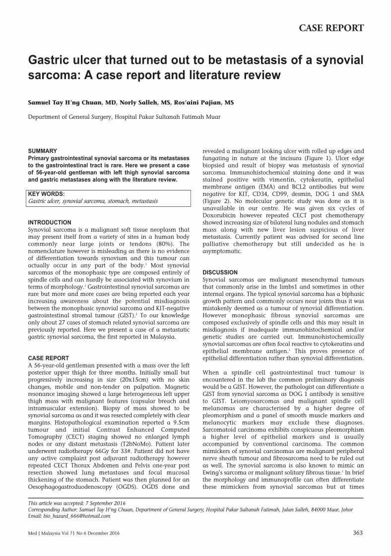
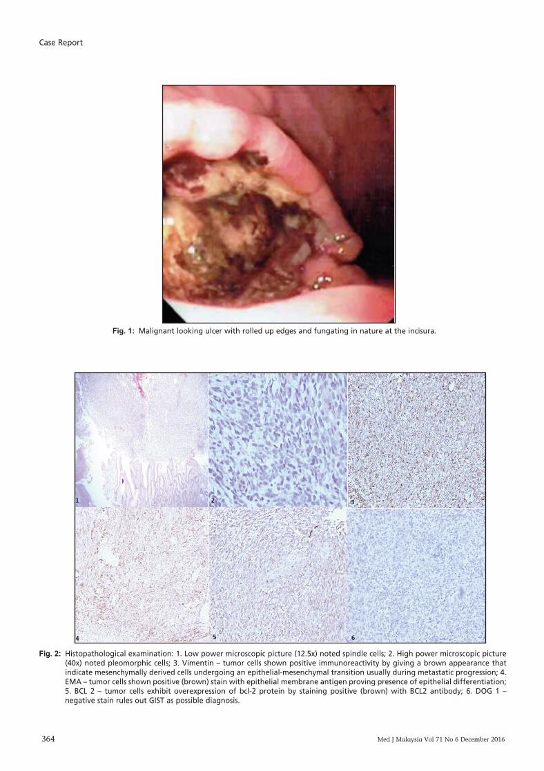
revealed a malignant looking ulcer with rolled up edges andfungating in nature at the incisura (Figure 1). Ulcer edgebiopsied and result of biopsy was metastasis of synovialsarcoma. Immunohistochemical staining done and it wasstained positive with vimentin, cytokeratin, epithelialmembrane antigen (EMA) and BCL2 antibodies but werenegative for KIT, CD34, CD99, desmin, DOG 1 and SMA(Figure 2). No molecular genetic study was done as it isunavailable in our centre. He was given six cycles ofDoxorubicin however repeated CECT post chemotherapyshowed increasing size of bilateral lung nodules and stomachmass along with new liver lesion suspicious of livermetastasis. Currently patient was advised for second linepalliative chemotherapy but still undecided as he isasymptomatic.
DISCUSSIONSynovial sarcomas are malignant mesenchymal tumoursthat commonly arise in the limbs1 and sometimes in otherinternal organs. The typical synovial sarcoma has a biphasicgrowth pattern and commonly occurs near joints thus it wasmistakenly deemed as a tumour of synovial differentiation.However monophasic fibrous synovial sarcomas arecomposed exclusively of spindle cells and this may result inmisdiagnosis if inadequate immunohistochemical and/orgenetic studies are carried out. Immunohistochemicallysynovial sarcomas are often focal reactive to cytokeratins andepithelial membrane antigen.1 This proves presence ofepithelial differentiation rather than synovial differentiation.
When a spindle cell gastrointestinal tract tumour isencountered in the lab the common preliminary diagnosiswould be a GIST. However, the pathologist can differentiate aGIST from synovial sarcoma as DOG 1 antibody is sensitiveto GIST. Leiomyosarcomas and malignant spindle cellmelanomas are characterised by a higher degree ofpleomorphism and a panel of smooth muscle markers andmelanocytic markers may exclude these diagnoses.Sarcomatoid carcinoma exhibits conspicuous pleomorphisma higher level of epithelial markers and is usuallyaccompanied by conventional carcinoma. The commonmimickers of synovial carcinomas are malignant peripheralnerve sheath tumour and fibrosarcoma need to be ruled outas well. The synovial sarcoma is also known to mimic anEwing’s sarcoma or malignant solitary fibrous tissue.1 In briefthe morphology and immunoprofile can often differentiatethese mimickers from synovial sarcomas but at times
Gastric ulcer that turned out to be metastasis of a synovialsarcoma: A case report and literature review
Samuel Tay H’ng Chuan, MD, Norly Salleh, MS, Ros’aini Pajian, MS
Department of General Surgery, Hospital Pakar Sultanah Fatimah Muar
CASE REPORT
This article was accepted: 7 September 2016Corresponding Author: Samuel Tay H'ng Chuan, Department of General Surgery, Hospital Pakar Sultanah Fatimah, Jalan Salleh, 84000 Muar, JohorEmail: [email protected]
14-Gastric00070_3-PRIMARY.qxd 1/5/17 1:05 AM Page 363
Case Report
364 Med J Malaysia Vol 71 No 6 December 2016
Fig. 1: Malignant looking ulcer with rolled up edges and fungating in nature at the incisura.
Fig. 2: Histopathological examination: 1. Low power microscopic picture (12.5x) noted spindle cells; 2. High power microscopic picture(40x) noted pleomorphic cells; 3. Vimentin – tumor cells shown positive immunoreactivity by giving a brown appearance thatindicate mesenchymally derived cells undergoing an epithelial-mesenchymal transition usually during metastatic progression; 4.EMA – tumor cells shown positive (brown) stain with epithelial membrane antigen proving presence of epithelial differentiation;5. BCL 2 – tumor cells exhibit overexpression of bcl-2 protein by staining positive (brown) with BCL2 antibody; 6. DOG 1 –negative stain rules out GIST as possible diagnosis.
14-Gastric00070_3-PRIMARY.qxd 1/5/17 1:05 AM Page 364
A case report and literature review
Med J Malaysia Vol 71 No 6 December 2016 365
molecular genetic study (SYT-SSX2 or SYT-SSX1) may berequired for confirmation in difficult cases. This proves achallenge for developing countries with limited resources andmay lead to misdiagnosis.
The main treatment for synovial sarcoma is surgical resectionwith/without radiotherapy to enhance local control.Chemotherapy (typically Doxorubicin and/or Ifosfamide)might be recommended in the treatment especially inadvanced/metastatic disease.2 Due to the rarity of thismalignant tumour there is still no consensus from experts onhow much the role of chemotherapy plays in preventingmetastases and improving survival.
The prognostic factors in a synovial sarcoma patient areinfluenced by the quality of surgery (clean margins) and thecharacteristics of the disease (size of tumour, local invasion,histology subtype, presence of metastases and lymph nodeinvolvement). There is excellent prognosis for patients withsmall tumour that managed to be resected with clearmargins. Patients with tumours more than 5 cm tend to havehigher risk in developing distant metastases.3 Patients withpoorly differentiated subtype and those already havingmetastases tend to have a poor prognosis.
CONCLUSIONSynovial sarcoma in the digestive tract is rare and prone tomisdiagnosis. The accurate and early diagnosis is crucial foreffective and appropriate therapy. The potential clinicalbenefits for a patient with metastatic synovial sarcoma toundergo another resection for gastric metastases are stillunclear. More cases should be reported in order to study itsdisease pattern and prevalence as only then clinical practiceand management guideline for this malignant disease maybe established.
REFERENCES1. Fisher C, De Bruijin DRH, Geurts van Kessel A. Synovial Sarcoma. In:
Fletcher CDM, Krishnan Unni K, Mertens F, editors. WHO Classification ofTumours of Soft Tissue and Bone, Fourth Edition, IARC Press: Lyon : 2013.
2. Eilber FC, Dry SM. Diagnosis and management of synovial sarcoma. J SurgOncol 2008; 97(4): 314-20.
3. Bergh P, Meis-Kindblom JM, Gherlinzoni F, Berlin 0, Bacchini P, Bertoni F,et al. Synovial sarcoma: Identification of low and high risk groups. Cancer1999; 85(12): 2596-607.
14-Gastric00070_3-PRIMARY.qxd 1/5/17 1:05 AM Page 365