Embed Size (px)
Citation preview
Gastroesophageal RefluxDisease in the Neonatal
Intensive Care Unit Infant Who Needs to Be Treated and What Approach IsBeneficial?
Ish K. Gulati, MDa,b,Sudarshan R. Jadcherla, MD, FRCP (Irel), DCHa,b,c,d,*
KEYWORDS
� GER � GERD � Preterm � Neonate � NICU
KEY POINTS
� Gastroesophageal reflux (GER) is defined as the retrograde passage of gastric contentsinto the esophagus and possibly the oral cavity, and when “troublesome symptoms”persist because of these events, it is called gastroesophageal reflux disease (GERD).
� Transient lower esophageal sphincter relaxation remains themost commonmechanism ofGER in neonates and infants.
� Neonatal presentations are distinct from clinical findings in older infants and children withGERD.
� Symptom-based diagnosis and empirical pharmacologic therapies are not appropriate inthe management of neonates with GERD.
Disclosures: S.R. Jadcherla’s efforts are supported in part by NIH R01 DK 068158.a Innovative Research Program in Neonatal Feeding Disorders; b The Neonatal and InfantFeeding Disorders Program, Nationwide Children’s Hospital, Columbus, OH, USA; c Division ofNeonatology, Department of Pediatrics, Center for Perinatal Research, WB 5211, The ResearchInstitute at Nationwide Children’s Hospital, The Ohio State University College of Medicine, 575Children’s Cross Roads, Columbus, OH 43215, USA; d Division of Pediatric Gastroenterology andNutrition, Department of Pediatrics, Center for Perinatal Research, WB 5211, The ResearchInstitute at Nationwide Children’s Hospital, The Ohio State University College of Medicine, 575Children’s Cross Roads, Columbus, OH 43215, USA* Corresponding author. Division of Neonatology, Center for Perinatal Research, WB 5211, TheResearch Institute at Nationwide Children’s Hospital, 575 Children’s Cross Roads, Columbus, OH43215.E-mail address: [email protected]
Pediatr Clin N Am - (2018) -–-https://doi.org/10.1016/j.pcl.2018.12.012 pediatric.theclinics.com0031-3955/18/ª 2018 Elsevier Inc. All rights reserved.
Gulati & Jadcherla2
INTRODUCTIONDefinition
Gastroesophageal reflux (GER) is defined as the retrograde passage of gastric con-tents into the esophagus and possibly the oral cavity, and when “troublesome symp-toms” persist because of these events, it is called gastroesophageal reflux disease(GERD).1–3 Infants in the neonatal intensive care unit (NICU) present with a multitudeof aerodigestive, cardiorespiratory, and somatic symptoms; it is often unclear whetherthese symptoms can be attributed to GER. In infants in the NICU or in nonverbal devel-opmentally challenged patients, it is common to associate the troublesome symptomsor cues that are witnessed by an observer with GERD; however, the definition of “trou-blesome” can be challenging. Based on subjective definitions, the use of pharmaco-logic and nonpharmacologic therapies to mitigate these symptoms has become acommon practice, although there is significant practice variation among providers.Many infants in the NICU are prescribed acid-suppressive therapies to treat a pre-sumed diagnosis of GERD.4,5 These and other pharmacologic approaches, includingprokinetics and antacids, have all been associated with serious short-term and long-term consequences.5–9 Furthermore, empirical and over-the-counter approved andunapproved therapies are commonly used, adding to the expense and contributingto unintended long-term consequences.10
Epidemiology and Burden
The exact burden of GERD in the NICU infant is not known, partly as a result of diversedefinitions. To complicate matters, GER is a normal occurrence in the neonate with 2to 3 episodes of reflux per hour,11 and is related to the infants’ frequent feeding cycles.The composition of gastric contents varies with feeding methods, and therefore thephysical and chemical properties of the gastric contents, vary within an infant’sfeeding cycle.12 Symptoms are based on the state of activity of the infant (ie, sleep-awake-activity states), with infants spending a considerable amount of time sleeping.Interventions that alter the sleep-awake-activity states may include, but are not limitedto, routine examination and providing care, nasogastric tube placement and feedingmethods, checking residuals, and suctioning aerodigestive tract secretions in sickerinfants. Therefore, changes in sleep patterns and interventions in NICU infants maymodify the symptoms and responses to reflux events.13,14
In an attempt to determine the burden of GERD, the authors studied 33 academicfreestanding children’s hospital NICUs in the United States. Using the definition ofGERD based on symptoms, they noted a 13-fold variation (2%–26%) in the diagnosisof GERD and found that infants with a diagnosis of GERD stayed 1 month longer in theNICU.12 Preterm infants who are diagnosed with GERD have longer hospital stays andhigher hospital costs than infants without this diagnostic label.12 It is estimated that thediagnosis of GERD in an NICU infant increases the NICU costs by wUS$70,000.12
Furthermore, many infants continue to be treated after they are discharged from theNICU.5,15
CONTROVERSIES SURROUNDING GASTROESOPHAGEAL REFLUX DISEASE IN THENEONATAL INTENSIVE CARE UNIT INFANT
Ambiguity in the diagnosis of GER or GERD in the NICU may be related to lack ofproper understanding and inability to differentiate normal (physiologic) from disease(pathologic) processes. In the absence of physiologic evidence, the diagnosis andmanagement approaches are often influenced by 4 factors.
GERD in the NICU 3
1. Symptoms and cues of the patient. In general, NICU infants have many types ofpresenting symptoms and cues; these can be classified into 4 groups: (a) gastroin-testinal (regurgitation, emesis, abdominal distention), (b) cardiorespiratory (spellscharacterized with bradycardia, tachycardia, apnea, periodic breathing, tachyp-nea, increased respiratory effort, desaturations), (c) somatosensory (irritability,back arching, crying, and grimace), and (d) aerodigestive (swallowing and feedingdifficulties, sneezing, coughing and choking, breathing disturbances) systems.Attributing such troublesome symptoms to reflux events in the absence of evidenceremains controversial. Often there is more than one category of presenting symp-toms and cues, which can occur with any provocation from within the airway, pul-monary, digestive, cardiac, or neurologic systems. However, the vagal response isa common attribute that can possibly link all of these 4 categories with nerve-mediated aggravating and ameliorating sensorimotor mechanisms that involvesympathetic and parasympathetic responses.
2. Perceptions of parents and providers. Parents and bedside care providers are oftenthe first responders to symptoms and clinical signs, and an initial workup for GERDis often based on their reports. Parental perception of GERD may be influenced byindividual experiences or readings from older literature. The presence or magnitudeof symptoms as a significant predictor of GERD has been evaluated in a survey, theInfant Gastroesophageal Reflux Questionnaire Revised (I-GERQ-R).16 The I-GERQ-R is a brief 12-item validated questionnaire completed by parents and physicianproviders to measure GERD symptoms in infants. This questionnaire validatesthe diagnosis of GERD in children aged 1 to 14 months by using abnormal pH-probe studies and abnormal esophageal biopsies as gold standards. AnI-GERQ-R score greater than 16 is suggestive of acid GERD. However, Salvatoreand colleagues17 found that the I-GERQ-R questionnaire is not reliable for predict-ing the severity of GERD. The questionnaire had no correlation with esophagealacid exposure as measured by pH-metry and with esophagitis as evaluated by his-tology of esophageal biopsies. The questionnaire also does not assess the antici-pated response to therapeutic interventions.17
3. NICU operational systems and processes. The NICU operating systems also playan important role in the supply chain of diet and feeding methods provided to hos-pitalized infants. For example, the processes involving infant diet, volume intake,milk type, position during feeding, caloric density, osmolality of feedings, use offeeding pumps and gavage tube, or transitional or oral feeding methods can influ-ence GER.12,18
4. Physician’s role in the definition of the GERD. Responsibility ultimately rests withthe physician as to whether to treat GERD empirically or wait, or to consider testsfor persistent feeding difficulties or troublesome symptoms, and seek alternativediagnoses. Such a determination can be challenging when several factors, asdescribed earlier, are at play. The absence of a highly sensitive and specific, easilyavailable crib-side test makes it more difficult to make a diagnosis based on objec-tive criteria.
DEVELOPMENTAL ANATOMY AND PHYSIOLOGY OF THE GASTROESOPHAGEALJUNCTION
The neonatal period is the only time when anatomic development and functional phys-iologic maturation of individual systems are rapidly evolving ex utero. This processfurther depends on the birth gestation, efficient nutrition and feeding methods, and in-terventions associated with coexisting morbidities. For the purpose of delineating the
Gulati & Jadcherla4
pathophysiological basis of GERD as related to NICU infants, it is important to under-stand the development and maturation of the gastroesophageal junction (GEJ) in earlyinfancy, because structural and functional abnormalities can influence the GERD diag-nosis particularly in the NICU setting.
Embryology and Clinical Implications
The neuroanatomic relationship between the airway and foregut can be explained bytheir embryologic origins from adjacent segments of the primitive foregut.19–22 Thetracheobronchial diverticulum, the pharynx, the esophagus, the stomach, and the dia-phragm are all derived from the primitive foregut or its mesenchyme and share similarcontrol systems. By 4 weeks’ gestation, the tracheobronchial diverticulum appears atthe ventral wall of the foregut, with the left vagus located anterior and the right vaguslocated posterior. The stomach is a fusiform tube with a growth rate of the dorsal sidethat is greater than the ventral side, thus creating greater and lesser curvatures. At7 weeks’ gestation, the stomach also rotates 90� clockwise, with the greater curvaturedisplaced to the left. By the sixth or seventh week of gestation, a structure superior tothe true vocal cords evolves to protect the vocal cords and lower airway. This superiorstructure consists of the epiglottis, aryepiglottic folds, false vocal cords, and the laryn-geal ventricles. The epiglottis starts as a hypobranchial eminence behind the futuretongue. By week 7, the epiglottis is separated from the tongue and 2 lateral foldsare connected to the base of the epiglottis, and the distal end of the lateral folds de-velops into the arytenoids cartilages. The larynx begins as a groove in the primitiveforegut, which folds on itself to become the laryngotracheal bud, the subsequent di-visions of which form the bronchopulmonary segments. From this phase, 20 genera-tions of conducting airways form. The first 8 generations constitute bronchi andacquire cartilaginous walls; the next 9 to 20 generations constitute the non-respiratory bronchioles, which are not cartilaginous and contain smooth muscle. Sub-sequent divisions form the bronchopulmonary segments. At 10 weeks’ gestation, theesophagus and the stomach are properly positioned; the circular and longitudinalmuscle layers and the ganglion cells are in place. The true vocal cords begin as glottalfolds.Thus, from 4 weeks to 24 weeks of intrauterine growth, rapid changes in develop-
ment, maturation, and functioning of the organs related to the pharyngoesophagealand cardiorespiratory apparatus occur. In the premature infant developing ex utero,further development and maturation of these inadequately developed organ systemscan influence the overlapping reflexes involving the 4 categories of symptomsdescribed earlier. Therefore, the structural maldevelopment of the aerodigestive tractand GEJ can result in situations predisposing to GER. Such predisposing conditionsfor a causal increase in GER events or maladaptive presenting symptomsmay include,but are not limited to, craniofacial anomalies, airway anomalies, esophageal atresiaand tracheoesophageal fistula, congenital diaphragmatic hernia, hiatal hernia,abdominal wall defects, malrotation, pyloric stenosis, atresia and stricture, and dupli-cation of the small intestine.
Neuromuscular Physiology of Gastroesophageal Junction and Clinical Implications
The pharynx, upper esophageal sphincter (UES), and proximal esophagus arecomposed of striated muscle. The UES is a high-pressure zone generated by the cri-copharyngeus, proximal cervical esophagus, and inferior pharyngeal constrictor, andis located between the pharynx and the esophagus.23 The UES is innervated by thevagus nerve via the branches of the pharyngoesophageal, superior laryngeal, andrecurrent laryngeal nerve, the glossopharyngeal nerve, and the sympathetic nerve
GERD in the NICU 5
fibers via the cranial nerve ganglia. The distal esophagus and the lower esophagealsphincter (LES) are composed of smooth muscle with an inner layer consisting of cir-cular muscle cells and an outer layer consisting of longitudinal muscle cells with amyenteric plexus in between. The LES is an autonomous contractile apparatus thatis tonically active and relaxes periodically to facilitate bolus transit. The integrity ofthe GEJ is augmented by the LES, diaphragmatic crural fibers, intra-abdominal esoph-agus, and sling fibers of the stomach.2
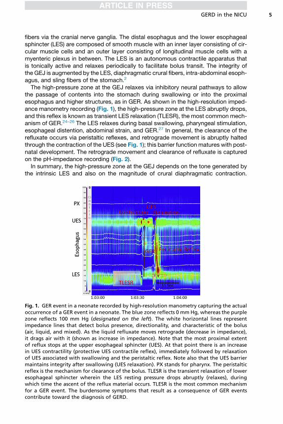
The high-pressure zone at the GEJ relaxes via inhibitory neural pathways to allowthe passage of contents into the stomach during swallowing or into the proximalesophagus and higher structures, as in GER. As shown in the high-resolution imped-ance manometry recording (Fig. 1), the high-pressure zone at the LES abruptly drops,and this reflex is known as transient LES relaxation (TLESR), the most common mech-anism of GER.24–26 The LES relaxes during basal swallowing, pharyngeal stimulation,esophageal distention, abdominal strain, and GER.27 In general, the clearance of therefluxate occurs via peristaltic reflexes, and retrograde movement is abruptly haltedthrough the contraction of the UES (see Fig. 1); this barrier function matures with post-natal development. The retrograde movement and clearance of refluxate is capturedon the pH-impedance recording (Fig. 2).In summary, the high-pressure zone at the GEJ depends on the tone generated by
the intrinsic LES and also on the magnitude of crural diaphragmatic contraction.
Fig. 1. GER event in a neonate recorded by high-resolution manometry capturing the actualoccurrence of a GER event in a neonate. The blue zone reflects 0 mm Hg, whereas the purplezone reflects 100 mm Hg (designated on the left). The white horizontal lines representimpedance lines that detect bolus presence, directionality, and characteristic of the bolus(air, liquid, and mixed). As the liquid refluxate moves retrograde (decrease in impedance),it drags air with it (shown as increase in impedance). Note that the most proximal extentof reflux stops at the upper esophageal sphincter (UES). At that point there is an increasein UES contractility (protective UES contractile reflex), immediately followed by relaxationof UES associated with swallowing and the peristaltic reflex. Note also that the UES barriermaintains integrity after swallowing (UES relaxation). PX stands for pharynx. The peristalticreflex is the mechanism for clearance of the bolus. TLESR is the transient relaxation of loweresophageal sphincter wherein the LES resting pressure drops abruptly (relaxes), duringwhich time the ascent of the reflux material occurs. TLESR is the most common mechanismfor a GER event. The burdensome symptoms that result as a consequence of GER eventscontribute toward the diagnosis of GERD.
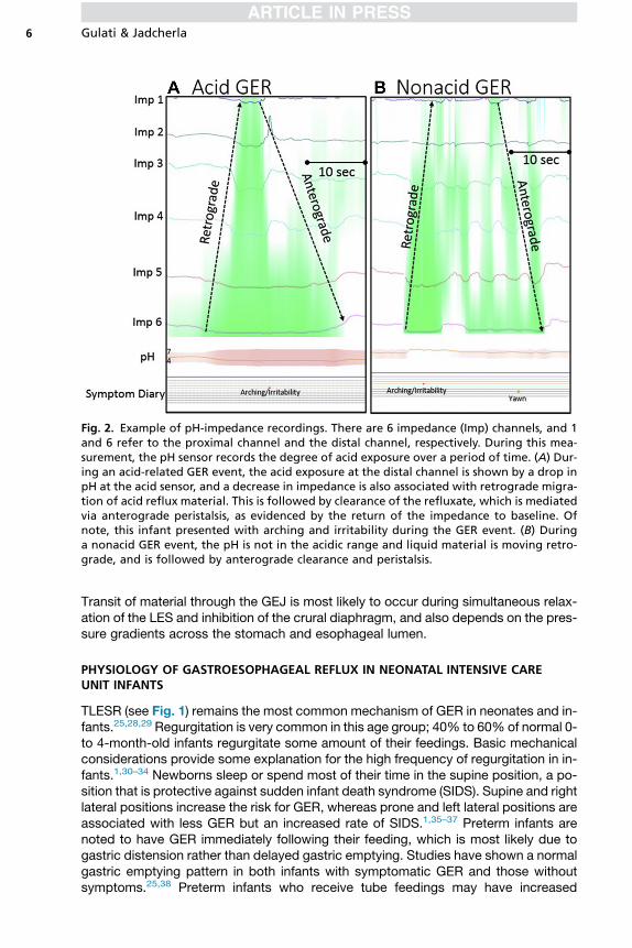
Fig. 2. Example of pH-impedance recordings. There are 6 impedance (Imp) channels, and 1and 6 refer to the proximal channel and the distal channel, respectively. During this mea-surement, the pH sensor records the degree of acid exposure over a period of time. (A) Dur-ing an acid-related GER event, the acid exposure at the distal channel is shown by a drop inpH at the acid sensor, and a decrease in impedance is also associated with retrograde migra-tion of acid reflux material. This is followed by clearance of the refluxate, which is mediatedvia anterograde peristalsis, as evidenced by the return of the impedance to baseline. Ofnote, this infant presented with arching and irritability during the GER event. (B) Duringa nonacid GER event, the pH is not in the acidic range and liquid material is moving retro-grade, and is followed by anterograde clearance and peristalsis.
Gulati & Jadcherla6
Transit of material through the GEJ is most likely to occur during simultaneous relax-ation of the LES and inhibition of the crural diaphragm, and also depends on the pres-sure gradients across the stomach and esophageal lumen.
PHYSIOLOGY OF GASTROESOPHAGEAL REFLUX IN NEONATAL INTENSIVE CAREUNIT INFANTS
TLESR (see Fig. 1) remains the most common mechanism of GER in neonates and in-fants.25,28,29 Regurgitation is very common in this age group; 40% to 60% of normal 0-to 4-month-old infants regurgitate some amount of their feedings. Basic mechanicalconsiderations provide some explanation for the high frequency of regurgitation in in-fants.1,30–34 Newborns sleep or spend most of their time in the supine position, a po-sition that is protective against sudden infant death syndrome (SIDS). Supine and rightlateral positions increase the risk for GER, whereas prone and left lateral positions areassociated with less GER but an increased rate of SIDS.1,35–37 Preterm infants arenoted to have GER immediately following their feeding, which is most likely due togastric distension rather than delayed gastric emptying. Studies have shown a normalgastric emptying pattern in both infants with symptomatic GER and those withoutsymptoms.25,38 Preterm infants who receive tube feedings may have increased
GERD in the NICU 7
episodes of GER because of incomplete closure of the LES secondary to the presenceof a feeding tube.39 However, on the contrary, in symptomatic dysphagic neonatesevaluated for suspected GERD using pH-impedance methods, the authors showedthat tube-fed infants had fewer GER events than the exclusively oral-fed group.12
The length of the infant’s esophagus and LES are short and increase with matura-tion.40 A term infant’s esophagus may be only 8 to 10 cm; the intra-abdominal esoph-agus develops during the first 6 months of life after full-term birth. Thus, refluxedmaterial has a greater chance of extending to a more proximal extent in preterm in-fants who are at 6 months corrected age.Manometric studies in both premature and term neonates have confirmed normal
primary esophageal peristalsis. However, premature infants at 30 to 34 weeks’ gesta-tional age have lower esophageal peristaltic velocity and amplitude than term in-fants,41,42 and preterm infants as young as 33 weeks’ postmenstrual age have areduced esophageal high-pressure zone, which increases with age.43–45 In responseto midesophageal liquid stimulus provocations, premature infants have a longer delayto LES relaxation, but once relaxation occurs it is of longer duration than that found interm infants.27 Premature infants have an elevated frequency of nonperistaltic esoph-ageal contractions in the absence of a swallow, and this lack of coordination may leadto inadequate clearance of refluxed material.43,45 As in adults, it seems that transientrelaxations of the high-pressure zone are the primary mechanism of GER inneonates.43,45,46
In summary, the most frequent mechanism for GER is TLESR, a common mecha-nism in neonates and adults. Factors unique to neonates include anatomic factors, po-sition, feeding methods, immaturity, esophageal clearance mechanisms, andpresence of inflammation or anomalies.
PATHOPHYSIOLOGY OF GASTROESOPHAGEAL REFLUX DISEASE
The esophageal and laryngeal reflexes that protect the esophagus and airway fromdamage caused by GER appear to be present in healthy preterm infants. Esophagealdistension from the reflux of gastric contents activates anterograde peristalsis reflex ofthe esophagus along with closure of the UES. This prevents the refluxate from reach-ing the pharynx. However, if the UES relaxes to allow the refluxate to reach the phar-ynx, the laryngeal chemosensitive receptors trigger the initiation of the laryngealchemoreflex to prevent aspiration of gastric contents by glottis closure, which is al-ways accompanied by a period of apnea (glottal closure reflex), although the durationof pause in breathing varies. In addition, primary peristalsis is triggered when therefluxate is present in the pharynx. Theoretically, GERD and retrograde aspirationcould result from the failure of these mechanisms. Abnormalities of all of these reflexesare unlikely in physiologically healthy infants, which is why most healthy infants areasymptomatic despite having frequent episodes of GER.47–49
RISK FACTORS FOR GASTROESOPHAGEAL REFLUX DISEASE
Several risk factors for GERD have been identified in infants, and the most commoncauses are listed in Fig. 3. Apart from the congenital causes and physiologic conse-quences of prematurity, it is important to analyze the causes of GER events that arisefrom complications of prematurity. Bronchopulmonary dysplasia is a major complica-tion of prematurity that affects 30% of extremely low birth weight infants.50 These in-fants have an increased risk of GER events secondary to increased respiratory effortand transient increase in intra-abdominal pressure resulting from coughing, airflowobstruction, and crying. This causes the LES tone to decrease and in turn contributes
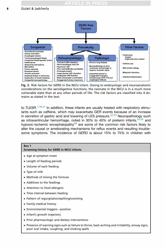
Fig. 3. Risk factors for GERD in the NICU infant. Owing to embryologic and neuroanatomicconsiderations on the aerodigestive functions, the neonate in the NICU is in a much morevulnerable state than at any other periods of life. The risk factors are classified into 4 do-mains as stated in the text.
Gulati & Jadcherla8
to TLESR.1,46,51 In addition, these infants are usually treated with respiratory stimu-lants such as caffeine, which may exacerbate GER events because of an increasein secretion of gastric acid and lowering of LES pressure.52,53 Neuropathology suchas intraventricular hemorrhage, noted in 30% to 40% of preterm infants,54,55 andhypoxic-ischemic encephalopathy56 are some of the common risk factors likely toalter the causal or ameliorating mechanisms for reflux events and resulting trouble-some symptoms. The incidence of GERD is about 15% to 75% in children with
Box 1
Screening history for GERD in NICU infants
� Age at symptom onset
� Length of feeding periods
� Volume of each feeding
� Type of milk
� Methods of mixing the formula
� Additives to the feedings
� Attention to food allergens
� Time interval between feeding
� Pattern of regurgitation/spitting/vomiting
� Family medical history
� Environmental triggers—position
� Infant’s growth trajectory
� Prior pharmacologic and dietary interventions
� Presence of warning signs such as failure to thrive, back arching and irritability, airway signs,poor oral intake, coughing, and choking spells
GERD in the NICU 9
neurologic impairment, and the prevalence of GERD in the presence of neuropa-thology is estimated to be 50%.57 Neuropathology contributes to GERD through dys-regulation of aerodigestive reflexes. Several other causes may contribute to GERD,including metabolic disorders, body positioning, milk protein allergy, and infections,to name a few. These conditions must be investigated in preterm infants who presentwith signs concerning GERD.
APPROACH TO THE PROBLEM THOUGHT TO BE DUE TO GASTROESOPHAGEALREFLUX DISEASE IN INFANTS IN THE NEONATAL INTENSIVE CARE UNIT
In 2018, the North American Society for Pediatric Gastroenterology, Hepatology andNutrition and the European Society for Pediatric Gastroenterology Hepatology andNutrition1 published guidelines on the approach to children presenting with GER orGERD. However, further research is needed because these guidelines are not entirelyclear about applicability to infants in the NICU. Infants in the NICU presenting with
Box 2
Signs and symptoms that may be associated with GERD in NICU infants
Symptoms
Gastrointestinal� Regurgitation� Spitting� Emesis� Abdominal distension
Aerodigestive� Swallowing problems� Feeding problems� Sneezing� Coughing� Choking� Wheezing� Stridor
Cardiorespiratory Spells� Bradycardia� Tachycardia� Apneas� Periodic breathing� Tachypnea� Increased respiratory effort� Desaturations
Somatosensory� Irritability� Back arching� Crying� Grimace
Signs
Aerodigestive� Esophagitis� Recurrent pneumonia with aspiration� Recurrent otitis media
General� Anemia� Failure to thrive
Gulati & Jadcherla10
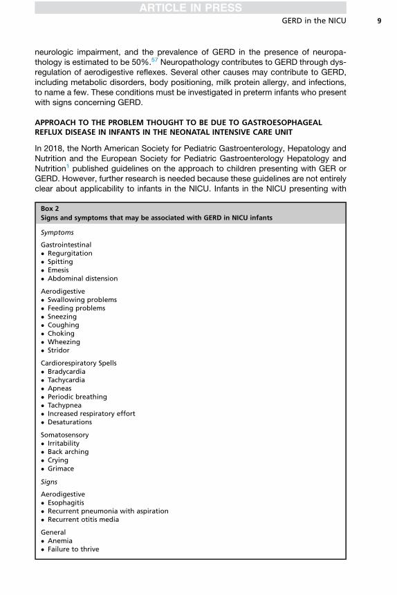
troublesome signs and symptoms suspected to be due to GERD should be evaluatedthoroughly for any findings suggestive of disorders other than GERD. A wide range ofclinical symptoms are attributed to GERD in NICU infants; however, the reliability ofthese symptoms as a manifestation of GERD is not clear. The evaluation of a neonatewith a suspicion for GERD begins with a thorough focused history (Box 1) and physicalexamination while paying attention to the pharyngoesophageal, cardiorespiratory, andneurologic systems, nutrition, feeding methods, and growth characteristics. In partic-ular, the evaluation should pay attention to signs and symptoms of aerodigestive prob-lems, nonspecific behavioral signs including arching and irritability, and feedingproblems that may be associated with GERD (Box 2). Additionally it is imperative toexclude any symptoms and signs that masquerade as GERD. Relevant risk factors(see Fig. 3) must be addressed. Initial management should include paying attentionto optimal nutrition and feeding methods, and continued breastfeeding. However, ifthere are no improvements a trial of protein hydrolysate or amino acid–based formulaor, in breastfed infants, elimination of cow’s milk in the maternal diet should be consid-ered for 2 to 4 weeks. If there are no improvements despite these interventions,gastrointestinal specialty testing using pH impedance with symptom correlationmethods, and/or manometry for pharyngoesophageal functional abnormalities, maybe considered when available to ascertain the causal and ameliorating mechanisms.In such situations, or if a referral for specialty testing is not possible, 4 to 8 weeks’ trialof acid suppression using proton-pump inhibitors may be considered with extremecaution, while weighing the benefits versus risks, and this can only be consideredwhen infants are at and beyond full term age.10 There are no safe prokinetic agentsfor use in premature infants. The role of antacids remains uncertain in the prematureinfant population.
SUMMARY
Diagnosis and management considerations for GER and GERD in the NICU infant canbe challenging. Neonatal presentations are not as typical as those seen in older infantsand children with GERD. Symptom-based diagnosis and empirical pharmacologictherapies are not appropriate. Developmental pathologies and maturational deficitsin the causal and ameliorating mechanisms of GER may be associated with GERDrisks. When relevant, structural anomalies and risk factors of GERD must beaddressed. Emphasis must first be placed on optimal nutrition, feeding methods,growth, conservative management, and reassurance. Because symptoms arenonspecific, other causes and diagnoses that masquerade as GERD must be consid-ered. Minimizing the use and duration of acid-suppressive therapies is appropriatewhile weighing benefits and risks. Further research is critically needed in this high-risk population of NICU infants, with relevance to screening, diagnostic algorithms,objective criteria, and nonpharmacologic and pharmacologic approaches, to manageobjectively determined acid and nonacid GERD and their consequences.
ACKNOWLEDGMENTS
The authors are thankful to Nour Hanandeh, BS, BME for help with figures, andKathryn A. Hasenstab, BS, BME for help with tables and references.
REFERENCES
1. Rosen R, Vandenplas Y, Singendonk M, et al. Pediatric gastroesophageal refluxclinical practice guidelines: joint recommendations of the North American Society
GERD in the NICU 11
for Pediatric Gastroenterology, Hepatology, and Nutrition and the European Soci-ety for Pediatric Gastroenterology, Hepatology, and Nutrition. J Pediatr Gastroen-terol Nutr 2018;66(3):516–54.
2. Jadcherla SR. Pathophysiology of gastroesophageal reflux. In: Polin RA,Abman SH, Rowitch D, et al, editors. Fetal and neonatal physiology. 5th edition.Philadelphia: Elsevier; 2017. p. 1643–52.
3. Eichenwald EC. Committee on fetus and newborn. Diagnosis and managementof gastroesophageal reflux in preterm infants. Pediatrics 2018;142(1):e20181061.
4. Slaughter JL, Stenger MR, Reagan PB, et al. Neonatal histamine-2 receptorantagonist and proton pump inhibitor treatment at United States Children’s Hos-pitals. J Pediatr 2016;174:63–70.
5. Malcolm WF, Cotton CM. Metoclopramide, H2 blockers, and proton pump inhib-itors: pharmacotherapy for gastroesophageal reflux in neonates. Clin Perinatol2012;39(1):99–109.
6. Hibbs AM, Lorch SA. Metoclopramide for the treatment of gastroesophageal re-flux disease in infants: a systematic review. Pediatrics 2006;118(2):746–52.
7. Guillet R, Stoll BJ, Cotton CM, et al. Association of H2-blocker therapy and higherincidence of necrotizing enterocolitis in very low birth weight infants. Pediatrics2006;117(2):e137–42.
8. Terrin G, Passariello A, DeCurtis M, et al. Ranitidine is associated with infections,necrotizing enterocolitis, and fatal outcome in newborns. Pediatrics 2012;129(1):e40–5.
9. Orenstein SR, Hassall E, Furmaga-Jablonska W, et al. Multicenter, double-blind,randomized, placebo-controlled trial assessing the efficacy and safety of protonpump inhibitor lansoprazole in infants with symptoms of gastroesophageal refluxdisease. J Pediatr 2009;154(4):514–20.
10. El-Mahdy MA, Mansoor FA, Jadcherla SR. Pharmacological management ofgastroesophageal reflux disease in infants: current opinions. Curr Opin Pharma-col 2017;37:112–7.
11. Lopez-Alonso M, Moya MJ, Cabo JA, et al. Twenty-four-hour esophagealimpedance-pH monitoring in healthy preterm neonates: rate and characteristicsof acid, weakly acidic, and weakly alkaline gastroesophageal reflux. Pediatrics2006;118(2):e299–308.
12. Jadcherla SR, Slaughter JL, Stenger MR, et al. Practice variance, prevalence,and economic burden of premature infants diagnosed with GERD. Hosp Pediatr2013;3(4):335–41.
13. Qureshi A, Malkar M, Splaingard M, et al. The role of sleep in the modulation ofgastroesophageal reflux and symptoms in NICU neonates. Pediatr Neurol2015;53(3):226–32.
14. Sankaran J, Qureshi AH, Woodley F, et al. Effect of severity of esophageal acid-ification on sleep vs wake periods in infants presenting with brief resolved unex-plained events. J Pediatr 2016;179:42–8.
15. Golski CA, Rome EW, Martin RJ, et al. Pediatric specialists’ beliefs about gastro-esophageal reflux disease in premature infants. Pediatrics 2010;125(1):96–104.
16. Kleinman L, Rothman M, Strauss R, et al. The infant gastroesophageal refluxquestionnaire revised: development and validation as an evaluative instrument.Clin Gastroenterol Hepatol 2006;4(5):588–96.
17. Salvatore S, Hauser B, Vandemaele K, et al. Gastroesophageal reflux disease ininfants: how much is predictable with questionnaires, pH-metry, endoscopy andhistology? J Pediatr Gastroenterol Nutr 2005;40(2):210–5.
Gulati & Jadcherla12
18. Levy DS, Osborn E, Hasenstab KA, et al. The effect of additives for reflux ordysphagia management on osmolality in ready-to-feed preterm formula: practiceimplications. JPEN J Parenter Enteral Nutr 2018. https://doi.org/10.1002/jpen.1418.
19. Mansfield LE. Embryonic origins of the relation of gastroesophageal reflux dis-ease and airway disease. Am J Med 2001;111(Suppl 8A):3S–7S.
20. Miller JL, Sonies BC, Macedonia C. Emergence of oropharyngeal, laryngeal andswallowing activity in the developing fetal upper aerodigestive tract: an ultra-sound evaluation. Early Hum Dev 2003;71(1):61–87.
21. Sadler TW. Respiratory system. In: Sadler TW, editor. Langman’s medical embry-ology. Baltimore (MD): Williams & Wilkins; 1995. p. 232–41.
22. Sadler TW. Digestive system. In: Sadler TW, editor. Langman’s medical embry-ology. Baltimore (MD): Williams & Wilkins; 1995. p. 208–29.
23. Lang IM, Shaker R. Anatomy and physiology of the upper esophageal sphincter.Am J Med 1997;103(5A):50S–5S.
24. Werlin SL, Dodds WJ, Hogan WJ, et al. Mechanisms of gastroesophageal refluxin children. J Pediatr 1980;97(2):244–9.
25. Omari TI, Barnett CP, Benninga MA, et al. Mechanisms of gastro-oesophageal re-flux in preterm and term infants with reflux disease. Gut 2002;51(4):475–9.
26. Dent J, Dodds WJ, Friedman RH, et al. Mechanism of gastroesophageal reflux inrecumbent asymptomatic human-subjects. J Clin Invest 1980;65(2):256–67.
27. Pena EM, Parks VN, Peng J, et al. Lower esophageal sphincter relaxation reflexkinetics: effects of peristaltic reflexes and maturation in human premature neo-nates. Am J Physiol Gastrointest Liver Physiol 2010;299(6):G1386–95.
28. Omari TI, Barnett C, Snel A, et al. Mechanisms of gastroesophageal reflux inhealthy premature infants. J Pediatr 1998;133(5):650–4.
29. Omari TI, Benninga MA, Barnett CP, et al. Characterization of esophageal bodyand lower esophageal sphincter motor function in the very premature neonate.J Pediatr 1999;135(4):517–21.
30. Hyman PE, Milla PJ, Benninga MA, et al. Childhood functional gastrointestinaldisorders: neonate/toddler. Gastroenterology 2006;130(5):1519–26.
31. Orenstein SR. Prone positioning in infant gastroesophageal reflux: is elevation ofthe head worth the trouble? J Pediatr 1990;117(2 Pt 1):184–7.
32. Sondheimer JM. Gastroesophageal reflux: update on pathogenesis and diag-nosis. Pediatr Clin North Am 1988;35(1):103–16.
33. Corvaglia L, Rotatori R, Ferlini M, et al. The effect of body positioning on gastro-esophageal reflux in premature infants: evaluation by combined impedance andpH monitoring. J Pediatr 2007;151(6):591–6.
34. Omari TI, Rommel N, Staunton E, et al. Paradoxical impact of body positioning ongastroesophageal reflux and gastric emptying in the premature neonate.J Pediatr 2004;145(2):194–200.
35. Jadcherla SR, Rudolph CD. Gastroesophageal reflux in the preterm neonate.Neoreviews 2005;6(2):e87–98.
36. Jadcherla SR. Gastroesophageal reflux in the neonate. Clin Perinatol 2002;29(1):135–58.
37. Jadcherla SR. Pathophysiology of aerodigestive pulmonary disorders in theneonate. Clin Perinatol 2012;39(3):639–54.
38. Ewer AK, Durbin GM, Morgan ME, et al. Gastric emptying and gastro-oesophageal reflux in preterm infants. Arch Dis Child Fetal Neonatal Ed 1996;75(2):F117–21.
GERD in the NICU 13
39. Peter CS, Sprodowski N, Bohnhorst B, et al. Gastroesophageal reflux and apneaof prematurity: no temporal relationship. Pediatrics 2002;109(1):8–11.
40. Gupta A, Jadcherla SR. The relationship between somatic growth and in vivoesophageal segmental and sphincteric growth in human neonates. J PediatrGastroenterol Nutr 2006;43(1):35–41.
41. Gupta A, Gulati P, Kim W, et al. Effect of postnatal maturation on the mechanismsof esophageal propulsion in preterm human neonates: primary and secondaryperistalsis. Am J Gastroenterol 2009;104(2):411–9.
42. Jadcherla SR, Duong HQ, Hofmann C, et al. Characteristics of upper oesopha-geal sphincter and oesophageal body during maturation in healthy human neo-nates compared with adults. Neurogastroenterol Motil 2005;17(5):663–70.
43. Omari TI, Miki K, Davidson G, et al. Characterisation of relaxation of the lower oe-sophageal sphincter in healthy premature infants. Gut 1997;40(3):370–5.
44. Kawahara H, Dent J, Davidson G. Mechanisms responsible for gastroesophagealreflux in children. Gastroenterology 1997;113(2):399–408.
45. Omari TI, Miki K, Fraser R, et al. Esophageal body and lower esophagealsphincter function in healthy premature infants. Gastroenterology 1995;109(6):1757–64.
46. Omari T, Barnett C, Snel A, et al. Mechanism of gastroesophageal reflux in pre-mature infants with chronic lung disease. J Pediatr Surg 1999;34(12):1795–8.
47. Thach BT. Reflux associated apnea in infants: evidence for a laryngeal chemore-flex. Am J Med 1997;103:120s–4s.
48. Jadcherla SR, Hoffmann RG, Shaker R. Effect of maturation of the magnitude ofmechanosensitive and chemosensitive reflexes in the premature human esoph-agus. J Pediatr 2006;149(1):77–82.
49. Jadcherla SR, Gupta A, Coley BD, et al. Esophago-glottal closure reflex in humaninfants: a novel reflex elicited with concurrent manometry and ultrasonography.Am J Gastroenterol 2007;102(10):2286–93.
50. Nobile S, Noviello C, Cobellis G, et al. Are infants with bronchopulmonarydysplasia prone to gastroesophageal reflux? a prospective observational studywith esophageal ph-impedance monitoring. J Pediatr 2015;167(2):279–85.
51. Orenstein SR, Orenstein DM. Gastroesophageal reflux and respiratory-disease inchildren. J Pediatr 1988;112(6):847–58.
52. Foster LJ, Trudeau WL, Goldman AL. Bronchodilator effects on gastric acidsecretion. JAMA 1979;241(24):2613–5.
53. Stein MR, Towner TG, Weber RW, et al. The effect of theophylline on the loweresophageal sphincter pressure. Ann Allergy 1980;45(4):238–41.
54. Ballabh P. Intraventricular hemorrhage in premature infants: mechanism of dis-ease. Pediatr Res 2010;67(1):1–8.
55. Payne AH, Hintz SR, Hibbs AM, et al. Neurodevelopmental outcomes ofextremely low-gestational-age neonates with low-grade periventricular-intraven-tricular hemorrhage. JAMA Pediatr 2013;167(5):451–9.
56. Hill CD, Jadcherla SR. Esophageal mechanosensitive mechanisms are impairedin neonates with hypoxic-ischemic encephalopathy. J Pediatr 2013;162(5):976–82.
57. Del Buono R, Wenzl TG, Rawat D, et al. Acid and nonacid gastro-oesophagealreflux in neurologically impaired children: investigation with the multiple intralumi-nal impedance procedure. J Pediatr Gastroenterol Nutr 2006;43(3):331–5.