Embed Size (px)
DESCRIPTION
Gastrointestinal First Aid Step 2 CK
Citation preview

151
H I G H - Y I E L D F A C T S I N
Gastrointestinal
Esophageal Disease 153
DYSPHAGIA/ODYNOPHAGIA 153
INFECTIOUS ESOPHAGITIS 153
DIFFUSE ESOPHAGEAL SPASM 153
ACHALASIA 154
ESOPHAGEAL DIVERTICULA 155
ESOPHAGEAL CANCER 155
GASTROESOPHAGEAL REFLUX DISEASE 155
HIATAL HERNIA 156
Disorders of the Stomach and Duodenum 156
GASTRITIS 156
GASTRIC CANCER 157
PEPTIC ULCER DISEASE 157
ZOLLINGER-ELLISON SYNDROME 158
Disorders of the Small Bowel 159
DIARRHEA 159
MALABSORPTION 161
LACTOSE INTOLERANCE 162
CARCINOID SYNDROME 162
IRRITABLE BOWEL SYNDROME 162
SMALL BOWEL OBSTRUCTION 163
ILEUS 164
MESENTERIC ISCHEMIA 165
APPENDICITIS 166
Disorders of the Large Bowel 166
DIVERTICULAR DISEASE 166
LARGE BOWEL OBSTRUCTION 167
COLON AND RECTAL CANCER 167
ISCHEMIC COLITIS 170
Gastrointestinal Bleeding 170
Infl ammatory Bowel Disease 170
Inguinal Hernias 172
504 _02-06_Gastro.indd 151 8/18/09 9:56:59 A

152
Biliary Disease 173
CHOLELITHIASIS AND BILIARY COLIC 173
ACUTE CHOLECYSTITIS 174
CHOLEDOCHOLITHIASIS 175
ASCENDING CHOLANGITIS 176
GALLSTONE ILEUS 176
PRIMARY SCLEROSING CHOLANGITIS 177
Liver Disease 177
ABNORMAL LIVER FUNCTION TESTS 177
HEPATITIS 177
CIRRHOSIS 180
PRIMARY BILIARY CIRRHOSIS 182
HEPATOCELLULAR CARCINOMA 182
HEMOCHROMATOSIS 183
WILSON’S DISEASE (HEPATOLENTICULAR DEGENERATION) 183
Pancreatic Disease 184
PANCREATITIS 184
PANCREATIC CANCER 185
504 _02-06_Gastro.indd 152 8/18/09 9:56:59 A

153
HIGH-YIELD FACTS
GASTROINTESTINAL
E SOPHAG EAL D ISEASE �
Dysphagia/Odynophagia
Diffi culty swallowing (dysphagia) or pain with swallowing (odynophagia) due to abnormalities of the oropharynx or esophagus.
HISTORY/PE
■ Presentation varies according to the location:■ Oropharyngeal dysphagia: Presents with diffi culty passing material
from the oropharynx to the esophagus. Usually involves liquids more than solids. Causes can be neurologic or muscular and include stroke, Parkinson’s disease, myasthenia gravis, prolonged intubation, and Zen-ker’s diverticula.
■ Esophageal dysphagia: Usually involves solids more than liquids for most obstructive causes (strictures, Schatzki rings, webs, carcinoma) and is generally progressive. Motility disorders (achalasia, scleroderma, esophageal spasm) present with both liquid and solid dysphagia.
■ Examine for masses (e.g., goiter, tumor) and anatomic defects.
DIAGNOSIS
■ Oropharyngeal dysphagia: Cine-esophagram.■ Esophageal dysphagia: Barium swallow followed by endoscopy, manom-
etry, and/or pH monitoring. If an obstructive lesion is suspected, proceed directly to endoscopy with biopsy.
■ Odynophagia: Upper endoscopy.
TREATMENT
Etiology dependent.
Infectious Esophagitis
Table 2.6-1 outlines the etiology, diagnosis, and treatment of infectious esophagitis.
Diffuse Esophageal Spasm
■ A motility disorder in which normal peristalsis is periodically interrupted by high-amplitude nonperistaltic contractions of unknown etiology. Also known as nutcracker esophagus.
Esophageal webs are
associated with iron defi ciency
anemia and glossitis
(Plummer-Vinson syndrome).
Candidal esophagitis is an
AIDS-defi ning illness.
T A B L E 2 . 6 - 1 . Causes of Infectious Esophagitis
ETIOLOGIC AGENT EXAM FINDINGS UPPER ENDOSCOPY TREATMENT
Candida albicans Oral thrush Yellow-white plaques adherent to the mucosa. Fluconazole PO
HSV Oral ulcers Small, deep ulcerations; multinucleated giant
cells with nuclear inclusions on biopsy.
Acyclovir IV
CMV Retinitis, colitis Large, superfi cial ulcerations; intranuclear
inclusions on biopsy.
Ganciclovir IV
504 _02-06_Gastro.indd 153 8/18/09 9:56:59 A

HIG
H-YI
ELD
FACT
SGA
STRO
INTE
STIN
AL
154
■ Hx/PE: Presents with chest pain, dysphagia, and odynophagia. Often pre-cipitated by ingestion of hot or cold liquids; relieved by nitroglycerin.
■ Dx: Barium swallow may show a corkscrew-shaped esophagus. Esopha-geal manometry reveals high-amplitude, simultaneous contractions.
■ Tx: Nitrates and calcium channel blockers (CCBs) for symptomatic relief; surgery (esophageal myotomy) for severe, incapacitating symptoms.
Achalasia
■ A motor disorder of the esophagus characterized by impaired relaxation of the lower esophageal sphincter (LES) and loss of peristalsis in the distal two-thirds of the esophagus. Thought to result from the loss or absence of inhibitory ganglion cells in the myenteric (Auerbach’s) plexus.
■ Hx/PE: Progressive dysphagia, chest pain, regurgitation of undigested food, weight loss, and nocturnal cough are common symptoms.
■ Dx:■ Barium swallow reveals esophageal dilation with a “bird’s beak” ta-
pering of the distal esophagus (see Figure 2.6-1).■ Manometry shows ↑ resting LES pressure, incomplete LES relax-
ation upon swallowing, and ↓ peristalsis in the body of the esophagus.■ Mechanical causes of obstruction must be ruled out by endoscopy.
■ Tx: Nitrates, CCBs, or endoscopic injection of botulinum toxin into the LES may provide short-term relief of symptoms. Pneumatic balloon dila-tion or surgical (Heller) myotomy are defi nitive treatment options.
The musculature of the upper
one-third of the esophagus is
skeletal, whereas that of the
lower two-thirds is smooth
muscle.
Malignancy may mimic
achalasia (pseudoachalasia).
F I G U R E 2 . 6 - 1 . Achalasia.
Barium esophagram reveals esophageal dilation with a “bird’s beak” tapering of the distal
esophagus. (Reproduced from Waters PF, DeMeester TR. Foregut motor disorders and their
surgical management. Med Clin North Am 65: 1235, © 1981, with permission from Elsevier.)
504 _02-06_Gastro.indd 154 8/18/09 9:57:00 A

155
HIGH-YIELD FACTS
GASTROINTESTINAL
Esophageal Diverticula
■ The second most common esophageal motility disorder (after achalasia).■ Cervical outpouching through the cricopharyngeal muscle is called Zen-
ker’s diverticulum. Diverticula can also occur in the middle and distal third of the esophagus.
■ Hx/PE: Patients complain of chest pain, dysphagia, halitosis, and regurgi-tation of undigested food.
■ Dx: Barium swallow will demonstrate outpouchings.■ Tx: If symptomatic, treat with surgical excision of the diverticulum. For
Zenker’s diverticulum, myotomy of the cricopharyngeus is required to re-lieve the high-pressure zone.
Esophageal Cancer
■ Squamous cell carcinoma (SCC) is the most common type of esophageal cancer worldwide, while adenocarcinoma is most prevalent in the United States, Europe, and Australia. The latter is associated with Barrett’s esopha-gus (columnar metaplasia of the distal esophagus 2° to chronic GERD).
■ Alcohol use and smoking have been identifi ed as major risk factors for SCC.
■ Hx/PE: Progressive dysphagia, initially to solids and later to liquids, is common. Weight loss, odynophagia, GERD, GI bleeding, and vomiting are also seen.
■ Dx: Barium study shows narrowing of the esophagus with an irregular bor-der protruding into the lumen. EGD and biopsy confi rm the diagnosis. CT and endoscopic ultrasound are used for staging.
■ Tx: Chemoradiation and surgical resection is the defi nitive treatment, but the prognosis is poor. Surgery is also offered for high-grade Barrett’s dyspla-sia. Patients who are not candidates for surgery may require an endoscopi-cally placed esophageal stent for palliation and improved quality of life.
Gastroesophageal Refl ux Disease (GERD)
Symptomatic refl ux of gastric contents into the esophagus, most commonly as a result of transient LES relaxation. Can be due to an incompetent LES, gastroparesis, or hiatal hernia.
HISTORY/PE
■ Patients present with heartburn that commonly occurs 30–90 minutes af-ter a meal, worsens with reclining, and often improves with antacids, sit-ting, or standing. Substernal chest pain can be diffi cult to distinguish from other causes.
■ Sour taste (“water brash”), globus, unexplained cough, and morning hoarseness can be clues.
■ Exam is usually normal unless a systemic disease (e.g., scleroderma) is present.
DIAGNOSIS
■ The history and clinical impression are important.■ An empiric trial of lifestyle modifi cation and medical treatment is often at-
tempted fi rst. Studies may include barium swallow (to look for hiatal her-nia), esophageal manometry, and 24-hour pH monitoring.
Squamous cell esophageal
cancer is associated with
tobacco and alcohol use.
Esophageal cancer
metastasizes early because
the esophagus lacks a serosa.
Risk factors for GERD include
hiatal hernia and ↑ intra-
abdominal pressure (e.g.,
obesity, pregnancy).
GERD can mimic cough-
variant asthma.
504 _02-06_Gastro.indd 155 8/18/09 9:57:00 A

HIG
H-YI
ELD
FACT
SGA
STRO
INTE
STIN
AL
156
■ EGD with biopsies should be performed in patients whose symptoms are unresponsive to initial empiric therapy, long-standing (to rule out Barrett’s esophagus and adenocarcinoma), or suggestive of complicated disease (e.g., anorexia, weight loss, dysphagia/odynophagia).
TREATMENT
■ Lifestyle: Weight loss, head-of-bed elevation, reduction of meal size, and avoidance of nocturnal meals and substances that ↓ LES tone.
■ Pharmacologic: Start with antacids in patients with mild, intermittent symptoms; use H2 receptor antagonists (cimetidine, ranitidine) or PPIs (omeprazole, lansoprazole) in patients with chronic and frequent symp-toms. PPIs are preferred for severe or erosive disease.
■ Surgical: For refractory or severe disease, Nissen fundoplication may offer signifi cant relief.
COMPLICATIONS
Esophagitis, esophageal stricture, aspiration of gastric contents, upper GI bleeding, Barrett’s esophagus.
Hiatal Hernia
■ Herniation of a portion of the stomach upward into the chest through a diaphragmatic opening. There are two common types:■ Sliding hiatal hernias (95%): The gastroesophageal junction and a
portion of the stomach are displaced above the diaphragm.■ Paraesophageal hiatal hernias (5%): The gastroesophageal junction re-
mains below the diaphragm, while a neighboring portion of the fundus herniates into the mediastinum.
■ Hx/PE: May be asymptomatic. Those with sliding hernias may present with GERD.
■ Dx: Commonly an incidental fi nding on CXR; also frequently diagnosed by barium swallow or EGD.
■ Tx:■ Sliding hernias: Medical therapy and lifestyle modifi cations to ↓
GERD symptoms.■ Paraesophageal hernias: Surgical gastropexy (attachment of the stom-
ach to the rectus sheath and closure of the hiatus) is recommended to prevent gastric volvulus.
DISORDE RS OF TH E STOMAC H AN D DUODE N U M �
Gastritis
Infl ammation of the stomach lining. Subtypes are as follows:
■ Acute gastritis: Rapidly developing, superfi cial lesions that are often due to NSAID use, alcohol, H. pylori infection, and stress from severe illness (e.g., burns, CNS injury).
■ Chronic gastritis:■ Type A (10%): Occurs in the fundus and is due to autoantibodies to
parietal cells. Causes pernicious anemia and is associated with other autoimmune disorders, such as thyroiditis. Also ↑ the risk of gastric ad-enocarcinoma.
Patients with GERD should
avoid caffeine, alcohol,
chocolate, garlic, onions,
mints, and nicotine.
BARRett’s—
BecomesAdenocarcinomaResults fromRefl ux
Type A gastritis is associated
with pernicious anemia due
to the lack of intrinsic factor
necessary for the absorption
of vitamin B12.
504 _02-06_Gastro.indd 156 8/18/09 9:57:00 A

157
HIGH-YIELD FACTS
GASTROINTESTINAL
■ Type B (90%): Occurs in the antrum and may be caused by NSAID use or H. pylori infection. Often asymptomatic, but associated with ↑ risk of PUD and gastric cancer.
HISTORY/PE
Patients may be asymptomatic or may complain of epigastric pain, nausea, vomiting, hematemesis, or melena.
DIAGNOSIS
■ Upper endoscopy can visualize the gastric lining.■ H. pylori infection can be detected by urease breath test, serum IgG an-
tibodies (which indicate exposure, not current infection), H. pylori stool antigen, or endoscopic biopsy.
TREATMENT
■ ↓ intake of offending agents. Antacids, sucralfate, H2 blockers, and/or PPIs may help.
■ Triple therapy (amoxicillin, clarithromycin, omeprazole) to treat H. pylori infection.
■ Give prophylactic H2 blockers or PPIs to patients at risk for stress ulcers (e.g., ICU patients).
Gastric Cancer
■ This malignant tumor is the second most common cause of cancer-related death worldwide and is particularly common in Korea and Japan. Tumors are generally adenocarcinomas, which exhibit two morphologic types:■ Intestinal type: Thought to arise from intestinal metaplasia of gas-
tric mucosal cells. Risk factors include a diet high in nitrites and salt and low in fresh vegetables (antioxidants), H. pylori colonization, and chronic gastritis.
■ Diffuse type: Tends to be poorly differentiated and not associated with H. pylori infection or chronic gastritis. Risk factors are largely unknown; signet ring cells are seen on pathology.
■ Hx/PE: Early signs are indigestion and loss of appetite. Advanced cases generally present with abdominal pain, weight loss, or upper GI bleeding.
■ Dx: Early gastric carcinoma is largely asymptomatic and is discovered ser-endipitously with endoscopic examination of high-risk individuals.
■ Tx: Successful treatment rests entirely on early detection and surgical re-moval of the tumor. Five-year survival is < 10% for advanced disease.
Peptic Ulcer Disease (PUD)
Damage to the gastric or duodenal mucosa caused by impaired mucosal defense and/or ↑ acidic gastric contents. H. pylori plays a causative role in > 90% of duodenal ulcers and 70% of gastric ulcers. Other risk factors include corticosteroid, NSAID, alcohol, and tobacco use. Males are affected more often than females.
HISTORY/PE
■ Classically presents with chronic or periodic dull, burning epigastric pain that improves with meals (especially duodenal ulcers), worsens 2–3 hours after eating, and can radiate to the back.
Gastric adenocarcinoma that
metastasizes to the ovary is
called a Krukenberg tumor.
Gastric cancer may present
with Virchow’s node (an
enlarged left supraclavicular
lymph node).
504 _02-06_Gastro.indd 157 8/18/09 9:57:01 A

HIG
H-YI
ELD
FACT
SGA
STRO
INTE
STIN
AL
158
■ Patients may also complain of nausea, hematemesis (“coffee-ground” em-esis), or blood in the stool (melena or hematochezia).
■ Exam may reveal varying degrees of epigastric tenderness and, if there is active bleeding, a � stool guaiac.
■ An acute perforation can present with a rigid abdomen, rebound tender-ness, guarding, or other signs of peritoneal irritation.
DIAGNOSIS
■ AXR to rule out perforation (free air under the diaphragm); CBC to as-sess for GI bleeding (low or ↓ hematocrit).
■ Upper endoscopy with biopsy to confi rm PUD and to rule out active bleeding or gastric adenocarcinoma (10% of gastric ulcers); barium swal-low is an alternative.
■ H. pylori testing.■ In recurrent or refractory cases, serum gastrin can be used to screen for
Zollinger-Ellison syndrome (patients must discontinue PPI use prior to testing).
TREATMENT
■ Acute:■ Rule out active bleeding with serial hematocrits, a rectal exam with
stool guaiac, and NG lavage.■ Monitor the patient’s hematocrit and BP and treat with IV hydration,
transfusion, IV PPIs, endoscopy, and surgery as needed for complica-tions.
■ If perforation is likely, emergent surgery is indicated.■ Pharmacologic:
■ Involves protecting the mucosa, decreasing acid production, and eradi-cating H. pylori infection.
■ Treat mild disease with antacids or with sucralfate, bismuth, and miso-prostol (a prostaglandin analog) for mucosal protection. PPIs or H2 re-ceptor antagonists may be used to ↓ acid secretion.
■ Patients with confi rmed H. pylori infection should receive triple ther-apy (amoxicillin, clarithromycin, and omeprazole).
■ Discontinue use of exacerbating agents. Patients with recurrent or se-vere disease may require chronic symptomatic therapy.
■ Endoscopy and surgery:■ Patients with symptomatic gastric ulcers for > 2 months that are refrac-
tory to medical therapy should have either endoscopy or an upper GI series with barium to rule out gastric adenocarcinoma.
■ Refractory cases may require a surgical procedure such as parietal cell vagotomy (the most selective and preferred surgical approach).
COMPLICATIONS
Hemorrhage (posterior ulcers that erode into the gastroduodenal artery), gas-tric outlet obstruction, perforation (usually anterior ulcers), intractable pain.
Zollinger-Ellison Syndrome
■ A rare condition characterized by gastrin-producing tumors in the duode-num and/or pancreas that lead to oversecretion of gastrin.
■ ↑ gastrin results in the production of high levels of gastric acid by the gas-tric mucosa, leading to recurrent/intractable ulcers in the stomach and duodenum (may occur more distally).
After a meal, pain from a
Gastric ulcer is Greater,
whereas Duodenal pain
Decreases.
Roughly 5–7% of lower GI
bleeds are from an upper GI
source.
Rule out Zollinger-Ellison
syndrome with serum gastrin
levels in cases of GERD and
PUD that are refractory to
medical management.
Misoprostol can help patients
with PUD who require NSAID
therapy (e.g., for arthritis).
Complications of PUD—
HOPI
HemorrhageObstructionPerforationIntractable pain
504 _02-06_Gastro.indd 158 8/18/09 9:57:01 A

159
HIGH-YIELD FACTS
GASTROINTESTINAL
■ In 20% of cases, gastrinomas are associated with MEN 1 (pancreas, pitu-itary, and parathyroid tumors).
■ Hx/PE: Patients may present with unresponsive, recurrent gnawing, burn-ing abdominal pain as well as with diarrhea, nausea, vomiting, fatigue, weakness, weight loss, and GI bleeding.
■ Dx: ↑ fasting serum gastrin levels are characteristic, as is ↑ gastrin follow-ing a secretin stimulation test. Octreotide scan can localize the tumor.
■ Tx:■ Requires ↓ acid production. H2 blockers are typically ineffective, but a
moderate- to high-dose PPI often controls symptoms.■ In light of the malignant potential of the tumor, surgical resection
should be attempted where possible.
DISORDE RS OF TH E SMALL BOWE L �
Diarrhea
Defi ned as the production of > 200 g of feces per day along with ↑ frequency or ↓ consistency of stool. Broadly, the four etiologic mechanisms are ↑ motility, ↑ secretion, ↑ luminal osmolarity, and infl ammation (see also Table 2.6-2).
HISTORY/PE
■ Acute diarrhea:■ Acute onset with < 2 weeks of symptoms; usually infectious and self-
limited.■ Causes include bacteria with preformed toxins (e.g., S. aureus, Bacillus
cereus), noninvasive bacteria (e.g., enterotoxigenic E. coli, Vibrio chol-erae, C. diffi cile), invasive bacteria (e.g., enteroinvasive E. coli, Salmo-nella, Shigella, Campylobacter, Yersinia), parasites (e.g., Giardia, Enta-moeba histolytica), and opportunistic organisms (e.g., Cryptosporidium, Isospora, Microsporidium, CMV).
■ One of the most common causes of pediatric diarrhea is rotavirus in-fection (most common during winter).
■ Chronic diarrhea:■ Insidious onset with > 4 weeks of symptoms.■ Can be due to ↑ intestinal secretion (e.g., carcinoid, VIPomas), mal-
absorption/osmotic diarrhea (e.g., bacterial overgrowth, pancreatic in-suffi ciency, mucosal abnormalities, lactose intolerance), infl ammatory bowel disease (IBD), or altered motility (e.g., IBS). ↑ stool osmotic gap and resolution of diarrhea with fasting suggest osmotic diarrhea. See Figure 2.6-2 for a diagnostic algorithm.
DIAGNOSIS
■ Acute diarrhea usually does not require laboratory investigation unless the patient has a high fever, bloody diarrhea, or diarrhea lasting > 4–5 days.
■ Send stool for fecal leukocytes, bacterial culture, C. diffi cile toxin, and O&P.■ Consider sigmoidoscopy in patients with bloody diarrhea.
TREATMENT
■ Acute diarrhea:■ When bacterial infection is not suspected, treat with antidiarrheals
(e.g., loperamide, bismuth salicylate) and oral rehydration solutions.
Zollinger-Ellison syndrome
is associated with MEN 1
syndrome in roughly 20%
of cases.
Acute diarrhea is generally
infectious and self-limited.
Organisms that cause bloody
diarrhea include Salmonella,
Shigella, E. coli, and
Campylobacter.
Diarrhea after ingestion
of raw eggs or dairy: think
Salmonella.
504 _02-06_Gastro.indd 159 8/18/09 9:57:01 A

HIG
H-YI
ELD
FACT
SGA
STRO
INTE
STIN
AL
160
T A B L E 2 . 6 - 2 . Causes of Infectious Diarrhea
INFECTIOUS AGENT HISTORY EXAM COMMENTS TREATMENT
Campylobacter The most common
etiology of infectious
diarrhea.
Ingestion of contaminated
food or water.
Affects young children and
young adults.
Generally lasts 7–10 days.
Fecal RBCs and WBCs.
Frequently bloody
diarrhea.
Rule out appendicitis
and IBD.
Erythromycin.
Clostridium
diffi cile
Recent treatment with
antibiotics (penicillins,
cephalosporins,
clindamycin).
Affects hospitalized adult
patients.
Watch for toxic
megacolon.
Fever, abdominal pain,
possible systemic
toxicity.
Fecal RBCs and WBCs.
Most commonly
causes colitis, but
can involve the
small bowel.
Identify C. diffi cile
toxin in the stool.
Sigmoidoscopy shows
pseudomembranes.
Cessation of the
inciting antibiotic.
PO metronidazole
or vancomycin; IV
metronidazole if
the patient cannot
tolerate PO.
Entamoeba
histolytica
Ingestion of contaminated
food or water; history
of travel in developing
countries.
Incubation period can last
up to three months.
Severe abdominal
pain, fever.
Fecal RBCs and WBCs.
Chronic amebic colitis
mimics IBD.
Steroids can lead to
fatal perforation.
Treat with
metronidazole.
E. coli O157:H7 Ingestion of contaminated
food (raw meat). Affects
children and the elderly.
Generally lasts 5–10 days.
Severe abdominal
pain, low-grade
fever, vomiting.
Fecal RBCs and WBCs.
It is important to rule
out GI bleed and
ischemic colitis.
HUS is a possible
complication.
Avoid antibiotic or
antidiarrheal therapy,
which ↑ HUS risk.
Salmonella Ingestion of contaminated
poultry or eggs.
Affects young children and
elderly patients.
Generally lasts 2–5 days.
Prodromal headache,
fever, myalgia,
abdominal pain.
Fecal WBCs.
Sepsis is a concern, as
5–10% of patients
become bacteremic.
Sickle cell patients
are susceptible
to invasive
disease leading to
osteomyelitis.
Treat bacteremia or
at-risk patients (e.g.,
sickle cell patients)
with oral quinolone or
TMP-SMX.
Shigella Extremely contagious;
transmitted between
people by the fecal-oral
route.
Affects young children
and institutionalized
patients.
Fecal RBCs and WBCs. May lead to severe
dehydration.
Can also cause febrile
seizures in the very
young.
Treat with TMP-SMX
to ↓ person-to-person
spread.
5040_02-06_Gastro.indd 160 8/18/09 9:57:02 A

161
HIGH-YIELD FACTS
GASTROINTESTINAL■ If the patient has evidence of systemic infection (e.g., fever, chills, mal-
aise), avoid antimotility agents and consider antibiotics after stool stud-ies have been sent.
■ Chronic diarrhea: Identify the underlying cause and treat symptoms with loperamide, opioids, octreotide, or cholestyramine.
■ Pediatric diarrhea: For children who cannot take medication or PO fl u-ids—hospitalize, give IV fl uids, replete electrolytes, and treat the underly-ing cause.
Malabsorption
■ Inability to absorb nutrients as a result of an underlying condition such as bile salt defi ciency (e.g., bacterial overgrowth, ileal disease), short bowel syndrome, mucosal abnormalities (e.g., celiac disease, Whipple’s disease, tropical sprue), and pancreatic insuffi ciency. The small bowel is most commonly involved.
CHRONIC DIARRHEA
Exclude: 1. Causes of acute diarrhea 2. Lactose intolerance 3. Previous gastric surgery or ileal resection 4. Parasitic infections 5. Medications 6. Systemic disease
Fecal leukocytes and occult bloodFlexible sigmoidoscopy with biopsyUpper GI series, barium enema
Abnormal
Inflammatory bowel disease Cancer
Normal
Stool electrolytes, osmolality,weight/24 h, quantitative fat
Increased osmotic gap
Increased fecal fat
Malabsorption syndromesPancreatic insufficiencyBacterial overgrowth
Normal fecal fat
Lactose intolerance Sorbitol, lactuloseLaxative abuse
Normal stool weight
Irritable bowel syndromeFactitious diarrhea
Increased stool weight
> 1000 g: Secretory Laxative abuse
Normal osmotic gap
F I G U R E 2 . 6 - 2 . Chronic diarrhea decision diagram.
(Reproduced, with permission, from McPhee SJ et al. Current Medical Diagnosis & Treatment, 48th ed. New York: McGraw-
Hill, 2009: Fig. 15-2.)
504 _02-06_Gastro.indd 161 8/18/09 9:57:02 A

HIG
H-YI
ELD
FACT
SGA
STRO
INTE
STIN
AL
162
■ May result in ↓ absorption of protein, fat, carbohydrates, and micronutri-ents (folate/B12/iron).
■ Hx/PE: Presents with frequent, loose, watery stools and/or pale, foul-smelling, bulky stools associated with abdominal pain, fl atus, bloating, weight loss, nutritional defi ciencies, and fatigue.
■ Tx: Etiology dependent. Institute a gluten-free diet for patients with celiac sprue. Severely affected patients may receive TPN, immunosuppressants, and anti-infl ammatory medications.
Lactose Intolerance
■ Results from a defi ciency of lactase, a brush-border enzyme that hydro-lyzes the disaccharide lactose into glucose and galactose.
■ Lactase defi ciency is common among populations of African, Asian, and Native American descent. It may also occur transiently 2° to an acute epi-sode of gastroenteritis or other disorders affecting the proximal small intes-tinal mucosa.
■ Hx/PE: Presents with abdominal bloating, fl atulence, cramping, and wa-tery diarrhea following milk ingestion.
■ Dx: Hydrogen breath test reveals ↑ breath hydrogen following ingestion of a lactose load (indicates metabolism of lactose by colonic bacteria). An empiric lactose-free diet that results in symptom resolution is highly sug-gestive of the diagnosis.
■ Tx: Avoidance of dairy products; lactase enzyme replacement.
Carcinoid Syndrome
■ Due to liver metastasis of carcinoid tumors (hormone-producing entero-chromaffi n cells) that most commonly arise from the ileum and appen-dix and produce vasoactive substances such as serotonin and substance P. Prior to metastasis, most secreted hormones undergo fi rst-pass metabolism by the liver and do not reach systemic circulation.
■ Hx/PE: Cutaneous fl ushing, diarrhea, abdominal cramps, wheezing, and right-sided cardiac valvular lesions are the most common manifesta-tions. Symptoms usually follow eating, exertion, or excitement.
■ Dx: High urine levels of the serotonin metabolite 5-HIAA are diagnostic. Octreotide scan can localize the tumor.
■ Tx: Treatment includes octreotide (for symptoms) and debulking of tumor mass.
Irritable Bowel Syndrome (IBS)
An idiopathic functional disorder that is characterized by changes in bowel habits that ↑ with stress as well as by abdominal pain that is relieved by bowel movements. It is most common in the second and third decades, but since the syndrome is chronic, patients may present at any age. Half of all IBS pa-tients who seek medical care have comorbid psychiatric disorders (e.g., de-pression, anxiety, fi bromyalgia).
HISTORY/PE
■ Patients present with abdominal pain, a change in bowel habits (diarrhea and/or constipation), abdominal distention, mucous stools, and relief of pain with a bowel movement.
Cutaneous fl ushing, diarrhea,
wheezing, and cardiac
valvular lesions are the most
common manifestations of
carcinoid tumors.
Half of all patients with IBS
have comorbid psychiatric
disturbances.
504 _02-06_Gastro.indd 162 8/18/09 9:57:02 A

163
HIGH-YIELD FACTS
GASTROINTESTINAL
■ IBS rarely awakens patients from sleep; vomiting, signifi cant weight loss, and constitutional symptoms are also uncommon.
■ Exam is usually unremarkable except for mild abdominal tenderness.
DIAGNOSIS
■ A diagnosis of exclusion based on clinical history.■ Tests to rule out other GI causes include CBC, TSH, electrolytes, stool
cultures, abdominal fi lms, and barium contrast studies.■ Manometry can assess sphincter function.
TREATMENT
■ Psychological: Patients need reassurance from their physicians. They should not be told that their symptoms are “all in their head.”
■ Dietary: Fiber supplements (psyllium) may help.■ Pharmacologic: Treat with TCAs, antidiarrheals (loperamide), and anti-
spasmodics (anticholinergics such as dicyclomine).
Small Bowel Obstruction (SBO)
Defi ned as blocked passage of bowel contents through the small bowel. Fluid and gas can build up proximal to the obstruction, leading to fl uid and electro-lyte imbalances and signifi cant abdominal discomfort. The obstruction can be complete or partial, and ischemia or necrosis of the bowel may occur. SBO may arise from adhesions from a prior abdominal surgery (60% of cases), her-nias (10–20%), neoplasms (10–20%), intussusception, gallstone ileus, stricture due to IBD, or volvulus.
HISTORY/PE
■ Patients typically experience cramping abdominal pain with a recurrent crescendo-decrescendo pattern at 5- to 10-minute intervals.
■ Vomiting typically follows the pain; early emesis is bilious and nonfecu-lent if the obstruction is proximal but feculent if it is distal.
■ In partial obstruction, there is continued passage of fl atus but no stool, whereas in complete obstruction, no fl atus or stool is passed (obstipation).
■ Abdominal exam often reveals distention, tenderness, prior surgical scars, or hernias.
■ Bowel sounds are characterized by high-pitched tinkles and peristaltic rushes.
■ Peristalsis may disappear later in disease progression. Fever, hypotension, rebound tenderness, and tachycardia suggest peritonitis, a surgical emer-gency.
DIAGNOSIS
■ CBC may demonstrate leukocytosis if there is ischemia or necrosis of bowel.
■ Labs often refl ect dehydration and metabolic alkalosis due to vomiting. Lactic acidosis is particularly worrisome, as it suggests necrotic bowel and the need for emergent surgical intervention.
■ Abdominal fi lms often demonstrate a stepladder pattern of dilated small-bowel loops, air-fl uid levels (see Figure 2.6-3), and a paucity of gas in the colon. The presence of radiopaque material at the cecum is suggestive of gallstone ileus.
IBS is a diagnosis of exclusion.
The leading cause of SBO
in children is hernias. The
leading cause of SBO in adults
is adhesions.
504 _02-06_Gastro.indd 163 8/18/09 9:57:02 A

HIG
H-YI
ELD
FACT
SGA
STRO
INTE
STIN
AL
164
TREATMENT
■ For partial obstruction, supportive care may be suffi cient and should in-clude NPO status, NG suction, IV hydration, correction of electrolyte ab-normalities, and Foley catheterization to monitor fl uid status.
■ Surgery is required in cases of complete SBO, vascular compromise (ne-crotic bowel), or symptoms lasting > 3 days without resolution.
■ Exploratory laparotomy may be performed with lysis of adhesions, resec-tion of necrotic bowel, and evaluation for stricture, IBD, and hernias.
■ There is a 2% mortality risk for a nonstrangulated SBO; strangulated SBO is associated with up to a 25% mortality rate depending on the time be-tween diagnosis and treatment.
■ A second-look laparotomy or laparoscopy may be performed 18–36 hours after initial surgical treatment to reevaluate bowel viability if ischemia is a concern.
Ileus
Loss of peristalsis without structural obstruction. Risk factors include recent surgery/GI procedures, severe medical illness, immobility, hypokalemia or other electrolyte imbalances, hypothyroidism, DM, and medications that slow GI motility (e.g., anticholinergics, opioids).
Never let the sun rise or set on
a complete SBO.
Anticholinergics, opioids, and
hypokalemia slow GI motility.
F I G U R E 2 . 6 - 3 . Acute mechanical obstruction of the small intestine (upright fi lm).
Note the air-fl uid levels, marked distention of bowel loops, and absence of colonic gas. (Repro-
duced, with permission, from Kasper DL et al. Harrison’s Principles of Internal Medicine, 16th
ed. New York: McGraw-Hill, 2005: 1804.)
504 _02-06_Gastro.indd 164 8/18/09 9:57:02 A

165
HIGH-YIELD FACTS
GASTROINTESTINAL
HISTORY/PE
■ Presenting symptoms include diffuse, constant, moderate abdominal dis-comfort; nausea and vomiting (especially with eating); and an absence of fl atulence or bowel movements.
■ Exam may reveal diffuse tenderness and abdominal distention, no perito-neal signs, and ↓ or absent bowel sounds.
■ A rectal exam is required to rule out fecal impaction in elderly patients.
DIAGNOSIS
■ Diffusely distended loops of small and large bowel are seen on supine AXR with air-fl uid levels on upright view.
■ A Gastrografi n study can rule out partial obstruction; CT can rule out neo-plasms.
TREATMENT
■ ↓ or discontinue the use of narcotics and any other drugs that reduce bowel motility.
■ Temporarily ↓ or discontinue oral feeds.■ Initiate NG suction/parenteral feeds as necessary.■ Replete electrolytes as needed.
Mesenteric Ischemia
↓ mesenteric blood supply leading to insuffi cient perfusion to intestinal tis-sue and ischemic injury. Causes include acute arterial occlusion (usually in-volving the SMA) from thrombosis (due to atherosclerosis) or embolism (due to atrial fi brillation or ↓ ejection fraction); nonocclusive arterial disease (low cardiac output, arteriolar vasospasm); and venous thrombosis (due to hyperco-agulable states).
HISTORY/PE
■ Patients present with sudden onset of severe abdominal pain out of pro-portion to the exam.
■ A history of prior episodes of similar abdominal pain after eating (“intesti-nal angina”) may be present.
■ Other symptoms may include nausea, vomiting, diarrhea, and bloody stools.
■ Early abdominal exam is often unremarkable; later fi ndings may include peritoneal signs (suggest bowel infarction).
DIAGNOSIS
■ Lab tests may show leukocytosis, metabolic acidosis with ↑ lactate, ↑ am-ylase, ↑ LDH, and ↑ CK.
■ AXR and CT may reveal bowel wall edema (“thumbprinting”) and air within the bowel wall (pneumatosis intestinalis).
■ Mesenteric angiography is the gold standard for arterial occlusive disease.
TREATMENT
■ Volume resuscitation, broad-spectrum antibiotics, optimization of hemo-dynamics, and avoidance of vasoconstrictors.
■ Anticoagulation for arterial or venous thrombosis or embolism.
In diagnosing ileus, look for
air throughout the small and
large bowel on AXR.
504 _02-06_Gastro.indd 165 8/18/09 9:57:03 A
HIG
H-YI
ELD
FACT
SGA
STRO
INTE
STIN
AL
166
■ Early laparotomy for acute arterial occlusive disease or if evidence of peri-tonitis or clinical deterioration is present.
■ Angioplasty and thrombectomy +/– endovascular stenting for acute arterial thrombosis.
■ Embolectomy for acute arterial embolism.■ Resection of infarcted bowel.
COMPLICATIONS
Sepsis/septic shock, multisystem organ failure, death.
Appendicitis
See the Emergency Medicine chapter.
DISORDE RS OF TH E LARG E BOWE L �
Diverticular Disease
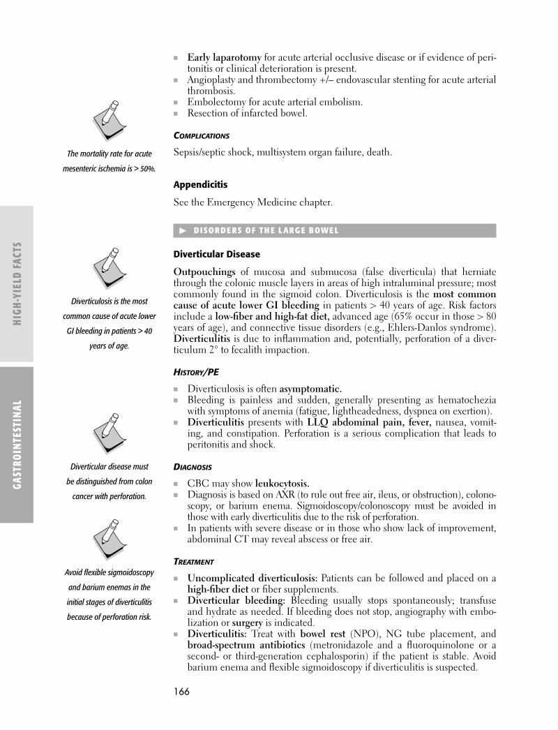
Outpouchings of mucosa and submucosa (false diverticula) that herniate through the colonic muscle layers in areas of high intraluminal pressure; most commonly found in the sigmoid colon. Diverticulosis is the most common cause of acute lower GI bleeding in patients > 40 years of age. Risk factors include a low-fi ber and high-fat diet, advanced age (65% occur in those > 80 years of age), and connective tissue disorders (e.g., Ehlers-Danlos syndrome). Diverticulitis is due to infl ammation and, potentially, perforation of a diver-ticulum 2° to fecalith impaction.
HISTORY/PE
■ Diverticulosis is often asymptomatic.■ Bleeding is painless and sudden, generally presenting as hematochezia
with symptoms of anemia (fatigue, lightheadedness, dyspnea on exertion).■ Diverticulitis presents with LLQ abdominal pain, fever, nausea, vomit-
ing, and constipation. Perforation is a serious complication that leads to peritonitis and shock.
DIAGNOSIS
■ CBC may show leukocytosis.■ Diagnosis is based on AXR (to rule out free air, ileus, or obstruction), colono-
scopy, or barium enema. Sigmoidoscopy/colonoscopy must be avoided in those with early diverticulitis due to the risk of perforation.
■ In patients with severe disease or in those who show lack of improvement, abdominal CT may reveal abscess or free air.
TREATMENT
■ Uncomplicated diverticulosis: Patients can be followed and placed on a high-fi ber diet or fi ber supplements.
■ Diverticular bleeding: Bleeding usually stops spontaneously; transfuse and hydrate as needed. If bleeding does not stop, angiography with embo-lization or surgery is indicated.
■ Diverticulitis: Treat with bowel rest (NPO), NG tube placement, and broad-spectrum antibiotics (metronidazole and a fl uoroquinolone or a second- or third-generation cephalosporin) if the patient is stable. Avoid barium enema and fl exible sigmoidoscopy if diverticulitis is suspected.
The mortality rate for acute
mesenteric ischemia is > 50%.
Diverticulosis is the most
common cause of acute lower
GI bleeding in patients > 40
years of age.
Diverticular disease must
be distinguished from colon
cancer with perforation.
Avoid fl exible sigmoidoscopy
and barium enemas in the
initial stages of diverticulitis
because of perforation risk.
504 _02-06_Gastro.indd 166 8/18/09 9:57:03 A

167
HIGH-YIELD FACTS
GASTROINTESTINAL
■ For perforation, perform immediate surgical resection of diseased bowel via a Hartmann’s procedure with a temporary colostomy.
Large Bowel Obstruction (LBO)
Table 2.6-3 describes features that distinguish SBO from LBO. Figure 2.6-4 demonstrates the classic radiographic fi ndings of LBO.
Colon and Rectal Cancer
The second leading cause of cancer mortality in the United States after lung cancer. There is an ↑ incidence with age, with a peak incidence at 70–80 years. There has also been an observed association between colon cancer
T A B L E 2 . 6 - 3 . Characteristics of Small and Large Bowel Obstruction
SBO LBO
History Moderate to severe acute abdominal pain; copious
emesis.
Cramping pain with distal SBO.
Fever, signs of dehydration, and hypotension may be
seen.
Constipation/obstipation, deep and cramping
abdominal pain (less intense than SBO), nausea/
vomiting (less than SBO but more commonly
feculent).
PE Abdominal distention (distal SBO), abdominal
tenderness, visible peristaltic waves, fever,
hypovolemia.
Look for surgical scars/hernias; perform a rectal
exam.
High-pitched “tinkly” bowel sounds; later, absence
of bowel sounds.
Signifi cant distention, tympany, and tenderness;
examine for peritoneal irritation or mass; fever or
signs of shock suggest perforation/peritonitis or
ischemia/necrosis.
High-pitched “tinkly” bowel sounds; later, absence
of bowel sounds.
Etiologies Adhesions (postsurgery), hernias, neoplasm,
volvulus, intussusception, gallstone ileus, foreign
body, Crohn’s disease, CF, stricture, hematoma.
Colon cancer, diverticulitis, volvulus, fecal impaction,
benign tumors.
Assume colon cancer until proven otherwise.
Differential LBO, paralytic ileus, gastroenteritis. SBO, paralytic ileus, appendicitis, IBD, Ogilvie’s
syndrome (pseudo-obstruction).
Diagnosis CBC, electrolytes, lactic acid, AXR (see Figure
2.6-3); contrast studies (determine if it is partial or
complete), CT scan.
CBC, electrolytes, lactic acid, AXR (see Figure
2.6-4), CT scan; water contrast enema (if perforation
is suspected); sigmoidoscopy/colonoscopy if stable.
Treatment Hospitalize. Partial SBO can be treated conservatively
with NG decompression and NPO status.
Patients with complete SBO should be managed
aggressively with NPO status, NG decompression,
IV fl uids, electrolyte replacement, and surgical
correction.
Hospitalize. Obstruction can be relieved with a
Gastrografi n enema, colonoscopy, or a rectal tube;
however, surgery is usually required. Ischemic
colon usually requires partial colectomy with a
diverting colostomy.
Treat the underlying cause (e.g., neoplasm).
504 _02-06_Gastro.indd 167 8/18/09 9:57:03 A

HIG
H-YI
ELD
FACT
SGA
STRO
INTE
STIN
AL
168
and Streptococcus bovis bacteremia. Risk factors and screening protocols are summarized in Table 2.6-4.
HISTORY/PE
■ In the absence of screening, colon and rectal cancer typically present with symptoms only after a prolonged period of silent growth.
F I G U R E 2 . 6 - 4 . Large bowel obstruction.
Barium study shows the “bird-beak” sign, with juxtaposed adjacent bowel walls in the dilated
loop pointing toward the site of obstruction. (Reproduced, with permission, from Way LW.
Current Surgical Diagnosis & Treatment, 10th ed. Stamford, CT: Appleton & Lange, 1994:
676.)
T A B L E 2 . 6 - 4 . Risk Factors and Screening for Colorectal Cancer
RISK FACTORS SCREENING
Age.
Hereditary syndromes—familial adenomatous polyposis
(100% risk by age 40), Gardner’s disease, hereditary
nonpolyposis colorectal cancer (HNPCC).
Family history.
IBD—ulcerative colitis carries a higher risk than does Crohn’s
disease.
Adenomatous polyps—villous polyps progress more often
than tubular polyps and sessile more than pedunculated
polyps. Lesions > 2 cm carry an ↑ risk.
Past history of colorectal cancer.
High-fat, low-fi ber diet.
A DRE should be performed yearly for patients ≥ 50 years of
age. Up to 10% of all lesions are palpable with DRE.
Stool guaiac should be performed every year for patients
≥ 50 years of age. Up to 50% of � guaiac tests are due to
colorectal cancer.
Colonoscopy every 10 years in those ≥ 50 years of age OR
yearly FOBT with sigmoidoscopy every 5 years.
Colonoscopy should be performed every 10 years in patients
≥ 40 years of age who have a fi rst-degree relative with
colorectal cancer or adenomatous polyps, or 10 years prior
to the age at diagnosis of the youngest family member.
504 _02-06_Gastro.indd 168 8/18/09 9:57:03 A
169
HIGH-YIELD FACTS
GASTROINTESTINAL
■ Although often asymptomatic, patients may present with unexplained ane-mia or vague abdominal pain. Other features depend on location:■ Right-sided lesions: Often bulky, ulcerating masses that lead to anemia
from chronic occult blood loss. Patients may complain of weight loss, anorexia, diarrhea, weakness, or vague abdominal pain. Obstruction is rare.
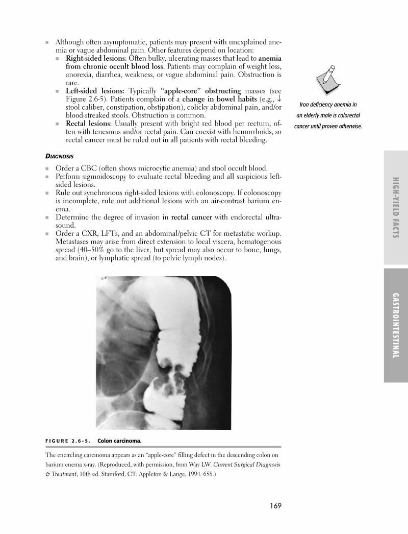
■ Left-sided lesions: Typically “apple-core” obstructing masses (see Figure 2.6-5). Patients complain of a change in bowel habits (e.g., ↓ stool caliber, constipation, obstipation), colicky abdominal pain, and/or blood-streaked stools. Obstruction is common.
■ Rectal lesions: Usually present with bright red blood per rectum, of-ten with tenesmus and/or rectal pain. Can coexist with hemorrhoids, so rectal cancer must be ruled out in all patients with rectal bleeding.
DIAGNOSIS
■ Order a CBC (often shows microcytic anemia) and stool occult blood.■ Perform sigmoidoscopy to evaluate rectal bleeding and all suspicious left-
sided lesions.■ Rule out synchronous right-sided lesions with colonoscopy. If colonoscopy
is incomplete, rule out additional lesions with an air-contrast barium en-ema.
■ Determine the degree of invasion in rectal cancer with endorectal ultra-sound.
■ Order a CXR, LFTs, and an abdominal/pelvic CT for metastatic workup. Metastases may arise from direct extension to local viscera, hematogenous spread (40–50% go to the liver, but spread may also occur to bone, lungs, and brain), or lymphatic spread (to pelvic lymph nodes).
Iron defi ciency anemia in
an elderly male is colorectal
cancer until proven otherwise.
F I G U R E 2 . 6 - 5 . Colon carcinoma.
The encircling carcinoma appears as an “apple-core” fi lling defect in the descending colon on
barium enema x-ray. (Reproduced, with permission, from Way LW. Current Surgical Diagnosis
& Treatment, 10th ed. Stamford, CT: Appleton & Lange, 1994: 658.)
504 _02-06_Gastro.indd 169 8/18/09 9:57:03 A

HIG
H-YI
ELD
FACT
SGA
STRO
INTE
STIN
AL
170
■ Staging is based on the depth of tumor penetration into the bowel wall and the presence of lymph node involvement and distant metastases.
TREATMENT
■ Surgical resection of the 1° cancer is the treatment of choice. Regional lymph node dissection should be performed for staging purposes.
■ For rectal lesions, the resection technique depends on the proximity of the lesion to the anal verge (the junction between the anal canal and the anal skin).■ Abdominoperineal resection: For distal lesions < 10 cm from the anal
verge, when the sphincter cannot be preserved, the rectum and anus are resected and a permanent colostomy is placed.
■ Low anterior resection: For proximal lesions > 10 cm from the anal verge, a 1° anastomosis is created between the colon and rectum.
■ Wide local excision: For small, low-grade, well-differentiated tumors in the lower third of the rectum.
■ Adjuvant chemotherapy: Used in cases of colon cancer with � lymph nodes. Can be considered for rectal cancers.
■ Follow with serial CEA levels (diagnostically nonspecifi c, but useful for monitoring recurrence), colonoscopy, LFTs, CXR, and abdominal CT (for metastasis).
Ischemic Colitis
■ Due to lack of arterial blood supply to the colon. Severity ranges from su-perfi cial mucosal involvement to full-thickness necrosis.
■ The most commonly affected site is the left colon, particularly the “water-shed area” at the splenic fl exure. Incidence ↑ with age.
■ Hx/PE: Presents with crampy lower abdominal pain associated with bloody diarrhea. Fever and peritoneal signs suggest infarction.
■ Dx:■ CBC may reveal leukocytosis.■ Flexible sigmoidoscopy or colonoscopy to assess colonic mucosa.
■ Tx:■ Supportive therapy with bowel rest, IV fl uids, and broad-spectrum anti-
biotics.■ Surgery with resection is indicated for infarction, fulminant colitis, or
obstruction 2° to ischemic stricture.
GASTROI NTESTI NAL BLE E DI NG �
Bleeding from the GI tract may present as hematemesis, hematochezia, and/or melena. Upper GI tract bleeding is defi ned as bleeding from lesions proximal to the ligament of Treitz (the anatomic boundary between the duodenum and jejunum). Table 2.6-5 presents the features of upper and lower GI bleeding.
I N FLAM MATORY BOWE L D ISEASE ( I BD) �
Comprises Crohn’s disease and ulcerative colitis (see Figure 2.6-6). Most common in Caucasians and Ashkenazi Jews, appearing most frequently dur-ing the teens to early 30s or in the 50s. Table 2.6-6 summarizes the features of IBD.
Ischemic colitis is a known
complication of AAA repair
due to sacrifi ce of the inferior
mesenteric artery.
One unit of PRBCs should ↑
hemoglobin by 1 unit and
hematocrit by 3–4 units.
504 _02-06_Gastro.indd 170 8/18/09 9:57:04 A

171
HIGH-YIELD FACTS
GASTROINTESTINAL
T A B L E 2 . 6 - 5 . Features of Upper and Lower GI Bleeding
VARIABLE UPPER GI BLEEDING LOWER GI BLEEDING
History/PE Hematemesis (”coffee-ground” emesis), melena
> hematochezia, depleted volume status (e.g.,
tachycardia, lightheadedness, hypotension).
Hematochezia > melena, but can be either.
Diagnosis NG tube and NG lavage; endoscopy if stable. Rule out upper GI bleed with NG lavage.
Anoscopy/sigmoidoscopy for patients < 45 years
of age with small-volume bleeding.
Colonoscopy if stable; arteriography or exploratory
laparotomy if unstable.
Etiologies PUD, esophagitis/gastritis, Mallory-Weiss tear,
esophageal varices.
Diverticulosis (60%), IBD, hemorrhoids/fi ssures,
neoplasm, AVM.
Initial
management
Protect the airway (may need intubation). Stabilize
the patient with IV fl uids and PRBCs (hematocrit
may be normal early in acute blood loss).
Similar to upper GI bleed.
Long-term
management
Endoscopy followed by therapy directed at the
underlying cause (e.g., high-dose PPIs for PUD;
octreotide and/or banding for varices).
Depends on the underlying etiology. Endoscopic
therapy (e.g., epinephrine injection), intra-arterial
vasopressin infusion or embolization, or surgery for
diverticular disease or angiodysplasia.
F I G U R E 2 . 6 - 6 . Infl ammatory bowel disease.
(A) Crohn’s disease. Barium enema x-ray reveals deep transverse fi ssures, ulcers, and edema of the bowel. (B) Ulcer-
ative colitis. Barium enema x-ray demonstrates shortening of the colon, loss of haustra (“lead pipe” appearance), and
fi ne serrations of the bowel edges from small ulcers. (Reproduced, with permission, from Stobo J et al. The Principles
and Practice of Medicine, 23rd ed. Stamford, CT: Appleton & Lange, 1996: 135.)
A B
504 _02-06_Gastro.indd 171 8/18/09 9:57:04 A

HIG
H-YI
ELD
FACT
SGA
STRO
INTE
STIN
AL
172
I NG U I NAL H E RN IAS �
Abnormal protrusions of abdominal contents (usually the small intestine) into the inguinal region through a weakness or defect in the abdominal wall. Defi ned as direct or indirect on the basis of their relationship to the inguinal canal.
■ Indirect: Herniation of abdominal contents through the internal and then external inguinal rings and eventually into the scrotum (in males).■ The most common hernia in both genders.■ Due to a congenital patent processus vaginalis.
T A B L E 2 . 6 - 6 . Features of Ulcerative Colitis and Crohn’s Disease
VARIABLE ULCERATIVE COLITIS CROHN’S DISEASE
Site of
involvement
The rectum is always involved. May extend
proximally in a continuous fashion.
Infl ammation and ulceration are limited to the
mucosa and submucosa.
May involve any portion of the GI tract, particularly
the ileocecal region, in a discontinuous pattern
(“skip lesions”). The rectum is often spared.
Transmural infl ammation is seen.
History/PE Bloody diarrhea, lower abdominal cramps,
tenesmus, urgency.
Exam may reveal orthostatic hypotension,
tachycardia, abdominal tenderness, frank
blood on rectal exam, and extraintestinal
manifestations.
Abdominal pain, abdominal mass, low-grade fever,
weight loss, watery diarrhea.
Exam may reveal fever, abdominal tenderness
or mass, perianal fi ssures, fi stulas, and
extraintestinal manifestations.
Extraintestinal
manifestations
Aphthous stomatitis, episcleritis/uveitis, arthritis,
primary sclerosing cholangitis, erythema
nodosum, and pyoderma gangrenosum.
The same as ulcerative colitis, as well as gallstones,
nephrolithiasis, and fi stulas to the skin, bladder, or
between bowel loops.
Diagnosis CBC, AXR, stool cultures, O&P, stool assay for
C. diffi cile.
Colonoscopy can show diffuse and continuous
rectal involvement, friability, edema, and
pseudopolyps.
Defi nitive diagnosis can be made with biopsy.
The same lab workup as ulcerative colitis. Upper GI
series with small bowel follow-through.
Colonoscopy may show aphthoid, linear, or stellate
ulcers, strictures, “cobblestoning,” and “skip
lesions.” “Creeping fat” may also be present
during laparotomy.
Defi nitive diagnosis can be made with biopsy.
Treatment 5-ASA agents (e.g., sulfasalazine, mesalamine),
topical or oral; corticosteroids and
immunomodulating agents (e.g., azathioprine)
for refractory disease.
Total proctocolectomy is curative for long-
standing or fulminant colitis or toxic megacolon;
it also ↓ cancer risk.
5-ASA agents; corticosteroids and
immunomodulating agents (e.g., azathioprine,
infl iximab) are indicated if no improvement is
seen.
Surgical resection may be necessary for suspected
perforation, stricture, fi stula, or abscess; may
recur anywhere in the GI tract.
Incidence of
cancer
Markedly ↑ risk of colorectal cancer in long-
standing cases (monitor with frequent fecal occult
blood screening and yearly colonoscopy with
multiple biopsies after eight years of disease).
Incidence of 2° malignancy is lower than in
ulcerative colitis, but greater than the general
population.
504 _02-06_Gastro.indd 172 8/18/09 9:57:04 A

173
HIGH-YIELD FACTS
GASTROINTESTINAL
■ Protrudes lateral to the inferior epigastric vessels.■ Direct: Herniation of abdominal contents through the fl oor of Hessel-
bach’s triangle. ■ Protrudes medial to the epigastric vessels.■ Hernial sac contents do not traverse the internal inguinal ring; they
herniate directly through the abdominal wall and are contained within the aponeurosis of the external oblique muscle.
■ Most often due to an acquired defect in the transversalis fascia from mechanical breakdown that ↑ with age.
TREATMENT
■ Because of the risk of incarceration and strangulation, surgical manage-ment (open or laparoscopic) is indicated unless specifi c contraindications are present.
■ Repair of a direct inguinal hernia involves correcting the defect in the transversalis fascia.
■ Indirect inguinal hernias are repaired by isolating and ligating the hernial sac and reducing the size of the internal inguinal ring to allow only the spermatic cord structures in males to pass through.
BI LIARY D ISEASE �
Cholelithiasis and Biliary Colic
Colic results from transient cystic duct blockage from impacted stones. Al-though risk factors include the 4 F’s—Female, Fat, Fertile, and Forty—the disorder is common and can occur in any patient. Flatulence can be thought of as a “5th F.” Other risk factors include OCP use, rapid weight loss, a � family history, chronic hemolysis (pigment stones in sickle cell disease), small bowel resection, and TPN.
HISTORY/PE
■ Patients present with postprandial abdominal pain (usually in the RUQ) that radiates to the right subscapular area or the epigastrium.
■ Pain is abrupt; is followed by gradual relief; and is often associated with nausea and vomiting, fatty food intolerance, dyspepsia, and fl atulence.
■ Gallstones may be asymptomatic in up to 80% of patients. Exam may re-veal RUQ tenderness and a palpable gallbladder.
DIAGNOSIS
■ Plain x-rays are rarely diagnostic; only 10–15% of stones are radiopaque.■ RUQ ultrasound is the imaging modality of choice (85–90% sensitive).■ Table 2.6-7 contrasts lab fi ndings with those of other forms of biliary dis-
ease.
TREATMENT
■ Cholecystectomy is curative and can be performed electively for symp-tomatic gallstones. It is generally performed laparoscopically. Asymptom-atic gallstones do not require any intervention.
■ Patients may require preoperative endoscopic retrograde cholangiopancre-atography (ERCP) for common bile duct stones.
■ Treat nonsurgical candidates with dietary modifi cation (avoid triggers such as fatty foods).
Hesselbach’s triangle is
an area bounded by the
inguinal ligament, the inferior
epigastric artery, and the
rectus abdominis.
Pigmented gallstones result
from hemolysis (black) or
infection (brown).
Most gallstones are
precipitations of cholesterol
and are not radiopaque.
504 _02-06_Gastro.indd 173 8/18/09 9:57:04 A

HIG
H-YI
ELD
FACT
SGA
STRO
INTE
STIN
AL
174
COMPLICATIONS
Recurrent biliary colic, acute cholecystitis, choledocholithiasis, ascending cholangitis, gallstone ileus, gallstone pancreatitis.
Acute Cholecystitis
Prolonged blockage of the cystic duct, usually by an impacted stone, that leads to obstructive distention, infl ammation, superinfection, and possi-bly gangrene of the gallbladder (acute gangrenous cholecystitis). Acalcu-lous cholecystitis occurs in the absence of cholelithiasis in patients who are chronically debilitated, those who are critically ill in the ICU or on TPN, and trauma or burn victims.
HISTORY/PE
■ Patients present with RUQ pain, nausea, vomiting, and fever. Symptoms are typically more severe and of longer duration than those of biliary colic.
■ RUQ tenderness, inspiratory arrest during deep palpation of the RUQ (Murphy’s sign), low-grade fever, mild icterus, and possibly guarding or rebound tenderness may be present on exam.
DIAGNOSIS
■ Fever is often present, and CBC shows leukocytosis (see Table 2.6-7).■ Ultrasound may demonstrate stones, bile sludge, pericholecystic fl uid, a
thickened gallbladder wall, gas in the gallbladder, and an ultrasonic Mur-phy’s sign (see Figure 2.6-7).
■ Obtain a HIDA scan when ultrasound is equivocal (see Figure 2.6-8); nonvisualization of the gallbladder on HIDA scan suggests acute cholecys-titis.
TREATMENT
■ Hospitalize patients, administer broad-spectrum IV antibiotics and IV fl u-ids, and replete electrolytes.
■ If diagnosed soon after onset, early cholecystectomy is indicated. ■ For stable patients or those with signifi cant medical problems, surgery can
be delayed for 4–6 weeks.
The cystic artery usually
passes through the anatomic
triangle of Calot, comprising
the common hepatic duct, the
cystic duct, and the inferior
border of the liver.
In patients with signifi cant
medical problems (including
DM), delay cholecystectomy
until acute infl ammation
resolves.
T A B L E 2 . 6 - 7 . Differential Diagnosis of Biliary Disease
FEVER/ELEVATED
WBC COUNT
ELEVATED TOTAL
BILIRUBIN/
ALKALINE
PHOSPHATASE
ELEVATED SERUM
AMYLASE
Cholelithiasis (colic) – – –
Acute cholecystitis + – –
Choledocholithiasis/
ascending cholangitis
+ + –
Gallstone pancreatitis + + +
504 _02-06_Gastro.indd 174 8/18/09 9:57:05 A

175
HIGH-YIELD FACTS
GASTROINTESTINAL
COMPLICATIONS
Gangrene, empyema, perforation, emphysematous gallbladder (due to infec-tion by gas-forming organisms), fi stulization, gallstone ileus, sepsis, abscess formation.
Choledocholithiasis
■ Gallstones in the common bile duct. Symptoms vary according to the de-gree of obstruction, the duration of the obstruction, and the extent of bac-terial infection.
F I G U R E 2 . 6 - 7 . Acute cholecystitis, ultrasound.
(A) Note the sludge-fi lled, thick-walled gallbladder with a hyperechoic stone and acoustic shadow (arrow). (B) This pa-
tient exhibits sludge and pericholecystic fl uid (arrow) but no gallstones. (Reproduced, with permission, from Grendell
J. Current Diagnosis & Treatment in Gastroenterology, 1st ed. Stamford, CT: Appleton & Lange, 1996: 212.)
A B
F I G U R E 2 . 6 - 8 . Acute cholecystitis, HIDA scan.
IV dye is taken up by hepatocytes and is conjugated and excreted into the common bile duct.
The gallbladder is not visualized, although activity is present in the liver, common duct, and
small bowel, suggesting cystic duct obstruction due to acute cholecystitis. (Reproduced, with
permission, from Grendell J. Current Diagnosis & Treatment in Gastroenterology, 1st ed. Stam-
ford, CT: Appleton & Lange, 1996: 217.)
504 _02-06_Gastro.indd 175 8/18/09 9:57:05 A

HIG
H-YI
ELD
FACT
SGA
STRO
INTE
STIN
AL
176
■ Hx/PE: Although sometimes asymptomatic, it often presents with biliary colic, jaundice, fever, and pancreatitis.
■ Dx: The hallmark is ↑ alkaline phosphatase and total bilirubin, which may be the only abnormal lab values (see Table 2.6-7).
■ Tx: Management generally consists of ERCP with sphincterotomy fol-lowed by semielective cholecystectomy. Common bile duct exploration may be necessary.
Ascending Cholangitis
An acute bacterial infection of the biliary tree that commonly occurs 2° to ob-struction, usually from gallstones (choledocholithiasis) or primary sclerosing cholangitis (progressive infl ammation of the biliary tree associated with ulcer-ative colitis). Other etiologies include bile duct stricture and malignancy (bil-iary or pancreatic). Gram-� enterics (e.g., E. coli, Enterobacter, Pseudomo-nas) are commonly identifi ed pathogens.
HISTORY/PE
■ Charcot’s triad—RUQ pain, jaundice, and fever/chills—is classic and seen in 50–70% of cases.
■ Reynolds’ pentad—Charcot’s triad plus septic shock and altered mental status—may be present in acute suppurative cholangitis and suggests sep-sis.
DIAGNOSIS
■ Look for leukocytosis, ↑ bilirubin, and ↑ alkaline phosphatase (see Table 2.6-7).
■ Obtain blood cultures to rule out sepsis. Ultrasound or CT may be a use-ful adjunct, but diagnosis is often clinical. Magnetic resonance cholan-giopancreatography (MRCP) is a noninvasive test that may also be useful.
■ ERCP is both diagnostic and therapeutic (biliary drainage).
TREATMENT
■ Patients often require ICU admission for monitoring, hydration, BP sup-port, and broad-spectrum IV antibiotic treatment.
■ Patients with acute suppurative cholangitis require emergent bile duct de-compression via ERCP/sphincterotomy, percutaneous transhepatic drain-age, or open decompression.
Gallstone Ileus
■ A mechanical obstruction resulting from the passage of a large (> 2.5-cm) stone into the bowel through a cholecystoduodenal fi stula. Obstruction is often at the terminal ileum (TI)/ileocecal valve.
■ Hx/PE: Many patients have no previous history of biliary symptoms and present as an SBO. The classic presentation is that of a subacute SBO in an elderly woman.
■ Dx: Pneumobilia (gas in the biliary tree) seen on imaging can confi rm, in addition to an upper GI series with small bowel follow-through showing a TI obstruction.
■ Tx: Laparotomy with stone extraction (enterolithotomy) or manipulation into the colon.
Gallstone pancreatitis
occurs when stones in the
ampulla also obstruct the
pancreatic duct.
Charcot’s triad consists
of RUQ pain, jaundice,
and fever/chills. Reynolds’
pentad consists of RUQ pain,
jaundice, fever/chills, shock,
and altered mental status.
504 _02-06_Gastro.indd 176 8/18/09 9:57:05 A

177
HIGH-YIELD FACTS
GASTROINTESTINAL
Primary Sclerosing Cholangitis
■ An idiopathic disorder characterized by infl ammation, fi brosis, and stric-tures of extra- and intrahepatic bile ducts. The disease usually presents in young men with IBD (most often ulcerative colitis).
■ Hx/PE: Presents with progressive jaundice, pruritus, and fatigue.■ Dx:
■ Laboratory fi ndings include ↑ alkaline phosphatase and ↑ bilirubin.■ MRCP/ERCP show multiple bile duct strictures with dilatations be-
tween strictures.■ Liver biopsy reveals periductal sclerosis (“onion skinning”).
■ Tx: High-dose ursodeoxycholic acid; endoscopic dilation and short-term stenting of bile duct strictures; liver transplantation. Patients are at ↑ risk for cholangiocarcinoma.
LIVE R D ISEASE �
Abnormal Liver Function Tests
Liver diseases can be divided into distinct patterns based on LFT results:
■ Hepatocellular injury: ↑ AST and ALT.■ Cholestasis: ↑ alkaline phosphatase and bilirubin.■ Isolated hyperbilirubinemia: ↑ bilirubin; normal aminotransferases and
alkaline phosphatase.
Jaundice, which can be seen in any of the patterns outlined above, is a clini-cal sign that arises when excess bilirubin (> 2.5 mg/dL) is circulating in the blood. Figures 2.6-9 and 2.6-10 summarize the clinical approaches toward cholestasis and isolated hyperbilirubinemia. Hepatocellular injury is de-scribed in the section that follows.
Hepatitis
Infl ammation of the liver leading to liver cell injury and necrosis. The causes of acute hepatitis include viruses (e.g., HAV, HBV, HCV, HDV, HEV) and drug-induced disease (e.g., alcohol, acetaminophen, INH, methyldopa). The
Primary sclerosing cholangitis
is signifi cantly associated with
ulcerative colitis.
Cholestasis ( alkaline phosphatase and bilirubin)
No Yes
MedicationsPostopSepsis
StoneStrictureCancer
Intrahepatic cholestasis Biliary obstruction
Ductal dilation?
F I G U R E 2 . 6 - 9 . Approach to cholestasis.
504 _02-06_Gastro.indd 177 8/18/09 9:57:05 A

HIG
H-YI
ELD
FACT
SGA
STRO
INTE
STIN
AL
178
causes of chronic hepatitis include viruses (e.g., HBV, HCV, HDV), alco-holic hepatitis, autoimmune hepatitis, ischemic hepatitis, and hereditary eti-ologies (e.g., Wilson’s disease, hemochromatosis, α1-antitrypsin defi ciency).
■ HAV and HEV are transmitted by the fecal-oral route.■ HBV and HCV are transmitted by bodily fl uids, although the risk of ac-
quiring HCV sexually is very low.
HISTORY/PE
■ Acute hepatitis often starts with a viral prodrome of nonspecifi c symptoms (e.g., malaise, fever, joint pain, fatigue, URI symptoms, nausea, vomiting, changes in bowel habits) followed by jaundice and RUQ tenderness.
■ Exam often reveals jaundice, scleral icterus, tender hepatomegaly, pos-sible splenomegaly, and lymphadenopathy.
■ Chronic hepatitis usually gives rise to symptoms indicative of chronic liver disease (jaundice, fatigue, hepatosplenomegaly). At least 80% of those in-fected with HCV and 10% of those with HBV will develop chronic hepatitis.
DIAGNOSIS
■ Dramatically ↑ ALT and AST and ↑ bilirubin/alkaline phosphatase are present in the acute form.
■ In chronic hepatitis, ALT and AST are ↑ for > 6 months with a concurrent ↑ in alkaline phosphatase/bilirubin and hypoalbuminemia. In severe cases, PT will be prolonged, as all clotting factors except factor VIII are produced by the liver.
■ The diagnosis of viral hepatitis is made by hepatitis serology (see Table 2.6-8 and Figure 2.6-11 for a description and timing of serologic markers) and by liver biopsy in chronic or severe cases.
■ ANA, anti–smooth muscle antibody, and antimitochondrial antibody point to autoimmune hepatitis. Iron saturation (hemochromatosis) and cerulo-plasmin (Wilson’s disease) can identify other causes.
TREATMENT
Treatment is etiology specifi c; monitor for resolution of symptoms over time.
■ Steroids for severe alcoholic hepatitis.■ Immunosuppression with steroids and other agents (azathioprine) for au-
toimmune hepatitis.
HCV is Chronic. Eighty
percent of patients with
HCV infection will develop
chronic hepatitis.
An AST/ALT ratio > 2 suggests
alcoholic hepatitis—you’re
toASTed.
Isolated hyperbilirubinemia ( bilirubin)
Unconjugated Conjugated
Overproduction
Hemolyticanemia
Defectiveconjugation
Gilbert's syndrome (< 5 mg/dL)Crigler-Najjar syndrome
Defectiveexcretion
Dubin-Johnson syndromeRotor's syndrome
F I G U R E 2 . 6 - 1 0 . Approach to isolated hyperbilirubinemia.
504 _02-06_Gastro.indd 178 8/18/09 9:57:05 A

179
HIGH-YIELD FACTS
GASTROINTESTINAL
■ IFN-α, lamivudine (3TC), or adefovir for chronic HBV infection; peginterferon and ribavirin for chronic HCV infection.
■ Liver transplantation is the treatment of choice for patients with end-stage liver failure.
■ ICU management and emergent transplant for fulminant hepatic failure.
The sequelae of chronic
hepatitis include cirrhosis, liver
failure, and hepatocellular
carcinoma.
T A B L E 2 . 6 - 8 . Key Hepatitis Serologic Markers
SEROLOGIC MARKER DESCRIPTION
IgM HAVAb IgM antibody to HAV; the best test to detect active hepatitis A.
HBsAg Antigen found on the surface of HBV; continued presence indicates
carrier state.
HBsAb Antibody to HBsAg; provides immunity to HBV.
HBcAg Antigen associated with core of HBV.
HBcAb Antibody to HBcAg; � during the window period. IgM HBcAb is an
indicator of recent disease.
HBeAg A second, different antigenic determinant in the HBV core. An
important indicator of transmissibility (BEware!).
HBeAb Antibody to e antigen; indicates low transmissibility.
HBsAg
HBeAg
Anti-HBs
Anti-HBe
DNA polymerase
HBV particles Anti-HBc
0 1 2 3 4 5 6 7 8Months after exposure
Level of detection
Tite
r
0 1 2 3 4 5 6 7 8
HBsAg HBsAg(anti-HBc)
Anti-HBc
Anti-HBs(anti-HBc)
Incubationperiod
Prodrome,acute disease Early
ConvalescenceImportant diagnostic tests
Late
Window period
F I G U R E 2 . 6 - 1 1 . Time course of hepatitis B with serologic markers.
504 _02-06_Gastro.indd 179 8/18/09 9:57:06 A

HIG
H-YI
ELD
FACT
SGA
STRO
INTE
STIN
AL
180
COMPLICATIONS
Cirrhosis, liver failure, hepatocellular carcinoma (3–5%).
Cirrhosis
Defi ned as fi brosis and nodular regeneration resulting from hepatocellular injury. Etiologies include causes of chronic hepatitis, biliary tract disease (e.g., primary biliary cirrhosis, primary sclerosing cholangitis), right-sided heart fail-ure, constrictive pericarditis, and Budd-Chiari syndrome (hepatic vein throm-bosis 2° to hypercoagulability).
HISTORY/PE
■ Presents with jaundice, ascites, spontaneous bacterial peritonitis, he-patic encephalopathy (e.g., asterixis, altered mental status), gastroesopha-geal varices, coagulopathy, and renal dysfunction. Weakness, anorexia, and weight loss are also seen in advanced disease.
■ Exam may reveal an enlarged, palpable, or fi rm liver. Stigmata of portal hypertension and signs of liver failure may be present (see Figures 2.6-12 and 2.6-13).
DIAGNOSIS
■ Lab studies show ↓ albumin, ↑ PT/PTT, and ↑ bilirubin. Anemia or thrombocytopenia (2° to hypersplenism) may also be seen.
■ Abdominal ultrasound with Doppler can assess liver size, the presence of ascites, and the patency of splenic and hepatic veins. The etiology of ascites can be established through measurement of the serum-ascites albumin gradient (SAAG = serum albumin – ascites albumin); see Table 2.6-9.
■ Obtain hepatitis serologies and autoimmune hepatitis studies.■ Serum ferritin, ceruloplasmin, and α1-antitrypsin may help identify ad-
ditional causes, such as hemochromatosis, Wilson’s disease, and α1-antitrypsin defi ciency, respectively.
■ Liver biopsy showing bridging fi brosis and nodular regeneration.
Alcoholism, chronic hepatitis,
and other chronic liver
diseases lead to cirrhosis.
Gut, butt, and caput—the three
anastomoses commonly seen
in cirrhosis.
Effects of liver cell failure• Coma• Scleral icterus• Fetor hepaticus (breath smells like a freshly opened corpse)
• Spider nevi
• Gynecomastia
• Jaundice
• Loss of sexual hair
• Liver "flap" = asterixis (coarse hand tremor)
• Bleeding tendency (decreased prothrombin)
• Anemia
• Ankle edema
Effects of portal hypertension
• Esophageal varices
Hematemesis
Pepticulcer
• Melena• Splenomegaly• Caput medusae• Ascites
• Hemorrhoids
• Testicular atrophy
F I G U R E 2 . 6 - 1 2 . Presentation of cirrhosis/portal hypertension.
(Adapted, with permission, from Chandrasoma P, Taylor CE. Concise Pathology, 3rd ed. Stam-
ford, CT: Appleton & Lange, 1998: 654.)
504 _02-06_Gastro.indd 180 8/18/09 9:57:06 A

181
HIGH-YIELD FACTS
GASTROINTESTINAL
TREATMENT
Aimed at ameliorating the complications of cirrhosis/portal hypertension.
■ Ascites:■ Sodium restriction and diuretics (furosemide and spironolactone).■ Rule out infectious and neoplastic causes; perform paracentesis to ob-
tain SAAG, cell count with differential, and cultures.■ If possible, treat underlying liver disease.
■ Spontaneous bacterial peritonitis:■ Presents with fever, abdominal pain, and altered mental status.■ Check peritoneal fl uid if there is a possibility of infection. The fl uid is
� if there are > 250 PMNs/mL or > 500 WBCs.■ Treat with IV antibiotics (e.g., third-generation cephalosporin) to cover
both gram-� (Enterococcus) and gram-� (E. coli, Klebsiella) organisms until a causative organism is identifi ed.
Noncirrhotic causes of
portal hypertension include
right heart failure, splenic
vein thrombosis, and
schistosomiasis.
Superior mesenteric v.
Gastroesophageal vv.
Superior-middle/inferior rectal vv.
Gastrorenal-splenorenal vv.
Paraumbilical v.
IVC
Systemic venousPortal venous
Portal v.
L. gastric v.
Retro-peritonealparavertebral v
Splenic v.
1
2
3
4
5
v.
F I G U R E 2 . 6 - 1 3 . Portosystemic anastomoses.
1. Left gastric–azygos → esophageal varices. 2. Superior–middle/inferior rectal → hemorrhoids.
3. Paraumbilical–inferior epigastric → caput medusae (navel). 4. Gastrorenal-splenorenal.
5. Retroperitoneal paravertebral.
T A B L E 2 . 6 - 9 . Serum-Ascites Albumin Gradient
SAAG > 1.1 SAAG < 1.1
Ascites is related to portal hypertension:
■ Presinusoidal: Splenic or portal vein
thrombosis, schistosomiasis
■ Sinusoidal: Cirrhosis, massive hepatic
metastases
■ Postsinusoidal: Right heart failure,
constrictive pericarditis, Budd-Chiari
syndrome
Ascites is due to protein leakage:
■ Nephrotic syndrome
■ Tuberculosis
■ Malignancy (e.g., ovarian cancer)
504 _02-06_Gastro.indd 181 8/18/09 9:57:06 A

HIG
H-YI
ELD
FACT
SGA
STRO
INTE
STIN
AL
182
■ Hepatorenal syndrome: A diagnosis of exclusion; diffi cult to treat and of-ten requires dialysis.
■ Hepatic encephalopathy:■ Due to ↓ clearance of ammonia.■ Often precipitated by dehydration, infection, electrolyte abnormalities,
and GI bleeding.■ Treat with dietary protein restriction, lactulose, and rifaximin.
■ Esophageal varices: ■ Monitor for GI bleeding; treat medically (β-blockers, octreotide), endo-
scopically (band ligation), or surgically (portocaval shunt).■ Consider liver transplantation for patients with advanced disease.
Primary Biliary Cirrhosis
■ An autoimmune disorder characterized by destruction of intrahepatic bile ducts. The disease most commonly presents in middle-aged women with other autoimmune conditions.
■ Hx/PE: Presents with progressive jaundice, pruritus, and malabsorption of the fat-soluble vitamins (A, D, E, K).
■ Dx: Laboratory fi ndings include ↑ alkaline phosphatase, ↑ bilirubin, � antimitochondrial antibody, and ↑ cholesterol.
■ Tx: Ursodeoxycholic acid (slows progression of disease); cholestyramine for pruritus; liver transplantation.
Hepatocellular Carcinoma
One of the most common cancers worldwide despite its relatively low inci-dence in the United States. 1° risk factors for the development of hepatocellu-lar carcinoma in the United States are cirrhosis and chronic hepatitis (HCV). In developing countries, afl atoxins (in various food sources) and HBV infec-tion are also major risk factors.
HISTORY/PE
■ Patients commonly present with RUQ tenderness, abdominal distention, and signs of chronic liver disease such as jaundice, easy bruisability, and coagulopathy. Cachexia and weakness may be present.
■ Exam may reveal tender enlargement of the liver.
DIAGNOSIS
■ Often suggested by the presence of a mass on ultrasound or CT as well as by abnormal LFTs and signifi cantly elevated α-fetoprotein (AFP) levels.
■ Liver biopsy for defi nitive diagnosis.
TREATMENT
■ For small tumors that are detected early, aggressive tumor resection or or-thotopic liver transplantation may be successful.
■ Chemotherapy and radiation are generally not effective, although they may be used to shrink large tumors prior to surgery (neoadjuvant ther-apy).
■ Monitor tumor recurrence with serial AFP levels. Prevent exposure to he-patic carcinogens and vaccinate against hepatitis in high-risk individuals.
Spontaneous bacterial
peritonitis is diagnosed by
> 250 PMNs/mL or > 500
WBCs in the ascitic fl uid.
Primary biliary cirrhosis is
an autoimmune disease
that presents with jaundice
and pruritus in middle-aged
women.
Complications of
hepatocellular carcinoma
include GI bleeding, liver
failure, and metastasis.
504 _02-06_Gastro.indd 182 8/18/09 9:57:06 A
183
HIGH-YIELD FACTS
GASTROINTESTINAL
Hemochromatosis
Caused by hyperabsorption of iron with parenchymal hemosiderin accumula-tion in the liver, pancreas, heart, adrenals, testes, pituitary, and kidneys. It is an autosomal-recessive disease that usually occurs in males of northern Eu-ropean descent and is rarely recognized before the fi fth decade. 2° hemochro-matosis may occur with iron overload and is common in patients receiving chronic transfusion therapy (e.g., for α-thalassemia) as well as in alcoholics(alcohol ↑ iron absorption).
HISTORY/PE
■ Patients may present with abdominal pain or symptoms of DM, hypogo-nadism, arthropathy of the MCP joints, heart failure, or cirrhosis.
■ Exam may reveal bronze skin pigmentation, pancreatic dysfunction, car-diac dysfunction (CHF), hepatomegaly, and testicular atrophy.
DIAGNOSIS
■ ↑ serum iron, percent saturation of iron, and ferritin with ↓ serum trans-ferrin.
■ Fasting transferrin saturation (serum iron divided by transferrin level) > 45% is the most sensitive diagnostic test.
■ Glucose intolerance and mildly elevated AST and alkaline phosphatase can be present.
■ Perform a liver biopsy (to determine hepatic iron index), hepatic MRI, or HFE gene mutation screen.
TREATMENT
■ Weekly phlebotomy; when serum iron levels ↓, perform maintenance phlebotomy every 2–4 months.
■ Deferoxamine can be used for maintenance therapy.
COMPLICATIONS
Cirrhosis, hepatocellular carcinoma, cardiomegaly leading to CHF and/or conduction defects, DM, impotence, arthropathy, hypopituitarism.
Wilson’s Disease (Hepatolenticular Degeneration)
■ ↓ ceruloplasmin and excessive deposition of copper in the liver and brain due to a defi cient copper-transporting protein. Linked to an autosomal- recessive defect on chromosome 13. Usually occurs in patients < 30 years of age; 50% of patients are symptomatic by age 15.
■ Hx: Patients with present hemolytic anemia, liver abnormalities (jaundice 2° to hepatitis/cirrhosis), and neurologic (loss of coordination, tremor, dys-phagia) as well as psychiatric (psychosis, anxiety, mania, depression) ab-normalities.
■ PE: May reveal Kayser-Fleischer rings in the cornea (green-to-brown de-posits of copper in Descemet’s membrane) as well as jaundice, hepatomeg-aly, asterixis, choreiform movements, and rigidity.
■ Dx: ↓ serum ceruloplasmin, ↑ urinary copper excretion, ↑ hepatic cop-per.
■ Tx: Dietary copper restriction (avoid shellfi sh, liver, legumes), pen-icillamine (a copper chelator that ↑ urinary copper excretion; administer with pyridoxine), and possibly oral zinc (↑ fecal excretion).
Wilson’s disease—
ABCD
AsterixisBasal ganglia
deteriorationCeruloplasmin ↓,
Cirrhosis, Copper ↑, Carcinoma (hepatocellular), Choreiform movements
Dementia
504 _02-06_Gastro.indd 183 8/18/09 9:57:07 A

HIG
H-YI
ELD
FACT
SGA
STRO
INTE
STIN
AL
184
PANC REATIC D ISEASE �
Pancreatitis
Table 2.6-10 outlines the important features of acute and chronic pancreati-tis. Table 2.6-11 lists Ranson’s criteria for predicting mortality associated with acute pancreatitis.
T A B L E 2 . 6 - 1 0 . Features of Acute and Chronic Pancreatitis
VARIABLE ACUTE PANCREATITIS CHRONIC PANCREATITIS
Pathophysiology Leakage of pancreatic enzymes into pancreatic and
peripancreatic tissue, often 2° to gallstone disease
or alcoholism.
Irreversible parenchymal destruction leading to
pancreatic dysfunction.
Time course Abrupt onset of severe pain. Persistent, recurrent episodes of severe pain.
Risk factors Gallstones, alcoholism, hypercalcemia,
hypertriglyceridemia, trauma, drug side effects
(thiazide diuretics), viral infections, post-ERCP,
scorpion bites.
Alcoholism (90%), gallstones,
hyperparathyroidism, hypercholesterolemia,
cystic fi brosis.
May also be idiopathic.
History/PE Severe epigastric pain (radiating to the back);
nausea, vomiting, weakness, fever, shock.
Flank discoloration (Grey Turner’s sign) and
periumbilical discoloration (Cullen’s sign) may
be evident on exam.
Recurrent episodes of persistent epigastric
pain; anorexia, nausea, constipation, fl atulence,
steatorrhea, weight loss, DM.
Diagnosis ↑ amylase, ↑ lipase, ↓ calcium if severe; “sentinel
loop” or “colon cutoff sign” on AXR.
Abdominal ultrasound or CT may show an enlarged
pancreas with stranding, abscess, hemorrhage,
necrosis, or pseudocyst.
↑ or normal amylase and lipase, ↓ stool elastase,
glycosuria, pancreatic calcifi cations, and mild
ileus on AXR and CT (“chain of lakes”).
Treatment Removal of the offending agent if possible.
Standard supportive measures: IV fl uids/electrolyte
replacement, analgesia, bowel rest, NG suction,
nutritional support, O2, “tincture of time.”
IV antibiotics, respiratory support, and surgical
debridement if necrotizing pancreatitis is present.
Analgesia, exogenous lipase/trypsin and medium-
chain fatty-acid diet, avoidance of causative agents
(EtOH), celiac nerve block, surgery for intractable
pain or structural causes.
Prognosis Roughly 85–90% are mild and self-limited; 10–
15% are severe, requiring ICU admission. Mortality
may approach 50% in severe cases.
Can have chronic pain and pancreatic exocrine and
endocrine dysfunction.
Complications Pancreatic pseudocyst, fi stula formation,
hypocalcemia, renal failure, pleural effusion,
chronic pancreatitis, sepsis.
Mortality 2° to acute pancreatitis can be predicted
with Ranson’s criteria (see Table 2.6-11).
Chronic pain, malnutrition/weight loss, pancreatic
cancer.
504 _02-06_Gastro.indd 184 8/18/09 9:57:07 A

185
HIGH-YIELD FACTS
GASTROINTESTINAL
Pancreatic Cancer
Roughly 75% are adenocarcinomas in the head of the pancreas. Risk factors include smoking, chronic pancreatitis, a fi rst-degree relative with pancreatic cancer, and a high-fat diet. Incidence rises after age 45; slightly more com-mon in men.
HISTORY/PE ■ Presents with abdominal pain radiating toward the back, as well as with
obstructive jaundice, loss of appetite, nausea, vomiting, weight loss, weak-ness, fatigue, and indigestion.
■ Often asymptomatic, and thus presents late in the disease course.■ Exam may reveal a palpable, nontender gallbladder (Courvoisier’s sign)
or migratory thrombophlebitis (Trousseau’s sign).
DIAGNOSIS
■ Use CT to detect a pancreatic mass, dilated pancreatic and bile ducts, the extent of vascular involvement (particularly the SMA, SMV, and portal vein), and metastases (hepatic).
■ If a mass is not visualized, use ERCP or endoscopic ultrasound for better visualization.
■ CA-19-9 is often elevated, but this measure lacks sensitivity and specifi city.
TREATMENT
■ Most patients present with metastatic disease, and treatment is palliative.■ Some 10–20% of pancreatic head tumors have no evidence of metastasis
or major vessel involvement and may be resected using the Whipple pro-cedure (pancreaticoduodenectomy).
■ Chemotherapy with 5-FU and gemcitabine may improve short-term sur-vival, but long-term prognosis is poor (5–10% fi ve-year survival).
The classic presentation of
pancreatic cancer is painless,
progressive obstructive
jaundice.
T A B L E 2 . 6 - 1 1 . Ranson’s Criteria for Acute Pancreatitis a
ON ADMISSION AFTER 48 HOURS
“GA LAW”:
Glucose > 200 mg/dL
Age > 55 years
LDH > 350 IU/L
AST > 250 IU/dL
WBC > 16,000/mL
“C HOBBS”:
Ca2+ < 8.0 mg/dL
Hematocrit ↓ by > 10%
PaO2 < 60 mmHg
Base excess > 4 mEq/L
BUN ↑ by > 5 mg/dL
Sequestered fl uid > 6 L
a The risk of mortality is 20% with 3–4 signs, 40% with 5–6 signs, and 100% with ≥ 7 signs.
504 _02-06_Gastro.indd 185 8/18/09 9:57:07 A

HIG
H-YI
ELD
FACT
SGA
STRO
INTE
STIN
AL
186
NOTES �
504 _02-06_Gastro.indd 186 8/18/09 9:57:08 A