Embed Size (px)
Citation preview

Gene Function
19 Jan, 2005


Transfer of information• DNA RNA polypeptide
• Complementary base pairing transfers information– during transcription to form RNA– during translation between codon and anticodon
• DNA binding proteins– recognize double- or single-stranded DNA– recognize specific nucleotide sequences– are coded by genes– have variety of important functions

RNA•Transcription: copying nucleotide sequence of DNA into RNA
–forms RNA transcript–DNA may be transcribed multiple times
•RNA–single-stranded polynucleotide–contains ribose sugar–contains the pyrimidine uracil (U)
•hydrogen bonds with A
–5’ and 3’ ends critically important

RNA Nucleotides

Transcription


Transcription steps• Initiation
– at 5’ end of gene– binding of RNA polymerase to promoter– unwinding of DNA
• Elongation– addition of nucleotides to 3’ end– rules of base pairing– requires Mg2+ – energy from NTP substrates
• Termination– at 3’ end of gene– terminator loop (prokaryote) or processing enzyme
coding region5’UTR 3’UTR

Promoters

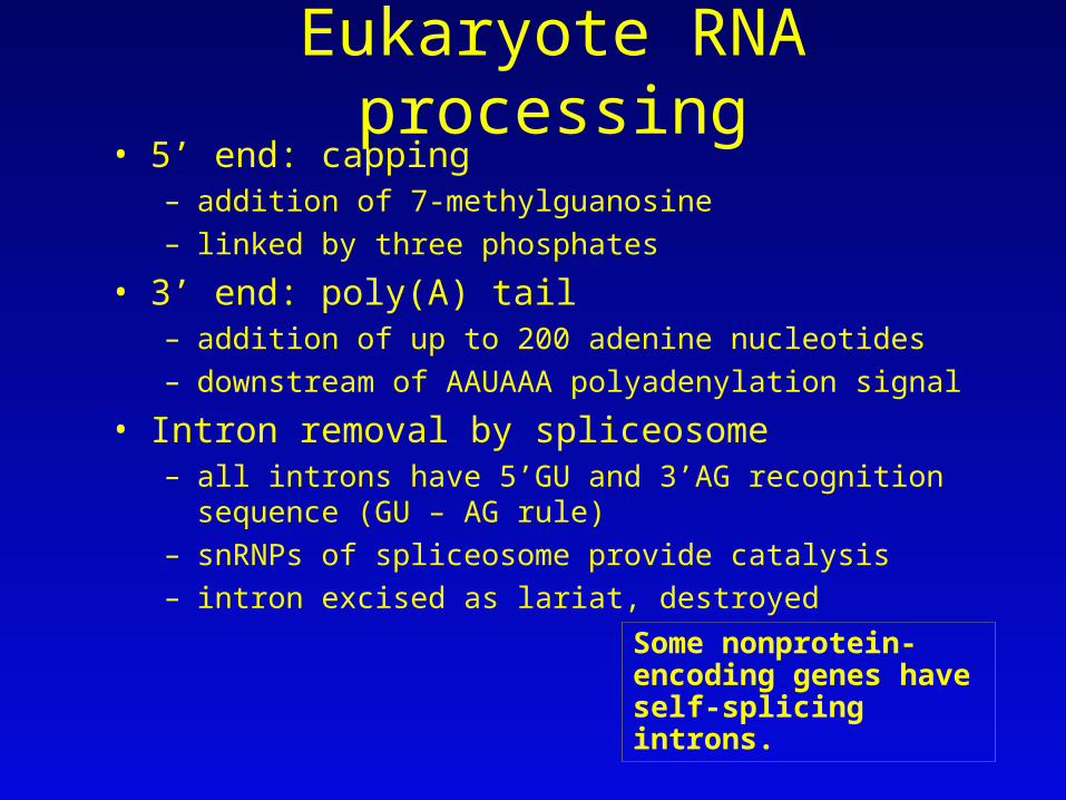
Eukaryote RNA processing• 5’ end: capping
– addition of 7-methylguanosine
– linked by three phosphates
• 3’ end: poly(A) tail – addition of up to 200 adenine nucleotides
– downstream of AAUAAA polyadenylation signal
• Intron removal by spliceosome– all introns have 5’GU and 3’AG recognition sequence (GU – AG
rule)
– snRNPs of spliceosome provide catalysis
– intron excised as lariat, destroyed
Some nonprotein- encoding genes have self-splicing introns.


Processing Overview

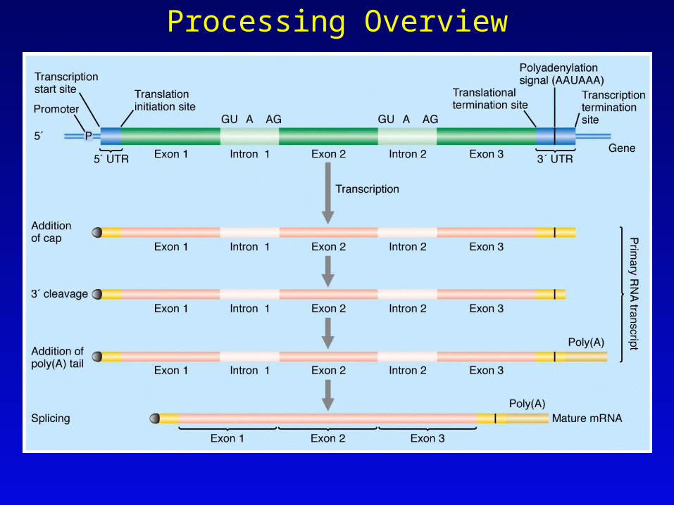
Protein structure• Protein is polymer of amino acids (polypeptide)
– each amino acid has R group conferring unique properties
– amino acids connected by peptide bond
– each polypeptide has amino end and carboxyl end
• Structures– primary: amino acid sequence
– secondary: hydrogen bonding, -helix and -sheet
– tertiary: folding of secondary structure
– quaternary: two or more tertiary structures
• Shape and function determined by primary structure encoded by gene


Translation•mRNA is translated by tRNA at ribosome
•nucleotide sequence is read three nucleotides at a time
–each triplet is called a codon–each amino acid has one or more codons–64 possible codons (4 4 4) = genetic code
•used by all organisms with few exceptions
•Genetic code specifies 20 different amino acids (sometimes selenocysteine)

Codon translation•tRNA
–anticodon consists of 3 nucleotides•base pairs with codon in antiparallel fashion
–3’ acceptor end attaches amino acid•attachment catalyzed by aminoacyl-tRNA synthetases
•one for each different tRNA
•Wobble hypothesis–permits third nucleotide of anticodon (5’ end) to hydrogen bond with alternative nucleotide–permits a tRNA to translate more than one codon


Translation at the ribosome•Ribosome
–large subunit–small subunit
•3 ribosomal sites–A site (amino site), accepts incoming charged tRNA–P site (polypeptide site), peptide bond–E site (exit site), tRNA exits ribosome
•Amino terminus synthesized first, beginning near 5’ end of mRNA


Protein function• Function determined by amino acid sequence
• Colinearity between DNA nucleotide sequence and amino acid sequence of protein
• Two broad types of protein– structural proteins– active proteins, including enzymes
• Proteins often have specialized domains


Malfunctioning alleles• Mutation alters gene function by altering
structure/function in product– wild-type: normal allele
• designated by plus (+) sign• example: arg-3+
– mutation: change in nucleotide sequence• sometimes designated by minus (–) sign
• Nutritional mutants– prototroph: wild-type, synthesizes nutrients– auxotroph: mutant, fails to make essential
nutrient (e.g., amino acid)


Types of mutation• Mutant site: area of nucleotide change
• Three types of mutation– substitution of different amino acid
• e.g., 5’GGA3’ 5’GAA3’, gly glu
– premature stop codon• e.g, 5’GGA3’ 5’UGA3’, gly stop
– frameshift• insertion or deletion of one or two nucleotides alters
reading frame from point of change
• all downstream codons altered

Effect of mutation• Often reduces or eliminates protein function
– leaky mutation: reduced function– null mutation: no function– silent mutation: no change in function, though
amino acid sequence may be changed
• Mutations in information transfer– mutations in exon-intron junction– mutations in promoter or regulatory sequences– mutations in UTRs

Dominance and recessiveness• Recessive genes typically produce little or no product. One
dose of wild-type gene produces sufficient product, resulting in dominant phenotype (haplo-sufficiency)
• Nomenclature
– recessive genes, lower case italicized letter, e.g., a
– dominant gene, upper case italicized letter, e.g., A
• Genotypes
– A/A, (a+/a+) homozygous dominant
– A/a, (a+/a) heterozygous
– a/a, homozygous recessive
normal phenotype


Haplo-insufficiency• Wild-type gene provides insufficient
product to fulfill normal cell function– in this case, defective gene is dominant
• B+/B+, homozygous recessive
• B/B+, heterozygous
• B/B, homozygous dominant defective phenotype


Assignment: Concept map, solved problems 1 and 2, basic problems 2, 8 through 12, challenging problems 18, 21, 23-25
Continue with web-based NCBI tutorial sections from Introduction to Using BLAST to compare sequences.