Embed Size (px)
Citation preview
AP Biology 2013
Chapter 18: Regulation of Gene Expression
Gene Regulation
✤ Prokaryotes and eukaryotes alter their gene expression in response to their changing environment
✤ In multicellular eukaryotes, gene expression regulates development and is responsible for differences in cell types
✤ RNA molecules play a role in regulating this
Bacteria
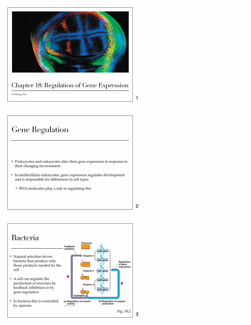
✤ Natural selection favors bacteria that produce only those products needed by the cell
✤ A cell can regulate the production of enzymes by feedback inhibition or by gene regulation
✤ In bacteria this is controlled by operons
Precursor Feedback inhibition
Enzyme 1
Enzyme 2
Enzyme 3
Tryptophan
(a) (b) Regulation of enzyme activity
Regulation of enzyme production
Regulation of gene expression
-
-
trpE gene
trpD gene
trpC gene
trpB gene
trpA gene
Fig. 18.2
1
2
3

Operons✤ Cluster of functionally related genes under coordinated control by a
single “on-off switch” that includes the operator, promoter, and genes they control
✤ Regulatory “switch” is a segment of DNA called an operator which is usually positioned within the promoter
✤ Operon can be switched off by a repressor protein (works by binding to the operator and blocking RNA polymerase)
✤ Repressor is the product of a separate regulatory gene
✤ Repressor can be in an active or inactive form depending on the presence of other molecules
✤ Corepressor is a molecule that cooperates with a repressor protein to shut off an operon
Ex. Trp Operon
✤ E. coli synthesizes the amino acid tryptophan
✤ Trp operon is on by default
✤ When tryptophan is present, it binds to the trp repressor protein which turns the operon off (acts as a corepressor)
Fig. 18.3
Promoter
DNA
Regulatory gene mRNA
trpR
5!
3!
Protein Inactive repressor
RNA polymerase
Promoter
trp operon
Genes of operon
Operator
mRNA 5!
Start codon Stop codon
trpE trpD trpC trpB trpA
E D C B A
Polypeptide subunits that make up enzymes for tryptophan synthesis
(a) Tryptophan absent, repressor inactive, operon on
(b) Tryptophan present, repressor active, operon off
DNA
mRNA
Protein
Tryptophan (corepressor)
Active repressor
No RNA made
Repressible and Inducible Operons
✤ Repressible operon is usually on (binding of a repressor shuts it off)
✤ Ex. trp operon
✤ Inducible operon is usually off (binding of an inducer inactivates the repressor and turns on transcription)
✤ Ex. lac operon - contains genes that code for enzymes used in hydrolysis and metabolism of lactose
(a) Lactose absent, repressor active, operon off
Regulatory gene
Promoter Operator
DNA lacZ lacI DNA
mRNA 5!
3!
No RNA made
RNA polymerase
Active repressor Protein
(b) Lactose present, repressor inactive, operon on
lacI
lac operon
lacZ lacY lacA DNA
mRNA 5!
3!
Protein
mRNA 5!
Inactive repressor
RNA polymerase
Allolactose (inducer)
β-Galactosidase Permease Transacetylase
Fig. 18.4
4
5
6
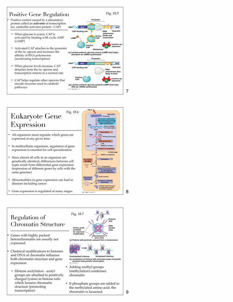
Positive Gene Regulation✤ Positive control caused by a stimulatory
protein called an activator of transcription (ex. catabolite activator protein - CAP)
✤ When glucose is scarce, CAP is activated by binding with cyclic AMP (cAMP)
✤ Activated CAP attaches to the promoter of the lac operon and increases the affinity of RNA polymerase (accelerating transcription)
✤ When glucose levels increase, CAP detaches from the lac operon and transcription returns to a normal rate
✤ CAP helps regulate other operons that encode enzymes used in catabolic pathways
Promoter
DNA
CAP-binding site
lacZ lacI
RNA polymerase binds and transcribes
Operator
cAMP Active CAP
Inactive CAP
Allolactose
Inactive lac repressor
(a) Lactose present, glucose scarce (cAMP level high): abundant lac mRNA synthesized
Promoter
DNA
CAP-binding site
lacZ lacI
Operator RNA polymerase less likely to bind
Inactive lac repressor
Inactive CAP
(b) Lactose present, glucose present (cAMP level low): little lac mRNA synthesized
Fig. 18.5
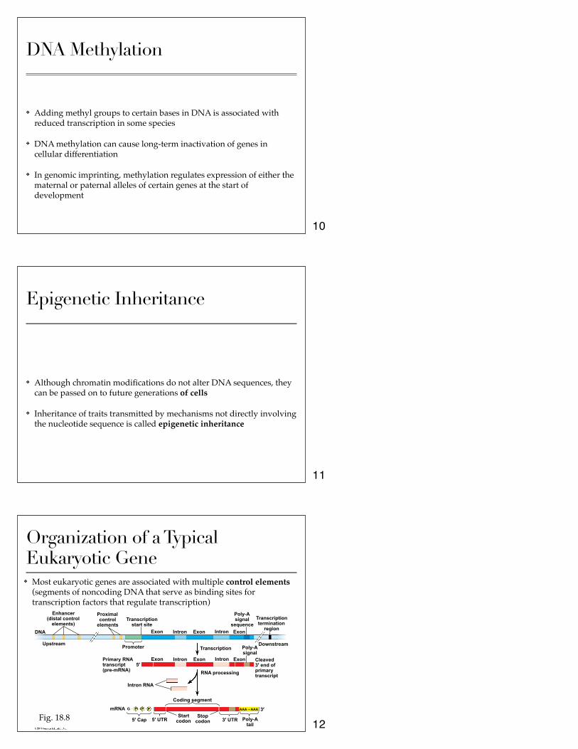
Eukaryote Gene Expression
✤ All organisms must regulate which genes are expressed at any given time
✤ In multicellular organisms, regulation of gene expression is essential for cell specialization
✤ Since almost all cells in an organism are genetically identical, differences between cell types result from differential gene expression (expression of different genes by cells with the same genome)
✤ Abnormalities in gene expression can lead to diseases including cancer
✤ Gene expression is regulated at many stages
Signal
NUCLEUS Chromatin
Chromatin modification: DNA unpacking involving histone acetylation and
DNA demethylation DNA
Gene
Gene available for transcription
RNA Exon Primary transcript
Transcription
Intron RNA processing
Cap Tail
mRNA in nucleus
Transport to cytoplasm
CYTOPLASM mRNA in cytoplasm
Translation Degradation of mRNA
Polypeptide Protein processing, such
as cleavage and chemical modification
Active protein Degradation
of protein Transport to cellular
destination
Cellular function (such as enzymatic activity, structural support)
Fig. 18.6
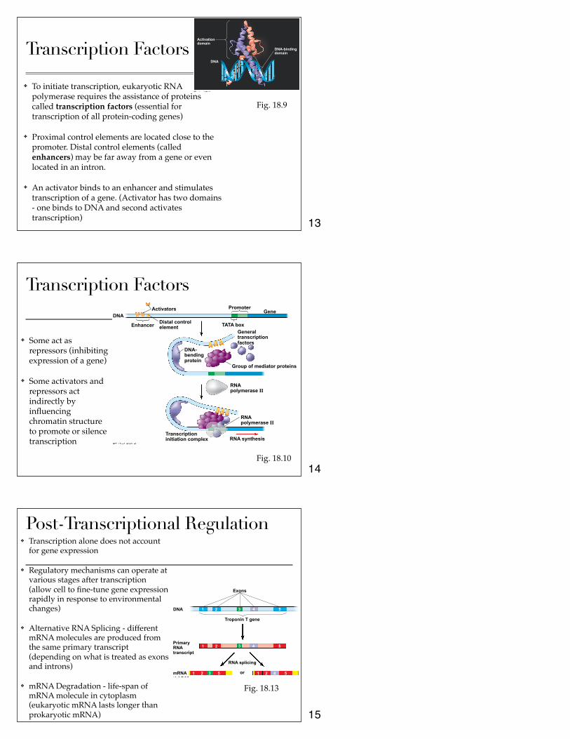
Regulation of Chromatin Structure
✤ Genes with highly packed heterochromatin are usually not expressed
✤ Chemical modifications to histones and DNA of chromatin influence both chromatin structure and gene expression
✤ Histone acetylation - acetyl groups are attached to positively charged lysines in histone tails which loosens chromatin structure (promoting transcription)
Amino acids available for chemical modification
Histone tails
DNA double helix
Nucleosome (end view)
(a) Histone tails protrude outward from a nucleosome
Unacetylated histones Acetylated histones (b) Acetylation of histone tails promotes loose chromatin
structure that permits transcription
Fig. 18.7
✤ Adding methyl groups (methylation) condenses chromatin
✤ If phosphate groups are added to the methylated amino acid, the chromatin is loosened.
7
8
9
DNA Methylation
✤ Adding methyl groups to certain bases in DNA is associated with reduced transcription in some species
✤ DNA methylation can cause long-term inactivation of genes in cellular differentiation
✤ In genomic imprinting, methylation regulates expression of either the maternal or paternal alleles of certain genes at the start of development
Epigenetic Inheritance
✤ Although chromatin modifications do not alter DNA sequences, they can be passed on to future generations of cells
✤ Inheritance of traits transmitted by mechanisms not directly involving the nucleotide sequence is called epigenetic inheritance
Organization of a Typical Eukaryotic Gene
✤ Most eukaryotic genes are associated with multiple control elements (segments of noncoding DNA that serve as binding sites for transcription factors that regulate transcription)
Enhancer (distal control
elements)
DNA
Upstream Promoter
Proximal control
elements Transcription
start site Exon Intron Exon Exon Intron
Poly-A signal
sequence Transcription termination
region
Downstream Poly-A signal
Exon Intron Exon Exon Intron
Transcription
Cleaved 3! end of primary transcript
5! Primary RNA transcript (pre-mRNA)
Intron RNA
RNA processing
mRNA
Coding segment
5! Cap 5! UTR Start
codon Stop
codon 3! UTR
3!
Poly-A tail
P P P G AAA ⋅⋅⋅ AAA
Fig. 18.8
10
11
12
Transcription Factors
✤ To initiate transcription, eukaryotic RNA polymerase requires the assistance of proteins called transcription factors (essential for transcription of all protein-coding genes)
✤ Proximal control elements are located close to the promoter. Distal control elements (called enhancers) may be far away from a gene or even located in an intron.
✤ An activator binds to an enhancer and stimulates transcription of a gene. (Activator has two domains - one binds to DNA and second activates transcription)
DNA
Activation domain
DNA-binding domain
Fig. 18.9
Transcription Factors
✤ Some act as repressors (inhibiting expression of a gene)
✤ Some activators and repressors act indirectly by influencing chromatin structure to promote or silence transcription
Activators DNA
Enhancer Distal control element
Promoter Gene
TATA box General transcription factors
DNA- bending protein
Group of mediator proteins
RNA polymerase II
RNA polymerase II
RNA synthesis Transcription initiation complex
Fig. 18.10
Post-Transcriptional Regulation✤ Transcription alone does not account
for gene expression
✤ Regulatory mechanisms can operate at various stages after transcription (allow cell to fine-tune gene expression rapidly in response to environmental changes)
✤ Alternative RNA Splicing - different mRNA molecules are produced from the same primary transcript (depending on what is treated as exons and introns)
✤ mRNA Degradation - life-span of mRNA molecule in cytoplasm (eukaryotic mRNA lasts longer than prokaryotic mRNA)
Exons
DNA
Troponin T gene
Primary RNA transcript
RNA splicing
or mRNA
1
1
1 1
2
2
2 2
3
3
3
4
4
4
5
5
5 5
Fig. 18.13
13
14
15
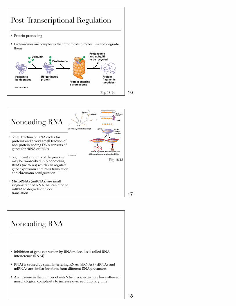
Post-Transcriptional Regulation
✤ Protein processing
✤ Proteasomes are complexes that bind protein molecules and degrade them
Protein to be degraded
Ubiquitin
Ubiquitinated protein
Proteasome
Protein entering a proteasome
Proteasome and ubiquitin to be recycled
Protein fragments (peptides)
Fig. 18.14
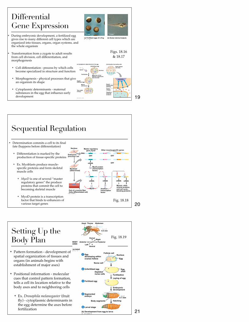
Noncoding RNA
✤ Small fraction of DNA codes for proteins and a very small fraction of non-protein-coding DNA consists of genes for rRNA or tRNA
✤ Significant amounts of the genome may be transcribed into noncoding RNAs (ncRNAs) which can regulate gene expression at mRNA translation and chromatin configuration
✤ MicroRNAs (miRNAs) are small single-stranded RNA that can bind to mRNA to degrade or block translation
(a) Primary miRNA transcript
Hairpin miRNA
miRNA
Hydrogen bond
Dicer
miRNA- protein complex
mRNA degraded Translation blocked (b) Generation and function of miRNAs
5! 3!
Fig. 18.15
Noncoding RNA
✤ Inhibition of gene expression by RNA molecules is called RNA interference (RNAi)
✤ RNAi is caused by small interfering RNAs (siRNAs) - siRNAs and miRNAs are similar but form from different RNA precursors
✤ An increase in the number of miRNAs in a species may have allowed morphological complexity to increase over evolutionary time
16
17
18
Differential Gene Expression
✤ During embryonic development, a fertilized egg gives rise to many different cell types which are organized into tissues, organs, organ systems, and the whole organism
✤ Transformation from a zygote to adult results from cell division, cell differentiation, and morphogenesis
✤ Cell differentiation - process by which cells become specialized in structure and function
✤ Morphogenesis - physical processes that give an organism its shape
✤ Cytoplasmic determinants - maternal substances in the egg that influence early development
(a) Cytoplasmic determinants in the egg (b) Induction by nearby cells
Unfertilized egg
Sperm
Fertilization
Zygote (fertilized egg)
Mitotic cell division
Two-celled embryo
Nucleus
Molecules of two different cytoplasmic determinants
Early embryo (32 cells)
NUCLEUS
Signal transduction pathway
Signal receptor
Signaling molecule (inducer)
(a) Fertilized eggs of a frog (b) Newly hatched tadpole
1 mm 2 mm
Figs. 18.16 & 18.17
Sequential Regulation
✤ Determination commits a cell to its final fate (happens before differentiation)
✤ Differentiation is marked by the production of tissue-specific proteins
✤ Ex. Myoblasts produce muscle-specific proteins and form skeletal muscle cells
✤ MyoD is one of several “master regulatory genes” the produce proteins that commit the cell to becoming skeletal muscle
✤ MyoD protein is a transcription factor that binds to enhancers of various target genes
Nucleus
Embryonic precursor cell
Myoblast (determined)
Part of a muscle fiber (fully differentiated cell)
DNA
Master regulatory gene myoD
OFF OFF
OFF mRNA
Other muscle-specific genes
MyoD protein (transcription factor)
mRNA mRNA mRNA mRNA
MyoD Another transcription factor
Myosin, other muscle proteins, and cell cycle– blocking proteins
Fig. 18.18
Setting Up the Body Plan
✤ Pattern formation - development of spatial organization of tissues and organs (in animals begins with establishment of major axes)
✤ Positional information - molecular cues that control pattern formation, tells a cell its location relative to the body axes and to neighboring cells
✤ Ex. Drosophila melanogaster (fruit fly) - cytoplasmic determinants in the egg determine the axes before fertilization
Head Thorax Abdomen
0.5 mm
BODY AXES
Anterior Left
Ventral
Dorsal Right
Posterior
(a) Adult
Egg developing within ovarian follicle
Follicle cell Nucleus
Nurse cell
Egg
Unfertilized egg Depleted nurse cells
Egg shell
Fertilization
Laying of egg Fertilized egg
Embryonic development
Segmented embryo
Body segments Hatching 0.1 mm
Larval stage
(b) Development from egg to larva
5
4
3
2
1
Fig. 18.19
19
20
21

Developmental Genes in Drosophila
✤ Homeotic genes - control pattern formation in late embryo, larva, and adult stages
✤ Embryonic lethals - mutations that cause death during embryogenesis
✤ Maternal effect genes - encode for cytoplasmic determinants that establish the axes (also called egg-polarity genes)
✤ Ex. Bicoid gene - if it is not functional, the fly will lack a front half and have duplicate posterior structures at both ends
Head Tail
Tail Tail
Wild-type larva
Mutant larva (bicoid)
250 µm
T1 T2 T3 A1 A2 A3 A4 A5 A6
A7 A8
A8 A7 A6 A7
A8
Fig. 18.21
Bicoid mRNA in mature unfertilized egg
Bicoid mRNA in mature unfertilized egg
Fertilization, translation of bicoid mRNA
Anterior end 100 µm
Bicoid protein in early embryo
Bicoid protein in early embryo
RESULTS
Fig. 18.22
Cancer✤ Gene regulation systems that go wrong in cancer are the same systems
involved in embryonic development
✤ Cancer is caused by mutations in genes that regulate cell growth and division
✤ Tumor viruses can also cause cancer in animals including humans
✤ Oncogenes - cancer-causing genes
✤ Proto-oncogenes - normal cellular genes responsible for normal cell growth and division
✤ Conversion of a proto-oncogene to an oncogene can lead to abnormal stimulation of the cell cycle
✤ Conversion can be caused by movement of DNA near a promoter, amplification of a proto-oncogene, point mutations in the proto-oncogene or its control elements
Conversion of a Proto-oncogene
Proto-oncogene
DNA
Translocation or transposition: gene moved to new locus, under new controls
Gene amplification: multiple copies of the gene
New promoter
Normal growth- stimulating protein in excess
Normal growth-stimulating protein in excess
Point mutation: within a control
element within
the gene
Oncogene Oncogene
Normal growth- stimulating protein in excess
Hyperactive or degradation- resistant protein
Fig. 18.23
22
23
24
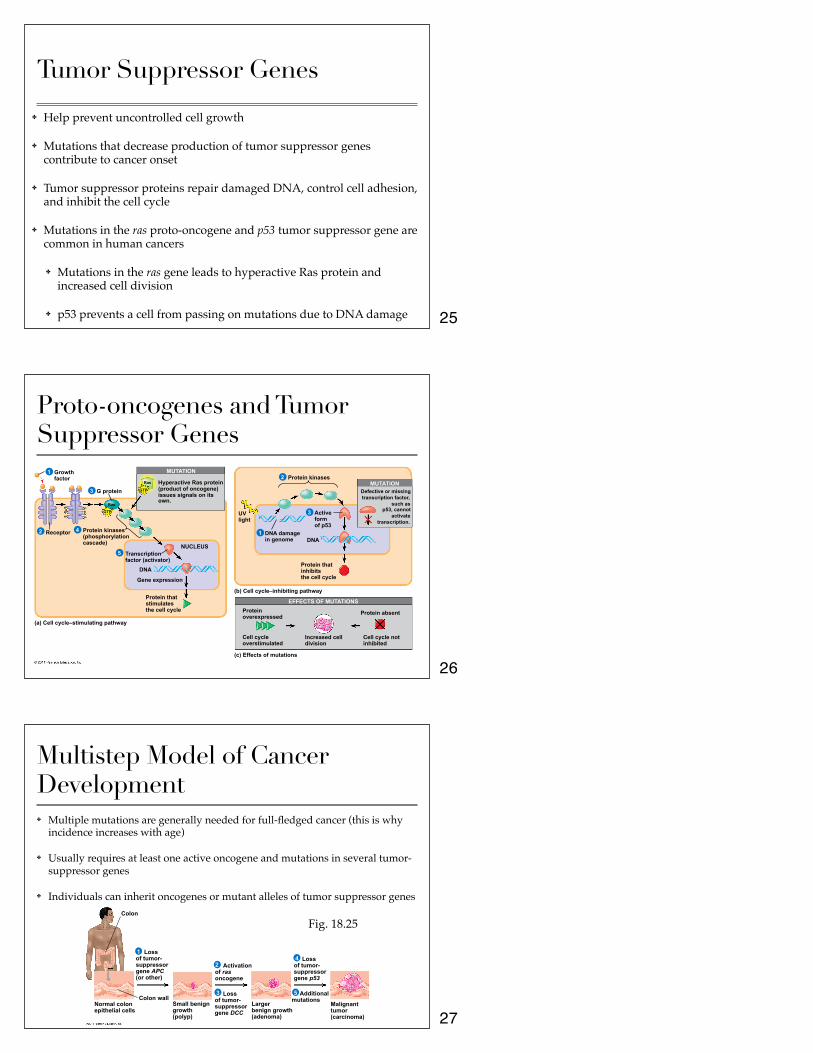
Tumor Suppressor Genes
✤ Help prevent uncontrolled cell growth
✤ Mutations that decrease production of tumor suppressor genes contribute to cancer onset
✤ Tumor suppressor proteins repair damaged DNA, control cell adhesion, and inhibit the cell cycle
✤ Mutations in the ras proto-oncogene and p53 tumor suppressor gene are common in human cancers
✤ Mutations in the ras gene leads to hyperactive Ras protein and increased cell division
✤ p53 prevents a cell from passing on mutations due to DNA damage
Proto-oncogenes and Tumor Suppressor Genes
Growth factor
1
2
3
4
5
1
2
Receptor
G protein
Protein kinases (phosphorylation cascade) NUCLEUS
Transcription factor (activator)
DNA
Gene expression
Protein that stimulates the cell cycle
Hyperactive Ras protein (product of oncogene) issues signals on its own.
(a) Cell cycle–stimulating pathway
MUTATION
Ras
Ras
GTP
GTP
P P P P
P P
(b) Cell cycle–inhibiting pathway
Protein kinases
UV light
DNA damage in genome
Active form of p53
DNA
Protein that inhibits the cell cycle
Defective or missing transcription factor,
such as p53, cannot
activate transcription.
MUTATION
EFFECTS OF MUTATIONS
(c) Effects of mutations
Protein overexpressed
Cell cycle overstimulated
Increased cell division
Protein absent
Cell cycle not inhibited
3
Multistep Model of Cancer Development✤ Multiple mutations are generally needed for full-fledged cancer (this is why
incidence increases with age)
✤ Usually requires at least one active oncogene and mutations in several tumor-suppressor genes
✤ Individuals can inherit oncogenes or mutant alleles of tumor suppressor genesColon
Normal colon epithelial cells
Loss of tumor- suppressor gene APC (or other)
1
2
3
4
5 Colon wall
Small benign growth (polyp)
Activation of ras oncogene
Loss of tumor- suppressor gene DCC
Loss of tumor- suppressor gene p53
Additional mutations
Malignant tumor (carcinoma)
Larger benign growth (adenoma)
Fig. 18.25
25
26
27