Embed Size (px)
Citation preview
• INTRODUCTION: slide 2
• BACKGROUND: slide 3, 4
• OBJECTIVES: slide 5
• ESPERIMENTAL PLAN:
1.Strategies: slide 6, 7
2. In vitro: slide 8, 9
3. In vivo: slide 10,11
• PITFALLS AND SOLUTIONS : slide 12
• CONCLUSIONS: slide 13
• MATERIALS AND COSTS: slide 13
• REFERENCES: slide 13
LM Genetica e Biologia molecolare nella Ricerca di Base e Biomedica aa 2013/2014
Corso di Terapia Genica Prof. Isabella Saggio
WERNER’S SYNDROME
GENE THERAPY DESIGN Sara Luzzi, Claudia Barbaccia, Simona Manni

INTRODUCTION
From Chun and Yee, Cancer Biol. Ther. 2010
From Matsumoto et al., Hum Genet, 1997
Werner syndrome (WS) is a rare autosomal recessive progeroid disease which affects the adults.
SYMPTOMS • Dermatologic pathologies (atrophy, tight skin, ulceration, hyperkeratosis) • Premature hair graying • Bilateral cataracts • Voice changes • Osteoporosis • Type II diabetes mellitus • Cardiovascular disease • Cancer (pancreas, skin, thyroid, colorectum) • Soft tissue sarcomas
CAUSES OF DEATH • Pancreatic cancer
• Myocardial infarctions • Cerebrovascular accidents
• Other neoplasms PANCREATIC DYSFUNCTIONS SEEMS TO BE THE PRIMARY CAUSE OF DEATH
The numbers indicate the patients reported
from 1904 to 1994.
At least one patient is reported for the countries in black
Matsumoto et al., Nat. Genet. 1997
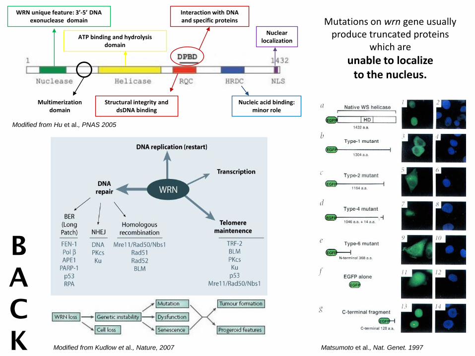
Mutations on wrn gene usually produce truncated proteins
which are
unable to localize to the nucleus.
Modified from Kudlow et al., Nature, 2007
WRN unique feature: 3’-5’ DNA exonuclease domain
ATP binding and hydrolysis domain
Structural integrity and dsDNA binding
Nucleic acid binding: minor role
Nuclear localization
Interaction with DNA and specific proteins
Modified from Hu et al., PNAS 2005
Multimerization domain
B
A
C
K

C>T in the coding sequence
Stop codon in exon 21
Modified from Yu et al., Science 1996, Matsumoto et al.,Hum. Genet. 1997
G>C on a site acceptor Loss of exon 26 and frameshift
Stop codon in exon 27
Modified from Ramirez et al.,Cell. Mol. Life Sci. 2007
From Huang et al., Hum. Mutat. 2006
Most frequent WS mutations
VI IV
G
R
O
U
N
D

EXON 21 SKIPPING R877-R909 deletion
UPSTREAM the RCQ domain
ADENO-ASSOCIATED VIRUS VECTORS
• Successfully transfered to the nucleus
• Persist as extrachromosomal elements
• Small size, small genome
• Expressed for a sustained period of time
• Lack of toxicity
• Weaker immunogenicity (see Pitfall and solutions)
D950 WRN RQC domain
EXON 26 RESCUE G1047-Q1059 deletion
i.e. part of α-helix5
NO CATALITIC CORE LOSS
Modified from Kitano et al., Structure 2010
CATALITIC
CORE
ITR ITR
U1 snRNA promoter
U1 snRNA transcript
artificial tail#1
ITR ITR
OBJECTIVES
U1 snRNA promoter
U1 snRNA transcript
artificial tail#2

ESE Region1 ESE Region2 ESE Region3
αESE-R1 αESE-R2 αESE-R3
#1 + +
#2 + +
#3 + +
20 5’ 3’ 21 22
3’
Prediction made with http://rulai.cshl.edu/cgi-bin/tools/ESE3/esefinder.cgi?process=home
20
21
22
TTA AAT AG– –A TTG GAT
L N R L D
Three possible constructs:
Probability of SRSF binding on Exon 21
U1snRNA-derived antisense
oligonucleotide
Loss of UGA stop codon
allows the recovery of the
full-length WRN protein
EXON SKIPPING

wt SA
Ex-25 ACG AAA AAG AGC CTC ATC
T K K S L I
Prediction made with http://www.umd.be/HSF/
NEW SA
“NEW”
Ex.26
“New” exon 26 inclusion
allows the recovery of the
full-length WRN protein
U1snRNA-derived antisense
oligonucleotide
EXON RESCUE

Day
s af
ter
infe
ctio
n
From Cheng et al., J. Biomed. Sci. 2007
AAV VECTOR
HEK 293T (expressing E1)
Purification: • Cell lysis with benzonase-containing buffer • Iodixanol density gradiend centrifugation
and heparin-sepharose affinity chromatography
OR CsCl gradients. • Dialysis Purity determined by silver-stained sds-page. Titer determined with a dot-blot assay
rAAV2/8 VECTOR
PERSISTENCE HIGH EXPRESSION SPECIFITY

RT-PCR: exon skipping and exon rescue
Western blot (αWRN N-term)
PCR products sequencing
ImmunoFluorescence (IF)
Biochemical assays • Exonuclease activity
• Helicase activity
• Binding with known interacting proteins (e.g. TRF2, PARP1, Rad52, p53)
Proliferation analyses
Single Telomere Length Assay (STeLA)
Adapted from
Cazzella et al., Mol. Ther. 2012
Duterte et al., Nat.Struct.Mol.Biol. 2010
Unskipped
Skipped
snRNA-derived constructs
GAPDH (normalizer)
Full-length WRN
Truncated WRN
Actin (normalizer)
AAV2/8 transduction of exocrine and endocrine pancreatic cells explanted from a WS patient who shows one of the two mentioned mutations.
Control cell line: Isolated human pancreatic islet cells according to Ricordi et al., Diabetes 1988.
Mock samples (shown just in IF example, but well considered for every method): WS pancreatic cells infected with an empty vector
Samples collected after 0-3-7-10 days after infection. STeLA, IF and proliferative analysis: also 2-3-4-5-6-7 weeks after infection.
αWRN
(N-term)
DAPI
MERGE
Control WS AAV infected Mock
1. An adapter
(telorette) is
ligated at the
end of
telomeres.
2. Selective
amplification of
X/Y telomeres.
3. Southern Blotting
Control or
AAV infected cells
WS or
mock
Chen et al., BJMedBiolRes 2013
Azzalin et al., PLoSOne 2012
Colocalization of TRF2, PARP1, Rad52 and p53 will be tested.
PANCREATIC CELLS INFECTION
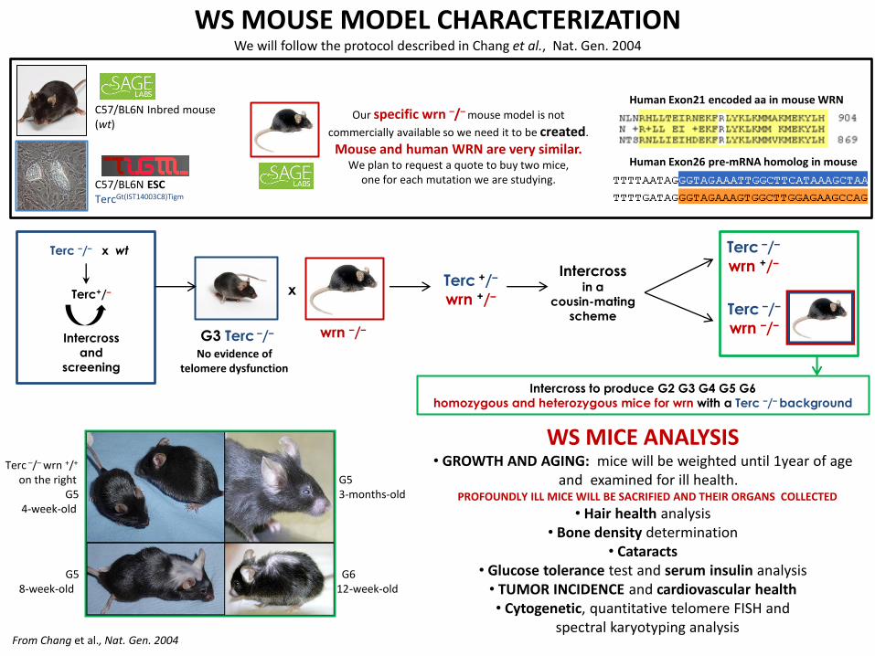
Terc –/– x wt
Terc+/–
Intercross
and
screening
G3 Terc –/–
x
wrn –/–
WS MOUSE MODEL CHARACTERIZATION We will follow the protocol described in Chang et al., Nat. Gen. 2004
Terc +/–
wrn +/–
No evidence of telomere dysfunction
Intercross in a
cousin-mating
scheme
Terc –/–
wrn +/–
Terc –/–
wrn –/–
Intercross to produce G2 G3 G4 G5 G6
homozygous and heterozygous mice for wrn with a Terc –/– background
WS MICE ANALYSIS • GROWTH AND AGING: mice will be weighted until 1year of age
and examined for ill health. PROFOUNDLY ILL MICE WILL BE SACRIFIED AND THEIR ORGANS COLLECTED
• Hair health analysis • Bone density determination
• Cataracts • Glucose tolerance test and serum insulin analysis • TUMOR INCIDENCE and cardiovascular health • Cytogenetic, quantitative telomere FISH and
spectral karyotyping analysis
C57/BL6N Inbred mouse (wt)
Terc –/– wrn +/+ on the right G5 G5 3-months-old 4-week-old
G5 G6 8-week-old 12-week-old
C57/BL6N ESC TercGt(IST14003C8)Tigm
Our specific wrn –/– mouse model is not
commercially available so we need it to be created.
Mouse and human WRN are very similar. We plan to request a quote to buy two mice,
one for each mutation we are studying.
Human Exon21 encoded aa in mouse WRN
Human Exon26 pre-mRNA homolog in mouse
From Chang et al., Nat. Gen. 2004

AAV2/8 infection of C57BL6 Terc–/Wrn– after anesthesia and a lateral incision on the left side of the abdominal cavity.
Efficient in vivo transduction of a control AAV2/8 vector expressing GFP will be tested in wt mice as described in Cheng et al., J Biomed Sci 2007
Control: wt C57BL6 mice
Mock: empty vector in C57BL6 Terc–/Wrn– mice
• Biopsy after 0-7-14 -28-56-150 days and after infection.
- RT-PCR for exon skipping and exon rescue analyses
- Western blot and IF with αWRN (N-terminal)
- Biochemical assays: catalitic analysis; IP and Western blot to assay
the binding of some known interacting proteins (e.g. TRF2, p53, PARP1, Rad52)
- Colocalization with the same interacting protein assayed with the IP analysis
- STeLA
- Proliferation and karyotype analyses
• Monitoring of mice well-being
- Measurement of blood glucose
- Insuline detection from mice pancreas sections
- Measurement of LDL (“bad” cholesterol) and HDL (“good” cholesterol)
- Noninvasive imaging such as micro-TC (Fig.1) and PET (Fig.2) (also for tumor incidence)
Fig. 1:Three-dimensional mouse rendering. Shown in yellow is the adipose tissue revealed by segmentation based on computed tomographic value of fat. Reconstruction with 93-μm voxel. (Grassi et al., Radiol Med. 2009)
Fig. 2: PET scans. Circles in A and B draw attention to lack and presence of pancreatic tumour respectively. (Grassi et al., Radiol Med. 2009)
WS MOUSE MODEL AAV INFECTION
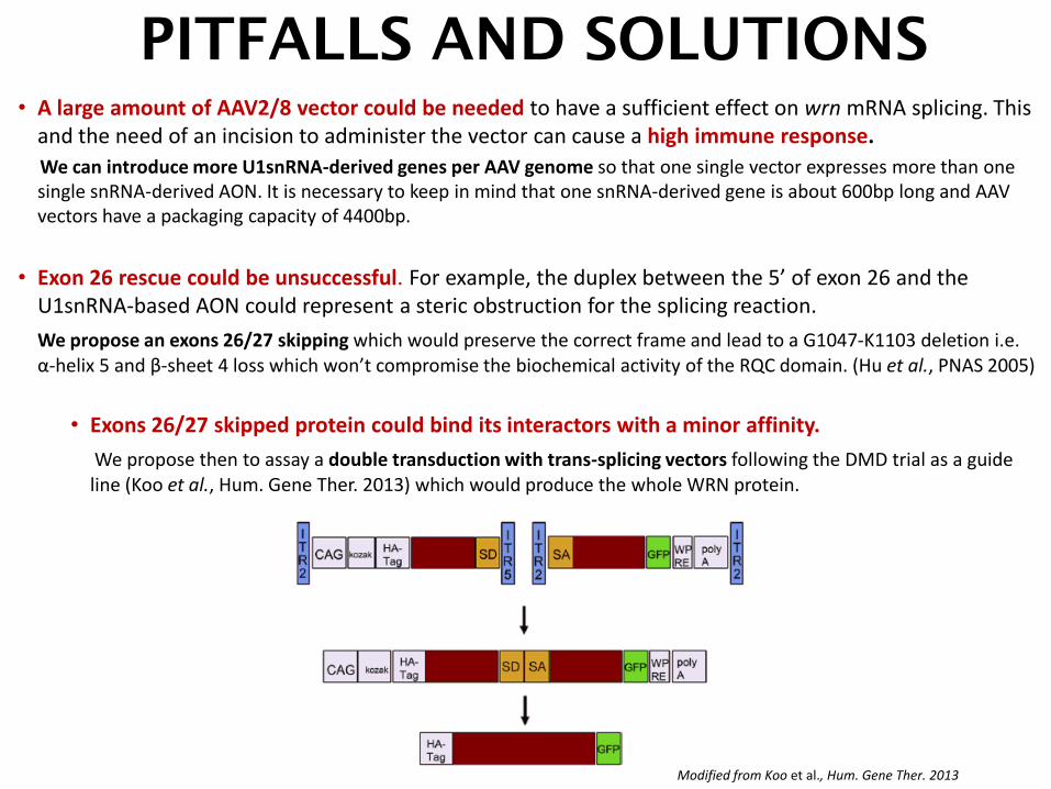
• A large amount of AAV2/8 vector could be needed to have a sufficient effect on wrn mRNA splicing. This and the need of an incision to administer the vector can cause a high immune response.
We can introduce more U1snRNA-derived genes per AAV genome so that one single vector expresses more than one single snRNA-derived AON. It is necessary to keep in mind that one snRNA-derived gene is about 600bp long and AAV vectors have a packaging capacity of 4400bp.
• Exon 26 rescue could be unsuccessful. For example, the duplex between the 5’ of exon 26 and the U1snRNA-based AON could represent a steric obstruction for the splicing reaction.
We propose an exons 26/27 skipping which would preserve the correct frame and lead to a G1047-K1103 deletion i.e. α-helix 5 and β-sheet 4 loss which won’t compromise the biochemical activity of the RQC domain. (Hu et al., PNAS 2005)
• Exons 26/27 skipped protein could bind its interactors with a minor affinity.
We propose then to assay a double transduction with trans-splicing vectors following the DMD trial as a guide line (Koo et al., Hum. Gene Ther. 2013) which would produce the whole WRN protein.
Modified from Koo et al., Hum. Gene Ther. 2013
PITFALLS AND SOLUTIONS

Once our tests on wrn mouse model will confirm the real efficacy of our therapeutic system, clinical trial on human patients will be able to start. It has to be reminded that immune response in humans represent the critical point of every gene therapy approach. Therefore the amount of AAV vector administrated to patients has to be chosen very carefully, also depending on the number of administrations needed. We finally suggest the use of both our treatment and classical therapeutic approaches to reduce also Werner’s Syndrome effects which are not connected to pancreas dysfunction (e.g., therapies for cataracts, pioglitazon for diabetes mellitus type II, thiazolidinetione and rosiglitazione to reduce insuline resistance).
CONCLUSIONS
MATERIALS
AND COSTS • 293AAV Cell Line, Cell Biolabs, 350$ every 106 cells + delivery costs • pAAV-MCS Promoterless Expression Vector, 455$ every 10μg + delivery costs • pAAV rep2/cap8, quote to be requested
• pAd-helper, quote to be requested • pAAV-GFP Control Vector, Cell Biolabs, 395$ every 10μg + delivery costs
• (eventually) AAV purification and quantification reagents • (eventually) AAV purification standard kit, Cell Biolabs, 230$ every kit + delivery costs • (eventually) AAV quantification kit, Cell Biolabs, 230$ every kit + delivery costs • about 900-1000$ per mouse • Stabulation costs • Cell cultures reagents • RT PCR, Western blot, IF, IP, biochemical assays, STeLA reagents (+ other assays ones) (e.g. Abcam Ab200 390€ every 100μl, Ab66601 380€ every 100μl) • PCR purification + sequencing, Biofab research, 13,30€ per sample • chemical reagents, plastics.
~12.000-14.000€/year (4-5 years predicted) (we excluded instruments and materials that can be possibly collected thanks to collaboration with medical department e.g. ws or wt cells, imaging instruments)
REFERENCES Cazzella et al., Mol. Ther. 2012 Chang et al., Nat. Gen. 2004 Cheng et al., J. Biom. Sci. 2007 Choi et al., Curr. Prot. Mol. Biol. 2007 Choudary et al., J. Biol. Chem. 2004 Chu et al., Nature, 2009 Chun et al., Can. Biol. Ther. 2010 Chun et al., Hawai’i Med. J. 2011 Daya and Berns, Clin. Microbiol. Rev. 2008 Flotte et al., Diabetes 2001 Friedrich et al., Hum. Genet. 2010 Grassi et al., Radiol. Med. 2009 Hammond and Wood, Trends Genet. 2011 High and Aubourg, Methods Mol. Biol. 2011 Hu et al., PNAS 2005 Kitano et al., Structure 2010 Kong et al., FEBS J. 2013 Koo et al., Hum. Gene Ther. 2013 Kudlow et al., Nat. Rev. 2007 Kyng et al., Oncogene, 2005 Li et al., Sci. World J. 2009 Matsumoto et al. Hum. Genet. 1997 Matsumoto et al., Nat. Genet. 1997 Perry et al., Nat. Struct. Mol. Biol. 2006 Perry et al., J. Biol. Chem. 2010 Ramirez et al., Cell. Mol. Life Sci. 2007 Ricordi et al., Diabetes 1988 Schulz et al., Hum. Genet. 1996 Thomas et al., J. Virol. 2004 Yu et al., Science 1996 Yu et al., Am. J. Hum. Genet. 2007