Embed Size (px)
Citation preview

General enquiries on this form should be made to:Defra, Procurements and Contracts Division (Science R&D Team)Telephone No. 0207 238 5734E-mail: [email protected]
SID 5 Research Project Final Report
NoteIn line with the Freedom of Information Act 2000, Defra aims to place the results of its completed research projects in the public domain wherever possible. The SID 5 (Research Project Final Report) is designed to capture the information on the results and outputs of Defra-funded research in a format that is easily publishable through the Defra website. A SID 5 must be completed for all projects.
This form is in Word format and the boxes may be expanded or reduced, as appropriate.
ACCESS TO INFORMATIONThe information collected on this form will be stored electronically and may be sent to any part of Defra, or to individual researchers or organisations outside Defra for the purposes of reviewing the project. Defra may also disclose the information to any outside organisation acting as an agent authorised by Defra to process final research reports on its behalf. Defra intends to publish this form on its website, unless there are strong reasons not to, which fully comply with exemptions under the Environmental Information Regulations or the Freedom of Information Act 2000.Defra may be required to release information, including personal data and commercial information, on request under the Environmental Information Regulations or the Freedom of Information Act 2000. However, Defra will not permit any unwarranted breach of confidentiality or act in contravention of its obligations under the Data Protection Act 1998. Defra or its appointed agents may use the name, address or other details on your form to contact you in connection with occasional customer research aimed at improving the processes through which Defra works with its contractors.
Project identification
1. Defra Project code OZ0151
2. Project title
A survey to determine the seroprevalence of infection with Toxoplasma gondii in sheep flocks in Great Britain
3. Contractororganisation(s)
Veterinary Laboratories Agency
54. Total Defra project costs £ 56,424(agreed fixed price)
5. Project: start date................ 07/05/2010
end date................. 28/02/11
SID 5 (Rev. 07/10) Page 1 of 13

6. It is Defra’s intention to publish this form. Please confirm your agreement to do so......................................................................................YES x NO (a) When preparing SID 5s contractors should bear in mind that Defra intends that they be made public. They
should be written in a clear and concise manner and represent a full account of the research project which someone not closely associated with the project can follow.Defra recognises that in a small minority of cases there may be information, such as intellectual property or commercially confidential data, used in or generated by the research project, which should not be disclosed. In these cases, such information should be detailed in a separate annex (not to be published) so that the SID 5 can be placed in the public domain. Where it is impossible to complete the Final Report without including references to any sensitive or confidential data, the information should be included and section (b) completed. NB: only in exceptional circumstances will Defra expect contractors to give a "No" answer.In all cases, reasons for withholding information must be fully in line with exemptions under the Environmental Information Regulations or the Freedom of Information Act 2000.
(b) If you have answered NO, please explain why the Final report should not be released into public domain
Executive Summary7. The executive summary must not exceed 2 sides in total of A4 and should be understandable to the
intelligent non-scientist. It should cover the main objectives, methods and findings of the research, together with any other significant events and options for new work.
Toxoplasma gondii is a single-celled parasite that occurs worldwide in both domesticated and wild animals. Members of the cat family act as definitive hosts for the parasite, although a large number of warm-blooded animals, including humans, can act as intermediate hosts. Human infection occurs principally through ingestion of food and water that has been contaminated with cat faeces, or ingestion of undercooked meat, particularly lamb and pork. Although toxoplasmosis is not generally associated with illness in animals, it is still an important cause of economic loss to the sheep industry since infection of non-immune pregnant sheep commonly results in abortion. Likewise, human infection is usually mild or asymptomatic but severe damage to the foetus can occur if a woman acquires infection for the first time during pregnancy. Life threatening encephalitis can also result from newly acquired or reactivated latent infection in immunocompromised patients, particularly those with HIV/AIDS.
Despite the economic and public health significance of toxoplasmosis, little is currently known about the levels of infection in sheep flocks in Great Britain. The aims of this study were to:
Estimate the proportion of flocks in GB that carry antibodies to toxoplasma Estimate the seroprevalence of ewes within GB that have been infected with toxoplasma Test whether any of the exposures recorded in the Brucella survey questionnaire are associated
with the risk of a flock being seropositive. Determine whether there is any evidence of spatial over-dispersion of toxoplasma seropositive
flocks.
Data was collected via the submission form used for the Brucella survey on location, number of breeding ewes present on farm, the presence of other species on farm, and the age and toxoplasma vaccination status of the animals sampled.
Of the 3539 blood samples collected from 227 flocks, 2619 (74.0 per cent) were found to be positive for antibody to toxoplasma. Details of vaccination status were returned for 3049 (86.1 per cent) of animals sampled. The results show that 6.2 per cent of the animals included in the survey were vaccinated, 57.2 per cent were unvaccinated and the remaining 36.5 per cent were of unknown vaccination status. Animal seroprevalence was estimated at 68.6 per cent, flock seroprevalence at 100 per cent and within flock seroprevalence at 68.6 per cent. Multilevel logistic modelling showed that the likelihood of an animal
SID 5 (Rev. 07/10) Page 2 of 13

testing positive for toxoplasma antibody increased with age and this effect appeared to be amplified in animals vaccinated against toxoplasma. Animals originating from farms where cattle were also kept were also more likely to be seropositive. The model did not reveal an association between vaccination status and risk of testing positive. There was no evidence of regional variation in the distribution of seropositive flocks.
Although the use of surplus material from unrelated studies provides an efficient and cost-effective method of collecting surveillance data, a number of limitations to this approach were identified. Of particular significance to this study was the lack of information relating to timing of vaccine administration and the possibility that vaccinal antibody might have produced false positive results. Previous studies have shown that mean vaccinal antibody titres peak at 1/64 1-2 weeks after vaccination and fall to 1/32 within 12 weeks (Maley and others 1997). The results should therefore be interpreted as maximum prevalences of toxoplasma infection at the individual animal and flock levels. Furthermore, considering that the majority of meat consumed in this country is derived from lambs rather than breeding animals, and the correlation between seropositivity and the presence of tissue cysts in meat has not been definitively proven, the results do not directly reflect the public health risk posed by infected meat products.
However, despite these constraints, this survey has provided a useful baseline measure of seroprevalence in sheep from which future trends can be monitored. Several other potential areas of investigation have also been highlighted as a result of this work, specifically seroprevalence studies in slaughter animals, determination of the level of subclinical congenital infection in sheep and the correlation of serology with direct methods of parasite detection such as PCR or histopathology.
Project Report to Defra8. As a guide this report should be no longer than 20 sides of A4. This report is to provide Defra with
details of the outputs of the research project for internal purposes; to meet the terms of the contract; and to allow Defra to publish details of the outputs to meet Environmental Information Regulation or Freedom of Information obligations. This short report to Defra does not preclude contractors from also seeking to publish a full, formal scientific report/paper in an appropriate scientific or other journal/publication. Indeed, Defra actively encourages such publications as part of the contract terms. The report to Defra should include: the scientific objectives as set out in the contract; the extent to which the objectives set out in the contract have been met; details of methods used and the results obtained, including statistical analysis (if appropriate); a discussion of the results and their reliability; the main implications of the findings; possible future work; and any action resulting from the research (e.g. IP, Knowledge Transfer).
The objectives of this study were to:
Estimate the proportion of flocks in GB that carry antibodies to toxoplasma Estimate the seroprevalence of ewes within GB that have been infected with toxoplasma Test whether any of the exposures recorded in the Brucella survey questionnaire are associated with the
risk of a flock being seropositive. Determine whether there is any evidence of spatial over-dispersion of toxoplasma seropositive flocks.
The findings of the study are presented below in the form of a draft publication:
ABSTRACT
A serological survey of Toxoplasma gondii infection in adult breeding sheep in Great Britain was conducted using surplus sera taken during a seroprevalence study of Brucella melitensis in 2009. Of the 3539 sera collected from 227 flocks, 2619 (74.0 per cent) were found to be positive for T. gondii specific antibody when tested using latex agglutination. Animal prevalence was estimated at 68.6 per cent, flock prevalence at 100 per cent and within flock prevalence at 68.6 per cent. Multilevel logistic modelling suggested that the likelihood of infection increased with age and this effect appeared to be amplified in animals vaccinated against T. gondii. The model also indicated
SID 5 (Rev. 07/10) Page 3 of 13

that the odds of sheep being seropositive were increased on premises where cattle were also kept. These results suggest that levels of toxoplasma infection in breeding sheep in GB are high and provide further evidence to implicate oocysts as the principal source of infection in sheep.
INTRODUCTION
Toxoplasmosis is the most common parasitic zoonosis worldwide and a major source of financial loss to the UK sheep industry (EFSA 2007). Cats are the definitive hosts of the parasite although many warm blooded animal species, including people, can act as intermediate hosts. The remarkable success of T. gondii as a parasite can be attributed to its broad host range, ability to exploit several routes of transmission and generally benign co-existence with the host, but its ubiquitous nature and complex life cycle also present significant challenges to disease control in both humans and animals. Despite the fact that undercooked meat is recognised as a major risk factor for human infection the contribution of infected lamb and mutton to cases of human disease has not been quantified (Cook and others 2000, Dubey and Jones 2008).
T.gondii exists in three distinct phases, all of which are capable of causing infection in both cats and intermediate hosts. Oocysts are produced when the parasite undergoes sexual reproduction in the intestinal epithelial cells of the definitive host, primarily young cats. Despite the fact that oocyst shedding is usually only transient, the enormous numbers produced and their resistance to destruction ensures widespread environmental contamination. Oocysts in freshly voided cat faeces are non-infective until sporulation occurs, usually after 1-5 days depending on environmental conditions (Dubey 1996, Dubey 2004, Dubey and Jones 2008).
Tachyzoites and bradyzoites are the asexually reproducing forms of the parasite that occur during the acute and chronic stages of the disease respectively. Proliferation of tachyzoites is progressively restricted by the host immune response, which induces their differentiation into the morphologically similar, but more slowly dividing bradyzoites that form a cyst wall, partly parasite and partly host in origin. Although organotropism of tissue cysts appears to vary between different intermediate host species, they commonly infect brain, skeletal muscle and heart tissue. Periodic release of bradyzoites from tissue cysts also induces further cyst formation and in some intermediate hosts, including humans and most livestock species, cysts may persist for the lifetime of the animal (Carruthers 2002, Montoya and Liesenfeld 2004, EFSA 2007).
People usually acquire infection through ingestion of tissue cysts in undercooked meat, or via food and water that has been contaminated with sporulated oocysts. The relative contribution of oocysts and tissue cysts to cases of human toxoplasmosis is unknown as in most cases the exact source of infection is not identified (Elsheikha 2008). Seroprevalence studies indicate that between 7 and 34 per cent of the population of the United Kingdom have been infected with toxoplasma (HPA 2010a). Although toxoplasmosis is usually asymptomatic in healthy immunocompetant individuals, more serious sequelae can occur in immunocompromised patients and pregnant women.
Congenital infection can occur if a non-immune woman becomes infected during pregnancy, allowing circulating tachyzoites to cross the placenta and infect the developing foetus. Frequency of congenital transmission and severity of disease are inversely related; the risk of vertical transmission is only about 10-15 per cent when infection is acquired in the early stages of gestation but rates of transmission can be as high as 60-90 per cent in the third trimester. Conversely, the severity of congenital disease tends to decrease with gestational age, and whereas foetal death or severe neonatal disease may result from maternal infection during the first and second trimester, infection in later pregnancy rarely causes physical abnormalities. Subclinical congenital infection, which may initially go unnoticed, often manifests as chorioretinitis or delayed growth later in life (Montoya and Liesenfeld 2004, Dubey and Jones 2008, Dubey and others 2008, Boothroyd 2009).
Disease in immunocompromised individuals such as those with human immunodeficiency virus (HIV) infection or patients undergoing chemotherapy or organ transplantation, can result from either newly acquired infection or more commonly, reactivation of tissue cysts under the influence of falling T-cell counts and interferon-gamma levels. Although reactivation of latent infection can occur in a variety of tissues it is most prominent in the central nervous system often causing toxoplasmic encephalitis (Elsheikha 2008, Henriquez and Roberts 2009). In recent years the incidence of toxoplasmic encephalitis in developed countries has declined significantly due to widespread use of highly active antiretroviral therapy (HAART). However, in underdeveloped regions of the world toxoplasmosis continues to present a serious threat to AIDS patients due to the emergence of drug resistant strains of HIV and limited access to anti-viral therapy (Carruthers 2002, Sukthana 2006, Dubey and Jones 2008, Boothroyd 2009, Dubey 2009, HPA 2010b).
More recently, evidence has emerged to suggest a possible link between chronic toxoplasmosis and behavioural disorders in a number of intermediate animal hosts, including people. Although this association has not been definitively proven, it has been speculated that T. gondii may influence the synthesis of neurotransmitters such as dopamine (Flegr and others 2000, Carruthers 2002, McAllister 2005, Dubey and Jones 2008).
SID 5 (Rev. 07/10) Page 4 of 13

Whilst infection in sheep is usually asymptomatic, significant losses can occur due to abortion if animals become infected during pregnancy. Veterinary Investigation Diagnosis Analysis (VIDA) data for 2009 show that toxoplasmosis accounted for 204 (23 per cent) of the 904 ovine abortion cases examined by the Veterinary Laboratories Agency (VLA) where a diagnosis was subsequently reached. It has also been estimated that over 0.5 million lambs may be lost due to toxoplasmosis each year in the UK at a cost of £12-24 million to the sheep industry (Defra n.d., Defra 2009, Innes and others 2009). As in people, the effect of the parasite on the developing foetus is largely dependent on the stage of gestation; infection of a susceptible ewe in the first trimester usually results in foetal death and resorption, whereas infection acquired later in pregnancy can cause abortion, stillbirth, infected weak lambs or infected but clinically normal lambs (Esteban-Redondo and Innes 1997, EFSA 2007, Dubey and Jones 2008).
Although subclinical congenital infection in lambs has been demonstrated using serology and bioassay, this is thought to be a relatively rare occurrence and infection in the vast majority of sheep is believed to result from ingestion of sporulated oocysts in the post natal period (Dubey 2009). The extensive nature of sheep husbandry and widespread contamination of the environment with oocysts limit the effectiveness of management practices aimed at controlling cats and reducing contamination of feed and water sources. Vaccines using attenuated live tachyzoites of the S48 strain of T. gondii have been licensed for use in sheep in Europe and New Zealand and protective immunity in vaccinated sheep isolated from natural exposure has been shown to last for approximately 18 months. Despite providing significant protection against toxoplasma abortion, it is not known whether vaccination has any effect on the development of tissue cysts in sheep (McAllister 2005, Innes and others 2009).
Seroprevalence data for toxoplasma infection in sheep is sparse. A comparison of studies from several European countries revealed a median seroprevalence of 30 per cent (affsa 2005), whilst UK-based studies have suggested seroprevalences of about 29-31per cent (Blewett 1983, Samad and Clarkson 1994). However, seroprevalence can be affected by several factors, including climate, vaccination and other husbandry practices. Comparison of results from different studies is further complicated by differences in serological tests, choice of threshold for positive cut-off and sampling strategy.
This paper describes the results of a seroprevalence survey of toxoplasma infection in sheep in Great Britain using surplus sera collected for an unrelated study. The aims of the study were to estimate the proportion of seropositive flocks and their spatial distribution, establish the overall seroprevalence of toxoplasma infection in breeding ewes and identify possible risk factors for infection.
MATERIALS AND METHODS
Study population and sample selection
Surplus sheep sera from a large, statistically structured survey of Brucella melitensis in sheep and goats in Great Britain were used for the survey. All samples were collected between June and December 2009.
Twenty animals from each flock were randomly sampled during the Brucella survey unless the flock size was less than twenty, in which case all animals were sampled. Of the 1380 sheep flocks (21,005 animals) sampled, owner’s permission was granted to test 1177 flocks (18,013 animals) for toxoplasma antibody.
Sample framework
An initial sift was performed on the Brucella sample submissions to remove those where either Rapid Analysis and Detection of Animal-Related Risks (RADAR) statistics for 2009 (Defra 2010) or data recorded on the Brucella survey questionnaire indicated that the required number of sheep had not been sampled. Subsequently 1108 submissions were considered eligible for random selection for toxoplasma serology.
A random sample of 227 flocks (3544 animals), stratified by Animal Health Office (AHO) region, was selected in order to estimate the seroprevalence of positive flocks, with a precision of +/- 5 per cent and 95 per cent confidence, assuming a true prevalence of 50 per cent. All available samples were tested from each flock, thereby providing 95 per cent confidence in detecting at least one true positive animal at a minimum within flock seroprevalence of 45 per cent. As relevant data relating to the use of LAT on sheep sera is lacking, results from human studies described by Barker and Holliman (1992) were used and a test sensitivity of 99 per cent and specificity of 81 per cent was assumed. Of the 3544 samples selected, 4 of 20 samples from one flock, and 1 of 19 from another were insufficient for testing. Results were therefore returned for 3539 animals.
SID 5 (Rev. 07/10) Page 5 of 13

Questionnaire
Data was collected via the submission form used for the Brucella survey. As well as location details this form recorded the number of breeding ewes present on farm, the presence of other species on farm, and the age and toxoplasma vaccination status of the animals sampled.
Serological testing
Serum samples were tested by Latex Agglutination Test (LAT); the standard ISO 17025 (UKAS) accredited test used by the Veterinary Laboratories Agency (VLA) to detect T. gondii specific IgG and IgM in animal serum samples. A series of three doubling dilutions (1:16, 1:32 and 1:64) was made for each serum sample. Samples exhibiting significant agglutination at a serum dilution factor (antibody titre) of 1:64 were defined as positive (Barker and Holliman 1992, MAST diagnostics 2004).
Statistical analysis
A multilevel logistic model was produced in MLwiN 2.2 (Rabash and others 2004) to investigate whether any variables recorded on the Brucella submission forms were significantly associated with the likelihood of an animal being infected with toxoplasma. A multilevel model was used to represent the hierarchical nature of the data (animals within flocks), and account for non-independence of animals within flocks. All potential explanatory variables were entered separately into the logistic model, with positive or negative (for toxoplasma) as the outcome variable. Significant variables (p < 0.05) were then entered into the model together and non-significant ones were removed. All other variables were then re-entered sequentially; significant variables were retained, and the process repeated until nothing else was significant. Interactions were investigated within the final model and with other variables where appropriate.
RESULTS
Animal age
Age of animal was returned for 2663 (75.3 per cent) of the 3539 samples tested. The mean age of seronegative animals was 2.97 years compared to a mean age of 3.29 years for seropositive animals (Table 1).
Table 1. Numbers, means and confidence intervals for age, stratified by toxoplasma status.
Toxoplasma negative Toxoplasma positiven Mean CI n Mean CI
Age (years) 712 2.97 2.83, 3.11 1951 3.29 3.22, 3.36
RADAR data provided an estimate of number of sheep in the flock for 131 out of 227 flocks sampled (mean flock size: 317.6 sheep), and number of breeding ewes on farm was recorded on the survey questionnaire for all 227 flocks (mean number of breeding ewes per flock: 161.1).
Table 2 shows the seroprevalence in different age groups of animals sampled. However, as the animals were not sampled in uniform age groups these values do not take into account the effect of mixing between age groups, and clustering on farm.
Table 2. Mean animal seroprevalence according to peerage group.
Age (years) n No. positive animals Mean seroprevalenceNone given 873 665 76.0%
<1 83 17 20.5%1 - <2 378 242 64.0%2 - <3 513 399 77.8%3 - <4 535 436 81.5%4 - <5 637 466 73.2%5 - <6 326 258 79.1%6 - <7 133 97 72.9%7 - <8 24 15 62.5%8 - <9 27 19 70.4%
9+ 10 5 50.0%Total 3539 2619
SID 5 (Rev. 07/10) Page 6 of 13

Vaccination status
Details of vaccination status were returned for 3049 (86.1 per cent) of animals sampled (Table 3). The results show that 6.2 per cent of the animals included in the survey were vaccinated, 57.2 per cent were unvaccinated and the remaining 36.5 per cent were of unknown vaccination status.
Table 3. Numbers of animals in each category of vaccination status, stratified by toxoplasma status.
Toxoplasma statusNegative Positive
Vaccinated 54 164Unvaccinated 549 1477Unknown 317 978Total 920 2619
Presence of other species on farm
Details of other livestock species present on farms included in the survey are presented in Table 4.
Table 4. Numbers of animals in each category sharing premises with other species, stratified by toxoplasma status.
Species on same premises
Toxoplasma statusNegative Positive
Horses YesNo
11909
92610
Poultry YesNo
224696
5542065
Camelids YesNo
5915
102609
Pigs YesNo
102818
2032416
Cattle YesNo
349571
12711348
Total 920 2619
Animal seroprevalence
In total, 2619 animals (218 flocks) tested positive for toxoplasma giving a crude animal seroprevalence of 74.0 per cent. Assuming a test sensitivity of 99 per cent and specificity of 81 per cent the true animal seroprevalence was estimated as 68.6 per cent (Confidence interval (CI): 67.8-69.4%). However, this estimate does not take into account clustering within flocks.
Between flock prevalence
The crude between flock prevalence was 96.0 per cent giving an estimated true between flock prevalence of 100 per cent (CI: 98.7-100%).
Within flock prevalence
Insufficient samples were collected from each flock to calculate seroprevalence within individual flocks. However, assuming an animal prevalence of 68.6 per cent and flock prevalence of 100 per cent, the within flock prevalence was approximately 68.6 per cent (calculated as animal prevalence/herd prevalence*100).
Risk Factors
Three variables significantly affected the likelihood of an animal being seropositive in the first instance: number of breeding ewes in the flock (for every 10 animal increase: OR: 1.01, Χ2 = 5.28 p = 0.022), presence of cattle on the holding (cattle compared to no cattle: OR: 1.65, Χ2 = 12.5 p < 0.001) and animal age (for every year’s increase in age: OR: 1.12, Χ2 = 10.2 p = 0.001). All variables significant at this stage were entered into a model together, and number of breeding ewes became insignificant (p > 0.05).
SID 5 (Rev. 07/10) Page 7 of 13

The final model is detailed in Table 5. The odds of an animal testing positive were higher where cattle were present on the same premises. Odds of testing positive also increased with age of animal in an interaction with vaccination status (Χ2 = 23.9 p < 0.001), as illustrated in Figure 1. The odds of an animal being test positive increased with age in all vaccination groups except those of unknown vaccination status, but the increase was more marked in vaccinated animals.
Table 5 Showing the final model of factors significantly affecting the odds of an animal being seropositive.
Variable OR Comparison OR CI Χ2p-
valueCattle on premises Yes (n=1919):no (n=1620) 1.63 1.39, 1.92 12.7 <0.001Age (years) Continuous variable (n=2663) 1.16 1.11, 1.21 11.8 0.001Toxoplasma vaccination status
Vaccinated (n=218):Unvaccinated (n=2026)Unknown (n=805):Unvaccinated (n=2026)
0.372.02
0.17, 0.811.48, 2.75
0.126.37
0.7260.012
Age*toxoplasma vaccination status
Age*Vaccinated:UnvaccinatedAge*Unknown:Unvaccinated
1.660.85
1.25, 2.210.78, 0.92
2.173.88
0.1400.049
Figure 1 Mean age of sheep by toxoplasma status, within vaccination status.
Spatial Analysis
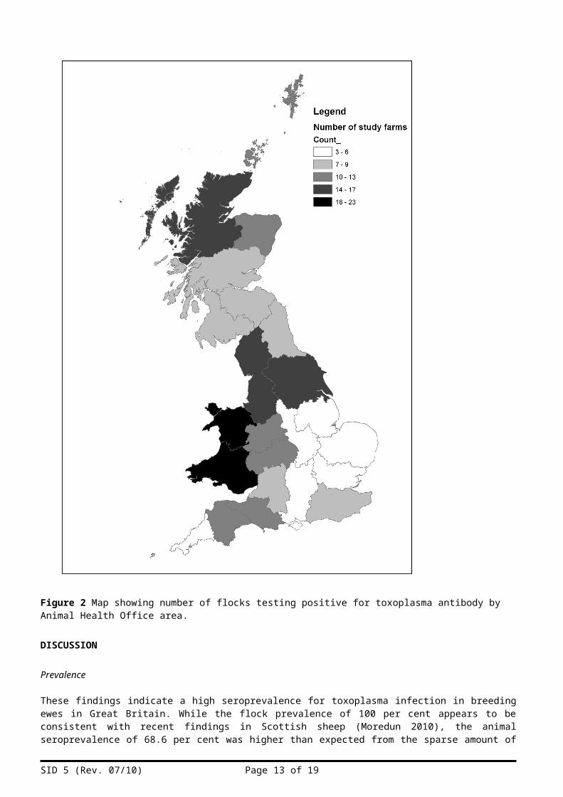
Figure 2 shows the spatial distribution of seropositive flocks. In view of the high seroprevalence there was no evidence of spatial heterogeneity.
SID 5 (Rev. 07/10) Page 8 of 13

Figure 2 Map showing number of flocks testing positive for toxoplasma antibody by Animal Health Office area.
DISCUSSION
Prevalence
These findings indicate a high seroprevalence for toxoplasma infection in breeding ewes in Great Britain. While the flock prevalence of 100 per cent appears to be consistent with recent findings in Scottish sheep (Moredun 2010), the animal seroprevalence of 68.6 per cent was higher than expected from the sparse amount of existing UK data. Although it is not possible to determine whether this discrepancy reflects increased exposure to the parasite or different sampling and testing methods, the results nevertheless provide a useful baseline measure of seroprevalence from which future trends can be monitored.
SID 5 (Rev. 07/10) Page 9 of 13

Risk factors
The fact that increasing age was identified as a risk factor for an animal testing seropositive is not surprising as this reflects increasing exposure to oocysts over time. Differences in exposure to environmental oocysts are also likely to account for the fact that the association between age of animal and likelihood of infection was amplified when animals were vaccinated. As vaccination is usually administered in response to previous episodes of toxoplasma abortion, it follows that these animals are more likely to originate from areas where oocyst exposure is greater and are therefore likely to be infected earlier in life. These findings may also have public health implications, as irrespective of whether vaccination has any effect on tissue cyst development, by remaining seronegative for longer, unvaccinated animals are more likely to produce congenitally infected lambs.
Although an intriguing finding, the association between seropositivity in sheep and the presence of cattle on the same farm is not one that can be readily explained in light of our current understanding of the epidemiology of toxoplasma. Rather than cattle acting as a reservoir of infection for sheep it is likely that confounding factors have given rise to this observation. For example, it is possible that farmers concentrating exclusively on sheep production may be less likely to have cats on their premises due to a heightened awareness of toxoplasma, or that regions suitable for cattle rearing may also be more favourable for oocyst survival.
It is acknowledged that by using data collected for an unrelated survey, a number of important potential risk factors associated with toxoplasma infection could not be investigated in this study. Most significantly, there was no information available relating to the presence of cats on farms, a factor which has previously been shown to increase the risk of infection in sheep (Buxton and others 2007). Other potential risk factors that were not explored include surface water drinking water sources, farm size and history of abortion in the flock (Dubey 2009).
Spatial distribution
The lack of spatial heterogeneity in seroprevalence suggests that toxoplasma infection in sheep is uniform throughout Great Britain. This is an interesting finding as it was anticipated that regional variation in seroprevalence could have arisen due to differences in local cat populations, husbandry practices and geographical features that affect oocyst survival and dispersal. However, whilst it is possible that such variations may have become apparent with a larger study population, a uniform risk of infection suggests that farmers who do not vaccinate are overlooking the threat of toxoplasma as a potential cause of loss. Had the data been available, it may have been useful to look at flock prevalence in relation to both vaccination status and confirmed toxoplasma abortion figures for the farms studied.
Although it is not possible to distinguish between acquired and congenital infection on the basis of a serological survey, and not withstanding the possible effects of clustering, it is interesting to note that the seroprevalence in animals less than 12 months of age was 20.5 per cent compared to 64.0 per cent in those aged 12-24 months. Considering the age-associated risk predicted by the model and the fact there was no evidence of variability in exposure in different areas of the country, these results support the theory that levels of sub-clinical congenital infection in sheep are generally low and that most sheep acquire infection after birth.
Choice of serological test
Since LAT is not capable of detecting serum antibody until several weeks after exposure the test can give false negative results in recently infected animals (Barker and Holliman 1992). Also, by limiting the number of serum dilutions to three the possibility of false negative results was increased slightly further due to interference from the ‘prozone’ effect, which can occur in highly positive sera (MAST 2004). Conversely, cross-reaction with other coccidian parasites may give rise to false positive results, since reactions with unspecified IgM antibody are known to occur with human serum (Barker and Holliman 1992, EFSA 2007).
Effects of vaccination
Despite the fact that LAT cannot differentiate between vaccine-induced and naturally-occurring antibody, vaccinated animals were included in the study since, as it was considered that they were more likely to have been exposed to infection, their exclusion could have led to an underestimated seroprevalence. A comparison of serological techniques by Maley and others (1997) found that mean LAT antibody titres in previously naïve sheep peak at 1/64 1-2 weeks after vaccination and fall to 1/32 or below within 12 weeks. Subsequent exposure of vaccinated animals induced a significant and prolonged rise in mean antibody titre of 1/128. Since the Brucella survey questionnaire did not include dates of vaccination the possibility of some false positive results due to vaccine-induced antibody cannot be discounted, particularly as the samples were taken during the pre-mating season when toxoplasma vaccine is commonly administered. However, in view of the relatively high proportion of unvaccinated animals in the study population and the fact that the model did not reveal an association between vaccination status and risk of testing positive, it could be argued that vaccination had a minimal effect on the serology results and that the seroprevalence figures broadly represent levels of natural infection.
SID 5 (Rev. 07/10) Page 10 of 13

Public health implications
Although serological studies can be useful in measuring the comparative risk of infection between species or groups of animals reared in different locations or husbandry systems, they do not directly relate to the risks posed to people by infected meat products. Focussing entirely on breeding animals poses further limitations, since the large majority of sheep meat consumed in this country is derived from lambs rather than older animals. Although both sales and exports of UK lamb and mutton have been increasing steadily in recent years, figures for 2009 show that approximately 2.1 million rams and ewes were slaughtered in this country compared to about 12.7 million lambs (EBLEX 2009, EBLEX 2010, Farmers Weekly 2009). Although supportive data is lacking, it could also be argued that since mutton is more likely to be served well cooked than lamb, any increased health risk associated with a higher tissue cyst burden will be negated (Kijlstra and Jongert 2008b).
Correlating seropositivity with the presence of viable tissue cysts is also highly problematic, particularly as there is no evidence to suggest that any of the serological tests for toxoplasma have been validated using parasite isolation (Dubey 2009). In view of this, the European Food Safety Authority (EFSA) has recommended that, ‘the correlation between serology and number of infective tissue cysts in edible parts of animals should be established’ (EFSA 2007).
Nevertheless, the high seroprevalence and ubiquitous distribution of infection identified in this study highlights the scale of the threat posed to the UK sheep industry and the importance of control measures aimed at preventing infection in pregnant animals. The complex epidemiology of toxoplasmosis, the commercial challenges associated with infection control at the level of primary production and the technical difficulties involved in detecting the parasite after slaughter are also of significance to public health and food safety advice aimed at reducing the risks of infection among the general public, and in particular vulnerable groups, should continue to be promoted.
Possible future work
A number of potential areas of future work became apparent as a result of this study:
Seroprevalence studies in slaughter animals, particularly sheep and pigs.
Seroprevalence studies in sheep in relation to vaccination status and confirmed toxoplasma abortion figures for the farms studied.
Assessment of subclinical congenital infection levels in sheep.
Correlation of serology with direct methods of parasite detection such as PCR or histopathology.
Outputs
These findings were presented to Defra at the VLA Food and Environmental Safety Programme Meeting on 17 th February 2011.
These findings have also been summarised as a draft paper entitled, ‘A survey to determine the seroprevalence of Toxoplasma gondii infection in British sheep flocks’, which will be submitted to a peer-reviewed journal.
ACKNOWLEDGEMENTS
This work was funded by the UK Department for Environment, Food and Rural Affairs through project OZ0151. The authors wish to thank the many colleagues within the VLA who were involved, particularly Daniel Barker, Johanne Ellis-Iverson, Karen Gillard, Anthony Kay, Sarah Lambton, Geoff Pritchard and Angus Wear.
References to published materialSID 5 (Rev. 07/10) Page 11 of 13

9. This section should be used to record links (hypertext links where possible) or references to other published material generated by, or relating to this project.
Agence Française de Sécurité Santaire des Aliments (afssa). (2005). Toxoplasmose : état des connaissances et évaluation du risque lié à l’alimentation. Rapport du groupe de travail « Toxoplasma gondii » de l’Afssa. Available: http://www.afssa.fr/Documents/MIC-Ra-Toxoplasmose.pdf. Last accessed 17/11/10.
Barker, K.F., Holliman, R.E. (1992). Laboratory techniques in the investigation of toxoplasmosis. Genitourinary Medicine. 68, 55-59.Buxton, D., Maley, S.W., Wright, S.E., Rodger, S., Bartley, P., Innes, E.A. (2007). Toxoplasma gondii and Blewett, D.A. (1983). The epidemiology of ovine toxoplasmosis. I. The interpretation of data for the
Boothroyd, J.C. (2009). Toxoplasma gondii: 25 years and 25 major advances for the field. International Journal for Parasitology. 39, 935–946.
Carruthers, V.B. (2002). Host cell invasion by the opportunistic pathogen Toxoplasma gondii. Acta Tropica. 81, 111–122.
Cook, A.J.C., Gilbert, R.E., Buffolano, W., Zufferey, J., Petersen, E., Jenum, P.A., Foulon, W., Semprini, A.E., Dunn, D.T. (2000). Sources of toxoplasma infection in pregnant women: European multicentre case-control study. British Medical Journal. 321, 142-147.
Defra. (2009). Non-Statutory Zoonoses Annual Report. Available:http:// www.defra.gov.uk/vla/reports/docs/rep_zoo0409.pdf. Last accessed 18/11/10.
Defra. (2010). Veterinary surveillance: Rapid Analysis and Detection of Animal-Related Risks (RADAR). Available:http://www.defra.gov.uk/foodfarm/farmanimal/diseases/vetsurveillance/radar/index.htm. Last accessed 03/03/11.
Defra. (n.d.). Summary profile for toxoplasmosis. Available:http://www.defra.gov.uk/foodfarm/farmanimal/diseases/vetsurveillance/profiles/documents/sp-toxoplasmosis.pdf//. Last accessed 05/01/11
Dubey, J.P. (1996). Strategies to reduce transmission of Toxoplasma gondii to animals and humans. Veterinary Parasitology. 64, 65-70.
Dubey, J.P. (2004). Toxoplasmosis – a waterborne zoonosis. Veterinary Parasitology . 126, 57–72.
Dubey, J.P., Jones, J.L. (2008). Toxoplasma gondii infection in humans and animals in the United States. International Journal for Parasitology . 38, 1257–1278.
Dubey, J.P., Sundar, N., Hill, D., Velmurugan, G.V., Bandini, L.A., Kwok, O.C.H., Majumdar, D., Su, C. (2008). High prevalence and abundant atypical genotypes of Toxoplasma gondii isolated from lambs destined for human consumption in the USA. International Journal for Parasitology. 38, 999–1006.
Dubey, J.P. (2009). Toxoplasmosis in sheep – The last 20 years. Veterinary Parasitology. Doi:10.1016/j.vetpar.2009.02.026.
EBLEX (English Beef and Lamb Executive). (2009). Beef and lamb exports continue to grow. Available:http://www.eblex.org.uk/news/continue-to-grow.aspx. Last accessed 28/12/10.
EBLEX (English Beef and Lamb Executive). (2010). UK monthly production. Available:http://www.eblex.org.uk/markets/other-reports. Last accessed 28/12/10.
Elsheikha, H.M. (2008). Congenital toxoplasmosis: Priorities for further health promotion action. Public Health. 122, 335–353.
Esteban-Redondo, I., Innes, E.A. (1997). Toxoplasma gondii infection in sheep and cattle. Comp. Immun. Microbiol. infect. Dis. 20 (2) 191-196.
European Food Safety Authority (EFSA). (2007). Scientific Opinion of the Panel on Biological Hazards on a request from EFSA on Surveillance and monitoring of Toxoplasma in humans, foods and animals. EFSA Journal. 583 1-64. Available:http://www.efsa.europa.eu/en/scdocs/doc/583.pdf. Last accessed 15/11/10.
Farmers Weekly. (2009). Food Chain: Make the most of opportunities. Available:
SID 5 (Rev. 07/10) Page 12 of 13
http://www.fwi.co.uk/Articles/2009/07/11/116568/Food-Chain-Make-the-most-of-lamb-opportunities.htm. Last accessed 28/12/10.
Flegr, J., Kodym, P., Tolarová, V. (2000). Correlation of duration of latent Toxoplasma gondii infection with personality changes in women. Biological Psychology. 53, 57–68.
Health Protection Agency. (2010a). Information for Health Professionals: Toxoplasmosis. Available: http://www.hpa.nhs.uk/webw/HPAweb&HPAwebStandard/HPAweb_C/1195733799638?p=1191942176127. Last accessed 30/11/10.
Health Protection Agency. (2010b). Health Protection Report Volume 4 Number 6. Available:http//www.hpa.org.uk/hpr/archives/2010/hpr0610.pdf. Last accessed 30/11/10.
Henriquez, F.L., Roberts.W. (2009). A century of Toxoplasma gondii research. Microbiology today. November 09. 192-195.
Innes, E.A., Bartley, P.M., Buxton, D., Katzer, F. (2009a). Ovine Toxoplasmosis. Parasitology. 136, 1887–1894.
Kijlstra, A., Jongert, E. (2008b). Control of the risk of human toxoplasmosis transmitted by meat. International Journal for Parasitology. 38, 1359–1370.
McAllister, M.M. (2005). A decade of discoveries in veterinary protozoology changes our concept of “subclinical” toxoplasmosis. Veterinary Parasitology. 132, 241-247.
Maley, S.W., Thompson, K.M., Bos, H.J., Buxton, D. (1997). Serological diagnosis of toxoplasmosis in sheep following vaccination and challenge. Veterinary Record. 140, 558-559.
MAST Diagnostics. (2004). Data sheet for Toxoreagent Kit. Available: http://www.mastgrp.com/catalogue_products_in_sublist.asp?SubProduct_Type=1394&cat=1. Last accessed. 13/12/10.
Montoya, J.G., Liesenfeld, O. (2003). Toxoplasmosis. Lancet. 363 (9425), 1965-1976.
Moredun. 2010. Toxoplasma Research at Moredun. Available: http://www.moredun.org.uk/research/research-@-moredun/reproductive-diseases/toxoplasma/toxoplasma-research-at-moredun. Last accessed 18/01/11.
Rasbash, J., Steele, F., Browne, W., Prosser, B. (2004) A user’s guide to MlwiN. Centre for Multilevel Modelling, Institute of Education; University of London: 2004.
Samad, M.A., Clarkson, M.J. (1994). Seroconversion to natural Toxoplasma gondii infection during reproductive cycle and its effect on reproduction in sheep. Bangladesh Veterinary Journal. 28, 1-6.
Sukthana, Y. (2006). Toxoplasmosis: beyond animals to humans. Trends in Parasitology. 22 (3), 137-142.
SID 5 (Rev. 07/10) Page 13 of 13