Embed Size (px)
Citation preview
General Introduction to the Course
Course outline and scheduleTextbookRules and procedures Exams and grades
What is a discipline?What is physiology? Study of systems while they are alive (Harvey, 1622) Experimental, rather than observational Subdisciplines, especially cell (= general) physiology
Cell physiology: basis, advantages, hazards
Basic elements of human physiology How does an organ perform its function? How are organs coordinated in time? What are the routes of communication between them? What are the control centers? What are the responses?
Homeostasis and homeostatic mechanisms: feedback loops Negative feedback - nearly universal in physiology Positive feedback – usually pathological (“vicious cycles”)
Intracellular and extracellular fluids ICF has high K+, low Na+ ECF has low K+, high Na+ So what?
Subcellular Organelles
Internal organelles (mitochondria, lysosomes, endoplasmic reticulum, etc.) perform basic cell functions. Please know names and basic functions of each, well presented in textbook.
Membrane Architecture
Continuous phase = lipid bilayerDiscontinuous phase = proteins embedded or floating in it Some proteins extend to both faces (intrinsic proteins) Some proteins only exposed on one side (extrinsic proteins)
Cell membrane = plasma membrane = plasmalemmaOther membranous organelles probably have similar architecture
Movement of Molecules and Ions in Solutions and Membranes
Fat soluble (“lipophilic = hydrophobic”) things cross membranes by dissolving into and out of it from either side
Water soluble (“lipophobic = hydrophilic”) things can cross membranes through aqueous pores if they’re smaller than pores
Special mechanisms (carrier-mediated) for some water soluble things that are too big for the pores
Diffusion
Random movement of molecules or ions in fluids, caused by thermal agitation: non-directional
If two fluids are separated by a membrane, the constituents that can cross that membrane by diffusion (fat soluble things and small water soluble things) will do so, in both directions
The diffusion rate across the membrane is the difference between the rates at which the substance crosses the membrane in each direction
Factors Determining Diffusion Rates
Difference in concentration of the diffusing substance
Size of diffusing substance
Area across which diffusion can occur
Viscosity of the membrane
Thickness of the membrane (= length of the diffusion path)
Temperature (in Kelvin degrees, not Fahrenheit; room temperature is about 300 degrees Kelvin)
Oxygen as an example
Cells use oxygen to make energy (ATP), converting it to CO2Oxygen is carried into the capillaries reversibly bound to hemoglobin, the red protein in red blood cellsThe oxygen content of cells is decreased because they use itTherefore, if oxygen can get off hemoglobin, diffusion will bring it into the cellsSince oxygen binding by hemoglobin is reversible, some is always off hemoglobin and in the plasmaOxygen diffuses into cells because the concentration in cells is lower than it is in plasma
Factors influencing rate of oxygen diffusion into cells
Oxygen is lipophilic, so it can diffuse through the lipid phase. The cross-sectional area through which it can diffuse is hugeOxygen is a small molecule, so it can diffuse rapidlyThe diffusion path (across the thickness of cell membranes) is smallTemperature is essentially constant (body temperature is about 310 degrees Kelvin, never varies by more than 2%)Viscosity of cell membrane lipid is modest, like a fairly thick liquid
If cellular rate of oxygen usage goes up (in an exercising muscle, for example), intracellular oxygen level goes down. This increases rate of oxygen diffusion into the cell, so supply nearly keeps up with modest increases in demand
Facilitated diffusion Membrane proteins include some that reversibly bind specific hydrophilic substances that are too big to diffuse through pores. These proteins constantly flip back and forth across the membrane, exposing their binding sites (or bound substances) to each side. Releasing a bound molecule occurs more readily when it’s exposed to the “downhill” side of the concentration gradient. Binding an unbound molecules occurs more readily on the “uphill” side. Hence, the proteins provide a path through which the substance diffuses.
This is called facilitated diffusion. The role of the carrier protein is to facilitate diffusion of a substance down its concentration gradient.

Active Transport
Some substance can cross cell membranes “uphill” in concentration – going from the side where the concentration is low to the side where its concentration is higher.
This requires energy. It’s called active transport for that reason.
The energy comes from converting ATP to ADP, a reaction that releases small amounts of energy that can be trapped and used to do work in cells.
Many substances cross cell membranes by active transport. Two that are especially relevant are K+ and Na+. Each has a steep concentration gradient across cell membranes, in opposite directions.
Na+, K+ and Membrane ATPase
Na+ and K+ can both diffuse across membranes. How come they don’t diffuse to equilibrium, with equal concentrations in the ICF and ECF?
A pump in cell membranes pumps Na+ out and K+ in. It’s got several names – Na+ pump, Na+/K+ ATPase, membrane ATPase, a few others.
As it creates concentration differences, diffusion in the opposite direction occurs. As concentration differences increase, the diffusion rates increase.
At some point, the diffusion rate and the pumping rate for each ion are equal. The net movement is equal, the concentrations are stable.
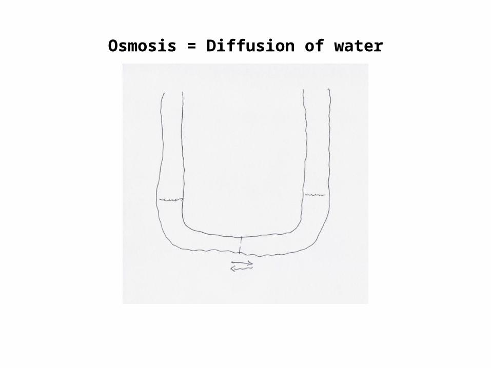
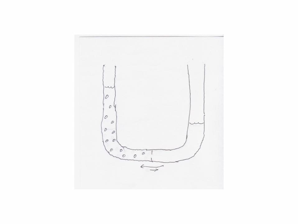
Osmosis = Diffusion of water

Osmosis = Diffusion of Water
Osmotic Pressure
Hydrostatic pressure is the pressure exerted by a column of a fluid. It increases with the height and with the density of the fluid. It can be expressed as pounds per square inch (most common in everyday life), cm of water, mm of Hg.
The osmotic driving force (diffusion of water) can be opposed by hydrostatic pressure in the opposite direction. When that pressure results in no net movement of fluid, it is equal but of opposite sign to the osmotic pressure.
van’t Hoff’s Law
Obviously, as the concentration difference of a solute across a membrane increases, the rate of diffusion of water increases and osmotic pressure increases.
A Dutch chemist, van’t Hoff, worked out the quantitative relationship between concentration difference and osmotic pressure. In simple terms, a concentration difference of 1 mole/L -> a pressure of 22.4 ATM
Since 1 ATM = 760 mm Hg, this is 17,000 mm Hg (compare that to arterial pressure of 100 mm Hg). He discovered something else that’s interesting and important: every molecule or ion in a solution makes an equal contribution to the total osmotic pressure. That is, you can get total osmotic pressure by summing the concentrations of every solute.
ICF and ECF both have hundreds of solutes, totaling about 0.310 moles per liter. Thus, each has osmotic pressure of about 6,000 mm Hg relative to water. It’s convenient to have a unit of concentration that applies to mixtures of molecules and ions. That’s osmolarity, or osmoles/L. Usually written as Osm or mOsm.
Since a difference in osmolarity of 1 Osm -> 17,000 mm Hg osmotic pressure, a difference of 1 mOsm/L -> 17 mm Hg. That’s more than enough pressure to push water across a membrane very rapidly, so when small differences in osmolarity exist, water diffuses across the membrane and corrects the difference.
When molecules or ions cross membranes, they create osmotic differences that force water to follow. This is the only significant mechanism for getting water from one side of a membrane to the other.
When you drink a fluid, it appears in the urine in a matter of hours. It has crossed membranes in your intestine to get into the bloodstream, crossed more membranes in your kidneys to get into the urine. All of that happens osmotically.
Cellular Neurophysiology
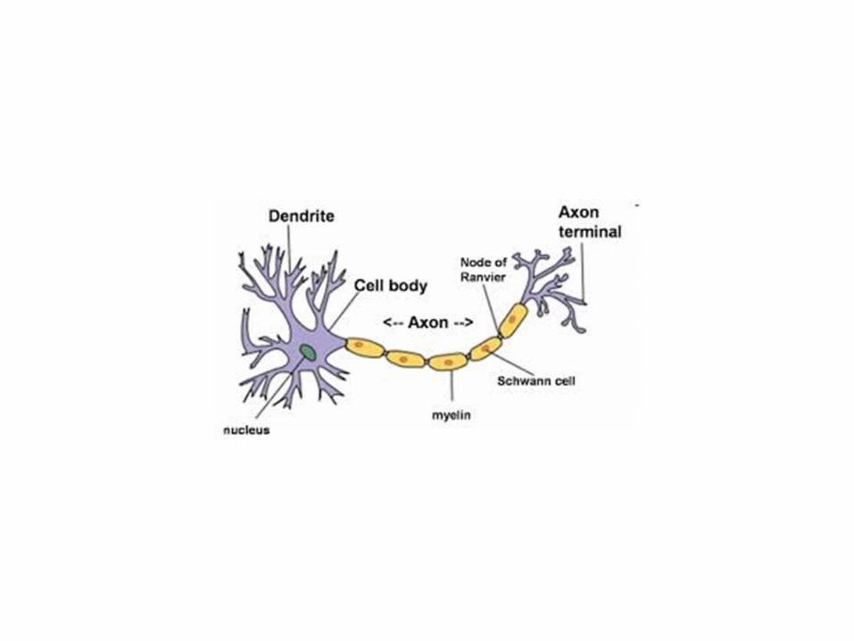
Two major categories of cells in the nervous system:
1. Neurons, which are specialized to conduct information, and2. Glia (“glue”), which optimize the environment for neurons
GLIAL CELLS
OLIGODENDROGLIA (in CNS) = SCHWANN CELLS (in PNS) (we’ll come back to these)
ASTROGLIA = Cells with many projections, usually contact a neuron and the nearest capillary. Presumably facilitate movement of materials back and forth
MICROGLIA = Very small cells. These accumulate at sites of injury, then disappear after healing. Presumably facilitate healing.
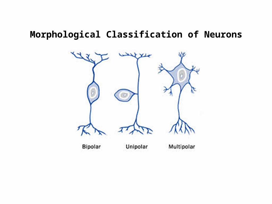
Morphological Classification of Neurons
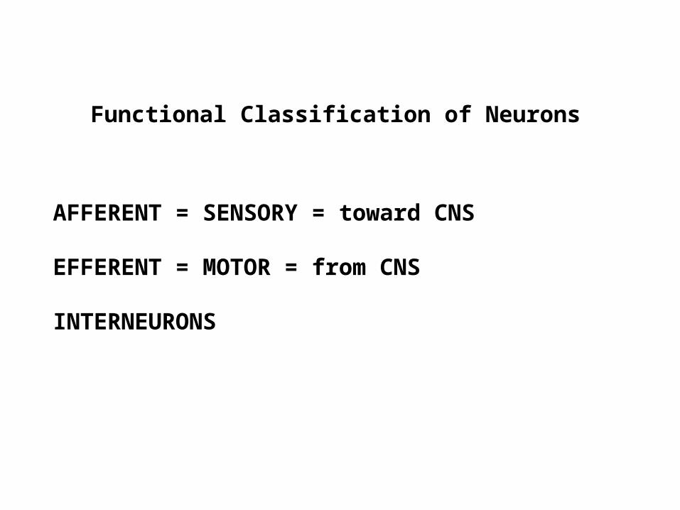
AFFERENT = SENSORY = toward CNS
EFFERENT = MOTOR = from CNS
INTERNEURONS
Functional Classification of Neurons

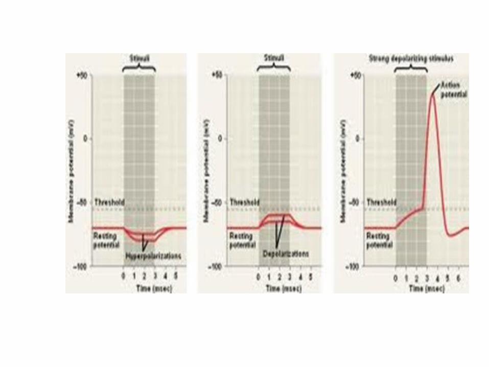
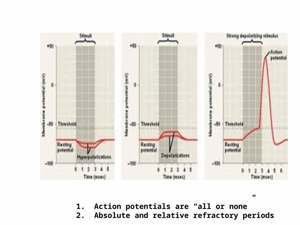
1. Action potentials are “all or none”2. Absolute and relative refractory periods

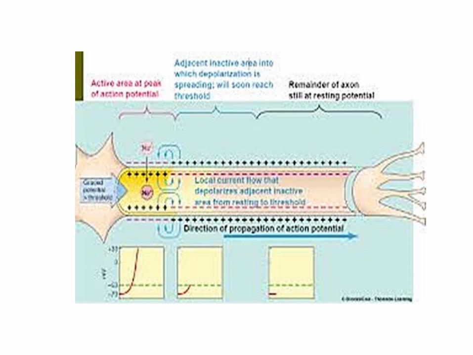
Conduction Velocity of Action Potentials
Non-myelinated Axons = around 1-2 meter/sec; increases with axon diameter
Myelinated Axons = around 20 meter/sec
Synaptic Transmission1. “Electrical “ (via gap junctions)2. Chemical (unique to neurons)
Neurotransmitters
Presynaptic and Postsynaptic Cells
Most common neurotransmitter is acetylcholine (= ACh)
Neurons that release it are cholinergic neurons; ACh receptors are cholinergic receptors.
Next most common neurotransmitters are biogenic amines, the mostwidespread being epinephrine and norepinephrine.
Neurons that release them are aminergic neurons; the receptors are aminergic receptors.
There are many other neurotransmitters; we’ll worry about them later in the course.
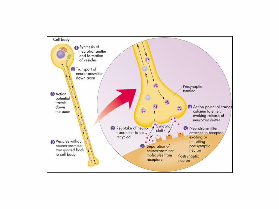
What happens after transmitter binds to receptor?1. Transmitter is destroyed a. Most common transmitter, acetylcholine ( = ACh), is broken into acetate and choline by a plasma enzyme, acetylcholinesterase. b. The choline is then taken up by the axon terminal and used to make more ACh2. What happens in postsynaptic cell? a. Binding to receptor initiates release of a “second messenger” into the cytoplasm of the postsynaptic cell. This is most often Ca ion, cyclic AMP (= cAMP), or cyclic GMP (= cGMP). b. The second messenger elicits response.

There are usually many neurons presynaptic to a single postsynapticneuron. A neuron releases only one kind of neurotransmitter, a neuron’s soma and dendrites often have many kinds of receptors.
Some neurotransmitters cause the postsynaptic cell to become more negative (inhibitory = membrane potential is further from threshold). Others cause the postsynaptic cell to become lessnegative (excitatory = membrane potential is closer to threshold).
The postsynaptic cell sums its inputs. If the result is to make the membrane potential more negative, the change from resting potential is an inhibitory postsynaptic potential (IPSP). If the result is to make the membrane potential less negative, it is an excitatory postsynaptic potential (EPSP).
When the EPSP brings it to threshold, action potentials happen.
Summation
1. When an IPSP or EPSP from a particular presynaptic neuron begins before an existing one has decayed, it is called temporal summation
2. When IPSP and/or EPSP are arriving at a postsynaptic cell from more than one presynaptic cell (a very common situation), it is called spatial summation
Many neurons being presynaptic to a single postsynaptic neuron isconvergence. Information is converging onto that postsynaptic neuron. There can be as many as 1,000 presynaptic neurons converging onto a single postsynaptic neuron.
A single neuron being presynaptic to many postsynaptic neurons (axon terminals branching extensively) is called divergence. Information from the presynaptic neuron is diverging – being spread out.
All of neural integration results from combinations of EPSP, IPSP, convergence and divergence in neural networks.