Embed Size (px)
Citation preview
wwwallsyllabuscom
wwwallsyllabuscom
EC2021-Medical Electronics Lesson Notes
Bio-Potential
Electrode ndash Electrolyte Interface
General Ionic Equations
If electrode has same material as cation then this material gets oxidized and enters the
electrolyte as a cation and electrons remain at the electrode and flow in the external
circuit
If anion can be oxidized at the electrode to form a neutral atom one or two electrons are
given to the electrode
The dominating reaction can be inferred from the following
Current flow from electrode to electrolyte Oxidation (Loss of e-)
Current flow from electrolyte to electrode Reduction (Gain of e-)
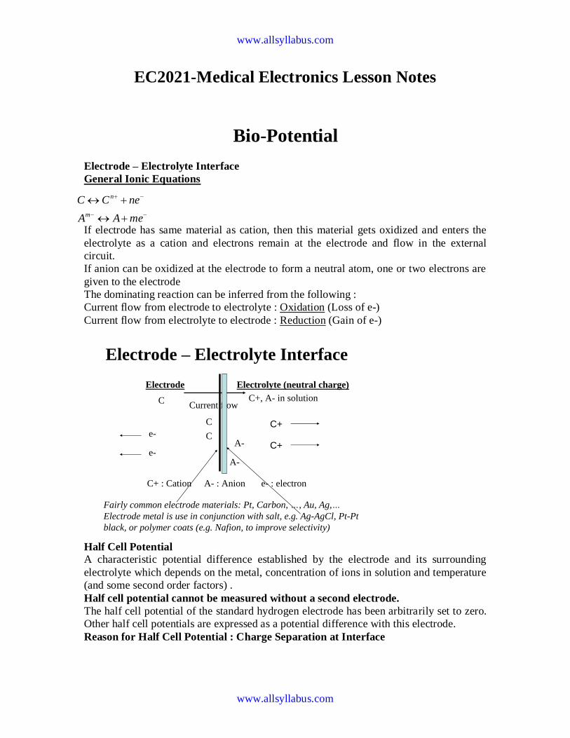
Electrode ndash Electrolyte Interface
Electrode Electrolyte (neutral charge)
C+ A- in solutionC
C
C
A-
A-
C+
C+
e-
e-
Current flow
C+ Cation A- Anion e- electron
Fairly common electrode materials Pt Carbon hellip Au Aghellip
Electrode metal is use in conjunction with salt eg Ag-AgCl Pt-Pt
black or polymer coats (eg Nafion to improve selectivity)
Half Cell Potential A characteristic potential difference established by the electrode and its surrounding
electrolyte which depends on the metal concentration of ions in solution and temperature
(and some second order factors)
Half cell potential cannot be measured without a second electrode
The half cell potential of the standard hydrogen electrode has been arbitrarily set to zero
Other half cell potentials are expressed as a potential difference with this electrode
Reason for Half Cell Potential Charge Separation at Interface
meAA
neCC
m
n
wwwallsyllabuscom
wwwallsyllabuscom
Oxidation or reduction reactions at the electrode-electrolyte interface lead to a double-
charge layer similar to that which exists along electrically active biological cell
membranes
Measuring Half Cell Potential
Polarization If there is a current between the electrode and electrolyte the observed half cell potential
is often altered due to polarization
Nernst Equation When two aqueous ionic solutions of different concentration are separated by an ion-
selective semi-permeable membrane an electric potential exists across the membrane
The Nernst equation for half cell potential is
where E0 Standard Half Cell Potential E Half Cell Potential
a Ionic Activity (generally same as concentration)
n Number of valence electrons involved
Polarizable and Non-Polarizable Electrodes
Perfectly Polarizable Electrodes
These are electrodes in which no actual charge crosses the electrode-electrolyte interface
when a current is applied The current across the interface is a displacement current and
the electrode behaves like a capacitor Example AgAgCl Electrode
Perfectly Non-Polarizable Electrode
These are electrodes where current passes freely across the electrode-electrolyte
interface requiring no energy to make the transition These electrodes see no
Over potentials Example Platinum electrode
Example Ag-AgCl is used in recording while Pt is use in stimulation
BA
DC
aa
aa
nF
RTEE ln0
wwwallsyllabuscom
wwwallsyllabuscom
Equivalent Circuit
Cd capacitance of electrode-eletrolyte interface
Rd resistance of electrode-eletrolyte interface
Rs resistance of electrode lead wire
Ecell cell potential for electrode
Electrode Skin Interface
Sweat glands and ducts
Ru
Rs
Rd Cd
Re
Ese EP
RP CP Ce
wwwallsyllabuscom
wwwallsyllabuscom
Motion Artifact
Why
When the electrode moves with respect to the electrolyte the distribution of the double
layer of charge on polarizable electrode interface changes This changes the half cell
potential temporarily
What
If a pair of electrodes is in an electrolyte and one moves with respect to the other a
potential difference appears across the electrodes known as the motion artifact This is a
source of noise and interference in biopotential measurements
Motion artifact is minimal for non-polarizable electrodes
Body Surface Recording Electrodes
1 Metal Plate Electrodes (historic)
2 Suction Electrodes
Electrode
Epidermis
Dermis and subcutaneous layer
Cd
Gel
Stratum Corneum
Capillary
wwwallsyllabuscom
wwwallsyllabuscom
(historic interest)
1 Floating Electrodes
2 Flexible Electrodes
Commonly Used Biopotential Electrodes
Metal plate electrodes
ndash Large surface Ancient therefore still used ECG
ndash Metal disk with stainless steel platinum or gold coated
ndash EMG EEG
ndash smaller diameters
ndash motion artifacts
ndash Disposable foam-pad Cheap
(a) Metal-plate electrode used for application to limbs
(b) Metal-disk electrode applied with surgical tape
(c)Disposable foam-pad electrodes often used with ECG
Commonly Used Biopotential Electrodes
Suction electrodes
- No straps or adhesives required
- precordial (chest) ECG
- can only be used for short periods
Floating electrodes
- metal disk is recessed
- swimming in the electrolyte gel
- not in contact with the skin
- reduces motion artifact
wwwallsyllabuscom
wwwallsyllabuscom
Commonly Used Biopotential Electrodes
Flexible electrodes
- Body contours are often irregular
- Regularly shaped rigid electrodes
may not always work
- Special case infants
- Material
- Polymer or nylon with silver
- Carbon filled silicon rubber
(Mylar film)
Double-sided
Adhesive-tape
ring Electrolyte gel
in recess
(a) (b)
(c)
Snap coated with Ag-AgCl External snap
Plastic cup
Tack
Plastic disk
Foam pad
Capillary loops
Dead cellular material
Germinating layer
Gel-coated sponge
Floating Electrodes
wwwallsyllabuscom
wwwallsyllabuscom
(a) Carbon-filled silicone rubber electrode
(b) Flexible thin-film neonatal electrode
(c) Cross-sectional view of the thin-film electrode in (b)
Electrodes in Biopotential Measurements
3 Describe the construction of commercial ECG electrode (not the cheap polymer
electrode used in the lab) What is the common electrode metal and why is it preferred
So you are an inventor who has a better idea Describe an improvement
bull to make the electrode cheaper
bull more suitable for lower noise measurement for EEG
bull circumvent patents that are based on plasticfoam electrode body
bull attractive to consumers for use with their ECG machines at home
bull reduce artifact (minimize the motion of skinelectrode) in ambulatory recording
4 In a research laboratory scientists want to record from single cells in a culture dish
They want to record action potentials from single isolated heart cells What kind of
electrode would they need to use (describe material and design) Give a simplified
schematic (circuit model of the electrode) described in the notes given to you
What is the challenge involved in designing an amplifier for use with a microelectrode
for single cell recording Ie what are the critical amplifier design characteristics and
specifications (hint this is not the usual differentialinstrumentation amplifier)
Electrodes and Microelectrodes (miscellaneous)
bull How would you detect bacteria or other microorganisms in water supply Make
sure that your method distinguishes inert particulate matter from living cellular
matter
bull Draw the equivalent circuit model of the skin and an ECG electrode Identify the
key sources of electrical interference and otherwise the elements that would likely
contribute to the poor quality of recordings
wwwallsyllabuscom
wwwallsyllabuscom
bull Design an amplifier interface for the following two applications Patch clamp ion
channel current amplifier Your goal is to amplify pA level current to produce 1
Volt output
bull Strain gauge sensor amplifier Your goal is to convert 10 ohm change in
resistance of a strain gauge to produce 1 volt output
Neural electrodesmicroelectrodes
You want to record from neurons in the brain However you want to record from dozens
of neurons all at once from several closely spaced microelectrodes What material and
process would you use to make the microelectrode array
bull What metal would you prefer to use to make electrode arrays of about 10 micron
square size to make electrical contacts with dozens of neurons
bull What metal would you prefer to use to stimulate dozens of neurons in a deep
brain microelectrode based stimulator
bull (which metal provides good recording vs stimulating properties ndash and at the same
time not be toxic to brain tissue)
bull You are asked to develop an experimental set up to record from rat brain cells
using microelectrodes What precautions would you take to minimize the
electrical interference in your recording set up
Biopotential Amplifiers bull These are very important part of modern medical instrumentation bull We need to amplify biopotentials which are generated in the body at low levels with a high source impedance
bull Biopotentials amplifiers are required to increase signal strength while maintaining fidelity Basic Requirements of Biopotential Amplifiers
Essential functions of a bioamplifier are bull To take a weak biopotential and increase its amplitude so that it can be processed recorded or displayed bull To amplify voltage but it could be considered as a power amplifier as well bull To amplify current since in some cases a biopotential amplifier is used to isolate the load from the source current gain only
Input Impedance (Zin)
bull All biopotential amplifiers must have high input impedance minimize loading (remember the characteristics of biopotential electrodes resulting into loading and distortion if input impedance of the amplifier is not high enough) ndash typical values of Zin over the frequency range of the measurand = 10 MΩ (remember the loading rule)
Protection amp Isolation
bull The input circuit of a biopotential amplifier must provide protection to the live measurand
wwwallsyllabuscom
wwwallsyllabuscom
Vbio
bull Any potential or current at amplifierrsquos input terminals can affect Vbio
bull Electric currents produced by the biopotential amplifier can result in microshock and
macroshock bull The bioamplifier must have isolation and protection circuitry so that the current through the electrodes can be kept at safe levels and any artifact generated by such current can be minimized Output Impedance (Zout)
bull The output circuit does not present any critical problems all it needs to do is to drive the load bull Output impedance must be low with respect to the load impedance and it must be capable of satisfying the power requirements of the load Bandwidth (BW)
Frequency response requirements bull The biopotential amplifier must be sensitive to important frequency components of the biosignal bull Since biopotentials are low level signals it is important to limit bandwidth optimize signal-to-noise ratio Gain (G)
bull Biopotential amplifiers have a gain of 1000 or greater
Mode of Operation
bull Very frequently biosignals are obtained from bipolar electrodes bull Electrodes symmetrically located with respect to ground need differential amplification bull High CMRR required because 1 Common mode signals much greater than the biosignal appear on bipolar electrodes 2 Symmetry with respect to ground is not perfect (mismatch between electrode impedances) ndash more on this later
Calibration Signal
bull Medical and clinical equipment require quick calibration bull The gain of the biopotential amplifier must be calibrated to provide us with an accurate indication of the signalrsquos amplitude bull Push button to apply standard signal to the input of the biopotential amplifier bull Adjustable gain switch carefully selects calibrated fixed gains (in microprocessorndashbased systems gain adjustment can be Electrocardiography
hearts A very widely used medical instrument which is utilized to diagnose and monitor cardiac beat
abnormalities is the electrocardiograph
hearts It measures the electrical activity of the heart (more precisely biopotential differences arising from the electrical activity of myocardium) Wersquove already talked about the genesis of the ECG signal hearts The ECG machine uses surface electrodes and high input impedance hearts Differential amplifiers with good common mode rejection ratio to record the electrocardiogram
hearts Normal ECG amplitude ranges between 05-4 mV Normal frequency content of ECG (for diagnostic purposes) is 005-100 Hz A typical ECG waveform is shown below
wwwallsyllabuscom
wwwallsyllabuscom
Obviously all human hearts are not the same and this leads into variability in different parts of the ECG signal Significant diagnostic features of the ECG signal are hearts Duration of component parts of the signal
hearts Polarities and magnitudes hearts The details of the ECG signal and the degree of variability in different parts of the ECG signal is shown below
The QRS amplitude polarity time duration the RR interval (indicator of heartbeat per min) and the T-wave amplitude are some very important and distinctive features of the ECG signal The heart rate in BPM = Beats Per Minute) is simply = 60 (RR interval in seconds)
Some ECG waveform abnormalities that may indicate illness are hearts An extended PR interval may be diagnosed as AV node block hearts A widening of the QRS complex may indicate conduction problems in the bundle of His hearts An elevated ST segment may indicate occurrence of myocardial Infarction (MI) hearts A negative polarity in the T wave may be due to coronary insufficiency ECG Leads
Normal ECG recordings for the standard lead connections leads I II and III (Lead II provides
the strongest signal) Obviously all human hearts are not the same and this results into a high degree of variability
wwwallsyllabuscom
wwwallsyllabuscom
hearts Note the degree of variability of different parts of the ECG Signal
Some abnormalities that may indicate illness hearts An extended P-R interval may be diagnosed as AV node block hearts Widening of the QRS complex conduction problems in the bundle of His hearts Elevated ST segment may indicate occurrence of MI
hearts Negative polarity T wave may be due to coronary insufficiency QRS amplitude polarity time domain PR interval (indicator of heat beat per min amp T-wave amplitude are some very important
distinctive features 1 Loss
2 Origin of the ECG signal
hearts We have already covered this concept extensively in the previous lectures (The Dipole filed of the heart the Eindhovenrsquos Triangle the electrical circuit model for the electrocardiographic problem etc)
hearts Standard Limb Leads (I II III)
wwwallsyllabuscom
wwwallsyllabuscom
hearts The The lead wires are color-coded according to some conventions One example is White ndashRA
(Right Arm) Black ndash LA (Left Arm) Green ndash RL (Right Leg) Red ndash LL (Left Leg) and Brown
ndash C (Chest) Note There is a CM (common mode) amplifier connected to the right leg We will discuss this in detail later Augmented Limb Leads
These leads offer a free 50 increase over leads VR VL and VF connections (unipolar leads) with respect to Wilson terminal AVR = -I ndash III2 AVL = I ndash II2 aVF = II ndash I2
wwwallsyllabuscom
wwwallsyllabuscom
Each measurement is made from the reflected limb and the average of the other two limbs Chest Leads (Precordial)
Chest Lead Anatomical Positions
V1 - 4th intercostal space ndash Right sternal margin
V2 - 4th intercostal space ndash Left sternal margin
V3 ndash Midway between V2 amp V4
V4 ndash 5th intercostal space on mid-clavicular line
V5 ndash Same as V4 on the anterior axillary line
V6 ndash Same as V5 on the mid-axillary line
12 Lead Clinical Electrocardiography
The ECG Machine
Most representative Specs
wwwallsyllabuscom
wwwallsyllabuscom
bull Zin = 10 MΩ bull Frequency response = 005 ndash100 Hz bull Strip Chart Recorder Speed = 25 mmsec bull Fast Speed = 100 mmsec
For detailed Specs Refer to the Table in your text ―Summary of performance requirements for electrocardiographs Location of the Heart bull The heart is located between the lungs behind the sternum and above the diaphragm bull It is surrounded by the pericardium bull Its size is about that of a fist and its weight is about 250-300 g bull Its center is located about 15 cm to the left of the midsagittal plane
Anatomy of the heart
bull The walls of the heart are composed of cardiac muscle called myocardium
bull It consists of four compartments ndash the right and left atria and ventricles
The Heart Valves
bull The tricuspid valve regulates blood flow between the right atrium and right
ventricle
bull The pulmonary valve controls blood flow from the right ventricle into the
pulmonary arteries
bull The mitral valve lets oxygen-rich blood from your lungs pass from the left atrium
into the left ventricle
bull The aortic valve lets oxygen-rich blood pass from the left ventricle into the aorta
then to the body
Blood circulation via heart
wwwallsyllabuscom
wwwallsyllabuscom
bull The blood returns from the systemic circulation to the right atrium and from
there goes through the tricuspid valve to the right ventricle
bull It is ejected from the right ventricle through the pulmonary valve to the lungs
bull Oxygenated blood returns from the lungs to the left atrium and from there
through the mitral valve to the left ventricle
bull Finally blood is pumped through the aortic valve to the aorta and the systemic
circulation
Electrical activation of the heart
bull In the heart muscle cell or myocyte electric activation takes place by means of
the same mechanism as in the nerve cell ie from the inflow of Na ions across
the cell membrane
bull The amplitude of the action potential is also similar being 100 mV for both nerve
and muscle
bull The duration of the cardiac impulse is however two orders of magnitude longer
than in either nerve cell or sceletal muscle cell
bull As in the nerve cell repolarization is a consequence of the outflow of K ions
bull The duration of the action impulse is about 300 ms
Mechanical contraction of Cardiac Muscle
bull Associated with the electric activation of cardiac muscle cell is its mechanical
contraction which occurs a little later
bull An important distinction between cardiac muscle tissue and skeletal muscle is
that in cardiac muscle activation can propagate from one cell to another
in any direction
bull Electrical signal begins in the sinoatrial (SA) node natural pacemaker causes
the atria to contract
bull The signal then passes through the atrioventricular (AV) node
ndash sends the signal to the ventricles via the ―bundle of His
ndash causes the ventricles to contract
The Conduction System
wwwallsyllabuscom
wwwallsyllabuscom
The Action Potential
Recording an AP requires the isolation of a single cell Microelectrodes (with tips a few μm across) are used to stimulate and record the response A typical AP is 2-4ms long with an amplitude of about 100Mv The Electroencephalogram EEG
EEG is the graphical representation of the electrical activity of the brain Very commonly used to diagnose certain neurological disorders such as epilepsy
More recently also investigated whether it can detect various forms of dementia or schizophrenia EEG is the specific recording obtained using the scalp electrodes from the surface of the skull
During surgery electrodes may also be placed directly on the cortex The resulting signal is then electrocorticogram (ECoG) Just like ECG EEG is also obtained using several different electrodes places on different regions of the head brain
The Event Related
wwwallsyllabuscom
wwwallsyllabuscom
Potentials ndash ERPs
ERPs are really EEGs obtained under a specific protocol that requires the patient to response to certain stimuli ndash hence event related potentials Also called evoked potentials these signals can be used to diagnose certain
neurological disorders such as dementia and they can also be used as a liedetector bull The oddball paradigm
bull The guilty knowledge test
Electroretinogram ERG
The ERG is the record of the retinal action currents produced by the retina in response to a light stimulus It measures the electrical responses of the light-sensitive cells (such as rods and cones)
The stimuli are often a series of light flashes or rotating patterns The ERG is recorded using contact lens electrode that the subject wears while watching the stimuli
Phonocardiogram ndash PCG
The PCG is the graphic record of the heart sounds and murmurs It is thus a mechanical audio signal rather than an electrical signal Can be easily heard using a stethoscope Or can be converted into an electrical signal using a transducer Typically used to determine the disorders related to the heart valve since their
routine opening and closing create the well-known sounds bull S1 sounds First heart sounds ndash ventricular contractions move blood into atria closing of the AV (mitral and tricuspid) valves then semilunar valves open and blood ejected out of ventricles ndash immediately follows the QRS complex bull S2 sounds Second heart sounds ndash Closure of semilunar (aortic and pulmonary) valves bull Any unexpected sound may indicate a malfunctioning valve that causes the blood flow into out of a chamber when it should not Also called heart murmurs
wwwallsyllabuscom
wwwallsyllabuscom
Define ultrasound
bull Mechanical waves in different modalities (longitudinallateral) needs medium
to be propagated (solid liquid gas)
bull gt 20 kHz
bull Continuouspulsed
bull Sphericalplanarnarrow beamsurface waveLamb-wave
Physical phenomena behind ultrasound measurements
Transmission
bull
bull reflection
bull transit time
bull differences in propagation velocities
bull returns to transit time
bull doppler-shift in frequency
bull flow velocity
bull change of acoustic impedance
bull comparing to reference
bull interference of ultrasound waves (holography)
bull interaction of ultrasound and light (photoacousticz)
bull ultrasound needs medium for propagation it doesnrsquot propagate in vacuum
bull because mechanical waves need moving massunits and spring forces between
them
bull in acoustic emission the medium creates ultrasound (for example during pressure
changes) which is received by sensors
bull pulsed mode more common than continuous
bull continuous reguires separate transducers for transmitting and receicving
bull in pulsed mode an ultrasound burst is sent to the object and the same transducer is
switched to listen echoes
bull standing wave problem
bull in us-therapy pulsed mode gives more effective care without too much heating The Doppler Equation describes the relationship of the Doppler frequency shift to target velocity
wwwallsyllabuscom
wwwallsyllabuscom
The frequency difference is equal to the reflected frequency (FR) minus the originating frequency (FT) If the resulting frequency is higher then there is a positive Doppler shift and the object is moving toward the transducer but if the resulting frequency is lower there is a negative Doppler shift and it is moving away from the transducer In its simplest form it would be calculated as if
the ultrasound was parallel to the targetrsquos direction as shown in diagram A below However this would be a rare occurrence in clinical practice because the transducer is rarely pointed head on to a blood vessel In real life the ultrasound waves would approach the target at an angle called the Doppler angle ( ) On the following page diagram B shows the Doppler
equation used in general clinical situations which includes the Doppler angle The Doppler Angle
The ultrasound beam usually approaches the moving target at an angle called the Doppler angle ( ) This reduces the frequency shift in proportion to the cosine of this
angle If this angle is known then the flow velocity can be calculated The equation used is The Doppler Equation
equiv Doppler shift frequency (the difference between the transmitted and received frequencies) equiv transmitted frequency equiv reflected frequency
V equiv velocity of the blood flow towards the transducer C equiv velocity of sound in tissue θ equiv the angle between the sound beam and the direction of moving blood Where
The Doppler angle ( ) is also known as the angle of insonation It is estimated by the sonographer by a process known as angle correction which involves aligning an indicator on the duplex image along the longitudinal axis of the vessel
There are a few considerations that affect the performance of a Doppler examination that are inherent in the Doppler equation which are ndash The cosine of 90deg is zero so if the ultrasound beam is perpendicular to the direction of blood flow there will be no Doppler shift and it will appear as if there is no flow in the vessel ndash Appropriate estimation of the angle of insonation or angle correction is essential for the accurate determination of Doppler shift and blood flow velocity The angle of insonation should also be less than 60deg at all times since the
cosine function has a steeper curve above this angle and errors in angle correction will be magnified
The simplest Doppler devices use continuous wave (CW Doppler) rather than the pulsed wave used in more complex devices CW Doppler uses two transducers (or a dual element transducer) that transmit and receive ultrasound continuously The transmit and receive beams overlap in a Doppler sample volume some distance from the transducer face as
shown in the diagram below
volume) is the region of transmitting and receiving beam overlap (shaded region) Because there is continuous transducer transmission and reception echoes from all depths within the area arrive at the transducer simultaneously So although CW Doppler can determine the direction of flow it cannot discriminate the different depths where the motion originates The usefulness of CW Doppler devices is limited but they are used clinically to confirm blood flow in superficial vessels as they are good at detecting low velocities As they are easily portable this
can be done at the bedside or in the operating room Most other clinical applications require pulsed wave Doppler Pulsed Wave Doppler (PW Doppler)
wwwallsyllabuscom
wwwallsyllabuscom
Pulsed wave Doppler (PW Doppler) uses a single-element transducer that emits brief pulses of ultrasound energy The time interval between transmitting and then receiving the echoing sound can be used to calculate the depth from where the echo arises The Doppler sample volume can be chosen as to shape depth and position in sampling the flow
data For example the depth is chosen by processing only the signals that return to the transducer in a stipulated time For this technique the ultrasound system transmits a short pulse The eceiver is opened to detect the returning echoes only after a controlled delay and only for a specific duration This time-based gating of the receiving channel allows the definition of a fixed easuring distance which is often referred to as the Sample volume or Doppler gate Then the next ultrasound wave is transmitted The number of pulses transmitted by the system within a second is referred to as the pulse repetition frequency (PRF) The upper PRF limit is given by the time interval required for the echoes to arrive from a sample volume located at a
certain depth The greater the sample-volume depth the longer the time before the echoes are returned and the longer the delay between pulse transmission The greater the samplevolume depth the lower will be the maximum PRF setting Errors in the accuracy of the information arise if the velocities exceed a certain speed The highest velocity accurately measured is called the Nyquist limit Beyond this limit the errors that occur are referred to as aliasing Volume and flow measurement
Flow ndash volume of a liquidgas passing some point over a given time Gases are compressible
Benedict Roth Spirometer
Widely used for physiological amp clinical studies
Light bell moves with the ptrsquos breathing
Movement recorded by a pen on a rotating drum
Water seal prevents leakage of gas
Small seal minimises volume of gas dissolved in water
Suitable for measuring limited gas vols (few litres)
Pneumotachograph
wwwallsyllabuscom
wwwallsyllabuscom
Cardiac Output
Defn vol of blood pumped by the heart per min
CO = SV x HR
Norm ~ 5 lmin
Cardiac index ndash corrected for body surface area
Affected by
Met Rate ndash pregnancy hyperthyroid septic
Preload contractility afterload
Clinical indicators of CO imprecise
Affected by anaesthetic agents used in everyday practice
Provides estimate of
whole body perfusion
oxygen delivery
left ventricular function
Persistently low CO assoc with poor outcome
Methods
Fick method
Dilution techniques ndash dye thermal lithium
Pulse contour analysis- LiDCO amp PiCCO
Oesophageal doppler
TOE
Transthoracic impedance plethysmography
Inert gas through flow
Non-invasive cardiac output measurement
Fick Principle measure volume displacement
1st proposed 1870
―the total uptake or release of a substance by an organ is the product of the
blood flow through that organ and the arteriovenous concentration
difference of the substance
CO = O2 consumption (mlmin) art ndash mixed
venous O2 conc (mll)
Limited by cumbersome equipment sampling errors need for invasive
monitoring and steady-state haemodynamic and metabolic conditions
Indicator dilution techniques
―An indicator mixed into a unit volume of constantly flowing blood can be used to
identify that volume of blood in time provided the indicator remains in the system
between injection and measurement and mixes completely in the blood
wwwallsyllabuscom
wwwallsyllabuscom
Dye dilution
Inert dye ndash indocyanin green
Injected into pulmonary artery and arterial conc measured using a
calibrated cuvette densitometer
Plot indicator dilution curve (see diagram)
CO derived from area under curve
Indicator Dilution Curve
Cardiac Output Measurement
wwwallsyllabuscom
wwwallsyllabuscom
Why bother to check blood pressure
For each 20 mm rise in systolic blood pressure or 10 mm rise in diastolic blood
pressure over 11575
Risk of stroke increases
Risk of heart disease doubles
Risk of renal failure increases
Systolic and diastolic blood pressure
Systolic blood pressure is the highest pressure in the arteries just after the heart
beats
Diastolic blood pressure is the lowest pressure in the arteries just before the heart
beats
Blood pressure is measured indirectly by blood pressure cuff
(sphygmomanometer)
Inflating cuff increases pressure until it cuts off arterial circulation to the arm
Deflating cuff decrease pressure by 2 to 3 mm of mercury per second until blood
first enters the artery creating turbulence this causes a sound with each heartbeat
Sounds continue with each heartbeat until pressure lowers to the lowest pressure
in the artery then turbulence stops so the sound stops
Systolic blood pressure is the cuff pressure at the first sounds diastolic is the cuff
pressure just before the sounds stop
Phase 1 sharp thuds start at systolic blood pressure
Phase 2 blowing sound may disappear entirely (the auscultatory gap )
Phase 3 crisp thud a bit quieter than phase 1
Phase 4 sounds become muffled
Phase 5 end of sounds -- ends at diastolic blood pressure
wwwallsyllabuscom
wwwallsyllabuscom
Make sure the cuff is the right size - its width should be at least 40 of the arms
circumference The cuff will overestimate blood pressure if too small and
underestimate if too large
Place the cuff snugly on patients proximal arm on skin ( not cloth) centered over
the brachial artery (most cuffs have markings)
Support the patients arm at heart level using your arm or a desk
Your patient should sit in the chair for 5 minutes before BP is measured and
should have no caffeine or nicotine for 30 minutes before (JAMA 273 p1211-
1218 1995)
With fingers palpating radial or brachial artery inflate cuff rapidly until you cant
feel the pulse then 20 mm higher
Release cuff at 2 to 3 mm Hg per second until you again feel the pulse this is the
palpable systolic pressure
Wait 30 seconds before measuring blood pressure
Measuring palpable pressure first avoids risk of seriously underestimating blood
pressure because of the auscultatory gap (mistaking Korotkoff phase 3 for phase
1) Many doctors skip this step for time reasons and instead pump cuff to 200 mm
Hg at the next step)
Phase 2 of the Korotkoff sounds can be inaudible - especially in older patients
with systolic hypertension who are at especially high risk of stroke Inflating the
cuff until you dont hear sounds can give you a reading of 14086 when the
patients actual blood pressure is 22086 Most physicians are pressed for time so
they instead inflate the cuff to 200 mm which is beyond the auscultatory gap in
most patients But palpable systolic blood pressure is according to research more
reliable
Place bell of stethoscope (diaphragm is acceptable) over brachial artery
Rapidly pump the cuff to 20 to 30 mm Hg above palpable systolic pressure
Release pressure in the cuff by 2 to 3 mm Hg per second and listen for Korotkoff
sounds including systolic (first) and diastolic (last)
Record as systolicdiastolic Check in both arms the first time you check a
patients blood pressure It may differ by 10 mm Hg or more
If the sounds continues to zero record diastolic blood pressure as the point when
sounds become muffled (phase 4) over zero eg 130700 or just as 13070
Rate
ndash Number of beats in 30 seconds x 2
Strength
ndash Bounding strong or weak (thready)
Regularity
ndash Regular or irregular
You need three readings on two occasions to diagnose hypertension unless blood
pressure is very high
Normal blood pressure in children is
wwwallsyllabuscom
wwwallsyllabuscom
ndash 10255 at 1 year 11269 at 5 years 11978 at 10 years
Blood pressures in adults (JNC VII JAMA 2892560-72 2003)
ndash Normal lt120lt80
ndash Prehypertensive 120-13980-89
ndash Stage 1 hypertension 140-15990-99
ndash Stage 2 hypertension gt160gt100
Adult 60 to 100
Newborn 120-170
1 year 80-160
3 years 80-120
6 years 75-115
10 years 70-110
How to measure observe rise and fall of chest
In infants count for 60 seconds in adults 15 or 30 seconds
Normal respiration Adults 12 to 20
Children
newborn 30-80
1 year 20-40
3 years 20-30
6 years 16-22
Rate
Number of breaths in 30 seconds x 2
Quality
Character of breathing
Rhythm
Regular or irregular
Effort
Normal or labored
Noisy respiration
Normal stridor wheezing snoring gurgling
Depth
Shallow or deep
pH electrode
wwwallsyllabuscom
wwwallsyllabuscom
Governing equation is the Nernst Equation
pCO2 Electrode
The measurement of pCO2 is based on its linear relationship with pH over the range of
10 to 90 mm Hg
The dissociation constant is given by
Taking logarithms
pH = log[HCO3-] ndash log k ndash log a ndash log pCO2
Diathermy
In the natural sciences the term diathermy means electrically induced heat and is commonly
used for muscle relaxation It is also a method of heating tissue electromagnetically or
ultrasonically for therapeutic purposes in medicineContents
[hide]
1 Heating uses 2 Surgical uses
3 Trivia
4 References
E
RT
nF
H
HH
i
ln
0
H O CO H CO H HCO2 2 2 3 3
k
H HCO
a pCO
3
2
wwwallsyllabuscom
wwwallsyllabuscom
Heating uses
Ultrasonic diathermy refers to heating of tissues by ultrasound for the purpose of
therapeutic deep heating No tissue is ordinarily damaged hence it is generally used in
biomedical applications
Electric diathermy uses high frequency alternating electric or magnetic fields sometimes
with no electrode or device contact to the skin to induce gentle deep tissue heating by
induction Again no tissue is ordinarily damaged
Surgical uses
Surgical diathermy is usually better known as electrosurgery (It is also referred to
occasionally as electrocautery but see disambiguation below) Electrosurgery and
surgical diathermy involve the use of high frequency AC electrical current in surgery as
either a cutting modality or else to cauterize small blood vessels to stop bleeding This
technique induces localized tissue burning and damage the zone of which is controlled
by the frequency and power of the device Some sources[1]
insist that electrosurgery be
applied to surgery accomplished by high frequency AC cutting and that
electrocautery be used only for the practice of cauterization with heated nichrome wires
powered by DC current as in the handheld battery-operated portable cautery tools
Trivia
Medical Diathermy devices were used to cause interference to German radio beams used
for targeting night time bombing raids in WWII during the Battle of the Beams
I Diathermy
A Therapeutic use
1 Generation of local heating by high-frequency electromagnetic waves
2 Capacitance techniquemdashbody is placed in an electric field
a Dipolesmdashstructures with positive and negative poles
b Structures with large numbers of dipoles have a greater capacitance to store an electrical charge
c Greatest heating occurs in tissues with fewer dipoles particularly fatty tissues
3 Rapid rotation of dipoles causing mechanical friction and movement of electrons results in local heating
4 Inductance techniquemdashbody is not placed in an electric field
a Magnetic waves generated by driving current through a coiled wire
b Magnetic field creates currents in tissues
wwwallsyllabuscom
wwwallsyllabuscom
c Greatest heating occurs in tissues with low impedance especially muscle
B Precautions and contraindications
1 Diathermy should not be used
a Over metal implants and cardiac pacemakersmdashmore research needed regarding its use over metallic fixations
b Near the uterus of a pregnant woman or near the abdomen or back of a woman who might be pregnant
c On individuals with infections
d On individuals with acute inflammation
e Over moist open wounds
f On patients with malignant tumors
g Over large joint effusions
C Pulsed electromagnetic fields and diathermy
1 Can be pulsed to decrease total energy transmitted to the tissues
2 Short-wave diathermy can be adjusted into a nonthermal range
a Classified as pulsed electromagnetic field (PEMF) or
b Pulsed radio frequency energy (PRFE)
3 Important reclassification as diathermy implies heating
D Efficiency of diathermy and PEMF therapy for musculoskeletal conditions
1 Current research is limited but results suggest that diathermy enhances
treatments directed at soft tissue stretching
2 Some studies suggest that PEMF may speed wound healing and promote healing of nonunion fractures
Diathermy is a modality that uses electromagnetic energy to heat deeper tissues
Diathermy is more effective than ultrasound at heating a larger area of deep tissues
The athletic trainer must identify and respect contraindications to application of ultrasound and diathermy
Pulsed ultrasound and diathermy are used to treat slow-to-heal lesions including skin ulcers and nonunion fractures and may be able to facilitate repair of other tissues including ligaments and tendons
Diathermy
Definition
In diathermy high-frequency electrical currents are used to heat deep muscular tissues The heat increases blood flow speeding up recovery Doctors also use diathermy in surgical procedures by sealing blood vessels with electrically heated probes
The term diathermy is derived from the Greek words therma meaning heat and dia meaning through Diathermy literally means heating through
Origins
The therapeutic effects of heat have long been recognized More than 2000 years ago the Romans took advantage of heat therapies by building hot-spring bathhouses Since then various
wwwallsyllabuscom
wwwallsyllabuscom
methods of using heat have evolved In the early 1890s French physiologist Arseacutene dArsonval began studying the medical application of high-frequency currents The term diathermy was coined by German physician Carl Franz Nagelschmidt who designed a prototype apparatus in 1906 Around 1925 United States doctor J W Schereschewsky began studying the physiological
effects of high-frequency electrical currents on animals It was several years however before the fundamentals of the therapy were understood and put into practice
Benefits
Diathermy can be used to treat arthritis bursitis and other conditions involving stiff painful joints It is also used to treat pelvic infections and sinusitis A benefit of diathermy is that it is a painless procedure that can be administered at a clinic Also if the treatment relieves pain then patients can discontinue pain killers and escape their high cost and side effects
Description
Diathermy involves heating deep muscular tissues When heat is applied to the painful area cellular metabolism speeds up and blood flow increases The increased metabolism and circulation accelerates tissue repair The heat helps the tissues relax and stretch thus alleviating
stiffness Heat also reduces nerve fiber sensitivity increasing the patients pain threshold
There are three methods of diathermy In each energy is delivered to the deep tissues where it is converted to heat The three methods are
Shortwave diathermy The body part to be treated is placed between two capacitor plates Heat is generated as the high-frequency waves travel through the body tissues between the plates Shortwave diathermy is most often used to treat areas like the hip which is covered with a dense tissue mass It is also used to treat pelvic infections and sinusitis The treatment reduces inflammation The Federal Communications Commission regulates the frequency allowed for short-wave diathermy treatment Most machines
function at 2733 megahertz Ultrasound diathermy In this method high-frequency acoustic vibrations are used to
generate heat in deep tissue Microwave diathermy This method uses radar waves to heat tissue This form is the
easiest to use but the microwaves cannot penetrate deep muscles
Diathermy is also used in surgical procedures Many doctors use electrically heated probes to seal blood vessels to prevent excessive bleeding This is particularly helpful in neurosurgery and eye surgery Doctors can also use diathermy to kill abnormal growths such as tumors warts and
infected tissues
Preparations
To keep patients from sweating patients are usually asked to remove clothing from the body part being treated If a patient sweats the electrical currents may pool in the area causing burns Also clothing containing metal must be removed as must earrings buttons barrettes or zippers that contain metal Watches and hearing aids should be removed because the therapy may affect their function
wwwallsyllabuscom
wwwallsyllabuscom
Practitioners of surgical diathermy should steer clear of alcohol-based solutions to prepare and cleanse the skin These preparations can create a flammable vapor and cause burns and fires
Precautions
Patients with metal implants should not undergo diathermy treatment because the metal can act as a conductor of heat and result in serious internal burns Female patients with metallic uterine implants such as an IUD should avoid treatment in the pelvic area Diathermy should not be
used in joints that have been replaced with a prosthesis or in those with sensory impairment who may not be able to tell if they are burning Furthermore pulsed shortwave diathermy should be avoided during pregnancy as it can lead to abnormal fetal development
Patients with hemophilia should avoid the treatment because the increased blood flow could cause them to hemorrhage
Side effects
Some patients may experience superficial burns Since the therapy involves creating heat care must be taken to avoid burns particularly in patients whose injuries have caused decreased sensitivity to heat Also diathermy may affect pacemaker function
Female patients who receive treatment in the lower back or pelvic area may experience an
increased menstrual flow
Research amp general acceptance
For years physiotherapists and physical therapists have used diathermy as a routine part of physical rehabilitation
Electrical Safety
Electrical safety is very important in hospitals as patients may be undergoing a diagnostic or treatment procedure where the protective effect of dry skin is reduced Also patients may be unattended unconscious or anaesthetised and may not respond normally to an electric current Further electrically conductive solutions such as blood and saline are often present in patient treatment areas and may drip or spill on electrical equipment
Electric Current Leakage Current
Extension Leads Double Adaptors Equipment Classification
Class I Class II Defibrillator-Proof
Protective Devices
Residual Current Devices (RCD) Line Isolation overload Monitors (LIMs) Equipment Earthing
Area Classification
wwwallsyllabuscom
wwwallsyllabuscom
Body Protection Area Cardiac Protected Area
Other Electrical Issues Extension Leads
Double Adapters Main Extension Devices Power Boards Installation of Additional Power Points
Electric Current
Injuries received from electric current are dependent on the magnitude of current the pathway
that it takes through the body and the time for which it flows
The nature of electricity flowing through a circuit is analogous to blood flowing through the circulatory system within the human body In this analogy the source of energy is represented by the heart and the blood flowing through arteries and veins is analagous to current flowing
through the conductors and other components of the electric circuit
The application of an electric potential to an electric circuit generates a flow of current through conductive pathways This is analogous to the changes in blood pressure caused by contraction of cardiac muscle that causes blood to flow into the circulatory system For electric current to flow there must be a continuous pathway from the source of potential through electrical components and back to the source
Leakage Current
Electrical components and systems are encased in non conducting insulation to ensure that the electric current is contained and follows the intended pathways If the insulation deteriorates or breaks down current will leak through the insulation barrier and flow to earth This may
be through the protective earth conductor or through the operator
Medical equipment and clinical areas are fitted with a number of protective devices to protect the patient and operator from harmful leakage currents
Extension Leads
Extension leads are not permitted in clinical areas of RCH organisations They may cause high
earth resistance and excessive earth leakage current An extension lead can allow equipment to be powered from areas other than the relevant protected treatment area The power from the other area may not be protected to the same level as the power in the treatment area
wwwallsyllabuscom
wwwallsyllabuscom
As the connection between the extension lead and the equipment mains cable is often on the floor there is a high danger from fluid spills tripping and damage to the mains cable by trolleys when an extension lead is used
Double Adaptors
Double adaptors must not be used in RCH organisations They may not sit securely in a wall
outlet may not be able to provide adequate earth protection and may cause overloading overheating fire or loss of electrical supply
EQUIPMENT CLASSIFICATIONS
There are several methods of providing protection for operators and patients from electrical faults
and harmful leakage current
Class I
Class I equipment is fitted with a three core mains cable containing a protective earth wire
Exposed metal parts on class I equipment are connected to this earth wire
Should a fault develop inside the equipment and the exposed metal comes into contact with the mains the earthing conductor will conduct the fault current to ground Regular testing procedures ensure that earthing conductors are intact as the integrity of the earth wire is of vital importance
Class II
Class II equipment is enclosed within a double insulated case and does not require earthing
conductors Class II equipment is usually fitted with a 2-pin mains plug An internal electrical fault is unlikely to be hazardous as the double insulation prevents any external parts from becoming alive Class II or double insulated equipment can be identified by the class II symbol on the cabinet
Class II Symbol
wwwallsyllabuscom
wwwallsyllabuscom
Defibrillator-Proof
Some medical equipment within the hospital is classified as defibrillator proof When a
defibrillator is discharged through a patient connected to defibrillator proof equipment the equipment will not be damaged by the defibrillators energy Defibrillator proof equipment can remain connected to the patient during defibrillation It is identified by one of the following
symbols
Defibrillator proof symbols
Body protected Cardiac protected
PROTECTIVE DEVICES
Most patient care areas in the hospital are fitted with protective devices These devices are
regularly tested in accordance with the relevant guidelines published by Standards Australia The level of protection provided is dependent upon the device and the area in which it is located
Residual Current Devices (RCD)
RCDs (safety switches) are used in patient treatment areas to monitor and protect the mains supply RCDs sense leakage currents flowing to earth from the equipment If a significant leakage current flows the RCD will detect it and shut off the power supplied to the equipment
within 40 milliseconds Hospital RCDs are more sensitive than those fitted in homes A hospital RCD will trip at 10 milliamperes leakage current
Power outlets supplied through an RCD have a Supply Available lamp The lamp will extinguish
when the RCD trips due to excessive leakage current
wwwallsyllabuscom
wwwallsyllabuscom
Resetting a RCD
Lamp indicates supply is no longer available Disconnect all equipment from the supply Operate the reset button or lever on the supply panel and the Supply Available lamp
should illuminate If not contact Biomedical Engineering
Connect an item of equipment If the RCD trips again then this is the faulty item and should be labelled and sent to Biomedical Engineering
If the RCD does not trip continue connecting equipment until the RCD trips The last piece of equipment connected to the supply is most likely to be faulty as it will have caused the RCD to trip Remove the faulty item from service label it and send to Biomedical Engineering as mentioned above
Line Isolation overload Monitors (LIMs)
In critical life support applications where loss of power supply cannot be tolerated special power outlets powered by isolation transformers are installed
Line Isolation Monitors are installed to continually monitor electrical leakage in the power
supply system If an electrical fault develops in a medical device connected to an isolated power outlet the LIM will detect the leakage current The LIM will alarm and indicate the level of leakage current but will not shut off the electric supply
The faulty equipment can be identified by un plugging one item of equipment at a time from the supply until the alarm stops sounding Equipment that is not faulty may be reconnected Faulty equipment should be appropriately labelled and sent to Biomedical Engineering for repair
The LIM also monitors how much power is being used by the equpiment connected to it If too much power is being used the LIM will alarm and indicate that there is an overload The power used must be reduced immediately by moving some equipment to another circuit as soon as possible until the alarm stops sounding Failure to reduce the load on the LIM will result in the circuit breaker tripping and loss of power to the circuit
wwwallsyllabuscom
wwwallsyllabuscom
Equipotential Earthing
Equipotential earthing is installed in rooms classified as Cardiac Protected electrical areas
Equipotential earthing in treatment areas used for cardiac procedures is intended to minimise any voltage differences between earthed parts of equipment and any other exposed metal in the room
This reduces the possibility of leakage currents that can cause microelectrocution when the patient comes into contact with multiple items of equipment or if the patient happens to come
into contact with metal items in the room whilr they are connected to a medical device
All conductive metal in an equipotential area is connected to a common equipotential earth point with special heavy duty cable
AREA CLASSIFICATIONS
Body Protected Area
These areas are designed for procedures in which patients are connected to equipment that lowers the natural resistance of the skin Applied parts such as electrode gels conductive fluids entering the patient metal needles and catheters provide an easy pathway for current to flow
The main occurrence of injury from Body-Type procedures is from high current levels causing electric shock A direct connection to the patients heart is not present so the risk of Microelectrocution - fibrillation from minute current levels - is reduced
Residual Current Devices (RCD) or Isolation Transformers and Line Isolation Monitors (LIMiacutes) are used in Body Protected areas to provide protection against electrocution from high leakage currents Body-Protected Areas are identified with this sign
Cardiac Protected Area
Where the procedure involves placing an electrical conductor within or near the heart protection against fibrillation induced from small leakage currents is required Electrical conductors used in these procedures include cardiac pacing electrodes intracardiac ECG electrodes and intracardiac catheters
wwwallsyllabuscom
wwwallsyllabuscom
Equipotential earthing in conjunction with RCDs or LIMs provides protection against microelectrocution in Cardiac-Type procedures
Fault currents are reduced to magnitudes that are unlikely to induce fibrillation Used in conjunction with RCDs or LIMs the magnitude and duration of any fault currents
sourced from equipment are limited Cardiac-Protected Areas are identified with this sign
Other electrical issues
This policy aims to provide guidance to those who find that they need more electrical outlets than
those available or that the existing electrical outlets are inconveniently located
As extension leads and multiple outlet power boards can introduce additional hazards into an area the following procedures should be observed
Extension leads
Approved extension leads (AS 3760 1996) may be used in some areas within the hospital but MUST NOT BE USED IN PATIENT AREAS All electrical extension leads must be tagged with an Engineering Department maintenance tag and require a yearly safety inspection and test via
the Engineering Department
Double adapters
Double adapters may cause overloading or equipment earthing problems and are not to be used in
WCH
Mains extension device
The only mains extension device that is to be used in Patient care areas is the 4-way or 8-way
portable Core Balance Unit
The Biomedical Engineering Department must approve all units prior to use These units contain a safety switch and can detect excessive leakage current and disconnect the power in the event of a hazardous situation
wwwallsyllabuscom
wwwallsyllabuscom
Care must be exercised in the use of a portable Core Balance Unit It should be located off the
floor and in a position that will protect it from physical abuse and possible entry of fluids These devices are expensive and easily damaged The device must be sent to Biomedical Engineering every 6 months for safety testing
Power boards
Approved multiple-outlet power boards can be used across RCH but must not be used in patient
care areas except areas approved by the Biomedical Engineering Department
The power boards must have overload protection be fitted with internal safety shutters that protect unused outlets and be fitted with an onoff switch for each outlet
Medical Laser Applications
The main research subjects of the group of Medical Laser Application are in the field of online
monitoring and diagnostics as well as the development of new therapheutic methods The main focus in research is based upon the use of ultrashort (fs) laser pulses
The main advantage of ultrashort laser pulses is the extrem short interaction time which suppress any unwanted side effects of the laser irradiation of the tissue Secondly the broad spectra of the fs laser pulses give the advantage to use the same laser pulses for diagnostic applications like the optical coherence tomography (OCT)
wwwallsyllabuscom
wwwallsyllabuscom
Following research subjects have special attention at the moment
Optical Coherence Tomographie (OCT)
A fs-laser light source can be used to obtain images from inside the tissue when the pulses are coherent superposed This group uses this application to obtain an inside view of the vocal fold and to measure the outline of the crystalline lens
Refractive Surgery
The principle of getting rid of glasses with the help of a laser surgery (fs-LASIK) is underlying rapid improvements concerning precision and safety New technologies were evaluated and transferred very fast in cooperation with an industrial partner
Treatment of Presbyopia
The flexibility of the crystalline lens can be increase by pricise cuts which are induce by fs laser pulse inside the lens The treatment of the presbyopia is thinkable
wwwallsyllabuscom
wwwallsyllabuscom
Ultrafast Physics
Whereas the applications of ultrashort laser pulses increase rapidly is the physics of the interaction between the laser pulses and tissue in many case not fully understood To obtain improved knowledge of the interaction for many fs-laser application this group preforms numerical simulations as well as fundamental experiments
Thermography
Thermography thermal imaging or thermal video is a type of infrared imaging Thermographic cameras detect radiation in the infrared range of the electromagnetic spectrum
(roughly 900ndash14000 nanometers or 09ndash14 microm) and produce images of that radiation Since infrared radiation is emitted by all objects based on their temperatures according to the black body radiation law thermography makes it possible to see ones environment with or without visible illumination The amount of radiation emitted by an object increases with temperature therefore thermography allows one to see variations in temperature (hence the name) When viewed by thermographic camera warm objects stand out well against cooler backgrounds humans and other warm-blooded animals become easily visible against the environment day or
night As a result thermographys extensive use can historically be ascribed to the military and security services
Thermal imaging photography finds many other uses For example firefighters use it to see through smoke find persons and localize the base of a fire With thermal imaging power lines maintenance technicians locate overheating joints and parts a telltale sign of their failure to eliminate potential hazards Where thermal insulation becomes faulty building construction technicians can see heat leaks to improve the efficiencies of cooling or heating air-conditioning Thermal imaging cameras are also installed in some luxury cars to aid the driver the first being
the 2000 Cadillac DeVille Some physiological activities particularly responses in human beings and other warm-blooded animals can also be monitored with thermographic imaging [1]
The appearance and operation of a modern thermographic camera is often similar to a camcorder Enabling the user to see in the infrared spectrum is a function so useful that ability to record their output is often optional A recording module is therefore not always built-in
Instead of CCD sensors most thermal imaging cameras use CMOS Focal Plane Array (FPA) The most common types are InSb InGaAs HgCdTe and QWIP FPA The newest technologies are using low cost and uncooled microbolometers FPA sensors Their resolution is considerably lower than of optical cameras mostly 160x120 or 320x240 pixels up to 640x512 for the most
expensive models Thermographic cameras are much more expensive than their visible-spectrum counterparts and higher-end models are often export-restricted Older bolometers or more
wwwallsyllabuscom
wwwallsyllabuscom
sensitive models as InSb require cryogenic cooling usually by a miniature Stirling cycle refrigerator or liquid nitrogen
Contents
[hide]
1 Difference between IR film and thermography 2 Advantages of Thermography
3 Limitations amp disadvantages of thermography 4 Applications 5 See also 6 External links
o 61 History of thermal imager manufacturers
[edit] Difference between IR film and thermography
Thermal imaging is going to be used on Mars to detect caves that could hold life
[edit] Advantages of Thermography
You get a visual picture so that you can compare temperatures over a large area
It is real time capable of catching moving targets Able to find deteriorating components prior to failure Measurement in areas inaccessible or hazardous for other methods It is a non-destructive test method
[edit] Limitations amp disadvantages of thermography
Quality cameras are expensive and are easily damaged Images can be hard to interpret accurately even with experience
Accurate temperature measurements are very hard to make because of emissivities Most cameras have plusmn2 or worse accuracy (not as accurate as contact) Training and staying proficient in IR scanning is time consuming Ability to only measure surface areas
[edit] Applications
Condition monitoring Medical imaging Night vision Research
Process control Non destructive testing Surveillance in security law enforcement and defense Chemical imaging
wwwallsyllabuscom
wwwallsyllabuscom
Thermal infrared imagers convert the energy in the infrared wavelength into a visible light video display All objects above 0 kelvins emit thermal infrared energy so thermal imagers can passively see all objects regardless of ambient light However most thermal imagers only see objects warmer than -50 degC
The spectrum and amount of thermal radiation depend strongly on an objects surface
temperature This makes it possible for a thermal camera to display an objects temperature However other factors also influence the radiation which limits the accuracy of this technique For example the radiation depends not only on the temperature of the object but is also a function of the emissivity of the object Also radiation also originates from the surroundings and is reflected in the object and the radiation from the object and the reflected radiation will also be influenced by the absorption of the atmosphere
Endoscopy
This page is about having an endoscopy - a test that looks at the inside of your digestive system
There is information on
What endoscopy is
Why you may have an endoscopy
Having an endoscopy
Your results
What an endoscopy is
An endoscopy is a test that looks inside the body The endoscope is a long flexible tube that can
be swallowed It has a camera and light inside it Some doctors call it a telescope
wwwallsyllabuscom
wwwallsyllabuscom
Why you may have an endoscopy
You are most likely to have an endoscopy to look at the inside of your
Gullet (oesophagus)
Stomach
Duodenum - the first part of the small bowel that attaches to the stomach
Large bowel (colon)
Your doctor may want to see inside because you have symptoms such as abnormal bleeding or
difficulty swallowing The doctor can look down the endoscope and see if there are any growths
or other abnormal looking areas Also through the endoscope the doctor can take samples
(biopsies) of any abnormal looking tissues
There is more detailed information about having a colonoscopy in the bowel cancer section of
CancerHelp UK Below is information about having other types of endoscopy
Having an endoscopy
You can have this test as an out patient Most people have a choice between having the test while
they are awake or after having a medicine to make them drowsy (a sedative) Your hospital may
not be happy for you to have a sedative if you live alone and will have no one to look after you
when you go home If you live alone but really want sedation your hospital may allow you to
stay overnight You should ask your doctor about this as early as possible as extra arrangements
will need to be made beforehand
If you dont have a sedative you will have a spray to numb the back of your throat and make it
easier for you to swallow the endoscopy tube
If you would prefer to be asleep during the test you will have an injection to make you very
drowsy just before the test You will need to take someone with you to the hospital appointment
You wont be able to drive for the rest of the day and should have someone to go home with you
You cant eat or drink for about 8 hours before the test so that your stomach and duodenum are
empty Your doctor will give you written instructions about this beforehand or they may arrive
with your appointment letter When you get to the clinic you may be asked to take your upper
clothing off and put on a hospital gown Some hospitals prefer to use gowns because your clothes
wont get messy Once you are ready you get onto the bed or X-ray couch When you are lying
comfortably you have the sedative injection to make you very drowsy Or your doctor will spray
the back of your throat to numb it
Once the sedative or throat spray has worked the doctor will pass the endoscope tube down your
wwwallsyllabuscom
wwwallsyllabuscom
throat to the area being investigated Your doctor will ask you to swallow as the tube goes down
but if youve had a sedative you wont remember that afterwards If there are any abnormalities
the doctor will take pieces of tissue from the abnormal looking area to send to the laboratory for
closer inspection under a microscope These tissue samples are called biopsies
When the test is over you will need to rest for a while If youve had a sedative you may not
remember much (if anything) about the test once you have come round You should be able to go
home the same day
The results
It can take time for test results to come through How long will depend on why you are having
the test Usually the doctor who carries out the endoscopy dictates a report straight way The
report is typed up by the department secretary and goes to your specialist who gives the results to
you If your GP has sent you for the test the results will go directly to the GP surgery
Understandably waiting for results can make you anxious It usually takes a couple of weeks for
the results to come through If your doctor needed them urgently it would have been noted on
the test request form and the results will be ready sooner than that Try to remember to ask your
doctor how long you should expect to wait for the results when you are first asked to go for the
test If it is not an emergency and you have not heard a couple of weeks after your test ring your
doctors secretary to check if they are back
Endoscopy
From Wikipedia the free encyclopedia
Jump to navigation search
Endoscopic images of a duodenal ulcer
wwwallsyllabuscom
wwwallsyllabuscom
A flexible endoscope
Endoscopy means looking inside and typically refers to looking inside the human body for medical reasons using an instrument called an endoscope Endoscopy can also refer to using a borescope in technical situations where direct line-of-sight observation is not feasible
Contents
[hide]
1 Overview 2 Components 3 Uses
4 History 5 Risks 6 After The Endoscopy 7 Recent developments 8 See also 9 References 10 Footnotes
11 External links
Overview
Endoscopy is a minimally invasive diagnostic medical procedure that is used to assess the interior surfaces of an organ by inserting a tube into the body The instrument may have a rigid or flexible tube and not only provide an image for visual inspection and photography but also enable taking
biopsies and retrieval of foreign objects Endoscopy is the vehicle for minimally invasive surgery
Many endoscopic procedures are considered to be relatively painless and at worst associated with mild discomfort for example in esophagogastroduodenoscopy most patients tolerate the procedure with only topical anaesthesia of the oropharynx using lignocaine spray[1] Complications are not common (only 5 of all operations)[citation needed] but can include perforation of the organ under inspection with the endoscope or biopsy instrument If that occurs open surgery may be required to repair the injury
wwwallsyllabuscom
wwwallsyllabuscom
Components
An endoscope can consist of
a rigid or flexible tube a light delivery system to illuminate the organ or object under inspection The light
source is normally outside the body and the light is typically directed via an optical fiber system
a lens system transmitting the image to the viewer from the fiberscope an additional channel to allow entry of medical instruments or manipulators
Uses
Endoscopy can involve
The gastrointestinal tract (GI tract) o esophagus stomach and duodenum (esophagogastroduodenoscopy) o small intestine o colon (colonoscopyproctosigmoidoscopy) o Bile duct
endoscopic retrograde cholangiopancreatography (ERCP) duodenoscope-assisted cholangiopancreatoscopy intraoperative cholangioscopy
The respiratory tract o The nose (rhinoscopy) o The lower respiratory tract (bronchoscopy)
The urinary tract (cystoscopy) The female reproductive system
o The cervix (colposcopy) o The uterus (hysteroscopy) o The Fallopian tubes (Falloscopy)
Normally closed body cavities (through a small incision) o The abdominal or pelvic cavity (laparoscopy) o The interior of a joint (arthroscopy) o Organs of the chest (thoracoscopy and mediastinoscopy)
During pregnancy o The amnion (amnioscopy) o The fetus (fetoscopy)
Plastic Surgery Panendoscopy (or triple endoscopy)
o Combines laryngoscopy esophagoscopy and bronchoscopy Non-medical uses for endoscopy
o The planning and architectural community have found the endoscope useful for
pre-visualization of scale models of proposed buildings and cities (architectural endoscopy)
o Internal inspection of complex technical systems (borescope) o Endoscopes are also a tool helpful in the examination of improvised explosive
devices by bomb disposal personnel
wwwallsyllabuscom
wwwallsyllabuscom
o The FBI uses endoscopes for conducting surveillance via tight spaces
History
The first endoscope of a kind was developed in 1806 by Philip Bozzini with his introduction of a
Lichtleiter (light conductor) for the examinations of the canals and cavities of the human body However the Vienna Medical Society disapproved of such curiosity An endoscope was first introduced into a human in 1822 by William Beaumont an army surgeon at Mackinac
Island Michigan[citation needed] The use of electric light was a major step in the improvement of endoscopy The first such lights were external Later smaller bulbs became available making internal light possible for instance in a hysteroscope by Charles David in 1908[citation needed] Hans Christian Jacobaeus has been given credit for early endoscopic explorations of the abdomen and the thorax with laparoscopy (1912) and thoracoscopy (1910)[citation needed] Laparoscopy was used in the diagnosis of liver and gallbladder disease by Heinz Kalk in the 1930s[citation needed] Hope reported in 1937 on the use of laparoscopy to diagnose ectopic pregnancy[citation needed] In 1944
Raoul Palmer placed his patients in the Trendelenburg position after gaseous distention of the abdomen and thus was able to reliably perform gynecologic laparoscopy[citation needed]
The first gastrocamera was released in 1950 by Olympus Optical Co Ltd The device took pictures on monochromatic film using a small light bulb that was triggered manually The device was of limited use however because it did not implement real-time optical capability Olympus continued its development of endoscopes by incorporating fiber optics in the early 1960s leading to the first useful endoscopes In 1964 it released a gastrocamera guided by a fiberscope[1] A few articles claim that DrBasil Hirschowitz of UnivOf MichiganAnn Arbor discussed the
endoscope in early 50s[2]
As endoscopic technology improved so did the methods of gastrointestinal endoscopy Owing primarily to the efforts of Dr Hiromi Shinya in the late 1960s GI endoscopy developed into what is more recognizable as todays colonoscopy While many doctors experimented with techniques to take advantage of the new iterations of endoscopes Dr Shinya focused on techniques that would allow for successful operation of the endoscope by an individual rejecting the common practice at the time of utilizing two people Consequently many of the fundamental methods and procedures of modern colonoscopy were developed by Dr Shinya
Dr Shinyas other great contribution was to therapeutic endoscopy in his invention of the
electrosurgical polypectomy snare with the aid of Olympus employee Hiroshi Ichikawa Shinya sketched his first plans for the device on January 8 1969 He envisioned a loop of wire attached to the end of a colonoscope that would allow for easy removal of polyps during investigation by passing a current through the wire By September of 1969 the first polypectomy using this device was performed Polypectomy has since become the most common therapeutic procedure performed with an endoscope (Sivak 2004)
By 1980 laparoscopy training was required by gynecologists to perform tubal ligation procedures and diagnostic evaluations of the pelvis The first laparoscopic cholecystectomy was performed in 1984 and the first video-laparoscopic cholecystectomy in 1987[citation needed] During the 1990s
laparoscopic surgery was extended to the appendix spleen colon stomach kidney and liver[citation needed] Wireless capsule endoscopy or Capsule Endoscopy is now approved in all the countries including Japan where government reimbusement will be available from Oct2007Capsule Endoscopy [3] increases detection of Small Bowel tumors where traditional Endoscopy is not very efficient
wwwallsyllabuscom
wwwallsyllabuscom
Risks
Infection
Punctured organs Allergic reactions due to Contrast agents or dyes (such as those used in a CT scan) Over-sedation
After The Endoscopy
After the procedure the patient will be observed and monitored by a qualified individual in the
endoscopy or a recovery area until a significant portion of the medication has worn off Occasionally a patient is left with a mild sore throat which promptly responds to saline gargles or a feeling of distention from the insufflated air that was used during the procedure Both problems are mild and fleeting When fully recovered the patient will be instructed when to resume hisher usual diet (probably within a few hours) and will be allowed to be taken home Because of the use of sedation most facilities mandate that the patient is taken home by another person and not to drive on hisher own or handle machinery for the remainder of the day
Recent developments
With the application of robotic systems telesurgery was introduced as the surgeon could operate
from a site physically removed from the patient The first transatlantic surgery has been called the Lindbergh Operation
wwwallsyllabuscom
wwwallsyllabuscom
Upper Endoscopy
Upper endoscopy enables the physician to look inside the esophagus stomach and duodenum (first part of the small intestine) The procedure might be used to discover the reason for swallowing difficulties nausea vomiting
reflux bleeding indigestion abdominal pain or chest pain Upper endoscopy is also called EGD which stands for esophagogastroduodenoscopy (eh-SAH-fuh-goh-GAS-troh-doo-AH-duh-NAH-skuh-pee)
For the procedure you will swallow a thin flexible lighted tube called an endoscope (EN-doh-skope) Right before the procedure the physician will spray your throat with a numbing agent that may help prevent gagging
You may also receive pain medicine and a sedative to help you relax during the exam The endoscope transmits an image of the inside of the esophagus stomach and duodenum so the physician can carefully examine the lining of these organs The scope also blows air into the stomach this expands the folds of tissue and makes it easier for the physician to examine the stomach
The physician can see abnormalities like inflammation or
bleeding through the endoscope that dont show up well on x rays The physician can also insert instruments into the scope to treat bleeding abnormalities or remove samples of tissue (biopsy) for further tests
Possible complications of upper endoscopy include bleeding and puncture of the stomach lining However such complications are rare Most people will probably have nothing more than a mild sore throat after the procedure
The procedure takes 20 to 30 minutes Because you will be sedated you will need to rest at the endoscopy facility for 1 to 2 hours until the medication wears off
Preparation
Your stomach and duodenum must be empty for the procedure to be thorough and safe so you will not be able to eat or drink anything for at least 6 hours beforehand Also you must
arrange for someone to take you homemdashyou will not be allowed to drive because of the sedatives Your physician may give you other special instructions
The digestive system
wwwallsyllabuscom
wwwallsyllabuscom
What is Endoscopy and Why is it Performed
Endoscopy allows physicians to peer through the bodys passageways Endoscopy is the examination and inspection of the interior of body organs joints or cavities through an endoscope An endoscope is a device that uses fiber optics and powerful lens systems to provide lighting and visualization of the interior of a joint The portion of the endoscope inserted into the body may be rigid or flexible depending upon the medical procedure
An endoscope uses two fiber optic lines A light fiber carries light into the body cavity and an image fiber carries the image of the body cavity back to the physicians viewing lens There is also a separate port to allow for administration of drugs suction and irrigation This port may also be used to introduce small folding instruments such as forceps scissors brushes snares and baskets for tissue excision (removal) sampling or other diagnostic and therapeutic work Endoscopes may be used in conjunction with a camera or video recorder to document images of the inside of the joint or chronicle an endoscopic procedure New endoscopes have digital capabilities for manipulating and enhancing the video images
This figure shows a rigid endoscope used for arthroscopy The image fiber leads from the ocular (eye piece) to the inserted end of the scope The light fiber is below and leads from the light source to the working end of the endoscope
Why Is Endoscopy Performed
Endoscopy can be used to diagnose various conditions by close examination of internal organ and body structures Endoscopy can also guide therapy and repair such as the removal of torn cartilage from the bearing surfaces of a joint Biopsy (tissue sampling for pathologic testing) may also be performed under endoscopic guidance Local or general anesthetic may be used during endoscopy depending upon the type of procedure being performed
Internal abnormalities revealed through endoscopy include abscesses biliary (liver) cirrhosis bleeding bronchitis cancer cysts degenerative disease gallbladder stones hernia inflammation metastatic cancer polyps tumors ulcers and other diseases and conditions
Endoscopy is a minimally invasive procedure and carries with it certain minor risks depending upon the type of procedure being performed However these risks are typically far outweighed by the diagnostic and therapeutic potential of the procedure
Prior to the widespread use of endoscopy and diagnostic imaging most internal conditions could only be diagnosed or treated with open surgery Until the last several decades exploratory surgery was routinely performed only when a patient was critically ill and the source of illness was not known For example in certain dire cases the patients thorax or abdomen were surgically opened and examined to try to determine the source of illness
wwwallsyllabuscom
wwwallsyllabuscom
Endoscopy can often be done on an outpatient basis Outpatient means that the procedure does not require hospital admission and acute care and observation and may be performed outside the premises of a hospital Outpatient procedures performed at hospitals or ambulatory centers allow the patient to go home or return to work within a short while after their procedure
Types of Endoscopy
Fiber optic endoscopes now have widespread use in medicine and guide a myriad of diagnostic and therapeutic procedures including
Arthroscopy examination of joints for diagnosis and treatment (arthroscopic surgery) Bronchoscopy examination of the trachea and lungs bronchial trees to reveal
abscesses bronchitis carcinoma tumors tuberculosis alveolitis infection inflammation Colonoscopy examination of the inside of the colon and large intestine to detect polyps
tumors ulceration inflammation colitis diverticula Chrohns disease and discovery and removal of foreign bodies
Colposcopy direct visualization of the vagina and cervix to detect cancer inflammation and other conditions
Cystoscopy examination of the bladder urethra urinary tract uteral orifices and
prostate (men) with insertion of the endoscope through the urethra
ERCP (endoscopic retrograde cholangio-pancreatography) uses endoscopic guidance to
place a catheter for x-ray fluorosocopy with contrast enhancement This technique is used to examine the livers biliary tree the gallbladder the pancreatic duct and other anatomy to check for stones other obstructions and disease X-ray contrast is introduced into these ducts via catheter and fluoroscopic x-ray images are taken to show any abnormality or blockage If disease is detected it can sometimes be treated at the same time or biopsy can be performed to test for cancer or other pathology ERCP can detect biliary cirrhosis
cancer of the bile ducts pancreatic cysts pseudocysts pancreatic tumors chronic pancreatitis and other conditions such as gallbladder stones
EGD (Esophogealgastroduodensoscopy) visual examination of the upper gastro-
intestinal (GI) tract (also referred to as gastroscopy) to reveal hemorrhage hiatal hernia inflammation of the esophagus gastric ulcers
Endoscopic biopsy is the removal of tissue specimens for pathologic examination and
analysis Gastroscopy examination of the lining of the esophagus stomach and duodenum
Gastroscopy is often used to diagnose ulcers and other sources of bleeding and to guide biopsy of suspect GI cancers
Laparoscopy visualization of the stomach liver and other abdominal organs including
the female reproductive organs for example the fallopian tubes Laryngoscopy examination of the larynx (voice box) Proctoscopy sigmoidoscopy proctosigmoidoscopy examination of the rectum and
sigmoid colon Thoracoscopy examination of the pleura (sac that covers the lungs) pleural spaces
mediastinum and pericardium
A Brief History of Endoscopy
In the early 1900s the first attempts to view inside the body with lighted telescopes were made These initial devices were often fully rigid In the 1930s semi-flexible endoscopes called gastroscopes were developed to view inside of the stomach Fiber-optic endoscopy was pioneered by South African-born physician Basil Hirschowitz at the University of Michigan in 1957 Widespread use of fiber optic endoscopes began in the 1960s
wwwallsyllabuscom
wwwallsyllabuscom
A fiber optic cable is simply a bundle of microscopic glass or plastic fibers that literally allows light and images to be transmitted through curved structures Fiber optic cables are also replacing metal wires as the backbone of the worlds telecommunications infrastructure This Internet page may have traveled through a fiber optic cable as a stream of digital data (bursts of light) on its way to your computer
Endoscopy Equipment
Endoscopes have many practical needs And HMB Endoscopy Products (Hollywood Florida) has been providing endoscopic equipment and educating people on the use of endoscopes for more than 17 years Be sure to Browse our Catalog for all the details on how to purchase these medical instruments that can examine any part of the body
Video
Systems
Fiber
Endoscopes
Video
Endoscopes
In the simplest terms Endoscopy equipment consists of instruments that can look at the inside of many different organs mdash these are small flexible or rigid tubes with a light or lenses on the end that can look into the esophagus stomach and colon mdash and in more general terms endoscopy equipment can help doctors look deep inside body structures and hollow organs
An endoscope and related endoscope products and equipment are usually composed of three components
An optic system that allows the doctor to look through the scope into the organ or
cavity or to attach a video camera to the scope
A fiberoptic cable to light up the bodily area
A lumen (eg the bore of a tube like a needle or catheter) to take tissue samples of the
area being viewed
The beauty of endoscopic products is that they perform dual functions mdash with both diagnostic and therapeutic capabilities For example this means that these endoscopic products and instruments can perform biopsies (eg to evaluate tissue samples) as well as provide sclerotherapy (a medical procedure used to treat varicose veins and ―spider veins) In truth these brief explanations only tell part of the story
wwwallsyllabuscom
wwwallsyllabuscom
TYPES OF ENDOSCOPES
Take a look at the different types of endoscopic equipment you can get with HMB Endoscopy Products Again in very general terms there are two main types of endoscopes
Rigid Flexible
1 rigid endoscopes ndash the majority of which use a convex (curving out like one half of a
circle) glass lens system in which the small glass lenses are separated by large air spaces
2 flexible endoscopes allow for just that mdash flexibility In the animal kingdom for example
a flexible endoscope would be perfect for examining the stomach area of a snake
The popularity of endoscopy equipment continues to grow Some of the industryrsquos household names include Olympus Pentax Funjinon and Storz mdash with HMB both selling and repairing pre-owned and completely refurbished endoscopic products from each of these major manufacturers Browse our Catalog to get the ones you need
THERMOGRAPH APPLICATION Thermographs are digital recording thermometers used to log
temperature in the
marine environment The instruments are attached to a computer to set up recording
parameters for
deployment downloading and display of data in graphic or numeric format These units
are placed in
underwater housings and attached to the bottom or suspended in the water column for
deployment
DESCRIPTION Two types of thermograph are currently available The Ryan
Tempmentor is a
reusable data logger that allows storage of a maximum of 6361 temperature
measurements in the
range of -32C to +70C with 01resolution and 03accuracy The instrument can be
programmed
to take measurements from once per second to once every other hour Instrument
dimensions are 3 x
6 x 13 and the unit weighs 11 ounces The Hobo Temp is a miniature reusable data
logger that
allows storage of 1800 measurements in the range of -20C to +70C with
03resolution and 07
wwwallsyllabuscom
wwwallsyllabuscom
accuracy Sampling intervals from 05 seconds to 48 hours are available Instrument
dimensions are
24 x 19 x 08 and the unit weighs 1 ounce Both units are battery powered with the
battery life of
the Tempmentor being approximately two years and the battery life of the Hobo Temp
being one year THERMOGRAPH APPLICATION Thermographs are digital recording thermometers used to log
temperature in the
marine environment The instruments are attached to a computer to set up recording
parameters for
deployment downloading and display of data in graphic or numeric format These units
are placed in
underwater housings and attached to the bottom or suspended in the water column for
deployment
DESCRIPTION Two types of thermograph are currently available The Ryan
Tempmentor is a
reusable data logger that allows storage of a maximum of 6361 temperature
measurements in the
range of -32C to +70C with 01resolution and 03accuracy The instrument can be
programmed
to take measurements from once per second to once every other hour Instrument
dimensions are 3 x
6 x 13 and the unit weighs 11 ounces The Hobo Temp is a miniature reusable data
logger that
allows storage of 1800 measurements in the range of -20C to +70C with
03resolution and 07
accuracy Sampling intervals from 05 seconds to 48 hours are available Instrument
dimensions are
24 x 19 x 08 and the unit weighs 1 ounce Both units are battery powered with the
battery life of
the Tempmentor being approximately two years and the battery life of the Hobo Temp
being one year THERMOGRAPH APPLICATION Thermographs are digital recording thermometers used to log
temperature in the
marine environment The instruments are attached to a computer to set up recording
parameters for
deployment downloading and display of data in graphic or numeric format These units
are placed in
underwater housings and attached to the bottom or suspended in the water column for
deployment
DESCRIPTION Two types of thermograph are currently available The Ryan
Tempmentor is a
reusable data logger that allows storage of a maximum of 6361 temperature
measurements in the
range of -32C to +70C with 01resolution and 03accuracy The instrument can be
programmed
wwwallsyllabuscom
wwwallsyllabuscom
to take measurements from once per second to once every other hour Instrument
dimensions are 3 x
6 x 13 and the unit weighs 11 ounces The Hobo Temp is a miniature reusable data
logger that
allows storage of 1800 measurements in the range of -20C to +70C with
03resolution and 07
accuracy Sampling intervals from 05 seconds to 48 hours are available Instrument
dimensions are
24 x 19 x 08 and the unit weighs 1 ounce Both units are battery powered with the
battery life of
the Tempmentor being approximately two years and the battery life of the Hobo Temp
being one year
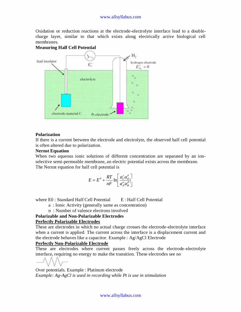
wwwallsyllabuscom
wwwallsyllabuscom
Oxidation or reduction reactions at the electrode-electrolyte interface lead to a double-
charge layer similar to that which exists along electrically active biological cell
membranes
Measuring Half Cell Potential
Polarization If there is a current between the electrode and electrolyte the observed half cell potential
is often altered due to polarization
Nernst Equation When two aqueous ionic solutions of different concentration are separated by an ion-
selective semi-permeable membrane an electric potential exists across the membrane
The Nernst equation for half cell potential is
where E0 Standard Half Cell Potential E Half Cell Potential
a Ionic Activity (generally same as concentration)
n Number of valence electrons involved
Polarizable and Non-Polarizable Electrodes
Perfectly Polarizable Electrodes
These are electrodes in which no actual charge crosses the electrode-electrolyte interface
when a current is applied The current across the interface is a displacement current and
the electrode behaves like a capacitor Example AgAgCl Electrode
Perfectly Non-Polarizable Electrode
These are electrodes where current passes freely across the electrode-electrolyte
interface requiring no energy to make the transition These electrodes see no
Over potentials Example Platinum electrode
Example Ag-AgCl is used in recording while Pt is use in stimulation
BA
DC
aa
aa
nF
RTEE ln0
wwwallsyllabuscom
wwwallsyllabuscom
Equivalent Circuit
Cd capacitance of electrode-eletrolyte interface
Rd resistance of electrode-eletrolyte interface
Rs resistance of electrode lead wire
Ecell cell potential for electrode
Electrode Skin Interface
Sweat glands and ducts
Ru
Rs
Rd Cd
Re
Ese EP
RP CP Ce
wwwallsyllabuscom
wwwallsyllabuscom
Motion Artifact
Why
When the electrode moves with respect to the electrolyte the distribution of the double
layer of charge on polarizable electrode interface changes This changes the half cell
potential temporarily
What
If a pair of electrodes is in an electrolyte and one moves with respect to the other a
potential difference appears across the electrodes known as the motion artifact This is a
source of noise and interference in biopotential measurements
Motion artifact is minimal for non-polarizable electrodes
Body Surface Recording Electrodes
1 Metal Plate Electrodes (historic)
2 Suction Electrodes
Electrode
Epidermis
Dermis and subcutaneous layer
Cd
Gel
Stratum Corneum
Capillary
wwwallsyllabuscom
wwwallsyllabuscom
(historic interest)
1 Floating Electrodes
2 Flexible Electrodes
Commonly Used Biopotential Electrodes
Metal plate electrodes
ndash Large surface Ancient therefore still used ECG
ndash Metal disk with stainless steel platinum or gold coated
ndash EMG EEG
ndash smaller diameters
ndash motion artifacts
ndash Disposable foam-pad Cheap
(a) Metal-plate electrode used for application to limbs
(b) Metal-disk electrode applied with surgical tape
(c)Disposable foam-pad electrodes often used with ECG
Commonly Used Biopotential Electrodes
Suction electrodes
- No straps or adhesives required
- precordial (chest) ECG
- can only be used for short periods
Floating electrodes
- metal disk is recessed
- swimming in the electrolyte gel
- not in contact with the skin
- reduces motion artifact
wwwallsyllabuscom
wwwallsyllabuscom
Commonly Used Biopotential Electrodes
Flexible electrodes
- Body contours are often irregular
- Regularly shaped rigid electrodes
may not always work
- Special case infants
- Material
- Polymer or nylon with silver
- Carbon filled silicon rubber
(Mylar film)
Double-sided
Adhesive-tape
ring Electrolyte gel
in recess
(a) (b)
(c)
Snap coated with Ag-AgCl External snap
Plastic cup
Tack
Plastic disk
Foam pad
Capillary loops
Dead cellular material
Germinating layer
Gel-coated sponge
Floating Electrodes
wwwallsyllabuscom
wwwallsyllabuscom
(a) Carbon-filled silicone rubber electrode
(b) Flexible thin-film neonatal electrode
(c) Cross-sectional view of the thin-film electrode in (b)
Electrodes in Biopotential Measurements
3 Describe the construction of commercial ECG electrode (not the cheap polymer
electrode used in the lab) What is the common electrode metal and why is it preferred
So you are an inventor who has a better idea Describe an improvement
bull to make the electrode cheaper
bull more suitable for lower noise measurement for EEG
bull circumvent patents that are based on plasticfoam electrode body
bull attractive to consumers for use with their ECG machines at home
bull reduce artifact (minimize the motion of skinelectrode) in ambulatory recording
4 In a research laboratory scientists want to record from single cells in a culture dish
They want to record action potentials from single isolated heart cells What kind of
electrode would they need to use (describe material and design) Give a simplified
schematic (circuit model of the electrode) described in the notes given to you
What is the challenge involved in designing an amplifier for use with a microelectrode
for single cell recording Ie what are the critical amplifier design characteristics and
specifications (hint this is not the usual differentialinstrumentation amplifier)
Electrodes and Microelectrodes (miscellaneous)
bull How would you detect bacteria or other microorganisms in water supply Make
sure that your method distinguishes inert particulate matter from living cellular
matter
bull Draw the equivalent circuit model of the skin and an ECG electrode Identify the
key sources of electrical interference and otherwise the elements that would likely
contribute to the poor quality of recordings
wwwallsyllabuscom
wwwallsyllabuscom
bull Design an amplifier interface for the following two applications Patch clamp ion
channel current amplifier Your goal is to amplify pA level current to produce 1
Volt output
bull Strain gauge sensor amplifier Your goal is to convert 10 ohm change in
resistance of a strain gauge to produce 1 volt output
Neural electrodesmicroelectrodes
You want to record from neurons in the brain However you want to record from dozens
of neurons all at once from several closely spaced microelectrodes What material and
process would you use to make the microelectrode array
bull What metal would you prefer to use to make electrode arrays of about 10 micron
square size to make electrical contacts with dozens of neurons
bull What metal would you prefer to use to stimulate dozens of neurons in a deep
brain microelectrode based stimulator
bull (which metal provides good recording vs stimulating properties ndash and at the same
time not be toxic to brain tissue)
bull You are asked to develop an experimental set up to record from rat brain cells
using microelectrodes What precautions would you take to minimize the
electrical interference in your recording set up
Biopotential Amplifiers bull These are very important part of modern medical instrumentation bull We need to amplify biopotentials which are generated in the body at low levels with a high source impedance
bull Biopotentials amplifiers are required to increase signal strength while maintaining fidelity Basic Requirements of Biopotential Amplifiers
Essential functions of a bioamplifier are bull To take a weak biopotential and increase its amplitude so that it can be processed recorded or displayed bull To amplify voltage but it could be considered as a power amplifier as well bull To amplify current since in some cases a biopotential amplifier is used to isolate the load from the source current gain only
Input Impedance (Zin)
bull All biopotential amplifiers must have high input impedance minimize loading (remember the characteristics of biopotential electrodes resulting into loading and distortion if input impedance of the amplifier is not high enough) ndash typical values of Zin over the frequency range of the measurand = 10 MΩ (remember the loading rule)
Protection amp Isolation
bull The input circuit of a biopotential amplifier must provide protection to the live measurand
wwwallsyllabuscom
wwwallsyllabuscom
Vbio
bull Any potential or current at amplifierrsquos input terminals can affect Vbio
bull Electric currents produced by the biopotential amplifier can result in microshock and
macroshock bull The bioamplifier must have isolation and protection circuitry so that the current through the electrodes can be kept at safe levels and any artifact generated by such current can be minimized Output Impedance (Zout)
bull The output circuit does not present any critical problems all it needs to do is to drive the load bull Output impedance must be low with respect to the load impedance and it must be capable of satisfying the power requirements of the load Bandwidth (BW)
Frequency response requirements bull The biopotential amplifier must be sensitive to important frequency components of the biosignal bull Since biopotentials are low level signals it is important to limit bandwidth optimize signal-to-noise ratio Gain (G)
bull Biopotential amplifiers have a gain of 1000 or greater
Mode of Operation
bull Very frequently biosignals are obtained from bipolar electrodes bull Electrodes symmetrically located with respect to ground need differential amplification bull High CMRR required because 1 Common mode signals much greater than the biosignal appear on bipolar electrodes 2 Symmetry with respect to ground is not perfect (mismatch between electrode impedances) ndash more on this later
Calibration Signal
bull Medical and clinical equipment require quick calibration bull The gain of the biopotential amplifier must be calibrated to provide us with an accurate indication of the signalrsquos amplitude bull Push button to apply standard signal to the input of the biopotential amplifier bull Adjustable gain switch carefully selects calibrated fixed gains (in microprocessorndashbased systems gain adjustment can be Electrocardiography
hearts A very widely used medical instrument which is utilized to diagnose and monitor cardiac beat
abnormalities is the electrocardiograph
hearts It measures the electrical activity of the heart (more precisely biopotential differences arising from the electrical activity of myocardium) Wersquove already talked about the genesis of the ECG signal hearts The ECG machine uses surface electrodes and high input impedance hearts Differential amplifiers with good common mode rejection ratio to record the electrocardiogram
hearts Normal ECG amplitude ranges between 05-4 mV Normal frequency content of ECG (for diagnostic purposes) is 005-100 Hz A typical ECG waveform is shown below
wwwallsyllabuscom
wwwallsyllabuscom
Obviously all human hearts are not the same and this leads into variability in different parts of the ECG signal Significant diagnostic features of the ECG signal are hearts Duration of component parts of the signal
hearts Polarities and magnitudes hearts The details of the ECG signal and the degree of variability in different parts of the ECG signal is shown below
The QRS amplitude polarity time duration the RR interval (indicator of heartbeat per min) and the T-wave amplitude are some very important and distinctive features of the ECG signal The heart rate in BPM = Beats Per Minute) is simply = 60 (RR interval in seconds)
Some ECG waveform abnormalities that may indicate illness are hearts An extended PR interval may be diagnosed as AV node block hearts A widening of the QRS complex may indicate conduction problems in the bundle of His hearts An elevated ST segment may indicate occurrence of myocardial Infarction (MI) hearts A negative polarity in the T wave may be due to coronary insufficiency ECG Leads
Normal ECG recordings for the standard lead connections leads I II and III (Lead II provides
the strongest signal) Obviously all human hearts are not the same and this results into a high degree of variability
wwwallsyllabuscom
wwwallsyllabuscom
hearts Note the degree of variability of different parts of the ECG Signal
Some abnormalities that may indicate illness hearts An extended P-R interval may be diagnosed as AV node block hearts Widening of the QRS complex conduction problems in the bundle of His hearts Elevated ST segment may indicate occurrence of MI
hearts Negative polarity T wave may be due to coronary insufficiency QRS amplitude polarity time domain PR interval (indicator of heat beat per min amp T-wave amplitude are some very important
distinctive features 1 Loss
2 Origin of the ECG signal
hearts We have already covered this concept extensively in the previous lectures (The Dipole filed of the heart the Eindhovenrsquos Triangle the electrical circuit model for the electrocardiographic problem etc)
hearts Standard Limb Leads (I II III)
wwwallsyllabuscom
wwwallsyllabuscom
hearts The The lead wires are color-coded according to some conventions One example is White ndashRA
(Right Arm) Black ndash LA (Left Arm) Green ndash RL (Right Leg) Red ndash LL (Left Leg) and Brown
ndash C (Chest) Note There is a CM (common mode) amplifier connected to the right leg We will discuss this in detail later Augmented Limb Leads
These leads offer a free 50 increase over leads VR VL and VF connections (unipolar leads) with respect to Wilson terminal AVR = -I ndash III2 AVL = I ndash II2 aVF = II ndash I2
wwwallsyllabuscom
wwwallsyllabuscom
Each measurement is made from the reflected limb and the average of the other two limbs Chest Leads (Precordial)
Chest Lead Anatomical Positions
V1 - 4th intercostal space ndash Right sternal margin
V2 - 4th intercostal space ndash Left sternal margin
V3 ndash Midway between V2 amp V4
V4 ndash 5th intercostal space on mid-clavicular line
V5 ndash Same as V4 on the anterior axillary line
V6 ndash Same as V5 on the mid-axillary line
12 Lead Clinical Electrocardiography
The ECG Machine
Most representative Specs
wwwallsyllabuscom
wwwallsyllabuscom
bull Zin = 10 MΩ bull Frequency response = 005 ndash100 Hz bull Strip Chart Recorder Speed = 25 mmsec bull Fast Speed = 100 mmsec
For detailed Specs Refer to the Table in your text ―Summary of performance requirements for electrocardiographs Location of the Heart bull The heart is located between the lungs behind the sternum and above the diaphragm bull It is surrounded by the pericardium bull Its size is about that of a fist and its weight is about 250-300 g bull Its center is located about 15 cm to the left of the midsagittal plane
Anatomy of the heart
bull The walls of the heart are composed of cardiac muscle called myocardium
bull It consists of four compartments ndash the right and left atria and ventricles
The Heart Valves
bull The tricuspid valve regulates blood flow between the right atrium and right
ventricle
bull The pulmonary valve controls blood flow from the right ventricle into the
pulmonary arteries
bull The mitral valve lets oxygen-rich blood from your lungs pass from the left atrium
into the left ventricle
bull The aortic valve lets oxygen-rich blood pass from the left ventricle into the aorta
then to the body
Blood circulation via heart
wwwallsyllabuscom
wwwallsyllabuscom
bull The blood returns from the systemic circulation to the right atrium and from
there goes through the tricuspid valve to the right ventricle
bull It is ejected from the right ventricle through the pulmonary valve to the lungs
bull Oxygenated blood returns from the lungs to the left atrium and from there
through the mitral valve to the left ventricle
bull Finally blood is pumped through the aortic valve to the aorta and the systemic
circulation
Electrical activation of the heart
bull In the heart muscle cell or myocyte electric activation takes place by means of
the same mechanism as in the nerve cell ie from the inflow of Na ions across
the cell membrane
bull The amplitude of the action potential is also similar being 100 mV for both nerve
and muscle
bull The duration of the cardiac impulse is however two orders of magnitude longer
than in either nerve cell or sceletal muscle cell
bull As in the nerve cell repolarization is a consequence of the outflow of K ions
bull The duration of the action impulse is about 300 ms
Mechanical contraction of Cardiac Muscle
bull Associated with the electric activation of cardiac muscle cell is its mechanical
contraction which occurs a little later
bull An important distinction between cardiac muscle tissue and skeletal muscle is
that in cardiac muscle activation can propagate from one cell to another
in any direction
bull Electrical signal begins in the sinoatrial (SA) node natural pacemaker causes
the atria to contract
bull The signal then passes through the atrioventricular (AV) node
ndash sends the signal to the ventricles via the ―bundle of His
ndash causes the ventricles to contract
The Conduction System
wwwallsyllabuscom
wwwallsyllabuscom
The Action Potential
Recording an AP requires the isolation of a single cell Microelectrodes (with tips a few μm across) are used to stimulate and record the response A typical AP is 2-4ms long with an amplitude of about 100Mv The Electroencephalogram EEG
EEG is the graphical representation of the electrical activity of the brain Very commonly used to diagnose certain neurological disorders such as epilepsy
More recently also investigated whether it can detect various forms of dementia or schizophrenia EEG is the specific recording obtained using the scalp electrodes from the surface of the skull
During surgery electrodes may also be placed directly on the cortex The resulting signal is then electrocorticogram (ECoG) Just like ECG EEG is also obtained using several different electrodes places on different regions of the head brain
The Event Related
wwwallsyllabuscom
wwwallsyllabuscom
Potentials ndash ERPs
ERPs are really EEGs obtained under a specific protocol that requires the patient to response to certain stimuli ndash hence event related potentials Also called evoked potentials these signals can be used to diagnose certain
neurological disorders such as dementia and they can also be used as a liedetector bull The oddball paradigm
bull The guilty knowledge test
Electroretinogram ERG
The ERG is the record of the retinal action currents produced by the retina in response to a light stimulus It measures the electrical responses of the light-sensitive cells (such as rods and cones)
The stimuli are often a series of light flashes or rotating patterns The ERG is recorded using contact lens electrode that the subject wears while watching the stimuli
Phonocardiogram ndash PCG
The PCG is the graphic record of the heart sounds and murmurs It is thus a mechanical audio signal rather than an electrical signal Can be easily heard using a stethoscope Or can be converted into an electrical signal using a transducer Typically used to determine the disorders related to the heart valve since their
routine opening and closing create the well-known sounds bull S1 sounds First heart sounds ndash ventricular contractions move blood into atria closing of the AV (mitral and tricuspid) valves then semilunar valves open and blood ejected out of ventricles ndash immediately follows the QRS complex bull S2 sounds Second heart sounds ndash Closure of semilunar (aortic and pulmonary) valves bull Any unexpected sound may indicate a malfunctioning valve that causes the blood flow into out of a chamber when it should not Also called heart murmurs
wwwallsyllabuscom
wwwallsyllabuscom
Define ultrasound
bull Mechanical waves in different modalities (longitudinallateral) needs medium
to be propagated (solid liquid gas)
bull gt 20 kHz
bull Continuouspulsed
bull Sphericalplanarnarrow beamsurface waveLamb-wave
Physical phenomena behind ultrasound measurements
Transmission
bull
bull reflection
bull transit time
bull differences in propagation velocities
bull returns to transit time
bull doppler-shift in frequency
bull flow velocity
bull change of acoustic impedance
bull comparing to reference
bull interference of ultrasound waves (holography)
bull interaction of ultrasound and light (photoacousticz)
bull ultrasound needs medium for propagation it doesnrsquot propagate in vacuum
bull because mechanical waves need moving massunits and spring forces between
them
bull in acoustic emission the medium creates ultrasound (for example during pressure
changes) which is received by sensors
bull pulsed mode more common than continuous
bull continuous reguires separate transducers for transmitting and receicving
bull in pulsed mode an ultrasound burst is sent to the object and the same transducer is
switched to listen echoes
bull standing wave problem
bull in us-therapy pulsed mode gives more effective care without too much heating The Doppler Equation describes the relationship of the Doppler frequency shift to target velocity
wwwallsyllabuscom
wwwallsyllabuscom
The frequency difference is equal to the reflected frequency (FR) minus the originating frequency (FT) If the resulting frequency is higher then there is a positive Doppler shift and the object is moving toward the transducer but if the resulting frequency is lower there is a negative Doppler shift and it is moving away from the transducer In its simplest form it would be calculated as if
the ultrasound was parallel to the targetrsquos direction as shown in diagram A below However this would be a rare occurrence in clinical practice because the transducer is rarely pointed head on to a blood vessel In real life the ultrasound waves would approach the target at an angle called the Doppler angle ( ) On the following page diagram B shows the Doppler
equation used in general clinical situations which includes the Doppler angle The Doppler Angle
The ultrasound beam usually approaches the moving target at an angle called the Doppler angle ( ) This reduces the frequency shift in proportion to the cosine of this
angle If this angle is known then the flow velocity can be calculated The equation used is The Doppler Equation
equiv Doppler shift frequency (the difference between the transmitted and received frequencies) equiv transmitted frequency equiv reflected frequency
V equiv velocity of the blood flow towards the transducer C equiv velocity of sound in tissue θ equiv the angle between the sound beam and the direction of moving blood Where
The Doppler angle ( ) is also known as the angle of insonation It is estimated by the sonographer by a process known as angle correction which involves aligning an indicator on the duplex image along the longitudinal axis of the vessel
There are a few considerations that affect the performance of a Doppler examination that are inherent in the Doppler equation which are ndash The cosine of 90deg is zero so if the ultrasound beam is perpendicular to the direction of blood flow there will be no Doppler shift and it will appear as if there is no flow in the vessel ndash Appropriate estimation of the angle of insonation or angle correction is essential for the accurate determination of Doppler shift and blood flow velocity The angle of insonation should also be less than 60deg at all times since the
cosine function has a steeper curve above this angle and errors in angle correction will be magnified
The simplest Doppler devices use continuous wave (CW Doppler) rather than the pulsed wave used in more complex devices CW Doppler uses two transducers (or a dual element transducer) that transmit and receive ultrasound continuously The transmit and receive beams overlap in a Doppler sample volume some distance from the transducer face as
shown in the diagram below
volume) is the region of transmitting and receiving beam overlap (shaded region) Because there is continuous transducer transmission and reception echoes from all depths within the area arrive at the transducer simultaneously So although CW Doppler can determine the direction of flow it cannot discriminate the different depths where the motion originates The usefulness of CW Doppler devices is limited but they are used clinically to confirm blood flow in superficial vessels as they are good at detecting low velocities As they are easily portable this
can be done at the bedside or in the operating room Most other clinical applications require pulsed wave Doppler Pulsed Wave Doppler (PW Doppler)
wwwallsyllabuscom
wwwallsyllabuscom
Pulsed wave Doppler (PW Doppler) uses a single-element transducer that emits brief pulses of ultrasound energy The time interval between transmitting and then receiving the echoing sound can be used to calculate the depth from where the echo arises The Doppler sample volume can be chosen as to shape depth and position in sampling the flow
data For example the depth is chosen by processing only the signals that return to the transducer in a stipulated time For this technique the ultrasound system transmits a short pulse The eceiver is opened to detect the returning echoes only after a controlled delay and only for a specific duration This time-based gating of the receiving channel allows the definition of a fixed easuring distance which is often referred to as the Sample volume or Doppler gate Then the next ultrasound wave is transmitted The number of pulses transmitted by the system within a second is referred to as the pulse repetition frequency (PRF) The upper PRF limit is given by the time interval required for the echoes to arrive from a sample volume located at a
certain depth The greater the sample-volume depth the longer the time before the echoes are returned and the longer the delay between pulse transmission The greater the samplevolume depth the lower will be the maximum PRF setting Errors in the accuracy of the information arise if the velocities exceed a certain speed The highest velocity accurately measured is called the Nyquist limit Beyond this limit the errors that occur are referred to as aliasing Volume and flow measurement
Flow ndash volume of a liquidgas passing some point over a given time Gases are compressible
Benedict Roth Spirometer
Widely used for physiological amp clinical studies
Light bell moves with the ptrsquos breathing
Movement recorded by a pen on a rotating drum
Water seal prevents leakage of gas
Small seal minimises volume of gas dissolved in water
Suitable for measuring limited gas vols (few litres)
Pneumotachograph
wwwallsyllabuscom
wwwallsyllabuscom
Cardiac Output
Defn vol of blood pumped by the heart per min
CO = SV x HR
Norm ~ 5 lmin
Cardiac index ndash corrected for body surface area
Affected by
Met Rate ndash pregnancy hyperthyroid septic
Preload contractility afterload
Clinical indicators of CO imprecise
Affected by anaesthetic agents used in everyday practice
Provides estimate of
whole body perfusion
oxygen delivery
left ventricular function
Persistently low CO assoc with poor outcome
Methods
Fick method
Dilution techniques ndash dye thermal lithium
Pulse contour analysis- LiDCO amp PiCCO
Oesophageal doppler
TOE
Transthoracic impedance plethysmography
Inert gas through flow
Non-invasive cardiac output measurement
Fick Principle measure volume displacement
1st proposed 1870
―the total uptake or release of a substance by an organ is the product of the
blood flow through that organ and the arteriovenous concentration
difference of the substance
CO = O2 consumption (mlmin) art ndash mixed
venous O2 conc (mll)
Limited by cumbersome equipment sampling errors need for invasive
monitoring and steady-state haemodynamic and metabolic conditions
Indicator dilution techniques
―An indicator mixed into a unit volume of constantly flowing blood can be used to
identify that volume of blood in time provided the indicator remains in the system
between injection and measurement and mixes completely in the blood
wwwallsyllabuscom
wwwallsyllabuscom
Dye dilution
Inert dye ndash indocyanin green
Injected into pulmonary artery and arterial conc measured using a
calibrated cuvette densitometer
Plot indicator dilution curve (see diagram)
CO derived from area under curve
Indicator Dilution Curve
Cardiac Output Measurement
wwwallsyllabuscom
wwwallsyllabuscom
Why bother to check blood pressure
For each 20 mm rise in systolic blood pressure or 10 mm rise in diastolic blood
pressure over 11575
Risk of stroke increases
Risk of heart disease doubles
Risk of renal failure increases
Systolic and diastolic blood pressure
Systolic blood pressure is the highest pressure in the arteries just after the heart
beats
Diastolic blood pressure is the lowest pressure in the arteries just before the heart
beats
Blood pressure is measured indirectly by blood pressure cuff
(sphygmomanometer)
Inflating cuff increases pressure until it cuts off arterial circulation to the arm
Deflating cuff decrease pressure by 2 to 3 mm of mercury per second until blood
first enters the artery creating turbulence this causes a sound with each heartbeat
Sounds continue with each heartbeat until pressure lowers to the lowest pressure
in the artery then turbulence stops so the sound stops
Systolic blood pressure is the cuff pressure at the first sounds diastolic is the cuff
pressure just before the sounds stop
Phase 1 sharp thuds start at systolic blood pressure
Phase 2 blowing sound may disappear entirely (the auscultatory gap )
Phase 3 crisp thud a bit quieter than phase 1
Phase 4 sounds become muffled
Phase 5 end of sounds -- ends at diastolic blood pressure
wwwallsyllabuscom
wwwallsyllabuscom
Make sure the cuff is the right size - its width should be at least 40 of the arms
circumference The cuff will overestimate blood pressure if too small and
underestimate if too large
Place the cuff snugly on patients proximal arm on skin ( not cloth) centered over
the brachial artery (most cuffs have markings)
Support the patients arm at heart level using your arm or a desk
Your patient should sit in the chair for 5 minutes before BP is measured and
should have no caffeine or nicotine for 30 minutes before (JAMA 273 p1211-
1218 1995)
With fingers palpating radial or brachial artery inflate cuff rapidly until you cant
feel the pulse then 20 mm higher
Release cuff at 2 to 3 mm Hg per second until you again feel the pulse this is the
palpable systolic pressure
Wait 30 seconds before measuring blood pressure
Measuring palpable pressure first avoids risk of seriously underestimating blood
pressure because of the auscultatory gap (mistaking Korotkoff phase 3 for phase
1) Many doctors skip this step for time reasons and instead pump cuff to 200 mm
Hg at the next step)
Phase 2 of the Korotkoff sounds can be inaudible - especially in older patients
with systolic hypertension who are at especially high risk of stroke Inflating the
cuff until you dont hear sounds can give you a reading of 14086 when the
patients actual blood pressure is 22086 Most physicians are pressed for time so
they instead inflate the cuff to 200 mm which is beyond the auscultatory gap in
most patients But palpable systolic blood pressure is according to research more
reliable
Place bell of stethoscope (diaphragm is acceptable) over brachial artery
Rapidly pump the cuff to 20 to 30 mm Hg above palpable systolic pressure
Release pressure in the cuff by 2 to 3 mm Hg per second and listen for Korotkoff
sounds including systolic (first) and diastolic (last)
Record as systolicdiastolic Check in both arms the first time you check a
patients blood pressure It may differ by 10 mm Hg or more
If the sounds continues to zero record diastolic blood pressure as the point when
sounds become muffled (phase 4) over zero eg 130700 or just as 13070
Rate
ndash Number of beats in 30 seconds x 2
Strength
ndash Bounding strong or weak (thready)
Regularity
ndash Regular or irregular
You need three readings on two occasions to diagnose hypertension unless blood
pressure is very high
Normal blood pressure in children is
wwwallsyllabuscom
wwwallsyllabuscom
ndash 10255 at 1 year 11269 at 5 years 11978 at 10 years
Blood pressures in adults (JNC VII JAMA 2892560-72 2003)
ndash Normal lt120lt80
ndash Prehypertensive 120-13980-89
ndash Stage 1 hypertension 140-15990-99
ndash Stage 2 hypertension gt160gt100
Adult 60 to 100
Newborn 120-170
1 year 80-160
3 years 80-120
6 years 75-115
10 years 70-110
How to measure observe rise and fall of chest
In infants count for 60 seconds in adults 15 or 30 seconds
Normal respiration Adults 12 to 20
Children
newborn 30-80
1 year 20-40
3 years 20-30
6 years 16-22
Rate
Number of breaths in 30 seconds x 2
Quality
Character of breathing
Rhythm
Regular or irregular
Effort
Normal or labored
Noisy respiration
Normal stridor wheezing snoring gurgling
Depth
Shallow or deep
pH electrode
wwwallsyllabuscom
wwwallsyllabuscom
Governing equation is the Nernst Equation
pCO2 Electrode
The measurement of pCO2 is based on its linear relationship with pH over the range of
10 to 90 mm Hg
The dissociation constant is given by
Taking logarithms
pH = log[HCO3-] ndash log k ndash log a ndash log pCO2
Diathermy
In the natural sciences the term diathermy means electrically induced heat and is commonly
used for muscle relaxation It is also a method of heating tissue electromagnetically or
ultrasonically for therapeutic purposes in medicineContents
[hide]
1 Heating uses 2 Surgical uses
3 Trivia
4 References
E
RT
nF
H
HH
i
ln
0
H O CO H CO H HCO2 2 2 3 3
k
H HCO
a pCO
3
2
wwwallsyllabuscom
wwwallsyllabuscom
Heating uses
Ultrasonic diathermy refers to heating of tissues by ultrasound for the purpose of
therapeutic deep heating No tissue is ordinarily damaged hence it is generally used in
biomedical applications
Electric diathermy uses high frequency alternating electric or magnetic fields sometimes
with no electrode or device contact to the skin to induce gentle deep tissue heating by
induction Again no tissue is ordinarily damaged
Surgical uses
Surgical diathermy is usually better known as electrosurgery (It is also referred to
occasionally as electrocautery but see disambiguation below) Electrosurgery and
surgical diathermy involve the use of high frequency AC electrical current in surgery as
either a cutting modality or else to cauterize small blood vessels to stop bleeding This
technique induces localized tissue burning and damage the zone of which is controlled
by the frequency and power of the device Some sources[1]
insist that electrosurgery be
applied to surgery accomplished by high frequency AC cutting and that
electrocautery be used only for the practice of cauterization with heated nichrome wires
powered by DC current as in the handheld battery-operated portable cautery tools
Trivia
Medical Diathermy devices were used to cause interference to German radio beams used
for targeting night time bombing raids in WWII during the Battle of the Beams
I Diathermy
A Therapeutic use
1 Generation of local heating by high-frequency electromagnetic waves
2 Capacitance techniquemdashbody is placed in an electric field
a Dipolesmdashstructures with positive and negative poles
b Structures with large numbers of dipoles have a greater capacitance to store an electrical charge
c Greatest heating occurs in tissues with fewer dipoles particularly fatty tissues
3 Rapid rotation of dipoles causing mechanical friction and movement of electrons results in local heating
4 Inductance techniquemdashbody is not placed in an electric field
a Magnetic waves generated by driving current through a coiled wire
b Magnetic field creates currents in tissues
wwwallsyllabuscom
wwwallsyllabuscom
c Greatest heating occurs in tissues with low impedance especially muscle
B Precautions and contraindications
1 Diathermy should not be used
a Over metal implants and cardiac pacemakersmdashmore research needed regarding its use over metallic fixations
b Near the uterus of a pregnant woman or near the abdomen or back of a woman who might be pregnant
c On individuals with infections
d On individuals with acute inflammation
e Over moist open wounds
f On patients with malignant tumors
g Over large joint effusions
C Pulsed electromagnetic fields and diathermy
1 Can be pulsed to decrease total energy transmitted to the tissues
2 Short-wave diathermy can be adjusted into a nonthermal range
a Classified as pulsed electromagnetic field (PEMF) or
b Pulsed radio frequency energy (PRFE)
3 Important reclassification as diathermy implies heating
D Efficiency of diathermy and PEMF therapy for musculoskeletal conditions
1 Current research is limited but results suggest that diathermy enhances
treatments directed at soft tissue stretching
2 Some studies suggest that PEMF may speed wound healing and promote healing of nonunion fractures
Diathermy is a modality that uses electromagnetic energy to heat deeper tissues
Diathermy is more effective than ultrasound at heating a larger area of deep tissues
The athletic trainer must identify and respect contraindications to application of ultrasound and diathermy
Pulsed ultrasound and diathermy are used to treat slow-to-heal lesions including skin ulcers and nonunion fractures and may be able to facilitate repair of other tissues including ligaments and tendons
Diathermy
Definition
In diathermy high-frequency electrical currents are used to heat deep muscular tissues The heat increases blood flow speeding up recovery Doctors also use diathermy in surgical procedures by sealing blood vessels with electrically heated probes
The term diathermy is derived from the Greek words therma meaning heat and dia meaning through Diathermy literally means heating through
Origins
The therapeutic effects of heat have long been recognized More than 2000 years ago the Romans took advantage of heat therapies by building hot-spring bathhouses Since then various
wwwallsyllabuscom
wwwallsyllabuscom
methods of using heat have evolved In the early 1890s French physiologist Arseacutene dArsonval began studying the medical application of high-frequency currents The term diathermy was coined by German physician Carl Franz Nagelschmidt who designed a prototype apparatus in 1906 Around 1925 United States doctor J W Schereschewsky began studying the physiological
effects of high-frequency electrical currents on animals It was several years however before the fundamentals of the therapy were understood and put into practice
Benefits
Diathermy can be used to treat arthritis bursitis and other conditions involving stiff painful joints It is also used to treat pelvic infections and sinusitis A benefit of diathermy is that it is a painless procedure that can be administered at a clinic Also if the treatment relieves pain then patients can discontinue pain killers and escape their high cost and side effects
Description
Diathermy involves heating deep muscular tissues When heat is applied to the painful area cellular metabolism speeds up and blood flow increases The increased metabolism and circulation accelerates tissue repair The heat helps the tissues relax and stretch thus alleviating
stiffness Heat also reduces nerve fiber sensitivity increasing the patients pain threshold
There are three methods of diathermy In each energy is delivered to the deep tissues where it is converted to heat The three methods are
Shortwave diathermy The body part to be treated is placed between two capacitor plates Heat is generated as the high-frequency waves travel through the body tissues between the plates Shortwave diathermy is most often used to treat areas like the hip which is covered with a dense tissue mass It is also used to treat pelvic infections and sinusitis The treatment reduces inflammation The Federal Communications Commission regulates the frequency allowed for short-wave diathermy treatment Most machines
function at 2733 megahertz Ultrasound diathermy In this method high-frequency acoustic vibrations are used to
generate heat in deep tissue Microwave diathermy This method uses radar waves to heat tissue This form is the
easiest to use but the microwaves cannot penetrate deep muscles
Diathermy is also used in surgical procedures Many doctors use electrically heated probes to seal blood vessels to prevent excessive bleeding This is particularly helpful in neurosurgery and eye surgery Doctors can also use diathermy to kill abnormal growths such as tumors warts and
infected tissues
Preparations
To keep patients from sweating patients are usually asked to remove clothing from the body part being treated If a patient sweats the electrical currents may pool in the area causing burns Also clothing containing metal must be removed as must earrings buttons barrettes or zippers that contain metal Watches and hearing aids should be removed because the therapy may affect their function
wwwallsyllabuscom
wwwallsyllabuscom
Practitioners of surgical diathermy should steer clear of alcohol-based solutions to prepare and cleanse the skin These preparations can create a flammable vapor and cause burns and fires
Precautions
Patients with metal implants should not undergo diathermy treatment because the metal can act as a conductor of heat and result in serious internal burns Female patients with metallic uterine implants such as an IUD should avoid treatment in the pelvic area Diathermy should not be
used in joints that have been replaced with a prosthesis or in those with sensory impairment who may not be able to tell if they are burning Furthermore pulsed shortwave diathermy should be avoided during pregnancy as it can lead to abnormal fetal development
Patients with hemophilia should avoid the treatment because the increased blood flow could cause them to hemorrhage
Side effects
Some patients may experience superficial burns Since the therapy involves creating heat care must be taken to avoid burns particularly in patients whose injuries have caused decreased sensitivity to heat Also diathermy may affect pacemaker function
Female patients who receive treatment in the lower back or pelvic area may experience an
increased menstrual flow
Research amp general acceptance
For years physiotherapists and physical therapists have used diathermy as a routine part of physical rehabilitation
Electrical Safety
Electrical safety is very important in hospitals as patients may be undergoing a diagnostic or treatment procedure where the protective effect of dry skin is reduced Also patients may be unattended unconscious or anaesthetised and may not respond normally to an electric current Further electrically conductive solutions such as blood and saline are often present in patient treatment areas and may drip or spill on electrical equipment
Electric Current Leakage Current
Extension Leads Double Adaptors Equipment Classification
Class I Class II Defibrillator-Proof
Protective Devices
Residual Current Devices (RCD) Line Isolation overload Monitors (LIMs) Equipment Earthing
Area Classification
wwwallsyllabuscom
wwwallsyllabuscom
Body Protection Area Cardiac Protected Area
Other Electrical Issues Extension Leads
Double Adapters Main Extension Devices Power Boards Installation of Additional Power Points
Electric Current
Injuries received from electric current are dependent on the magnitude of current the pathway
that it takes through the body and the time for which it flows
The nature of electricity flowing through a circuit is analogous to blood flowing through the circulatory system within the human body In this analogy the source of energy is represented by the heart and the blood flowing through arteries and veins is analagous to current flowing
through the conductors and other components of the electric circuit
The application of an electric potential to an electric circuit generates a flow of current through conductive pathways This is analogous to the changes in blood pressure caused by contraction of cardiac muscle that causes blood to flow into the circulatory system For electric current to flow there must be a continuous pathway from the source of potential through electrical components and back to the source
Leakage Current
Electrical components and systems are encased in non conducting insulation to ensure that the electric current is contained and follows the intended pathways If the insulation deteriorates or breaks down current will leak through the insulation barrier and flow to earth This may
be through the protective earth conductor or through the operator
Medical equipment and clinical areas are fitted with a number of protective devices to protect the patient and operator from harmful leakage currents
Extension Leads
Extension leads are not permitted in clinical areas of RCH organisations They may cause high
earth resistance and excessive earth leakage current An extension lead can allow equipment to be powered from areas other than the relevant protected treatment area The power from the other area may not be protected to the same level as the power in the treatment area
wwwallsyllabuscom
wwwallsyllabuscom
As the connection between the extension lead and the equipment mains cable is often on the floor there is a high danger from fluid spills tripping and damage to the mains cable by trolleys when an extension lead is used
Double Adaptors
Double adaptors must not be used in RCH organisations They may not sit securely in a wall
outlet may not be able to provide adequate earth protection and may cause overloading overheating fire or loss of electrical supply
EQUIPMENT CLASSIFICATIONS
There are several methods of providing protection for operators and patients from electrical faults
and harmful leakage current
Class I
Class I equipment is fitted with a three core mains cable containing a protective earth wire
Exposed metal parts on class I equipment are connected to this earth wire
Should a fault develop inside the equipment and the exposed metal comes into contact with the mains the earthing conductor will conduct the fault current to ground Regular testing procedures ensure that earthing conductors are intact as the integrity of the earth wire is of vital importance
Class II
Class II equipment is enclosed within a double insulated case and does not require earthing
conductors Class II equipment is usually fitted with a 2-pin mains plug An internal electrical fault is unlikely to be hazardous as the double insulation prevents any external parts from becoming alive Class II or double insulated equipment can be identified by the class II symbol on the cabinet
Class II Symbol
wwwallsyllabuscom
wwwallsyllabuscom
Defibrillator-Proof
Some medical equipment within the hospital is classified as defibrillator proof When a
defibrillator is discharged through a patient connected to defibrillator proof equipment the equipment will not be damaged by the defibrillators energy Defibrillator proof equipment can remain connected to the patient during defibrillation It is identified by one of the following
symbols
Defibrillator proof symbols
Body protected Cardiac protected
PROTECTIVE DEVICES
Most patient care areas in the hospital are fitted with protective devices These devices are
regularly tested in accordance with the relevant guidelines published by Standards Australia The level of protection provided is dependent upon the device and the area in which it is located
Residual Current Devices (RCD)
RCDs (safety switches) are used in patient treatment areas to monitor and protect the mains supply RCDs sense leakage currents flowing to earth from the equipment If a significant leakage current flows the RCD will detect it and shut off the power supplied to the equipment
within 40 milliseconds Hospital RCDs are more sensitive than those fitted in homes A hospital RCD will trip at 10 milliamperes leakage current
Power outlets supplied through an RCD have a Supply Available lamp The lamp will extinguish
when the RCD trips due to excessive leakage current
wwwallsyllabuscom
wwwallsyllabuscom
Resetting a RCD
Lamp indicates supply is no longer available Disconnect all equipment from the supply Operate the reset button or lever on the supply panel and the Supply Available lamp
should illuminate If not contact Biomedical Engineering
Connect an item of equipment If the RCD trips again then this is the faulty item and should be labelled and sent to Biomedical Engineering
If the RCD does not trip continue connecting equipment until the RCD trips The last piece of equipment connected to the supply is most likely to be faulty as it will have caused the RCD to trip Remove the faulty item from service label it and send to Biomedical Engineering as mentioned above
Line Isolation overload Monitors (LIMs)
In critical life support applications where loss of power supply cannot be tolerated special power outlets powered by isolation transformers are installed
Line Isolation Monitors are installed to continually monitor electrical leakage in the power
supply system If an electrical fault develops in a medical device connected to an isolated power outlet the LIM will detect the leakage current The LIM will alarm and indicate the level of leakage current but will not shut off the electric supply
The faulty equipment can be identified by un plugging one item of equipment at a time from the supply until the alarm stops sounding Equipment that is not faulty may be reconnected Faulty equipment should be appropriately labelled and sent to Biomedical Engineering for repair
The LIM also monitors how much power is being used by the equpiment connected to it If too much power is being used the LIM will alarm and indicate that there is an overload The power used must be reduced immediately by moving some equipment to another circuit as soon as possible until the alarm stops sounding Failure to reduce the load on the LIM will result in the circuit breaker tripping and loss of power to the circuit
wwwallsyllabuscom
wwwallsyllabuscom
Equipotential Earthing
Equipotential earthing is installed in rooms classified as Cardiac Protected electrical areas
Equipotential earthing in treatment areas used for cardiac procedures is intended to minimise any voltage differences between earthed parts of equipment and any other exposed metal in the room
This reduces the possibility of leakage currents that can cause microelectrocution when the patient comes into contact with multiple items of equipment or if the patient happens to come
into contact with metal items in the room whilr they are connected to a medical device
All conductive metal in an equipotential area is connected to a common equipotential earth point with special heavy duty cable
AREA CLASSIFICATIONS
Body Protected Area
These areas are designed for procedures in which patients are connected to equipment that lowers the natural resistance of the skin Applied parts such as electrode gels conductive fluids entering the patient metal needles and catheters provide an easy pathway for current to flow
The main occurrence of injury from Body-Type procedures is from high current levels causing electric shock A direct connection to the patients heart is not present so the risk of Microelectrocution - fibrillation from minute current levels - is reduced
Residual Current Devices (RCD) or Isolation Transformers and Line Isolation Monitors (LIMiacutes) are used in Body Protected areas to provide protection against electrocution from high leakage currents Body-Protected Areas are identified with this sign
Cardiac Protected Area
Where the procedure involves placing an electrical conductor within or near the heart protection against fibrillation induced from small leakage currents is required Electrical conductors used in these procedures include cardiac pacing electrodes intracardiac ECG electrodes and intracardiac catheters
wwwallsyllabuscom
wwwallsyllabuscom
Equipotential earthing in conjunction with RCDs or LIMs provides protection against microelectrocution in Cardiac-Type procedures
Fault currents are reduced to magnitudes that are unlikely to induce fibrillation Used in conjunction with RCDs or LIMs the magnitude and duration of any fault currents
sourced from equipment are limited Cardiac-Protected Areas are identified with this sign
Other electrical issues
This policy aims to provide guidance to those who find that they need more electrical outlets than
those available or that the existing electrical outlets are inconveniently located
As extension leads and multiple outlet power boards can introduce additional hazards into an area the following procedures should be observed
Extension leads
Approved extension leads (AS 3760 1996) may be used in some areas within the hospital but MUST NOT BE USED IN PATIENT AREAS All electrical extension leads must be tagged with an Engineering Department maintenance tag and require a yearly safety inspection and test via
the Engineering Department
Double adapters
Double adapters may cause overloading or equipment earthing problems and are not to be used in
WCH
Mains extension device
The only mains extension device that is to be used in Patient care areas is the 4-way or 8-way
portable Core Balance Unit
The Biomedical Engineering Department must approve all units prior to use These units contain a safety switch and can detect excessive leakage current and disconnect the power in the event of a hazardous situation
wwwallsyllabuscom
wwwallsyllabuscom
Care must be exercised in the use of a portable Core Balance Unit It should be located off the
floor and in a position that will protect it from physical abuse and possible entry of fluids These devices are expensive and easily damaged The device must be sent to Biomedical Engineering every 6 months for safety testing
Power boards
Approved multiple-outlet power boards can be used across RCH but must not be used in patient
care areas except areas approved by the Biomedical Engineering Department
The power boards must have overload protection be fitted with internal safety shutters that protect unused outlets and be fitted with an onoff switch for each outlet
Medical Laser Applications
The main research subjects of the group of Medical Laser Application are in the field of online
monitoring and diagnostics as well as the development of new therapheutic methods The main focus in research is based upon the use of ultrashort (fs) laser pulses
The main advantage of ultrashort laser pulses is the extrem short interaction time which suppress any unwanted side effects of the laser irradiation of the tissue Secondly the broad spectra of the fs laser pulses give the advantage to use the same laser pulses for diagnostic applications like the optical coherence tomography (OCT)
wwwallsyllabuscom
wwwallsyllabuscom
Following research subjects have special attention at the moment
Optical Coherence Tomographie (OCT)
A fs-laser light source can be used to obtain images from inside the tissue when the pulses are coherent superposed This group uses this application to obtain an inside view of the vocal fold and to measure the outline of the crystalline lens
Refractive Surgery
The principle of getting rid of glasses with the help of a laser surgery (fs-LASIK) is underlying rapid improvements concerning precision and safety New technologies were evaluated and transferred very fast in cooperation with an industrial partner
Treatment of Presbyopia
The flexibility of the crystalline lens can be increase by pricise cuts which are induce by fs laser pulse inside the lens The treatment of the presbyopia is thinkable
wwwallsyllabuscom
wwwallsyllabuscom
Ultrafast Physics
Whereas the applications of ultrashort laser pulses increase rapidly is the physics of the interaction between the laser pulses and tissue in many case not fully understood To obtain improved knowledge of the interaction for many fs-laser application this group preforms numerical simulations as well as fundamental experiments
Thermography
Thermography thermal imaging or thermal video is a type of infrared imaging Thermographic cameras detect radiation in the infrared range of the electromagnetic spectrum
(roughly 900ndash14000 nanometers or 09ndash14 microm) and produce images of that radiation Since infrared radiation is emitted by all objects based on their temperatures according to the black body radiation law thermography makes it possible to see ones environment with or without visible illumination The amount of radiation emitted by an object increases with temperature therefore thermography allows one to see variations in temperature (hence the name) When viewed by thermographic camera warm objects stand out well against cooler backgrounds humans and other warm-blooded animals become easily visible against the environment day or
night As a result thermographys extensive use can historically be ascribed to the military and security services
Thermal imaging photography finds many other uses For example firefighters use it to see through smoke find persons and localize the base of a fire With thermal imaging power lines maintenance technicians locate overheating joints and parts a telltale sign of their failure to eliminate potential hazards Where thermal insulation becomes faulty building construction technicians can see heat leaks to improve the efficiencies of cooling or heating air-conditioning Thermal imaging cameras are also installed in some luxury cars to aid the driver the first being
the 2000 Cadillac DeVille Some physiological activities particularly responses in human beings and other warm-blooded animals can also be monitored with thermographic imaging [1]
The appearance and operation of a modern thermographic camera is often similar to a camcorder Enabling the user to see in the infrared spectrum is a function so useful that ability to record their output is often optional A recording module is therefore not always built-in
Instead of CCD sensors most thermal imaging cameras use CMOS Focal Plane Array (FPA) The most common types are InSb InGaAs HgCdTe and QWIP FPA The newest technologies are using low cost and uncooled microbolometers FPA sensors Their resolution is considerably lower than of optical cameras mostly 160x120 or 320x240 pixels up to 640x512 for the most
expensive models Thermographic cameras are much more expensive than their visible-spectrum counterparts and higher-end models are often export-restricted Older bolometers or more
wwwallsyllabuscom
wwwallsyllabuscom
sensitive models as InSb require cryogenic cooling usually by a miniature Stirling cycle refrigerator or liquid nitrogen
Contents
[hide]
1 Difference between IR film and thermography 2 Advantages of Thermography
3 Limitations amp disadvantages of thermography 4 Applications 5 See also 6 External links
o 61 History of thermal imager manufacturers
[edit] Difference between IR film and thermography
Thermal imaging is going to be used on Mars to detect caves that could hold life
[edit] Advantages of Thermography
You get a visual picture so that you can compare temperatures over a large area
It is real time capable of catching moving targets Able to find deteriorating components prior to failure Measurement in areas inaccessible or hazardous for other methods It is a non-destructive test method
[edit] Limitations amp disadvantages of thermography
Quality cameras are expensive and are easily damaged Images can be hard to interpret accurately even with experience
Accurate temperature measurements are very hard to make because of emissivities Most cameras have plusmn2 or worse accuracy (not as accurate as contact) Training and staying proficient in IR scanning is time consuming Ability to only measure surface areas
[edit] Applications
Condition monitoring Medical imaging Night vision Research
Process control Non destructive testing Surveillance in security law enforcement and defense Chemical imaging
wwwallsyllabuscom
wwwallsyllabuscom
Thermal infrared imagers convert the energy in the infrared wavelength into a visible light video display All objects above 0 kelvins emit thermal infrared energy so thermal imagers can passively see all objects regardless of ambient light However most thermal imagers only see objects warmer than -50 degC
The spectrum and amount of thermal radiation depend strongly on an objects surface
temperature This makes it possible for a thermal camera to display an objects temperature However other factors also influence the radiation which limits the accuracy of this technique For example the radiation depends not only on the temperature of the object but is also a function of the emissivity of the object Also radiation also originates from the surroundings and is reflected in the object and the radiation from the object and the reflected radiation will also be influenced by the absorption of the atmosphere
Endoscopy
This page is about having an endoscopy - a test that looks at the inside of your digestive system
There is information on
What endoscopy is
Why you may have an endoscopy
Having an endoscopy
Your results
What an endoscopy is
An endoscopy is a test that looks inside the body The endoscope is a long flexible tube that can
be swallowed It has a camera and light inside it Some doctors call it a telescope
wwwallsyllabuscom
wwwallsyllabuscom
Why you may have an endoscopy
You are most likely to have an endoscopy to look at the inside of your
Gullet (oesophagus)
Stomach
Duodenum - the first part of the small bowel that attaches to the stomach
Large bowel (colon)
Your doctor may want to see inside because you have symptoms such as abnormal bleeding or
difficulty swallowing The doctor can look down the endoscope and see if there are any growths
or other abnormal looking areas Also through the endoscope the doctor can take samples
(biopsies) of any abnormal looking tissues
There is more detailed information about having a colonoscopy in the bowel cancer section of
CancerHelp UK Below is information about having other types of endoscopy
Having an endoscopy
You can have this test as an out patient Most people have a choice between having the test while
they are awake or after having a medicine to make them drowsy (a sedative) Your hospital may
not be happy for you to have a sedative if you live alone and will have no one to look after you
when you go home If you live alone but really want sedation your hospital may allow you to
stay overnight You should ask your doctor about this as early as possible as extra arrangements
will need to be made beforehand
If you dont have a sedative you will have a spray to numb the back of your throat and make it
easier for you to swallow the endoscopy tube
If you would prefer to be asleep during the test you will have an injection to make you very
drowsy just before the test You will need to take someone with you to the hospital appointment
You wont be able to drive for the rest of the day and should have someone to go home with you
You cant eat or drink for about 8 hours before the test so that your stomach and duodenum are
empty Your doctor will give you written instructions about this beforehand or they may arrive
with your appointment letter When you get to the clinic you may be asked to take your upper
clothing off and put on a hospital gown Some hospitals prefer to use gowns because your clothes
wont get messy Once you are ready you get onto the bed or X-ray couch When you are lying
comfortably you have the sedative injection to make you very drowsy Or your doctor will spray
the back of your throat to numb it
Once the sedative or throat spray has worked the doctor will pass the endoscope tube down your
wwwallsyllabuscom
wwwallsyllabuscom
throat to the area being investigated Your doctor will ask you to swallow as the tube goes down
but if youve had a sedative you wont remember that afterwards If there are any abnormalities
the doctor will take pieces of tissue from the abnormal looking area to send to the laboratory for
closer inspection under a microscope These tissue samples are called biopsies
When the test is over you will need to rest for a while If youve had a sedative you may not
remember much (if anything) about the test once you have come round You should be able to go
home the same day
The results
It can take time for test results to come through How long will depend on why you are having
the test Usually the doctor who carries out the endoscopy dictates a report straight way The
report is typed up by the department secretary and goes to your specialist who gives the results to
you If your GP has sent you for the test the results will go directly to the GP surgery
Understandably waiting for results can make you anxious It usually takes a couple of weeks for
the results to come through If your doctor needed them urgently it would have been noted on
the test request form and the results will be ready sooner than that Try to remember to ask your
doctor how long you should expect to wait for the results when you are first asked to go for the
test If it is not an emergency and you have not heard a couple of weeks after your test ring your
doctors secretary to check if they are back
Endoscopy
From Wikipedia the free encyclopedia
Jump to navigation search
Endoscopic images of a duodenal ulcer
wwwallsyllabuscom
wwwallsyllabuscom
A flexible endoscope
Endoscopy means looking inside and typically refers to looking inside the human body for medical reasons using an instrument called an endoscope Endoscopy can also refer to using a borescope in technical situations where direct line-of-sight observation is not feasible
Contents
[hide]
1 Overview 2 Components 3 Uses
4 History 5 Risks 6 After The Endoscopy 7 Recent developments 8 See also 9 References 10 Footnotes
11 External links
Overview
Endoscopy is a minimally invasive diagnostic medical procedure that is used to assess the interior surfaces of an organ by inserting a tube into the body The instrument may have a rigid or flexible tube and not only provide an image for visual inspection and photography but also enable taking
biopsies and retrieval of foreign objects Endoscopy is the vehicle for minimally invasive surgery
Many endoscopic procedures are considered to be relatively painless and at worst associated with mild discomfort for example in esophagogastroduodenoscopy most patients tolerate the procedure with only topical anaesthesia of the oropharynx using lignocaine spray[1] Complications are not common (only 5 of all operations)[citation needed] but can include perforation of the organ under inspection with the endoscope or biopsy instrument If that occurs open surgery may be required to repair the injury
wwwallsyllabuscom
wwwallsyllabuscom
Components
An endoscope can consist of
a rigid or flexible tube a light delivery system to illuminate the organ or object under inspection The light
source is normally outside the body and the light is typically directed via an optical fiber system
a lens system transmitting the image to the viewer from the fiberscope an additional channel to allow entry of medical instruments or manipulators
Uses
Endoscopy can involve
The gastrointestinal tract (GI tract) o esophagus stomach and duodenum (esophagogastroduodenoscopy) o small intestine o colon (colonoscopyproctosigmoidoscopy) o Bile duct
endoscopic retrograde cholangiopancreatography (ERCP) duodenoscope-assisted cholangiopancreatoscopy intraoperative cholangioscopy
The respiratory tract o The nose (rhinoscopy) o The lower respiratory tract (bronchoscopy)
The urinary tract (cystoscopy) The female reproductive system
o The cervix (colposcopy) o The uterus (hysteroscopy) o The Fallopian tubes (Falloscopy)
Normally closed body cavities (through a small incision) o The abdominal or pelvic cavity (laparoscopy) o The interior of a joint (arthroscopy) o Organs of the chest (thoracoscopy and mediastinoscopy)
During pregnancy o The amnion (amnioscopy) o The fetus (fetoscopy)
Plastic Surgery Panendoscopy (or triple endoscopy)
o Combines laryngoscopy esophagoscopy and bronchoscopy Non-medical uses for endoscopy
o The planning and architectural community have found the endoscope useful for
pre-visualization of scale models of proposed buildings and cities (architectural endoscopy)
o Internal inspection of complex technical systems (borescope) o Endoscopes are also a tool helpful in the examination of improvised explosive
devices by bomb disposal personnel
wwwallsyllabuscom
wwwallsyllabuscom
o The FBI uses endoscopes for conducting surveillance via tight spaces
History
The first endoscope of a kind was developed in 1806 by Philip Bozzini with his introduction of a
Lichtleiter (light conductor) for the examinations of the canals and cavities of the human body However the Vienna Medical Society disapproved of such curiosity An endoscope was first introduced into a human in 1822 by William Beaumont an army surgeon at Mackinac
Island Michigan[citation needed] The use of electric light was a major step in the improvement of endoscopy The first such lights were external Later smaller bulbs became available making internal light possible for instance in a hysteroscope by Charles David in 1908[citation needed] Hans Christian Jacobaeus has been given credit for early endoscopic explorations of the abdomen and the thorax with laparoscopy (1912) and thoracoscopy (1910)[citation needed] Laparoscopy was used in the diagnosis of liver and gallbladder disease by Heinz Kalk in the 1930s[citation needed] Hope reported in 1937 on the use of laparoscopy to diagnose ectopic pregnancy[citation needed] In 1944
Raoul Palmer placed his patients in the Trendelenburg position after gaseous distention of the abdomen and thus was able to reliably perform gynecologic laparoscopy[citation needed]
The first gastrocamera was released in 1950 by Olympus Optical Co Ltd The device took pictures on monochromatic film using a small light bulb that was triggered manually The device was of limited use however because it did not implement real-time optical capability Olympus continued its development of endoscopes by incorporating fiber optics in the early 1960s leading to the first useful endoscopes In 1964 it released a gastrocamera guided by a fiberscope[1] A few articles claim that DrBasil Hirschowitz of UnivOf MichiganAnn Arbor discussed the
endoscope in early 50s[2]
As endoscopic technology improved so did the methods of gastrointestinal endoscopy Owing primarily to the efforts of Dr Hiromi Shinya in the late 1960s GI endoscopy developed into what is more recognizable as todays colonoscopy While many doctors experimented with techniques to take advantage of the new iterations of endoscopes Dr Shinya focused on techniques that would allow for successful operation of the endoscope by an individual rejecting the common practice at the time of utilizing two people Consequently many of the fundamental methods and procedures of modern colonoscopy were developed by Dr Shinya
Dr Shinyas other great contribution was to therapeutic endoscopy in his invention of the
electrosurgical polypectomy snare with the aid of Olympus employee Hiroshi Ichikawa Shinya sketched his first plans for the device on January 8 1969 He envisioned a loop of wire attached to the end of a colonoscope that would allow for easy removal of polyps during investigation by passing a current through the wire By September of 1969 the first polypectomy using this device was performed Polypectomy has since become the most common therapeutic procedure performed with an endoscope (Sivak 2004)
By 1980 laparoscopy training was required by gynecologists to perform tubal ligation procedures and diagnostic evaluations of the pelvis The first laparoscopic cholecystectomy was performed in 1984 and the first video-laparoscopic cholecystectomy in 1987[citation needed] During the 1990s
laparoscopic surgery was extended to the appendix spleen colon stomach kidney and liver[citation needed] Wireless capsule endoscopy or Capsule Endoscopy is now approved in all the countries including Japan where government reimbusement will be available from Oct2007Capsule Endoscopy [3] increases detection of Small Bowel tumors where traditional Endoscopy is not very efficient
wwwallsyllabuscom
wwwallsyllabuscom
Risks
Infection
Punctured organs Allergic reactions due to Contrast agents or dyes (such as those used in a CT scan) Over-sedation
After The Endoscopy
After the procedure the patient will be observed and monitored by a qualified individual in the
endoscopy or a recovery area until a significant portion of the medication has worn off Occasionally a patient is left with a mild sore throat which promptly responds to saline gargles or a feeling of distention from the insufflated air that was used during the procedure Both problems are mild and fleeting When fully recovered the patient will be instructed when to resume hisher usual diet (probably within a few hours) and will be allowed to be taken home Because of the use of sedation most facilities mandate that the patient is taken home by another person and not to drive on hisher own or handle machinery for the remainder of the day
Recent developments
With the application of robotic systems telesurgery was introduced as the surgeon could operate
from a site physically removed from the patient The first transatlantic surgery has been called the Lindbergh Operation
wwwallsyllabuscom
wwwallsyllabuscom
Upper Endoscopy
Upper endoscopy enables the physician to look inside the esophagus stomach and duodenum (first part of the small intestine) The procedure might be used to discover the reason for swallowing difficulties nausea vomiting
reflux bleeding indigestion abdominal pain or chest pain Upper endoscopy is also called EGD which stands for esophagogastroduodenoscopy (eh-SAH-fuh-goh-GAS-troh-doo-AH-duh-NAH-skuh-pee)
For the procedure you will swallow a thin flexible lighted tube called an endoscope (EN-doh-skope) Right before the procedure the physician will spray your throat with a numbing agent that may help prevent gagging
You may also receive pain medicine and a sedative to help you relax during the exam The endoscope transmits an image of the inside of the esophagus stomach and duodenum so the physician can carefully examine the lining of these organs The scope also blows air into the stomach this expands the folds of tissue and makes it easier for the physician to examine the stomach
The physician can see abnormalities like inflammation or
bleeding through the endoscope that dont show up well on x rays The physician can also insert instruments into the scope to treat bleeding abnormalities or remove samples of tissue (biopsy) for further tests
Possible complications of upper endoscopy include bleeding and puncture of the stomach lining However such complications are rare Most people will probably have nothing more than a mild sore throat after the procedure
The procedure takes 20 to 30 minutes Because you will be sedated you will need to rest at the endoscopy facility for 1 to 2 hours until the medication wears off
Preparation
Your stomach and duodenum must be empty for the procedure to be thorough and safe so you will not be able to eat or drink anything for at least 6 hours beforehand Also you must
arrange for someone to take you homemdashyou will not be allowed to drive because of the sedatives Your physician may give you other special instructions
The digestive system
wwwallsyllabuscom
wwwallsyllabuscom
What is Endoscopy and Why is it Performed
Endoscopy allows physicians to peer through the bodys passageways Endoscopy is the examination and inspection of the interior of body organs joints or cavities through an endoscope An endoscope is a device that uses fiber optics and powerful lens systems to provide lighting and visualization of the interior of a joint The portion of the endoscope inserted into the body may be rigid or flexible depending upon the medical procedure
An endoscope uses two fiber optic lines A light fiber carries light into the body cavity and an image fiber carries the image of the body cavity back to the physicians viewing lens There is also a separate port to allow for administration of drugs suction and irrigation This port may also be used to introduce small folding instruments such as forceps scissors brushes snares and baskets for tissue excision (removal) sampling or other diagnostic and therapeutic work Endoscopes may be used in conjunction with a camera or video recorder to document images of the inside of the joint or chronicle an endoscopic procedure New endoscopes have digital capabilities for manipulating and enhancing the video images
This figure shows a rigid endoscope used for arthroscopy The image fiber leads from the ocular (eye piece) to the inserted end of the scope The light fiber is below and leads from the light source to the working end of the endoscope
Why Is Endoscopy Performed
Endoscopy can be used to diagnose various conditions by close examination of internal organ and body structures Endoscopy can also guide therapy and repair such as the removal of torn cartilage from the bearing surfaces of a joint Biopsy (tissue sampling for pathologic testing) may also be performed under endoscopic guidance Local or general anesthetic may be used during endoscopy depending upon the type of procedure being performed
Internal abnormalities revealed through endoscopy include abscesses biliary (liver) cirrhosis bleeding bronchitis cancer cysts degenerative disease gallbladder stones hernia inflammation metastatic cancer polyps tumors ulcers and other diseases and conditions
Endoscopy is a minimally invasive procedure and carries with it certain minor risks depending upon the type of procedure being performed However these risks are typically far outweighed by the diagnostic and therapeutic potential of the procedure
Prior to the widespread use of endoscopy and diagnostic imaging most internal conditions could only be diagnosed or treated with open surgery Until the last several decades exploratory surgery was routinely performed only when a patient was critically ill and the source of illness was not known For example in certain dire cases the patients thorax or abdomen were surgically opened and examined to try to determine the source of illness
wwwallsyllabuscom
wwwallsyllabuscom
Endoscopy can often be done on an outpatient basis Outpatient means that the procedure does not require hospital admission and acute care and observation and may be performed outside the premises of a hospital Outpatient procedures performed at hospitals or ambulatory centers allow the patient to go home or return to work within a short while after their procedure
Types of Endoscopy
Fiber optic endoscopes now have widespread use in medicine and guide a myriad of diagnostic and therapeutic procedures including
Arthroscopy examination of joints for diagnosis and treatment (arthroscopic surgery) Bronchoscopy examination of the trachea and lungs bronchial trees to reveal
abscesses bronchitis carcinoma tumors tuberculosis alveolitis infection inflammation Colonoscopy examination of the inside of the colon and large intestine to detect polyps
tumors ulceration inflammation colitis diverticula Chrohns disease and discovery and removal of foreign bodies
Colposcopy direct visualization of the vagina and cervix to detect cancer inflammation and other conditions
Cystoscopy examination of the bladder urethra urinary tract uteral orifices and
prostate (men) with insertion of the endoscope through the urethra
ERCP (endoscopic retrograde cholangio-pancreatography) uses endoscopic guidance to
place a catheter for x-ray fluorosocopy with contrast enhancement This technique is used to examine the livers biliary tree the gallbladder the pancreatic duct and other anatomy to check for stones other obstructions and disease X-ray contrast is introduced into these ducts via catheter and fluoroscopic x-ray images are taken to show any abnormality or blockage If disease is detected it can sometimes be treated at the same time or biopsy can be performed to test for cancer or other pathology ERCP can detect biliary cirrhosis
cancer of the bile ducts pancreatic cysts pseudocysts pancreatic tumors chronic pancreatitis and other conditions such as gallbladder stones
EGD (Esophogealgastroduodensoscopy) visual examination of the upper gastro-
intestinal (GI) tract (also referred to as gastroscopy) to reveal hemorrhage hiatal hernia inflammation of the esophagus gastric ulcers
Endoscopic biopsy is the removal of tissue specimens for pathologic examination and
analysis Gastroscopy examination of the lining of the esophagus stomach and duodenum
Gastroscopy is often used to diagnose ulcers and other sources of bleeding and to guide biopsy of suspect GI cancers
Laparoscopy visualization of the stomach liver and other abdominal organs including
the female reproductive organs for example the fallopian tubes Laryngoscopy examination of the larynx (voice box) Proctoscopy sigmoidoscopy proctosigmoidoscopy examination of the rectum and
sigmoid colon Thoracoscopy examination of the pleura (sac that covers the lungs) pleural spaces
mediastinum and pericardium
A Brief History of Endoscopy
In the early 1900s the first attempts to view inside the body with lighted telescopes were made These initial devices were often fully rigid In the 1930s semi-flexible endoscopes called gastroscopes were developed to view inside of the stomach Fiber-optic endoscopy was pioneered by South African-born physician Basil Hirschowitz at the University of Michigan in 1957 Widespread use of fiber optic endoscopes began in the 1960s
wwwallsyllabuscom
wwwallsyllabuscom
A fiber optic cable is simply a bundle of microscopic glass or plastic fibers that literally allows light and images to be transmitted through curved structures Fiber optic cables are also replacing metal wires as the backbone of the worlds telecommunications infrastructure This Internet page may have traveled through a fiber optic cable as a stream of digital data (bursts of light) on its way to your computer
Endoscopy Equipment
Endoscopes have many practical needs And HMB Endoscopy Products (Hollywood Florida) has been providing endoscopic equipment and educating people on the use of endoscopes for more than 17 years Be sure to Browse our Catalog for all the details on how to purchase these medical instruments that can examine any part of the body
Video
Systems
Fiber
Endoscopes
Video
Endoscopes
In the simplest terms Endoscopy equipment consists of instruments that can look at the inside of many different organs mdash these are small flexible or rigid tubes with a light or lenses on the end that can look into the esophagus stomach and colon mdash and in more general terms endoscopy equipment can help doctors look deep inside body structures and hollow organs
An endoscope and related endoscope products and equipment are usually composed of three components
An optic system that allows the doctor to look through the scope into the organ or
cavity or to attach a video camera to the scope
A fiberoptic cable to light up the bodily area
A lumen (eg the bore of a tube like a needle or catheter) to take tissue samples of the
area being viewed
The beauty of endoscopic products is that they perform dual functions mdash with both diagnostic and therapeutic capabilities For example this means that these endoscopic products and instruments can perform biopsies (eg to evaluate tissue samples) as well as provide sclerotherapy (a medical procedure used to treat varicose veins and ―spider veins) In truth these brief explanations only tell part of the story
wwwallsyllabuscom
wwwallsyllabuscom
TYPES OF ENDOSCOPES
Take a look at the different types of endoscopic equipment you can get with HMB Endoscopy Products Again in very general terms there are two main types of endoscopes
Rigid Flexible
1 rigid endoscopes ndash the majority of which use a convex (curving out like one half of a
circle) glass lens system in which the small glass lenses are separated by large air spaces
2 flexible endoscopes allow for just that mdash flexibility In the animal kingdom for example
a flexible endoscope would be perfect for examining the stomach area of a snake
The popularity of endoscopy equipment continues to grow Some of the industryrsquos household names include Olympus Pentax Funjinon and Storz mdash with HMB both selling and repairing pre-owned and completely refurbished endoscopic products from each of these major manufacturers Browse our Catalog to get the ones you need
THERMOGRAPH APPLICATION Thermographs are digital recording thermometers used to log
temperature in the
marine environment The instruments are attached to a computer to set up recording
parameters for
deployment downloading and display of data in graphic or numeric format These units
are placed in
underwater housings and attached to the bottom or suspended in the water column for
deployment
DESCRIPTION Two types of thermograph are currently available The Ryan
Tempmentor is a
reusable data logger that allows storage of a maximum of 6361 temperature
measurements in the
range of -32C to +70C with 01resolution and 03accuracy The instrument can be
programmed
to take measurements from once per second to once every other hour Instrument
dimensions are 3 x
6 x 13 and the unit weighs 11 ounces The Hobo Temp is a miniature reusable data
logger that
allows storage of 1800 measurements in the range of -20C to +70C with
03resolution and 07
wwwallsyllabuscom
wwwallsyllabuscom
accuracy Sampling intervals from 05 seconds to 48 hours are available Instrument
dimensions are
24 x 19 x 08 and the unit weighs 1 ounce Both units are battery powered with the
battery life of
the Tempmentor being approximately two years and the battery life of the Hobo Temp
being one year THERMOGRAPH APPLICATION Thermographs are digital recording thermometers used to log
temperature in the
marine environment The instruments are attached to a computer to set up recording
parameters for
deployment downloading and display of data in graphic or numeric format These units
are placed in
underwater housings and attached to the bottom or suspended in the water column for
deployment
DESCRIPTION Two types of thermograph are currently available The Ryan
Tempmentor is a
reusable data logger that allows storage of a maximum of 6361 temperature
measurements in the
range of -32C to +70C with 01resolution and 03accuracy The instrument can be
programmed
to take measurements from once per second to once every other hour Instrument
dimensions are 3 x
6 x 13 and the unit weighs 11 ounces The Hobo Temp is a miniature reusable data
logger that
allows storage of 1800 measurements in the range of -20C to +70C with
03resolution and 07
accuracy Sampling intervals from 05 seconds to 48 hours are available Instrument
dimensions are
24 x 19 x 08 and the unit weighs 1 ounce Both units are battery powered with the
battery life of
the Tempmentor being approximately two years and the battery life of the Hobo Temp
being one year THERMOGRAPH APPLICATION Thermographs are digital recording thermometers used to log
temperature in the
marine environment The instruments are attached to a computer to set up recording
parameters for
deployment downloading and display of data in graphic or numeric format These units
are placed in
underwater housings and attached to the bottom or suspended in the water column for
deployment
DESCRIPTION Two types of thermograph are currently available The Ryan
Tempmentor is a
reusable data logger that allows storage of a maximum of 6361 temperature
measurements in the
range of -32C to +70C with 01resolution and 03accuracy The instrument can be
programmed
wwwallsyllabuscom
wwwallsyllabuscom
to take measurements from once per second to once every other hour Instrument
dimensions are 3 x
6 x 13 and the unit weighs 11 ounces The Hobo Temp is a miniature reusable data
logger that
allows storage of 1800 measurements in the range of -20C to +70C with
03resolution and 07
accuracy Sampling intervals from 05 seconds to 48 hours are available Instrument
dimensions are
24 x 19 x 08 and the unit weighs 1 ounce Both units are battery powered with the
battery life of
the Tempmentor being approximately two years and the battery life of the Hobo Temp
being one year
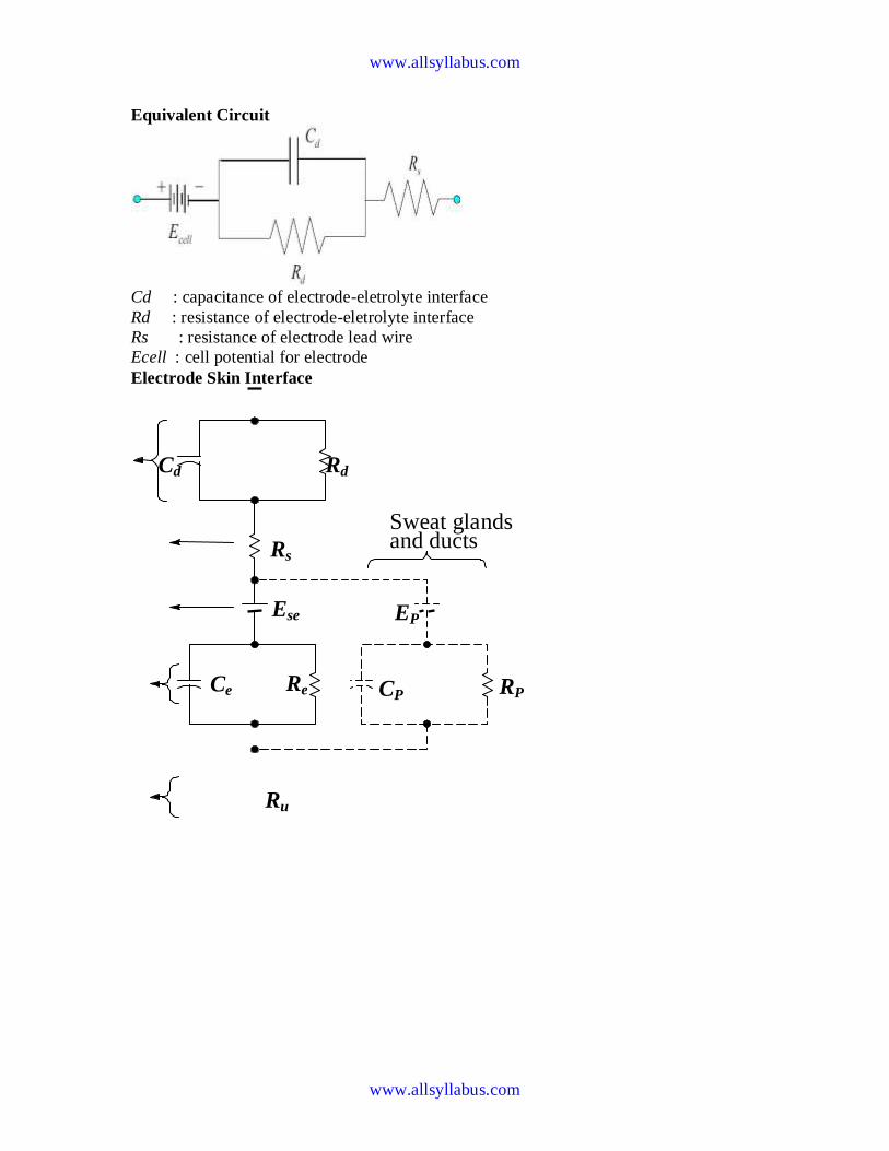
wwwallsyllabuscom
wwwallsyllabuscom
Equivalent Circuit
Cd capacitance of electrode-eletrolyte interface
Rd resistance of electrode-eletrolyte interface
Rs resistance of electrode lead wire
Ecell cell potential for electrode
Electrode Skin Interface
Sweat glands and ducts
Ru
Rs
Rd Cd
Re
Ese EP
RP CP Ce
wwwallsyllabuscom
wwwallsyllabuscom
Motion Artifact
Why
When the electrode moves with respect to the electrolyte the distribution of the double
layer of charge on polarizable electrode interface changes This changes the half cell
potential temporarily
What
If a pair of electrodes is in an electrolyte and one moves with respect to the other a
potential difference appears across the electrodes known as the motion artifact This is a
source of noise and interference in biopotential measurements
Motion artifact is minimal for non-polarizable electrodes
Body Surface Recording Electrodes
1 Metal Plate Electrodes (historic)
2 Suction Electrodes
Electrode
Epidermis
Dermis and subcutaneous layer
Cd
Gel
Stratum Corneum
Capillary
wwwallsyllabuscom
wwwallsyllabuscom
(historic interest)
1 Floating Electrodes
2 Flexible Electrodes
Commonly Used Biopotential Electrodes
Metal plate electrodes
ndash Large surface Ancient therefore still used ECG
ndash Metal disk with stainless steel platinum or gold coated
ndash EMG EEG
ndash smaller diameters
ndash motion artifacts
ndash Disposable foam-pad Cheap
(a) Metal-plate electrode used for application to limbs
(b) Metal-disk electrode applied with surgical tape
(c)Disposable foam-pad electrodes often used with ECG
Commonly Used Biopotential Electrodes
Suction electrodes
- No straps or adhesives required
- precordial (chest) ECG
- can only be used for short periods
Floating electrodes
- metal disk is recessed
- swimming in the electrolyte gel
- not in contact with the skin
- reduces motion artifact
wwwallsyllabuscom
wwwallsyllabuscom
Commonly Used Biopotential Electrodes
Flexible electrodes
- Body contours are often irregular
- Regularly shaped rigid electrodes
may not always work
- Special case infants
- Material
- Polymer or nylon with silver
- Carbon filled silicon rubber
(Mylar film)
Double-sided
Adhesive-tape
ring Electrolyte gel
in recess
(a) (b)
(c)
Snap coated with Ag-AgCl External snap
Plastic cup
Tack
Plastic disk
Foam pad
Capillary loops
Dead cellular material
Germinating layer
Gel-coated sponge
Floating Electrodes
wwwallsyllabuscom
wwwallsyllabuscom
(a) Carbon-filled silicone rubber electrode
(b) Flexible thin-film neonatal electrode
(c) Cross-sectional view of the thin-film electrode in (b)
Electrodes in Biopotential Measurements
3 Describe the construction of commercial ECG electrode (not the cheap polymer
electrode used in the lab) What is the common electrode metal and why is it preferred
So you are an inventor who has a better idea Describe an improvement
bull to make the electrode cheaper
bull more suitable for lower noise measurement for EEG
bull circumvent patents that are based on plasticfoam electrode body
bull attractive to consumers for use with their ECG machines at home
bull reduce artifact (minimize the motion of skinelectrode) in ambulatory recording
4 In a research laboratory scientists want to record from single cells in a culture dish
They want to record action potentials from single isolated heart cells What kind of
electrode would they need to use (describe material and design) Give a simplified
schematic (circuit model of the electrode) described in the notes given to you
What is the challenge involved in designing an amplifier for use with a microelectrode
for single cell recording Ie what are the critical amplifier design characteristics and
specifications (hint this is not the usual differentialinstrumentation amplifier)
Electrodes and Microelectrodes (miscellaneous)
bull How would you detect bacteria or other microorganisms in water supply Make
sure that your method distinguishes inert particulate matter from living cellular
matter
bull Draw the equivalent circuit model of the skin and an ECG electrode Identify the
key sources of electrical interference and otherwise the elements that would likely
contribute to the poor quality of recordings
wwwallsyllabuscom
wwwallsyllabuscom
bull Design an amplifier interface for the following two applications Patch clamp ion
channel current amplifier Your goal is to amplify pA level current to produce 1
Volt output
bull Strain gauge sensor amplifier Your goal is to convert 10 ohm change in
resistance of a strain gauge to produce 1 volt output
Neural electrodesmicroelectrodes
You want to record from neurons in the brain However you want to record from dozens
of neurons all at once from several closely spaced microelectrodes What material and
process would you use to make the microelectrode array
bull What metal would you prefer to use to make electrode arrays of about 10 micron
square size to make electrical contacts with dozens of neurons
bull What metal would you prefer to use to stimulate dozens of neurons in a deep
brain microelectrode based stimulator
bull (which metal provides good recording vs stimulating properties ndash and at the same
time not be toxic to brain tissue)
bull You are asked to develop an experimental set up to record from rat brain cells
using microelectrodes What precautions would you take to minimize the
electrical interference in your recording set up
Biopotential Amplifiers bull These are very important part of modern medical instrumentation bull We need to amplify biopotentials which are generated in the body at low levels with a high source impedance
bull Biopotentials amplifiers are required to increase signal strength while maintaining fidelity Basic Requirements of Biopotential Amplifiers
Essential functions of a bioamplifier are bull To take a weak biopotential and increase its amplitude so that it can be processed recorded or displayed bull To amplify voltage but it could be considered as a power amplifier as well bull To amplify current since in some cases a biopotential amplifier is used to isolate the load from the source current gain only
Input Impedance (Zin)
bull All biopotential amplifiers must have high input impedance minimize loading (remember the characteristics of biopotential electrodes resulting into loading and distortion if input impedance of the amplifier is not high enough) ndash typical values of Zin over the frequency range of the measurand = 10 MΩ (remember the loading rule)
Protection amp Isolation
bull The input circuit of a biopotential amplifier must provide protection to the live measurand
wwwallsyllabuscom
wwwallsyllabuscom
Vbio
bull Any potential or current at amplifierrsquos input terminals can affect Vbio
bull Electric currents produced by the biopotential amplifier can result in microshock and
macroshock bull The bioamplifier must have isolation and protection circuitry so that the current through the electrodes can be kept at safe levels and any artifact generated by such current can be minimized Output Impedance (Zout)
bull The output circuit does not present any critical problems all it needs to do is to drive the load bull Output impedance must be low with respect to the load impedance and it must be capable of satisfying the power requirements of the load Bandwidth (BW)
Frequency response requirements bull The biopotential amplifier must be sensitive to important frequency components of the biosignal bull Since biopotentials are low level signals it is important to limit bandwidth optimize signal-to-noise ratio Gain (G)
bull Biopotential amplifiers have a gain of 1000 or greater
Mode of Operation
bull Very frequently biosignals are obtained from bipolar electrodes bull Electrodes symmetrically located with respect to ground need differential amplification bull High CMRR required because 1 Common mode signals much greater than the biosignal appear on bipolar electrodes 2 Symmetry with respect to ground is not perfect (mismatch between electrode impedances) ndash more on this later
Calibration Signal
bull Medical and clinical equipment require quick calibration bull The gain of the biopotential amplifier must be calibrated to provide us with an accurate indication of the signalrsquos amplitude bull Push button to apply standard signal to the input of the biopotential amplifier bull Adjustable gain switch carefully selects calibrated fixed gains (in microprocessorndashbased systems gain adjustment can be Electrocardiography
hearts A very widely used medical instrument which is utilized to diagnose and monitor cardiac beat
abnormalities is the electrocardiograph
hearts It measures the electrical activity of the heart (more precisely biopotential differences arising from the electrical activity of myocardium) Wersquove already talked about the genesis of the ECG signal hearts The ECG machine uses surface electrodes and high input impedance hearts Differential amplifiers with good common mode rejection ratio to record the electrocardiogram
hearts Normal ECG amplitude ranges between 05-4 mV Normal frequency content of ECG (for diagnostic purposes) is 005-100 Hz A typical ECG waveform is shown below
wwwallsyllabuscom
wwwallsyllabuscom
Obviously all human hearts are not the same and this leads into variability in different parts of the ECG signal Significant diagnostic features of the ECG signal are hearts Duration of component parts of the signal
hearts Polarities and magnitudes hearts The details of the ECG signal and the degree of variability in different parts of the ECG signal is shown below
The QRS amplitude polarity time duration the RR interval (indicator of heartbeat per min) and the T-wave amplitude are some very important and distinctive features of the ECG signal The heart rate in BPM = Beats Per Minute) is simply = 60 (RR interval in seconds)
Some ECG waveform abnormalities that may indicate illness are hearts An extended PR interval may be diagnosed as AV node block hearts A widening of the QRS complex may indicate conduction problems in the bundle of His hearts An elevated ST segment may indicate occurrence of myocardial Infarction (MI) hearts A negative polarity in the T wave may be due to coronary insufficiency ECG Leads
Normal ECG recordings for the standard lead connections leads I II and III (Lead II provides
the strongest signal) Obviously all human hearts are not the same and this results into a high degree of variability
wwwallsyllabuscom
wwwallsyllabuscom
hearts Note the degree of variability of different parts of the ECG Signal
Some abnormalities that may indicate illness hearts An extended P-R interval may be diagnosed as AV node block hearts Widening of the QRS complex conduction problems in the bundle of His hearts Elevated ST segment may indicate occurrence of MI
hearts Negative polarity T wave may be due to coronary insufficiency QRS amplitude polarity time domain PR interval (indicator of heat beat per min amp T-wave amplitude are some very important
distinctive features 1 Loss
2 Origin of the ECG signal
hearts We have already covered this concept extensively in the previous lectures (The Dipole filed of the heart the Eindhovenrsquos Triangle the electrical circuit model for the electrocardiographic problem etc)
hearts Standard Limb Leads (I II III)
wwwallsyllabuscom
wwwallsyllabuscom
hearts The The lead wires are color-coded according to some conventions One example is White ndashRA
(Right Arm) Black ndash LA (Left Arm) Green ndash RL (Right Leg) Red ndash LL (Left Leg) and Brown
ndash C (Chest) Note There is a CM (common mode) amplifier connected to the right leg We will discuss this in detail later Augmented Limb Leads
These leads offer a free 50 increase over leads VR VL and VF connections (unipolar leads) with respect to Wilson terminal AVR = -I ndash III2 AVL = I ndash II2 aVF = II ndash I2
wwwallsyllabuscom
wwwallsyllabuscom
Each measurement is made from the reflected limb and the average of the other two limbs Chest Leads (Precordial)
Chest Lead Anatomical Positions
V1 - 4th intercostal space ndash Right sternal margin
V2 - 4th intercostal space ndash Left sternal margin
V3 ndash Midway between V2 amp V4
V4 ndash 5th intercostal space on mid-clavicular line
V5 ndash Same as V4 on the anterior axillary line
V6 ndash Same as V5 on the mid-axillary line
12 Lead Clinical Electrocardiography
The ECG Machine
Most representative Specs
wwwallsyllabuscom
wwwallsyllabuscom
bull Zin = 10 MΩ bull Frequency response = 005 ndash100 Hz bull Strip Chart Recorder Speed = 25 mmsec bull Fast Speed = 100 mmsec
For detailed Specs Refer to the Table in your text ―Summary of performance requirements for electrocardiographs Location of the Heart bull The heart is located between the lungs behind the sternum and above the diaphragm bull It is surrounded by the pericardium bull Its size is about that of a fist and its weight is about 250-300 g bull Its center is located about 15 cm to the left of the midsagittal plane
Anatomy of the heart
bull The walls of the heart are composed of cardiac muscle called myocardium
bull It consists of four compartments ndash the right and left atria and ventricles
The Heart Valves
bull The tricuspid valve regulates blood flow between the right atrium and right
ventricle
bull The pulmonary valve controls blood flow from the right ventricle into the
pulmonary arteries
bull The mitral valve lets oxygen-rich blood from your lungs pass from the left atrium
into the left ventricle
bull The aortic valve lets oxygen-rich blood pass from the left ventricle into the aorta
then to the body
Blood circulation via heart
wwwallsyllabuscom
wwwallsyllabuscom
bull The blood returns from the systemic circulation to the right atrium and from
there goes through the tricuspid valve to the right ventricle
bull It is ejected from the right ventricle through the pulmonary valve to the lungs
bull Oxygenated blood returns from the lungs to the left atrium and from there
through the mitral valve to the left ventricle
bull Finally blood is pumped through the aortic valve to the aorta and the systemic
circulation
Electrical activation of the heart
bull In the heart muscle cell or myocyte electric activation takes place by means of
the same mechanism as in the nerve cell ie from the inflow of Na ions across
the cell membrane
bull The amplitude of the action potential is also similar being 100 mV for both nerve
and muscle
bull The duration of the cardiac impulse is however two orders of magnitude longer
than in either nerve cell or sceletal muscle cell
bull As in the nerve cell repolarization is a consequence of the outflow of K ions
bull The duration of the action impulse is about 300 ms
Mechanical contraction of Cardiac Muscle
bull Associated with the electric activation of cardiac muscle cell is its mechanical
contraction which occurs a little later
bull An important distinction between cardiac muscle tissue and skeletal muscle is
that in cardiac muscle activation can propagate from one cell to another
in any direction
bull Electrical signal begins in the sinoatrial (SA) node natural pacemaker causes
the atria to contract
bull The signal then passes through the atrioventricular (AV) node
ndash sends the signal to the ventricles via the ―bundle of His
ndash causes the ventricles to contract
The Conduction System
wwwallsyllabuscom
wwwallsyllabuscom
The Action Potential
Recording an AP requires the isolation of a single cell Microelectrodes (with tips a few μm across) are used to stimulate and record the response A typical AP is 2-4ms long with an amplitude of about 100Mv The Electroencephalogram EEG
EEG is the graphical representation of the electrical activity of the brain Very commonly used to diagnose certain neurological disorders such as epilepsy
More recently also investigated whether it can detect various forms of dementia or schizophrenia EEG is the specific recording obtained using the scalp electrodes from the surface of the skull
During surgery electrodes may also be placed directly on the cortex The resulting signal is then electrocorticogram (ECoG) Just like ECG EEG is also obtained using several different electrodes places on different regions of the head brain
The Event Related
wwwallsyllabuscom
wwwallsyllabuscom
Potentials ndash ERPs
ERPs are really EEGs obtained under a specific protocol that requires the patient to response to certain stimuli ndash hence event related potentials Also called evoked potentials these signals can be used to diagnose certain
neurological disorders such as dementia and they can also be used as a liedetector bull The oddball paradigm
bull The guilty knowledge test
Electroretinogram ERG
The ERG is the record of the retinal action currents produced by the retina in response to a light stimulus It measures the electrical responses of the light-sensitive cells (such as rods and cones)
The stimuli are often a series of light flashes or rotating patterns The ERG is recorded using contact lens electrode that the subject wears while watching the stimuli
Phonocardiogram ndash PCG
The PCG is the graphic record of the heart sounds and murmurs It is thus a mechanical audio signal rather than an electrical signal Can be easily heard using a stethoscope Or can be converted into an electrical signal using a transducer Typically used to determine the disorders related to the heart valve since their
routine opening and closing create the well-known sounds bull S1 sounds First heart sounds ndash ventricular contractions move blood into atria closing of the AV (mitral and tricuspid) valves then semilunar valves open and blood ejected out of ventricles ndash immediately follows the QRS complex bull S2 sounds Second heart sounds ndash Closure of semilunar (aortic and pulmonary) valves bull Any unexpected sound may indicate a malfunctioning valve that causes the blood flow into out of a chamber when it should not Also called heart murmurs
wwwallsyllabuscom
wwwallsyllabuscom
Define ultrasound
bull Mechanical waves in different modalities (longitudinallateral) needs medium
to be propagated (solid liquid gas)
bull gt 20 kHz
bull Continuouspulsed
bull Sphericalplanarnarrow beamsurface waveLamb-wave
Physical phenomena behind ultrasound measurements
Transmission
bull
bull reflection
bull transit time
bull differences in propagation velocities
bull returns to transit time
bull doppler-shift in frequency
bull flow velocity
bull change of acoustic impedance
bull comparing to reference
bull interference of ultrasound waves (holography)
bull interaction of ultrasound and light (photoacousticz)
bull ultrasound needs medium for propagation it doesnrsquot propagate in vacuum
bull because mechanical waves need moving massunits and spring forces between
them
bull in acoustic emission the medium creates ultrasound (for example during pressure
changes) which is received by sensors
bull pulsed mode more common than continuous
bull continuous reguires separate transducers for transmitting and receicving
bull in pulsed mode an ultrasound burst is sent to the object and the same transducer is
switched to listen echoes
bull standing wave problem
bull in us-therapy pulsed mode gives more effective care without too much heating The Doppler Equation describes the relationship of the Doppler frequency shift to target velocity
wwwallsyllabuscom
wwwallsyllabuscom
The frequency difference is equal to the reflected frequency (FR) minus the originating frequency (FT) If the resulting frequency is higher then there is a positive Doppler shift and the object is moving toward the transducer but if the resulting frequency is lower there is a negative Doppler shift and it is moving away from the transducer In its simplest form it would be calculated as if
the ultrasound was parallel to the targetrsquos direction as shown in diagram A below However this would be a rare occurrence in clinical practice because the transducer is rarely pointed head on to a blood vessel In real life the ultrasound waves would approach the target at an angle called the Doppler angle ( ) On the following page diagram B shows the Doppler
equation used in general clinical situations which includes the Doppler angle The Doppler Angle
The ultrasound beam usually approaches the moving target at an angle called the Doppler angle ( ) This reduces the frequency shift in proportion to the cosine of this
angle If this angle is known then the flow velocity can be calculated The equation used is The Doppler Equation
equiv Doppler shift frequency (the difference between the transmitted and received frequencies) equiv transmitted frequency equiv reflected frequency
V equiv velocity of the blood flow towards the transducer C equiv velocity of sound in tissue θ equiv the angle between the sound beam and the direction of moving blood Where
The Doppler angle ( ) is also known as the angle of insonation It is estimated by the sonographer by a process known as angle correction which involves aligning an indicator on the duplex image along the longitudinal axis of the vessel
There are a few considerations that affect the performance of a Doppler examination that are inherent in the Doppler equation which are ndash The cosine of 90deg is zero so if the ultrasound beam is perpendicular to the direction of blood flow there will be no Doppler shift and it will appear as if there is no flow in the vessel ndash Appropriate estimation of the angle of insonation or angle correction is essential for the accurate determination of Doppler shift and blood flow velocity The angle of insonation should also be less than 60deg at all times since the
cosine function has a steeper curve above this angle and errors in angle correction will be magnified
The simplest Doppler devices use continuous wave (CW Doppler) rather than the pulsed wave used in more complex devices CW Doppler uses two transducers (or a dual element transducer) that transmit and receive ultrasound continuously The transmit and receive beams overlap in a Doppler sample volume some distance from the transducer face as
shown in the diagram below
volume) is the region of transmitting and receiving beam overlap (shaded region) Because there is continuous transducer transmission and reception echoes from all depths within the area arrive at the transducer simultaneously So although CW Doppler can determine the direction of flow it cannot discriminate the different depths where the motion originates The usefulness of CW Doppler devices is limited but they are used clinically to confirm blood flow in superficial vessels as they are good at detecting low velocities As they are easily portable this
can be done at the bedside or in the operating room Most other clinical applications require pulsed wave Doppler Pulsed Wave Doppler (PW Doppler)
wwwallsyllabuscom
wwwallsyllabuscom
Pulsed wave Doppler (PW Doppler) uses a single-element transducer that emits brief pulses of ultrasound energy The time interval between transmitting and then receiving the echoing sound can be used to calculate the depth from where the echo arises The Doppler sample volume can be chosen as to shape depth and position in sampling the flow
data For example the depth is chosen by processing only the signals that return to the transducer in a stipulated time For this technique the ultrasound system transmits a short pulse The eceiver is opened to detect the returning echoes only after a controlled delay and only for a specific duration This time-based gating of the receiving channel allows the definition of a fixed easuring distance which is often referred to as the Sample volume or Doppler gate Then the next ultrasound wave is transmitted The number of pulses transmitted by the system within a second is referred to as the pulse repetition frequency (PRF) The upper PRF limit is given by the time interval required for the echoes to arrive from a sample volume located at a
certain depth The greater the sample-volume depth the longer the time before the echoes are returned and the longer the delay between pulse transmission The greater the samplevolume depth the lower will be the maximum PRF setting Errors in the accuracy of the information arise if the velocities exceed a certain speed The highest velocity accurately measured is called the Nyquist limit Beyond this limit the errors that occur are referred to as aliasing Volume and flow measurement
Flow ndash volume of a liquidgas passing some point over a given time Gases are compressible
Benedict Roth Spirometer
Widely used for physiological amp clinical studies
Light bell moves with the ptrsquos breathing
Movement recorded by a pen on a rotating drum
Water seal prevents leakage of gas
Small seal minimises volume of gas dissolved in water
Suitable for measuring limited gas vols (few litres)
Pneumotachograph
wwwallsyllabuscom
wwwallsyllabuscom
Cardiac Output
Defn vol of blood pumped by the heart per min
CO = SV x HR
Norm ~ 5 lmin
Cardiac index ndash corrected for body surface area
Affected by
Met Rate ndash pregnancy hyperthyroid septic
Preload contractility afterload
Clinical indicators of CO imprecise
Affected by anaesthetic agents used in everyday practice
Provides estimate of
whole body perfusion
oxygen delivery
left ventricular function
Persistently low CO assoc with poor outcome
Methods
Fick method
Dilution techniques ndash dye thermal lithium
Pulse contour analysis- LiDCO amp PiCCO
Oesophageal doppler
TOE
Transthoracic impedance plethysmography
Inert gas through flow
Non-invasive cardiac output measurement
Fick Principle measure volume displacement
1st proposed 1870
―the total uptake or release of a substance by an organ is the product of the
blood flow through that organ and the arteriovenous concentration
difference of the substance
CO = O2 consumption (mlmin) art ndash mixed
venous O2 conc (mll)
Limited by cumbersome equipment sampling errors need for invasive
monitoring and steady-state haemodynamic and metabolic conditions
Indicator dilution techniques
―An indicator mixed into a unit volume of constantly flowing blood can be used to
identify that volume of blood in time provided the indicator remains in the system
between injection and measurement and mixes completely in the blood
wwwallsyllabuscom
wwwallsyllabuscom
Dye dilution
Inert dye ndash indocyanin green
Injected into pulmonary artery and arterial conc measured using a
calibrated cuvette densitometer
Plot indicator dilution curve (see diagram)
CO derived from area under curve
Indicator Dilution Curve
Cardiac Output Measurement
wwwallsyllabuscom
wwwallsyllabuscom
Why bother to check blood pressure
For each 20 mm rise in systolic blood pressure or 10 mm rise in diastolic blood
pressure over 11575
Risk of stroke increases
Risk of heart disease doubles
Risk of renal failure increases
Systolic and diastolic blood pressure
Systolic blood pressure is the highest pressure in the arteries just after the heart
beats
Diastolic blood pressure is the lowest pressure in the arteries just before the heart
beats
Blood pressure is measured indirectly by blood pressure cuff
(sphygmomanometer)
Inflating cuff increases pressure until it cuts off arterial circulation to the arm
Deflating cuff decrease pressure by 2 to 3 mm of mercury per second until blood
first enters the artery creating turbulence this causes a sound with each heartbeat
Sounds continue with each heartbeat until pressure lowers to the lowest pressure
in the artery then turbulence stops so the sound stops
Systolic blood pressure is the cuff pressure at the first sounds diastolic is the cuff
pressure just before the sounds stop
Phase 1 sharp thuds start at systolic blood pressure
Phase 2 blowing sound may disappear entirely (the auscultatory gap )
Phase 3 crisp thud a bit quieter than phase 1
Phase 4 sounds become muffled
Phase 5 end of sounds -- ends at diastolic blood pressure
wwwallsyllabuscom
wwwallsyllabuscom
Make sure the cuff is the right size - its width should be at least 40 of the arms
circumference The cuff will overestimate blood pressure if too small and
underestimate if too large
Place the cuff snugly on patients proximal arm on skin ( not cloth) centered over
the brachial artery (most cuffs have markings)
Support the patients arm at heart level using your arm or a desk
Your patient should sit in the chair for 5 minutes before BP is measured and
should have no caffeine or nicotine for 30 minutes before (JAMA 273 p1211-
1218 1995)
With fingers palpating radial or brachial artery inflate cuff rapidly until you cant
feel the pulse then 20 mm higher
Release cuff at 2 to 3 mm Hg per second until you again feel the pulse this is the
palpable systolic pressure
Wait 30 seconds before measuring blood pressure
Measuring palpable pressure first avoids risk of seriously underestimating blood
pressure because of the auscultatory gap (mistaking Korotkoff phase 3 for phase
1) Many doctors skip this step for time reasons and instead pump cuff to 200 mm
Hg at the next step)
Phase 2 of the Korotkoff sounds can be inaudible - especially in older patients
with systolic hypertension who are at especially high risk of stroke Inflating the
cuff until you dont hear sounds can give you a reading of 14086 when the
patients actual blood pressure is 22086 Most physicians are pressed for time so
they instead inflate the cuff to 200 mm which is beyond the auscultatory gap in
most patients But palpable systolic blood pressure is according to research more
reliable
Place bell of stethoscope (diaphragm is acceptable) over brachial artery
Rapidly pump the cuff to 20 to 30 mm Hg above palpable systolic pressure
Release pressure in the cuff by 2 to 3 mm Hg per second and listen for Korotkoff
sounds including systolic (first) and diastolic (last)
Record as systolicdiastolic Check in both arms the first time you check a
patients blood pressure It may differ by 10 mm Hg or more
If the sounds continues to zero record diastolic blood pressure as the point when
sounds become muffled (phase 4) over zero eg 130700 or just as 13070
Rate
ndash Number of beats in 30 seconds x 2
Strength
ndash Bounding strong or weak (thready)
Regularity
ndash Regular or irregular
You need three readings on two occasions to diagnose hypertension unless blood
pressure is very high
Normal blood pressure in children is
wwwallsyllabuscom
wwwallsyllabuscom
ndash 10255 at 1 year 11269 at 5 years 11978 at 10 years
Blood pressures in adults (JNC VII JAMA 2892560-72 2003)
ndash Normal lt120lt80
ndash Prehypertensive 120-13980-89
ndash Stage 1 hypertension 140-15990-99
ndash Stage 2 hypertension gt160gt100
Adult 60 to 100
Newborn 120-170
1 year 80-160
3 years 80-120
6 years 75-115
10 years 70-110
How to measure observe rise and fall of chest
In infants count for 60 seconds in adults 15 or 30 seconds
Normal respiration Adults 12 to 20
Children
newborn 30-80
1 year 20-40
3 years 20-30
6 years 16-22
Rate
Number of breaths in 30 seconds x 2
Quality
Character of breathing
Rhythm
Regular or irregular
Effort
Normal or labored
Noisy respiration
Normal stridor wheezing snoring gurgling
Depth
Shallow or deep
pH electrode
wwwallsyllabuscom
wwwallsyllabuscom
Governing equation is the Nernst Equation
pCO2 Electrode
The measurement of pCO2 is based on its linear relationship with pH over the range of
10 to 90 mm Hg
The dissociation constant is given by
Taking logarithms
pH = log[HCO3-] ndash log k ndash log a ndash log pCO2
Diathermy
In the natural sciences the term diathermy means electrically induced heat and is commonly
used for muscle relaxation It is also a method of heating tissue electromagnetically or
ultrasonically for therapeutic purposes in medicineContents
[hide]
1 Heating uses 2 Surgical uses
3 Trivia
4 References
E
RT
nF
H
HH
i
ln
0
H O CO H CO H HCO2 2 2 3 3
k
H HCO
a pCO
3
2
wwwallsyllabuscom
wwwallsyllabuscom
Heating uses
Ultrasonic diathermy refers to heating of tissues by ultrasound for the purpose of
therapeutic deep heating No tissue is ordinarily damaged hence it is generally used in
biomedical applications
Electric diathermy uses high frequency alternating electric or magnetic fields sometimes
with no electrode or device contact to the skin to induce gentle deep tissue heating by
induction Again no tissue is ordinarily damaged
Surgical uses
Surgical diathermy is usually better known as electrosurgery (It is also referred to
occasionally as electrocautery but see disambiguation below) Electrosurgery and
surgical diathermy involve the use of high frequency AC electrical current in surgery as
either a cutting modality or else to cauterize small blood vessels to stop bleeding This
technique induces localized tissue burning and damage the zone of which is controlled
by the frequency and power of the device Some sources[1]
insist that electrosurgery be
applied to surgery accomplished by high frequency AC cutting and that
electrocautery be used only for the practice of cauterization with heated nichrome wires
powered by DC current as in the handheld battery-operated portable cautery tools
Trivia
Medical Diathermy devices were used to cause interference to German radio beams used
for targeting night time bombing raids in WWII during the Battle of the Beams
I Diathermy
A Therapeutic use
1 Generation of local heating by high-frequency electromagnetic waves
2 Capacitance techniquemdashbody is placed in an electric field
a Dipolesmdashstructures with positive and negative poles
b Structures with large numbers of dipoles have a greater capacitance to store an electrical charge
c Greatest heating occurs in tissues with fewer dipoles particularly fatty tissues
3 Rapid rotation of dipoles causing mechanical friction and movement of electrons results in local heating
4 Inductance techniquemdashbody is not placed in an electric field
a Magnetic waves generated by driving current through a coiled wire
b Magnetic field creates currents in tissues
wwwallsyllabuscom
wwwallsyllabuscom
c Greatest heating occurs in tissues with low impedance especially muscle
B Precautions and contraindications
1 Diathermy should not be used
a Over metal implants and cardiac pacemakersmdashmore research needed regarding its use over metallic fixations
b Near the uterus of a pregnant woman or near the abdomen or back of a woman who might be pregnant
c On individuals with infections
d On individuals with acute inflammation
e Over moist open wounds
f On patients with malignant tumors
g Over large joint effusions
C Pulsed electromagnetic fields and diathermy
1 Can be pulsed to decrease total energy transmitted to the tissues
2 Short-wave diathermy can be adjusted into a nonthermal range
a Classified as pulsed electromagnetic field (PEMF) or
b Pulsed radio frequency energy (PRFE)
3 Important reclassification as diathermy implies heating
D Efficiency of diathermy and PEMF therapy for musculoskeletal conditions
1 Current research is limited but results suggest that diathermy enhances
treatments directed at soft tissue stretching
2 Some studies suggest that PEMF may speed wound healing and promote healing of nonunion fractures
Diathermy is a modality that uses electromagnetic energy to heat deeper tissues
Diathermy is more effective than ultrasound at heating a larger area of deep tissues
The athletic trainer must identify and respect contraindications to application of ultrasound and diathermy
Pulsed ultrasound and diathermy are used to treat slow-to-heal lesions including skin ulcers and nonunion fractures and may be able to facilitate repair of other tissues including ligaments and tendons
Diathermy
Definition
In diathermy high-frequency electrical currents are used to heat deep muscular tissues The heat increases blood flow speeding up recovery Doctors also use diathermy in surgical procedures by sealing blood vessels with electrically heated probes
The term diathermy is derived from the Greek words therma meaning heat and dia meaning through Diathermy literally means heating through
Origins
The therapeutic effects of heat have long been recognized More than 2000 years ago the Romans took advantage of heat therapies by building hot-spring bathhouses Since then various
wwwallsyllabuscom
wwwallsyllabuscom
methods of using heat have evolved In the early 1890s French physiologist Arseacutene dArsonval began studying the medical application of high-frequency currents The term diathermy was coined by German physician Carl Franz Nagelschmidt who designed a prototype apparatus in 1906 Around 1925 United States doctor J W Schereschewsky began studying the physiological
effects of high-frequency electrical currents on animals It was several years however before the fundamentals of the therapy were understood and put into practice
Benefits
Diathermy can be used to treat arthritis bursitis and other conditions involving stiff painful joints It is also used to treat pelvic infections and sinusitis A benefit of diathermy is that it is a painless procedure that can be administered at a clinic Also if the treatment relieves pain then patients can discontinue pain killers and escape their high cost and side effects
Description
Diathermy involves heating deep muscular tissues When heat is applied to the painful area cellular metabolism speeds up and blood flow increases The increased metabolism and circulation accelerates tissue repair The heat helps the tissues relax and stretch thus alleviating
stiffness Heat also reduces nerve fiber sensitivity increasing the patients pain threshold
There are three methods of diathermy In each energy is delivered to the deep tissues where it is converted to heat The three methods are
Shortwave diathermy The body part to be treated is placed between two capacitor plates Heat is generated as the high-frequency waves travel through the body tissues between the plates Shortwave diathermy is most often used to treat areas like the hip which is covered with a dense tissue mass It is also used to treat pelvic infections and sinusitis The treatment reduces inflammation The Federal Communications Commission regulates the frequency allowed for short-wave diathermy treatment Most machines
function at 2733 megahertz Ultrasound diathermy In this method high-frequency acoustic vibrations are used to
generate heat in deep tissue Microwave diathermy This method uses radar waves to heat tissue This form is the
easiest to use but the microwaves cannot penetrate deep muscles
Diathermy is also used in surgical procedures Many doctors use electrically heated probes to seal blood vessels to prevent excessive bleeding This is particularly helpful in neurosurgery and eye surgery Doctors can also use diathermy to kill abnormal growths such as tumors warts and
infected tissues
Preparations
To keep patients from sweating patients are usually asked to remove clothing from the body part being treated If a patient sweats the electrical currents may pool in the area causing burns Also clothing containing metal must be removed as must earrings buttons barrettes or zippers that contain metal Watches and hearing aids should be removed because the therapy may affect their function
wwwallsyllabuscom
wwwallsyllabuscom
Practitioners of surgical diathermy should steer clear of alcohol-based solutions to prepare and cleanse the skin These preparations can create a flammable vapor and cause burns and fires
Precautions
Patients with metal implants should not undergo diathermy treatment because the metal can act as a conductor of heat and result in serious internal burns Female patients with metallic uterine implants such as an IUD should avoid treatment in the pelvic area Diathermy should not be
used in joints that have been replaced with a prosthesis or in those with sensory impairment who may not be able to tell if they are burning Furthermore pulsed shortwave diathermy should be avoided during pregnancy as it can lead to abnormal fetal development
Patients with hemophilia should avoid the treatment because the increased blood flow could cause them to hemorrhage
Side effects
Some patients may experience superficial burns Since the therapy involves creating heat care must be taken to avoid burns particularly in patients whose injuries have caused decreased sensitivity to heat Also diathermy may affect pacemaker function
Female patients who receive treatment in the lower back or pelvic area may experience an
increased menstrual flow
Research amp general acceptance
For years physiotherapists and physical therapists have used diathermy as a routine part of physical rehabilitation
Electrical Safety
Electrical safety is very important in hospitals as patients may be undergoing a diagnostic or treatment procedure where the protective effect of dry skin is reduced Also patients may be unattended unconscious or anaesthetised and may not respond normally to an electric current Further electrically conductive solutions such as blood and saline are often present in patient treatment areas and may drip or spill on electrical equipment
Electric Current Leakage Current
Extension Leads Double Adaptors Equipment Classification
Class I Class II Defibrillator-Proof
Protective Devices
Residual Current Devices (RCD) Line Isolation overload Monitors (LIMs) Equipment Earthing
Area Classification
wwwallsyllabuscom
wwwallsyllabuscom
Body Protection Area Cardiac Protected Area
Other Electrical Issues Extension Leads
Double Adapters Main Extension Devices Power Boards Installation of Additional Power Points
Electric Current
Injuries received from electric current are dependent on the magnitude of current the pathway
that it takes through the body and the time for which it flows
The nature of electricity flowing through a circuit is analogous to blood flowing through the circulatory system within the human body In this analogy the source of energy is represented by the heart and the blood flowing through arteries and veins is analagous to current flowing
through the conductors and other components of the electric circuit
The application of an electric potential to an electric circuit generates a flow of current through conductive pathways This is analogous to the changes in blood pressure caused by contraction of cardiac muscle that causes blood to flow into the circulatory system For electric current to flow there must be a continuous pathway from the source of potential through electrical components and back to the source
Leakage Current
Electrical components and systems are encased in non conducting insulation to ensure that the electric current is contained and follows the intended pathways If the insulation deteriorates or breaks down current will leak through the insulation barrier and flow to earth This may
be through the protective earth conductor or through the operator
Medical equipment and clinical areas are fitted with a number of protective devices to protect the patient and operator from harmful leakage currents
Extension Leads
Extension leads are not permitted in clinical areas of RCH organisations They may cause high
earth resistance and excessive earth leakage current An extension lead can allow equipment to be powered from areas other than the relevant protected treatment area The power from the other area may not be protected to the same level as the power in the treatment area
wwwallsyllabuscom
wwwallsyllabuscom
As the connection between the extension lead and the equipment mains cable is often on the floor there is a high danger from fluid spills tripping and damage to the mains cable by trolleys when an extension lead is used
Double Adaptors
Double adaptors must not be used in RCH organisations They may not sit securely in a wall
outlet may not be able to provide adequate earth protection and may cause overloading overheating fire or loss of electrical supply
EQUIPMENT CLASSIFICATIONS
There are several methods of providing protection for operators and patients from electrical faults
and harmful leakage current
Class I
Class I equipment is fitted with a three core mains cable containing a protective earth wire
Exposed metal parts on class I equipment are connected to this earth wire
Should a fault develop inside the equipment and the exposed metal comes into contact with the mains the earthing conductor will conduct the fault current to ground Regular testing procedures ensure that earthing conductors are intact as the integrity of the earth wire is of vital importance
Class II
Class II equipment is enclosed within a double insulated case and does not require earthing
conductors Class II equipment is usually fitted with a 2-pin mains plug An internal electrical fault is unlikely to be hazardous as the double insulation prevents any external parts from becoming alive Class II or double insulated equipment can be identified by the class II symbol on the cabinet
Class II Symbol
wwwallsyllabuscom
wwwallsyllabuscom
Defibrillator-Proof
Some medical equipment within the hospital is classified as defibrillator proof When a
defibrillator is discharged through a patient connected to defibrillator proof equipment the equipment will not be damaged by the defibrillators energy Defibrillator proof equipment can remain connected to the patient during defibrillation It is identified by one of the following
symbols
Defibrillator proof symbols
Body protected Cardiac protected
PROTECTIVE DEVICES
Most patient care areas in the hospital are fitted with protective devices These devices are
regularly tested in accordance with the relevant guidelines published by Standards Australia The level of protection provided is dependent upon the device and the area in which it is located
Residual Current Devices (RCD)
RCDs (safety switches) are used in patient treatment areas to monitor and protect the mains supply RCDs sense leakage currents flowing to earth from the equipment If a significant leakage current flows the RCD will detect it and shut off the power supplied to the equipment
within 40 milliseconds Hospital RCDs are more sensitive than those fitted in homes A hospital RCD will trip at 10 milliamperes leakage current
Power outlets supplied through an RCD have a Supply Available lamp The lamp will extinguish
when the RCD trips due to excessive leakage current
wwwallsyllabuscom
wwwallsyllabuscom
Resetting a RCD
Lamp indicates supply is no longer available Disconnect all equipment from the supply Operate the reset button or lever on the supply panel and the Supply Available lamp
should illuminate If not contact Biomedical Engineering
Connect an item of equipment If the RCD trips again then this is the faulty item and should be labelled and sent to Biomedical Engineering
If the RCD does not trip continue connecting equipment until the RCD trips The last piece of equipment connected to the supply is most likely to be faulty as it will have caused the RCD to trip Remove the faulty item from service label it and send to Biomedical Engineering as mentioned above
Line Isolation overload Monitors (LIMs)
In critical life support applications where loss of power supply cannot be tolerated special power outlets powered by isolation transformers are installed
Line Isolation Monitors are installed to continually monitor electrical leakage in the power
supply system If an electrical fault develops in a medical device connected to an isolated power outlet the LIM will detect the leakage current The LIM will alarm and indicate the level of leakage current but will not shut off the electric supply
The faulty equipment can be identified by un plugging one item of equipment at a time from the supply until the alarm stops sounding Equipment that is not faulty may be reconnected Faulty equipment should be appropriately labelled and sent to Biomedical Engineering for repair
The LIM also monitors how much power is being used by the equpiment connected to it If too much power is being used the LIM will alarm and indicate that there is an overload The power used must be reduced immediately by moving some equipment to another circuit as soon as possible until the alarm stops sounding Failure to reduce the load on the LIM will result in the circuit breaker tripping and loss of power to the circuit
wwwallsyllabuscom
wwwallsyllabuscom
Equipotential Earthing
Equipotential earthing is installed in rooms classified as Cardiac Protected electrical areas
Equipotential earthing in treatment areas used for cardiac procedures is intended to minimise any voltage differences between earthed parts of equipment and any other exposed metal in the room
This reduces the possibility of leakage currents that can cause microelectrocution when the patient comes into contact with multiple items of equipment or if the patient happens to come
into contact with metal items in the room whilr they are connected to a medical device
All conductive metal in an equipotential area is connected to a common equipotential earth point with special heavy duty cable
AREA CLASSIFICATIONS
Body Protected Area
These areas are designed for procedures in which patients are connected to equipment that lowers the natural resistance of the skin Applied parts such as electrode gels conductive fluids entering the patient metal needles and catheters provide an easy pathway for current to flow
The main occurrence of injury from Body-Type procedures is from high current levels causing electric shock A direct connection to the patients heart is not present so the risk of Microelectrocution - fibrillation from minute current levels - is reduced
Residual Current Devices (RCD) or Isolation Transformers and Line Isolation Monitors (LIMiacutes) are used in Body Protected areas to provide protection against electrocution from high leakage currents Body-Protected Areas are identified with this sign
Cardiac Protected Area
Where the procedure involves placing an electrical conductor within or near the heart protection against fibrillation induced from small leakage currents is required Electrical conductors used in these procedures include cardiac pacing electrodes intracardiac ECG electrodes and intracardiac catheters
wwwallsyllabuscom
wwwallsyllabuscom
Equipotential earthing in conjunction with RCDs or LIMs provides protection against microelectrocution in Cardiac-Type procedures
Fault currents are reduced to magnitudes that are unlikely to induce fibrillation Used in conjunction with RCDs or LIMs the magnitude and duration of any fault currents
sourced from equipment are limited Cardiac-Protected Areas are identified with this sign
Other electrical issues
This policy aims to provide guidance to those who find that they need more electrical outlets than
those available or that the existing electrical outlets are inconveniently located
As extension leads and multiple outlet power boards can introduce additional hazards into an area the following procedures should be observed
Extension leads
Approved extension leads (AS 3760 1996) may be used in some areas within the hospital but MUST NOT BE USED IN PATIENT AREAS All electrical extension leads must be tagged with an Engineering Department maintenance tag and require a yearly safety inspection and test via
the Engineering Department
Double adapters
Double adapters may cause overloading or equipment earthing problems and are not to be used in
WCH
Mains extension device
The only mains extension device that is to be used in Patient care areas is the 4-way or 8-way
portable Core Balance Unit
The Biomedical Engineering Department must approve all units prior to use These units contain a safety switch and can detect excessive leakage current and disconnect the power in the event of a hazardous situation
wwwallsyllabuscom
wwwallsyllabuscom
Care must be exercised in the use of a portable Core Balance Unit It should be located off the
floor and in a position that will protect it from physical abuse and possible entry of fluids These devices are expensive and easily damaged The device must be sent to Biomedical Engineering every 6 months for safety testing
Power boards
Approved multiple-outlet power boards can be used across RCH but must not be used in patient
care areas except areas approved by the Biomedical Engineering Department
The power boards must have overload protection be fitted with internal safety shutters that protect unused outlets and be fitted with an onoff switch for each outlet
Medical Laser Applications
The main research subjects of the group of Medical Laser Application are in the field of online
monitoring and diagnostics as well as the development of new therapheutic methods The main focus in research is based upon the use of ultrashort (fs) laser pulses
The main advantage of ultrashort laser pulses is the extrem short interaction time which suppress any unwanted side effects of the laser irradiation of the tissue Secondly the broad spectra of the fs laser pulses give the advantage to use the same laser pulses for diagnostic applications like the optical coherence tomography (OCT)
wwwallsyllabuscom
wwwallsyllabuscom
Following research subjects have special attention at the moment
Optical Coherence Tomographie (OCT)
A fs-laser light source can be used to obtain images from inside the tissue when the pulses are coherent superposed This group uses this application to obtain an inside view of the vocal fold and to measure the outline of the crystalline lens
Refractive Surgery
The principle of getting rid of glasses with the help of a laser surgery (fs-LASIK) is underlying rapid improvements concerning precision and safety New technologies were evaluated and transferred very fast in cooperation with an industrial partner
Treatment of Presbyopia
The flexibility of the crystalline lens can be increase by pricise cuts which are induce by fs laser pulse inside the lens The treatment of the presbyopia is thinkable
wwwallsyllabuscom
wwwallsyllabuscom
Ultrafast Physics
Whereas the applications of ultrashort laser pulses increase rapidly is the physics of the interaction between the laser pulses and tissue in many case not fully understood To obtain improved knowledge of the interaction for many fs-laser application this group preforms numerical simulations as well as fundamental experiments
Thermography
Thermography thermal imaging or thermal video is a type of infrared imaging Thermographic cameras detect radiation in the infrared range of the electromagnetic spectrum
(roughly 900ndash14000 nanometers or 09ndash14 microm) and produce images of that radiation Since infrared radiation is emitted by all objects based on their temperatures according to the black body radiation law thermography makes it possible to see ones environment with or without visible illumination The amount of radiation emitted by an object increases with temperature therefore thermography allows one to see variations in temperature (hence the name) When viewed by thermographic camera warm objects stand out well against cooler backgrounds humans and other warm-blooded animals become easily visible against the environment day or
night As a result thermographys extensive use can historically be ascribed to the military and security services
Thermal imaging photography finds many other uses For example firefighters use it to see through smoke find persons and localize the base of a fire With thermal imaging power lines maintenance technicians locate overheating joints and parts a telltale sign of their failure to eliminate potential hazards Where thermal insulation becomes faulty building construction technicians can see heat leaks to improve the efficiencies of cooling or heating air-conditioning Thermal imaging cameras are also installed in some luxury cars to aid the driver the first being
the 2000 Cadillac DeVille Some physiological activities particularly responses in human beings and other warm-blooded animals can also be monitored with thermographic imaging [1]
The appearance and operation of a modern thermographic camera is often similar to a camcorder Enabling the user to see in the infrared spectrum is a function so useful that ability to record their output is often optional A recording module is therefore not always built-in
Instead of CCD sensors most thermal imaging cameras use CMOS Focal Plane Array (FPA) The most common types are InSb InGaAs HgCdTe and QWIP FPA The newest technologies are using low cost and uncooled microbolometers FPA sensors Their resolution is considerably lower than of optical cameras mostly 160x120 or 320x240 pixels up to 640x512 for the most
expensive models Thermographic cameras are much more expensive than their visible-spectrum counterparts and higher-end models are often export-restricted Older bolometers or more
wwwallsyllabuscom
wwwallsyllabuscom
sensitive models as InSb require cryogenic cooling usually by a miniature Stirling cycle refrigerator or liquid nitrogen
Contents
[hide]
1 Difference between IR film and thermography 2 Advantages of Thermography
3 Limitations amp disadvantages of thermography 4 Applications 5 See also 6 External links
o 61 History of thermal imager manufacturers
[edit] Difference between IR film and thermography
Thermal imaging is going to be used on Mars to detect caves that could hold life
[edit] Advantages of Thermography
You get a visual picture so that you can compare temperatures over a large area
It is real time capable of catching moving targets Able to find deteriorating components prior to failure Measurement in areas inaccessible or hazardous for other methods It is a non-destructive test method
[edit] Limitations amp disadvantages of thermography
Quality cameras are expensive and are easily damaged Images can be hard to interpret accurately even with experience
Accurate temperature measurements are very hard to make because of emissivities Most cameras have plusmn2 or worse accuracy (not as accurate as contact) Training and staying proficient in IR scanning is time consuming Ability to only measure surface areas
[edit] Applications
Condition monitoring Medical imaging Night vision Research
Process control Non destructive testing Surveillance in security law enforcement and defense Chemical imaging
wwwallsyllabuscom
wwwallsyllabuscom
Thermal infrared imagers convert the energy in the infrared wavelength into a visible light video display All objects above 0 kelvins emit thermal infrared energy so thermal imagers can passively see all objects regardless of ambient light However most thermal imagers only see objects warmer than -50 degC
The spectrum and amount of thermal radiation depend strongly on an objects surface
temperature This makes it possible for a thermal camera to display an objects temperature However other factors also influence the radiation which limits the accuracy of this technique For example the radiation depends not only on the temperature of the object but is also a function of the emissivity of the object Also radiation also originates from the surroundings and is reflected in the object and the radiation from the object and the reflected radiation will also be influenced by the absorption of the atmosphere
Endoscopy
This page is about having an endoscopy - a test that looks at the inside of your digestive system
There is information on
What endoscopy is
Why you may have an endoscopy
Having an endoscopy
Your results
What an endoscopy is
An endoscopy is a test that looks inside the body The endoscope is a long flexible tube that can
be swallowed It has a camera and light inside it Some doctors call it a telescope
wwwallsyllabuscom
wwwallsyllabuscom
Why you may have an endoscopy
You are most likely to have an endoscopy to look at the inside of your
Gullet (oesophagus)
Stomach
Duodenum - the first part of the small bowel that attaches to the stomach
Large bowel (colon)
Your doctor may want to see inside because you have symptoms such as abnormal bleeding or
difficulty swallowing The doctor can look down the endoscope and see if there are any growths
or other abnormal looking areas Also through the endoscope the doctor can take samples
(biopsies) of any abnormal looking tissues
There is more detailed information about having a colonoscopy in the bowel cancer section of
CancerHelp UK Below is information about having other types of endoscopy
Having an endoscopy
You can have this test as an out patient Most people have a choice between having the test while
they are awake or after having a medicine to make them drowsy (a sedative) Your hospital may
not be happy for you to have a sedative if you live alone and will have no one to look after you
when you go home If you live alone but really want sedation your hospital may allow you to
stay overnight You should ask your doctor about this as early as possible as extra arrangements
will need to be made beforehand
If you dont have a sedative you will have a spray to numb the back of your throat and make it
easier for you to swallow the endoscopy tube
If you would prefer to be asleep during the test you will have an injection to make you very
drowsy just before the test You will need to take someone with you to the hospital appointment
You wont be able to drive for the rest of the day and should have someone to go home with you
You cant eat or drink for about 8 hours before the test so that your stomach and duodenum are
empty Your doctor will give you written instructions about this beforehand or they may arrive
with your appointment letter When you get to the clinic you may be asked to take your upper
clothing off and put on a hospital gown Some hospitals prefer to use gowns because your clothes
wont get messy Once you are ready you get onto the bed or X-ray couch When you are lying
comfortably you have the sedative injection to make you very drowsy Or your doctor will spray
the back of your throat to numb it
Once the sedative or throat spray has worked the doctor will pass the endoscope tube down your
wwwallsyllabuscom
wwwallsyllabuscom
throat to the area being investigated Your doctor will ask you to swallow as the tube goes down
but if youve had a sedative you wont remember that afterwards If there are any abnormalities
the doctor will take pieces of tissue from the abnormal looking area to send to the laboratory for
closer inspection under a microscope These tissue samples are called biopsies
When the test is over you will need to rest for a while If youve had a sedative you may not
remember much (if anything) about the test once you have come round You should be able to go
home the same day
The results
It can take time for test results to come through How long will depend on why you are having
the test Usually the doctor who carries out the endoscopy dictates a report straight way The
report is typed up by the department secretary and goes to your specialist who gives the results to
you If your GP has sent you for the test the results will go directly to the GP surgery
Understandably waiting for results can make you anxious It usually takes a couple of weeks for
the results to come through If your doctor needed them urgently it would have been noted on
the test request form and the results will be ready sooner than that Try to remember to ask your
doctor how long you should expect to wait for the results when you are first asked to go for the
test If it is not an emergency and you have not heard a couple of weeks after your test ring your
doctors secretary to check if they are back
Endoscopy
From Wikipedia the free encyclopedia
Jump to navigation search
Endoscopic images of a duodenal ulcer
wwwallsyllabuscom
wwwallsyllabuscom
A flexible endoscope
Endoscopy means looking inside and typically refers to looking inside the human body for medical reasons using an instrument called an endoscope Endoscopy can also refer to using a borescope in technical situations where direct line-of-sight observation is not feasible
Contents
[hide]
1 Overview 2 Components 3 Uses
4 History 5 Risks 6 After The Endoscopy 7 Recent developments 8 See also 9 References 10 Footnotes
11 External links
Overview
Endoscopy is a minimally invasive diagnostic medical procedure that is used to assess the interior surfaces of an organ by inserting a tube into the body The instrument may have a rigid or flexible tube and not only provide an image for visual inspection and photography but also enable taking
biopsies and retrieval of foreign objects Endoscopy is the vehicle for minimally invasive surgery
Many endoscopic procedures are considered to be relatively painless and at worst associated with mild discomfort for example in esophagogastroduodenoscopy most patients tolerate the procedure with only topical anaesthesia of the oropharynx using lignocaine spray[1] Complications are not common (only 5 of all operations)[citation needed] but can include perforation of the organ under inspection with the endoscope or biopsy instrument If that occurs open surgery may be required to repair the injury
wwwallsyllabuscom
wwwallsyllabuscom
Components
An endoscope can consist of
a rigid or flexible tube a light delivery system to illuminate the organ or object under inspection The light
source is normally outside the body and the light is typically directed via an optical fiber system
a lens system transmitting the image to the viewer from the fiberscope an additional channel to allow entry of medical instruments or manipulators
Uses
Endoscopy can involve
The gastrointestinal tract (GI tract) o esophagus stomach and duodenum (esophagogastroduodenoscopy) o small intestine o colon (colonoscopyproctosigmoidoscopy) o Bile duct
endoscopic retrograde cholangiopancreatography (ERCP) duodenoscope-assisted cholangiopancreatoscopy intraoperative cholangioscopy
The respiratory tract o The nose (rhinoscopy) o The lower respiratory tract (bronchoscopy)
The urinary tract (cystoscopy) The female reproductive system
o The cervix (colposcopy) o The uterus (hysteroscopy) o The Fallopian tubes (Falloscopy)
Normally closed body cavities (through a small incision) o The abdominal or pelvic cavity (laparoscopy) o The interior of a joint (arthroscopy) o Organs of the chest (thoracoscopy and mediastinoscopy)
During pregnancy o The amnion (amnioscopy) o The fetus (fetoscopy)
Plastic Surgery Panendoscopy (or triple endoscopy)
o Combines laryngoscopy esophagoscopy and bronchoscopy Non-medical uses for endoscopy
o The planning and architectural community have found the endoscope useful for
pre-visualization of scale models of proposed buildings and cities (architectural endoscopy)
o Internal inspection of complex technical systems (borescope) o Endoscopes are also a tool helpful in the examination of improvised explosive
devices by bomb disposal personnel
wwwallsyllabuscom
wwwallsyllabuscom
o The FBI uses endoscopes for conducting surveillance via tight spaces
History
The first endoscope of a kind was developed in 1806 by Philip Bozzini with his introduction of a
Lichtleiter (light conductor) for the examinations of the canals and cavities of the human body However the Vienna Medical Society disapproved of such curiosity An endoscope was first introduced into a human in 1822 by William Beaumont an army surgeon at Mackinac
Island Michigan[citation needed] The use of electric light was a major step in the improvement of endoscopy The first such lights were external Later smaller bulbs became available making internal light possible for instance in a hysteroscope by Charles David in 1908[citation needed] Hans Christian Jacobaeus has been given credit for early endoscopic explorations of the abdomen and the thorax with laparoscopy (1912) and thoracoscopy (1910)[citation needed] Laparoscopy was used in the diagnosis of liver and gallbladder disease by Heinz Kalk in the 1930s[citation needed] Hope reported in 1937 on the use of laparoscopy to diagnose ectopic pregnancy[citation needed] In 1944
Raoul Palmer placed his patients in the Trendelenburg position after gaseous distention of the abdomen and thus was able to reliably perform gynecologic laparoscopy[citation needed]
The first gastrocamera was released in 1950 by Olympus Optical Co Ltd The device took pictures on monochromatic film using a small light bulb that was triggered manually The device was of limited use however because it did not implement real-time optical capability Olympus continued its development of endoscopes by incorporating fiber optics in the early 1960s leading to the first useful endoscopes In 1964 it released a gastrocamera guided by a fiberscope[1] A few articles claim that DrBasil Hirschowitz of UnivOf MichiganAnn Arbor discussed the
endoscope in early 50s[2]
As endoscopic technology improved so did the methods of gastrointestinal endoscopy Owing primarily to the efforts of Dr Hiromi Shinya in the late 1960s GI endoscopy developed into what is more recognizable as todays colonoscopy While many doctors experimented with techniques to take advantage of the new iterations of endoscopes Dr Shinya focused on techniques that would allow for successful operation of the endoscope by an individual rejecting the common practice at the time of utilizing two people Consequently many of the fundamental methods and procedures of modern colonoscopy were developed by Dr Shinya
Dr Shinyas other great contribution was to therapeutic endoscopy in his invention of the
electrosurgical polypectomy snare with the aid of Olympus employee Hiroshi Ichikawa Shinya sketched his first plans for the device on January 8 1969 He envisioned a loop of wire attached to the end of a colonoscope that would allow for easy removal of polyps during investigation by passing a current through the wire By September of 1969 the first polypectomy using this device was performed Polypectomy has since become the most common therapeutic procedure performed with an endoscope (Sivak 2004)
By 1980 laparoscopy training was required by gynecologists to perform tubal ligation procedures and diagnostic evaluations of the pelvis The first laparoscopic cholecystectomy was performed in 1984 and the first video-laparoscopic cholecystectomy in 1987[citation needed] During the 1990s
laparoscopic surgery was extended to the appendix spleen colon stomach kidney and liver[citation needed] Wireless capsule endoscopy or Capsule Endoscopy is now approved in all the countries including Japan where government reimbusement will be available from Oct2007Capsule Endoscopy [3] increases detection of Small Bowel tumors where traditional Endoscopy is not very efficient
wwwallsyllabuscom
wwwallsyllabuscom
Risks
Infection
Punctured organs Allergic reactions due to Contrast agents or dyes (such as those used in a CT scan) Over-sedation
After The Endoscopy
After the procedure the patient will be observed and monitored by a qualified individual in the
endoscopy or a recovery area until a significant portion of the medication has worn off Occasionally a patient is left with a mild sore throat which promptly responds to saline gargles or a feeling of distention from the insufflated air that was used during the procedure Both problems are mild and fleeting When fully recovered the patient will be instructed when to resume hisher usual diet (probably within a few hours) and will be allowed to be taken home Because of the use of sedation most facilities mandate that the patient is taken home by another person and not to drive on hisher own or handle machinery for the remainder of the day
Recent developments
With the application of robotic systems telesurgery was introduced as the surgeon could operate
from a site physically removed from the patient The first transatlantic surgery has been called the Lindbergh Operation
wwwallsyllabuscom
wwwallsyllabuscom
Upper Endoscopy
Upper endoscopy enables the physician to look inside the esophagus stomach and duodenum (first part of the small intestine) The procedure might be used to discover the reason for swallowing difficulties nausea vomiting
reflux bleeding indigestion abdominal pain or chest pain Upper endoscopy is also called EGD which stands for esophagogastroduodenoscopy (eh-SAH-fuh-goh-GAS-troh-doo-AH-duh-NAH-skuh-pee)
For the procedure you will swallow a thin flexible lighted tube called an endoscope (EN-doh-skope) Right before the procedure the physician will spray your throat with a numbing agent that may help prevent gagging
You may also receive pain medicine and a sedative to help you relax during the exam The endoscope transmits an image of the inside of the esophagus stomach and duodenum so the physician can carefully examine the lining of these organs The scope also blows air into the stomach this expands the folds of tissue and makes it easier for the physician to examine the stomach
The physician can see abnormalities like inflammation or
bleeding through the endoscope that dont show up well on x rays The physician can also insert instruments into the scope to treat bleeding abnormalities or remove samples of tissue (biopsy) for further tests
Possible complications of upper endoscopy include bleeding and puncture of the stomach lining However such complications are rare Most people will probably have nothing more than a mild sore throat after the procedure
The procedure takes 20 to 30 minutes Because you will be sedated you will need to rest at the endoscopy facility for 1 to 2 hours until the medication wears off
Preparation
Your stomach and duodenum must be empty for the procedure to be thorough and safe so you will not be able to eat or drink anything for at least 6 hours beforehand Also you must
arrange for someone to take you homemdashyou will not be allowed to drive because of the sedatives Your physician may give you other special instructions
The digestive system
wwwallsyllabuscom
wwwallsyllabuscom
What is Endoscopy and Why is it Performed
Endoscopy allows physicians to peer through the bodys passageways Endoscopy is the examination and inspection of the interior of body organs joints or cavities through an endoscope An endoscope is a device that uses fiber optics and powerful lens systems to provide lighting and visualization of the interior of a joint The portion of the endoscope inserted into the body may be rigid or flexible depending upon the medical procedure
An endoscope uses two fiber optic lines A light fiber carries light into the body cavity and an image fiber carries the image of the body cavity back to the physicians viewing lens There is also a separate port to allow for administration of drugs suction and irrigation This port may also be used to introduce small folding instruments such as forceps scissors brushes snares and baskets for tissue excision (removal) sampling or other diagnostic and therapeutic work Endoscopes may be used in conjunction with a camera or video recorder to document images of the inside of the joint or chronicle an endoscopic procedure New endoscopes have digital capabilities for manipulating and enhancing the video images
This figure shows a rigid endoscope used for arthroscopy The image fiber leads from the ocular (eye piece) to the inserted end of the scope The light fiber is below and leads from the light source to the working end of the endoscope
Why Is Endoscopy Performed
Endoscopy can be used to diagnose various conditions by close examination of internal organ and body structures Endoscopy can also guide therapy and repair such as the removal of torn cartilage from the bearing surfaces of a joint Biopsy (tissue sampling for pathologic testing) may also be performed under endoscopic guidance Local or general anesthetic may be used during endoscopy depending upon the type of procedure being performed
Internal abnormalities revealed through endoscopy include abscesses biliary (liver) cirrhosis bleeding bronchitis cancer cysts degenerative disease gallbladder stones hernia inflammation metastatic cancer polyps tumors ulcers and other diseases and conditions
Endoscopy is a minimally invasive procedure and carries with it certain minor risks depending upon the type of procedure being performed However these risks are typically far outweighed by the diagnostic and therapeutic potential of the procedure
Prior to the widespread use of endoscopy and diagnostic imaging most internal conditions could only be diagnosed or treated with open surgery Until the last several decades exploratory surgery was routinely performed only when a patient was critically ill and the source of illness was not known For example in certain dire cases the patients thorax or abdomen were surgically opened and examined to try to determine the source of illness
wwwallsyllabuscom
wwwallsyllabuscom
Endoscopy can often be done on an outpatient basis Outpatient means that the procedure does not require hospital admission and acute care and observation and may be performed outside the premises of a hospital Outpatient procedures performed at hospitals or ambulatory centers allow the patient to go home or return to work within a short while after their procedure
Types of Endoscopy
Fiber optic endoscopes now have widespread use in medicine and guide a myriad of diagnostic and therapeutic procedures including
Arthroscopy examination of joints for diagnosis and treatment (arthroscopic surgery) Bronchoscopy examination of the trachea and lungs bronchial trees to reveal
abscesses bronchitis carcinoma tumors tuberculosis alveolitis infection inflammation Colonoscopy examination of the inside of the colon and large intestine to detect polyps
tumors ulceration inflammation colitis diverticula Chrohns disease and discovery and removal of foreign bodies
Colposcopy direct visualization of the vagina and cervix to detect cancer inflammation and other conditions
Cystoscopy examination of the bladder urethra urinary tract uteral orifices and
prostate (men) with insertion of the endoscope through the urethra
ERCP (endoscopic retrograde cholangio-pancreatography) uses endoscopic guidance to
place a catheter for x-ray fluorosocopy with contrast enhancement This technique is used to examine the livers biliary tree the gallbladder the pancreatic duct and other anatomy to check for stones other obstructions and disease X-ray contrast is introduced into these ducts via catheter and fluoroscopic x-ray images are taken to show any abnormality or blockage If disease is detected it can sometimes be treated at the same time or biopsy can be performed to test for cancer or other pathology ERCP can detect biliary cirrhosis
cancer of the bile ducts pancreatic cysts pseudocysts pancreatic tumors chronic pancreatitis and other conditions such as gallbladder stones
EGD (Esophogealgastroduodensoscopy) visual examination of the upper gastro-
intestinal (GI) tract (also referred to as gastroscopy) to reveal hemorrhage hiatal hernia inflammation of the esophagus gastric ulcers
Endoscopic biopsy is the removal of tissue specimens for pathologic examination and
analysis Gastroscopy examination of the lining of the esophagus stomach and duodenum
Gastroscopy is often used to diagnose ulcers and other sources of bleeding and to guide biopsy of suspect GI cancers
Laparoscopy visualization of the stomach liver and other abdominal organs including
the female reproductive organs for example the fallopian tubes Laryngoscopy examination of the larynx (voice box) Proctoscopy sigmoidoscopy proctosigmoidoscopy examination of the rectum and
sigmoid colon Thoracoscopy examination of the pleura (sac that covers the lungs) pleural spaces
mediastinum and pericardium
A Brief History of Endoscopy
In the early 1900s the first attempts to view inside the body with lighted telescopes were made These initial devices were often fully rigid In the 1930s semi-flexible endoscopes called gastroscopes were developed to view inside of the stomach Fiber-optic endoscopy was pioneered by South African-born physician Basil Hirschowitz at the University of Michigan in 1957 Widespread use of fiber optic endoscopes began in the 1960s
wwwallsyllabuscom
wwwallsyllabuscom
A fiber optic cable is simply a bundle of microscopic glass or plastic fibers that literally allows light and images to be transmitted through curved structures Fiber optic cables are also replacing metal wires as the backbone of the worlds telecommunications infrastructure This Internet page may have traveled through a fiber optic cable as a stream of digital data (bursts of light) on its way to your computer
Endoscopy Equipment
Endoscopes have many practical needs And HMB Endoscopy Products (Hollywood Florida) has been providing endoscopic equipment and educating people on the use of endoscopes for more than 17 years Be sure to Browse our Catalog for all the details on how to purchase these medical instruments that can examine any part of the body
Video
Systems
Fiber
Endoscopes
Video
Endoscopes
In the simplest terms Endoscopy equipment consists of instruments that can look at the inside of many different organs mdash these are small flexible or rigid tubes with a light or lenses on the end that can look into the esophagus stomach and colon mdash and in more general terms endoscopy equipment can help doctors look deep inside body structures and hollow organs
An endoscope and related endoscope products and equipment are usually composed of three components
An optic system that allows the doctor to look through the scope into the organ or
cavity or to attach a video camera to the scope
A fiberoptic cable to light up the bodily area
A lumen (eg the bore of a tube like a needle or catheter) to take tissue samples of the
area being viewed
The beauty of endoscopic products is that they perform dual functions mdash with both diagnostic and therapeutic capabilities For example this means that these endoscopic products and instruments can perform biopsies (eg to evaluate tissue samples) as well as provide sclerotherapy (a medical procedure used to treat varicose veins and ―spider veins) In truth these brief explanations only tell part of the story
wwwallsyllabuscom
wwwallsyllabuscom
TYPES OF ENDOSCOPES
Take a look at the different types of endoscopic equipment you can get with HMB Endoscopy Products Again in very general terms there are two main types of endoscopes
Rigid Flexible
1 rigid endoscopes ndash the majority of which use a convex (curving out like one half of a
circle) glass lens system in which the small glass lenses are separated by large air spaces
2 flexible endoscopes allow for just that mdash flexibility In the animal kingdom for example
a flexible endoscope would be perfect for examining the stomach area of a snake
The popularity of endoscopy equipment continues to grow Some of the industryrsquos household names include Olympus Pentax Funjinon and Storz mdash with HMB both selling and repairing pre-owned and completely refurbished endoscopic products from each of these major manufacturers Browse our Catalog to get the ones you need
THERMOGRAPH APPLICATION Thermographs are digital recording thermometers used to log
temperature in the
marine environment The instruments are attached to a computer to set up recording
parameters for
deployment downloading and display of data in graphic or numeric format These units
are placed in
underwater housings and attached to the bottom or suspended in the water column for
deployment
DESCRIPTION Two types of thermograph are currently available The Ryan
Tempmentor is a
reusable data logger that allows storage of a maximum of 6361 temperature
measurements in the
range of -32C to +70C with 01resolution and 03accuracy The instrument can be
programmed
to take measurements from once per second to once every other hour Instrument
dimensions are 3 x
6 x 13 and the unit weighs 11 ounces The Hobo Temp is a miniature reusable data
logger that
allows storage of 1800 measurements in the range of -20C to +70C with
03resolution and 07
wwwallsyllabuscom
wwwallsyllabuscom
accuracy Sampling intervals from 05 seconds to 48 hours are available Instrument
dimensions are
24 x 19 x 08 and the unit weighs 1 ounce Both units are battery powered with the
battery life of
the Tempmentor being approximately two years and the battery life of the Hobo Temp
being one year THERMOGRAPH APPLICATION Thermographs are digital recording thermometers used to log
temperature in the
marine environment The instruments are attached to a computer to set up recording
parameters for
deployment downloading and display of data in graphic or numeric format These units
are placed in
underwater housings and attached to the bottom or suspended in the water column for
deployment
DESCRIPTION Two types of thermograph are currently available The Ryan
Tempmentor is a
reusable data logger that allows storage of a maximum of 6361 temperature
measurements in the
range of -32C to +70C with 01resolution and 03accuracy The instrument can be
programmed
to take measurements from once per second to once every other hour Instrument
dimensions are 3 x
6 x 13 and the unit weighs 11 ounces The Hobo Temp is a miniature reusable data
logger that
allows storage of 1800 measurements in the range of -20C to +70C with
03resolution and 07
accuracy Sampling intervals from 05 seconds to 48 hours are available Instrument
dimensions are
24 x 19 x 08 and the unit weighs 1 ounce Both units are battery powered with the
battery life of
the Tempmentor being approximately two years and the battery life of the Hobo Temp
being one year THERMOGRAPH APPLICATION Thermographs are digital recording thermometers used to log
temperature in the
marine environment The instruments are attached to a computer to set up recording
parameters for
deployment downloading and display of data in graphic or numeric format These units
are placed in
underwater housings and attached to the bottom or suspended in the water column for
deployment
DESCRIPTION Two types of thermograph are currently available The Ryan
Tempmentor is a
reusable data logger that allows storage of a maximum of 6361 temperature
measurements in the
range of -32C to +70C with 01resolution and 03accuracy The instrument can be
programmed
wwwallsyllabuscom
wwwallsyllabuscom
to take measurements from once per second to once every other hour Instrument
dimensions are 3 x
6 x 13 and the unit weighs 11 ounces The Hobo Temp is a miniature reusable data
logger that
allows storage of 1800 measurements in the range of -20C to +70C with
03resolution and 07
accuracy Sampling intervals from 05 seconds to 48 hours are available Instrument
dimensions are
24 x 19 x 08 and the unit weighs 1 ounce Both units are battery powered with the
battery life of
the Tempmentor being approximately two years and the battery life of the Hobo Temp
being one year
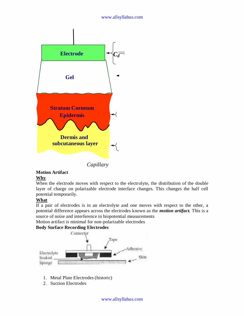
wwwallsyllabuscom
wwwallsyllabuscom
Motion Artifact
Why
When the electrode moves with respect to the electrolyte the distribution of the double
layer of charge on polarizable electrode interface changes This changes the half cell
potential temporarily
What
If a pair of electrodes is in an electrolyte and one moves with respect to the other a
potential difference appears across the electrodes known as the motion artifact This is a
source of noise and interference in biopotential measurements
Motion artifact is minimal for non-polarizable electrodes
Body Surface Recording Electrodes
1 Metal Plate Electrodes (historic)
2 Suction Electrodes
Electrode
Epidermis
Dermis and subcutaneous layer
Cd
Gel
Stratum Corneum
Capillary
wwwallsyllabuscom
wwwallsyllabuscom
(historic interest)
1 Floating Electrodes
2 Flexible Electrodes
Commonly Used Biopotential Electrodes
Metal plate electrodes
ndash Large surface Ancient therefore still used ECG
ndash Metal disk with stainless steel platinum or gold coated
ndash EMG EEG
ndash smaller diameters
ndash motion artifacts
ndash Disposable foam-pad Cheap
(a) Metal-plate electrode used for application to limbs
(b) Metal-disk electrode applied with surgical tape
(c)Disposable foam-pad electrodes often used with ECG
Commonly Used Biopotential Electrodes
Suction electrodes
- No straps or adhesives required
- precordial (chest) ECG
- can only be used for short periods
Floating electrodes
- metal disk is recessed
- swimming in the electrolyte gel
- not in contact with the skin
- reduces motion artifact
wwwallsyllabuscom
wwwallsyllabuscom
Commonly Used Biopotential Electrodes
Flexible electrodes
- Body contours are often irregular
- Regularly shaped rigid electrodes
may not always work
- Special case infants
- Material
- Polymer or nylon with silver
- Carbon filled silicon rubber
(Mylar film)
Double-sided
Adhesive-tape
ring Electrolyte gel
in recess
(a) (b)
(c)
Snap coated with Ag-AgCl External snap
Plastic cup
Tack
Plastic disk
Foam pad
Capillary loops
Dead cellular material
Germinating layer
Gel-coated sponge
Floating Electrodes
wwwallsyllabuscom
wwwallsyllabuscom
(a) Carbon-filled silicone rubber electrode
(b) Flexible thin-film neonatal electrode
(c) Cross-sectional view of the thin-film electrode in (b)
Electrodes in Biopotential Measurements
3 Describe the construction of commercial ECG electrode (not the cheap polymer
electrode used in the lab) What is the common electrode metal and why is it preferred
So you are an inventor who has a better idea Describe an improvement
bull to make the electrode cheaper
bull more suitable for lower noise measurement for EEG
bull circumvent patents that are based on plasticfoam electrode body
bull attractive to consumers for use with their ECG machines at home
bull reduce artifact (minimize the motion of skinelectrode) in ambulatory recording
4 In a research laboratory scientists want to record from single cells in a culture dish
They want to record action potentials from single isolated heart cells What kind of
electrode would they need to use (describe material and design) Give a simplified
schematic (circuit model of the electrode) described in the notes given to you
What is the challenge involved in designing an amplifier for use with a microelectrode
for single cell recording Ie what are the critical amplifier design characteristics and
specifications (hint this is not the usual differentialinstrumentation amplifier)
Electrodes and Microelectrodes (miscellaneous)
bull How would you detect bacteria or other microorganisms in water supply Make
sure that your method distinguishes inert particulate matter from living cellular
matter
bull Draw the equivalent circuit model of the skin and an ECG electrode Identify the
key sources of electrical interference and otherwise the elements that would likely
contribute to the poor quality of recordings
wwwallsyllabuscom
wwwallsyllabuscom
bull Design an amplifier interface for the following two applications Patch clamp ion
channel current amplifier Your goal is to amplify pA level current to produce 1
Volt output
bull Strain gauge sensor amplifier Your goal is to convert 10 ohm change in
resistance of a strain gauge to produce 1 volt output
Neural electrodesmicroelectrodes
You want to record from neurons in the brain However you want to record from dozens
of neurons all at once from several closely spaced microelectrodes What material and
process would you use to make the microelectrode array
bull What metal would you prefer to use to make electrode arrays of about 10 micron
square size to make electrical contacts with dozens of neurons
bull What metal would you prefer to use to stimulate dozens of neurons in a deep
brain microelectrode based stimulator
bull (which metal provides good recording vs stimulating properties ndash and at the same
time not be toxic to brain tissue)
bull You are asked to develop an experimental set up to record from rat brain cells
using microelectrodes What precautions would you take to minimize the
electrical interference in your recording set up
Biopotential Amplifiers bull These are very important part of modern medical instrumentation bull We need to amplify biopotentials which are generated in the body at low levels with a high source impedance
bull Biopotentials amplifiers are required to increase signal strength while maintaining fidelity Basic Requirements of Biopotential Amplifiers
Essential functions of a bioamplifier are bull To take a weak biopotential and increase its amplitude so that it can be processed recorded or displayed bull To amplify voltage but it could be considered as a power amplifier as well bull To amplify current since in some cases a biopotential amplifier is used to isolate the load from the source current gain only
Input Impedance (Zin)
bull All biopotential amplifiers must have high input impedance minimize loading (remember the characteristics of biopotential electrodes resulting into loading and distortion if input impedance of the amplifier is not high enough) ndash typical values of Zin over the frequency range of the measurand = 10 MΩ (remember the loading rule)
Protection amp Isolation
bull The input circuit of a biopotential amplifier must provide protection to the live measurand
wwwallsyllabuscom
wwwallsyllabuscom
Vbio
bull Any potential or current at amplifierrsquos input terminals can affect Vbio
bull Electric currents produced by the biopotential amplifier can result in microshock and
macroshock bull The bioamplifier must have isolation and protection circuitry so that the current through the electrodes can be kept at safe levels and any artifact generated by such current can be minimized Output Impedance (Zout)
bull The output circuit does not present any critical problems all it needs to do is to drive the load bull Output impedance must be low with respect to the load impedance and it must be capable of satisfying the power requirements of the load Bandwidth (BW)
Frequency response requirements bull The biopotential amplifier must be sensitive to important frequency components of the biosignal bull Since biopotentials are low level signals it is important to limit bandwidth optimize signal-to-noise ratio Gain (G)
bull Biopotential amplifiers have a gain of 1000 or greater
Mode of Operation
bull Very frequently biosignals are obtained from bipolar electrodes bull Electrodes symmetrically located with respect to ground need differential amplification bull High CMRR required because 1 Common mode signals much greater than the biosignal appear on bipolar electrodes 2 Symmetry with respect to ground is not perfect (mismatch between electrode impedances) ndash more on this later
Calibration Signal
bull Medical and clinical equipment require quick calibration bull The gain of the biopotential amplifier must be calibrated to provide us with an accurate indication of the signalrsquos amplitude bull Push button to apply standard signal to the input of the biopotential amplifier bull Adjustable gain switch carefully selects calibrated fixed gains (in microprocessorndashbased systems gain adjustment can be Electrocardiography
hearts A very widely used medical instrument which is utilized to diagnose and monitor cardiac beat
abnormalities is the electrocardiograph
hearts It measures the electrical activity of the heart (more precisely biopotential differences arising from the electrical activity of myocardium) Wersquove already talked about the genesis of the ECG signal hearts The ECG machine uses surface electrodes and high input impedance hearts Differential amplifiers with good common mode rejection ratio to record the electrocardiogram
hearts Normal ECG amplitude ranges between 05-4 mV Normal frequency content of ECG (for diagnostic purposes) is 005-100 Hz A typical ECG waveform is shown below
wwwallsyllabuscom
wwwallsyllabuscom
Obviously all human hearts are not the same and this leads into variability in different parts of the ECG signal Significant diagnostic features of the ECG signal are hearts Duration of component parts of the signal
hearts Polarities and magnitudes hearts The details of the ECG signal and the degree of variability in different parts of the ECG signal is shown below
The QRS amplitude polarity time duration the RR interval (indicator of heartbeat per min) and the T-wave amplitude are some very important and distinctive features of the ECG signal The heart rate in BPM = Beats Per Minute) is simply = 60 (RR interval in seconds)
Some ECG waveform abnormalities that may indicate illness are hearts An extended PR interval may be diagnosed as AV node block hearts A widening of the QRS complex may indicate conduction problems in the bundle of His hearts An elevated ST segment may indicate occurrence of myocardial Infarction (MI) hearts A negative polarity in the T wave may be due to coronary insufficiency ECG Leads
Normal ECG recordings for the standard lead connections leads I II and III (Lead II provides
the strongest signal) Obviously all human hearts are not the same and this results into a high degree of variability
wwwallsyllabuscom
wwwallsyllabuscom
hearts Note the degree of variability of different parts of the ECG Signal
Some abnormalities that may indicate illness hearts An extended P-R interval may be diagnosed as AV node block hearts Widening of the QRS complex conduction problems in the bundle of His hearts Elevated ST segment may indicate occurrence of MI
hearts Negative polarity T wave may be due to coronary insufficiency QRS amplitude polarity time domain PR interval (indicator of heat beat per min amp T-wave amplitude are some very important
distinctive features 1 Loss
2 Origin of the ECG signal
hearts We have already covered this concept extensively in the previous lectures (The Dipole filed of the heart the Eindhovenrsquos Triangle the electrical circuit model for the electrocardiographic problem etc)
hearts Standard Limb Leads (I II III)
wwwallsyllabuscom
wwwallsyllabuscom
hearts The The lead wires are color-coded according to some conventions One example is White ndashRA
(Right Arm) Black ndash LA (Left Arm) Green ndash RL (Right Leg) Red ndash LL (Left Leg) and Brown
ndash C (Chest) Note There is a CM (common mode) amplifier connected to the right leg We will discuss this in detail later Augmented Limb Leads
These leads offer a free 50 increase over leads VR VL and VF connections (unipolar leads) with respect to Wilson terminal AVR = -I ndash III2 AVL = I ndash II2 aVF = II ndash I2
wwwallsyllabuscom
wwwallsyllabuscom
Each measurement is made from the reflected limb and the average of the other two limbs Chest Leads (Precordial)
Chest Lead Anatomical Positions
V1 - 4th intercostal space ndash Right sternal margin
V2 - 4th intercostal space ndash Left sternal margin
V3 ndash Midway between V2 amp V4
V4 ndash 5th intercostal space on mid-clavicular line
V5 ndash Same as V4 on the anterior axillary line
V6 ndash Same as V5 on the mid-axillary line
12 Lead Clinical Electrocardiography
The ECG Machine
Most representative Specs
wwwallsyllabuscom
wwwallsyllabuscom
bull Zin = 10 MΩ bull Frequency response = 005 ndash100 Hz bull Strip Chart Recorder Speed = 25 mmsec bull Fast Speed = 100 mmsec
For detailed Specs Refer to the Table in your text ―Summary of performance requirements for electrocardiographs Location of the Heart bull The heart is located between the lungs behind the sternum and above the diaphragm bull It is surrounded by the pericardium bull Its size is about that of a fist and its weight is about 250-300 g bull Its center is located about 15 cm to the left of the midsagittal plane
Anatomy of the heart
bull The walls of the heart are composed of cardiac muscle called myocardium
bull It consists of four compartments ndash the right and left atria and ventricles
The Heart Valves
bull The tricuspid valve regulates blood flow between the right atrium and right
ventricle
bull The pulmonary valve controls blood flow from the right ventricle into the
pulmonary arteries
bull The mitral valve lets oxygen-rich blood from your lungs pass from the left atrium
into the left ventricle
bull The aortic valve lets oxygen-rich blood pass from the left ventricle into the aorta
then to the body
Blood circulation via heart
wwwallsyllabuscom
wwwallsyllabuscom
bull The blood returns from the systemic circulation to the right atrium and from
there goes through the tricuspid valve to the right ventricle
bull It is ejected from the right ventricle through the pulmonary valve to the lungs
bull Oxygenated blood returns from the lungs to the left atrium and from there
through the mitral valve to the left ventricle
bull Finally blood is pumped through the aortic valve to the aorta and the systemic
circulation
Electrical activation of the heart
bull In the heart muscle cell or myocyte electric activation takes place by means of
the same mechanism as in the nerve cell ie from the inflow of Na ions across
the cell membrane
bull The amplitude of the action potential is also similar being 100 mV for both nerve
and muscle
bull The duration of the cardiac impulse is however two orders of magnitude longer
than in either nerve cell or sceletal muscle cell
bull As in the nerve cell repolarization is a consequence of the outflow of K ions
bull The duration of the action impulse is about 300 ms
Mechanical contraction of Cardiac Muscle
bull Associated with the electric activation of cardiac muscle cell is its mechanical
contraction which occurs a little later
bull An important distinction between cardiac muscle tissue and skeletal muscle is
that in cardiac muscle activation can propagate from one cell to another
in any direction
bull Electrical signal begins in the sinoatrial (SA) node natural pacemaker causes
the atria to contract
bull The signal then passes through the atrioventricular (AV) node
ndash sends the signal to the ventricles via the ―bundle of His
ndash causes the ventricles to contract
The Conduction System
wwwallsyllabuscom
wwwallsyllabuscom
The Action Potential
Recording an AP requires the isolation of a single cell Microelectrodes (with tips a few μm across) are used to stimulate and record the response A typical AP is 2-4ms long with an amplitude of about 100Mv The Electroencephalogram EEG
EEG is the graphical representation of the electrical activity of the brain Very commonly used to diagnose certain neurological disorders such as epilepsy
More recently also investigated whether it can detect various forms of dementia or schizophrenia EEG is the specific recording obtained using the scalp electrodes from the surface of the skull
During surgery electrodes may also be placed directly on the cortex The resulting signal is then electrocorticogram (ECoG) Just like ECG EEG is also obtained using several different electrodes places on different regions of the head brain
The Event Related
wwwallsyllabuscom
wwwallsyllabuscom
Potentials ndash ERPs
ERPs are really EEGs obtained under a specific protocol that requires the patient to response to certain stimuli ndash hence event related potentials Also called evoked potentials these signals can be used to diagnose certain
neurological disorders such as dementia and they can also be used as a liedetector bull The oddball paradigm
bull The guilty knowledge test
Electroretinogram ERG
The ERG is the record of the retinal action currents produced by the retina in response to a light stimulus It measures the electrical responses of the light-sensitive cells (such as rods and cones)
The stimuli are often a series of light flashes or rotating patterns The ERG is recorded using contact lens electrode that the subject wears while watching the stimuli
Phonocardiogram ndash PCG
The PCG is the graphic record of the heart sounds and murmurs It is thus a mechanical audio signal rather than an electrical signal Can be easily heard using a stethoscope Or can be converted into an electrical signal using a transducer Typically used to determine the disorders related to the heart valve since their
routine opening and closing create the well-known sounds bull S1 sounds First heart sounds ndash ventricular contractions move blood into atria closing of the AV (mitral and tricuspid) valves then semilunar valves open and blood ejected out of ventricles ndash immediately follows the QRS complex bull S2 sounds Second heart sounds ndash Closure of semilunar (aortic and pulmonary) valves bull Any unexpected sound may indicate a malfunctioning valve that causes the blood flow into out of a chamber when it should not Also called heart murmurs
wwwallsyllabuscom
wwwallsyllabuscom
Define ultrasound
bull Mechanical waves in different modalities (longitudinallateral) needs medium
to be propagated (solid liquid gas)
bull gt 20 kHz
bull Continuouspulsed
bull Sphericalplanarnarrow beamsurface waveLamb-wave
Physical phenomena behind ultrasound measurements
Transmission
bull
bull reflection
bull transit time
bull differences in propagation velocities
bull returns to transit time
bull doppler-shift in frequency
bull flow velocity
bull change of acoustic impedance
bull comparing to reference
bull interference of ultrasound waves (holography)
bull interaction of ultrasound and light (photoacousticz)
bull ultrasound needs medium for propagation it doesnrsquot propagate in vacuum
bull because mechanical waves need moving massunits and spring forces between
them
bull in acoustic emission the medium creates ultrasound (for example during pressure
changes) which is received by sensors
bull pulsed mode more common than continuous
bull continuous reguires separate transducers for transmitting and receicving
bull in pulsed mode an ultrasound burst is sent to the object and the same transducer is
switched to listen echoes
bull standing wave problem
bull in us-therapy pulsed mode gives more effective care without too much heating The Doppler Equation describes the relationship of the Doppler frequency shift to target velocity
wwwallsyllabuscom
wwwallsyllabuscom
The frequency difference is equal to the reflected frequency (FR) minus the originating frequency (FT) If the resulting frequency is higher then there is a positive Doppler shift and the object is moving toward the transducer but if the resulting frequency is lower there is a negative Doppler shift and it is moving away from the transducer In its simplest form it would be calculated as if
the ultrasound was parallel to the targetrsquos direction as shown in diagram A below However this would be a rare occurrence in clinical practice because the transducer is rarely pointed head on to a blood vessel In real life the ultrasound waves would approach the target at an angle called the Doppler angle ( ) On the following page diagram B shows the Doppler
equation used in general clinical situations which includes the Doppler angle The Doppler Angle
The ultrasound beam usually approaches the moving target at an angle called the Doppler angle ( ) This reduces the frequency shift in proportion to the cosine of this
angle If this angle is known then the flow velocity can be calculated The equation used is The Doppler Equation
equiv Doppler shift frequency (the difference between the transmitted and received frequencies) equiv transmitted frequency equiv reflected frequency
V equiv velocity of the blood flow towards the transducer C equiv velocity of sound in tissue θ equiv the angle between the sound beam and the direction of moving blood Where
The Doppler angle ( ) is also known as the angle of insonation It is estimated by the sonographer by a process known as angle correction which involves aligning an indicator on the duplex image along the longitudinal axis of the vessel
There are a few considerations that affect the performance of a Doppler examination that are inherent in the Doppler equation which are ndash The cosine of 90deg is zero so if the ultrasound beam is perpendicular to the direction of blood flow there will be no Doppler shift and it will appear as if there is no flow in the vessel ndash Appropriate estimation of the angle of insonation or angle correction is essential for the accurate determination of Doppler shift and blood flow velocity The angle of insonation should also be less than 60deg at all times since the
cosine function has a steeper curve above this angle and errors in angle correction will be magnified
The simplest Doppler devices use continuous wave (CW Doppler) rather than the pulsed wave used in more complex devices CW Doppler uses two transducers (or a dual element transducer) that transmit and receive ultrasound continuously The transmit and receive beams overlap in a Doppler sample volume some distance from the transducer face as
shown in the diagram below
volume) is the region of transmitting and receiving beam overlap (shaded region) Because there is continuous transducer transmission and reception echoes from all depths within the area arrive at the transducer simultaneously So although CW Doppler can determine the direction of flow it cannot discriminate the different depths where the motion originates The usefulness of CW Doppler devices is limited but they are used clinically to confirm blood flow in superficial vessels as they are good at detecting low velocities As they are easily portable this
can be done at the bedside or in the operating room Most other clinical applications require pulsed wave Doppler Pulsed Wave Doppler (PW Doppler)
wwwallsyllabuscom
wwwallsyllabuscom
Pulsed wave Doppler (PW Doppler) uses a single-element transducer that emits brief pulses of ultrasound energy The time interval between transmitting and then receiving the echoing sound can be used to calculate the depth from where the echo arises The Doppler sample volume can be chosen as to shape depth and position in sampling the flow
data For example the depth is chosen by processing only the signals that return to the transducer in a stipulated time For this technique the ultrasound system transmits a short pulse The eceiver is opened to detect the returning echoes only after a controlled delay and only for a specific duration This time-based gating of the receiving channel allows the definition of a fixed easuring distance which is often referred to as the Sample volume or Doppler gate Then the next ultrasound wave is transmitted The number of pulses transmitted by the system within a second is referred to as the pulse repetition frequency (PRF) The upper PRF limit is given by the time interval required for the echoes to arrive from a sample volume located at a
certain depth The greater the sample-volume depth the longer the time before the echoes are returned and the longer the delay between pulse transmission The greater the samplevolume depth the lower will be the maximum PRF setting Errors in the accuracy of the information arise if the velocities exceed a certain speed The highest velocity accurately measured is called the Nyquist limit Beyond this limit the errors that occur are referred to as aliasing Volume and flow measurement
Flow ndash volume of a liquidgas passing some point over a given time Gases are compressible
Benedict Roth Spirometer
Widely used for physiological amp clinical studies
Light bell moves with the ptrsquos breathing
Movement recorded by a pen on a rotating drum
Water seal prevents leakage of gas
Small seal minimises volume of gas dissolved in water
Suitable for measuring limited gas vols (few litres)
Pneumotachograph
wwwallsyllabuscom
wwwallsyllabuscom
Cardiac Output
Defn vol of blood pumped by the heart per min
CO = SV x HR
Norm ~ 5 lmin
Cardiac index ndash corrected for body surface area
Affected by
Met Rate ndash pregnancy hyperthyroid septic
Preload contractility afterload
Clinical indicators of CO imprecise
Affected by anaesthetic agents used in everyday practice
Provides estimate of
whole body perfusion
oxygen delivery
left ventricular function
Persistently low CO assoc with poor outcome
Methods
Fick method
Dilution techniques ndash dye thermal lithium
Pulse contour analysis- LiDCO amp PiCCO
Oesophageal doppler
TOE
Transthoracic impedance plethysmography
Inert gas through flow
Non-invasive cardiac output measurement
Fick Principle measure volume displacement
1st proposed 1870
―the total uptake or release of a substance by an organ is the product of the
blood flow through that organ and the arteriovenous concentration
difference of the substance
CO = O2 consumption (mlmin) art ndash mixed
venous O2 conc (mll)
Limited by cumbersome equipment sampling errors need for invasive
monitoring and steady-state haemodynamic and metabolic conditions
Indicator dilution techniques
―An indicator mixed into a unit volume of constantly flowing blood can be used to
identify that volume of blood in time provided the indicator remains in the system
between injection and measurement and mixes completely in the blood
wwwallsyllabuscom
wwwallsyllabuscom
Dye dilution
Inert dye ndash indocyanin green
Injected into pulmonary artery and arterial conc measured using a
calibrated cuvette densitometer
Plot indicator dilution curve (see diagram)
CO derived from area under curve
Indicator Dilution Curve
Cardiac Output Measurement
wwwallsyllabuscom
wwwallsyllabuscom
Why bother to check blood pressure
For each 20 mm rise in systolic blood pressure or 10 mm rise in diastolic blood
pressure over 11575
Risk of stroke increases
Risk of heart disease doubles
Risk of renal failure increases
Systolic and diastolic blood pressure
Systolic blood pressure is the highest pressure in the arteries just after the heart
beats
Diastolic blood pressure is the lowest pressure in the arteries just before the heart
beats
Blood pressure is measured indirectly by blood pressure cuff
(sphygmomanometer)
Inflating cuff increases pressure until it cuts off arterial circulation to the arm
Deflating cuff decrease pressure by 2 to 3 mm of mercury per second until blood
first enters the artery creating turbulence this causes a sound with each heartbeat
Sounds continue with each heartbeat until pressure lowers to the lowest pressure
in the artery then turbulence stops so the sound stops
Systolic blood pressure is the cuff pressure at the first sounds diastolic is the cuff
pressure just before the sounds stop
Phase 1 sharp thuds start at systolic blood pressure
Phase 2 blowing sound may disappear entirely (the auscultatory gap )
Phase 3 crisp thud a bit quieter than phase 1
Phase 4 sounds become muffled
Phase 5 end of sounds -- ends at diastolic blood pressure
wwwallsyllabuscom
wwwallsyllabuscom
Make sure the cuff is the right size - its width should be at least 40 of the arms
circumference The cuff will overestimate blood pressure if too small and
underestimate if too large
Place the cuff snugly on patients proximal arm on skin ( not cloth) centered over
the brachial artery (most cuffs have markings)
Support the patients arm at heart level using your arm or a desk
Your patient should sit in the chair for 5 minutes before BP is measured and
should have no caffeine or nicotine for 30 minutes before (JAMA 273 p1211-
1218 1995)
With fingers palpating radial or brachial artery inflate cuff rapidly until you cant
feel the pulse then 20 mm higher
Release cuff at 2 to 3 mm Hg per second until you again feel the pulse this is the
palpable systolic pressure
Wait 30 seconds before measuring blood pressure
Measuring palpable pressure first avoids risk of seriously underestimating blood
pressure because of the auscultatory gap (mistaking Korotkoff phase 3 for phase
1) Many doctors skip this step for time reasons and instead pump cuff to 200 mm
Hg at the next step)
Phase 2 of the Korotkoff sounds can be inaudible - especially in older patients
with systolic hypertension who are at especially high risk of stroke Inflating the
cuff until you dont hear sounds can give you a reading of 14086 when the
patients actual blood pressure is 22086 Most physicians are pressed for time so
they instead inflate the cuff to 200 mm which is beyond the auscultatory gap in
most patients But palpable systolic blood pressure is according to research more
reliable
Place bell of stethoscope (diaphragm is acceptable) over brachial artery
Rapidly pump the cuff to 20 to 30 mm Hg above palpable systolic pressure
Release pressure in the cuff by 2 to 3 mm Hg per second and listen for Korotkoff
sounds including systolic (first) and diastolic (last)
Record as systolicdiastolic Check in both arms the first time you check a
patients blood pressure It may differ by 10 mm Hg or more
If the sounds continues to zero record diastolic blood pressure as the point when
sounds become muffled (phase 4) over zero eg 130700 or just as 13070
Rate
ndash Number of beats in 30 seconds x 2
Strength
ndash Bounding strong or weak (thready)
Regularity
ndash Regular or irregular
You need three readings on two occasions to diagnose hypertension unless blood
pressure is very high
Normal blood pressure in children is
wwwallsyllabuscom
wwwallsyllabuscom
ndash 10255 at 1 year 11269 at 5 years 11978 at 10 years
Blood pressures in adults (JNC VII JAMA 2892560-72 2003)
ndash Normal lt120lt80
ndash Prehypertensive 120-13980-89
ndash Stage 1 hypertension 140-15990-99
ndash Stage 2 hypertension gt160gt100
Adult 60 to 100
Newborn 120-170
1 year 80-160
3 years 80-120
6 years 75-115
10 years 70-110
How to measure observe rise and fall of chest
In infants count for 60 seconds in adults 15 or 30 seconds
Normal respiration Adults 12 to 20
Children
newborn 30-80
1 year 20-40
3 years 20-30
6 years 16-22
Rate
Number of breaths in 30 seconds x 2
Quality
Character of breathing
Rhythm
Regular or irregular
Effort
Normal or labored
Noisy respiration
Normal stridor wheezing snoring gurgling
Depth
Shallow or deep
pH electrode
wwwallsyllabuscom
wwwallsyllabuscom
Governing equation is the Nernst Equation
pCO2 Electrode
The measurement of pCO2 is based on its linear relationship with pH over the range of
10 to 90 mm Hg
The dissociation constant is given by
Taking logarithms
pH = log[HCO3-] ndash log k ndash log a ndash log pCO2
Diathermy
In the natural sciences the term diathermy means electrically induced heat and is commonly
used for muscle relaxation It is also a method of heating tissue electromagnetically or
ultrasonically for therapeutic purposes in medicineContents
[hide]
1 Heating uses 2 Surgical uses
3 Trivia
4 References
E
RT
nF
H
HH
i
ln
0
H O CO H CO H HCO2 2 2 3 3
k
H HCO
a pCO
3
2
wwwallsyllabuscom
wwwallsyllabuscom
Heating uses
Ultrasonic diathermy refers to heating of tissues by ultrasound for the purpose of
therapeutic deep heating No tissue is ordinarily damaged hence it is generally used in
biomedical applications
Electric diathermy uses high frequency alternating electric or magnetic fields sometimes
with no electrode or device contact to the skin to induce gentle deep tissue heating by
induction Again no tissue is ordinarily damaged
Surgical uses
Surgical diathermy is usually better known as electrosurgery (It is also referred to
occasionally as electrocautery but see disambiguation below) Electrosurgery and
surgical diathermy involve the use of high frequency AC electrical current in surgery as
either a cutting modality or else to cauterize small blood vessels to stop bleeding This
technique induces localized tissue burning and damage the zone of which is controlled
by the frequency and power of the device Some sources[1]
insist that electrosurgery be
applied to surgery accomplished by high frequency AC cutting and that
electrocautery be used only for the practice of cauterization with heated nichrome wires
powered by DC current as in the handheld battery-operated portable cautery tools
Trivia
Medical Diathermy devices were used to cause interference to German radio beams used
for targeting night time bombing raids in WWII during the Battle of the Beams
I Diathermy
A Therapeutic use
1 Generation of local heating by high-frequency electromagnetic waves
2 Capacitance techniquemdashbody is placed in an electric field
a Dipolesmdashstructures with positive and negative poles
b Structures with large numbers of dipoles have a greater capacitance to store an electrical charge
c Greatest heating occurs in tissues with fewer dipoles particularly fatty tissues
3 Rapid rotation of dipoles causing mechanical friction and movement of electrons results in local heating
4 Inductance techniquemdashbody is not placed in an electric field
a Magnetic waves generated by driving current through a coiled wire
b Magnetic field creates currents in tissues
wwwallsyllabuscom
wwwallsyllabuscom
c Greatest heating occurs in tissues with low impedance especially muscle
B Precautions and contraindications
1 Diathermy should not be used
a Over metal implants and cardiac pacemakersmdashmore research needed regarding its use over metallic fixations
b Near the uterus of a pregnant woman or near the abdomen or back of a woman who might be pregnant
c On individuals with infections
d On individuals with acute inflammation
e Over moist open wounds
f On patients with malignant tumors
g Over large joint effusions
C Pulsed electromagnetic fields and diathermy
1 Can be pulsed to decrease total energy transmitted to the tissues
2 Short-wave diathermy can be adjusted into a nonthermal range
a Classified as pulsed electromagnetic field (PEMF) or
b Pulsed radio frequency energy (PRFE)
3 Important reclassification as diathermy implies heating
D Efficiency of diathermy and PEMF therapy for musculoskeletal conditions
1 Current research is limited but results suggest that diathermy enhances
treatments directed at soft tissue stretching
2 Some studies suggest that PEMF may speed wound healing and promote healing of nonunion fractures
Diathermy is a modality that uses electromagnetic energy to heat deeper tissues
Diathermy is more effective than ultrasound at heating a larger area of deep tissues
The athletic trainer must identify and respect contraindications to application of ultrasound and diathermy
Pulsed ultrasound and diathermy are used to treat slow-to-heal lesions including skin ulcers and nonunion fractures and may be able to facilitate repair of other tissues including ligaments and tendons
Diathermy
Definition
In diathermy high-frequency electrical currents are used to heat deep muscular tissues The heat increases blood flow speeding up recovery Doctors also use diathermy in surgical procedures by sealing blood vessels with electrically heated probes
The term diathermy is derived from the Greek words therma meaning heat and dia meaning through Diathermy literally means heating through
Origins
The therapeutic effects of heat have long been recognized More than 2000 years ago the Romans took advantage of heat therapies by building hot-spring bathhouses Since then various
wwwallsyllabuscom
wwwallsyllabuscom
methods of using heat have evolved In the early 1890s French physiologist Arseacutene dArsonval began studying the medical application of high-frequency currents The term diathermy was coined by German physician Carl Franz Nagelschmidt who designed a prototype apparatus in 1906 Around 1925 United States doctor J W Schereschewsky began studying the physiological
effects of high-frequency electrical currents on animals It was several years however before the fundamentals of the therapy were understood and put into practice
Benefits
Diathermy can be used to treat arthritis bursitis and other conditions involving stiff painful joints It is also used to treat pelvic infections and sinusitis A benefit of diathermy is that it is a painless procedure that can be administered at a clinic Also if the treatment relieves pain then patients can discontinue pain killers and escape their high cost and side effects
Description
Diathermy involves heating deep muscular tissues When heat is applied to the painful area cellular metabolism speeds up and blood flow increases The increased metabolism and circulation accelerates tissue repair The heat helps the tissues relax and stretch thus alleviating
stiffness Heat also reduces nerve fiber sensitivity increasing the patients pain threshold
There are three methods of diathermy In each energy is delivered to the deep tissues where it is converted to heat The three methods are
Shortwave diathermy The body part to be treated is placed between two capacitor plates Heat is generated as the high-frequency waves travel through the body tissues between the plates Shortwave diathermy is most often used to treat areas like the hip which is covered with a dense tissue mass It is also used to treat pelvic infections and sinusitis The treatment reduces inflammation The Federal Communications Commission regulates the frequency allowed for short-wave diathermy treatment Most machines
function at 2733 megahertz Ultrasound diathermy In this method high-frequency acoustic vibrations are used to
generate heat in deep tissue Microwave diathermy This method uses radar waves to heat tissue This form is the
easiest to use but the microwaves cannot penetrate deep muscles
Diathermy is also used in surgical procedures Many doctors use electrically heated probes to seal blood vessels to prevent excessive bleeding This is particularly helpful in neurosurgery and eye surgery Doctors can also use diathermy to kill abnormal growths such as tumors warts and
infected tissues
Preparations
To keep patients from sweating patients are usually asked to remove clothing from the body part being treated If a patient sweats the electrical currents may pool in the area causing burns Also clothing containing metal must be removed as must earrings buttons barrettes or zippers that contain metal Watches and hearing aids should be removed because the therapy may affect their function
wwwallsyllabuscom
wwwallsyllabuscom
Practitioners of surgical diathermy should steer clear of alcohol-based solutions to prepare and cleanse the skin These preparations can create a flammable vapor and cause burns and fires
Precautions
Patients with metal implants should not undergo diathermy treatment because the metal can act as a conductor of heat and result in serious internal burns Female patients with metallic uterine implants such as an IUD should avoid treatment in the pelvic area Diathermy should not be
used in joints that have been replaced with a prosthesis or in those with sensory impairment who may not be able to tell if they are burning Furthermore pulsed shortwave diathermy should be avoided during pregnancy as it can lead to abnormal fetal development
Patients with hemophilia should avoid the treatment because the increased blood flow could cause them to hemorrhage
Side effects
Some patients may experience superficial burns Since the therapy involves creating heat care must be taken to avoid burns particularly in patients whose injuries have caused decreased sensitivity to heat Also diathermy may affect pacemaker function
Female patients who receive treatment in the lower back or pelvic area may experience an
increased menstrual flow
Research amp general acceptance
For years physiotherapists and physical therapists have used diathermy as a routine part of physical rehabilitation
Electrical Safety
Electrical safety is very important in hospitals as patients may be undergoing a diagnostic or treatment procedure where the protective effect of dry skin is reduced Also patients may be unattended unconscious or anaesthetised and may not respond normally to an electric current Further electrically conductive solutions such as blood and saline are often present in patient treatment areas and may drip or spill on electrical equipment
Electric Current Leakage Current
Extension Leads Double Adaptors Equipment Classification
Class I Class II Defibrillator-Proof
Protective Devices
Residual Current Devices (RCD) Line Isolation overload Monitors (LIMs) Equipment Earthing
Area Classification
wwwallsyllabuscom
wwwallsyllabuscom
Body Protection Area Cardiac Protected Area
Other Electrical Issues Extension Leads
Double Adapters Main Extension Devices Power Boards Installation of Additional Power Points
Electric Current
Injuries received from electric current are dependent on the magnitude of current the pathway
that it takes through the body and the time for which it flows
The nature of electricity flowing through a circuit is analogous to blood flowing through the circulatory system within the human body In this analogy the source of energy is represented by the heart and the blood flowing through arteries and veins is analagous to current flowing
through the conductors and other components of the electric circuit
The application of an electric potential to an electric circuit generates a flow of current through conductive pathways This is analogous to the changes in blood pressure caused by contraction of cardiac muscle that causes blood to flow into the circulatory system For electric current to flow there must be a continuous pathway from the source of potential through electrical components and back to the source
Leakage Current
Electrical components and systems are encased in non conducting insulation to ensure that the electric current is contained and follows the intended pathways If the insulation deteriorates or breaks down current will leak through the insulation barrier and flow to earth This may
be through the protective earth conductor or through the operator
Medical equipment and clinical areas are fitted with a number of protective devices to protect the patient and operator from harmful leakage currents
Extension Leads
Extension leads are not permitted in clinical areas of RCH organisations They may cause high
earth resistance and excessive earth leakage current An extension lead can allow equipment to be powered from areas other than the relevant protected treatment area The power from the other area may not be protected to the same level as the power in the treatment area
wwwallsyllabuscom
wwwallsyllabuscom
As the connection between the extension lead and the equipment mains cable is often on the floor there is a high danger from fluid spills tripping and damage to the mains cable by trolleys when an extension lead is used
Double Adaptors
Double adaptors must not be used in RCH organisations They may not sit securely in a wall
outlet may not be able to provide adequate earth protection and may cause overloading overheating fire or loss of electrical supply
EQUIPMENT CLASSIFICATIONS
There are several methods of providing protection for operators and patients from electrical faults
and harmful leakage current
Class I
Class I equipment is fitted with a three core mains cable containing a protective earth wire
Exposed metal parts on class I equipment are connected to this earth wire
Should a fault develop inside the equipment and the exposed metal comes into contact with the mains the earthing conductor will conduct the fault current to ground Regular testing procedures ensure that earthing conductors are intact as the integrity of the earth wire is of vital importance
Class II
Class II equipment is enclosed within a double insulated case and does not require earthing
conductors Class II equipment is usually fitted with a 2-pin mains plug An internal electrical fault is unlikely to be hazardous as the double insulation prevents any external parts from becoming alive Class II or double insulated equipment can be identified by the class II symbol on the cabinet
Class II Symbol
wwwallsyllabuscom
wwwallsyllabuscom
Defibrillator-Proof
Some medical equipment within the hospital is classified as defibrillator proof When a
defibrillator is discharged through a patient connected to defibrillator proof equipment the equipment will not be damaged by the defibrillators energy Defibrillator proof equipment can remain connected to the patient during defibrillation It is identified by one of the following
symbols
Defibrillator proof symbols
Body protected Cardiac protected
PROTECTIVE DEVICES
Most patient care areas in the hospital are fitted with protective devices These devices are
regularly tested in accordance with the relevant guidelines published by Standards Australia The level of protection provided is dependent upon the device and the area in which it is located
Residual Current Devices (RCD)
RCDs (safety switches) are used in patient treatment areas to monitor and protect the mains supply RCDs sense leakage currents flowing to earth from the equipment If a significant leakage current flows the RCD will detect it and shut off the power supplied to the equipment
within 40 milliseconds Hospital RCDs are more sensitive than those fitted in homes A hospital RCD will trip at 10 milliamperes leakage current
Power outlets supplied through an RCD have a Supply Available lamp The lamp will extinguish
when the RCD trips due to excessive leakage current
wwwallsyllabuscom
wwwallsyllabuscom
Resetting a RCD
Lamp indicates supply is no longer available Disconnect all equipment from the supply Operate the reset button or lever on the supply panel and the Supply Available lamp
should illuminate If not contact Biomedical Engineering
Connect an item of equipment If the RCD trips again then this is the faulty item and should be labelled and sent to Biomedical Engineering
If the RCD does not trip continue connecting equipment until the RCD trips The last piece of equipment connected to the supply is most likely to be faulty as it will have caused the RCD to trip Remove the faulty item from service label it and send to Biomedical Engineering as mentioned above
Line Isolation overload Monitors (LIMs)
In critical life support applications where loss of power supply cannot be tolerated special power outlets powered by isolation transformers are installed
Line Isolation Monitors are installed to continually monitor electrical leakage in the power
supply system If an electrical fault develops in a medical device connected to an isolated power outlet the LIM will detect the leakage current The LIM will alarm and indicate the level of leakage current but will not shut off the electric supply
The faulty equipment can be identified by un plugging one item of equipment at a time from the supply until the alarm stops sounding Equipment that is not faulty may be reconnected Faulty equipment should be appropriately labelled and sent to Biomedical Engineering for repair
The LIM also monitors how much power is being used by the equpiment connected to it If too much power is being used the LIM will alarm and indicate that there is an overload The power used must be reduced immediately by moving some equipment to another circuit as soon as possible until the alarm stops sounding Failure to reduce the load on the LIM will result in the circuit breaker tripping and loss of power to the circuit
wwwallsyllabuscom
wwwallsyllabuscom
Equipotential Earthing
Equipotential earthing is installed in rooms classified as Cardiac Protected electrical areas
Equipotential earthing in treatment areas used for cardiac procedures is intended to minimise any voltage differences between earthed parts of equipment and any other exposed metal in the room
This reduces the possibility of leakage currents that can cause microelectrocution when the patient comes into contact with multiple items of equipment or if the patient happens to come
into contact with metal items in the room whilr they are connected to a medical device
All conductive metal in an equipotential area is connected to a common equipotential earth point with special heavy duty cable
AREA CLASSIFICATIONS
Body Protected Area
These areas are designed for procedures in which patients are connected to equipment that lowers the natural resistance of the skin Applied parts such as electrode gels conductive fluids entering the patient metal needles and catheters provide an easy pathway for current to flow
The main occurrence of injury from Body-Type procedures is from high current levels causing electric shock A direct connection to the patients heart is not present so the risk of Microelectrocution - fibrillation from minute current levels - is reduced
Residual Current Devices (RCD) or Isolation Transformers and Line Isolation Monitors (LIMiacutes) are used in Body Protected areas to provide protection against electrocution from high leakage currents Body-Protected Areas are identified with this sign
Cardiac Protected Area
Where the procedure involves placing an electrical conductor within or near the heart protection against fibrillation induced from small leakage currents is required Electrical conductors used in these procedures include cardiac pacing electrodes intracardiac ECG electrodes and intracardiac catheters
wwwallsyllabuscom
wwwallsyllabuscom
Equipotential earthing in conjunction with RCDs or LIMs provides protection against microelectrocution in Cardiac-Type procedures
Fault currents are reduced to magnitudes that are unlikely to induce fibrillation Used in conjunction with RCDs or LIMs the magnitude and duration of any fault currents
sourced from equipment are limited Cardiac-Protected Areas are identified with this sign
Other electrical issues
This policy aims to provide guidance to those who find that they need more electrical outlets than
those available or that the existing electrical outlets are inconveniently located
As extension leads and multiple outlet power boards can introduce additional hazards into an area the following procedures should be observed
Extension leads
Approved extension leads (AS 3760 1996) may be used in some areas within the hospital but MUST NOT BE USED IN PATIENT AREAS All electrical extension leads must be tagged with an Engineering Department maintenance tag and require a yearly safety inspection and test via
the Engineering Department
Double adapters
Double adapters may cause overloading or equipment earthing problems and are not to be used in
WCH
Mains extension device
The only mains extension device that is to be used in Patient care areas is the 4-way or 8-way
portable Core Balance Unit
The Biomedical Engineering Department must approve all units prior to use These units contain a safety switch and can detect excessive leakage current and disconnect the power in the event of a hazardous situation
wwwallsyllabuscom
wwwallsyllabuscom
Care must be exercised in the use of a portable Core Balance Unit It should be located off the
floor and in a position that will protect it from physical abuse and possible entry of fluids These devices are expensive and easily damaged The device must be sent to Biomedical Engineering every 6 months for safety testing
Power boards
Approved multiple-outlet power boards can be used across RCH but must not be used in patient
care areas except areas approved by the Biomedical Engineering Department
The power boards must have overload protection be fitted with internal safety shutters that protect unused outlets and be fitted with an onoff switch for each outlet
Medical Laser Applications
The main research subjects of the group of Medical Laser Application are in the field of online
monitoring and diagnostics as well as the development of new therapheutic methods The main focus in research is based upon the use of ultrashort (fs) laser pulses
The main advantage of ultrashort laser pulses is the extrem short interaction time which suppress any unwanted side effects of the laser irradiation of the tissue Secondly the broad spectra of the fs laser pulses give the advantage to use the same laser pulses for diagnostic applications like the optical coherence tomography (OCT)
wwwallsyllabuscom
wwwallsyllabuscom
Following research subjects have special attention at the moment
Optical Coherence Tomographie (OCT)
A fs-laser light source can be used to obtain images from inside the tissue when the pulses are coherent superposed This group uses this application to obtain an inside view of the vocal fold and to measure the outline of the crystalline lens
Refractive Surgery
The principle of getting rid of glasses with the help of a laser surgery (fs-LASIK) is underlying rapid improvements concerning precision and safety New technologies were evaluated and transferred very fast in cooperation with an industrial partner
Treatment of Presbyopia
The flexibility of the crystalline lens can be increase by pricise cuts which are induce by fs laser pulse inside the lens The treatment of the presbyopia is thinkable
wwwallsyllabuscom
wwwallsyllabuscom
Ultrafast Physics
Whereas the applications of ultrashort laser pulses increase rapidly is the physics of the interaction between the laser pulses and tissue in many case not fully understood To obtain improved knowledge of the interaction for many fs-laser application this group preforms numerical simulations as well as fundamental experiments
Thermography
Thermography thermal imaging or thermal video is a type of infrared imaging Thermographic cameras detect radiation in the infrared range of the electromagnetic spectrum
(roughly 900ndash14000 nanometers or 09ndash14 microm) and produce images of that radiation Since infrared radiation is emitted by all objects based on their temperatures according to the black body radiation law thermography makes it possible to see ones environment with or without visible illumination The amount of radiation emitted by an object increases with temperature therefore thermography allows one to see variations in temperature (hence the name) When viewed by thermographic camera warm objects stand out well against cooler backgrounds humans and other warm-blooded animals become easily visible against the environment day or
night As a result thermographys extensive use can historically be ascribed to the military and security services
Thermal imaging photography finds many other uses For example firefighters use it to see through smoke find persons and localize the base of a fire With thermal imaging power lines maintenance technicians locate overheating joints and parts a telltale sign of their failure to eliminate potential hazards Where thermal insulation becomes faulty building construction technicians can see heat leaks to improve the efficiencies of cooling or heating air-conditioning Thermal imaging cameras are also installed in some luxury cars to aid the driver the first being
the 2000 Cadillac DeVille Some physiological activities particularly responses in human beings and other warm-blooded animals can also be monitored with thermographic imaging [1]
The appearance and operation of a modern thermographic camera is often similar to a camcorder Enabling the user to see in the infrared spectrum is a function so useful that ability to record their output is often optional A recording module is therefore not always built-in
Instead of CCD sensors most thermal imaging cameras use CMOS Focal Plane Array (FPA) The most common types are InSb InGaAs HgCdTe and QWIP FPA The newest technologies are using low cost and uncooled microbolometers FPA sensors Their resolution is considerably lower than of optical cameras mostly 160x120 or 320x240 pixels up to 640x512 for the most
expensive models Thermographic cameras are much more expensive than their visible-spectrum counterparts and higher-end models are often export-restricted Older bolometers or more
wwwallsyllabuscom
wwwallsyllabuscom
sensitive models as InSb require cryogenic cooling usually by a miniature Stirling cycle refrigerator or liquid nitrogen
Contents
[hide]
1 Difference between IR film and thermography 2 Advantages of Thermography
3 Limitations amp disadvantages of thermography 4 Applications 5 See also 6 External links
o 61 History of thermal imager manufacturers
[edit] Difference between IR film and thermography
Thermal imaging is going to be used on Mars to detect caves that could hold life
[edit] Advantages of Thermography
You get a visual picture so that you can compare temperatures over a large area
It is real time capable of catching moving targets Able to find deteriorating components prior to failure Measurement in areas inaccessible or hazardous for other methods It is a non-destructive test method
[edit] Limitations amp disadvantages of thermography
Quality cameras are expensive and are easily damaged Images can be hard to interpret accurately even with experience
Accurate temperature measurements are very hard to make because of emissivities Most cameras have plusmn2 or worse accuracy (not as accurate as contact) Training and staying proficient in IR scanning is time consuming Ability to only measure surface areas
[edit] Applications
Condition monitoring Medical imaging Night vision Research
Process control Non destructive testing Surveillance in security law enforcement and defense Chemical imaging
wwwallsyllabuscom
wwwallsyllabuscom
Thermal infrared imagers convert the energy in the infrared wavelength into a visible light video display All objects above 0 kelvins emit thermal infrared energy so thermal imagers can passively see all objects regardless of ambient light However most thermal imagers only see objects warmer than -50 degC
The spectrum and amount of thermal radiation depend strongly on an objects surface
temperature This makes it possible for a thermal camera to display an objects temperature However other factors also influence the radiation which limits the accuracy of this technique For example the radiation depends not only on the temperature of the object but is also a function of the emissivity of the object Also radiation also originates from the surroundings and is reflected in the object and the radiation from the object and the reflected radiation will also be influenced by the absorption of the atmosphere
Endoscopy
This page is about having an endoscopy - a test that looks at the inside of your digestive system
There is information on
What endoscopy is
Why you may have an endoscopy
Having an endoscopy
Your results
What an endoscopy is
An endoscopy is a test that looks inside the body The endoscope is a long flexible tube that can
be swallowed It has a camera and light inside it Some doctors call it a telescope
wwwallsyllabuscom
wwwallsyllabuscom
Why you may have an endoscopy
You are most likely to have an endoscopy to look at the inside of your
Gullet (oesophagus)
Stomach
Duodenum - the first part of the small bowel that attaches to the stomach
Large bowel (colon)
Your doctor may want to see inside because you have symptoms such as abnormal bleeding or
difficulty swallowing The doctor can look down the endoscope and see if there are any growths
or other abnormal looking areas Also through the endoscope the doctor can take samples
(biopsies) of any abnormal looking tissues
There is more detailed information about having a colonoscopy in the bowel cancer section of
CancerHelp UK Below is information about having other types of endoscopy
Having an endoscopy
You can have this test as an out patient Most people have a choice between having the test while
they are awake or after having a medicine to make them drowsy (a sedative) Your hospital may
not be happy for you to have a sedative if you live alone and will have no one to look after you
when you go home If you live alone but really want sedation your hospital may allow you to
stay overnight You should ask your doctor about this as early as possible as extra arrangements
will need to be made beforehand
If you dont have a sedative you will have a spray to numb the back of your throat and make it
easier for you to swallow the endoscopy tube
If you would prefer to be asleep during the test you will have an injection to make you very
drowsy just before the test You will need to take someone with you to the hospital appointment
You wont be able to drive for the rest of the day and should have someone to go home with you
You cant eat or drink for about 8 hours before the test so that your stomach and duodenum are
empty Your doctor will give you written instructions about this beforehand or they may arrive
with your appointment letter When you get to the clinic you may be asked to take your upper
clothing off and put on a hospital gown Some hospitals prefer to use gowns because your clothes
wont get messy Once you are ready you get onto the bed or X-ray couch When you are lying
comfortably you have the sedative injection to make you very drowsy Or your doctor will spray
the back of your throat to numb it
Once the sedative or throat spray has worked the doctor will pass the endoscope tube down your
wwwallsyllabuscom
wwwallsyllabuscom
throat to the area being investigated Your doctor will ask you to swallow as the tube goes down
but if youve had a sedative you wont remember that afterwards If there are any abnormalities
the doctor will take pieces of tissue from the abnormal looking area to send to the laboratory for
closer inspection under a microscope These tissue samples are called biopsies
When the test is over you will need to rest for a while If youve had a sedative you may not
remember much (if anything) about the test once you have come round You should be able to go
home the same day
The results
It can take time for test results to come through How long will depend on why you are having
the test Usually the doctor who carries out the endoscopy dictates a report straight way The
report is typed up by the department secretary and goes to your specialist who gives the results to
you If your GP has sent you for the test the results will go directly to the GP surgery
Understandably waiting for results can make you anxious It usually takes a couple of weeks for
the results to come through If your doctor needed them urgently it would have been noted on
the test request form and the results will be ready sooner than that Try to remember to ask your
doctor how long you should expect to wait for the results when you are first asked to go for the
test If it is not an emergency and you have not heard a couple of weeks after your test ring your
doctors secretary to check if they are back
Endoscopy
From Wikipedia the free encyclopedia
Jump to navigation search
Endoscopic images of a duodenal ulcer
wwwallsyllabuscom
wwwallsyllabuscom
A flexible endoscope
Endoscopy means looking inside and typically refers to looking inside the human body for medical reasons using an instrument called an endoscope Endoscopy can also refer to using a borescope in technical situations where direct line-of-sight observation is not feasible
Contents
[hide]
1 Overview 2 Components 3 Uses
4 History 5 Risks 6 After The Endoscopy 7 Recent developments 8 See also 9 References 10 Footnotes
11 External links
Overview
Endoscopy is a minimally invasive diagnostic medical procedure that is used to assess the interior surfaces of an organ by inserting a tube into the body The instrument may have a rigid or flexible tube and not only provide an image for visual inspection and photography but also enable taking
biopsies and retrieval of foreign objects Endoscopy is the vehicle for minimally invasive surgery
Many endoscopic procedures are considered to be relatively painless and at worst associated with mild discomfort for example in esophagogastroduodenoscopy most patients tolerate the procedure with only topical anaesthesia of the oropharynx using lignocaine spray[1] Complications are not common (only 5 of all operations)[citation needed] but can include perforation of the organ under inspection with the endoscope or biopsy instrument If that occurs open surgery may be required to repair the injury
wwwallsyllabuscom
wwwallsyllabuscom
Components
An endoscope can consist of
a rigid or flexible tube a light delivery system to illuminate the organ or object under inspection The light
source is normally outside the body and the light is typically directed via an optical fiber system
a lens system transmitting the image to the viewer from the fiberscope an additional channel to allow entry of medical instruments or manipulators
Uses
Endoscopy can involve
The gastrointestinal tract (GI tract) o esophagus stomach and duodenum (esophagogastroduodenoscopy) o small intestine o colon (colonoscopyproctosigmoidoscopy) o Bile duct
endoscopic retrograde cholangiopancreatography (ERCP) duodenoscope-assisted cholangiopancreatoscopy intraoperative cholangioscopy
The respiratory tract o The nose (rhinoscopy) o The lower respiratory tract (bronchoscopy)
The urinary tract (cystoscopy) The female reproductive system
o The cervix (colposcopy) o The uterus (hysteroscopy) o The Fallopian tubes (Falloscopy)
Normally closed body cavities (through a small incision) o The abdominal or pelvic cavity (laparoscopy) o The interior of a joint (arthroscopy) o Organs of the chest (thoracoscopy and mediastinoscopy)
During pregnancy o The amnion (amnioscopy) o The fetus (fetoscopy)
Plastic Surgery Panendoscopy (or triple endoscopy)
o Combines laryngoscopy esophagoscopy and bronchoscopy Non-medical uses for endoscopy
o The planning and architectural community have found the endoscope useful for
pre-visualization of scale models of proposed buildings and cities (architectural endoscopy)
o Internal inspection of complex technical systems (borescope) o Endoscopes are also a tool helpful in the examination of improvised explosive
devices by bomb disposal personnel
wwwallsyllabuscom
wwwallsyllabuscom
o The FBI uses endoscopes for conducting surveillance via tight spaces
History
The first endoscope of a kind was developed in 1806 by Philip Bozzini with his introduction of a
Lichtleiter (light conductor) for the examinations of the canals and cavities of the human body However the Vienna Medical Society disapproved of such curiosity An endoscope was first introduced into a human in 1822 by William Beaumont an army surgeon at Mackinac
Island Michigan[citation needed] The use of electric light was a major step in the improvement of endoscopy The first such lights were external Later smaller bulbs became available making internal light possible for instance in a hysteroscope by Charles David in 1908[citation needed] Hans Christian Jacobaeus has been given credit for early endoscopic explorations of the abdomen and the thorax with laparoscopy (1912) and thoracoscopy (1910)[citation needed] Laparoscopy was used in the diagnosis of liver and gallbladder disease by Heinz Kalk in the 1930s[citation needed] Hope reported in 1937 on the use of laparoscopy to diagnose ectopic pregnancy[citation needed] In 1944
Raoul Palmer placed his patients in the Trendelenburg position after gaseous distention of the abdomen and thus was able to reliably perform gynecologic laparoscopy[citation needed]
The first gastrocamera was released in 1950 by Olympus Optical Co Ltd The device took pictures on monochromatic film using a small light bulb that was triggered manually The device was of limited use however because it did not implement real-time optical capability Olympus continued its development of endoscopes by incorporating fiber optics in the early 1960s leading to the first useful endoscopes In 1964 it released a gastrocamera guided by a fiberscope[1] A few articles claim that DrBasil Hirschowitz of UnivOf MichiganAnn Arbor discussed the
endoscope in early 50s[2]
As endoscopic technology improved so did the methods of gastrointestinal endoscopy Owing primarily to the efforts of Dr Hiromi Shinya in the late 1960s GI endoscopy developed into what is more recognizable as todays colonoscopy While many doctors experimented with techniques to take advantage of the new iterations of endoscopes Dr Shinya focused on techniques that would allow for successful operation of the endoscope by an individual rejecting the common practice at the time of utilizing two people Consequently many of the fundamental methods and procedures of modern colonoscopy were developed by Dr Shinya
Dr Shinyas other great contribution was to therapeutic endoscopy in his invention of the
electrosurgical polypectomy snare with the aid of Olympus employee Hiroshi Ichikawa Shinya sketched his first plans for the device on January 8 1969 He envisioned a loop of wire attached to the end of a colonoscope that would allow for easy removal of polyps during investigation by passing a current through the wire By September of 1969 the first polypectomy using this device was performed Polypectomy has since become the most common therapeutic procedure performed with an endoscope (Sivak 2004)
By 1980 laparoscopy training was required by gynecologists to perform tubal ligation procedures and diagnostic evaluations of the pelvis The first laparoscopic cholecystectomy was performed in 1984 and the first video-laparoscopic cholecystectomy in 1987[citation needed] During the 1990s
laparoscopic surgery was extended to the appendix spleen colon stomach kidney and liver[citation needed] Wireless capsule endoscopy or Capsule Endoscopy is now approved in all the countries including Japan where government reimbusement will be available from Oct2007Capsule Endoscopy [3] increases detection of Small Bowel tumors where traditional Endoscopy is not very efficient
wwwallsyllabuscom
wwwallsyllabuscom
Risks
Infection
Punctured organs Allergic reactions due to Contrast agents or dyes (such as those used in a CT scan) Over-sedation
After The Endoscopy
After the procedure the patient will be observed and monitored by a qualified individual in the
endoscopy or a recovery area until a significant portion of the medication has worn off Occasionally a patient is left with a mild sore throat which promptly responds to saline gargles or a feeling of distention from the insufflated air that was used during the procedure Both problems are mild and fleeting When fully recovered the patient will be instructed when to resume hisher usual diet (probably within a few hours) and will be allowed to be taken home Because of the use of sedation most facilities mandate that the patient is taken home by another person and not to drive on hisher own or handle machinery for the remainder of the day
Recent developments
With the application of robotic systems telesurgery was introduced as the surgeon could operate
from a site physically removed from the patient The first transatlantic surgery has been called the Lindbergh Operation
wwwallsyllabuscom
wwwallsyllabuscom
Upper Endoscopy
Upper endoscopy enables the physician to look inside the esophagus stomach and duodenum (first part of the small intestine) The procedure might be used to discover the reason for swallowing difficulties nausea vomiting
reflux bleeding indigestion abdominal pain or chest pain Upper endoscopy is also called EGD which stands for esophagogastroduodenoscopy (eh-SAH-fuh-goh-GAS-troh-doo-AH-duh-NAH-skuh-pee)
For the procedure you will swallow a thin flexible lighted tube called an endoscope (EN-doh-skope) Right before the procedure the physician will spray your throat with a numbing agent that may help prevent gagging
You may also receive pain medicine and a sedative to help you relax during the exam The endoscope transmits an image of the inside of the esophagus stomach and duodenum so the physician can carefully examine the lining of these organs The scope also blows air into the stomach this expands the folds of tissue and makes it easier for the physician to examine the stomach
The physician can see abnormalities like inflammation or
bleeding through the endoscope that dont show up well on x rays The physician can also insert instruments into the scope to treat bleeding abnormalities or remove samples of tissue (biopsy) for further tests
Possible complications of upper endoscopy include bleeding and puncture of the stomach lining However such complications are rare Most people will probably have nothing more than a mild sore throat after the procedure
The procedure takes 20 to 30 minutes Because you will be sedated you will need to rest at the endoscopy facility for 1 to 2 hours until the medication wears off
Preparation
Your stomach and duodenum must be empty for the procedure to be thorough and safe so you will not be able to eat or drink anything for at least 6 hours beforehand Also you must
arrange for someone to take you homemdashyou will not be allowed to drive because of the sedatives Your physician may give you other special instructions
The digestive system
wwwallsyllabuscom
wwwallsyllabuscom
What is Endoscopy and Why is it Performed
Endoscopy allows physicians to peer through the bodys passageways Endoscopy is the examination and inspection of the interior of body organs joints or cavities through an endoscope An endoscope is a device that uses fiber optics and powerful lens systems to provide lighting and visualization of the interior of a joint The portion of the endoscope inserted into the body may be rigid or flexible depending upon the medical procedure
An endoscope uses two fiber optic lines A light fiber carries light into the body cavity and an image fiber carries the image of the body cavity back to the physicians viewing lens There is also a separate port to allow for administration of drugs suction and irrigation This port may also be used to introduce small folding instruments such as forceps scissors brushes snares and baskets for tissue excision (removal) sampling or other diagnostic and therapeutic work Endoscopes may be used in conjunction with a camera or video recorder to document images of the inside of the joint or chronicle an endoscopic procedure New endoscopes have digital capabilities for manipulating and enhancing the video images
This figure shows a rigid endoscope used for arthroscopy The image fiber leads from the ocular (eye piece) to the inserted end of the scope The light fiber is below and leads from the light source to the working end of the endoscope
Why Is Endoscopy Performed
Endoscopy can be used to diagnose various conditions by close examination of internal organ and body structures Endoscopy can also guide therapy and repair such as the removal of torn cartilage from the bearing surfaces of a joint Biopsy (tissue sampling for pathologic testing) may also be performed under endoscopic guidance Local or general anesthetic may be used during endoscopy depending upon the type of procedure being performed
Internal abnormalities revealed through endoscopy include abscesses biliary (liver) cirrhosis bleeding bronchitis cancer cysts degenerative disease gallbladder stones hernia inflammation metastatic cancer polyps tumors ulcers and other diseases and conditions
Endoscopy is a minimally invasive procedure and carries with it certain minor risks depending upon the type of procedure being performed However these risks are typically far outweighed by the diagnostic and therapeutic potential of the procedure
Prior to the widespread use of endoscopy and diagnostic imaging most internal conditions could only be diagnosed or treated with open surgery Until the last several decades exploratory surgery was routinely performed only when a patient was critically ill and the source of illness was not known For example in certain dire cases the patients thorax or abdomen were surgically opened and examined to try to determine the source of illness
wwwallsyllabuscom
wwwallsyllabuscom
Endoscopy can often be done on an outpatient basis Outpatient means that the procedure does not require hospital admission and acute care and observation and may be performed outside the premises of a hospital Outpatient procedures performed at hospitals or ambulatory centers allow the patient to go home or return to work within a short while after their procedure
Types of Endoscopy
Fiber optic endoscopes now have widespread use in medicine and guide a myriad of diagnostic and therapeutic procedures including
Arthroscopy examination of joints for diagnosis and treatment (arthroscopic surgery) Bronchoscopy examination of the trachea and lungs bronchial trees to reveal
abscesses bronchitis carcinoma tumors tuberculosis alveolitis infection inflammation Colonoscopy examination of the inside of the colon and large intestine to detect polyps
tumors ulceration inflammation colitis diverticula Chrohns disease and discovery and removal of foreign bodies
Colposcopy direct visualization of the vagina and cervix to detect cancer inflammation and other conditions
Cystoscopy examination of the bladder urethra urinary tract uteral orifices and
prostate (men) with insertion of the endoscope through the urethra
ERCP (endoscopic retrograde cholangio-pancreatography) uses endoscopic guidance to
place a catheter for x-ray fluorosocopy with contrast enhancement This technique is used to examine the livers biliary tree the gallbladder the pancreatic duct and other anatomy to check for stones other obstructions and disease X-ray contrast is introduced into these ducts via catheter and fluoroscopic x-ray images are taken to show any abnormality or blockage If disease is detected it can sometimes be treated at the same time or biopsy can be performed to test for cancer or other pathology ERCP can detect biliary cirrhosis
cancer of the bile ducts pancreatic cysts pseudocysts pancreatic tumors chronic pancreatitis and other conditions such as gallbladder stones
EGD (Esophogealgastroduodensoscopy) visual examination of the upper gastro-
intestinal (GI) tract (also referred to as gastroscopy) to reveal hemorrhage hiatal hernia inflammation of the esophagus gastric ulcers
Endoscopic biopsy is the removal of tissue specimens for pathologic examination and
analysis Gastroscopy examination of the lining of the esophagus stomach and duodenum
Gastroscopy is often used to diagnose ulcers and other sources of bleeding and to guide biopsy of suspect GI cancers
Laparoscopy visualization of the stomach liver and other abdominal organs including
the female reproductive organs for example the fallopian tubes Laryngoscopy examination of the larynx (voice box) Proctoscopy sigmoidoscopy proctosigmoidoscopy examination of the rectum and
sigmoid colon Thoracoscopy examination of the pleura (sac that covers the lungs) pleural spaces
mediastinum and pericardium
A Brief History of Endoscopy
In the early 1900s the first attempts to view inside the body with lighted telescopes were made These initial devices were often fully rigid In the 1930s semi-flexible endoscopes called gastroscopes were developed to view inside of the stomach Fiber-optic endoscopy was pioneered by South African-born physician Basil Hirschowitz at the University of Michigan in 1957 Widespread use of fiber optic endoscopes began in the 1960s
wwwallsyllabuscom
wwwallsyllabuscom
A fiber optic cable is simply a bundle of microscopic glass or plastic fibers that literally allows light and images to be transmitted through curved structures Fiber optic cables are also replacing metal wires as the backbone of the worlds telecommunications infrastructure This Internet page may have traveled through a fiber optic cable as a stream of digital data (bursts of light) on its way to your computer
Endoscopy Equipment
Endoscopes have many practical needs And HMB Endoscopy Products (Hollywood Florida) has been providing endoscopic equipment and educating people on the use of endoscopes for more than 17 years Be sure to Browse our Catalog for all the details on how to purchase these medical instruments that can examine any part of the body
Video
Systems
Fiber
Endoscopes
Video
Endoscopes
In the simplest terms Endoscopy equipment consists of instruments that can look at the inside of many different organs mdash these are small flexible or rigid tubes with a light or lenses on the end that can look into the esophagus stomach and colon mdash and in more general terms endoscopy equipment can help doctors look deep inside body structures and hollow organs
An endoscope and related endoscope products and equipment are usually composed of three components
An optic system that allows the doctor to look through the scope into the organ or
cavity or to attach a video camera to the scope
A fiberoptic cable to light up the bodily area
A lumen (eg the bore of a tube like a needle or catheter) to take tissue samples of the
area being viewed
The beauty of endoscopic products is that they perform dual functions mdash with both diagnostic and therapeutic capabilities For example this means that these endoscopic products and instruments can perform biopsies (eg to evaluate tissue samples) as well as provide sclerotherapy (a medical procedure used to treat varicose veins and ―spider veins) In truth these brief explanations only tell part of the story
wwwallsyllabuscom
wwwallsyllabuscom
TYPES OF ENDOSCOPES
Take a look at the different types of endoscopic equipment you can get with HMB Endoscopy Products Again in very general terms there are two main types of endoscopes
Rigid Flexible
1 rigid endoscopes ndash the majority of which use a convex (curving out like one half of a
circle) glass lens system in which the small glass lenses are separated by large air spaces
2 flexible endoscopes allow for just that mdash flexibility In the animal kingdom for example
a flexible endoscope would be perfect for examining the stomach area of a snake
The popularity of endoscopy equipment continues to grow Some of the industryrsquos household names include Olympus Pentax Funjinon and Storz mdash with HMB both selling and repairing pre-owned and completely refurbished endoscopic products from each of these major manufacturers Browse our Catalog to get the ones you need
THERMOGRAPH APPLICATION Thermographs are digital recording thermometers used to log
temperature in the
marine environment The instruments are attached to a computer to set up recording
parameters for
deployment downloading and display of data in graphic or numeric format These units
are placed in
underwater housings and attached to the bottom or suspended in the water column for
deployment
DESCRIPTION Two types of thermograph are currently available The Ryan
Tempmentor is a
reusable data logger that allows storage of a maximum of 6361 temperature
measurements in the
range of -32C to +70C with 01resolution and 03accuracy The instrument can be
programmed
to take measurements from once per second to once every other hour Instrument
dimensions are 3 x
6 x 13 and the unit weighs 11 ounces The Hobo Temp is a miniature reusable data
logger that
allows storage of 1800 measurements in the range of -20C to +70C with
03resolution and 07
wwwallsyllabuscom
wwwallsyllabuscom
accuracy Sampling intervals from 05 seconds to 48 hours are available Instrument
dimensions are
24 x 19 x 08 and the unit weighs 1 ounce Both units are battery powered with the
battery life of
the Tempmentor being approximately two years and the battery life of the Hobo Temp
being one year THERMOGRAPH APPLICATION Thermographs are digital recording thermometers used to log
temperature in the
marine environment The instruments are attached to a computer to set up recording
parameters for
deployment downloading and display of data in graphic or numeric format These units
are placed in
underwater housings and attached to the bottom or suspended in the water column for
deployment
DESCRIPTION Two types of thermograph are currently available The Ryan
Tempmentor is a
reusable data logger that allows storage of a maximum of 6361 temperature
measurements in the
range of -32C to +70C with 01resolution and 03accuracy The instrument can be
programmed
to take measurements from once per second to once every other hour Instrument
dimensions are 3 x
6 x 13 and the unit weighs 11 ounces The Hobo Temp is a miniature reusable data
logger that
allows storage of 1800 measurements in the range of -20C to +70C with
03resolution and 07
accuracy Sampling intervals from 05 seconds to 48 hours are available Instrument
dimensions are
24 x 19 x 08 and the unit weighs 1 ounce Both units are battery powered with the
battery life of
the Tempmentor being approximately two years and the battery life of the Hobo Temp
being one year THERMOGRAPH APPLICATION Thermographs are digital recording thermometers used to log
temperature in the
marine environment The instruments are attached to a computer to set up recording
parameters for
deployment downloading and display of data in graphic or numeric format These units
are placed in
underwater housings and attached to the bottom or suspended in the water column for
deployment
DESCRIPTION Two types of thermograph are currently available The Ryan
Tempmentor is a
reusable data logger that allows storage of a maximum of 6361 temperature
measurements in the
range of -32C to +70C with 01resolution and 03accuracy The instrument can be
programmed
wwwallsyllabuscom
wwwallsyllabuscom
to take measurements from once per second to once every other hour Instrument
dimensions are 3 x
6 x 13 and the unit weighs 11 ounces The Hobo Temp is a miniature reusable data
logger that
allows storage of 1800 measurements in the range of -20C to +70C with
03resolution and 07
accuracy Sampling intervals from 05 seconds to 48 hours are available Instrument
dimensions are
24 x 19 x 08 and the unit weighs 1 ounce Both units are battery powered with the
battery life of
the Tempmentor being approximately two years and the battery life of the Hobo Temp
being one year
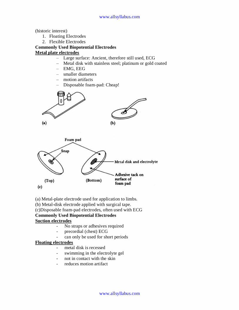
wwwallsyllabuscom
wwwallsyllabuscom
(historic interest)
1 Floating Electrodes
2 Flexible Electrodes
Commonly Used Biopotential Electrodes
Metal plate electrodes
ndash Large surface Ancient therefore still used ECG
ndash Metal disk with stainless steel platinum or gold coated
ndash EMG EEG
ndash smaller diameters
ndash motion artifacts
ndash Disposable foam-pad Cheap
(a) Metal-plate electrode used for application to limbs
(b) Metal-disk electrode applied with surgical tape
(c)Disposable foam-pad electrodes often used with ECG
Commonly Used Biopotential Electrodes
Suction electrodes
- No straps or adhesives required
- precordial (chest) ECG
- can only be used for short periods
Floating electrodes
- metal disk is recessed
- swimming in the electrolyte gel
- not in contact with the skin
- reduces motion artifact
wwwallsyllabuscom
wwwallsyllabuscom
Commonly Used Biopotential Electrodes
Flexible electrodes
- Body contours are often irregular
- Regularly shaped rigid electrodes
may not always work
- Special case infants
- Material
- Polymer or nylon with silver
- Carbon filled silicon rubber
(Mylar film)
Double-sided
Adhesive-tape
ring Electrolyte gel
in recess
(a) (b)
(c)
Snap coated with Ag-AgCl External snap
Plastic cup
Tack
Plastic disk
Foam pad
Capillary loops
Dead cellular material
Germinating layer
Gel-coated sponge
Floating Electrodes
wwwallsyllabuscom
wwwallsyllabuscom
(a) Carbon-filled silicone rubber electrode
(b) Flexible thin-film neonatal electrode
(c) Cross-sectional view of the thin-film electrode in (b)
Electrodes in Biopotential Measurements
3 Describe the construction of commercial ECG electrode (not the cheap polymer
electrode used in the lab) What is the common electrode metal and why is it preferred
So you are an inventor who has a better idea Describe an improvement
bull to make the electrode cheaper
bull more suitable for lower noise measurement for EEG
bull circumvent patents that are based on plasticfoam electrode body
bull attractive to consumers for use with their ECG machines at home
bull reduce artifact (minimize the motion of skinelectrode) in ambulatory recording
4 In a research laboratory scientists want to record from single cells in a culture dish
They want to record action potentials from single isolated heart cells What kind of
electrode would they need to use (describe material and design) Give a simplified
schematic (circuit model of the electrode) described in the notes given to you
What is the challenge involved in designing an amplifier for use with a microelectrode
for single cell recording Ie what are the critical amplifier design characteristics and
specifications (hint this is not the usual differentialinstrumentation amplifier)
Electrodes and Microelectrodes (miscellaneous)
bull How would you detect bacteria or other microorganisms in water supply Make
sure that your method distinguishes inert particulate matter from living cellular
matter
bull Draw the equivalent circuit model of the skin and an ECG electrode Identify the
key sources of electrical interference and otherwise the elements that would likely
contribute to the poor quality of recordings
wwwallsyllabuscom
wwwallsyllabuscom
bull Design an amplifier interface for the following two applications Patch clamp ion
channel current amplifier Your goal is to amplify pA level current to produce 1
Volt output
bull Strain gauge sensor amplifier Your goal is to convert 10 ohm change in
resistance of a strain gauge to produce 1 volt output
Neural electrodesmicroelectrodes
You want to record from neurons in the brain However you want to record from dozens
of neurons all at once from several closely spaced microelectrodes What material and
process would you use to make the microelectrode array
bull What metal would you prefer to use to make electrode arrays of about 10 micron
square size to make electrical contacts with dozens of neurons
bull What metal would you prefer to use to stimulate dozens of neurons in a deep
brain microelectrode based stimulator
bull (which metal provides good recording vs stimulating properties ndash and at the same
time not be toxic to brain tissue)
bull You are asked to develop an experimental set up to record from rat brain cells
using microelectrodes What precautions would you take to minimize the
electrical interference in your recording set up
Biopotential Amplifiers bull These are very important part of modern medical instrumentation bull We need to amplify biopotentials which are generated in the body at low levels with a high source impedance
bull Biopotentials amplifiers are required to increase signal strength while maintaining fidelity Basic Requirements of Biopotential Amplifiers
Essential functions of a bioamplifier are bull To take a weak biopotential and increase its amplitude so that it can be processed recorded or displayed bull To amplify voltage but it could be considered as a power amplifier as well bull To amplify current since in some cases a biopotential amplifier is used to isolate the load from the source current gain only
Input Impedance (Zin)
bull All biopotential amplifiers must have high input impedance minimize loading (remember the characteristics of biopotential electrodes resulting into loading and distortion if input impedance of the amplifier is not high enough) ndash typical values of Zin over the frequency range of the measurand = 10 MΩ (remember the loading rule)
Protection amp Isolation
bull The input circuit of a biopotential amplifier must provide protection to the live measurand
wwwallsyllabuscom
wwwallsyllabuscom
Vbio
bull Any potential or current at amplifierrsquos input terminals can affect Vbio
bull Electric currents produced by the biopotential amplifier can result in microshock and
macroshock bull The bioamplifier must have isolation and protection circuitry so that the current through the electrodes can be kept at safe levels and any artifact generated by such current can be minimized Output Impedance (Zout)
bull The output circuit does not present any critical problems all it needs to do is to drive the load bull Output impedance must be low with respect to the load impedance and it must be capable of satisfying the power requirements of the load Bandwidth (BW)
Frequency response requirements bull The biopotential amplifier must be sensitive to important frequency components of the biosignal bull Since biopotentials are low level signals it is important to limit bandwidth optimize signal-to-noise ratio Gain (G)
bull Biopotential amplifiers have a gain of 1000 or greater
Mode of Operation
bull Very frequently biosignals are obtained from bipolar electrodes bull Electrodes symmetrically located with respect to ground need differential amplification bull High CMRR required because 1 Common mode signals much greater than the biosignal appear on bipolar electrodes 2 Symmetry with respect to ground is not perfect (mismatch between electrode impedances) ndash more on this later
Calibration Signal
bull Medical and clinical equipment require quick calibration bull The gain of the biopotential amplifier must be calibrated to provide us with an accurate indication of the signalrsquos amplitude bull Push button to apply standard signal to the input of the biopotential amplifier bull Adjustable gain switch carefully selects calibrated fixed gains (in microprocessorndashbased systems gain adjustment can be Electrocardiography
hearts A very widely used medical instrument which is utilized to diagnose and monitor cardiac beat
abnormalities is the electrocardiograph
hearts It measures the electrical activity of the heart (more precisely biopotential differences arising from the electrical activity of myocardium) Wersquove already talked about the genesis of the ECG signal hearts The ECG machine uses surface electrodes and high input impedance hearts Differential amplifiers with good common mode rejection ratio to record the electrocardiogram
hearts Normal ECG amplitude ranges between 05-4 mV Normal frequency content of ECG (for diagnostic purposes) is 005-100 Hz A typical ECG waveform is shown below
wwwallsyllabuscom
wwwallsyllabuscom
Obviously all human hearts are not the same and this leads into variability in different parts of the ECG signal Significant diagnostic features of the ECG signal are hearts Duration of component parts of the signal
hearts Polarities and magnitudes hearts The details of the ECG signal and the degree of variability in different parts of the ECG signal is shown below
The QRS amplitude polarity time duration the RR interval (indicator of heartbeat per min) and the T-wave amplitude are some very important and distinctive features of the ECG signal The heart rate in BPM = Beats Per Minute) is simply = 60 (RR interval in seconds)
Some ECG waveform abnormalities that may indicate illness are hearts An extended PR interval may be diagnosed as AV node block hearts A widening of the QRS complex may indicate conduction problems in the bundle of His hearts An elevated ST segment may indicate occurrence of myocardial Infarction (MI) hearts A negative polarity in the T wave may be due to coronary insufficiency ECG Leads
Normal ECG recordings for the standard lead connections leads I II and III (Lead II provides
the strongest signal) Obviously all human hearts are not the same and this results into a high degree of variability
wwwallsyllabuscom
wwwallsyllabuscom
hearts Note the degree of variability of different parts of the ECG Signal
Some abnormalities that may indicate illness hearts An extended P-R interval may be diagnosed as AV node block hearts Widening of the QRS complex conduction problems in the bundle of His hearts Elevated ST segment may indicate occurrence of MI
hearts Negative polarity T wave may be due to coronary insufficiency QRS amplitude polarity time domain PR interval (indicator of heat beat per min amp T-wave amplitude are some very important
distinctive features 1 Loss
2 Origin of the ECG signal
hearts We have already covered this concept extensively in the previous lectures (The Dipole filed of the heart the Eindhovenrsquos Triangle the electrical circuit model for the electrocardiographic problem etc)
hearts Standard Limb Leads (I II III)
wwwallsyllabuscom
wwwallsyllabuscom
hearts The The lead wires are color-coded according to some conventions One example is White ndashRA
(Right Arm) Black ndash LA (Left Arm) Green ndash RL (Right Leg) Red ndash LL (Left Leg) and Brown
ndash C (Chest) Note There is a CM (common mode) amplifier connected to the right leg We will discuss this in detail later Augmented Limb Leads
These leads offer a free 50 increase over leads VR VL and VF connections (unipolar leads) with respect to Wilson terminal AVR = -I ndash III2 AVL = I ndash II2 aVF = II ndash I2
wwwallsyllabuscom
wwwallsyllabuscom
Each measurement is made from the reflected limb and the average of the other two limbs Chest Leads (Precordial)
Chest Lead Anatomical Positions
V1 - 4th intercostal space ndash Right sternal margin
V2 - 4th intercostal space ndash Left sternal margin
V3 ndash Midway between V2 amp V4
V4 ndash 5th intercostal space on mid-clavicular line
V5 ndash Same as V4 on the anterior axillary line
V6 ndash Same as V5 on the mid-axillary line
12 Lead Clinical Electrocardiography
The ECG Machine
Most representative Specs
wwwallsyllabuscom
wwwallsyllabuscom
bull Zin = 10 MΩ bull Frequency response = 005 ndash100 Hz bull Strip Chart Recorder Speed = 25 mmsec bull Fast Speed = 100 mmsec
For detailed Specs Refer to the Table in your text ―Summary of performance requirements for electrocardiographs Location of the Heart bull The heart is located between the lungs behind the sternum and above the diaphragm bull It is surrounded by the pericardium bull Its size is about that of a fist and its weight is about 250-300 g bull Its center is located about 15 cm to the left of the midsagittal plane
Anatomy of the heart
bull The walls of the heart are composed of cardiac muscle called myocardium
bull It consists of four compartments ndash the right and left atria and ventricles
The Heart Valves
bull The tricuspid valve regulates blood flow between the right atrium and right
ventricle
bull The pulmonary valve controls blood flow from the right ventricle into the
pulmonary arteries
bull The mitral valve lets oxygen-rich blood from your lungs pass from the left atrium
into the left ventricle
bull The aortic valve lets oxygen-rich blood pass from the left ventricle into the aorta
then to the body
Blood circulation via heart
wwwallsyllabuscom
wwwallsyllabuscom
bull The blood returns from the systemic circulation to the right atrium and from
there goes through the tricuspid valve to the right ventricle
bull It is ejected from the right ventricle through the pulmonary valve to the lungs
bull Oxygenated blood returns from the lungs to the left atrium and from there
through the mitral valve to the left ventricle
bull Finally blood is pumped through the aortic valve to the aorta and the systemic
circulation
Electrical activation of the heart
bull In the heart muscle cell or myocyte electric activation takes place by means of
the same mechanism as in the nerve cell ie from the inflow of Na ions across
the cell membrane
bull The amplitude of the action potential is also similar being 100 mV for both nerve
and muscle
bull The duration of the cardiac impulse is however two orders of magnitude longer
than in either nerve cell or sceletal muscle cell
bull As in the nerve cell repolarization is a consequence of the outflow of K ions
bull The duration of the action impulse is about 300 ms
Mechanical contraction of Cardiac Muscle
bull Associated with the electric activation of cardiac muscle cell is its mechanical
contraction which occurs a little later
bull An important distinction between cardiac muscle tissue and skeletal muscle is
that in cardiac muscle activation can propagate from one cell to another
in any direction
bull Electrical signal begins in the sinoatrial (SA) node natural pacemaker causes
the atria to contract
bull The signal then passes through the atrioventricular (AV) node
ndash sends the signal to the ventricles via the ―bundle of His
ndash causes the ventricles to contract
The Conduction System
wwwallsyllabuscom
wwwallsyllabuscom
The Action Potential
Recording an AP requires the isolation of a single cell Microelectrodes (with tips a few μm across) are used to stimulate and record the response A typical AP is 2-4ms long with an amplitude of about 100Mv The Electroencephalogram EEG
EEG is the graphical representation of the electrical activity of the brain Very commonly used to diagnose certain neurological disorders such as epilepsy
More recently also investigated whether it can detect various forms of dementia or schizophrenia EEG is the specific recording obtained using the scalp electrodes from the surface of the skull
During surgery electrodes may also be placed directly on the cortex The resulting signal is then electrocorticogram (ECoG) Just like ECG EEG is also obtained using several different electrodes places on different regions of the head brain
The Event Related
wwwallsyllabuscom
wwwallsyllabuscom
Potentials ndash ERPs
ERPs are really EEGs obtained under a specific protocol that requires the patient to response to certain stimuli ndash hence event related potentials Also called evoked potentials these signals can be used to diagnose certain
neurological disorders such as dementia and they can also be used as a liedetector bull The oddball paradigm
bull The guilty knowledge test
Electroretinogram ERG
The ERG is the record of the retinal action currents produced by the retina in response to a light stimulus It measures the electrical responses of the light-sensitive cells (such as rods and cones)
The stimuli are often a series of light flashes or rotating patterns The ERG is recorded using contact lens electrode that the subject wears while watching the stimuli
Phonocardiogram ndash PCG
The PCG is the graphic record of the heart sounds and murmurs It is thus a mechanical audio signal rather than an electrical signal Can be easily heard using a stethoscope Or can be converted into an electrical signal using a transducer Typically used to determine the disorders related to the heart valve since their
routine opening and closing create the well-known sounds bull S1 sounds First heart sounds ndash ventricular contractions move blood into atria closing of the AV (mitral and tricuspid) valves then semilunar valves open and blood ejected out of ventricles ndash immediately follows the QRS complex bull S2 sounds Second heart sounds ndash Closure of semilunar (aortic and pulmonary) valves bull Any unexpected sound may indicate a malfunctioning valve that causes the blood flow into out of a chamber when it should not Also called heart murmurs
wwwallsyllabuscom
wwwallsyllabuscom
Define ultrasound
bull Mechanical waves in different modalities (longitudinallateral) needs medium
to be propagated (solid liquid gas)
bull gt 20 kHz
bull Continuouspulsed
bull Sphericalplanarnarrow beamsurface waveLamb-wave
Physical phenomena behind ultrasound measurements
Transmission
bull
bull reflection
bull transit time
bull differences in propagation velocities
bull returns to transit time
bull doppler-shift in frequency
bull flow velocity
bull change of acoustic impedance
bull comparing to reference
bull interference of ultrasound waves (holography)
bull interaction of ultrasound and light (photoacousticz)
bull ultrasound needs medium for propagation it doesnrsquot propagate in vacuum
bull because mechanical waves need moving massunits and spring forces between
them
bull in acoustic emission the medium creates ultrasound (for example during pressure
changes) which is received by sensors
bull pulsed mode more common than continuous
bull continuous reguires separate transducers for transmitting and receicving
bull in pulsed mode an ultrasound burst is sent to the object and the same transducer is
switched to listen echoes
bull standing wave problem
bull in us-therapy pulsed mode gives more effective care without too much heating The Doppler Equation describes the relationship of the Doppler frequency shift to target velocity
wwwallsyllabuscom
wwwallsyllabuscom
The frequency difference is equal to the reflected frequency (FR) minus the originating frequency (FT) If the resulting frequency is higher then there is a positive Doppler shift and the object is moving toward the transducer but if the resulting frequency is lower there is a negative Doppler shift and it is moving away from the transducer In its simplest form it would be calculated as if
the ultrasound was parallel to the targetrsquos direction as shown in diagram A below However this would be a rare occurrence in clinical practice because the transducer is rarely pointed head on to a blood vessel In real life the ultrasound waves would approach the target at an angle called the Doppler angle ( ) On the following page diagram B shows the Doppler
equation used in general clinical situations which includes the Doppler angle The Doppler Angle
The ultrasound beam usually approaches the moving target at an angle called the Doppler angle ( ) This reduces the frequency shift in proportion to the cosine of this
angle If this angle is known then the flow velocity can be calculated The equation used is The Doppler Equation
equiv Doppler shift frequency (the difference between the transmitted and received frequencies) equiv transmitted frequency equiv reflected frequency
V equiv velocity of the blood flow towards the transducer C equiv velocity of sound in tissue θ equiv the angle between the sound beam and the direction of moving blood Where
The Doppler angle ( ) is also known as the angle of insonation It is estimated by the sonographer by a process known as angle correction which involves aligning an indicator on the duplex image along the longitudinal axis of the vessel
There are a few considerations that affect the performance of a Doppler examination that are inherent in the Doppler equation which are ndash The cosine of 90deg is zero so if the ultrasound beam is perpendicular to the direction of blood flow there will be no Doppler shift and it will appear as if there is no flow in the vessel ndash Appropriate estimation of the angle of insonation or angle correction is essential for the accurate determination of Doppler shift and blood flow velocity The angle of insonation should also be less than 60deg at all times since the
cosine function has a steeper curve above this angle and errors in angle correction will be magnified
The simplest Doppler devices use continuous wave (CW Doppler) rather than the pulsed wave used in more complex devices CW Doppler uses two transducers (or a dual element transducer) that transmit and receive ultrasound continuously The transmit and receive beams overlap in a Doppler sample volume some distance from the transducer face as
shown in the diagram below
volume) is the region of transmitting and receiving beam overlap (shaded region) Because there is continuous transducer transmission and reception echoes from all depths within the area arrive at the transducer simultaneously So although CW Doppler can determine the direction of flow it cannot discriminate the different depths where the motion originates The usefulness of CW Doppler devices is limited but they are used clinically to confirm blood flow in superficial vessels as they are good at detecting low velocities As they are easily portable this
can be done at the bedside or in the operating room Most other clinical applications require pulsed wave Doppler Pulsed Wave Doppler (PW Doppler)
wwwallsyllabuscom
wwwallsyllabuscom
Pulsed wave Doppler (PW Doppler) uses a single-element transducer that emits brief pulses of ultrasound energy The time interval between transmitting and then receiving the echoing sound can be used to calculate the depth from where the echo arises The Doppler sample volume can be chosen as to shape depth and position in sampling the flow
data For example the depth is chosen by processing only the signals that return to the transducer in a stipulated time For this technique the ultrasound system transmits a short pulse The eceiver is opened to detect the returning echoes only after a controlled delay and only for a specific duration This time-based gating of the receiving channel allows the definition of a fixed easuring distance which is often referred to as the Sample volume or Doppler gate Then the next ultrasound wave is transmitted The number of pulses transmitted by the system within a second is referred to as the pulse repetition frequency (PRF) The upper PRF limit is given by the time interval required for the echoes to arrive from a sample volume located at a
certain depth The greater the sample-volume depth the longer the time before the echoes are returned and the longer the delay between pulse transmission The greater the samplevolume depth the lower will be the maximum PRF setting Errors in the accuracy of the information arise if the velocities exceed a certain speed The highest velocity accurately measured is called the Nyquist limit Beyond this limit the errors that occur are referred to as aliasing Volume and flow measurement
Flow ndash volume of a liquidgas passing some point over a given time Gases are compressible
Benedict Roth Spirometer
Widely used for physiological amp clinical studies
Light bell moves with the ptrsquos breathing
Movement recorded by a pen on a rotating drum
Water seal prevents leakage of gas
Small seal minimises volume of gas dissolved in water
Suitable for measuring limited gas vols (few litres)
Pneumotachograph
wwwallsyllabuscom
wwwallsyllabuscom
Cardiac Output
Defn vol of blood pumped by the heart per min
CO = SV x HR
Norm ~ 5 lmin
Cardiac index ndash corrected for body surface area
Affected by
Met Rate ndash pregnancy hyperthyroid septic
Preload contractility afterload
Clinical indicators of CO imprecise
Affected by anaesthetic agents used in everyday practice
Provides estimate of
whole body perfusion
oxygen delivery
left ventricular function
Persistently low CO assoc with poor outcome
Methods
Fick method
Dilution techniques ndash dye thermal lithium
Pulse contour analysis- LiDCO amp PiCCO
Oesophageal doppler
TOE
Transthoracic impedance plethysmography
Inert gas through flow
Non-invasive cardiac output measurement
Fick Principle measure volume displacement
1st proposed 1870
―the total uptake or release of a substance by an organ is the product of the
blood flow through that organ and the arteriovenous concentration
difference of the substance
CO = O2 consumption (mlmin) art ndash mixed
venous O2 conc (mll)
Limited by cumbersome equipment sampling errors need for invasive
monitoring and steady-state haemodynamic and metabolic conditions
Indicator dilution techniques
―An indicator mixed into a unit volume of constantly flowing blood can be used to
identify that volume of blood in time provided the indicator remains in the system
between injection and measurement and mixes completely in the blood
wwwallsyllabuscom
wwwallsyllabuscom
Dye dilution
Inert dye ndash indocyanin green
Injected into pulmonary artery and arterial conc measured using a
calibrated cuvette densitometer
Plot indicator dilution curve (see diagram)
CO derived from area under curve
Indicator Dilution Curve
Cardiac Output Measurement
wwwallsyllabuscom
wwwallsyllabuscom
Why bother to check blood pressure
For each 20 mm rise in systolic blood pressure or 10 mm rise in diastolic blood
pressure over 11575
Risk of stroke increases
Risk of heart disease doubles
Risk of renal failure increases
Systolic and diastolic blood pressure
Systolic blood pressure is the highest pressure in the arteries just after the heart
beats
Diastolic blood pressure is the lowest pressure in the arteries just before the heart
beats
Blood pressure is measured indirectly by blood pressure cuff
(sphygmomanometer)
Inflating cuff increases pressure until it cuts off arterial circulation to the arm
Deflating cuff decrease pressure by 2 to 3 mm of mercury per second until blood
first enters the artery creating turbulence this causes a sound with each heartbeat
Sounds continue with each heartbeat until pressure lowers to the lowest pressure
in the artery then turbulence stops so the sound stops
Systolic blood pressure is the cuff pressure at the first sounds diastolic is the cuff
pressure just before the sounds stop
Phase 1 sharp thuds start at systolic blood pressure
Phase 2 blowing sound may disappear entirely (the auscultatory gap )
Phase 3 crisp thud a bit quieter than phase 1
Phase 4 sounds become muffled
Phase 5 end of sounds -- ends at diastolic blood pressure
wwwallsyllabuscom
wwwallsyllabuscom
Make sure the cuff is the right size - its width should be at least 40 of the arms
circumference The cuff will overestimate blood pressure if too small and
underestimate if too large
Place the cuff snugly on patients proximal arm on skin ( not cloth) centered over
the brachial artery (most cuffs have markings)
Support the patients arm at heart level using your arm or a desk
Your patient should sit in the chair for 5 minutes before BP is measured and
should have no caffeine or nicotine for 30 minutes before (JAMA 273 p1211-
1218 1995)
With fingers palpating radial or brachial artery inflate cuff rapidly until you cant
feel the pulse then 20 mm higher
Release cuff at 2 to 3 mm Hg per second until you again feel the pulse this is the
palpable systolic pressure
Wait 30 seconds before measuring blood pressure
Measuring palpable pressure first avoids risk of seriously underestimating blood
pressure because of the auscultatory gap (mistaking Korotkoff phase 3 for phase
1) Many doctors skip this step for time reasons and instead pump cuff to 200 mm
Hg at the next step)
Phase 2 of the Korotkoff sounds can be inaudible - especially in older patients
with systolic hypertension who are at especially high risk of stroke Inflating the
cuff until you dont hear sounds can give you a reading of 14086 when the
patients actual blood pressure is 22086 Most physicians are pressed for time so
they instead inflate the cuff to 200 mm which is beyond the auscultatory gap in
most patients But palpable systolic blood pressure is according to research more
reliable
Place bell of stethoscope (diaphragm is acceptable) over brachial artery
Rapidly pump the cuff to 20 to 30 mm Hg above palpable systolic pressure
Release pressure in the cuff by 2 to 3 mm Hg per second and listen for Korotkoff
sounds including systolic (first) and diastolic (last)
Record as systolicdiastolic Check in both arms the first time you check a
patients blood pressure It may differ by 10 mm Hg or more
If the sounds continues to zero record diastolic blood pressure as the point when
sounds become muffled (phase 4) over zero eg 130700 or just as 13070
Rate
ndash Number of beats in 30 seconds x 2
Strength
ndash Bounding strong or weak (thready)
Regularity
ndash Regular or irregular
You need three readings on two occasions to diagnose hypertension unless blood
pressure is very high
Normal blood pressure in children is
wwwallsyllabuscom
wwwallsyllabuscom
ndash 10255 at 1 year 11269 at 5 years 11978 at 10 years
Blood pressures in adults (JNC VII JAMA 2892560-72 2003)
ndash Normal lt120lt80
ndash Prehypertensive 120-13980-89
ndash Stage 1 hypertension 140-15990-99
ndash Stage 2 hypertension gt160gt100
Adult 60 to 100
Newborn 120-170
1 year 80-160
3 years 80-120
6 years 75-115
10 years 70-110
How to measure observe rise and fall of chest
In infants count for 60 seconds in adults 15 or 30 seconds
Normal respiration Adults 12 to 20
Children
newborn 30-80
1 year 20-40
3 years 20-30
6 years 16-22
Rate
Number of breaths in 30 seconds x 2
Quality
Character of breathing
Rhythm
Regular or irregular
Effort
Normal or labored
Noisy respiration
Normal stridor wheezing snoring gurgling
Depth
Shallow or deep
pH electrode
wwwallsyllabuscom
wwwallsyllabuscom
Governing equation is the Nernst Equation
pCO2 Electrode
The measurement of pCO2 is based on its linear relationship with pH over the range of
10 to 90 mm Hg
The dissociation constant is given by
Taking logarithms
pH = log[HCO3-] ndash log k ndash log a ndash log pCO2
Diathermy
In the natural sciences the term diathermy means electrically induced heat and is commonly
used for muscle relaxation It is also a method of heating tissue electromagnetically or
ultrasonically for therapeutic purposes in medicineContents
[hide]
1 Heating uses 2 Surgical uses
3 Trivia
4 References
E
RT
nF
H
HH
i
ln
0
H O CO H CO H HCO2 2 2 3 3
k
H HCO
a pCO
3
2
wwwallsyllabuscom
wwwallsyllabuscom
Heating uses
Ultrasonic diathermy refers to heating of tissues by ultrasound for the purpose of
therapeutic deep heating No tissue is ordinarily damaged hence it is generally used in
biomedical applications
Electric diathermy uses high frequency alternating electric or magnetic fields sometimes
with no electrode or device contact to the skin to induce gentle deep tissue heating by
induction Again no tissue is ordinarily damaged
Surgical uses
Surgical diathermy is usually better known as electrosurgery (It is also referred to
occasionally as electrocautery but see disambiguation below) Electrosurgery and
surgical diathermy involve the use of high frequency AC electrical current in surgery as
either a cutting modality or else to cauterize small blood vessels to stop bleeding This
technique induces localized tissue burning and damage the zone of which is controlled
by the frequency and power of the device Some sources[1]
insist that electrosurgery be
applied to surgery accomplished by high frequency AC cutting and that
electrocautery be used only for the practice of cauterization with heated nichrome wires
powered by DC current as in the handheld battery-operated portable cautery tools
Trivia
Medical Diathermy devices were used to cause interference to German radio beams used
for targeting night time bombing raids in WWII during the Battle of the Beams
I Diathermy
A Therapeutic use
1 Generation of local heating by high-frequency electromagnetic waves
2 Capacitance techniquemdashbody is placed in an electric field
a Dipolesmdashstructures with positive and negative poles
b Structures with large numbers of dipoles have a greater capacitance to store an electrical charge
c Greatest heating occurs in tissues with fewer dipoles particularly fatty tissues
3 Rapid rotation of dipoles causing mechanical friction and movement of electrons results in local heating
4 Inductance techniquemdashbody is not placed in an electric field
a Magnetic waves generated by driving current through a coiled wire
b Magnetic field creates currents in tissues
wwwallsyllabuscom
wwwallsyllabuscom
c Greatest heating occurs in tissues with low impedance especially muscle
B Precautions and contraindications
1 Diathermy should not be used
a Over metal implants and cardiac pacemakersmdashmore research needed regarding its use over metallic fixations
b Near the uterus of a pregnant woman or near the abdomen or back of a woman who might be pregnant
c On individuals with infections
d On individuals with acute inflammation
e Over moist open wounds
f On patients with malignant tumors
g Over large joint effusions
C Pulsed electromagnetic fields and diathermy
1 Can be pulsed to decrease total energy transmitted to the tissues
2 Short-wave diathermy can be adjusted into a nonthermal range
a Classified as pulsed electromagnetic field (PEMF) or
b Pulsed radio frequency energy (PRFE)
3 Important reclassification as diathermy implies heating
D Efficiency of diathermy and PEMF therapy for musculoskeletal conditions
1 Current research is limited but results suggest that diathermy enhances
treatments directed at soft tissue stretching
2 Some studies suggest that PEMF may speed wound healing and promote healing of nonunion fractures
Diathermy is a modality that uses electromagnetic energy to heat deeper tissues
Diathermy is more effective than ultrasound at heating a larger area of deep tissues
The athletic trainer must identify and respect contraindications to application of ultrasound and diathermy
Pulsed ultrasound and diathermy are used to treat slow-to-heal lesions including skin ulcers and nonunion fractures and may be able to facilitate repair of other tissues including ligaments and tendons
Diathermy
Definition
In diathermy high-frequency electrical currents are used to heat deep muscular tissues The heat increases blood flow speeding up recovery Doctors also use diathermy in surgical procedures by sealing blood vessels with electrically heated probes
The term diathermy is derived from the Greek words therma meaning heat and dia meaning through Diathermy literally means heating through
Origins
The therapeutic effects of heat have long been recognized More than 2000 years ago the Romans took advantage of heat therapies by building hot-spring bathhouses Since then various
wwwallsyllabuscom
wwwallsyllabuscom
methods of using heat have evolved In the early 1890s French physiologist Arseacutene dArsonval began studying the medical application of high-frequency currents The term diathermy was coined by German physician Carl Franz Nagelschmidt who designed a prototype apparatus in 1906 Around 1925 United States doctor J W Schereschewsky began studying the physiological
effects of high-frequency electrical currents on animals It was several years however before the fundamentals of the therapy were understood and put into practice
Benefits
Diathermy can be used to treat arthritis bursitis and other conditions involving stiff painful joints It is also used to treat pelvic infections and sinusitis A benefit of diathermy is that it is a painless procedure that can be administered at a clinic Also if the treatment relieves pain then patients can discontinue pain killers and escape their high cost and side effects
Description
Diathermy involves heating deep muscular tissues When heat is applied to the painful area cellular metabolism speeds up and blood flow increases The increased metabolism and circulation accelerates tissue repair The heat helps the tissues relax and stretch thus alleviating
stiffness Heat also reduces nerve fiber sensitivity increasing the patients pain threshold
There are three methods of diathermy In each energy is delivered to the deep tissues where it is converted to heat The three methods are
Shortwave diathermy The body part to be treated is placed between two capacitor plates Heat is generated as the high-frequency waves travel through the body tissues between the plates Shortwave diathermy is most often used to treat areas like the hip which is covered with a dense tissue mass It is also used to treat pelvic infections and sinusitis The treatment reduces inflammation The Federal Communications Commission regulates the frequency allowed for short-wave diathermy treatment Most machines
function at 2733 megahertz Ultrasound diathermy In this method high-frequency acoustic vibrations are used to
generate heat in deep tissue Microwave diathermy This method uses radar waves to heat tissue This form is the
easiest to use but the microwaves cannot penetrate deep muscles
Diathermy is also used in surgical procedures Many doctors use electrically heated probes to seal blood vessels to prevent excessive bleeding This is particularly helpful in neurosurgery and eye surgery Doctors can also use diathermy to kill abnormal growths such as tumors warts and
infected tissues
Preparations
To keep patients from sweating patients are usually asked to remove clothing from the body part being treated If a patient sweats the electrical currents may pool in the area causing burns Also clothing containing metal must be removed as must earrings buttons barrettes or zippers that contain metal Watches and hearing aids should be removed because the therapy may affect their function
wwwallsyllabuscom
wwwallsyllabuscom
Practitioners of surgical diathermy should steer clear of alcohol-based solutions to prepare and cleanse the skin These preparations can create a flammable vapor and cause burns and fires
Precautions
Patients with metal implants should not undergo diathermy treatment because the metal can act as a conductor of heat and result in serious internal burns Female patients with metallic uterine implants such as an IUD should avoid treatment in the pelvic area Diathermy should not be
used in joints that have been replaced with a prosthesis or in those with sensory impairment who may not be able to tell if they are burning Furthermore pulsed shortwave diathermy should be avoided during pregnancy as it can lead to abnormal fetal development
Patients with hemophilia should avoid the treatment because the increased blood flow could cause them to hemorrhage
Side effects
Some patients may experience superficial burns Since the therapy involves creating heat care must be taken to avoid burns particularly in patients whose injuries have caused decreased sensitivity to heat Also diathermy may affect pacemaker function
Female patients who receive treatment in the lower back or pelvic area may experience an
increased menstrual flow
Research amp general acceptance
For years physiotherapists and physical therapists have used diathermy as a routine part of physical rehabilitation
Electrical Safety
Electrical safety is very important in hospitals as patients may be undergoing a diagnostic or treatment procedure where the protective effect of dry skin is reduced Also patients may be unattended unconscious or anaesthetised and may not respond normally to an electric current Further electrically conductive solutions such as blood and saline are often present in patient treatment areas and may drip or spill on electrical equipment
Electric Current Leakage Current
Extension Leads Double Adaptors Equipment Classification
Class I Class II Defibrillator-Proof
Protective Devices
Residual Current Devices (RCD) Line Isolation overload Monitors (LIMs) Equipment Earthing
Area Classification
wwwallsyllabuscom
wwwallsyllabuscom
Body Protection Area Cardiac Protected Area
Other Electrical Issues Extension Leads
Double Adapters Main Extension Devices Power Boards Installation of Additional Power Points
Electric Current
Injuries received from electric current are dependent on the magnitude of current the pathway
that it takes through the body and the time for which it flows
The nature of electricity flowing through a circuit is analogous to blood flowing through the circulatory system within the human body In this analogy the source of energy is represented by the heart and the blood flowing through arteries and veins is analagous to current flowing
through the conductors and other components of the electric circuit
The application of an electric potential to an electric circuit generates a flow of current through conductive pathways This is analogous to the changes in blood pressure caused by contraction of cardiac muscle that causes blood to flow into the circulatory system For electric current to flow there must be a continuous pathway from the source of potential through electrical components and back to the source
Leakage Current
Electrical components and systems are encased in non conducting insulation to ensure that the electric current is contained and follows the intended pathways If the insulation deteriorates or breaks down current will leak through the insulation barrier and flow to earth This may
be through the protective earth conductor or through the operator
Medical equipment and clinical areas are fitted with a number of protective devices to protect the patient and operator from harmful leakage currents
Extension Leads
Extension leads are not permitted in clinical areas of RCH organisations They may cause high
earth resistance and excessive earth leakage current An extension lead can allow equipment to be powered from areas other than the relevant protected treatment area The power from the other area may not be protected to the same level as the power in the treatment area
wwwallsyllabuscom
wwwallsyllabuscom
As the connection between the extension lead and the equipment mains cable is often on the floor there is a high danger from fluid spills tripping and damage to the mains cable by trolleys when an extension lead is used
Double Adaptors
Double adaptors must not be used in RCH organisations They may not sit securely in a wall
outlet may not be able to provide adequate earth protection and may cause overloading overheating fire or loss of electrical supply
EQUIPMENT CLASSIFICATIONS
There are several methods of providing protection for operators and patients from electrical faults
and harmful leakage current
Class I
Class I equipment is fitted with a three core mains cable containing a protective earth wire
Exposed metal parts on class I equipment are connected to this earth wire
Should a fault develop inside the equipment and the exposed metal comes into contact with the mains the earthing conductor will conduct the fault current to ground Regular testing procedures ensure that earthing conductors are intact as the integrity of the earth wire is of vital importance
Class II
Class II equipment is enclosed within a double insulated case and does not require earthing
conductors Class II equipment is usually fitted with a 2-pin mains plug An internal electrical fault is unlikely to be hazardous as the double insulation prevents any external parts from becoming alive Class II or double insulated equipment can be identified by the class II symbol on the cabinet
Class II Symbol
wwwallsyllabuscom
wwwallsyllabuscom
Defibrillator-Proof
Some medical equipment within the hospital is classified as defibrillator proof When a
defibrillator is discharged through a patient connected to defibrillator proof equipment the equipment will not be damaged by the defibrillators energy Defibrillator proof equipment can remain connected to the patient during defibrillation It is identified by one of the following
symbols
Defibrillator proof symbols
Body protected Cardiac protected
PROTECTIVE DEVICES
Most patient care areas in the hospital are fitted with protective devices These devices are
regularly tested in accordance with the relevant guidelines published by Standards Australia The level of protection provided is dependent upon the device and the area in which it is located
Residual Current Devices (RCD)
RCDs (safety switches) are used in patient treatment areas to monitor and protect the mains supply RCDs sense leakage currents flowing to earth from the equipment If a significant leakage current flows the RCD will detect it and shut off the power supplied to the equipment
within 40 milliseconds Hospital RCDs are more sensitive than those fitted in homes A hospital RCD will trip at 10 milliamperes leakage current
Power outlets supplied through an RCD have a Supply Available lamp The lamp will extinguish
when the RCD trips due to excessive leakage current
wwwallsyllabuscom
wwwallsyllabuscom
Resetting a RCD
Lamp indicates supply is no longer available Disconnect all equipment from the supply Operate the reset button or lever on the supply panel and the Supply Available lamp
should illuminate If not contact Biomedical Engineering
Connect an item of equipment If the RCD trips again then this is the faulty item and should be labelled and sent to Biomedical Engineering
If the RCD does not trip continue connecting equipment until the RCD trips The last piece of equipment connected to the supply is most likely to be faulty as it will have caused the RCD to trip Remove the faulty item from service label it and send to Biomedical Engineering as mentioned above
Line Isolation overload Monitors (LIMs)
In critical life support applications where loss of power supply cannot be tolerated special power outlets powered by isolation transformers are installed
Line Isolation Monitors are installed to continually monitor electrical leakage in the power
supply system If an electrical fault develops in a medical device connected to an isolated power outlet the LIM will detect the leakage current The LIM will alarm and indicate the level of leakage current but will not shut off the electric supply
The faulty equipment can be identified by un plugging one item of equipment at a time from the supply until the alarm stops sounding Equipment that is not faulty may be reconnected Faulty equipment should be appropriately labelled and sent to Biomedical Engineering for repair
The LIM also monitors how much power is being used by the equpiment connected to it If too much power is being used the LIM will alarm and indicate that there is an overload The power used must be reduced immediately by moving some equipment to another circuit as soon as possible until the alarm stops sounding Failure to reduce the load on the LIM will result in the circuit breaker tripping and loss of power to the circuit
wwwallsyllabuscom
wwwallsyllabuscom
Equipotential Earthing
Equipotential earthing is installed in rooms classified as Cardiac Protected electrical areas
Equipotential earthing in treatment areas used for cardiac procedures is intended to minimise any voltage differences between earthed parts of equipment and any other exposed metal in the room
This reduces the possibility of leakage currents that can cause microelectrocution when the patient comes into contact with multiple items of equipment or if the patient happens to come
into contact with metal items in the room whilr they are connected to a medical device
All conductive metal in an equipotential area is connected to a common equipotential earth point with special heavy duty cable
AREA CLASSIFICATIONS
Body Protected Area
These areas are designed for procedures in which patients are connected to equipment that lowers the natural resistance of the skin Applied parts such as electrode gels conductive fluids entering the patient metal needles and catheters provide an easy pathway for current to flow
The main occurrence of injury from Body-Type procedures is from high current levels causing electric shock A direct connection to the patients heart is not present so the risk of Microelectrocution - fibrillation from minute current levels - is reduced
Residual Current Devices (RCD) or Isolation Transformers and Line Isolation Monitors (LIMiacutes) are used in Body Protected areas to provide protection against electrocution from high leakage currents Body-Protected Areas are identified with this sign
Cardiac Protected Area
Where the procedure involves placing an electrical conductor within or near the heart protection against fibrillation induced from small leakage currents is required Electrical conductors used in these procedures include cardiac pacing electrodes intracardiac ECG electrodes and intracardiac catheters
wwwallsyllabuscom
wwwallsyllabuscom
Equipotential earthing in conjunction with RCDs or LIMs provides protection against microelectrocution in Cardiac-Type procedures
Fault currents are reduced to magnitudes that are unlikely to induce fibrillation Used in conjunction with RCDs or LIMs the magnitude and duration of any fault currents
sourced from equipment are limited Cardiac-Protected Areas are identified with this sign
Other electrical issues
This policy aims to provide guidance to those who find that they need more electrical outlets than
those available or that the existing electrical outlets are inconveniently located
As extension leads and multiple outlet power boards can introduce additional hazards into an area the following procedures should be observed
Extension leads
Approved extension leads (AS 3760 1996) may be used in some areas within the hospital but MUST NOT BE USED IN PATIENT AREAS All electrical extension leads must be tagged with an Engineering Department maintenance tag and require a yearly safety inspection and test via
the Engineering Department
Double adapters
Double adapters may cause overloading or equipment earthing problems and are not to be used in
WCH
Mains extension device
The only mains extension device that is to be used in Patient care areas is the 4-way or 8-way
portable Core Balance Unit
The Biomedical Engineering Department must approve all units prior to use These units contain a safety switch and can detect excessive leakage current and disconnect the power in the event of a hazardous situation
wwwallsyllabuscom
wwwallsyllabuscom
Care must be exercised in the use of a portable Core Balance Unit It should be located off the
floor and in a position that will protect it from physical abuse and possible entry of fluids These devices are expensive and easily damaged The device must be sent to Biomedical Engineering every 6 months for safety testing
Power boards
Approved multiple-outlet power boards can be used across RCH but must not be used in patient
care areas except areas approved by the Biomedical Engineering Department
The power boards must have overload protection be fitted with internal safety shutters that protect unused outlets and be fitted with an onoff switch for each outlet
Medical Laser Applications
The main research subjects of the group of Medical Laser Application are in the field of online
monitoring and diagnostics as well as the development of new therapheutic methods The main focus in research is based upon the use of ultrashort (fs) laser pulses
The main advantage of ultrashort laser pulses is the extrem short interaction time which suppress any unwanted side effects of the laser irradiation of the tissue Secondly the broad spectra of the fs laser pulses give the advantage to use the same laser pulses for diagnostic applications like the optical coherence tomography (OCT)
wwwallsyllabuscom
wwwallsyllabuscom
Following research subjects have special attention at the moment
Optical Coherence Tomographie (OCT)
A fs-laser light source can be used to obtain images from inside the tissue when the pulses are coherent superposed This group uses this application to obtain an inside view of the vocal fold and to measure the outline of the crystalline lens
Refractive Surgery
The principle of getting rid of glasses with the help of a laser surgery (fs-LASIK) is underlying rapid improvements concerning precision and safety New technologies were evaluated and transferred very fast in cooperation with an industrial partner
Treatment of Presbyopia
The flexibility of the crystalline lens can be increase by pricise cuts which are induce by fs laser pulse inside the lens The treatment of the presbyopia is thinkable
wwwallsyllabuscom
wwwallsyllabuscom
Ultrafast Physics
Whereas the applications of ultrashort laser pulses increase rapidly is the physics of the interaction between the laser pulses and tissue in many case not fully understood To obtain improved knowledge of the interaction for many fs-laser application this group preforms numerical simulations as well as fundamental experiments
Thermography
Thermography thermal imaging or thermal video is a type of infrared imaging Thermographic cameras detect radiation in the infrared range of the electromagnetic spectrum
(roughly 900ndash14000 nanometers or 09ndash14 microm) and produce images of that radiation Since infrared radiation is emitted by all objects based on their temperatures according to the black body radiation law thermography makes it possible to see ones environment with or without visible illumination The amount of radiation emitted by an object increases with temperature therefore thermography allows one to see variations in temperature (hence the name) When viewed by thermographic camera warm objects stand out well against cooler backgrounds humans and other warm-blooded animals become easily visible against the environment day or
night As a result thermographys extensive use can historically be ascribed to the military and security services
Thermal imaging photography finds many other uses For example firefighters use it to see through smoke find persons and localize the base of a fire With thermal imaging power lines maintenance technicians locate overheating joints and parts a telltale sign of their failure to eliminate potential hazards Where thermal insulation becomes faulty building construction technicians can see heat leaks to improve the efficiencies of cooling or heating air-conditioning Thermal imaging cameras are also installed in some luxury cars to aid the driver the first being
the 2000 Cadillac DeVille Some physiological activities particularly responses in human beings and other warm-blooded animals can also be monitored with thermographic imaging [1]
The appearance and operation of a modern thermographic camera is often similar to a camcorder Enabling the user to see in the infrared spectrum is a function so useful that ability to record their output is often optional A recording module is therefore not always built-in
Instead of CCD sensors most thermal imaging cameras use CMOS Focal Plane Array (FPA) The most common types are InSb InGaAs HgCdTe and QWIP FPA The newest technologies are using low cost and uncooled microbolometers FPA sensors Their resolution is considerably lower than of optical cameras mostly 160x120 or 320x240 pixels up to 640x512 for the most
expensive models Thermographic cameras are much more expensive than their visible-spectrum counterparts and higher-end models are often export-restricted Older bolometers or more
wwwallsyllabuscom
wwwallsyllabuscom
sensitive models as InSb require cryogenic cooling usually by a miniature Stirling cycle refrigerator or liquid nitrogen
Contents
[hide]
1 Difference between IR film and thermography 2 Advantages of Thermography
3 Limitations amp disadvantages of thermography 4 Applications 5 See also 6 External links
o 61 History of thermal imager manufacturers
[edit] Difference between IR film and thermography
Thermal imaging is going to be used on Mars to detect caves that could hold life
[edit] Advantages of Thermography
You get a visual picture so that you can compare temperatures over a large area
It is real time capable of catching moving targets Able to find deteriorating components prior to failure Measurement in areas inaccessible or hazardous for other methods It is a non-destructive test method
[edit] Limitations amp disadvantages of thermography
Quality cameras are expensive and are easily damaged Images can be hard to interpret accurately even with experience
Accurate temperature measurements are very hard to make because of emissivities Most cameras have plusmn2 or worse accuracy (not as accurate as contact) Training and staying proficient in IR scanning is time consuming Ability to only measure surface areas
[edit] Applications
Condition monitoring Medical imaging Night vision Research
Process control Non destructive testing Surveillance in security law enforcement and defense Chemical imaging
wwwallsyllabuscom
wwwallsyllabuscom
Thermal infrared imagers convert the energy in the infrared wavelength into a visible light video display All objects above 0 kelvins emit thermal infrared energy so thermal imagers can passively see all objects regardless of ambient light However most thermal imagers only see objects warmer than -50 degC
The spectrum and amount of thermal radiation depend strongly on an objects surface
temperature This makes it possible for a thermal camera to display an objects temperature However other factors also influence the radiation which limits the accuracy of this technique For example the radiation depends not only on the temperature of the object but is also a function of the emissivity of the object Also radiation also originates from the surroundings and is reflected in the object and the radiation from the object and the reflected radiation will also be influenced by the absorption of the atmosphere
Endoscopy
This page is about having an endoscopy - a test that looks at the inside of your digestive system
There is information on
What endoscopy is
Why you may have an endoscopy
Having an endoscopy
Your results
What an endoscopy is
An endoscopy is a test that looks inside the body The endoscope is a long flexible tube that can
be swallowed It has a camera and light inside it Some doctors call it a telescope
wwwallsyllabuscom
wwwallsyllabuscom
Why you may have an endoscopy
You are most likely to have an endoscopy to look at the inside of your
Gullet (oesophagus)
Stomach
Duodenum - the first part of the small bowel that attaches to the stomach
Large bowel (colon)
Your doctor may want to see inside because you have symptoms such as abnormal bleeding or
difficulty swallowing The doctor can look down the endoscope and see if there are any growths
or other abnormal looking areas Also through the endoscope the doctor can take samples
(biopsies) of any abnormal looking tissues
There is more detailed information about having a colonoscopy in the bowel cancer section of
CancerHelp UK Below is information about having other types of endoscopy
Having an endoscopy
You can have this test as an out patient Most people have a choice between having the test while
they are awake or after having a medicine to make them drowsy (a sedative) Your hospital may
not be happy for you to have a sedative if you live alone and will have no one to look after you
when you go home If you live alone but really want sedation your hospital may allow you to
stay overnight You should ask your doctor about this as early as possible as extra arrangements
will need to be made beforehand
If you dont have a sedative you will have a spray to numb the back of your throat and make it
easier for you to swallow the endoscopy tube
If you would prefer to be asleep during the test you will have an injection to make you very
drowsy just before the test You will need to take someone with you to the hospital appointment
You wont be able to drive for the rest of the day and should have someone to go home with you
You cant eat or drink for about 8 hours before the test so that your stomach and duodenum are
empty Your doctor will give you written instructions about this beforehand or they may arrive
with your appointment letter When you get to the clinic you may be asked to take your upper
clothing off and put on a hospital gown Some hospitals prefer to use gowns because your clothes
wont get messy Once you are ready you get onto the bed or X-ray couch When you are lying
comfortably you have the sedative injection to make you very drowsy Or your doctor will spray
the back of your throat to numb it
Once the sedative or throat spray has worked the doctor will pass the endoscope tube down your
wwwallsyllabuscom
wwwallsyllabuscom
throat to the area being investigated Your doctor will ask you to swallow as the tube goes down
but if youve had a sedative you wont remember that afterwards If there are any abnormalities
the doctor will take pieces of tissue from the abnormal looking area to send to the laboratory for
closer inspection under a microscope These tissue samples are called biopsies
When the test is over you will need to rest for a while If youve had a sedative you may not
remember much (if anything) about the test once you have come round You should be able to go
home the same day
The results
It can take time for test results to come through How long will depend on why you are having
the test Usually the doctor who carries out the endoscopy dictates a report straight way The
report is typed up by the department secretary and goes to your specialist who gives the results to
you If your GP has sent you for the test the results will go directly to the GP surgery
Understandably waiting for results can make you anxious It usually takes a couple of weeks for
the results to come through If your doctor needed them urgently it would have been noted on
the test request form and the results will be ready sooner than that Try to remember to ask your
doctor how long you should expect to wait for the results when you are first asked to go for the
test If it is not an emergency and you have not heard a couple of weeks after your test ring your
doctors secretary to check if they are back
Endoscopy
From Wikipedia the free encyclopedia
Jump to navigation search
Endoscopic images of a duodenal ulcer
wwwallsyllabuscom
wwwallsyllabuscom
A flexible endoscope
Endoscopy means looking inside and typically refers to looking inside the human body for medical reasons using an instrument called an endoscope Endoscopy can also refer to using a borescope in technical situations where direct line-of-sight observation is not feasible
Contents
[hide]
1 Overview 2 Components 3 Uses
4 History 5 Risks 6 After The Endoscopy 7 Recent developments 8 See also 9 References 10 Footnotes
11 External links
Overview
Endoscopy is a minimally invasive diagnostic medical procedure that is used to assess the interior surfaces of an organ by inserting a tube into the body The instrument may have a rigid or flexible tube and not only provide an image for visual inspection and photography but also enable taking
biopsies and retrieval of foreign objects Endoscopy is the vehicle for minimally invasive surgery
Many endoscopic procedures are considered to be relatively painless and at worst associated with mild discomfort for example in esophagogastroduodenoscopy most patients tolerate the procedure with only topical anaesthesia of the oropharynx using lignocaine spray[1] Complications are not common (only 5 of all operations)[citation needed] but can include perforation of the organ under inspection with the endoscope or biopsy instrument If that occurs open surgery may be required to repair the injury
wwwallsyllabuscom
wwwallsyllabuscom
Components
An endoscope can consist of
a rigid or flexible tube a light delivery system to illuminate the organ or object under inspection The light
source is normally outside the body and the light is typically directed via an optical fiber system
a lens system transmitting the image to the viewer from the fiberscope an additional channel to allow entry of medical instruments or manipulators
Uses
Endoscopy can involve
The gastrointestinal tract (GI tract) o esophagus stomach and duodenum (esophagogastroduodenoscopy) o small intestine o colon (colonoscopyproctosigmoidoscopy) o Bile duct
endoscopic retrograde cholangiopancreatography (ERCP) duodenoscope-assisted cholangiopancreatoscopy intraoperative cholangioscopy
The respiratory tract o The nose (rhinoscopy) o The lower respiratory tract (bronchoscopy)
The urinary tract (cystoscopy) The female reproductive system
o The cervix (colposcopy) o The uterus (hysteroscopy) o The Fallopian tubes (Falloscopy)
Normally closed body cavities (through a small incision) o The abdominal or pelvic cavity (laparoscopy) o The interior of a joint (arthroscopy) o Organs of the chest (thoracoscopy and mediastinoscopy)
During pregnancy o The amnion (amnioscopy) o The fetus (fetoscopy)
Plastic Surgery Panendoscopy (or triple endoscopy)
o Combines laryngoscopy esophagoscopy and bronchoscopy Non-medical uses for endoscopy
o The planning and architectural community have found the endoscope useful for
pre-visualization of scale models of proposed buildings and cities (architectural endoscopy)
o Internal inspection of complex technical systems (borescope) o Endoscopes are also a tool helpful in the examination of improvised explosive
devices by bomb disposal personnel
wwwallsyllabuscom
wwwallsyllabuscom
o The FBI uses endoscopes for conducting surveillance via tight spaces
History
The first endoscope of a kind was developed in 1806 by Philip Bozzini with his introduction of a
Lichtleiter (light conductor) for the examinations of the canals and cavities of the human body However the Vienna Medical Society disapproved of such curiosity An endoscope was first introduced into a human in 1822 by William Beaumont an army surgeon at Mackinac
Island Michigan[citation needed] The use of electric light was a major step in the improvement of endoscopy The first such lights were external Later smaller bulbs became available making internal light possible for instance in a hysteroscope by Charles David in 1908[citation needed] Hans Christian Jacobaeus has been given credit for early endoscopic explorations of the abdomen and the thorax with laparoscopy (1912) and thoracoscopy (1910)[citation needed] Laparoscopy was used in the diagnosis of liver and gallbladder disease by Heinz Kalk in the 1930s[citation needed] Hope reported in 1937 on the use of laparoscopy to diagnose ectopic pregnancy[citation needed] In 1944
Raoul Palmer placed his patients in the Trendelenburg position after gaseous distention of the abdomen and thus was able to reliably perform gynecologic laparoscopy[citation needed]
The first gastrocamera was released in 1950 by Olympus Optical Co Ltd The device took pictures on monochromatic film using a small light bulb that was triggered manually The device was of limited use however because it did not implement real-time optical capability Olympus continued its development of endoscopes by incorporating fiber optics in the early 1960s leading to the first useful endoscopes In 1964 it released a gastrocamera guided by a fiberscope[1] A few articles claim that DrBasil Hirschowitz of UnivOf MichiganAnn Arbor discussed the
endoscope in early 50s[2]
As endoscopic technology improved so did the methods of gastrointestinal endoscopy Owing primarily to the efforts of Dr Hiromi Shinya in the late 1960s GI endoscopy developed into what is more recognizable as todays colonoscopy While many doctors experimented with techniques to take advantage of the new iterations of endoscopes Dr Shinya focused on techniques that would allow for successful operation of the endoscope by an individual rejecting the common practice at the time of utilizing two people Consequently many of the fundamental methods and procedures of modern colonoscopy were developed by Dr Shinya
Dr Shinyas other great contribution was to therapeutic endoscopy in his invention of the
electrosurgical polypectomy snare with the aid of Olympus employee Hiroshi Ichikawa Shinya sketched his first plans for the device on January 8 1969 He envisioned a loop of wire attached to the end of a colonoscope that would allow for easy removal of polyps during investigation by passing a current through the wire By September of 1969 the first polypectomy using this device was performed Polypectomy has since become the most common therapeutic procedure performed with an endoscope (Sivak 2004)
By 1980 laparoscopy training was required by gynecologists to perform tubal ligation procedures and diagnostic evaluations of the pelvis The first laparoscopic cholecystectomy was performed in 1984 and the first video-laparoscopic cholecystectomy in 1987[citation needed] During the 1990s
laparoscopic surgery was extended to the appendix spleen colon stomach kidney and liver[citation needed] Wireless capsule endoscopy or Capsule Endoscopy is now approved in all the countries including Japan where government reimbusement will be available from Oct2007Capsule Endoscopy [3] increases detection of Small Bowel tumors where traditional Endoscopy is not very efficient
wwwallsyllabuscom
wwwallsyllabuscom
Risks
Infection
Punctured organs Allergic reactions due to Contrast agents or dyes (such as those used in a CT scan) Over-sedation
After The Endoscopy
After the procedure the patient will be observed and monitored by a qualified individual in the
endoscopy or a recovery area until a significant portion of the medication has worn off Occasionally a patient is left with a mild sore throat which promptly responds to saline gargles or a feeling of distention from the insufflated air that was used during the procedure Both problems are mild and fleeting When fully recovered the patient will be instructed when to resume hisher usual diet (probably within a few hours) and will be allowed to be taken home Because of the use of sedation most facilities mandate that the patient is taken home by another person and not to drive on hisher own or handle machinery for the remainder of the day
Recent developments
With the application of robotic systems telesurgery was introduced as the surgeon could operate
from a site physically removed from the patient The first transatlantic surgery has been called the Lindbergh Operation
wwwallsyllabuscom
wwwallsyllabuscom
Upper Endoscopy
Upper endoscopy enables the physician to look inside the esophagus stomach and duodenum (first part of the small intestine) The procedure might be used to discover the reason for swallowing difficulties nausea vomiting
reflux bleeding indigestion abdominal pain or chest pain Upper endoscopy is also called EGD which stands for esophagogastroduodenoscopy (eh-SAH-fuh-goh-GAS-troh-doo-AH-duh-NAH-skuh-pee)
For the procedure you will swallow a thin flexible lighted tube called an endoscope (EN-doh-skope) Right before the procedure the physician will spray your throat with a numbing agent that may help prevent gagging
You may also receive pain medicine and a sedative to help you relax during the exam The endoscope transmits an image of the inside of the esophagus stomach and duodenum so the physician can carefully examine the lining of these organs The scope also blows air into the stomach this expands the folds of tissue and makes it easier for the physician to examine the stomach
The physician can see abnormalities like inflammation or
bleeding through the endoscope that dont show up well on x rays The physician can also insert instruments into the scope to treat bleeding abnormalities or remove samples of tissue (biopsy) for further tests
Possible complications of upper endoscopy include bleeding and puncture of the stomach lining However such complications are rare Most people will probably have nothing more than a mild sore throat after the procedure
The procedure takes 20 to 30 minutes Because you will be sedated you will need to rest at the endoscopy facility for 1 to 2 hours until the medication wears off
Preparation
Your stomach and duodenum must be empty for the procedure to be thorough and safe so you will not be able to eat or drink anything for at least 6 hours beforehand Also you must
arrange for someone to take you homemdashyou will not be allowed to drive because of the sedatives Your physician may give you other special instructions
The digestive system
wwwallsyllabuscom
wwwallsyllabuscom
What is Endoscopy and Why is it Performed
Endoscopy allows physicians to peer through the bodys passageways Endoscopy is the examination and inspection of the interior of body organs joints or cavities through an endoscope An endoscope is a device that uses fiber optics and powerful lens systems to provide lighting and visualization of the interior of a joint The portion of the endoscope inserted into the body may be rigid or flexible depending upon the medical procedure
An endoscope uses two fiber optic lines A light fiber carries light into the body cavity and an image fiber carries the image of the body cavity back to the physicians viewing lens There is also a separate port to allow for administration of drugs suction and irrigation This port may also be used to introduce small folding instruments such as forceps scissors brushes snares and baskets for tissue excision (removal) sampling or other diagnostic and therapeutic work Endoscopes may be used in conjunction with a camera or video recorder to document images of the inside of the joint or chronicle an endoscopic procedure New endoscopes have digital capabilities for manipulating and enhancing the video images
This figure shows a rigid endoscope used for arthroscopy The image fiber leads from the ocular (eye piece) to the inserted end of the scope The light fiber is below and leads from the light source to the working end of the endoscope
Why Is Endoscopy Performed
Endoscopy can be used to diagnose various conditions by close examination of internal organ and body structures Endoscopy can also guide therapy and repair such as the removal of torn cartilage from the bearing surfaces of a joint Biopsy (tissue sampling for pathologic testing) may also be performed under endoscopic guidance Local or general anesthetic may be used during endoscopy depending upon the type of procedure being performed
Internal abnormalities revealed through endoscopy include abscesses biliary (liver) cirrhosis bleeding bronchitis cancer cysts degenerative disease gallbladder stones hernia inflammation metastatic cancer polyps tumors ulcers and other diseases and conditions
Endoscopy is a minimally invasive procedure and carries with it certain minor risks depending upon the type of procedure being performed However these risks are typically far outweighed by the diagnostic and therapeutic potential of the procedure
Prior to the widespread use of endoscopy and diagnostic imaging most internal conditions could only be diagnosed or treated with open surgery Until the last several decades exploratory surgery was routinely performed only when a patient was critically ill and the source of illness was not known For example in certain dire cases the patients thorax or abdomen were surgically opened and examined to try to determine the source of illness
wwwallsyllabuscom
wwwallsyllabuscom
Endoscopy can often be done on an outpatient basis Outpatient means that the procedure does not require hospital admission and acute care and observation and may be performed outside the premises of a hospital Outpatient procedures performed at hospitals or ambulatory centers allow the patient to go home or return to work within a short while after their procedure
Types of Endoscopy
Fiber optic endoscopes now have widespread use in medicine and guide a myriad of diagnostic and therapeutic procedures including
Arthroscopy examination of joints for diagnosis and treatment (arthroscopic surgery) Bronchoscopy examination of the trachea and lungs bronchial trees to reveal
abscesses bronchitis carcinoma tumors tuberculosis alveolitis infection inflammation Colonoscopy examination of the inside of the colon and large intestine to detect polyps
tumors ulceration inflammation colitis diverticula Chrohns disease and discovery and removal of foreign bodies
Colposcopy direct visualization of the vagina and cervix to detect cancer inflammation and other conditions
Cystoscopy examination of the bladder urethra urinary tract uteral orifices and
prostate (men) with insertion of the endoscope through the urethra
ERCP (endoscopic retrograde cholangio-pancreatography) uses endoscopic guidance to
place a catheter for x-ray fluorosocopy with contrast enhancement This technique is used to examine the livers biliary tree the gallbladder the pancreatic duct and other anatomy to check for stones other obstructions and disease X-ray contrast is introduced into these ducts via catheter and fluoroscopic x-ray images are taken to show any abnormality or blockage If disease is detected it can sometimes be treated at the same time or biopsy can be performed to test for cancer or other pathology ERCP can detect biliary cirrhosis
cancer of the bile ducts pancreatic cysts pseudocysts pancreatic tumors chronic pancreatitis and other conditions such as gallbladder stones
EGD (Esophogealgastroduodensoscopy) visual examination of the upper gastro-
intestinal (GI) tract (also referred to as gastroscopy) to reveal hemorrhage hiatal hernia inflammation of the esophagus gastric ulcers
Endoscopic biopsy is the removal of tissue specimens for pathologic examination and
analysis Gastroscopy examination of the lining of the esophagus stomach and duodenum
Gastroscopy is often used to diagnose ulcers and other sources of bleeding and to guide biopsy of suspect GI cancers
Laparoscopy visualization of the stomach liver and other abdominal organs including
the female reproductive organs for example the fallopian tubes Laryngoscopy examination of the larynx (voice box) Proctoscopy sigmoidoscopy proctosigmoidoscopy examination of the rectum and
sigmoid colon Thoracoscopy examination of the pleura (sac that covers the lungs) pleural spaces
mediastinum and pericardium
A Brief History of Endoscopy
In the early 1900s the first attempts to view inside the body with lighted telescopes were made These initial devices were often fully rigid In the 1930s semi-flexible endoscopes called gastroscopes were developed to view inside of the stomach Fiber-optic endoscopy was pioneered by South African-born physician Basil Hirschowitz at the University of Michigan in 1957 Widespread use of fiber optic endoscopes began in the 1960s
wwwallsyllabuscom
wwwallsyllabuscom
A fiber optic cable is simply a bundle of microscopic glass or plastic fibers that literally allows light and images to be transmitted through curved structures Fiber optic cables are also replacing metal wires as the backbone of the worlds telecommunications infrastructure This Internet page may have traveled through a fiber optic cable as a stream of digital data (bursts of light) on its way to your computer
Endoscopy Equipment
Endoscopes have many practical needs And HMB Endoscopy Products (Hollywood Florida) has been providing endoscopic equipment and educating people on the use of endoscopes for more than 17 years Be sure to Browse our Catalog for all the details on how to purchase these medical instruments that can examine any part of the body
Video
Systems
Fiber
Endoscopes
Video
Endoscopes
In the simplest terms Endoscopy equipment consists of instruments that can look at the inside of many different organs mdash these are small flexible or rigid tubes with a light or lenses on the end that can look into the esophagus stomach and colon mdash and in more general terms endoscopy equipment can help doctors look deep inside body structures and hollow organs
An endoscope and related endoscope products and equipment are usually composed of three components
An optic system that allows the doctor to look through the scope into the organ or
cavity or to attach a video camera to the scope
A fiberoptic cable to light up the bodily area
A lumen (eg the bore of a tube like a needle or catheter) to take tissue samples of the
area being viewed
The beauty of endoscopic products is that they perform dual functions mdash with both diagnostic and therapeutic capabilities For example this means that these endoscopic products and instruments can perform biopsies (eg to evaluate tissue samples) as well as provide sclerotherapy (a medical procedure used to treat varicose veins and ―spider veins) In truth these brief explanations only tell part of the story
wwwallsyllabuscom
wwwallsyllabuscom
TYPES OF ENDOSCOPES
Take a look at the different types of endoscopic equipment you can get with HMB Endoscopy Products Again in very general terms there are two main types of endoscopes
Rigid Flexible
1 rigid endoscopes ndash the majority of which use a convex (curving out like one half of a
circle) glass lens system in which the small glass lenses are separated by large air spaces
2 flexible endoscopes allow for just that mdash flexibility In the animal kingdom for example
a flexible endoscope would be perfect for examining the stomach area of a snake
The popularity of endoscopy equipment continues to grow Some of the industryrsquos household names include Olympus Pentax Funjinon and Storz mdash with HMB both selling and repairing pre-owned and completely refurbished endoscopic products from each of these major manufacturers Browse our Catalog to get the ones you need
THERMOGRAPH APPLICATION Thermographs are digital recording thermometers used to log
temperature in the
marine environment The instruments are attached to a computer to set up recording
parameters for
deployment downloading and display of data in graphic or numeric format These units
are placed in
underwater housings and attached to the bottom or suspended in the water column for
deployment
DESCRIPTION Two types of thermograph are currently available The Ryan
Tempmentor is a
reusable data logger that allows storage of a maximum of 6361 temperature
measurements in the
range of -32C to +70C with 01resolution and 03accuracy The instrument can be
programmed
to take measurements from once per second to once every other hour Instrument
dimensions are 3 x
6 x 13 and the unit weighs 11 ounces The Hobo Temp is a miniature reusable data
logger that
allows storage of 1800 measurements in the range of -20C to +70C with
03resolution and 07
wwwallsyllabuscom
wwwallsyllabuscom
accuracy Sampling intervals from 05 seconds to 48 hours are available Instrument
dimensions are
24 x 19 x 08 and the unit weighs 1 ounce Both units are battery powered with the
battery life of
the Tempmentor being approximately two years and the battery life of the Hobo Temp
being one year THERMOGRAPH APPLICATION Thermographs are digital recording thermometers used to log
temperature in the
marine environment The instruments are attached to a computer to set up recording
parameters for
deployment downloading and display of data in graphic or numeric format These units
are placed in
underwater housings and attached to the bottom or suspended in the water column for
deployment
DESCRIPTION Two types of thermograph are currently available The Ryan
Tempmentor is a
reusable data logger that allows storage of a maximum of 6361 temperature
measurements in the
range of -32C to +70C with 01resolution and 03accuracy The instrument can be
programmed
to take measurements from once per second to once every other hour Instrument
dimensions are 3 x
6 x 13 and the unit weighs 11 ounces The Hobo Temp is a miniature reusable data
logger that
allows storage of 1800 measurements in the range of -20C to +70C with
03resolution and 07
accuracy Sampling intervals from 05 seconds to 48 hours are available Instrument
dimensions are
24 x 19 x 08 and the unit weighs 1 ounce Both units are battery powered with the
battery life of
the Tempmentor being approximately two years and the battery life of the Hobo Temp
being one year THERMOGRAPH APPLICATION Thermographs are digital recording thermometers used to log
temperature in the
marine environment The instruments are attached to a computer to set up recording
parameters for
deployment downloading and display of data in graphic or numeric format These units
are placed in
underwater housings and attached to the bottom or suspended in the water column for
deployment
DESCRIPTION Two types of thermograph are currently available The Ryan
Tempmentor is a
reusable data logger that allows storage of a maximum of 6361 temperature
measurements in the
range of -32C to +70C with 01resolution and 03accuracy The instrument can be
programmed
wwwallsyllabuscom
wwwallsyllabuscom
to take measurements from once per second to once every other hour Instrument
dimensions are 3 x
6 x 13 and the unit weighs 11 ounces The Hobo Temp is a miniature reusable data
logger that
allows storage of 1800 measurements in the range of -20C to +70C with
03resolution and 07
accuracy Sampling intervals from 05 seconds to 48 hours are available Instrument
dimensions are
24 x 19 x 08 and the unit weighs 1 ounce Both units are battery powered with the
battery life of
the Tempmentor being approximately two years and the battery life of the Hobo Temp
being one year
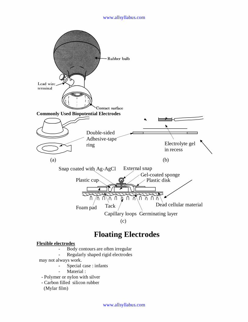
wwwallsyllabuscom
wwwallsyllabuscom
(a) Carbon-filled silicone rubber electrode
(b) Flexible thin-film neonatal electrode
(c) Cross-sectional view of the thin-film electrode in (b)
Electrodes in Biopotential Measurements
3 Describe the construction of commercial ECG electrode (not the cheap polymer
electrode used in the lab) What is the common electrode metal and why is it preferred
So you are an inventor who has a better idea Describe an improvement
bull to make the electrode cheaper
bull more suitable for lower noise measurement for EEG
bull circumvent patents that are based on plasticfoam electrode body
bull attractive to consumers for use with their ECG machines at home
bull reduce artifact (minimize the motion of skinelectrode) in ambulatory recording
4 In a research laboratory scientists want to record from single cells in a culture dish
They want to record action potentials from single isolated heart cells What kind of
electrode would they need to use (describe material and design) Give a simplified
schematic (circuit model of the electrode) described in the notes given to you
What is the challenge involved in designing an amplifier for use with a microelectrode
for single cell recording Ie what are the critical amplifier design characteristics and
specifications (hint this is not the usual differentialinstrumentation amplifier)
Electrodes and Microelectrodes (miscellaneous)
bull How would you detect bacteria or other microorganisms in water supply Make
sure that your method distinguishes inert particulate matter from living cellular
matter
bull Draw the equivalent circuit model of the skin and an ECG electrode Identify the
key sources of electrical interference and otherwise the elements that would likely
contribute to the poor quality of recordings
wwwallsyllabuscom
wwwallsyllabuscom
bull Design an amplifier interface for the following two applications Patch clamp ion
channel current amplifier Your goal is to amplify pA level current to produce 1
Volt output
bull Strain gauge sensor amplifier Your goal is to convert 10 ohm change in
resistance of a strain gauge to produce 1 volt output
Neural electrodesmicroelectrodes
You want to record from neurons in the brain However you want to record from dozens
of neurons all at once from several closely spaced microelectrodes What material and
process would you use to make the microelectrode array
bull What metal would you prefer to use to make electrode arrays of about 10 micron
square size to make electrical contacts with dozens of neurons
bull What metal would you prefer to use to stimulate dozens of neurons in a deep
brain microelectrode based stimulator
bull (which metal provides good recording vs stimulating properties ndash and at the same
time not be toxic to brain tissue)
bull You are asked to develop an experimental set up to record from rat brain cells
using microelectrodes What precautions would you take to minimize the
electrical interference in your recording set up
Biopotential Amplifiers bull These are very important part of modern medical instrumentation bull We need to amplify biopotentials which are generated in the body at low levels with a high source impedance
bull Biopotentials amplifiers are required to increase signal strength while maintaining fidelity Basic Requirements of Biopotential Amplifiers
Essential functions of a bioamplifier are bull To take a weak biopotential and increase its amplitude so that it can be processed recorded or displayed bull To amplify voltage but it could be considered as a power amplifier as well bull To amplify current since in some cases a biopotential amplifier is used to isolate the load from the source current gain only
Input Impedance (Zin)
bull All biopotential amplifiers must have high input impedance minimize loading (remember the characteristics of biopotential electrodes resulting into loading and distortion if input impedance of the amplifier is not high enough) ndash typical values of Zin over the frequency range of the measurand = 10 MΩ (remember the loading rule)
Protection amp Isolation
bull The input circuit of a biopotential amplifier must provide protection to the live measurand
wwwallsyllabuscom
wwwallsyllabuscom
Vbio
bull Any potential or current at amplifierrsquos input terminals can affect Vbio
bull Electric currents produced by the biopotential amplifier can result in microshock and
macroshock bull The bioamplifier must have isolation and protection circuitry so that the current through the electrodes can be kept at safe levels and any artifact generated by such current can be minimized Output Impedance (Zout)
bull The output circuit does not present any critical problems all it needs to do is to drive the load bull Output impedance must be low with respect to the load impedance and it must be capable of satisfying the power requirements of the load Bandwidth (BW)
Frequency response requirements bull The biopotential amplifier must be sensitive to important frequency components of the biosignal bull Since biopotentials are low level signals it is important to limit bandwidth optimize signal-to-noise ratio Gain (G)
bull Biopotential amplifiers have a gain of 1000 or greater
Mode of Operation
bull Very frequently biosignals are obtained from bipolar electrodes bull Electrodes symmetrically located with respect to ground need differential amplification bull High CMRR required because 1 Common mode signals much greater than the biosignal appear on bipolar electrodes 2 Symmetry with respect to ground is not perfect (mismatch between electrode impedances) ndash more on this later
Calibration Signal
bull Medical and clinical equipment require quick calibration bull The gain of the biopotential amplifier must be calibrated to provide us with an accurate indication of the signalrsquos amplitude bull Push button to apply standard signal to the input of the biopotential amplifier bull Adjustable gain switch carefully selects calibrated fixed gains (in microprocessorndashbased systems gain adjustment can be Electrocardiography
hearts A very widely used medical instrument which is utilized to diagnose and monitor cardiac beat
abnormalities is the electrocardiograph
hearts It measures the electrical activity of the heart (more precisely biopotential differences arising from the electrical activity of myocardium) Wersquove already talked about the genesis of the ECG signal hearts The ECG machine uses surface electrodes and high input impedance hearts Differential amplifiers with good common mode rejection ratio to record the electrocardiogram
hearts Normal ECG amplitude ranges between 05-4 mV Normal frequency content of ECG (for diagnostic purposes) is 005-100 Hz A typical ECG waveform is shown below
wwwallsyllabuscom
wwwallsyllabuscom
Obviously all human hearts are not the same and this leads into variability in different parts of the ECG signal Significant diagnostic features of the ECG signal are hearts Duration of component parts of the signal
hearts Polarities and magnitudes hearts The details of the ECG signal and the degree of variability in different parts of the ECG signal is shown below
The QRS amplitude polarity time duration the RR interval (indicator of heartbeat per min) and the T-wave amplitude are some very important and distinctive features of the ECG signal The heart rate in BPM = Beats Per Minute) is simply = 60 (RR interval in seconds)
Some ECG waveform abnormalities that may indicate illness are hearts An extended PR interval may be diagnosed as AV node block hearts A widening of the QRS complex may indicate conduction problems in the bundle of His hearts An elevated ST segment may indicate occurrence of myocardial Infarction (MI) hearts A negative polarity in the T wave may be due to coronary insufficiency ECG Leads
Normal ECG recordings for the standard lead connections leads I II and III (Lead II provides
the strongest signal) Obviously all human hearts are not the same and this results into a high degree of variability
wwwallsyllabuscom
wwwallsyllabuscom
hearts Note the degree of variability of different parts of the ECG Signal
Some abnormalities that may indicate illness hearts An extended P-R interval may be diagnosed as AV node block hearts Widening of the QRS complex conduction problems in the bundle of His hearts Elevated ST segment may indicate occurrence of MI
hearts Negative polarity T wave may be due to coronary insufficiency QRS amplitude polarity time domain PR interval (indicator of heat beat per min amp T-wave amplitude are some very important
distinctive features 1 Loss
2 Origin of the ECG signal
hearts We have already covered this concept extensively in the previous lectures (The Dipole filed of the heart the Eindhovenrsquos Triangle the electrical circuit model for the electrocardiographic problem etc)
hearts Standard Limb Leads (I II III)
wwwallsyllabuscom
wwwallsyllabuscom
hearts The The lead wires are color-coded according to some conventions One example is White ndashRA
(Right Arm) Black ndash LA (Left Arm) Green ndash RL (Right Leg) Red ndash LL (Left Leg) and Brown
ndash C (Chest) Note There is a CM (common mode) amplifier connected to the right leg We will discuss this in detail later Augmented Limb Leads
These leads offer a free 50 increase over leads VR VL and VF connections (unipolar leads) with respect to Wilson terminal AVR = -I ndash III2 AVL = I ndash II2 aVF = II ndash I2
wwwallsyllabuscom
wwwallsyllabuscom
Each measurement is made from the reflected limb and the average of the other two limbs Chest Leads (Precordial)
Chest Lead Anatomical Positions
V1 - 4th intercostal space ndash Right sternal margin
V2 - 4th intercostal space ndash Left sternal margin
V3 ndash Midway between V2 amp V4
V4 ndash 5th intercostal space on mid-clavicular line
V5 ndash Same as V4 on the anterior axillary line
V6 ndash Same as V5 on the mid-axillary line
12 Lead Clinical Electrocardiography
The ECG Machine
Most representative Specs
wwwallsyllabuscom
wwwallsyllabuscom
bull Zin = 10 MΩ bull Frequency response = 005 ndash100 Hz bull Strip Chart Recorder Speed = 25 mmsec bull Fast Speed = 100 mmsec
For detailed Specs Refer to the Table in your text ―Summary of performance requirements for electrocardiographs Location of the Heart bull The heart is located between the lungs behind the sternum and above the diaphragm bull It is surrounded by the pericardium bull Its size is about that of a fist and its weight is about 250-300 g bull Its center is located about 15 cm to the left of the midsagittal plane
Anatomy of the heart
bull The walls of the heart are composed of cardiac muscle called myocardium
bull It consists of four compartments ndash the right and left atria and ventricles
The Heart Valves
bull The tricuspid valve regulates blood flow between the right atrium and right
ventricle
bull The pulmonary valve controls blood flow from the right ventricle into the
pulmonary arteries
bull The mitral valve lets oxygen-rich blood from your lungs pass from the left atrium
into the left ventricle
bull The aortic valve lets oxygen-rich blood pass from the left ventricle into the aorta
then to the body
Blood circulation via heart
wwwallsyllabuscom
wwwallsyllabuscom
bull The blood returns from the systemic circulation to the right atrium and from
there goes through the tricuspid valve to the right ventricle
bull It is ejected from the right ventricle through the pulmonary valve to the lungs
bull Oxygenated blood returns from the lungs to the left atrium and from there
through the mitral valve to the left ventricle
bull Finally blood is pumped through the aortic valve to the aorta and the systemic
circulation
Electrical activation of the heart
bull In the heart muscle cell or myocyte electric activation takes place by means of
the same mechanism as in the nerve cell ie from the inflow of Na ions across
the cell membrane
bull The amplitude of the action potential is also similar being 100 mV for both nerve
and muscle
bull The duration of the cardiac impulse is however two orders of magnitude longer
than in either nerve cell or sceletal muscle cell
bull As in the nerve cell repolarization is a consequence of the outflow of K ions
bull The duration of the action impulse is about 300 ms
Mechanical contraction of Cardiac Muscle
bull Associated with the electric activation of cardiac muscle cell is its mechanical
contraction which occurs a little later
bull An important distinction between cardiac muscle tissue and skeletal muscle is
that in cardiac muscle activation can propagate from one cell to another
in any direction
bull Electrical signal begins in the sinoatrial (SA) node natural pacemaker causes
the atria to contract
bull The signal then passes through the atrioventricular (AV) node
ndash sends the signal to the ventricles via the ―bundle of His
ndash causes the ventricles to contract
The Conduction System
wwwallsyllabuscom
wwwallsyllabuscom
The Action Potential
Recording an AP requires the isolation of a single cell Microelectrodes (with tips a few μm across) are used to stimulate and record the response A typical AP is 2-4ms long with an amplitude of about 100Mv The Electroencephalogram EEG
EEG is the graphical representation of the electrical activity of the brain Very commonly used to diagnose certain neurological disorders such as epilepsy
More recently also investigated whether it can detect various forms of dementia or schizophrenia EEG is the specific recording obtained using the scalp electrodes from the surface of the skull
During surgery electrodes may also be placed directly on the cortex The resulting signal is then electrocorticogram (ECoG) Just like ECG EEG is also obtained using several different electrodes places on different regions of the head brain
The Event Related
wwwallsyllabuscom
wwwallsyllabuscom
Potentials ndash ERPs
ERPs are really EEGs obtained under a specific protocol that requires the patient to response to certain stimuli ndash hence event related potentials Also called evoked potentials these signals can be used to diagnose certain
neurological disorders such as dementia and they can also be used as a liedetector bull The oddball paradigm
bull The guilty knowledge test
Electroretinogram ERG
The ERG is the record of the retinal action currents produced by the retina in response to a light stimulus It measures the electrical responses of the light-sensitive cells (such as rods and cones)
The stimuli are often a series of light flashes or rotating patterns The ERG is recorded using contact lens electrode that the subject wears while watching the stimuli
Phonocardiogram ndash PCG
The PCG is the graphic record of the heart sounds and murmurs It is thus a mechanical audio signal rather than an electrical signal Can be easily heard using a stethoscope Or can be converted into an electrical signal using a transducer Typically used to determine the disorders related to the heart valve since their
routine opening and closing create the well-known sounds bull S1 sounds First heart sounds ndash ventricular contractions move blood into atria closing of the AV (mitral and tricuspid) valves then semilunar valves open and blood ejected out of ventricles ndash immediately follows the QRS complex bull S2 sounds Second heart sounds ndash Closure of semilunar (aortic and pulmonary) valves bull Any unexpected sound may indicate a malfunctioning valve that causes the blood flow into out of a chamber when it should not Also called heart murmurs
wwwallsyllabuscom
wwwallsyllabuscom
Define ultrasound
bull Mechanical waves in different modalities (longitudinallateral) needs medium
to be propagated (solid liquid gas)
bull gt 20 kHz
bull Continuouspulsed
bull Sphericalplanarnarrow beamsurface waveLamb-wave
Physical phenomena behind ultrasound measurements
Transmission
bull
bull reflection
bull transit time
bull differences in propagation velocities
bull returns to transit time
bull doppler-shift in frequency
bull flow velocity
bull change of acoustic impedance
bull comparing to reference
bull interference of ultrasound waves (holography)
bull interaction of ultrasound and light (photoacousticz)
bull ultrasound needs medium for propagation it doesnrsquot propagate in vacuum
bull because mechanical waves need moving massunits and spring forces between
them
bull in acoustic emission the medium creates ultrasound (for example during pressure
changes) which is received by sensors
bull pulsed mode more common than continuous
bull continuous reguires separate transducers for transmitting and receicving
bull in pulsed mode an ultrasound burst is sent to the object and the same transducer is
switched to listen echoes
bull standing wave problem
bull in us-therapy pulsed mode gives more effective care without too much heating The Doppler Equation describes the relationship of the Doppler frequency shift to target velocity
wwwallsyllabuscom
wwwallsyllabuscom
The frequency difference is equal to the reflected frequency (FR) minus the originating frequency (FT) If the resulting frequency is higher then there is a positive Doppler shift and the object is moving toward the transducer but if the resulting frequency is lower there is a negative Doppler shift and it is moving away from the transducer In its simplest form it would be calculated as if
the ultrasound was parallel to the targetrsquos direction as shown in diagram A below However this would be a rare occurrence in clinical practice because the transducer is rarely pointed head on to a blood vessel In real life the ultrasound waves would approach the target at an angle called the Doppler angle ( ) On the following page diagram B shows the Doppler
equation used in general clinical situations which includes the Doppler angle The Doppler Angle
The ultrasound beam usually approaches the moving target at an angle called the Doppler angle ( ) This reduces the frequency shift in proportion to the cosine of this
angle If this angle is known then the flow velocity can be calculated The equation used is The Doppler Equation
equiv Doppler shift frequency (the difference between the transmitted and received frequencies) equiv transmitted frequency equiv reflected frequency
V equiv velocity of the blood flow towards the transducer C equiv velocity of sound in tissue θ equiv the angle between the sound beam and the direction of moving blood Where
The Doppler angle ( ) is also known as the angle of insonation It is estimated by the sonographer by a process known as angle correction which involves aligning an indicator on the duplex image along the longitudinal axis of the vessel
There are a few considerations that affect the performance of a Doppler examination that are inherent in the Doppler equation which are ndash The cosine of 90deg is zero so if the ultrasound beam is perpendicular to the direction of blood flow there will be no Doppler shift and it will appear as if there is no flow in the vessel ndash Appropriate estimation of the angle of insonation or angle correction is essential for the accurate determination of Doppler shift and blood flow velocity The angle of insonation should also be less than 60deg at all times since the
cosine function has a steeper curve above this angle and errors in angle correction will be magnified
The simplest Doppler devices use continuous wave (CW Doppler) rather than the pulsed wave used in more complex devices CW Doppler uses two transducers (or a dual element transducer) that transmit and receive ultrasound continuously The transmit and receive beams overlap in a Doppler sample volume some distance from the transducer face as
shown in the diagram below
volume) is the region of transmitting and receiving beam overlap (shaded region) Because there is continuous transducer transmission and reception echoes from all depths within the area arrive at the transducer simultaneously So although CW Doppler can determine the direction of flow it cannot discriminate the different depths where the motion originates The usefulness of CW Doppler devices is limited but they are used clinically to confirm blood flow in superficial vessels as they are good at detecting low velocities As they are easily portable this
can be done at the bedside or in the operating room Most other clinical applications require pulsed wave Doppler Pulsed Wave Doppler (PW Doppler)
wwwallsyllabuscom
wwwallsyllabuscom
Pulsed wave Doppler (PW Doppler) uses a single-element transducer that emits brief pulses of ultrasound energy The time interval between transmitting and then receiving the echoing sound can be used to calculate the depth from where the echo arises The Doppler sample volume can be chosen as to shape depth and position in sampling the flow
data For example the depth is chosen by processing only the signals that return to the transducer in a stipulated time For this technique the ultrasound system transmits a short pulse The eceiver is opened to detect the returning echoes only after a controlled delay and only for a specific duration This time-based gating of the receiving channel allows the definition of a fixed easuring distance which is often referred to as the Sample volume or Doppler gate Then the next ultrasound wave is transmitted The number of pulses transmitted by the system within a second is referred to as the pulse repetition frequency (PRF) The upper PRF limit is given by the time interval required for the echoes to arrive from a sample volume located at a
certain depth The greater the sample-volume depth the longer the time before the echoes are returned and the longer the delay between pulse transmission The greater the samplevolume depth the lower will be the maximum PRF setting Errors in the accuracy of the information arise if the velocities exceed a certain speed The highest velocity accurately measured is called the Nyquist limit Beyond this limit the errors that occur are referred to as aliasing Volume and flow measurement
Flow ndash volume of a liquidgas passing some point over a given time Gases are compressible
Benedict Roth Spirometer
Widely used for physiological amp clinical studies
Light bell moves with the ptrsquos breathing
Movement recorded by a pen on a rotating drum
Water seal prevents leakage of gas
Small seal minimises volume of gas dissolved in water
Suitable for measuring limited gas vols (few litres)
Pneumotachograph
wwwallsyllabuscom
wwwallsyllabuscom
Cardiac Output
Defn vol of blood pumped by the heart per min
CO = SV x HR
Norm ~ 5 lmin
Cardiac index ndash corrected for body surface area
Affected by
Met Rate ndash pregnancy hyperthyroid septic
Preload contractility afterload
Clinical indicators of CO imprecise
Affected by anaesthetic agents used in everyday practice
Provides estimate of
whole body perfusion
oxygen delivery
left ventricular function
Persistently low CO assoc with poor outcome
Methods
Fick method
Dilution techniques ndash dye thermal lithium
Pulse contour analysis- LiDCO amp PiCCO
Oesophageal doppler
TOE
Transthoracic impedance plethysmography
Inert gas through flow
Non-invasive cardiac output measurement
Fick Principle measure volume displacement
1st proposed 1870
―the total uptake or release of a substance by an organ is the product of the
blood flow through that organ and the arteriovenous concentration
difference of the substance
CO = O2 consumption (mlmin) art ndash mixed
venous O2 conc (mll)
Limited by cumbersome equipment sampling errors need for invasive
monitoring and steady-state haemodynamic and metabolic conditions
Indicator dilution techniques
―An indicator mixed into a unit volume of constantly flowing blood can be used to
identify that volume of blood in time provided the indicator remains in the system
between injection and measurement and mixes completely in the blood
wwwallsyllabuscom
wwwallsyllabuscom
Dye dilution
Inert dye ndash indocyanin green
Injected into pulmonary artery and arterial conc measured using a
calibrated cuvette densitometer
Plot indicator dilution curve (see diagram)
CO derived from area under curve
Indicator Dilution Curve
Cardiac Output Measurement
wwwallsyllabuscom
wwwallsyllabuscom
Why bother to check blood pressure
For each 20 mm rise in systolic blood pressure or 10 mm rise in diastolic blood
pressure over 11575
Risk of stroke increases
Risk of heart disease doubles
Risk of renal failure increases
Systolic and diastolic blood pressure
Systolic blood pressure is the highest pressure in the arteries just after the heart
beats
Diastolic blood pressure is the lowest pressure in the arteries just before the heart
beats
Blood pressure is measured indirectly by blood pressure cuff
(sphygmomanometer)
Inflating cuff increases pressure until it cuts off arterial circulation to the arm
Deflating cuff decrease pressure by 2 to 3 mm of mercury per second until blood
first enters the artery creating turbulence this causes a sound with each heartbeat
Sounds continue with each heartbeat until pressure lowers to the lowest pressure
in the artery then turbulence stops so the sound stops
Systolic blood pressure is the cuff pressure at the first sounds diastolic is the cuff
pressure just before the sounds stop
Phase 1 sharp thuds start at systolic blood pressure
Phase 2 blowing sound may disappear entirely (the auscultatory gap )
Phase 3 crisp thud a bit quieter than phase 1
Phase 4 sounds become muffled
Phase 5 end of sounds -- ends at diastolic blood pressure
wwwallsyllabuscom
wwwallsyllabuscom
Make sure the cuff is the right size - its width should be at least 40 of the arms
circumference The cuff will overestimate blood pressure if too small and
underestimate if too large
Place the cuff snugly on patients proximal arm on skin ( not cloth) centered over
the brachial artery (most cuffs have markings)
Support the patients arm at heart level using your arm or a desk
Your patient should sit in the chair for 5 minutes before BP is measured and
should have no caffeine or nicotine for 30 minutes before (JAMA 273 p1211-
1218 1995)
With fingers palpating radial or brachial artery inflate cuff rapidly until you cant
feel the pulse then 20 mm higher
Release cuff at 2 to 3 mm Hg per second until you again feel the pulse this is the
palpable systolic pressure
Wait 30 seconds before measuring blood pressure
Measuring palpable pressure first avoids risk of seriously underestimating blood
pressure because of the auscultatory gap (mistaking Korotkoff phase 3 for phase
1) Many doctors skip this step for time reasons and instead pump cuff to 200 mm
Hg at the next step)
Phase 2 of the Korotkoff sounds can be inaudible - especially in older patients
with systolic hypertension who are at especially high risk of stroke Inflating the
cuff until you dont hear sounds can give you a reading of 14086 when the
patients actual blood pressure is 22086 Most physicians are pressed for time so
they instead inflate the cuff to 200 mm which is beyond the auscultatory gap in
most patients But palpable systolic blood pressure is according to research more
reliable
Place bell of stethoscope (diaphragm is acceptable) over brachial artery
Rapidly pump the cuff to 20 to 30 mm Hg above palpable systolic pressure
Release pressure in the cuff by 2 to 3 mm Hg per second and listen for Korotkoff
sounds including systolic (first) and diastolic (last)
Record as systolicdiastolic Check in both arms the first time you check a
patients blood pressure It may differ by 10 mm Hg or more
If the sounds continues to zero record diastolic blood pressure as the point when
sounds become muffled (phase 4) over zero eg 130700 or just as 13070
Rate
ndash Number of beats in 30 seconds x 2
Strength
ndash Bounding strong or weak (thready)
Regularity
ndash Regular or irregular
You need three readings on two occasions to diagnose hypertension unless blood
pressure is very high
Normal blood pressure in children is
wwwallsyllabuscom
wwwallsyllabuscom
ndash 10255 at 1 year 11269 at 5 years 11978 at 10 years
Blood pressures in adults (JNC VII JAMA 2892560-72 2003)
ndash Normal lt120lt80
ndash Prehypertensive 120-13980-89
ndash Stage 1 hypertension 140-15990-99
ndash Stage 2 hypertension gt160gt100
Adult 60 to 100
Newborn 120-170
1 year 80-160
3 years 80-120
6 years 75-115
10 years 70-110
How to measure observe rise and fall of chest
In infants count for 60 seconds in adults 15 or 30 seconds
Normal respiration Adults 12 to 20
Children
newborn 30-80
1 year 20-40
3 years 20-30
6 years 16-22
Rate
Number of breaths in 30 seconds x 2
Quality
Character of breathing
Rhythm
Regular or irregular
Effort
Normal or labored
Noisy respiration
Normal stridor wheezing snoring gurgling
Depth
Shallow or deep
pH electrode
wwwallsyllabuscom
wwwallsyllabuscom
Governing equation is the Nernst Equation
pCO2 Electrode
The measurement of pCO2 is based on its linear relationship with pH over the range of
10 to 90 mm Hg
The dissociation constant is given by
Taking logarithms
pH = log[HCO3-] ndash log k ndash log a ndash log pCO2
Diathermy
In the natural sciences the term diathermy means electrically induced heat and is commonly
used for muscle relaxation It is also a method of heating tissue electromagnetically or
ultrasonically for therapeutic purposes in medicineContents
[hide]
1 Heating uses 2 Surgical uses
3 Trivia
4 References
E
RT
nF
H
HH
i
ln
0
H O CO H CO H HCO2 2 2 3 3
k
H HCO
a pCO
3
2
wwwallsyllabuscom
wwwallsyllabuscom
Heating uses
Ultrasonic diathermy refers to heating of tissues by ultrasound for the purpose of
therapeutic deep heating No tissue is ordinarily damaged hence it is generally used in
biomedical applications
Electric diathermy uses high frequency alternating electric or magnetic fields sometimes
with no electrode or device contact to the skin to induce gentle deep tissue heating by
induction Again no tissue is ordinarily damaged
Surgical uses
Surgical diathermy is usually better known as electrosurgery (It is also referred to
occasionally as electrocautery but see disambiguation below) Electrosurgery and
surgical diathermy involve the use of high frequency AC electrical current in surgery as
either a cutting modality or else to cauterize small blood vessels to stop bleeding This
technique induces localized tissue burning and damage the zone of which is controlled
by the frequency and power of the device Some sources[1]
insist that electrosurgery be
applied to surgery accomplished by high frequency AC cutting and that
electrocautery be used only for the practice of cauterization with heated nichrome wires
powered by DC current as in the handheld battery-operated portable cautery tools
Trivia
Medical Diathermy devices were used to cause interference to German radio beams used
for targeting night time bombing raids in WWII during the Battle of the Beams
I Diathermy
A Therapeutic use
1 Generation of local heating by high-frequency electromagnetic waves
2 Capacitance techniquemdashbody is placed in an electric field
a Dipolesmdashstructures with positive and negative poles
b Structures with large numbers of dipoles have a greater capacitance to store an electrical charge
c Greatest heating occurs in tissues with fewer dipoles particularly fatty tissues
3 Rapid rotation of dipoles causing mechanical friction and movement of electrons results in local heating
4 Inductance techniquemdashbody is not placed in an electric field
a Magnetic waves generated by driving current through a coiled wire
b Magnetic field creates currents in tissues
wwwallsyllabuscom
wwwallsyllabuscom
c Greatest heating occurs in tissues with low impedance especially muscle
B Precautions and contraindications
1 Diathermy should not be used
a Over metal implants and cardiac pacemakersmdashmore research needed regarding its use over metallic fixations
b Near the uterus of a pregnant woman or near the abdomen or back of a woman who might be pregnant
c On individuals with infections
d On individuals with acute inflammation
e Over moist open wounds
f On patients with malignant tumors
g Over large joint effusions
C Pulsed electromagnetic fields and diathermy
1 Can be pulsed to decrease total energy transmitted to the tissues
2 Short-wave diathermy can be adjusted into a nonthermal range
a Classified as pulsed electromagnetic field (PEMF) or
b Pulsed radio frequency energy (PRFE)
3 Important reclassification as diathermy implies heating
D Efficiency of diathermy and PEMF therapy for musculoskeletal conditions
1 Current research is limited but results suggest that diathermy enhances
treatments directed at soft tissue stretching
2 Some studies suggest that PEMF may speed wound healing and promote healing of nonunion fractures
Diathermy is a modality that uses electromagnetic energy to heat deeper tissues
Diathermy is more effective than ultrasound at heating a larger area of deep tissues
The athletic trainer must identify and respect contraindications to application of ultrasound and diathermy
Pulsed ultrasound and diathermy are used to treat slow-to-heal lesions including skin ulcers and nonunion fractures and may be able to facilitate repair of other tissues including ligaments and tendons
Diathermy
Definition
In diathermy high-frequency electrical currents are used to heat deep muscular tissues The heat increases blood flow speeding up recovery Doctors also use diathermy in surgical procedures by sealing blood vessels with electrically heated probes
The term diathermy is derived from the Greek words therma meaning heat and dia meaning through Diathermy literally means heating through
Origins
The therapeutic effects of heat have long been recognized More than 2000 years ago the Romans took advantage of heat therapies by building hot-spring bathhouses Since then various
wwwallsyllabuscom
wwwallsyllabuscom
methods of using heat have evolved In the early 1890s French physiologist Arseacutene dArsonval began studying the medical application of high-frequency currents The term diathermy was coined by German physician Carl Franz Nagelschmidt who designed a prototype apparatus in 1906 Around 1925 United States doctor J W Schereschewsky began studying the physiological
effects of high-frequency electrical currents on animals It was several years however before the fundamentals of the therapy were understood and put into practice
Benefits
Diathermy can be used to treat arthritis bursitis and other conditions involving stiff painful joints It is also used to treat pelvic infections and sinusitis A benefit of diathermy is that it is a painless procedure that can be administered at a clinic Also if the treatment relieves pain then patients can discontinue pain killers and escape their high cost and side effects
Description
Diathermy involves heating deep muscular tissues When heat is applied to the painful area cellular metabolism speeds up and blood flow increases The increased metabolism and circulation accelerates tissue repair The heat helps the tissues relax and stretch thus alleviating
stiffness Heat also reduces nerve fiber sensitivity increasing the patients pain threshold
There are three methods of diathermy In each energy is delivered to the deep tissues where it is converted to heat The three methods are
Shortwave diathermy The body part to be treated is placed between two capacitor plates Heat is generated as the high-frequency waves travel through the body tissues between the plates Shortwave diathermy is most often used to treat areas like the hip which is covered with a dense tissue mass It is also used to treat pelvic infections and sinusitis The treatment reduces inflammation The Federal Communications Commission regulates the frequency allowed for short-wave diathermy treatment Most machines
function at 2733 megahertz Ultrasound diathermy In this method high-frequency acoustic vibrations are used to
generate heat in deep tissue Microwave diathermy This method uses radar waves to heat tissue This form is the
easiest to use but the microwaves cannot penetrate deep muscles
Diathermy is also used in surgical procedures Many doctors use electrically heated probes to seal blood vessels to prevent excessive bleeding This is particularly helpful in neurosurgery and eye surgery Doctors can also use diathermy to kill abnormal growths such as tumors warts and
infected tissues
Preparations
To keep patients from sweating patients are usually asked to remove clothing from the body part being treated If a patient sweats the electrical currents may pool in the area causing burns Also clothing containing metal must be removed as must earrings buttons barrettes or zippers that contain metal Watches and hearing aids should be removed because the therapy may affect their function
wwwallsyllabuscom
wwwallsyllabuscom
Practitioners of surgical diathermy should steer clear of alcohol-based solutions to prepare and cleanse the skin These preparations can create a flammable vapor and cause burns and fires
Precautions
Patients with metal implants should not undergo diathermy treatment because the metal can act as a conductor of heat and result in serious internal burns Female patients with metallic uterine implants such as an IUD should avoid treatment in the pelvic area Diathermy should not be
used in joints that have been replaced with a prosthesis or in those with sensory impairment who may not be able to tell if they are burning Furthermore pulsed shortwave diathermy should be avoided during pregnancy as it can lead to abnormal fetal development
Patients with hemophilia should avoid the treatment because the increased blood flow could cause them to hemorrhage
Side effects
Some patients may experience superficial burns Since the therapy involves creating heat care must be taken to avoid burns particularly in patients whose injuries have caused decreased sensitivity to heat Also diathermy may affect pacemaker function
Female patients who receive treatment in the lower back or pelvic area may experience an
increased menstrual flow
Research amp general acceptance
For years physiotherapists and physical therapists have used diathermy as a routine part of physical rehabilitation
Electrical Safety
Electrical safety is very important in hospitals as patients may be undergoing a diagnostic or treatment procedure where the protective effect of dry skin is reduced Also patients may be unattended unconscious or anaesthetised and may not respond normally to an electric current Further electrically conductive solutions such as blood and saline are often present in patient treatment areas and may drip or spill on electrical equipment
Electric Current Leakage Current
Extension Leads Double Adaptors Equipment Classification
Class I Class II Defibrillator-Proof
Protective Devices
Residual Current Devices (RCD) Line Isolation overload Monitors (LIMs) Equipment Earthing
Area Classification
wwwallsyllabuscom
wwwallsyllabuscom
Body Protection Area Cardiac Protected Area
Other Electrical Issues Extension Leads
Double Adapters Main Extension Devices Power Boards Installation of Additional Power Points
Electric Current
Injuries received from electric current are dependent on the magnitude of current the pathway
that it takes through the body and the time for which it flows
The nature of electricity flowing through a circuit is analogous to blood flowing through the circulatory system within the human body In this analogy the source of energy is represented by the heart and the blood flowing through arteries and veins is analagous to current flowing
through the conductors and other components of the electric circuit
The application of an electric potential to an electric circuit generates a flow of current through conductive pathways This is analogous to the changes in blood pressure caused by contraction of cardiac muscle that causes blood to flow into the circulatory system For electric current to flow there must be a continuous pathway from the source of potential through electrical components and back to the source
Leakage Current
Electrical components and systems are encased in non conducting insulation to ensure that the electric current is contained and follows the intended pathways If the insulation deteriorates or breaks down current will leak through the insulation barrier and flow to earth This may
be through the protective earth conductor or through the operator
Medical equipment and clinical areas are fitted with a number of protective devices to protect the patient and operator from harmful leakage currents
Extension Leads
Extension leads are not permitted in clinical areas of RCH organisations They may cause high
earth resistance and excessive earth leakage current An extension lead can allow equipment to be powered from areas other than the relevant protected treatment area The power from the other area may not be protected to the same level as the power in the treatment area
wwwallsyllabuscom
wwwallsyllabuscom
As the connection between the extension lead and the equipment mains cable is often on the floor there is a high danger from fluid spills tripping and damage to the mains cable by trolleys when an extension lead is used
Double Adaptors
Double adaptors must not be used in RCH organisations They may not sit securely in a wall
outlet may not be able to provide adequate earth protection and may cause overloading overheating fire or loss of electrical supply
EQUIPMENT CLASSIFICATIONS
There are several methods of providing protection for operators and patients from electrical faults
and harmful leakage current
Class I
Class I equipment is fitted with a three core mains cable containing a protective earth wire
Exposed metal parts on class I equipment are connected to this earth wire
Should a fault develop inside the equipment and the exposed metal comes into contact with the mains the earthing conductor will conduct the fault current to ground Regular testing procedures ensure that earthing conductors are intact as the integrity of the earth wire is of vital importance
Class II
Class II equipment is enclosed within a double insulated case and does not require earthing
conductors Class II equipment is usually fitted with a 2-pin mains plug An internal electrical fault is unlikely to be hazardous as the double insulation prevents any external parts from becoming alive Class II or double insulated equipment can be identified by the class II symbol on the cabinet
Class II Symbol
wwwallsyllabuscom
wwwallsyllabuscom
Defibrillator-Proof
Some medical equipment within the hospital is classified as defibrillator proof When a
defibrillator is discharged through a patient connected to defibrillator proof equipment the equipment will not be damaged by the defibrillators energy Defibrillator proof equipment can remain connected to the patient during defibrillation It is identified by one of the following
symbols
Defibrillator proof symbols
Body protected Cardiac protected
PROTECTIVE DEVICES
Most patient care areas in the hospital are fitted with protective devices These devices are
regularly tested in accordance with the relevant guidelines published by Standards Australia The level of protection provided is dependent upon the device and the area in which it is located
Residual Current Devices (RCD)
RCDs (safety switches) are used in patient treatment areas to monitor and protect the mains supply RCDs sense leakage currents flowing to earth from the equipment If a significant leakage current flows the RCD will detect it and shut off the power supplied to the equipment
within 40 milliseconds Hospital RCDs are more sensitive than those fitted in homes A hospital RCD will trip at 10 milliamperes leakage current
Power outlets supplied through an RCD have a Supply Available lamp The lamp will extinguish
when the RCD trips due to excessive leakage current
wwwallsyllabuscom
wwwallsyllabuscom
Resetting a RCD
Lamp indicates supply is no longer available Disconnect all equipment from the supply Operate the reset button or lever on the supply panel and the Supply Available lamp
should illuminate If not contact Biomedical Engineering
Connect an item of equipment If the RCD trips again then this is the faulty item and should be labelled and sent to Biomedical Engineering
If the RCD does not trip continue connecting equipment until the RCD trips The last piece of equipment connected to the supply is most likely to be faulty as it will have caused the RCD to trip Remove the faulty item from service label it and send to Biomedical Engineering as mentioned above
Line Isolation overload Monitors (LIMs)
In critical life support applications where loss of power supply cannot be tolerated special power outlets powered by isolation transformers are installed
Line Isolation Monitors are installed to continually monitor electrical leakage in the power
supply system If an electrical fault develops in a medical device connected to an isolated power outlet the LIM will detect the leakage current The LIM will alarm and indicate the level of leakage current but will not shut off the electric supply
The faulty equipment can be identified by un plugging one item of equipment at a time from the supply until the alarm stops sounding Equipment that is not faulty may be reconnected Faulty equipment should be appropriately labelled and sent to Biomedical Engineering for repair
The LIM also monitors how much power is being used by the equpiment connected to it If too much power is being used the LIM will alarm and indicate that there is an overload The power used must be reduced immediately by moving some equipment to another circuit as soon as possible until the alarm stops sounding Failure to reduce the load on the LIM will result in the circuit breaker tripping and loss of power to the circuit
wwwallsyllabuscom
wwwallsyllabuscom
Equipotential Earthing
Equipotential earthing is installed in rooms classified as Cardiac Protected electrical areas
Equipotential earthing in treatment areas used for cardiac procedures is intended to minimise any voltage differences between earthed parts of equipment and any other exposed metal in the room
This reduces the possibility of leakage currents that can cause microelectrocution when the patient comes into contact with multiple items of equipment or if the patient happens to come
into contact with metal items in the room whilr they are connected to a medical device
All conductive metal in an equipotential area is connected to a common equipotential earth point with special heavy duty cable
AREA CLASSIFICATIONS
Body Protected Area
These areas are designed for procedures in which patients are connected to equipment that lowers the natural resistance of the skin Applied parts such as electrode gels conductive fluids entering the patient metal needles and catheters provide an easy pathway for current to flow
The main occurrence of injury from Body-Type procedures is from high current levels causing electric shock A direct connection to the patients heart is not present so the risk of Microelectrocution - fibrillation from minute current levels - is reduced
Residual Current Devices (RCD) or Isolation Transformers and Line Isolation Monitors (LIMiacutes) are used in Body Protected areas to provide protection against electrocution from high leakage currents Body-Protected Areas are identified with this sign
Cardiac Protected Area
Where the procedure involves placing an electrical conductor within or near the heart protection against fibrillation induced from small leakage currents is required Electrical conductors used in these procedures include cardiac pacing electrodes intracardiac ECG electrodes and intracardiac catheters
wwwallsyllabuscom
wwwallsyllabuscom
Equipotential earthing in conjunction with RCDs or LIMs provides protection against microelectrocution in Cardiac-Type procedures
Fault currents are reduced to magnitudes that are unlikely to induce fibrillation Used in conjunction with RCDs or LIMs the magnitude and duration of any fault currents
sourced from equipment are limited Cardiac-Protected Areas are identified with this sign
Other electrical issues
This policy aims to provide guidance to those who find that they need more electrical outlets than
those available or that the existing electrical outlets are inconveniently located
As extension leads and multiple outlet power boards can introduce additional hazards into an area the following procedures should be observed
Extension leads
Approved extension leads (AS 3760 1996) may be used in some areas within the hospital but MUST NOT BE USED IN PATIENT AREAS All electrical extension leads must be tagged with an Engineering Department maintenance tag and require a yearly safety inspection and test via
the Engineering Department
Double adapters
Double adapters may cause overloading or equipment earthing problems and are not to be used in
WCH
Mains extension device
The only mains extension device that is to be used in Patient care areas is the 4-way or 8-way
portable Core Balance Unit
The Biomedical Engineering Department must approve all units prior to use These units contain a safety switch and can detect excessive leakage current and disconnect the power in the event of a hazardous situation
wwwallsyllabuscom
wwwallsyllabuscom
Care must be exercised in the use of a portable Core Balance Unit It should be located off the
floor and in a position that will protect it from physical abuse and possible entry of fluids These devices are expensive and easily damaged The device must be sent to Biomedical Engineering every 6 months for safety testing
Power boards
Approved multiple-outlet power boards can be used across RCH but must not be used in patient
care areas except areas approved by the Biomedical Engineering Department
The power boards must have overload protection be fitted with internal safety shutters that protect unused outlets and be fitted with an onoff switch for each outlet
Medical Laser Applications
The main research subjects of the group of Medical Laser Application are in the field of online
monitoring and diagnostics as well as the development of new therapheutic methods The main focus in research is based upon the use of ultrashort (fs) laser pulses
The main advantage of ultrashort laser pulses is the extrem short interaction time which suppress any unwanted side effects of the laser irradiation of the tissue Secondly the broad spectra of the fs laser pulses give the advantage to use the same laser pulses for diagnostic applications like the optical coherence tomography (OCT)
wwwallsyllabuscom
wwwallsyllabuscom
Following research subjects have special attention at the moment
Optical Coherence Tomographie (OCT)
A fs-laser light source can be used to obtain images from inside the tissue when the pulses are coherent superposed This group uses this application to obtain an inside view of the vocal fold and to measure the outline of the crystalline lens
Refractive Surgery
The principle of getting rid of glasses with the help of a laser surgery (fs-LASIK) is underlying rapid improvements concerning precision and safety New technologies were evaluated and transferred very fast in cooperation with an industrial partner
Treatment of Presbyopia
The flexibility of the crystalline lens can be increase by pricise cuts which are induce by fs laser pulse inside the lens The treatment of the presbyopia is thinkable
wwwallsyllabuscom
wwwallsyllabuscom
Ultrafast Physics
Whereas the applications of ultrashort laser pulses increase rapidly is the physics of the interaction between the laser pulses and tissue in many case not fully understood To obtain improved knowledge of the interaction for many fs-laser application this group preforms numerical simulations as well as fundamental experiments
Thermography
Thermography thermal imaging or thermal video is a type of infrared imaging Thermographic cameras detect radiation in the infrared range of the electromagnetic spectrum
(roughly 900ndash14000 nanometers or 09ndash14 microm) and produce images of that radiation Since infrared radiation is emitted by all objects based on their temperatures according to the black body radiation law thermography makes it possible to see ones environment with or without visible illumination The amount of radiation emitted by an object increases with temperature therefore thermography allows one to see variations in temperature (hence the name) When viewed by thermographic camera warm objects stand out well against cooler backgrounds humans and other warm-blooded animals become easily visible against the environment day or
night As a result thermographys extensive use can historically be ascribed to the military and security services
Thermal imaging photography finds many other uses For example firefighters use it to see through smoke find persons and localize the base of a fire With thermal imaging power lines maintenance technicians locate overheating joints and parts a telltale sign of their failure to eliminate potential hazards Where thermal insulation becomes faulty building construction technicians can see heat leaks to improve the efficiencies of cooling or heating air-conditioning Thermal imaging cameras are also installed in some luxury cars to aid the driver the first being
the 2000 Cadillac DeVille Some physiological activities particularly responses in human beings and other warm-blooded animals can also be monitored with thermographic imaging [1]
The appearance and operation of a modern thermographic camera is often similar to a camcorder Enabling the user to see in the infrared spectrum is a function so useful that ability to record their output is often optional A recording module is therefore not always built-in
Instead of CCD sensors most thermal imaging cameras use CMOS Focal Plane Array (FPA) The most common types are InSb InGaAs HgCdTe and QWIP FPA The newest technologies are using low cost and uncooled microbolometers FPA sensors Their resolution is considerably lower than of optical cameras mostly 160x120 or 320x240 pixels up to 640x512 for the most
expensive models Thermographic cameras are much more expensive than their visible-spectrum counterparts and higher-end models are often export-restricted Older bolometers or more
wwwallsyllabuscom
wwwallsyllabuscom
sensitive models as InSb require cryogenic cooling usually by a miniature Stirling cycle refrigerator or liquid nitrogen
Contents
[hide]
1 Difference between IR film and thermography 2 Advantages of Thermography
3 Limitations amp disadvantages of thermography 4 Applications 5 See also 6 External links
o 61 History of thermal imager manufacturers
[edit] Difference between IR film and thermography
Thermal imaging is going to be used on Mars to detect caves that could hold life
[edit] Advantages of Thermography
You get a visual picture so that you can compare temperatures over a large area
It is real time capable of catching moving targets Able to find deteriorating components prior to failure Measurement in areas inaccessible or hazardous for other methods It is a non-destructive test method
[edit] Limitations amp disadvantages of thermography
Quality cameras are expensive and are easily damaged Images can be hard to interpret accurately even with experience
Accurate temperature measurements are very hard to make because of emissivities Most cameras have plusmn2 or worse accuracy (not as accurate as contact) Training and staying proficient in IR scanning is time consuming Ability to only measure surface areas
[edit] Applications
Condition monitoring Medical imaging Night vision Research
Process control Non destructive testing Surveillance in security law enforcement and defense Chemical imaging
wwwallsyllabuscom
wwwallsyllabuscom
Thermal infrared imagers convert the energy in the infrared wavelength into a visible light video display All objects above 0 kelvins emit thermal infrared energy so thermal imagers can passively see all objects regardless of ambient light However most thermal imagers only see objects warmer than -50 degC
The spectrum and amount of thermal radiation depend strongly on an objects surface
temperature This makes it possible for a thermal camera to display an objects temperature However other factors also influence the radiation which limits the accuracy of this technique For example the radiation depends not only on the temperature of the object but is also a function of the emissivity of the object Also radiation also originates from the surroundings and is reflected in the object and the radiation from the object and the reflected radiation will also be influenced by the absorption of the atmosphere
Endoscopy
This page is about having an endoscopy - a test that looks at the inside of your digestive system
There is information on
What endoscopy is
Why you may have an endoscopy
Having an endoscopy
Your results
What an endoscopy is
An endoscopy is a test that looks inside the body The endoscope is a long flexible tube that can
be swallowed It has a camera and light inside it Some doctors call it a telescope
wwwallsyllabuscom
wwwallsyllabuscom
Why you may have an endoscopy
You are most likely to have an endoscopy to look at the inside of your
Gullet (oesophagus)
Stomach
Duodenum - the first part of the small bowel that attaches to the stomach
Large bowel (colon)
Your doctor may want to see inside because you have symptoms such as abnormal bleeding or
difficulty swallowing The doctor can look down the endoscope and see if there are any growths
or other abnormal looking areas Also through the endoscope the doctor can take samples
(biopsies) of any abnormal looking tissues
There is more detailed information about having a colonoscopy in the bowel cancer section of
CancerHelp UK Below is information about having other types of endoscopy
Having an endoscopy
You can have this test as an out patient Most people have a choice between having the test while
they are awake or after having a medicine to make them drowsy (a sedative) Your hospital may
not be happy for you to have a sedative if you live alone and will have no one to look after you
when you go home If you live alone but really want sedation your hospital may allow you to
stay overnight You should ask your doctor about this as early as possible as extra arrangements
will need to be made beforehand
If you dont have a sedative you will have a spray to numb the back of your throat and make it
easier for you to swallow the endoscopy tube
If you would prefer to be asleep during the test you will have an injection to make you very
drowsy just before the test You will need to take someone with you to the hospital appointment
You wont be able to drive for the rest of the day and should have someone to go home with you
You cant eat or drink for about 8 hours before the test so that your stomach and duodenum are
empty Your doctor will give you written instructions about this beforehand or they may arrive
with your appointment letter When you get to the clinic you may be asked to take your upper
clothing off and put on a hospital gown Some hospitals prefer to use gowns because your clothes
wont get messy Once you are ready you get onto the bed or X-ray couch When you are lying
comfortably you have the sedative injection to make you very drowsy Or your doctor will spray
the back of your throat to numb it
Once the sedative or throat spray has worked the doctor will pass the endoscope tube down your
wwwallsyllabuscom
wwwallsyllabuscom
throat to the area being investigated Your doctor will ask you to swallow as the tube goes down
but if youve had a sedative you wont remember that afterwards If there are any abnormalities
the doctor will take pieces of tissue from the abnormal looking area to send to the laboratory for
closer inspection under a microscope These tissue samples are called biopsies
When the test is over you will need to rest for a while If youve had a sedative you may not
remember much (if anything) about the test once you have come round You should be able to go
home the same day
The results
It can take time for test results to come through How long will depend on why you are having
the test Usually the doctor who carries out the endoscopy dictates a report straight way The
report is typed up by the department secretary and goes to your specialist who gives the results to
you If your GP has sent you for the test the results will go directly to the GP surgery
Understandably waiting for results can make you anxious It usually takes a couple of weeks for
the results to come through If your doctor needed them urgently it would have been noted on
the test request form and the results will be ready sooner than that Try to remember to ask your
doctor how long you should expect to wait for the results when you are first asked to go for the
test If it is not an emergency and you have not heard a couple of weeks after your test ring your
doctors secretary to check if they are back
Endoscopy
From Wikipedia the free encyclopedia
Jump to navigation search
Endoscopic images of a duodenal ulcer
wwwallsyllabuscom
wwwallsyllabuscom
A flexible endoscope
Endoscopy means looking inside and typically refers to looking inside the human body for medical reasons using an instrument called an endoscope Endoscopy can also refer to using a borescope in technical situations where direct line-of-sight observation is not feasible
Contents
[hide]
1 Overview 2 Components 3 Uses
4 History 5 Risks 6 After The Endoscopy 7 Recent developments 8 See also 9 References 10 Footnotes
11 External links
Overview
Endoscopy is a minimally invasive diagnostic medical procedure that is used to assess the interior surfaces of an organ by inserting a tube into the body The instrument may have a rigid or flexible tube and not only provide an image for visual inspection and photography but also enable taking
biopsies and retrieval of foreign objects Endoscopy is the vehicle for minimally invasive surgery
Many endoscopic procedures are considered to be relatively painless and at worst associated with mild discomfort for example in esophagogastroduodenoscopy most patients tolerate the procedure with only topical anaesthesia of the oropharynx using lignocaine spray[1] Complications are not common (only 5 of all operations)[citation needed] but can include perforation of the organ under inspection with the endoscope or biopsy instrument If that occurs open surgery may be required to repair the injury
wwwallsyllabuscom
wwwallsyllabuscom
Components
An endoscope can consist of
a rigid or flexible tube a light delivery system to illuminate the organ or object under inspection The light
source is normally outside the body and the light is typically directed via an optical fiber system
a lens system transmitting the image to the viewer from the fiberscope an additional channel to allow entry of medical instruments or manipulators
Uses
Endoscopy can involve
The gastrointestinal tract (GI tract) o esophagus stomach and duodenum (esophagogastroduodenoscopy) o small intestine o colon (colonoscopyproctosigmoidoscopy) o Bile duct
endoscopic retrograde cholangiopancreatography (ERCP) duodenoscope-assisted cholangiopancreatoscopy intraoperative cholangioscopy
The respiratory tract o The nose (rhinoscopy) o The lower respiratory tract (bronchoscopy)
The urinary tract (cystoscopy) The female reproductive system
o The cervix (colposcopy) o The uterus (hysteroscopy) o The Fallopian tubes (Falloscopy)
Normally closed body cavities (through a small incision) o The abdominal or pelvic cavity (laparoscopy) o The interior of a joint (arthroscopy) o Organs of the chest (thoracoscopy and mediastinoscopy)
During pregnancy o The amnion (amnioscopy) o The fetus (fetoscopy)
Plastic Surgery Panendoscopy (or triple endoscopy)
o Combines laryngoscopy esophagoscopy and bronchoscopy Non-medical uses for endoscopy
o The planning and architectural community have found the endoscope useful for
pre-visualization of scale models of proposed buildings and cities (architectural endoscopy)
o Internal inspection of complex technical systems (borescope) o Endoscopes are also a tool helpful in the examination of improvised explosive
devices by bomb disposal personnel
wwwallsyllabuscom
wwwallsyllabuscom
o The FBI uses endoscopes for conducting surveillance via tight spaces
History
The first endoscope of a kind was developed in 1806 by Philip Bozzini with his introduction of a
Lichtleiter (light conductor) for the examinations of the canals and cavities of the human body However the Vienna Medical Society disapproved of such curiosity An endoscope was first introduced into a human in 1822 by William Beaumont an army surgeon at Mackinac
Island Michigan[citation needed] The use of electric light was a major step in the improvement of endoscopy The first such lights were external Later smaller bulbs became available making internal light possible for instance in a hysteroscope by Charles David in 1908[citation needed] Hans Christian Jacobaeus has been given credit for early endoscopic explorations of the abdomen and the thorax with laparoscopy (1912) and thoracoscopy (1910)[citation needed] Laparoscopy was used in the diagnosis of liver and gallbladder disease by Heinz Kalk in the 1930s[citation needed] Hope reported in 1937 on the use of laparoscopy to diagnose ectopic pregnancy[citation needed] In 1944
Raoul Palmer placed his patients in the Trendelenburg position after gaseous distention of the abdomen and thus was able to reliably perform gynecologic laparoscopy[citation needed]
The first gastrocamera was released in 1950 by Olympus Optical Co Ltd The device took pictures on monochromatic film using a small light bulb that was triggered manually The device was of limited use however because it did not implement real-time optical capability Olympus continued its development of endoscopes by incorporating fiber optics in the early 1960s leading to the first useful endoscopes In 1964 it released a gastrocamera guided by a fiberscope[1] A few articles claim that DrBasil Hirschowitz of UnivOf MichiganAnn Arbor discussed the
endoscope in early 50s[2]
As endoscopic technology improved so did the methods of gastrointestinal endoscopy Owing primarily to the efforts of Dr Hiromi Shinya in the late 1960s GI endoscopy developed into what is more recognizable as todays colonoscopy While many doctors experimented with techniques to take advantage of the new iterations of endoscopes Dr Shinya focused on techniques that would allow for successful operation of the endoscope by an individual rejecting the common practice at the time of utilizing two people Consequently many of the fundamental methods and procedures of modern colonoscopy were developed by Dr Shinya
Dr Shinyas other great contribution was to therapeutic endoscopy in his invention of the
electrosurgical polypectomy snare with the aid of Olympus employee Hiroshi Ichikawa Shinya sketched his first plans for the device on January 8 1969 He envisioned a loop of wire attached to the end of a colonoscope that would allow for easy removal of polyps during investigation by passing a current through the wire By September of 1969 the first polypectomy using this device was performed Polypectomy has since become the most common therapeutic procedure performed with an endoscope (Sivak 2004)
By 1980 laparoscopy training was required by gynecologists to perform tubal ligation procedures and diagnostic evaluations of the pelvis The first laparoscopic cholecystectomy was performed in 1984 and the first video-laparoscopic cholecystectomy in 1987[citation needed] During the 1990s
laparoscopic surgery was extended to the appendix spleen colon stomach kidney and liver[citation needed] Wireless capsule endoscopy or Capsule Endoscopy is now approved in all the countries including Japan where government reimbusement will be available from Oct2007Capsule Endoscopy [3] increases detection of Small Bowel tumors where traditional Endoscopy is not very efficient
wwwallsyllabuscom
wwwallsyllabuscom
Risks
Infection
Punctured organs Allergic reactions due to Contrast agents or dyes (such as those used in a CT scan) Over-sedation
After The Endoscopy
After the procedure the patient will be observed and monitored by a qualified individual in the
endoscopy or a recovery area until a significant portion of the medication has worn off Occasionally a patient is left with a mild sore throat which promptly responds to saline gargles or a feeling of distention from the insufflated air that was used during the procedure Both problems are mild and fleeting When fully recovered the patient will be instructed when to resume hisher usual diet (probably within a few hours) and will be allowed to be taken home Because of the use of sedation most facilities mandate that the patient is taken home by another person and not to drive on hisher own or handle machinery for the remainder of the day
Recent developments
With the application of robotic systems telesurgery was introduced as the surgeon could operate
from a site physically removed from the patient The first transatlantic surgery has been called the Lindbergh Operation
wwwallsyllabuscom
wwwallsyllabuscom
Upper Endoscopy
Upper endoscopy enables the physician to look inside the esophagus stomach and duodenum (first part of the small intestine) The procedure might be used to discover the reason for swallowing difficulties nausea vomiting
reflux bleeding indigestion abdominal pain or chest pain Upper endoscopy is also called EGD which stands for esophagogastroduodenoscopy (eh-SAH-fuh-goh-GAS-troh-doo-AH-duh-NAH-skuh-pee)
For the procedure you will swallow a thin flexible lighted tube called an endoscope (EN-doh-skope) Right before the procedure the physician will spray your throat with a numbing agent that may help prevent gagging
You may also receive pain medicine and a sedative to help you relax during the exam The endoscope transmits an image of the inside of the esophagus stomach and duodenum so the physician can carefully examine the lining of these organs The scope also blows air into the stomach this expands the folds of tissue and makes it easier for the physician to examine the stomach
The physician can see abnormalities like inflammation or
bleeding through the endoscope that dont show up well on x rays The physician can also insert instruments into the scope to treat bleeding abnormalities or remove samples of tissue (biopsy) for further tests
Possible complications of upper endoscopy include bleeding and puncture of the stomach lining However such complications are rare Most people will probably have nothing more than a mild sore throat after the procedure
The procedure takes 20 to 30 minutes Because you will be sedated you will need to rest at the endoscopy facility for 1 to 2 hours until the medication wears off
Preparation
Your stomach and duodenum must be empty for the procedure to be thorough and safe so you will not be able to eat or drink anything for at least 6 hours beforehand Also you must
arrange for someone to take you homemdashyou will not be allowed to drive because of the sedatives Your physician may give you other special instructions
The digestive system
wwwallsyllabuscom
wwwallsyllabuscom
What is Endoscopy and Why is it Performed
Endoscopy allows physicians to peer through the bodys passageways Endoscopy is the examination and inspection of the interior of body organs joints or cavities through an endoscope An endoscope is a device that uses fiber optics and powerful lens systems to provide lighting and visualization of the interior of a joint The portion of the endoscope inserted into the body may be rigid or flexible depending upon the medical procedure
An endoscope uses two fiber optic lines A light fiber carries light into the body cavity and an image fiber carries the image of the body cavity back to the physicians viewing lens There is also a separate port to allow for administration of drugs suction and irrigation This port may also be used to introduce small folding instruments such as forceps scissors brushes snares and baskets for tissue excision (removal) sampling or other diagnostic and therapeutic work Endoscopes may be used in conjunction with a camera or video recorder to document images of the inside of the joint or chronicle an endoscopic procedure New endoscopes have digital capabilities for manipulating and enhancing the video images
This figure shows a rigid endoscope used for arthroscopy The image fiber leads from the ocular (eye piece) to the inserted end of the scope The light fiber is below and leads from the light source to the working end of the endoscope
Why Is Endoscopy Performed
Endoscopy can be used to diagnose various conditions by close examination of internal organ and body structures Endoscopy can also guide therapy and repair such as the removal of torn cartilage from the bearing surfaces of a joint Biopsy (tissue sampling for pathologic testing) may also be performed under endoscopic guidance Local or general anesthetic may be used during endoscopy depending upon the type of procedure being performed
Internal abnormalities revealed through endoscopy include abscesses biliary (liver) cirrhosis bleeding bronchitis cancer cysts degenerative disease gallbladder stones hernia inflammation metastatic cancer polyps tumors ulcers and other diseases and conditions
Endoscopy is a minimally invasive procedure and carries with it certain minor risks depending upon the type of procedure being performed However these risks are typically far outweighed by the diagnostic and therapeutic potential of the procedure
Prior to the widespread use of endoscopy and diagnostic imaging most internal conditions could only be diagnosed or treated with open surgery Until the last several decades exploratory surgery was routinely performed only when a patient was critically ill and the source of illness was not known For example in certain dire cases the patients thorax or abdomen were surgically opened and examined to try to determine the source of illness
wwwallsyllabuscom
wwwallsyllabuscom
Endoscopy can often be done on an outpatient basis Outpatient means that the procedure does not require hospital admission and acute care and observation and may be performed outside the premises of a hospital Outpatient procedures performed at hospitals or ambulatory centers allow the patient to go home or return to work within a short while after their procedure
Types of Endoscopy
Fiber optic endoscopes now have widespread use in medicine and guide a myriad of diagnostic and therapeutic procedures including
Arthroscopy examination of joints for diagnosis and treatment (arthroscopic surgery) Bronchoscopy examination of the trachea and lungs bronchial trees to reveal
abscesses bronchitis carcinoma tumors tuberculosis alveolitis infection inflammation Colonoscopy examination of the inside of the colon and large intestine to detect polyps
tumors ulceration inflammation colitis diverticula Chrohns disease and discovery and removal of foreign bodies
Colposcopy direct visualization of the vagina and cervix to detect cancer inflammation and other conditions
Cystoscopy examination of the bladder urethra urinary tract uteral orifices and
prostate (men) with insertion of the endoscope through the urethra
ERCP (endoscopic retrograde cholangio-pancreatography) uses endoscopic guidance to
place a catheter for x-ray fluorosocopy with contrast enhancement This technique is used to examine the livers biliary tree the gallbladder the pancreatic duct and other anatomy to check for stones other obstructions and disease X-ray contrast is introduced into these ducts via catheter and fluoroscopic x-ray images are taken to show any abnormality or blockage If disease is detected it can sometimes be treated at the same time or biopsy can be performed to test for cancer or other pathology ERCP can detect biliary cirrhosis
cancer of the bile ducts pancreatic cysts pseudocysts pancreatic tumors chronic pancreatitis and other conditions such as gallbladder stones
EGD (Esophogealgastroduodensoscopy) visual examination of the upper gastro-
intestinal (GI) tract (also referred to as gastroscopy) to reveal hemorrhage hiatal hernia inflammation of the esophagus gastric ulcers
Endoscopic biopsy is the removal of tissue specimens for pathologic examination and
analysis Gastroscopy examination of the lining of the esophagus stomach and duodenum
Gastroscopy is often used to diagnose ulcers and other sources of bleeding and to guide biopsy of suspect GI cancers
Laparoscopy visualization of the stomach liver and other abdominal organs including
the female reproductive organs for example the fallopian tubes Laryngoscopy examination of the larynx (voice box) Proctoscopy sigmoidoscopy proctosigmoidoscopy examination of the rectum and
sigmoid colon Thoracoscopy examination of the pleura (sac that covers the lungs) pleural spaces
mediastinum and pericardium
A Brief History of Endoscopy
In the early 1900s the first attempts to view inside the body with lighted telescopes were made These initial devices were often fully rigid In the 1930s semi-flexible endoscopes called gastroscopes were developed to view inside of the stomach Fiber-optic endoscopy was pioneered by South African-born physician Basil Hirschowitz at the University of Michigan in 1957 Widespread use of fiber optic endoscopes began in the 1960s
wwwallsyllabuscom
wwwallsyllabuscom
A fiber optic cable is simply a bundle of microscopic glass or plastic fibers that literally allows light and images to be transmitted through curved structures Fiber optic cables are also replacing metal wires as the backbone of the worlds telecommunications infrastructure This Internet page may have traveled through a fiber optic cable as a stream of digital data (bursts of light) on its way to your computer
Endoscopy Equipment
Endoscopes have many practical needs And HMB Endoscopy Products (Hollywood Florida) has been providing endoscopic equipment and educating people on the use of endoscopes for more than 17 years Be sure to Browse our Catalog for all the details on how to purchase these medical instruments that can examine any part of the body
Video
Systems
Fiber
Endoscopes
Video
Endoscopes
In the simplest terms Endoscopy equipment consists of instruments that can look at the inside of many different organs mdash these are small flexible or rigid tubes with a light or lenses on the end that can look into the esophagus stomach and colon mdash and in more general terms endoscopy equipment can help doctors look deep inside body structures and hollow organs
An endoscope and related endoscope products and equipment are usually composed of three components
An optic system that allows the doctor to look through the scope into the organ or
cavity or to attach a video camera to the scope
A fiberoptic cable to light up the bodily area
A lumen (eg the bore of a tube like a needle or catheter) to take tissue samples of the
area being viewed
The beauty of endoscopic products is that they perform dual functions mdash with both diagnostic and therapeutic capabilities For example this means that these endoscopic products and instruments can perform biopsies (eg to evaluate tissue samples) as well as provide sclerotherapy (a medical procedure used to treat varicose veins and ―spider veins) In truth these brief explanations only tell part of the story
wwwallsyllabuscom
wwwallsyllabuscom
TYPES OF ENDOSCOPES
Take a look at the different types of endoscopic equipment you can get with HMB Endoscopy Products Again in very general terms there are two main types of endoscopes
Rigid Flexible
1 rigid endoscopes ndash the majority of which use a convex (curving out like one half of a
circle) glass lens system in which the small glass lenses are separated by large air spaces
2 flexible endoscopes allow for just that mdash flexibility In the animal kingdom for example
a flexible endoscope would be perfect for examining the stomach area of a snake
The popularity of endoscopy equipment continues to grow Some of the industryrsquos household names include Olympus Pentax Funjinon and Storz mdash with HMB both selling and repairing pre-owned and completely refurbished endoscopic products from each of these major manufacturers Browse our Catalog to get the ones you need
THERMOGRAPH APPLICATION Thermographs are digital recording thermometers used to log
temperature in the
marine environment The instruments are attached to a computer to set up recording
parameters for
deployment downloading and display of data in graphic or numeric format These units
are placed in
underwater housings and attached to the bottom or suspended in the water column for
deployment
DESCRIPTION Two types of thermograph are currently available The Ryan
Tempmentor is a
reusable data logger that allows storage of a maximum of 6361 temperature
measurements in the
range of -32C to +70C with 01resolution and 03accuracy The instrument can be
programmed
to take measurements from once per second to once every other hour Instrument
dimensions are 3 x
6 x 13 and the unit weighs 11 ounces The Hobo Temp is a miniature reusable data
logger that
allows storage of 1800 measurements in the range of -20C to +70C with
03resolution and 07
wwwallsyllabuscom
wwwallsyllabuscom
accuracy Sampling intervals from 05 seconds to 48 hours are available Instrument
dimensions are
24 x 19 x 08 and the unit weighs 1 ounce Both units are battery powered with the
battery life of
the Tempmentor being approximately two years and the battery life of the Hobo Temp
being one year THERMOGRAPH APPLICATION Thermographs are digital recording thermometers used to log
temperature in the
marine environment The instruments are attached to a computer to set up recording
parameters for
deployment downloading and display of data in graphic or numeric format These units
are placed in
underwater housings and attached to the bottom or suspended in the water column for
deployment
DESCRIPTION Two types of thermograph are currently available The Ryan
Tempmentor is a
reusable data logger that allows storage of a maximum of 6361 temperature
measurements in the
range of -32C to +70C with 01resolution and 03accuracy The instrument can be
programmed
to take measurements from once per second to once every other hour Instrument
dimensions are 3 x
6 x 13 and the unit weighs 11 ounces The Hobo Temp is a miniature reusable data
logger that
allows storage of 1800 measurements in the range of -20C to +70C with
03resolution and 07
accuracy Sampling intervals from 05 seconds to 48 hours are available Instrument
dimensions are
24 x 19 x 08 and the unit weighs 1 ounce Both units are battery powered with the
battery life of
the Tempmentor being approximately two years and the battery life of the Hobo Temp
being one year THERMOGRAPH APPLICATION Thermographs are digital recording thermometers used to log
temperature in the
marine environment The instruments are attached to a computer to set up recording
parameters for
deployment downloading and display of data in graphic or numeric format These units
are placed in
underwater housings and attached to the bottom or suspended in the water column for
deployment
DESCRIPTION Two types of thermograph are currently available The Ryan
Tempmentor is a
reusable data logger that allows storage of a maximum of 6361 temperature
measurements in the
range of -32C to +70C with 01resolution and 03accuracy The instrument can be
programmed
wwwallsyllabuscom
wwwallsyllabuscom
to take measurements from once per second to once every other hour Instrument
dimensions are 3 x
6 x 13 and the unit weighs 11 ounces The Hobo Temp is a miniature reusable data
logger that
allows storage of 1800 measurements in the range of -20C to +70C with
03resolution and 07
accuracy Sampling intervals from 05 seconds to 48 hours are available Instrument
dimensions are
24 x 19 x 08 and the unit weighs 1 ounce Both units are battery powered with the
battery life of
the Tempmentor being approximately two years and the battery life of the Hobo Temp
being one year
wwwallsyllabuscom
wwwallsyllabuscom
bull Design an amplifier interface for the following two applications Patch clamp ion
channel current amplifier Your goal is to amplify pA level current to produce 1
Volt output
bull Strain gauge sensor amplifier Your goal is to convert 10 ohm change in
resistance of a strain gauge to produce 1 volt output
Neural electrodesmicroelectrodes
You want to record from neurons in the brain However you want to record from dozens
of neurons all at once from several closely spaced microelectrodes What material and
process would you use to make the microelectrode array
bull What metal would you prefer to use to make electrode arrays of about 10 micron
square size to make electrical contacts with dozens of neurons
bull What metal would you prefer to use to stimulate dozens of neurons in a deep
brain microelectrode based stimulator
bull (which metal provides good recording vs stimulating properties ndash and at the same
time not be toxic to brain tissue)
bull You are asked to develop an experimental set up to record from rat brain cells
using microelectrodes What precautions would you take to minimize the
electrical interference in your recording set up
Biopotential Amplifiers bull These are very important part of modern medical instrumentation bull We need to amplify biopotentials which are generated in the body at low levels with a high source impedance
bull Biopotentials amplifiers are required to increase signal strength while maintaining fidelity Basic Requirements of Biopotential Amplifiers
Essential functions of a bioamplifier are bull To take a weak biopotential and increase its amplitude so that it can be processed recorded or displayed bull To amplify voltage but it could be considered as a power amplifier as well bull To amplify current since in some cases a biopotential amplifier is used to isolate the load from the source current gain only
Input Impedance (Zin)
bull All biopotential amplifiers must have high input impedance minimize loading (remember the characteristics of biopotential electrodes resulting into loading and distortion if input impedance of the amplifier is not high enough) ndash typical values of Zin over the frequency range of the measurand = 10 MΩ (remember the loading rule)
Protection amp Isolation
bull The input circuit of a biopotential amplifier must provide protection to the live measurand
wwwallsyllabuscom
wwwallsyllabuscom
Vbio
bull Any potential or current at amplifierrsquos input terminals can affect Vbio
bull Electric currents produced by the biopotential amplifier can result in microshock and
macroshock bull The bioamplifier must have isolation and protection circuitry so that the current through the electrodes can be kept at safe levels and any artifact generated by such current can be minimized Output Impedance (Zout)
bull The output circuit does not present any critical problems all it needs to do is to drive the load bull Output impedance must be low with respect to the load impedance and it must be capable of satisfying the power requirements of the load Bandwidth (BW)
Frequency response requirements bull The biopotential amplifier must be sensitive to important frequency components of the biosignal bull Since biopotentials are low level signals it is important to limit bandwidth optimize signal-to-noise ratio Gain (G)
bull Biopotential amplifiers have a gain of 1000 or greater
Mode of Operation
bull Very frequently biosignals are obtained from bipolar electrodes bull Electrodes symmetrically located with respect to ground need differential amplification bull High CMRR required because 1 Common mode signals much greater than the biosignal appear on bipolar electrodes 2 Symmetry with respect to ground is not perfect (mismatch between electrode impedances) ndash more on this later
Calibration Signal
bull Medical and clinical equipment require quick calibration bull The gain of the biopotential amplifier must be calibrated to provide us with an accurate indication of the signalrsquos amplitude bull Push button to apply standard signal to the input of the biopotential amplifier bull Adjustable gain switch carefully selects calibrated fixed gains (in microprocessorndashbased systems gain adjustment can be Electrocardiography
hearts A very widely used medical instrument which is utilized to diagnose and monitor cardiac beat
abnormalities is the electrocardiograph
hearts It measures the electrical activity of the heart (more precisely biopotential differences arising from the electrical activity of myocardium) Wersquove already talked about the genesis of the ECG signal hearts The ECG machine uses surface electrodes and high input impedance hearts Differential amplifiers with good common mode rejection ratio to record the electrocardiogram
hearts Normal ECG amplitude ranges between 05-4 mV Normal frequency content of ECG (for diagnostic purposes) is 005-100 Hz A typical ECG waveform is shown below
wwwallsyllabuscom
wwwallsyllabuscom
Obviously all human hearts are not the same and this leads into variability in different parts of the ECG signal Significant diagnostic features of the ECG signal are hearts Duration of component parts of the signal
hearts Polarities and magnitudes hearts The details of the ECG signal and the degree of variability in different parts of the ECG signal is shown below
The QRS amplitude polarity time duration the RR interval (indicator of heartbeat per min) and the T-wave amplitude are some very important and distinctive features of the ECG signal The heart rate in BPM = Beats Per Minute) is simply = 60 (RR interval in seconds)
Some ECG waveform abnormalities that may indicate illness are hearts An extended PR interval may be diagnosed as AV node block hearts A widening of the QRS complex may indicate conduction problems in the bundle of His hearts An elevated ST segment may indicate occurrence of myocardial Infarction (MI) hearts A negative polarity in the T wave may be due to coronary insufficiency ECG Leads
Normal ECG recordings for the standard lead connections leads I II and III (Lead II provides
the strongest signal) Obviously all human hearts are not the same and this results into a high degree of variability
wwwallsyllabuscom
wwwallsyllabuscom
hearts Note the degree of variability of different parts of the ECG Signal
Some abnormalities that may indicate illness hearts An extended P-R interval may be diagnosed as AV node block hearts Widening of the QRS complex conduction problems in the bundle of His hearts Elevated ST segment may indicate occurrence of MI
hearts Negative polarity T wave may be due to coronary insufficiency QRS amplitude polarity time domain PR interval (indicator of heat beat per min amp T-wave amplitude are some very important
distinctive features 1 Loss
2 Origin of the ECG signal
hearts We have already covered this concept extensively in the previous lectures (The Dipole filed of the heart the Eindhovenrsquos Triangle the electrical circuit model for the electrocardiographic problem etc)
hearts Standard Limb Leads (I II III)
wwwallsyllabuscom
wwwallsyllabuscom
hearts The The lead wires are color-coded according to some conventions One example is White ndashRA
(Right Arm) Black ndash LA (Left Arm) Green ndash RL (Right Leg) Red ndash LL (Left Leg) and Brown
ndash C (Chest) Note There is a CM (common mode) amplifier connected to the right leg We will discuss this in detail later Augmented Limb Leads
These leads offer a free 50 increase over leads VR VL and VF connections (unipolar leads) with respect to Wilson terminal AVR = -I ndash III2 AVL = I ndash II2 aVF = II ndash I2
wwwallsyllabuscom
wwwallsyllabuscom
Each measurement is made from the reflected limb and the average of the other two limbs Chest Leads (Precordial)
Chest Lead Anatomical Positions
V1 - 4th intercostal space ndash Right sternal margin
V2 - 4th intercostal space ndash Left sternal margin
V3 ndash Midway between V2 amp V4
V4 ndash 5th intercostal space on mid-clavicular line
V5 ndash Same as V4 on the anterior axillary line
V6 ndash Same as V5 on the mid-axillary line
12 Lead Clinical Electrocardiography
The ECG Machine
Most representative Specs
wwwallsyllabuscom
wwwallsyllabuscom
bull Zin = 10 MΩ bull Frequency response = 005 ndash100 Hz bull Strip Chart Recorder Speed = 25 mmsec bull Fast Speed = 100 mmsec
For detailed Specs Refer to the Table in your text ―Summary of performance requirements for electrocardiographs Location of the Heart bull The heart is located between the lungs behind the sternum and above the diaphragm bull It is surrounded by the pericardium bull Its size is about that of a fist and its weight is about 250-300 g bull Its center is located about 15 cm to the left of the midsagittal plane
Anatomy of the heart
bull The walls of the heart are composed of cardiac muscle called myocardium
bull It consists of four compartments ndash the right and left atria and ventricles
The Heart Valves
bull The tricuspid valve regulates blood flow between the right atrium and right
ventricle
bull The pulmonary valve controls blood flow from the right ventricle into the
pulmonary arteries
bull The mitral valve lets oxygen-rich blood from your lungs pass from the left atrium
into the left ventricle
bull The aortic valve lets oxygen-rich blood pass from the left ventricle into the aorta
then to the body
Blood circulation via heart
wwwallsyllabuscom
wwwallsyllabuscom
bull The blood returns from the systemic circulation to the right atrium and from
there goes through the tricuspid valve to the right ventricle
bull It is ejected from the right ventricle through the pulmonary valve to the lungs
bull Oxygenated blood returns from the lungs to the left atrium and from there
through the mitral valve to the left ventricle
bull Finally blood is pumped through the aortic valve to the aorta and the systemic
circulation
Electrical activation of the heart
bull In the heart muscle cell or myocyte electric activation takes place by means of
the same mechanism as in the nerve cell ie from the inflow of Na ions across
the cell membrane
bull The amplitude of the action potential is also similar being 100 mV for both nerve
and muscle
bull The duration of the cardiac impulse is however two orders of magnitude longer
than in either nerve cell or sceletal muscle cell
bull As in the nerve cell repolarization is a consequence of the outflow of K ions
bull The duration of the action impulse is about 300 ms
Mechanical contraction of Cardiac Muscle
bull Associated with the electric activation of cardiac muscle cell is its mechanical
contraction which occurs a little later
bull An important distinction between cardiac muscle tissue and skeletal muscle is
that in cardiac muscle activation can propagate from one cell to another
in any direction
bull Electrical signal begins in the sinoatrial (SA) node natural pacemaker causes
the atria to contract
bull The signal then passes through the atrioventricular (AV) node
ndash sends the signal to the ventricles via the ―bundle of His
ndash causes the ventricles to contract
The Conduction System
wwwallsyllabuscom
wwwallsyllabuscom
The Action Potential
Recording an AP requires the isolation of a single cell Microelectrodes (with tips a few μm across) are used to stimulate and record the response A typical AP is 2-4ms long with an amplitude of about 100Mv The Electroencephalogram EEG
EEG is the graphical representation of the electrical activity of the brain Very commonly used to diagnose certain neurological disorders such as epilepsy
More recently also investigated whether it can detect various forms of dementia or schizophrenia EEG is the specific recording obtained using the scalp electrodes from the surface of the skull
During surgery electrodes may also be placed directly on the cortex The resulting signal is then electrocorticogram (ECoG) Just like ECG EEG is also obtained using several different electrodes places on different regions of the head brain
The Event Related
wwwallsyllabuscom
wwwallsyllabuscom
Potentials ndash ERPs
ERPs are really EEGs obtained under a specific protocol that requires the patient to response to certain stimuli ndash hence event related potentials Also called evoked potentials these signals can be used to diagnose certain
neurological disorders such as dementia and they can also be used as a liedetector bull The oddball paradigm
bull The guilty knowledge test
Electroretinogram ERG
The ERG is the record of the retinal action currents produced by the retina in response to a light stimulus It measures the electrical responses of the light-sensitive cells (such as rods and cones)
The stimuli are often a series of light flashes or rotating patterns The ERG is recorded using contact lens electrode that the subject wears while watching the stimuli
Phonocardiogram ndash PCG
The PCG is the graphic record of the heart sounds and murmurs It is thus a mechanical audio signal rather than an electrical signal Can be easily heard using a stethoscope Or can be converted into an electrical signal using a transducer Typically used to determine the disorders related to the heart valve since their
routine opening and closing create the well-known sounds bull S1 sounds First heart sounds ndash ventricular contractions move blood into atria closing of the AV (mitral and tricuspid) valves then semilunar valves open and blood ejected out of ventricles ndash immediately follows the QRS complex bull S2 sounds Second heart sounds ndash Closure of semilunar (aortic and pulmonary) valves bull Any unexpected sound may indicate a malfunctioning valve that causes the blood flow into out of a chamber when it should not Also called heart murmurs
wwwallsyllabuscom
wwwallsyllabuscom
Define ultrasound
bull Mechanical waves in different modalities (longitudinallateral) needs medium
to be propagated (solid liquid gas)
bull gt 20 kHz
bull Continuouspulsed
bull Sphericalplanarnarrow beamsurface waveLamb-wave
Physical phenomena behind ultrasound measurements
Transmission
bull
bull reflection
bull transit time
bull differences in propagation velocities
bull returns to transit time
bull doppler-shift in frequency
bull flow velocity
bull change of acoustic impedance
bull comparing to reference
bull interference of ultrasound waves (holography)
bull interaction of ultrasound and light (photoacousticz)
bull ultrasound needs medium for propagation it doesnrsquot propagate in vacuum
bull because mechanical waves need moving massunits and spring forces between
them
bull in acoustic emission the medium creates ultrasound (for example during pressure
changes) which is received by sensors
bull pulsed mode more common than continuous
bull continuous reguires separate transducers for transmitting and receicving
bull in pulsed mode an ultrasound burst is sent to the object and the same transducer is
switched to listen echoes
bull standing wave problem
bull in us-therapy pulsed mode gives more effective care without too much heating The Doppler Equation describes the relationship of the Doppler frequency shift to target velocity
wwwallsyllabuscom
wwwallsyllabuscom
The frequency difference is equal to the reflected frequency (FR) minus the originating frequency (FT) If the resulting frequency is higher then there is a positive Doppler shift and the object is moving toward the transducer but if the resulting frequency is lower there is a negative Doppler shift and it is moving away from the transducer In its simplest form it would be calculated as if
the ultrasound was parallel to the targetrsquos direction as shown in diagram A below However this would be a rare occurrence in clinical practice because the transducer is rarely pointed head on to a blood vessel In real life the ultrasound waves would approach the target at an angle called the Doppler angle ( ) On the following page diagram B shows the Doppler
equation used in general clinical situations which includes the Doppler angle The Doppler Angle
The ultrasound beam usually approaches the moving target at an angle called the Doppler angle ( ) This reduces the frequency shift in proportion to the cosine of this
angle If this angle is known then the flow velocity can be calculated The equation used is The Doppler Equation
equiv Doppler shift frequency (the difference between the transmitted and received frequencies) equiv transmitted frequency equiv reflected frequency
V equiv velocity of the blood flow towards the transducer C equiv velocity of sound in tissue θ equiv the angle between the sound beam and the direction of moving blood Where
The Doppler angle ( ) is also known as the angle of insonation It is estimated by the sonographer by a process known as angle correction which involves aligning an indicator on the duplex image along the longitudinal axis of the vessel
There are a few considerations that affect the performance of a Doppler examination that are inherent in the Doppler equation which are ndash The cosine of 90deg is zero so if the ultrasound beam is perpendicular to the direction of blood flow there will be no Doppler shift and it will appear as if there is no flow in the vessel ndash Appropriate estimation of the angle of insonation or angle correction is essential for the accurate determination of Doppler shift and blood flow velocity The angle of insonation should also be less than 60deg at all times since the
cosine function has a steeper curve above this angle and errors in angle correction will be magnified
The simplest Doppler devices use continuous wave (CW Doppler) rather than the pulsed wave used in more complex devices CW Doppler uses two transducers (or a dual element transducer) that transmit and receive ultrasound continuously The transmit and receive beams overlap in a Doppler sample volume some distance from the transducer face as
shown in the diagram below
volume) is the region of transmitting and receiving beam overlap (shaded region) Because there is continuous transducer transmission and reception echoes from all depths within the area arrive at the transducer simultaneously So although CW Doppler can determine the direction of flow it cannot discriminate the different depths where the motion originates The usefulness of CW Doppler devices is limited but they are used clinically to confirm blood flow in superficial vessels as they are good at detecting low velocities As they are easily portable this
can be done at the bedside or in the operating room Most other clinical applications require pulsed wave Doppler Pulsed Wave Doppler (PW Doppler)
wwwallsyllabuscom
wwwallsyllabuscom
Pulsed wave Doppler (PW Doppler) uses a single-element transducer that emits brief pulses of ultrasound energy The time interval between transmitting and then receiving the echoing sound can be used to calculate the depth from where the echo arises The Doppler sample volume can be chosen as to shape depth and position in sampling the flow
data For example the depth is chosen by processing only the signals that return to the transducer in a stipulated time For this technique the ultrasound system transmits a short pulse The eceiver is opened to detect the returning echoes only after a controlled delay and only for a specific duration This time-based gating of the receiving channel allows the definition of a fixed easuring distance which is often referred to as the Sample volume or Doppler gate Then the next ultrasound wave is transmitted The number of pulses transmitted by the system within a second is referred to as the pulse repetition frequency (PRF) The upper PRF limit is given by the time interval required for the echoes to arrive from a sample volume located at a
certain depth The greater the sample-volume depth the longer the time before the echoes are returned and the longer the delay between pulse transmission The greater the samplevolume depth the lower will be the maximum PRF setting Errors in the accuracy of the information arise if the velocities exceed a certain speed The highest velocity accurately measured is called the Nyquist limit Beyond this limit the errors that occur are referred to as aliasing Volume and flow measurement
Flow ndash volume of a liquidgas passing some point over a given time Gases are compressible
Benedict Roth Spirometer
Widely used for physiological amp clinical studies
Light bell moves with the ptrsquos breathing
Movement recorded by a pen on a rotating drum
Water seal prevents leakage of gas
Small seal minimises volume of gas dissolved in water
Suitable for measuring limited gas vols (few litres)
Pneumotachograph
wwwallsyllabuscom
wwwallsyllabuscom
Cardiac Output
Defn vol of blood pumped by the heart per min
CO = SV x HR
Norm ~ 5 lmin
Cardiac index ndash corrected for body surface area
Affected by
Met Rate ndash pregnancy hyperthyroid septic
Preload contractility afterload
Clinical indicators of CO imprecise
Affected by anaesthetic agents used in everyday practice
Provides estimate of
whole body perfusion
oxygen delivery
left ventricular function
Persistently low CO assoc with poor outcome
Methods
Fick method
Dilution techniques ndash dye thermal lithium
Pulse contour analysis- LiDCO amp PiCCO
Oesophageal doppler
TOE
Transthoracic impedance plethysmography
Inert gas through flow
Non-invasive cardiac output measurement
Fick Principle measure volume displacement
1st proposed 1870
―the total uptake or release of a substance by an organ is the product of the
blood flow through that organ and the arteriovenous concentration
difference of the substance
CO = O2 consumption (mlmin) art ndash mixed
venous O2 conc (mll)
Limited by cumbersome equipment sampling errors need for invasive
monitoring and steady-state haemodynamic and metabolic conditions
Indicator dilution techniques
―An indicator mixed into a unit volume of constantly flowing blood can be used to
identify that volume of blood in time provided the indicator remains in the system
between injection and measurement and mixes completely in the blood
wwwallsyllabuscom
wwwallsyllabuscom
Dye dilution
Inert dye ndash indocyanin green
Injected into pulmonary artery and arterial conc measured using a
calibrated cuvette densitometer
Plot indicator dilution curve (see diagram)
CO derived from area under curve
Indicator Dilution Curve
Cardiac Output Measurement
wwwallsyllabuscom
wwwallsyllabuscom
Why bother to check blood pressure
For each 20 mm rise in systolic blood pressure or 10 mm rise in diastolic blood
pressure over 11575
Risk of stroke increases
Risk of heart disease doubles
Risk of renal failure increases
Systolic and diastolic blood pressure
Systolic blood pressure is the highest pressure in the arteries just after the heart
beats
Diastolic blood pressure is the lowest pressure in the arteries just before the heart
beats
Blood pressure is measured indirectly by blood pressure cuff
(sphygmomanometer)
Inflating cuff increases pressure until it cuts off arterial circulation to the arm
Deflating cuff decrease pressure by 2 to 3 mm of mercury per second until blood
first enters the artery creating turbulence this causes a sound with each heartbeat
Sounds continue with each heartbeat until pressure lowers to the lowest pressure
in the artery then turbulence stops so the sound stops
Systolic blood pressure is the cuff pressure at the first sounds diastolic is the cuff
pressure just before the sounds stop
Phase 1 sharp thuds start at systolic blood pressure
Phase 2 blowing sound may disappear entirely (the auscultatory gap )
Phase 3 crisp thud a bit quieter than phase 1
Phase 4 sounds become muffled
Phase 5 end of sounds -- ends at diastolic blood pressure
wwwallsyllabuscom
wwwallsyllabuscom
Make sure the cuff is the right size - its width should be at least 40 of the arms
circumference The cuff will overestimate blood pressure if too small and
underestimate if too large
Place the cuff snugly on patients proximal arm on skin ( not cloth) centered over
the brachial artery (most cuffs have markings)
Support the patients arm at heart level using your arm or a desk
Your patient should sit in the chair for 5 minutes before BP is measured and
should have no caffeine or nicotine for 30 minutes before (JAMA 273 p1211-
1218 1995)
With fingers palpating radial or brachial artery inflate cuff rapidly until you cant
feel the pulse then 20 mm higher
Release cuff at 2 to 3 mm Hg per second until you again feel the pulse this is the
palpable systolic pressure
Wait 30 seconds before measuring blood pressure
Measuring palpable pressure first avoids risk of seriously underestimating blood
pressure because of the auscultatory gap (mistaking Korotkoff phase 3 for phase
1) Many doctors skip this step for time reasons and instead pump cuff to 200 mm
Hg at the next step)
Phase 2 of the Korotkoff sounds can be inaudible - especially in older patients
with systolic hypertension who are at especially high risk of stroke Inflating the
cuff until you dont hear sounds can give you a reading of 14086 when the
patients actual blood pressure is 22086 Most physicians are pressed for time so
they instead inflate the cuff to 200 mm which is beyond the auscultatory gap in
most patients But palpable systolic blood pressure is according to research more
reliable
Place bell of stethoscope (diaphragm is acceptable) over brachial artery
Rapidly pump the cuff to 20 to 30 mm Hg above palpable systolic pressure
Release pressure in the cuff by 2 to 3 mm Hg per second and listen for Korotkoff
sounds including systolic (first) and diastolic (last)
Record as systolicdiastolic Check in both arms the first time you check a
patients blood pressure It may differ by 10 mm Hg or more
If the sounds continues to zero record diastolic blood pressure as the point when
sounds become muffled (phase 4) over zero eg 130700 or just as 13070
Rate
ndash Number of beats in 30 seconds x 2
Strength
ndash Bounding strong or weak (thready)
Regularity
ndash Regular or irregular
You need three readings on two occasions to diagnose hypertension unless blood
pressure is very high
Normal blood pressure in children is
wwwallsyllabuscom
wwwallsyllabuscom
ndash 10255 at 1 year 11269 at 5 years 11978 at 10 years
Blood pressures in adults (JNC VII JAMA 2892560-72 2003)
ndash Normal lt120lt80
ndash Prehypertensive 120-13980-89
ndash Stage 1 hypertension 140-15990-99
ndash Stage 2 hypertension gt160gt100
Adult 60 to 100
Newborn 120-170
1 year 80-160
3 years 80-120
6 years 75-115
10 years 70-110
How to measure observe rise and fall of chest
In infants count for 60 seconds in adults 15 or 30 seconds
Normal respiration Adults 12 to 20
Children
newborn 30-80
1 year 20-40
3 years 20-30
6 years 16-22
Rate
Number of breaths in 30 seconds x 2
Quality
Character of breathing
Rhythm
Regular or irregular
Effort
Normal or labored
Noisy respiration
Normal stridor wheezing snoring gurgling
Depth
Shallow or deep
pH electrode
wwwallsyllabuscom
wwwallsyllabuscom
Governing equation is the Nernst Equation
pCO2 Electrode
The measurement of pCO2 is based on its linear relationship with pH over the range of
10 to 90 mm Hg
The dissociation constant is given by
Taking logarithms
pH = log[HCO3-] ndash log k ndash log a ndash log pCO2
Diathermy
In the natural sciences the term diathermy means electrically induced heat and is commonly
used for muscle relaxation It is also a method of heating tissue electromagnetically or
ultrasonically for therapeutic purposes in medicineContents
[hide]
1 Heating uses 2 Surgical uses
3 Trivia
4 References
E
RT
nF
H
HH
i
ln
0
H O CO H CO H HCO2 2 2 3 3
k
H HCO
a pCO
3
2
wwwallsyllabuscom
wwwallsyllabuscom
Heating uses
Ultrasonic diathermy refers to heating of tissues by ultrasound for the purpose of
therapeutic deep heating No tissue is ordinarily damaged hence it is generally used in
biomedical applications
Electric diathermy uses high frequency alternating electric or magnetic fields sometimes
with no electrode or device contact to the skin to induce gentle deep tissue heating by
induction Again no tissue is ordinarily damaged
Surgical uses
Surgical diathermy is usually better known as electrosurgery (It is also referred to
occasionally as electrocautery but see disambiguation below) Electrosurgery and
surgical diathermy involve the use of high frequency AC electrical current in surgery as
either a cutting modality or else to cauterize small blood vessels to stop bleeding This
technique induces localized tissue burning and damage the zone of which is controlled
by the frequency and power of the device Some sources[1]
insist that electrosurgery be
applied to surgery accomplished by high frequency AC cutting and that
electrocautery be used only for the practice of cauterization with heated nichrome wires
powered by DC current as in the handheld battery-operated portable cautery tools
Trivia
Medical Diathermy devices were used to cause interference to German radio beams used
for targeting night time bombing raids in WWII during the Battle of the Beams
I Diathermy
A Therapeutic use
1 Generation of local heating by high-frequency electromagnetic waves
2 Capacitance techniquemdashbody is placed in an electric field
a Dipolesmdashstructures with positive and negative poles
b Structures with large numbers of dipoles have a greater capacitance to store an electrical charge
c Greatest heating occurs in tissues with fewer dipoles particularly fatty tissues
3 Rapid rotation of dipoles causing mechanical friction and movement of electrons results in local heating
4 Inductance techniquemdashbody is not placed in an electric field
a Magnetic waves generated by driving current through a coiled wire
b Magnetic field creates currents in tissues
wwwallsyllabuscom
wwwallsyllabuscom
c Greatest heating occurs in tissues with low impedance especially muscle
B Precautions and contraindications
1 Diathermy should not be used
a Over metal implants and cardiac pacemakersmdashmore research needed regarding its use over metallic fixations
b Near the uterus of a pregnant woman or near the abdomen or back of a woman who might be pregnant
c On individuals with infections
d On individuals with acute inflammation
e Over moist open wounds
f On patients with malignant tumors
g Over large joint effusions
C Pulsed electromagnetic fields and diathermy
1 Can be pulsed to decrease total energy transmitted to the tissues
2 Short-wave diathermy can be adjusted into a nonthermal range
a Classified as pulsed electromagnetic field (PEMF) or
b Pulsed radio frequency energy (PRFE)
3 Important reclassification as diathermy implies heating
D Efficiency of diathermy and PEMF therapy for musculoskeletal conditions
1 Current research is limited but results suggest that diathermy enhances
treatments directed at soft tissue stretching
2 Some studies suggest that PEMF may speed wound healing and promote healing of nonunion fractures
Diathermy is a modality that uses electromagnetic energy to heat deeper tissues
Diathermy is more effective than ultrasound at heating a larger area of deep tissues
The athletic trainer must identify and respect contraindications to application of ultrasound and diathermy
Pulsed ultrasound and diathermy are used to treat slow-to-heal lesions including skin ulcers and nonunion fractures and may be able to facilitate repair of other tissues including ligaments and tendons
Diathermy
Definition
In diathermy high-frequency electrical currents are used to heat deep muscular tissues The heat increases blood flow speeding up recovery Doctors also use diathermy in surgical procedures by sealing blood vessels with electrically heated probes
The term diathermy is derived from the Greek words therma meaning heat and dia meaning through Diathermy literally means heating through
Origins
The therapeutic effects of heat have long been recognized More than 2000 years ago the Romans took advantage of heat therapies by building hot-spring bathhouses Since then various
wwwallsyllabuscom
wwwallsyllabuscom
methods of using heat have evolved In the early 1890s French physiologist Arseacutene dArsonval began studying the medical application of high-frequency currents The term diathermy was coined by German physician Carl Franz Nagelschmidt who designed a prototype apparatus in 1906 Around 1925 United States doctor J W Schereschewsky began studying the physiological
effects of high-frequency electrical currents on animals It was several years however before the fundamentals of the therapy were understood and put into practice
Benefits
Diathermy can be used to treat arthritis bursitis and other conditions involving stiff painful joints It is also used to treat pelvic infections and sinusitis A benefit of diathermy is that it is a painless procedure that can be administered at a clinic Also if the treatment relieves pain then patients can discontinue pain killers and escape their high cost and side effects
Description
Diathermy involves heating deep muscular tissues When heat is applied to the painful area cellular metabolism speeds up and blood flow increases The increased metabolism and circulation accelerates tissue repair The heat helps the tissues relax and stretch thus alleviating
stiffness Heat also reduces nerve fiber sensitivity increasing the patients pain threshold
There are three methods of diathermy In each energy is delivered to the deep tissues where it is converted to heat The three methods are
Shortwave diathermy The body part to be treated is placed between two capacitor plates Heat is generated as the high-frequency waves travel through the body tissues between the plates Shortwave diathermy is most often used to treat areas like the hip which is covered with a dense tissue mass It is also used to treat pelvic infections and sinusitis The treatment reduces inflammation The Federal Communications Commission regulates the frequency allowed for short-wave diathermy treatment Most machines
function at 2733 megahertz Ultrasound diathermy In this method high-frequency acoustic vibrations are used to
generate heat in deep tissue Microwave diathermy This method uses radar waves to heat tissue This form is the
easiest to use but the microwaves cannot penetrate deep muscles
Diathermy is also used in surgical procedures Many doctors use electrically heated probes to seal blood vessels to prevent excessive bleeding This is particularly helpful in neurosurgery and eye surgery Doctors can also use diathermy to kill abnormal growths such as tumors warts and
infected tissues
Preparations
To keep patients from sweating patients are usually asked to remove clothing from the body part being treated If a patient sweats the electrical currents may pool in the area causing burns Also clothing containing metal must be removed as must earrings buttons barrettes or zippers that contain metal Watches and hearing aids should be removed because the therapy may affect their function
wwwallsyllabuscom
wwwallsyllabuscom
Practitioners of surgical diathermy should steer clear of alcohol-based solutions to prepare and cleanse the skin These preparations can create a flammable vapor and cause burns and fires
Precautions
Patients with metal implants should not undergo diathermy treatment because the metal can act as a conductor of heat and result in serious internal burns Female patients with metallic uterine implants such as an IUD should avoid treatment in the pelvic area Diathermy should not be
used in joints that have been replaced with a prosthesis or in those with sensory impairment who may not be able to tell if they are burning Furthermore pulsed shortwave diathermy should be avoided during pregnancy as it can lead to abnormal fetal development
Patients with hemophilia should avoid the treatment because the increased blood flow could cause them to hemorrhage
Side effects
Some patients may experience superficial burns Since the therapy involves creating heat care must be taken to avoid burns particularly in patients whose injuries have caused decreased sensitivity to heat Also diathermy may affect pacemaker function
Female patients who receive treatment in the lower back or pelvic area may experience an
increased menstrual flow
Research amp general acceptance
For years physiotherapists and physical therapists have used diathermy as a routine part of physical rehabilitation
Electrical Safety
Electrical safety is very important in hospitals as patients may be undergoing a diagnostic or treatment procedure where the protective effect of dry skin is reduced Also patients may be unattended unconscious or anaesthetised and may not respond normally to an electric current Further electrically conductive solutions such as blood and saline are often present in patient treatment areas and may drip or spill on electrical equipment
Electric Current Leakage Current
Extension Leads Double Adaptors Equipment Classification
Class I Class II Defibrillator-Proof
Protective Devices
Residual Current Devices (RCD) Line Isolation overload Monitors (LIMs) Equipment Earthing
Area Classification
wwwallsyllabuscom
wwwallsyllabuscom
Body Protection Area Cardiac Protected Area
Other Electrical Issues Extension Leads
Double Adapters Main Extension Devices Power Boards Installation of Additional Power Points
Electric Current
Injuries received from electric current are dependent on the magnitude of current the pathway
that it takes through the body and the time for which it flows
The nature of electricity flowing through a circuit is analogous to blood flowing through the circulatory system within the human body In this analogy the source of energy is represented by the heart and the blood flowing through arteries and veins is analagous to current flowing
through the conductors and other components of the electric circuit
The application of an electric potential to an electric circuit generates a flow of current through conductive pathways This is analogous to the changes in blood pressure caused by contraction of cardiac muscle that causes blood to flow into the circulatory system For electric current to flow there must be a continuous pathway from the source of potential through electrical components and back to the source
Leakage Current
Electrical components and systems are encased in non conducting insulation to ensure that the electric current is contained and follows the intended pathways If the insulation deteriorates or breaks down current will leak through the insulation barrier and flow to earth This may
be through the protective earth conductor or through the operator
Medical equipment and clinical areas are fitted with a number of protective devices to protect the patient and operator from harmful leakage currents
Extension Leads
Extension leads are not permitted in clinical areas of RCH organisations They may cause high
earth resistance and excessive earth leakage current An extension lead can allow equipment to be powered from areas other than the relevant protected treatment area The power from the other area may not be protected to the same level as the power in the treatment area
wwwallsyllabuscom
wwwallsyllabuscom
As the connection between the extension lead and the equipment mains cable is often on the floor there is a high danger from fluid spills tripping and damage to the mains cable by trolleys when an extension lead is used
Double Adaptors
Double adaptors must not be used in RCH organisations They may not sit securely in a wall
outlet may not be able to provide adequate earth protection and may cause overloading overheating fire or loss of electrical supply
EQUIPMENT CLASSIFICATIONS
There are several methods of providing protection for operators and patients from electrical faults
and harmful leakage current
Class I
Class I equipment is fitted with a three core mains cable containing a protective earth wire
Exposed metal parts on class I equipment are connected to this earth wire
Should a fault develop inside the equipment and the exposed metal comes into contact with the mains the earthing conductor will conduct the fault current to ground Regular testing procedures ensure that earthing conductors are intact as the integrity of the earth wire is of vital importance
Class II
Class II equipment is enclosed within a double insulated case and does not require earthing
conductors Class II equipment is usually fitted with a 2-pin mains plug An internal electrical fault is unlikely to be hazardous as the double insulation prevents any external parts from becoming alive Class II or double insulated equipment can be identified by the class II symbol on the cabinet
Class II Symbol
wwwallsyllabuscom
wwwallsyllabuscom
Defibrillator-Proof
Some medical equipment within the hospital is classified as defibrillator proof When a
defibrillator is discharged through a patient connected to defibrillator proof equipment the equipment will not be damaged by the defibrillators energy Defibrillator proof equipment can remain connected to the patient during defibrillation It is identified by one of the following
symbols
Defibrillator proof symbols
Body protected Cardiac protected
PROTECTIVE DEVICES
Most patient care areas in the hospital are fitted with protective devices These devices are
regularly tested in accordance with the relevant guidelines published by Standards Australia The level of protection provided is dependent upon the device and the area in which it is located
Residual Current Devices (RCD)
RCDs (safety switches) are used in patient treatment areas to monitor and protect the mains supply RCDs sense leakage currents flowing to earth from the equipment If a significant leakage current flows the RCD will detect it and shut off the power supplied to the equipment
within 40 milliseconds Hospital RCDs are more sensitive than those fitted in homes A hospital RCD will trip at 10 milliamperes leakage current
Power outlets supplied through an RCD have a Supply Available lamp The lamp will extinguish
when the RCD trips due to excessive leakage current
wwwallsyllabuscom
wwwallsyllabuscom
Resetting a RCD
Lamp indicates supply is no longer available Disconnect all equipment from the supply Operate the reset button or lever on the supply panel and the Supply Available lamp
should illuminate If not contact Biomedical Engineering
Connect an item of equipment If the RCD trips again then this is the faulty item and should be labelled and sent to Biomedical Engineering
If the RCD does not trip continue connecting equipment until the RCD trips The last piece of equipment connected to the supply is most likely to be faulty as it will have caused the RCD to trip Remove the faulty item from service label it and send to Biomedical Engineering as mentioned above
Line Isolation overload Monitors (LIMs)
In critical life support applications where loss of power supply cannot be tolerated special power outlets powered by isolation transformers are installed
Line Isolation Monitors are installed to continually monitor electrical leakage in the power
supply system If an electrical fault develops in a medical device connected to an isolated power outlet the LIM will detect the leakage current The LIM will alarm and indicate the level of leakage current but will not shut off the electric supply
The faulty equipment can be identified by un plugging one item of equipment at a time from the supply until the alarm stops sounding Equipment that is not faulty may be reconnected Faulty equipment should be appropriately labelled and sent to Biomedical Engineering for repair
The LIM also monitors how much power is being used by the equpiment connected to it If too much power is being used the LIM will alarm and indicate that there is an overload The power used must be reduced immediately by moving some equipment to another circuit as soon as possible until the alarm stops sounding Failure to reduce the load on the LIM will result in the circuit breaker tripping and loss of power to the circuit
wwwallsyllabuscom
wwwallsyllabuscom
Equipotential Earthing
Equipotential earthing is installed in rooms classified as Cardiac Protected electrical areas
Equipotential earthing in treatment areas used for cardiac procedures is intended to minimise any voltage differences between earthed parts of equipment and any other exposed metal in the room
This reduces the possibility of leakage currents that can cause microelectrocution when the patient comes into contact with multiple items of equipment or if the patient happens to come
into contact with metal items in the room whilr they are connected to a medical device
All conductive metal in an equipotential area is connected to a common equipotential earth point with special heavy duty cable
AREA CLASSIFICATIONS
Body Protected Area
These areas are designed for procedures in which patients are connected to equipment that lowers the natural resistance of the skin Applied parts such as electrode gels conductive fluids entering the patient metal needles and catheters provide an easy pathway for current to flow
The main occurrence of injury from Body-Type procedures is from high current levels causing electric shock A direct connection to the patients heart is not present so the risk of Microelectrocution - fibrillation from minute current levels - is reduced
Residual Current Devices (RCD) or Isolation Transformers and Line Isolation Monitors (LIMiacutes) are used in Body Protected areas to provide protection against electrocution from high leakage currents Body-Protected Areas are identified with this sign
Cardiac Protected Area
Where the procedure involves placing an electrical conductor within or near the heart protection against fibrillation induced from small leakage currents is required Electrical conductors used in these procedures include cardiac pacing electrodes intracardiac ECG electrodes and intracardiac catheters
wwwallsyllabuscom
wwwallsyllabuscom
Equipotential earthing in conjunction with RCDs or LIMs provides protection against microelectrocution in Cardiac-Type procedures
Fault currents are reduced to magnitudes that are unlikely to induce fibrillation Used in conjunction with RCDs or LIMs the magnitude and duration of any fault currents
sourced from equipment are limited Cardiac-Protected Areas are identified with this sign
Other electrical issues
This policy aims to provide guidance to those who find that they need more electrical outlets than
those available or that the existing electrical outlets are inconveniently located
As extension leads and multiple outlet power boards can introduce additional hazards into an area the following procedures should be observed
Extension leads
Approved extension leads (AS 3760 1996) may be used in some areas within the hospital but MUST NOT BE USED IN PATIENT AREAS All electrical extension leads must be tagged with an Engineering Department maintenance tag and require a yearly safety inspection and test via
the Engineering Department
Double adapters
Double adapters may cause overloading or equipment earthing problems and are not to be used in
WCH
Mains extension device
The only mains extension device that is to be used in Patient care areas is the 4-way or 8-way
portable Core Balance Unit
The Biomedical Engineering Department must approve all units prior to use These units contain a safety switch and can detect excessive leakage current and disconnect the power in the event of a hazardous situation
wwwallsyllabuscom
wwwallsyllabuscom
Care must be exercised in the use of a portable Core Balance Unit It should be located off the
floor and in a position that will protect it from physical abuse and possible entry of fluids These devices are expensive and easily damaged The device must be sent to Biomedical Engineering every 6 months for safety testing
Power boards
Approved multiple-outlet power boards can be used across RCH but must not be used in patient
care areas except areas approved by the Biomedical Engineering Department
The power boards must have overload protection be fitted with internal safety shutters that protect unused outlets and be fitted with an onoff switch for each outlet
Medical Laser Applications
The main research subjects of the group of Medical Laser Application are in the field of online
monitoring and diagnostics as well as the development of new therapheutic methods The main focus in research is based upon the use of ultrashort (fs) laser pulses
The main advantage of ultrashort laser pulses is the extrem short interaction time which suppress any unwanted side effects of the laser irradiation of the tissue Secondly the broad spectra of the fs laser pulses give the advantage to use the same laser pulses for diagnostic applications like the optical coherence tomography (OCT)
wwwallsyllabuscom
wwwallsyllabuscom
Following research subjects have special attention at the moment
Optical Coherence Tomographie (OCT)
A fs-laser light source can be used to obtain images from inside the tissue when the pulses are coherent superposed This group uses this application to obtain an inside view of the vocal fold and to measure the outline of the crystalline lens
Refractive Surgery
The principle of getting rid of glasses with the help of a laser surgery (fs-LASIK) is underlying rapid improvements concerning precision and safety New technologies were evaluated and transferred very fast in cooperation with an industrial partner
Treatment of Presbyopia
The flexibility of the crystalline lens can be increase by pricise cuts which are induce by fs laser pulse inside the lens The treatment of the presbyopia is thinkable
wwwallsyllabuscom
wwwallsyllabuscom
Ultrafast Physics
Whereas the applications of ultrashort laser pulses increase rapidly is the physics of the interaction between the laser pulses and tissue in many case not fully understood To obtain improved knowledge of the interaction for many fs-laser application this group preforms numerical simulations as well as fundamental experiments
Thermography
Thermography thermal imaging or thermal video is a type of infrared imaging Thermographic cameras detect radiation in the infrared range of the electromagnetic spectrum
(roughly 900ndash14000 nanometers or 09ndash14 microm) and produce images of that radiation Since infrared radiation is emitted by all objects based on their temperatures according to the black body radiation law thermography makes it possible to see ones environment with or without visible illumination The amount of radiation emitted by an object increases with temperature therefore thermography allows one to see variations in temperature (hence the name) When viewed by thermographic camera warm objects stand out well against cooler backgrounds humans and other warm-blooded animals become easily visible against the environment day or
night As a result thermographys extensive use can historically be ascribed to the military and security services
Thermal imaging photography finds many other uses For example firefighters use it to see through smoke find persons and localize the base of a fire With thermal imaging power lines maintenance technicians locate overheating joints and parts a telltale sign of their failure to eliminate potential hazards Where thermal insulation becomes faulty building construction technicians can see heat leaks to improve the efficiencies of cooling or heating air-conditioning Thermal imaging cameras are also installed in some luxury cars to aid the driver the first being
the 2000 Cadillac DeVille Some physiological activities particularly responses in human beings and other warm-blooded animals can also be monitored with thermographic imaging [1]
The appearance and operation of a modern thermographic camera is often similar to a camcorder Enabling the user to see in the infrared spectrum is a function so useful that ability to record their output is often optional A recording module is therefore not always built-in
Instead of CCD sensors most thermal imaging cameras use CMOS Focal Plane Array (FPA) The most common types are InSb InGaAs HgCdTe and QWIP FPA The newest technologies are using low cost and uncooled microbolometers FPA sensors Their resolution is considerably lower than of optical cameras mostly 160x120 or 320x240 pixels up to 640x512 for the most
expensive models Thermographic cameras are much more expensive than their visible-spectrum counterparts and higher-end models are often export-restricted Older bolometers or more
wwwallsyllabuscom
wwwallsyllabuscom
sensitive models as InSb require cryogenic cooling usually by a miniature Stirling cycle refrigerator or liquid nitrogen
Contents
[hide]
1 Difference between IR film and thermography 2 Advantages of Thermography
3 Limitations amp disadvantages of thermography 4 Applications 5 See also 6 External links
o 61 History of thermal imager manufacturers
[edit] Difference between IR film and thermography
Thermal imaging is going to be used on Mars to detect caves that could hold life
[edit] Advantages of Thermography
You get a visual picture so that you can compare temperatures over a large area
It is real time capable of catching moving targets Able to find deteriorating components prior to failure Measurement in areas inaccessible or hazardous for other methods It is a non-destructive test method
[edit] Limitations amp disadvantages of thermography
Quality cameras are expensive and are easily damaged Images can be hard to interpret accurately even with experience
Accurate temperature measurements are very hard to make because of emissivities Most cameras have plusmn2 or worse accuracy (not as accurate as contact) Training and staying proficient in IR scanning is time consuming Ability to only measure surface areas
[edit] Applications
Condition monitoring Medical imaging Night vision Research
Process control Non destructive testing Surveillance in security law enforcement and defense Chemical imaging
wwwallsyllabuscom
wwwallsyllabuscom
Thermal infrared imagers convert the energy in the infrared wavelength into a visible light video display All objects above 0 kelvins emit thermal infrared energy so thermal imagers can passively see all objects regardless of ambient light However most thermal imagers only see objects warmer than -50 degC
The spectrum and amount of thermal radiation depend strongly on an objects surface
temperature This makes it possible for a thermal camera to display an objects temperature However other factors also influence the radiation which limits the accuracy of this technique For example the radiation depends not only on the temperature of the object but is also a function of the emissivity of the object Also radiation also originates from the surroundings and is reflected in the object and the radiation from the object and the reflected radiation will also be influenced by the absorption of the atmosphere
Endoscopy
This page is about having an endoscopy - a test that looks at the inside of your digestive system
There is information on
What endoscopy is
Why you may have an endoscopy
Having an endoscopy
Your results
What an endoscopy is
An endoscopy is a test that looks inside the body The endoscope is a long flexible tube that can
be swallowed It has a camera and light inside it Some doctors call it a telescope
wwwallsyllabuscom
wwwallsyllabuscom
Why you may have an endoscopy
You are most likely to have an endoscopy to look at the inside of your
Gullet (oesophagus)
Stomach
Duodenum - the first part of the small bowel that attaches to the stomach
Large bowel (colon)
Your doctor may want to see inside because you have symptoms such as abnormal bleeding or
difficulty swallowing The doctor can look down the endoscope and see if there are any growths
or other abnormal looking areas Also through the endoscope the doctor can take samples
(biopsies) of any abnormal looking tissues
There is more detailed information about having a colonoscopy in the bowel cancer section of
CancerHelp UK Below is information about having other types of endoscopy
Having an endoscopy
You can have this test as an out patient Most people have a choice between having the test while
they are awake or after having a medicine to make them drowsy (a sedative) Your hospital may
not be happy for you to have a sedative if you live alone and will have no one to look after you
when you go home If you live alone but really want sedation your hospital may allow you to
stay overnight You should ask your doctor about this as early as possible as extra arrangements
will need to be made beforehand
If you dont have a sedative you will have a spray to numb the back of your throat and make it
easier for you to swallow the endoscopy tube
If you would prefer to be asleep during the test you will have an injection to make you very
drowsy just before the test You will need to take someone with you to the hospital appointment
You wont be able to drive for the rest of the day and should have someone to go home with you
You cant eat or drink for about 8 hours before the test so that your stomach and duodenum are
empty Your doctor will give you written instructions about this beforehand or they may arrive
with your appointment letter When you get to the clinic you may be asked to take your upper
clothing off and put on a hospital gown Some hospitals prefer to use gowns because your clothes
wont get messy Once you are ready you get onto the bed or X-ray couch When you are lying
comfortably you have the sedative injection to make you very drowsy Or your doctor will spray
the back of your throat to numb it
Once the sedative or throat spray has worked the doctor will pass the endoscope tube down your
wwwallsyllabuscom
wwwallsyllabuscom
throat to the area being investigated Your doctor will ask you to swallow as the tube goes down
but if youve had a sedative you wont remember that afterwards If there are any abnormalities
the doctor will take pieces of tissue from the abnormal looking area to send to the laboratory for
closer inspection under a microscope These tissue samples are called biopsies
When the test is over you will need to rest for a while If youve had a sedative you may not
remember much (if anything) about the test once you have come round You should be able to go
home the same day
The results
It can take time for test results to come through How long will depend on why you are having
the test Usually the doctor who carries out the endoscopy dictates a report straight way The
report is typed up by the department secretary and goes to your specialist who gives the results to
you If your GP has sent you for the test the results will go directly to the GP surgery
Understandably waiting for results can make you anxious It usually takes a couple of weeks for
the results to come through If your doctor needed them urgently it would have been noted on
the test request form and the results will be ready sooner than that Try to remember to ask your
doctor how long you should expect to wait for the results when you are first asked to go for the
test If it is not an emergency and you have not heard a couple of weeks after your test ring your
doctors secretary to check if they are back
Endoscopy
From Wikipedia the free encyclopedia
Jump to navigation search
Endoscopic images of a duodenal ulcer
wwwallsyllabuscom
wwwallsyllabuscom
A flexible endoscope
Endoscopy means looking inside and typically refers to looking inside the human body for medical reasons using an instrument called an endoscope Endoscopy can also refer to using a borescope in technical situations where direct line-of-sight observation is not feasible
Contents
[hide]
1 Overview 2 Components 3 Uses
4 History 5 Risks 6 After The Endoscopy 7 Recent developments 8 See also 9 References 10 Footnotes
11 External links
Overview
Endoscopy is a minimally invasive diagnostic medical procedure that is used to assess the interior surfaces of an organ by inserting a tube into the body The instrument may have a rigid or flexible tube and not only provide an image for visual inspection and photography but also enable taking
biopsies and retrieval of foreign objects Endoscopy is the vehicle for minimally invasive surgery
Many endoscopic procedures are considered to be relatively painless and at worst associated with mild discomfort for example in esophagogastroduodenoscopy most patients tolerate the procedure with only topical anaesthesia of the oropharynx using lignocaine spray[1] Complications are not common (only 5 of all operations)[citation needed] but can include perforation of the organ under inspection with the endoscope or biopsy instrument If that occurs open surgery may be required to repair the injury
wwwallsyllabuscom
wwwallsyllabuscom
Components
An endoscope can consist of
a rigid or flexible tube a light delivery system to illuminate the organ or object under inspection The light
source is normally outside the body and the light is typically directed via an optical fiber system
a lens system transmitting the image to the viewer from the fiberscope an additional channel to allow entry of medical instruments or manipulators
Uses
Endoscopy can involve
The gastrointestinal tract (GI tract) o esophagus stomach and duodenum (esophagogastroduodenoscopy) o small intestine o colon (colonoscopyproctosigmoidoscopy) o Bile duct
endoscopic retrograde cholangiopancreatography (ERCP) duodenoscope-assisted cholangiopancreatoscopy intraoperative cholangioscopy
The respiratory tract o The nose (rhinoscopy) o The lower respiratory tract (bronchoscopy)
The urinary tract (cystoscopy) The female reproductive system
o The cervix (colposcopy) o The uterus (hysteroscopy) o The Fallopian tubes (Falloscopy)
Normally closed body cavities (through a small incision) o The abdominal or pelvic cavity (laparoscopy) o The interior of a joint (arthroscopy) o Organs of the chest (thoracoscopy and mediastinoscopy)
During pregnancy o The amnion (amnioscopy) o The fetus (fetoscopy)
Plastic Surgery Panendoscopy (or triple endoscopy)
o Combines laryngoscopy esophagoscopy and bronchoscopy Non-medical uses for endoscopy
o The planning and architectural community have found the endoscope useful for
pre-visualization of scale models of proposed buildings and cities (architectural endoscopy)
o Internal inspection of complex technical systems (borescope) o Endoscopes are also a tool helpful in the examination of improvised explosive
devices by bomb disposal personnel
wwwallsyllabuscom
wwwallsyllabuscom
o The FBI uses endoscopes for conducting surveillance via tight spaces
History
The first endoscope of a kind was developed in 1806 by Philip Bozzini with his introduction of a
Lichtleiter (light conductor) for the examinations of the canals and cavities of the human body However the Vienna Medical Society disapproved of such curiosity An endoscope was first introduced into a human in 1822 by William Beaumont an army surgeon at Mackinac
Island Michigan[citation needed] The use of electric light was a major step in the improvement of endoscopy The first such lights were external Later smaller bulbs became available making internal light possible for instance in a hysteroscope by Charles David in 1908[citation needed] Hans Christian Jacobaeus has been given credit for early endoscopic explorations of the abdomen and the thorax with laparoscopy (1912) and thoracoscopy (1910)[citation needed] Laparoscopy was used in the diagnosis of liver and gallbladder disease by Heinz Kalk in the 1930s[citation needed] Hope reported in 1937 on the use of laparoscopy to diagnose ectopic pregnancy[citation needed] In 1944
Raoul Palmer placed his patients in the Trendelenburg position after gaseous distention of the abdomen and thus was able to reliably perform gynecologic laparoscopy[citation needed]
The first gastrocamera was released in 1950 by Olympus Optical Co Ltd The device took pictures on monochromatic film using a small light bulb that was triggered manually The device was of limited use however because it did not implement real-time optical capability Olympus continued its development of endoscopes by incorporating fiber optics in the early 1960s leading to the first useful endoscopes In 1964 it released a gastrocamera guided by a fiberscope[1] A few articles claim that DrBasil Hirschowitz of UnivOf MichiganAnn Arbor discussed the
endoscope in early 50s[2]
As endoscopic technology improved so did the methods of gastrointestinal endoscopy Owing primarily to the efforts of Dr Hiromi Shinya in the late 1960s GI endoscopy developed into what is more recognizable as todays colonoscopy While many doctors experimented with techniques to take advantage of the new iterations of endoscopes Dr Shinya focused on techniques that would allow for successful operation of the endoscope by an individual rejecting the common practice at the time of utilizing two people Consequently many of the fundamental methods and procedures of modern colonoscopy were developed by Dr Shinya
Dr Shinyas other great contribution was to therapeutic endoscopy in his invention of the
electrosurgical polypectomy snare with the aid of Olympus employee Hiroshi Ichikawa Shinya sketched his first plans for the device on January 8 1969 He envisioned a loop of wire attached to the end of a colonoscope that would allow for easy removal of polyps during investigation by passing a current through the wire By September of 1969 the first polypectomy using this device was performed Polypectomy has since become the most common therapeutic procedure performed with an endoscope (Sivak 2004)
By 1980 laparoscopy training was required by gynecologists to perform tubal ligation procedures and diagnostic evaluations of the pelvis The first laparoscopic cholecystectomy was performed in 1984 and the first video-laparoscopic cholecystectomy in 1987[citation needed] During the 1990s
laparoscopic surgery was extended to the appendix spleen colon stomach kidney and liver[citation needed] Wireless capsule endoscopy or Capsule Endoscopy is now approved in all the countries including Japan where government reimbusement will be available from Oct2007Capsule Endoscopy [3] increases detection of Small Bowel tumors where traditional Endoscopy is not very efficient
wwwallsyllabuscom
wwwallsyllabuscom
Risks
Infection
Punctured organs Allergic reactions due to Contrast agents or dyes (such as those used in a CT scan) Over-sedation
After The Endoscopy
After the procedure the patient will be observed and monitored by a qualified individual in the
endoscopy or a recovery area until a significant portion of the medication has worn off Occasionally a patient is left with a mild sore throat which promptly responds to saline gargles or a feeling of distention from the insufflated air that was used during the procedure Both problems are mild and fleeting When fully recovered the patient will be instructed when to resume hisher usual diet (probably within a few hours) and will be allowed to be taken home Because of the use of sedation most facilities mandate that the patient is taken home by another person and not to drive on hisher own or handle machinery for the remainder of the day
Recent developments
With the application of robotic systems telesurgery was introduced as the surgeon could operate
from a site physically removed from the patient The first transatlantic surgery has been called the Lindbergh Operation
wwwallsyllabuscom
wwwallsyllabuscom
Upper Endoscopy
Upper endoscopy enables the physician to look inside the esophagus stomach and duodenum (first part of the small intestine) The procedure might be used to discover the reason for swallowing difficulties nausea vomiting
reflux bleeding indigestion abdominal pain or chest pain Upper endoscopy is also called EGD which stands for esophagogastroduodenoscopy (eh-SAH-fuh-goh-GAS-troh-doo-AH-duh-NAH-skuh-pee)
For the procedure you will swallow a thin flexible lighted tube called an endoscope (EN-doh-skope) Right before the procedure the physician will spray your throat with a numbing agent that may help prevent gagging
You may also receive pain medicine and a sedative to help you relax during the exam The endoscope transmits an image of the inside of the esophagus stomach and duodenum so the physician can carefully examine the lining of these organs The scope also blows air into the stomach this expands the folds of tissue and makes it easier for the physician to examine the stomach
The physician can see abnormalities like inflammation or
bleeding through the endoscope that dont show up well on x rays The physician can also insert instruments into the scope to treat bleeding abnormalities or remove samples of tissue (biopsy) for further tests
Possible complications of upper endoscopy include bleeding and puncture of the stomach lining However such complications are rare Most people will probably have nothing more than a mild sore throat after the procedure
The procedure takes 20 to 30 minutes Because you will be sedated you will need to rest at the endoscopy facility for 1 to 2 hours until the medication wears off
Preparation
Your stomach and duodenum must be empty for the procedure to be thorough and safe so you will not be able to eat or drink anything for at least 6 hours beforehand Also you must
arrange for someone to take you homemdashyou will not be allowed to drive because of the sedatives Your physician may give you other special instructions
The digestive system
wwwallsyllabuscom
wwwallsyllabuscom
What is Endoscopy and Why is it Performed
Endoscopy allows physicians to peer through the bodys passageways Endoscopy is the examination and inspection of the interior of body organs joints or cavities through an endoscope An endoscope is a device that uses fiber optics and powerful lens systems to provide lighting and visualization of the interior of a joint The portion of the endoscope inserted into the body may be rigid or flexible depending upon the medical procedure
An endoscope uses two fiber optic lines A light fiber carries light into the body cavity and an image fiber carries the image of the body cavity back to the physicians viewing lens There is also a separate port to allow for administration of drugs suction and irrigation This port may also be used to introduce small folding instruments such as forceps scissors brushes snares and baskets for tissue excision (removal) sampling or other diagnostic and therapeutic work Endoscopes may be used in conjunction with a camera or video recorder to document images of the inside of the joint or chronicle an endoscopic procedure New endoscopes have digital capabilities for manipulating and enhancing the video images
This figure shows a rigid endoscope used for arthroscopy The image fiber leads from the ocular (eye piece) to the inserted end of the scope The light fiber is below and leads from the light source to the working end of the endoscope
Why Is Endoscopy Performed
Endoscopy can be used to diagnose various conditions by close examination of internal organ and body structures Endoscopy can also guide therapy and repair such as the removal of torn cartilage from the bearing surfaces of a joint Biopsy (tissue sampling for pathologic testing) may also be performed under endoscopic guidance Local or general anesthetic may be used during endoscopy depending upon the type of procedure being performed
Internal abnormalities revealed through endoscopy include abscesses biliary (liver) cirrhosis bleeding bronchitis cancer cysts degenerative disease gallbladder stones hernia inflammation metastatic cancer polyps tumors ulcers and other diseases and conditions
Endoscopy is a minimally invasive procedure and carries with it certain minor risks depending upon the type of procedure being performed However these risks are typically far outweighed by the diagnostic and therapeutic potential of the procedure
Prior to the widespread use of endoscopy and diagnostic imaging most internal conditions could only be diagnosed or treated with open surgery Until the last several decades exploratory surgery was routinely performed only when a patient was critically ill and the source of illness was not known For example in certain dire cases the patients thorax or abdomen were surgically opened and examined to try to determine the source of illness
wwwallsyllabuscom
wwwallsyllabuscom
Endoscopy can often be done on an outpatient basis Outpatient means that the procedure does not require hospital admission and acute care and observation and may be performed outside the premises of a hospital Outpatient procedures performed at hospitals or ambulatory centers allow the patient to go home or return to work within a short while after their procedure
Types of Endoscopy
Fiber optic endoscopes now have widespread use in medicine and guide a myriad of diagnostic and therapeutic procedures including
Arthroscopy examination of joints for diagnosis and treatment (arthroscopic surgery) Bronchoscopy examination of the trachea and lungs bronchial trees to reveal
abscesses bronchitis carcinoma tumors tuberculosis alveolitis infection inflammation Colonoscopy examination of the inside of the colon and large intestine to detect polyps
tumors ulceration inflammation colitis diverticula Chrohns disease and discovery and removal of foreign bodies
Colposcopy direct visualization of the vagina and cervix to detect cancer inflammation and other conditions
Cystoscopy examination of the bladder urethra urinary tract uteral orifices and
prostate (men) with insertion of the endoscope through the urethra
ERCP (endoscopic retrograde cholangio-pancreatography) uses endoscopic guidance to
place a catheter for x-ray fluorosocopy with contrast enhancement This technique is used to examine the livers biliary tree the gallbladder the pancreatic duct and other anatomy to check for stones other obstructions and disease X-ray contrast is introduced into these ducts via catheter and fluoroscopic x-ray images are taken to show any abnormality or blockage If disease is detected it can sometimes be treated at the same time or biopsy can be performed to test for cancer or other pathology ERCP can detect biliary cirrhosis
cancer of the bile ducts pancreatic cysts pseudocysts pancreatic tumors chronic pancreatitis and other conditions such as gallbladder stones
EGD (Esophogealgastroduodensoscopy) visual examination of the upper gastro-
intestinal (GI) tract (also referred to as gastroscopy) to reveal hemorrhage hiatal hernia inflammation of the esophagus gastric ulcers
Endoscopic biopsy is the removal of tissue specimens for pathologic examination and
analysis Gastroscopy examination of the lining of the esophagus stomach and duodenum
Gastroscopy is often used to diagnose ulcers and other sources of bleeding and to guide biopsy of suspect GI cancers
Laparoscopy visualization of the stomach liver and other abdominal organs including
the female reproductive organs for example the fallopian tubes Laryngoscopy examination of the larynx (voice box) Proctoscopy sigmoidoscopy proctosigmoidoscopy examination of the rectum and
sigmoid colon Thoracoscopy examination of the pleura (sac that covers the lungs) pleural spaces
mediastinum and pericardium
A Brief History of Endoscopy
In the early 1900s the first attempts to view inside the body with lighted telescopes were made These initial devices were often fully rigid In the 1930s semi-flexible endoscopes called gastroscopes were developed to view inside of the stomach Fiber-optic endoscopy was pioneered by South African-born physician Basil Hirschowitz at the University of Michigan in 1957 Widespread use of fiber optic endoscopes began in the 1960s
wwwallsyllabuscom
wwwallsyllabuscom
A fiber optic cable is simply a bundle of microscopic glass or plastic fibers that literally allows light and images to be transmitted through curved structures Fiber optic cables are also replacing metal wires as the backbone of the worlds telecommunications infrastructure This Internet page may have traveled through a fiber optic cable as a stream of digital data (bursts of light) on its way to your computer
Endoscopy Equipment
Endoscopes have many practical needs And HMB Endoscopy Products (Hollywood Florida) has been providing endoscopic equipment and educating people on the use of endoscopes for more than 17 years Be sure to Browse our Catalog for all the details on how to purchase these medical instruments that can examine any part of the body
Video
Systems
Fiber
Endoscopes
Video
Endoscopes
In the simplest terms Endoscopy equipment consists of instruments that can look at the inside of many different organs mdash these are small flexible or rigid tubes with a light or lenses on the end that can look into the esophagus stomach and colon mdash and in more general terms endoscopy equipment can help doctors look deep inside body structures and hollow organs
An endoscope and related endoscope products and equipment are usually composed of three components
An optic system that allows the doctor to look through the scope into the organ or
cavity or to attach a video camera to the scope
A fiberoptic cable to light up the bodily area
A lumen (eg the bore of a tube like a needle or catheter) to take tissue samples of the
area being viewed
The beauty of endoscopic products is that they perform dual functions mdash with both diagnostic and therapeutic capabilities For example this means that these endoscopic products and instruments can perform biopsies (eg to evaluate tissue samples) as well as provide sclerotherapy (a medical procedure used to treat varicose veins and ―spider veins) In truth these brief explanations only tell part of the story
wwwallsyllabuscom
wwwallsyllabuscom
TYPES OF ENDOSCOPES
Take a look at the different types of endoscopic equipment you can get with HMB Endoscopy Products Again in very general terms there are two main types of endoscopes
Rigid Flexible
1 rigid endoscopes ndash the majority of which use a convex (curving out like one half of a
circle) glass lens system in which the small glass lenses are separated by large air spaces
2 flexible endoscopes allow for just that mdash flexibility In the animal kingdom for example
a flexible endoscope would be perfect for examining the stomach area of a snake
The popularity of endoscopy equipment continues to grow Some of the industryrsquos household names include Olympus Pentax Funjinon and Storz mdash with HMB both selling and repairing pre-owned and completely refurbished endoscopic products from each of these major manufacturers Browse our Catalog to get the ones you need
THERMOGRAPH APPLICATION Thermographs are digital recording thermometers used to log
temperature in the
marine environment The instruments are attached to a computer to set up recording
parameters for
deployment downloading and display of data in graphic or numeric format These units
are placed in
underwater housings and attached to the bottom or suspended in the water column for
deployment
DESCRIPTION Two types of thermograph are currently available The Ryan
Tempmentor is a
reusable data logger that allows storage of a maximum of 6361 temperature
measurements in the
range of -32C to +70C with 01resolution and 03accuracy The instrument can be
programmed
to take measurements from once per second to once every other hour Instrument
dimensions are 3 x
6 x 13 and the unit weighs 11 ounces The Hobo Temp is a miniature reusable data
logger that
allows storage of 1800 measurements in the range of -20C to +70C with
03resolution and 07
wwwallsyllabuscom
wwwallsyllabuscom
accuracy Sampling intervals from 05 seconds to 48 hours are available Instrument
dimensions are
24 x 19 x 08 and the unit weighs 1 ounce Both units are battery powered with the
battery life of
the Tempmentor being approximately two years and the battery life of the Hobo Temp
being one year THERMOGRAPH APPLICATION Thermographs are digital recording thermometers used to log
temperature in the
marine environment The instruments are attached to a computer to set up recording
parameters for
deployment downloading and display of data in graphic or numeric format These units
are placed in
underwater housings and attached to the bottom or suspended in the water column for
deployment
DESCRIPTION Two types of thermograph are currently available The Ryan
Tempmentor is a
reusable data logger that allows storage of a maximum of 6361 temperature
measurements in the
range of -32C to +70C with 01resolution and 03accuracy The instrument can be
programmed
to take measurements from once per second to once every other hour Instrument
dimensions are 3 x
6 x 13 and the unit weighs 11 ounces The Hobo Temp is a miniature reusable data
logger that
allows storage of 1800 measurements in the range of -20C to +70C with
03resolution and 07
accuracy Sampling intervals from 05 seconds to 48 hours are available Instrument
dimensions are
24 x 19 x 08 and the unit weighs 1 ounce Both units are battery powered with the
battery life of
the Tempmentor being approximately two years and the battery life of the Hobo Temp
being one year THERMOGRAPH APPLICATION Thermographs are digital recording thermometers used to log
temperature in the
marine environment The instruments are attached to a computer to set up recording
parameters for
deployment downloading and display of data in graphic or numeric format These units
are placed in
underwater housings and attached to the bottom or suspended in the water column for
deployment
DESCRIPTION Two types of thermograph are currently available The Ryan
Tempmentor is a
reusable data logger that allows storage of a maximum of 6361 temperature
measurements in the
range of -32C to +70C with 01resolution and 03accuracy The instrument can be
programmed
wwwallsyllabuscom
wwwallsyllabuscom
to take measurements from once per second to once every other hour Instrument
dimensions are 3 x
6 x 13 and the unit weighs 11 ounces The Hobo Temp is a miniature reusable data
logger that
allows storage of 1800 measurements in the range of -20C to +70C with
03resolution and 07
accuracy Sampling intervals from 05 seconds to 48 hours are available Instrument
dimensions are
24 x 19 x 08 and the unit weighs 1 ounce Both units are battery powered with the
battery life of
the Tempmentor being approximately two years and the battery life of the Hobo Temp
being one year
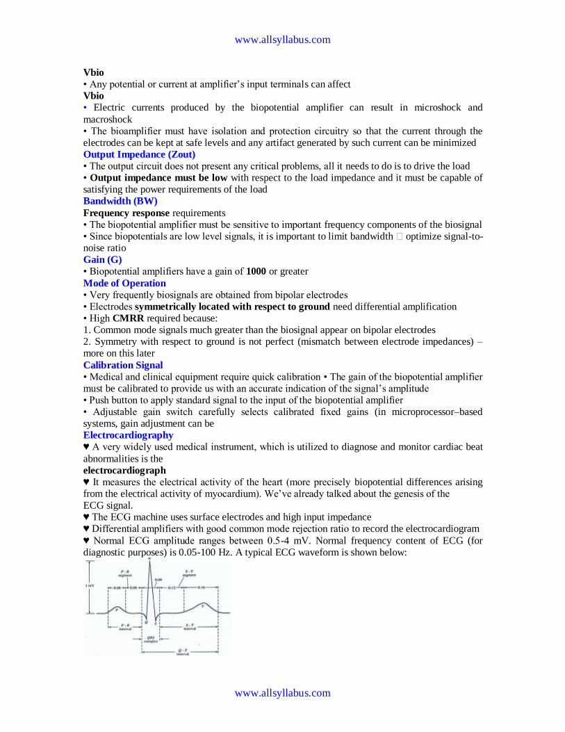
wwwallsyllabuscom
wwwallsyllabuscom
Vbio
bull Any potential or current at amplifierrsquos input terminals can affect Vbio
bull Electric currents produced by the biopotential amplifier can result in microshock and
macroshock bull The bioamplifier must have isolation and protection circuitry so that the current through the electrodes can be kept at safe levels and any artifact generated by such current can be minimized Output Impedance (Zout)
bull The output circuit does not present any critical problems all it needs to do is to drive the load bull Output impedance must be low with respect to the load impedance and it must be capable of satisfying the power requirements of the load Bandwidth (BW)
Frequency response requirements bull The biopotential amplifier must be sensitive to important frequency components of the biosignal bull Since biopotentials are low level signals it is important to limit bandwidth optimize signal-to-noise ratio Gain (G)
bull Biopotential amplifiers have a gain of 1000 or greater
Mode of Operation
bull Very frequently biosignals are obtained from bipolar electrodes bull Electrodes symmetrically located with respect to ground need differential amplification bull High CMRR required because 1 Common mode signals much greater than the biosignal appear on bipolar electrodes 2 Symmetry with respect to ground is not perfect (mismatch between electrode impedances) ndash more on this later
Calibration Signal
bull Medical and clinical equipment require quick calibration bull The gain of the biopotential amplifier must be calibrated to provide us with an accurate indication of the signalrsquos amplitude bull Push button to apply standard signal to the input of the biopotential amplifier bull Adjustable gain switch carefully selects calibrated fixed gains (in microprocessorndashbased systems gain adjustment can be Electrocardiography
hearts A very widely used medical instrument which is utilized to diagnose and monitor cardiac beat
abnormalities is the electrocardiograph
hearts It measures the electrical activity of the heart (more precisely biopotential differences arising from the electrical activity of myocardium) Wersquove already talked about the genesis of the ECG signal hearts The ECG machine uses surface electrodes and high input impedance hearts Differential amplifiers with good common mode rejection ratio to record the electrocardiogram
hearts Normal ECG amplitude ranges between 05-4 mV Normal frequency content of ECG (for diagnostic purposes) is 005-100 Hz A typical ECG waveform is shown below
wwwallsyllabuscom
wwwallsyllabuscom
Obviously all human hearts are not the same and this leads into variability in different parts of the ECG signal Significant diagnostic features of the ECG signal are hearts Duration of component parts of the signal
hearts Polarities and magnitudes hearts The details of the ECG signal and the degree of variability in different parts of the ECG signal is shown below
The QRS amplitude polarity time duration the RR interval (indicator of heartbeat per min) and the T-wave amplitude are some very important and distinctive features of the ECG signal The heart rate in BPM = Beats Per Minute) is simply = 60 (RR interval in seconds)
Some ECG waveform abnormalities that may indicate illness are hearts An extended PR interval may be diagnosed as AV node block hearts A widening of the QRS complex may indicate conduction problems in the bundle of His hearts An elevated ST segment may indicate occurrence of myocardial Infarction (MI) hearts A negative polarity in the T wave may be due to coronary insufficiency ECG Leads
Normal ECG recordings for the standard lead connections leads I II and III (Lead II provides
the strongest signal) Obviously all human hearts are not the same and this results into a high degree of variability
wwwallsyllabuscom
wwwallsyllabuscom
hearts Note the degree of variability of different parts of the ECG Signal
Some abnormalities that may indicate illness hearts An extended P-R interval may be diagnosed as AV node block hearts Widening of the QRS complex conduction problems in the bundle of His hearts Elevated ST segment may indicate occurrence of MI
hearts Negative polarity T wave may be due to coronary insufficiency QRS amplitude polarity time domain PR interval (indicator of heat beat per min amp T-wave amplitude are some very important
distinctive features 1 Loss
2 Origin of the ECG signal
hearts We have already covered this concept extensively in the previous lectures (The Dipole filed of the heart the Eindhovenrsquos Triangle the electrical circuit model for the electrocardiographic problem etc)
hearts Standard Limb Leads (I II III)
wwwallsyllabuscom
wwwallsyllabuscom
hearts The The lead wires are color-coded according to some conventions One example is White ndashRA
(Right Arm) Black ndash LA (Left Arm) Green ndash RL (Right Leg) Red ndash LL (Left Leg) and Brown
ndash C (Chest) Note There is a CM (common mode) amplifier connected to the right leg We will discuss this in detail later Augmented Limb Leads
These leads offer a free 50 increase over leads VR VL and VF connections (unipolar leads) with respect to Wilson terminal AVR = -I ndash III2 AVL = I ndash II2 aVF = II ndash I2
wwwallsyllabuscom
wwwallsyllabuscom
Each measurement is made from the reflected limb and the average of the other two limbs Chest Leads (Precordial)
Chest Lead Anatomical Positions
V1 - 4th intercostal space ndash Right sternal margin
V2 - 4th intercostal space ndash Left sternal margin
V3 ndash Midway between V2 amp V4
V4 ndash 5th intercostal space on mid-clavicular line
V5 ndash Same as V4 on the anterior axillary line
V6 ndash Same as V5 on the mid-axillary line
12 Lead Clinical Electrocardiography
The ECG Machine
Most representative Specs
wwwallsyllabuscom
wwwallsyllabuscom
bull Zin = 10 MΩ bull Frequency response = 005 ndash100 Hz bull Strip Chart Recorder Speed = 25 mmsec bull Fast Speed = 100 mmsec
For detailed Specs Refer to the Table in your text ―Summary of performance requirements for electrocardiographs Location of the Heart bull The heart is located between the lungs behind the sternum and above the diaphragm bull It is surrounded by the pericardium bull Its size is about that of a fist and its weight is about 250-300 g bull Its center is located about 15 cm to the left of the midsagittal plane
Anatomy of the heart
bull The walls of the heart are composed of cardiac muscle called myocardium
bull It consists of four compartments ndash the right and left atria and ventricles
The Heart Valves
bull The tricuspid valve regulates blood flow between the right atrium and right
ventricle
bull The pulmonary valve controls blood flow from the right ventricle into the
pulmonary arteries
bull The mitral valve lets oxygen-rich blood from your lungs pass from the left atrium
into the left ventricle
bull The aortic valve lets oxygen-rich blood pass from the left ventricle into the aorta
then to the body
Blood circulation via heart
wwwallsyllabuscom
wwwallsyllabuscom
bull The blood returns from the systemic circulation to the right atrium and from
there goes through the tricuspid valve to the right ventricle
bull It is ejected from the right ventricle through the pulmonary valve to the lungs
bull Oxygenated blood returns from the lungs to the left atrium and from there
through the mitral valve to the left ventricle
bull Finally blood is pumped through the aortic valve to the aorta and the systemic
circulation
Electrical activation of the heart
bull In the heart muscle cell or myocyte electric activation takes place by means of
the same mechanism as in the nerve cell ie from the inflow of Na ions across
the cell membrane
bull The amplitude of the action potential is also similar being 100 mV for both nerve
and muscle
bull The duration of the cardiac impulse is however two orders of magnitude longer
than in either nerve cell or sceletal muscle cell
bull As in the nerve cell repolarization is a consequence of the outflow of K ions
bull The duration of the action impulse is about 300 ms
Mechanical contraction of Cardiac Muscle
bull Associated with the electric activation of cardiac muscle cell is its mechanical
contraction which occurs a little later
bull An important distinction between cardiac muscle tissue and skeletal muscle is
that in cardiac muscle activation can propagate from one cell to another
in any direction
bull Electrical signal begins in the sinoatrial (SA) node natural pacemaker causes
the atria to contract
bull The signal then passes through the atrioventricular (AV) node
ndash sends the signal to the ventricles via the ―bundle of His
ndash causes the ventricles to contract
The Conduction System
wwwallsyllabuscom
wwwallsyllabuscom
The Action Potential
Recording an AP requires the isolation of a single cell Microelectrodes (with tips a few μm across) are used to stimulate and record the response A typical AP is 2-4ms long with an amplitude of about 100Mv The Electroencephalogram EEG
EEG is the graphical representation of the electrical activity of the brain Very commonly used to diagnose certain neurological disorders such as epilepsy
More recently also investigated whether it can detect various forms of dementia or schizophrenia EEG is the specific recording obtained using the scalp electrodes from the surface of the skull
During surgery electrodes may also be placed directly on the cortex The resulting signal is then electrocorticogram (ECoG) Just like ECG EEG is also obtained using several different electrodes places on different regions of the head brain
The Event Related
wwwallsyllabuscom
wwwallsyllabuscom
Potentials ndash ERPs
ERPs are really EEGs obtained under a specific protocol that requires the patient to response to certain stimuli ndash hence event related potentials Also called evoked potentials these signals can be used to diagnose certain
neurological disorders such as dementia and they can also be used as a liedetector bull The oddball paradigm
bull The guilty knowledge test
Electroretinogram ERG
The ERG is the record of the retinal action currents produced by the retina in response to a light stimulus It measures the electrical responses of the light-sensitive cells (such as rods and cones)
The stimuli are often a series of light flashes or rotating patterns The ERG is recorded using contact lens electrode that the subject wears while watching the stimuli
Phonocardiogram ndash PCG
The PCG is the graphic record of the heart sounds and murmurs It is thus a mechanical audio signal rather than an electrical signal Can be easily heard using a stethoscope Or can be converted into an electrical signal using a transducer Typically used to determine the disorders related to the heart valve since their
routine opening and closing create the well-known sounds bull S1 sounds First heart sounds ndash ventricular contractions move blood into atria closing of the AV (mitral and tricuspid) valves then semilunar valves open and blood ejected out of ventricles ndash immediately follows the QRS complex bull S2 sounds Second heart sounds ndash Closure of semilunar (aortic and pulmonary) valves bull Any unexpected sound may indicate a malfunctioning valve that causes the blood flow into out of a chamber when it should not Also called heart murmurs
wwwallsyllabuscom
wwwallsyllabuscom
Define ultrasound
bull Mechanical waves in different modalities (longitudinallateral) needs medium
to be propagated (solid liquid gas)
bull gt 20 kHz
bull Continuouspulsed
bull Sphericalplanarnarrow beamsurface waveLamb-wave
Physical phenomena behind ultrasound measurements
Transmission
bull
bull reflection
bull transit time
bull differences in propagation velocities
bull returns to transit time
bull doppler-shift in frequency
bull flow velocity
bull change of acoustic impedance
bull comparing to reference
bull interference of ultrasound waves (holography)
bull interaction of ultrasound and light (photoacousticz)
bull ultrasound needs medium for propagation it doesnrsquot propagate in vacuum
bull because mechanical waves need moving massunits and spring forces between
them
bull in acoustic emission the medium creates ultrasound (for example during pressure
changes) which is received by sensors
bull pulsed mode more common than continuous
bull continuous reguires separate transducers for transmitting and receicving
bull in pulsed mode an ultrasound burst is sent to the object and the same transducer is
switched to listen echoes
bull standing wave problem
bull in us-therapy pulsed mode gives more effective care without too much heating The Doppler Equation describes the relationship of the Doppler frequency shift to target velocity
wwwallsyllabuscom
wwwallsyllabuscom
The frequency difference is equal to the reflected frequency (FR) minus the originating frequency (FT) If the resulting frequency is higher then there is a positive Doppler shift and the object is moving toward the transducer but if the resulting frequency is lower there is a negative Doppler shift and it is moving away from the transducer In its simplest form it would be calculated as if
the ultrasound was parallel to the targetrsquos direction as shown in diagram A below However this would be a rare occurrence in clinical practice because the transducer is rarely pointed head on to a blood vessel In real life the ultrasound waves would approach the target at an angle called the Doppler angle ( ) On the following page diagram B shows the Doppler
equation used in general clinical situations which includes the Doppler angle The Doppler Angle
The ultrasound beam usually approaches the moving target at an angle called the Doppler angle ( ) This reduces the frequency shift in proportion to the cosine of this
angle If this angle is known then the flow velocity can be calculated The equation used is The Doppler Equation
equiv Doppler shift frequency (the difference between the transmitted and received frequencies) equiv transmitted frequency equiv reflected frequency
V equiv velocity of the blood flow towards the transducer C equiv velocity of sound in tissue θ equiv the angle between the sound beam and the direction of moving blood Where
The Doppler angle ( ) is also known as the angle of insonation It is estimated by the sonographer by a process known as angle correction which involves aligning an indicator on the duplex image along the longitudinal axis of the vessel
There are a few considerations that affect the performance of a Doppler examination that are inherent in the Doppler equation which are ndash The cosine of 90deg is zero so if the ultrasound beam is perpendicular to the direction of blood flow there will be no Doppler shift and it will appear as if there is no flow in the vessel ndash Appropriate estimation of the angle of insonation or angle correction is essential for the accurate determination of Doppler shift and blood flow velocity The angle of insonation should also be less than 60deg at all times since the
cosine function has a steeper curve above this angle and errors in angle correction will be magnified
The simplest Doppler devices use continuous wave (CW Doppler) rather than the pulsed wave used in more complex devices CW Doppler uses two transducers (or a dual element transducer) that transmit and receive ultrasound continuously The transmit and receive beams overlap in a Doppler sample volume some distance from the transducer face as
shown in the diagram below
volume) is the region of transmitting and receiving beam overlap (shaded region) Because there is continuous transducer transmission and reception echoes from all depths within the area arrive at the transducer simultaneously So although CW Doppler can determine the direction of flow it cannot discriminate the different depths where the motion originates The usefulness of CW Doppler devices is limited but they are used clinically to confirm blood flow in superficial vessels as they are good at detecting low velocities As they are easily portable this
can be done at the bedside or in the operating room Most other clinical applications require pulsed wave Doppler Pulsed Wave Doppler (PW Doppler)
wwwallsyllabuscom
wwwallsyllabuscom
Pulsed wave Doppler (PW Doppler) uses a single-element transducer that emits brief pulses of ultrasound energy The time interval between transmitting and then receiving the echoing sound can be used to calculate the depth from where the echo arises The Doppler sample volume can be chosen as to shape depth and position in sampling the flow
data For example the depth is chosen by processing only the signals that return to the transducer in a stipulated time For this technique the ultrasound system transmits a short pulse The eceiver is opened to detect the returning echoes only after a controlled delay and only for a specific duration This time-based gating of the receiving channel allows the definition of a fixed easuring distance which is often referred to as the Sample volume or Doppler gate Then the next ultrasound wave is transmitted The number of pulses transmitted by the system within a second is referred to as the pulse repetition frequency (PRF) The upper PRF limit is given by the time interval required for the echoes to arrive from a sample volume located at a
certain depth The greater the sample-volume depth the longer the time before the echoes are returned and the longer the delay between pulse transmission The greater the samplevolume depth the lower will be the maximum PRF setting Errors in the accuracy of the information arise if the velocities exceed a certain speed The highest velocity accurately measured is called the Nyquist limit Beyond this limit the errors that occur are referred to as aliasing Volume and flow measurement
Flow ndash volume of a liquidgas passing some point over a given time Gases are compressible
Benedict Roth Spirometer
Widely used for physiological amp clinical studies
Light bell moves with the ptrsquos breathing
Movement recorded by a pen on a rotating drum
Water seal prevents leakage of gas
Small seal minimises volume of gas dissolved in water
Suitable for measuring limited gas vols (few litres)
Pneumotachograph
wwwallsyllabuscom
wwwallsyllabuscom
Cardiac Output
Defn vol of blood pumped by the heart per min
CO = SV x HR
Norm ~ 5 lmin
Cardiac index ndash corrected for body surface area
Affected by
Met Rate ndash pregnancy hyperthyroid septic
Preload contractility afterload
Clinical indicators of CO imprecise
Affected by anaesthetic agents used in everyday practice
Provides estimate of
whole body perfusion
oxygen delivery
left ventricular function
Persistently low CO assoc with poor outcome
Methods
Fick method
Dilution techniques ndash dye thermal lithium
Pulse contour analysis- LiDCO amp PiCCO
Oesophageal doppler
TOE
Transthoracic impedance plethysmography
Inert gas through flow
Non-invasive cardiac output measurement
Fick Principle measure volume displacement
1st proposed 1870
―the total uptake or release of a substance by an organ is the product of the
blood flow through that organ and the arteriovenous concentration
difference of the substance
CO = O2 consumption (mlmin) art ndash mixed
venous O2 conc (mll)
Limited by cumbersome equipment sampling errors need for invasive
monitoring and steady-state haemodynamic and metabolic conditions
Indicator dilution techniques
―An indicator mixed into a unit volume of constantly flowing blood can be used to
identify that volume of blood in time provided the indicator remains in the system
between injection and measurement and mixes completely in the blood
wwwallsyllabuscom
wwwallsyllabuscom
Dye dilution
Inert dye ndash indocyanin green
Injected into pulmonary artery and arterial conc measured using a
calibrated cuvette densitometer
Plot indicator dilution curve (see diagram)
CO derived from area under curve
Indicator Dilution Curve
Cardiac Output Measurement
wwwallsyllabuscom
wwwallsyllabuscom
Why bother to check blood pressure
For each 20 mm rise in systolic blood pressure or 10 mm rise in diastolic blood
pressure over 11575
Risk of stroke increases
Risk of heart disease doubles
Risk of renal failure increases
Systolic and diastolic blood pressure
Systolic blood pressure is the highest pressure in the arteries just after the heart
beats
Diastolic blood pressure is the lowest pressure in the arteries just before the heart
beats
Blood pressure is measured indirectly by blood pressure cuff
(sphygmomanometer)
Inflating cuff increases pressure until it cuts off arterial circulation to the arm
Deflating cuff decrease pressure by 2 to 3 mm of mercury per second until blood
first enters the artery creating turbulence this causes a sound with each heartbeat
Sounds continue with each heartbeat until pressure lowers to the lowest pressure
in the artery then turbulence stops so the sound stops
Systolic blood pressure is the cuff pressure at the first sounds diastolic is the cuff
pressure just before the sounds stop
Phase 1 sharp thuds start at systolic blood pressure
Phase 2 blowing sound may disappear entirely (the auscultatory gap )
Phase 3 crisp thud a bit quieter than phase 1
Phase 4 sounds become muffled
Phase 5 end of sounds -- ends at diastolic blood pressure
wwwallsyllabuscom
wwwallsyllabuscom
Make sure the cuff is the right size - its width should be at least 40 of the arms
circumference The cuff will overestimate blood pressure if too small and
underestimate if too large
Place the cuff snugly on patients proximal arm on skin ( not cloth) centered over
the brachial artery (most cuffs have markings)
Support the patients arm at heart level using your arm or a desk
Your patient should sit in the chair for 5 minutes before BP is measured and
should have no caffeine or nicotine for 30 minutes before (JAMA 273 p1211-
1218 1995)
With fingers palpating radial or brachial artery inflate cuff rapidly until you cant
feel the pulse then 20 mm higher
Release cuff at 2 to 3 mm Hg per second until you again feel the pulse this is the
palpable systolic pressure
Wait 30 seconds before measuring blood pressure
Measuring palpable pressure first avoids risk of seriously underestimating blood
pressure because of the auscultatory gap (mistaking Korotkoff phase 3 for phase
1) Many doctors skip this step for time reasons and instead pump cuff to 200 mm
Hg at the next step)
Phase 2 of the Korotkoff sounds can be inaudible - especially in older patients
with systolic hypertension who are at especially high risk of stroke Inflating the
cuff until you dont hear sounds can give you a reading of 14086 when the
patients actual blood pressure is 22086 Most physicians are pressed for time so
they instead inflate the cuff to 200 mm which is beyond the auscultatory gap in
most patients But palpable systolic blood pressure is according to research more
reliable
Place bell of stethoscope (diaphragm is acceptable) over brachial artery
Rapidly pump the cuff to 20 to 30 mm Hg above palpable systolic pressure
Release pressure in the cuff by 2 to 3 mm Hg per second and listen for Korotkoff
sounds including systolic (first) and diastolic (last)
Record as systolicdiastolic Check in both arms the first time you check a
patients blood pressure It may differ by 10 mm Hg or more
If the sounds continues to zero record diastolic blood pressure as the point when
sounds become muffled (phase 4) over zero eg 130700 or just as 13070
Rate
ndash Number of beats in 30 seconds x 2
Strength
ndash Bounding strong or weak (thready)
Regularity
ndash Regular or irregular
You need three readings on two occasions to diagnose hypertension unless blood
pressure is very high
Normal blood pressure in children is
wwwallsyllabuscom
wwwallsyllabuscom
ndash 10255 at 1 year 11269 at 5 years 11978 at 10 years
Blood pressures in adults (JNC VII JAMA 2892560-72 2003)
ndash Normal lt120lt80
ndash Prehypertensive 120-13980-89
ndash Stage 1 hypertension 140-15990-99
ndash Stage 2 hypertension gt160gt100
Adult 60 to 100
Newborn 120-170
1 year 80-160
3 years 80-120
6 years 75-115
10 years 70-110
How to measure observe rise and fall of chest
In infants count for 60 seconds in adults 15 or 30 seconds
Normal respiration Adults 12 to 20
Children
newborn 30-80
1 year 20-40
3 years 20-30
6 years 16-22
Rate
Number of breaths in 30 seconds x 2
Quality
Character of breathing
Rhythm
Regular or irregular
Effort
Normal or labored
Noisy respiration
Normal stridor wheezing snoring gurgling
Depth
Shallow or deep
pH electrode
wwwallsyllabuscom
wwwallsyllabuscom
Governing equation is the Nernst Equation
pCO2 Electrode
The measurement of pCO2 is based on its linear relationship with pH over the range of
10 to 90 mm Hg
The dissociation constant is given by
Taking logarithms
pH = log[HCO3-] ndash log k ndash log a ndash log pCO2
Diathermy
In the natural sciences the term diathermy means electrically induced heat and is commonly
used for muscle relaxation It is also a method of heating tissue electromagnetically or
ultrasonically for therapeutic purposes in medicineContents
[hide]
1 Heating uses 2 Surgical uses
3 Trivia
4 References
E
RT
nF
H
HH
i
ln
0
H O CO H CO H HCO2 2 2 3 3
k
H HCO
a pCO
3
2
wwwallsyllabuscom
wwwallsyllabuscom
Heating uses
Ultrasonic diathermy refers to heating of tissues by ultrasound for the purpose of
therapeutic deep heating No tissue is ordinarily damaged hence it is generally used in
biomedical applications
Electric diathermy uses high frequency alternating electric or magnetic fields sometimes
with no electrode or device contact to the skin to induce gentle deep tissue heating by
induction Again no tissue is ordinarily damaged
Surgical uses
Surgical diathermy is usually better known as electrosurgery (It is also referred to
occasionally as electrocautery but see disambiguation below) Electrosurgery and
surgical diathermy involve the use of high frequency AC electrical current in surgery as
either a cutting modality or else to cauterize small blood vessels to stop bleeding This
technique induces localized tissue burning and damage the zone of which is controlled
by the frequency and power of the device Some sources[1]
insist that electrosurgery be
applied to surgery accomplished by high frequency AC cutting and that
electrocautery be used only for the practice of cauterization with heated nichrome wires
powered by DC current as in the handheld battery-operated portable cautery tools
Trivia
Medical Diathermy devices were used to cause interference to German radio beams used
for targeting night time bombing raids in WWII during the Battle of the Beams
I Diathermy
A Therapeutic use
1 Generation of local heating by high-frequency electromagnetic waves
2 Capacitance techniquemdashbody is placed in an electric field
a Dipolesmdashstructures with positive and negative poles
b Structures with large numbers of dipoles have a greater capacitance to store an electrical charge
c Greatest heating occurs in tissues with fewer dipoles particularly fatty tissues
3 Rapid rotation of dipoles causing mechanical friction and movement of electrons results in local heating
4 Inductance techniquemdashbody is not placed in an electric field
a Magnetic waves generated by driving current through a coiled wire
b Magnetic field creates currents in tissues
wwwallsyllabuscom
wwwallsyllabuscom
c Greatest heating occurs in tissues with low impedance especially muscle
B Precautions and contraindications
1 Diathermy should not be used
a Over metal implants and cardiac pacemakersmdashmore research needed regarding its use over metallic fixations
b Near the uterus of a pregnant woman or near the abdomen or back of a woman who might be pregnant
c On individuals with infections
d On individuals with acute inflammation
e Over moist open wounds
f On patients with malignant tumors
g Over large joint effusions
C Pulsed electromagnetic fields and diathermy
1 Can be pulsed to decrease total energy transmitted to the tissues
2 Short-wave diathermy can be adjusted into a nonthermal range
a Classified as pulsed electromagnetic field (PEMF) or
b Pulsed radio frequency energy (PRFE)
3 Important reclassification as diathermy implies heating
D Efficiency of diathermy and PEMF therapy for musculoskeletal conditions
1 Current research is limited but results suggest that diathermy enhances
treatments directed at soft tissue stretching
2 Some studies suggest that PEMF may speed wound healing and promote healing of nonunion fractures
Diathermy is a modality that uses electromagnetic energy to heat deeper tissues
Diathermy is more effective than ultrasound at heating a larger area of deep tissues
The athletic trainer must identify and respect contraindications to application of ultrasound and diathermy
Pulsed ultrasound and diathermy are used to treat slow-to-heal lesions including skin ulcers and nonunion fractures and may be able to facilitate repair of other tissues including ligaments and tendons
Diathermy
Definition
In diathermy high-frequency electrical currents are used to heat deep muscular tissues The heat increases blood flow speeding up recovery Doctors also use diathermy in surgical procedures by sealing blood vessels with electrically heated probes
The term diathermy is derived from the Greek words therma meaning heat and dia meaning through Diathermy literally means heating through
Origins
The therapeutic effects of heat have long been recognized More than 2000 years ago the Romans took advantage of heat therapies by building hot-spring bathhouses Since then various
wwwallsyllabuscom
wwwallsyllabuscom
methods of using heat have evolved In the early 1890s French physiologist Arseacutene dArsonval began studying the medical application of high-frequency currents The term diathermy was coined by German physician Carl Franz Nagelschmidt who designed a prototype apparatus in 1906 Around 1925 United States doctor J W Schereschewsky began studying the physiological
effects of high-frequency electrical currents on animals It was several years however before the fundamentals of the therapy were understood and put into practice
Benefits
Diathermy can be used to treat arthritis bursitis and other conditions involving stiff painful joints It is also used to treat pelvic infections and sinusitis A benefit of diathermy is that it is a painless procedure that can be administered at a clinic Also if the treatment relieves pain then patients can discontinue pain killers and escape their high cost and side effects
Description
Diathermy involves heating deep muscular tissues When heat is applied to the painful area cellular metabolism speeds up and blood flow increases The increased metabolism and circulation accelerates tissue repair The heat helps the tissues relax and stretch thus alleviating
stiffness Heat also reduces nerve fiber sensitivity increasing the patients pain threshold
There are three methods of diathermy In each energy is delivered to the deep tissues where it is converted to heat The three methods are
Shortwave diathermy The body part to be treated is placed between two capacitor plates Heat is generated as the high-frequency waves travel through the body tissues between the plates Shortwave diathermy is most often used to treat areas like the hip which is covered with a dense tissue mass It is also used to treat pelvic infections and sinusitis The treatment reduces inflammation The Federal Communications Commission regulates the frequency allowed for short-wave diathermy treatment Most machines
function at 2733 megahertz Ultrasound diathermy In this method high-frequency acoustic vibrations are used to
generate heat in deep tissue Microwave diathermy This method uses radar waves to heat tissue This form is the
easiest to use but the microwaves cannot penetrate deep muscles
Diathermy is also used in surgical procedures Many doctors use electrically heated probes to seal blood vessels to prevent excessive bleeding This is particularly helpful in neurosurgery and eye surgery Doctors can also use diathermy to kill abnormal growths such as tumors warts and
infected tissues
Preparations
To keep patients from sweating patients are usually asked to remove clothing from the body part being treated If a patient sweats the electrical currents may pool in the area causing burns Also clothing containing metal must be removed as must earrings buttons barrettes or zippers that contain metal Watches and hearing aids should be removed because the therapy may affect their function
wwwallsyllabuscom
wwwallsyllabuscom
Practitioners of surgical diathermy should steer clear of alcohol-based solutions to prepare and cleanse the skin These preparations can create a flammable vapor and cause burns and fires
Precautions
Patients with metal implants should not undergo diathermy treatment because the metal can act as a conductor of heat and result in serious internal burns Female patients with metallic uterine implants such as an IUD should avoid treatment in the pelvic area Diathermy should not be
used in joints that have been replaced with a prosthesis or in those with sensory impairment who may not be able to tell if they are burning Furthermore pulsed shortwave diathermy should be avoided during pregnancy as it can lead to abnormal fetal development
Patients with hemophilia should avoid the treatment because the increased blood flow could cause them to hemorrhage
Side effects
Some patients may experience superficial burns Since the therapy involves creating heat care must be taken to avoid burns particularly in patients whose injuries have caused decreased sensitivity to heat Also diathermy may affect pacemaker function
Female patients who receive treatment in the lower back or pelvic area may experience an
increased menstrual flow
Research amp general acceptance
For years physiotherapists and physical therapists have used diathermy as a routine part of physical rehabilitation
Electrical Safety
Electrical safety is very important in hospitals as patients may be undergoing a diagnostic or treatment procedure where the protective effect of dry skin is reduced Also patients may be unattended unconscious or anaesthetised and may not respond normally to an electric current Further electrically conductive solutions such as blood and saline are often present in patient treatment areas and may drip or spill on electrical equipment
Electric Current Leakage Current
Extension Leads Double Adaptors Equipment Classification
Class I Class II Defibrillator-Proof
Protective Devices
Residual Current Devices (RCD) Line Isolation overload Monitors (LIMs) Equipment Earthing
Area Classification
wwwallsyllabuscom
wwwallsyllabuscom
Body Protection Area Cardiac Protected Area
Other Electrical Issues Extension Leads
Double Adapters Main Extension Devices Power Boards Installation of Additional Power Points
Electric Current
Injuries received from electric current are dependent on the magnitude of current the pathway
that it takes through the body and the time for which it flows
The nature of electricity flowing through a circuit is analogous to blood flowing through the circulatory system within the human body In this analogy the source of energy is represented by the heart and the blood flowing through arteries and veins is analagous to current flowing
through the conductors and other components of the electric circuit
The application of an electric potential to an electric circuit generates a flow of current through conductive pathways This is analogous to the changes in blood pressure caused by contraction of cardiac muscle that causes blood to flow into the circulatory system For electric current to flow there must be a continuous pathway from the source of potential through electrical components and back to the source
Leakage Current
Electrical components and systems are encased in non conducting insulation to ensure that the electric current is contained and follows the intended pathways If the insulation deteriorates or breaks down current will leak through the insulation barrier and flow to earth This may
be through the protective earth conductor or through the operator
Medical equipment and clinical areas are fitted with a number of protective devices to protect the patient and operator from harmful leakage currents
Extension Leads
Extension leads are not permitted in clinical areas of RCH organisations They may cause high
earth resistance and excessive earth leakage current An extension lead can allow equipment to be powered from areas other than the relevant protected treatment area The power from the other area may not be protected to the same level as the power in the treatment area
wwwallsyllabuscom
wwwallsyllabuscom
As the connection between the extension lead and the equipment mains cable is often on the floor there is a high danger from fluid spills tripping and damage to the mains cable by trolleys when an extension lead is used
Double Adaptors
Double adaptors must not be used in RCH organisations They may not sit securely in a wall
outlet may not be able to provide adequate earth protection and may cause overloading overheating fire or loss of electrical supply
EQUIPMENT CLASSIFICATIONS
There are several methods of providing protection for operators and patients from electrical faults
and harmful leakage current
Class I
Class I equipment is fitted with a three core mains cable containing a protective earth wire
Exposed metal parts on class I equipment are connected to this earth wire
Should a fault develop inside the equipment and the exposed metal comes into contact with the mains the earthing conductor will conduct the fault current to ground Regular testing procedures ensure that earthing conductors are intact as the integrity of the earth wire is of vital importance
Class II
Class II equipment is enclosed within a double insulated case and does not require earthing
conductors Class II equipment is usually fitted with a 2-pin mains plug An internal electrical fault is unlikely to be hazardous as the double insulation prevents any external parts from becoming alive Class II or double insulated equipment can be identified by the class II symbol on the cabinet
Class II Symbol
wwwallsyllabuscom
wwwallsyllabuscom
Defibrillator-Proof
Some medical equipment within the hospital is classified as defibrillator proof When a
defibrillator is discharged through a patient connected to defibrillator proof equipment the equipment will not be damaged by the defibrillators energy Defibrillator proof equipment can remain connected to the patient during defibrillation It is identified by one of the following
symbols
Defibrillator proof symbols
Body protected Cardiac protected
PROTECTIVE DEVICES
Most patient care areas in the hospital are fitted with protective devices These devices are
regularly tested in accordance with the relevant guidelines published by Standards Australia The level of protection provided is dependent upon the device and the area in which it is located
Residual Current Devices (RCD)
RCDs (safety switches) are used in patient treatment areas to monitor and protect the mains supply RCDs sense leakage currents flowing to earth from the equipment If a significant leakage current flows the RCD will detect it and shut off the power supplied to the equipment
within 40 milliseconds Hospital RCDs are more sensitive than those fitted in homes A hospital RCD will trip at 10 milliamperes leakage current
Power outlets supplied through an RCD have a Supply Available lamp The lamp will extinguish
when the RCD trips due to excessive leakage current
wwwallsyllabuscom
wwwallsyllabuscom
Resetting a RCD
Lamp indicates supply is no longer available Disconnect all equipment from the supply Operate the reset button or lever on the supply panel and the Supply Available lamp
should illuminate If not contact Biomedical Engineering
Connect an item of equipment If the RCD trips again then this is the faulty item and should be labelled and sent to Biomedical Engineering
If the RCD does not trip continue connecting equipment until the RCD trips The last piece of equipment connected to the supply is most likely to be faulty as it will have caused the RCD to trip Remove the faulty item from service label it and send to Biomedical Engineering as mentioned above
Line Isolation overload Monitors (LIMs)
In critical life support applications where loss of power supply cannot be tolerated special power outlets powered by isolation transformers are installed
Line Isolation Monitors are installed to continually monitor electrical leakage in the power
supply system If an electrical fault develops in a medical device connected to an isolated power outlet the LIM will detect the leakage current The LIM will alarm and indicate the level of leakage current but will not shut off the electric supply
The faulty equipment can be identified by un plugging one item of equipment at a time from the supply until the alarm stops sounding Equipment that is not faulty may be reconnected Faulty equipment should be appropriately labelled and sent to Biomedical Engineering for repair
The LIM also monitors how much power is being used by the equpiment connected to it If too much power is being used the LIM will alarm and indicate that there is an overload The power used must be reduced immediately by moving some equipment to another circuit as soon as possible until the alarm stops sounding Failure to reduce the load on the LIM will result in the circuit breaker tripping and loss of power to the circuit
wwwallsyllabuscom
wwwallsyllabuscom
Equipotential Earthing
Equipotential earthing is installed in rooms classified as Cardiac Protected electrical areas
Equipotential earthing in treatment areas used for cardiac procedures is intended to minimise any voltage differences between earthed parts of equipment and any other exposed metal in the room
This reduces the possibility of leakage currents that can cause microelectrocution when the patient comes into contact with multiple items of equipment or if the patient happens to come
into contact with metal items in the room whilr they are connected to a medical device
All conductive metal in an equipotential area is connected to a common equipotential earth point with special heavy duty cable
AREA CLASSIFICATIONS
Body Protected Area
These areas are designed for procedures in which patients are connected to equipment that lowers the natural resistance of the skin Applied parts such as electrode gels conductive fluids entering the patient metal needles and catheters provide an easy pathway for current to flow
The main occurrence of injury from Body-Type procedures is from high current levels causing electric shock A direct connection to the patients heart is not present so the risk of Microelectrocution - fibrillation from minute current levels - is reduced
Residual Current Devices (RCD) or Isolation Transformers and Line Isolation Monitors (LIMiacutes) are used in Body Protected areas to provide protection against electrocution from high leakage currents Body-Protected Areas are identified with this sign
Cardiac Protected Area
Where the procedure involves placing an electrical conductor within or near the heart protection against fibrillation induced from small leakage currents is required Electrical conductors used in these procedures include cardiac pacing electrodes intracardiac ECG electrodes and intracardiac catheters
wwwallsyllabuscom
wwwallsyllabuscom
Equipotential earthing in conjunction with RCDs or LIMs provides protection against microelectrocution in Cardiac-Type procedures
Fault currents are reduced to magnitudes that are unlikely to induce fibrillation Used in conjunction with RCDs or LIMs the magnitude and duration of any fault currents
sourced from equipment are limited Cardiac-Protected Areas are identified with this sign
Other electrical issues
This policy aims to provide guidance to those who find that they need more electrical outlets than
those available or that the existing electrical outlets are inconveniently located
As extension leads and multiple outlet power boards can introduce additional hazards into an area the following procedures should be observed
Extension leads
Approved extension leads (AS 3760 1996) may be used in some areas within the hospital but MUST NOT BE USED IN PATIENT AREAS All electrical extension leads must be tagged with an Engineering Department maintenance tag and require a yearly safety inspection and test via
the Engineering Department
Double adapters
Double adapters may cause overloading or equipment earthing problems and are not to be used in
WCH
Mains extension device
The only mains extension device that is to be used in Patient care areas is the 4-way or 8-way
portable Core Balance Unit
The Biomedical Engineering Department must approve all units prior to use These units contain a safety switch and can detect excessive leakage current and disconnect the power in the event of a hazardous situation
wwwallsyllabuscom
wwwallsyllabuscom
Care must be exercised in the use of a portable Core Balance Unit It should be located off the
floor and in a position that will protect it from physical abuse and possible entry of fluids These devices are expensive and easily damaged The device must be sent to Biomedical Engineering every 6 months for safety testing
Power boards
Approved multiple-outlet power boards can be used across RCH but must not be used in patient
care areas except areas approved by the Biomedical Engineering Department
The power boards must have overload protection be fitted with internal safety shutters that protect unused outlets and be fitted with an onoff switch for each outlet
Medical Laser Applications
The main research subjects of the group of Medical Laser Application are in the field of online
monitoring and diagnostics as well as the development of new therapheutic methods The main focus in research is based upon the use of ultrashort (fs) laser pulses
The main advantage of ultrashort laser pulses is the extrem short interaction time which suppress any unwanted side effects of the laser irradiation of the tissue Secondly the broad spectra of the fs laser pulses give the advantage to use the same laser pulses for diagnostic applications like the optical coherence tomography (OCT)
wwwallsyllabuscom
wwwallsyllabuscom
Following research subjects have special attention at the moment
Optical Coherence Tomographie (OCT)
A fs-laser light source can be used to obtain images from inside the tissue when the pulses are coherent superposed This group uses this application to obtain an inside view of the vocal fold and to measure the outline of the crystalline lens
Refractive Surgery
The principle of getting rid of glasses with the help of a laser surgery (fs-LASIK) is underlying rapid improvements concerning precision and safety New technologies were evaluated and transferred very fast in cooperation with an industrial partner
Treatment of Presbyopia
The flexibility of the crystalline lens can be increase by pricise cuts which are induce by fs laser pulse inside the lens The treatment of the presbyopia is thinkable
wwwallsyllabuscom
wwwallsyllabuscom
Ultrafast Physics
Whereas the applications of ultrashort laser pulses increase rapidly is the physics of the interaction between the laser pulses and tissue in many case not fully understood To obtain improved knowledge of the interaction for many fs-laser application this group preforms numerical simulations as well as fundamental experiments
Thermography
Thermography thermal imaging or thermal video is a type of infrared imaging Thermographic cameras detect radiation in the infrared range of the electromagnetic spectrum
(roughly 900ndash14000 nanometers or 09ndash14 microm) and produce images of that radiation Since infrared radiation is emitted by all objects based on their temperatures according to the black body radiation law thermography makes it possible to see ones environment with or without visible illumination The amount of radiation emitted by an object increases with temperature therefore thermography allows one to see variations in temperature (hence the name) When viewed by thermographic camera warm objects stand out well against cooler backgrounds humans and other warm-blooded animals become easily visible against the environment day or
night As a result thermographys extensive use can historically be ascribed to the military and security services
Thermal imaging photography finds many other uses For example firefighters use it to see through smoke find persons and localize the base of a fire With thermal imaging power lines maintenance technicians locate overheating joints and parts a telltale sign of their failure to eliminate potential hazards Where thermal insulation becomes faulty building construction technicians can see heat leaks to improve the efficiencies of cooling or heating air-conditioning Thermal imaging cameras are also installed in some luxury cars to aid the driver the first being
the 2000 Cadillac DeVille Some physiological activities particularly responses in human beings and other warm-blooded animals can also be monitored with thermographic imaging [1]
The appearance and operation of a modern thermographic camera is often similar to a camcorder Enabling the user to see in the infrared spectrum is a function so useful that ability to record their output is often optional A recording module is therefore not always built-in
Instead of CCD sensors most thermal imaging cameras use CMOS Focal Plane Array (FPA) The most common types are InSb InGaAs HgCdTe and QWIP FPA The newest technologies are using low cost and uncooled microbolometers FPA sensors Their resolution is considerably lower than of optical cameras mostly 160x120 or 320x240 pixels up to 640x512 for the most
expensive models Thermographic cameras are much more expensive than their visible-spectrum counterparts and higher-end models are often export-restricted Older bolometers or more
wwwallsyllabuscom
wwwallsyllabuscom
sensitive models as InSb require cryogenic cooling usually by a miniature Stirling cycle refrigerator or liquid nitrogen
Contents
[hide]
1 Difference between IR film and thermography 2 Advantages of Thermography
3 Limitations amp disadvantages of thermography 4 Applications 5 See also 6 External links
o 61 History of thermal imager manufacturers
[edit] Difference between IR film and thermography
Thermal imaging is going to be used on Mars to detect caves that could hold life
[edit] Advantages of Thermography
You get a visual picture so that you can compare temperatures over a large area
It is real time capable of catching moving targets Able to find deteriorating components prior to failure Measurement in areas inaccessible or hazardous for other methods It is a non-destructive test method
[edit] Limitations amp disadvantages of thermography
Quality cameras are expensive and are easily damaged Images can be hard to interpret accurately even with experience
Accurate temperature measurements are very hard to make because of emissivities Most cameras have plusmn2 or worse accuracy (not as accurate as contact) Training and staying proficient in IR scanning is time consuming Ability to only measure surface areas
[edit] Applications
Condition monitoring Medical imaging Night vision Research
Process control Non destructive testing Surveillance in security law enforcement and defense Chemical imaging
wwwallsyllabuscom
wwwallsyllabuscom
Thermal infrared imagers convert the energy in the infrared wavelength into a visible light video display All objects above 0 kelvins emit thermal infrared energy so thermal imagers can passively see all objects regardless of ambient light However most thermal imagers only see objects warmer than -50 degC
The spectrum and amount of thermal radiation depend strongly on an objects surface
temperature This makes it possible for a thermal camera to display an objects temperature However other factors also influence the radiation which limits the accuracy of this technique For example the radiation depends not only on the temperature of the object but is also a function of the emissivity of the object Also radiation also originates from the surroundings and is reflected in the object and the radiation from the object and the reflected radiation will also be influenced by the absorption of the atmosphere
Endoscopy
This page is about having an endoscopy - a test that looks at the inside of your digestive system
There is information on
What endoscopy is
Why you may have an endoscopy
Having an endoscopy
Your results
What an endoscopy is
An endoscopy is a test that looks inside the body The endoscope is a long flexible tube that can
be swallowed It has a camera and light inside it Some doctors call it a telescope
wwwallsyllabuscom
wwwallsyllabuscom
Why you may have an endoscopy
You are most likely to have an endoscopy to look at the inside of your
Gullet (oesophagus)
Stomach
Duodenum - the first part of the small bowel that attaches to the stomach
Large bowel (colon)
Your doctor may want to see inside because you have symptoms such as abnormal bleeding or
difficulty swallowing The doctor can look down the endoscope and see if there are any growths
or other abnormal looking areas Also through the endoscope the doctor can take samples
(biopsies) of any abnormal looking tissues
There is more detailed information about having a colonoscopy in the bowel cancer section of
CancerHelp UK Below is information about having other types of endoscopy
Having an endoscopy
You can have this test as an out patient Most people have a choice between having the test while
they are awake or after having a medicine to make them drowsy (a sedative) Your hospital may
not be happy for you to have a sedative if you live alone and will have no one to look after you
when you go home If you live alone but really want sedation your hospital may allow you to
stay overnight You should ask your doctor about this as early as possible as extra arrangements
will need to be made beforehand
If you dont have a sedative you will have a spray to numb the back of your throat and make it
easier for you to swallow the endoscopy tube
If you would prefer to be asleep during the test you will have an injection to make you very
drowsy just before the test You will need to take someone with you to the hospital appointment
You wont be able to drive for the rest of the day and should have someone to go home with you
You cant eat or drink for about 8 hours before the test so that your stomach and duodenum are
empty Your doctor will give you written instructions about this beforehand or they may arrive
with your appointment letter When you get to the clinic you may be asked to take your upper
clothing off and put on a hospital gown Some hospitals prefer to use gowns because your clothes
wont get messy Once you are ready you get onto the bed or X-ray couch When you are lying
comfortably you have the sedative injection to make you very drowsy Or your doctor will spray
the back of your throat to numb it
Once the sedative or throat spray has worked the doctor will pass the endoscope tube down your
wwwallsyllabuscom
wwwallsyllabuscom
throat to the area being investigated Your doctor will ask you to swallow as the tube goes down
but if youve had a sedative you wont remember that afterwards If there are any abnormalities
the doctor will take pieces of tissue from the abnormal looking area to send to the laboratory for
closer inspection under a microscope These tissue samples are called biopsies
When the test is over you will need to rest for a while If youve had a sedative you may not
remember much (if anything) about the test once you have come round You should be able to go
home the same day
The results
It can take time for test results to come through How long will depend on why you are having
the test Usually the doctor who carries out the endoscopy dictates a report straight way The
report is typed up by the department secretary and goes to your specialist who gives the results to
you If your GP has sent you for the test the results will go directly to the GP surgery
Understandably waiting for results can make you anxious It usually takes a couple of weeks for
the results to come through If your doctor needed them urgently it would have been noted on
the test request form and the results will be ready sooner than that Try to remember to ask your
doctor how long you should expect to wait for the results when you are first asked to go for the
test If it is not an emergency and you have not heard a couple of weeks after your test ring your
doctors secretary to check if they are back
Endoscopy
From Wikipedia the free encyclopedia
Jump to navigation search
Endoscopic images of a duodenal ulcer
wwwallsyllabuscom
wwwallsyllabuscom
A flexible endoscope
Endoscopy means looking inside and typically refers to looking inside the human body for medical reasons using an instrument called an endoscope Endoscopy can also refer to using a borescope in technical situations where direct line-of-sight observation is not feasible
Contents
[hide]
1 Overview 2 Components 3 Uses
4 History 5 Risks 6 After The Endoscopy 7 Recent developments 8 See also 9 References 10 Footnotes
11 External links
Overview
Endoscopy is a minimally invasive diagnostic medical procedure that is used to assess the interior surfaces of an organ by inserting a tube into the body The instrument may have a rigid or flexible tube and not only provide an image for visual inspection and photography but also enable taking
biopsies and retrieval of foreign objects Endoscopy is the vehicle for minimally invasive surgery
Many endoscopic procedures are considered to be relatively painless and at worst associated with mild discomfort for example in esophagogastroduodenoscopy most patients tolerate the procedure with only topical anaesthesia of the oropharynx using lignocaine spray[1] Complications are not common (only 5 of all operations)[citation needed] but can include perforation of the organ under inspection with the endoscope or biopsy instrument If that occurs open surgery may be required to repair the injury
wwwallsyllabuscom
wwwallsyllabuscom
Components
An endoscope can consist of
a rigid or flexible tube a light delivery system to illuminate the organ or object under inspection The light
source is normally outside the body and the light is typically directed via an optical fiber system
a lens system transmitting the image to the viewer from the fiberscope an additional channel to allow entry of medical instruments or manipulators
Uses
Endoscopy can involve
The gastrointestinal tract (GI tract) o esophagus stomach and duodenum (esophagogastroduodenoscopy) o small intestine o colon (colonoscopyproctosigmoidoscopy) o Bile duct
endoscopic retrograde cholangiopancreatography (ERCP) duodenoscope-assisted cholangiopancreatoscopy intraoperative cholangioscopy
The respiratory tract o The nose (rhinoscopy) o The lower respiratory tract (bronchoscopy)
The urinary tract (cystoscopy) The female reproductive system
o The cervix (colposcopy) o The uterus (hysteroscopy) o The Fallopian tubes (Falloscopy)
Normally closed body cavities (through a small incision) o The abdominal or pelvic cavity (laparoscopy) o The interior of a joint (arthroscopy) o Organs of the chest (thoracoscopy and mediastinoscopy)
During pregnancy o The amnion (amnioscopy) o The fetus (fetoscopy)
Plastic Surgery Panendoscopy (or triple endoscopy)
o Combines laryngoscopy esophagoscopy and bronchoscopy Non-medical uses for endoscopy
o The planning and architectural community have found the endoscope useful for
pre-visualization of scale models of proposed buildings and cities (architectural endoscopy)
o Internal inspection of complex technical systems (borescope) o Endoscopes are also a tool helpful in the examination of improvised explosive
devices by bomb disposal personnel
wwwallsyllabuscom
wwwallsyllabuscom
o The FBI uses endoscopes for conducting surveillance via tight spaces
History
The first endoscope of a kind was developed in 1806 by Philip Bozzini with his introduction of a
Lichtleiter (light conductor) for the examinations of the canals and cavities of the human body However the Vienna Medical Society disapproved of such curiosity An endoscope was first introduced into a human in 1822 by William Beaumont an army surgeon at Mackinac
Island Michigan[citation needed] The use of electric light was a major step in the improvement of endoscopy The first such lights were external Later smaller bulbs became available making internal light possible for instance in a hysteroscope by Charles David in 1908[citation needed] Hans Christian Jacobaeus has been given credit for early endoscopic explorations of the abdomen and the thorax with laparoscopy (1912) and thoracoscopy (1910)[citation needed] Laparoscopy was used in the diagnosis of liver and gallbladder disease by Heinz Kalk in the 1930s[citation needed] Hope reported in 1937 on the use of laparoscopy to diagnose ectopic pregnancy[citation needed] In 1944
Raoul Palmer placed his patients in the Trendelenburg position after gaseous distention of the abdomen and thus was able to reliably perform gynecologic laparoscopy[citation needed]
The first gastrocamera was released in 1950 by Olympus Optical Co Ltd The device took pictures on monochromatic film using a small light bulb that was triggered manually The device was of limited use however because it did not implement real-time optical capability Olympus continued its development of endoscopes by incorporating fiber optics in the early 1960s leading to the first useful endoscopes In 1964 it released a gastrocamera guided by a fiberscope[1] A few articles claim that DrBasil Hirschowitz of UnivOf MichiganAnn Arbor discussed the
endoscope in early 50s[2]
As endoscopic technology improved so did the methods of gastrointestinal endoscopy Owing primarily to the efforts of Dr Hiromi Shinya in the late 1960s GI endoscopy developed into what is more recognizable as todays colonoscopy While many doctors experimented with techniques to take advantage of the new iterations of endoscopes Dr Shinya focused on techniques that would allow for successful operation of the endoscope by an individual rejecting the common practice at the time of utilizing two people Consequently many of the fundamental methods and procedures of modern colonoscopy were developed by Dr Shinya
Dr Shinyas other great contribution was to therapeutic endoscopy in his invention of the
electrosurgical polypectomy snare with the aid of Olympus employee Hiroshi Ichikawa Shinya sketched his first plans for the device on January 8 1969 He envisioned a loop of wire attached to the end of a colonoscope that would allow for easy removal of polyps during investigation by passing a current through the wire By September of 1969 the first polypectomy using this device was performed Polypectomy has since become the most common therapeutic procedure performed with an endoscope (Sivak 2004)
By 1980 laparoscopy training was required by gynecologists to perform tubal ligation procedures and diagnostic evaluations of the pelvis The first laparoscopic cholecystectomy was performed in 1984 and the first video-laparoscopic cholecystectomy in 1987[citation needed] During the 1990s
laparoscopic surgery was extended to the appendix spleen colon stomach kidney and liver[citation needed] Wireless capsule endoscopy or Capsule Endoscopy is now approved in all the countries including Japan where government reimbusement will be available from Oct2007Capsule Endoscopy [3] increases detection of Small Bowel tumors where traditional Endoscopy is not very efficient
wwwallsyllabuscom
wwwallsyllabuscom
Risks
Infection
Punctured organs Allergic reactions due to Contrast agents or dyes (such as those used in a CT scan) Over-sedation
After The Endoscopy
After the procedure the patient will be observed and monitored by a qualified individual in the
endoscopy or a recovery area until a significant portion of the medication has worn off Occasionally a patient is left with a mild sore throat which promptly responds to saline gargles or a feeling of distention from the insufflated air that was used during the procedure Both problems are mild and fleeting When fully recovered the patient will be instructed when to resume hisher usual diet (probably within a few hours) and will be allowed to be taken home Because of the use of sedation most facilities mandate that the patient is taken home by another person and not to drive on hisher own or handle machinery for the remainder of the day
Recent developments
With the application of robotic systems telesurgery was introduced as the surgeon could operate
from a site physically removed from the patient The first transatlantic surgery has been called the Lindbergh Operation
wwwallsyllabuscom
wwwallsyllabuscom
Upper Endoscopy
Upper endoscopy enables the physician to look inside the esophagus stomach and duodenum (first part of the small intestine) The procedure might be used to discover the reason for swallowing difficulties nausea vomiting
reflux bleeding indigestion abdominal pain or chest pain Upper endoscopy is also called EGD which stands for esophagogastroduodenoscopy (eh-SAH-fuh-goh-GAS-troh-doo-AH-duh-NAH-skuh-pee)
For the procedure you will swallow a thin flexible lighted tube called an endoscope (EN-doh-skope) Right before the procedure the physician will spray your throat with a numbing agent that may help prevent gagging
You may also receive pain medicine and a sedative to help you relax during the exam The endoscope transmits an image of the inside of the esophagus stomach and duodenum so the physician can carefully examine the lining of these organs The scope also blows air into the stomach this expands the folds of tissue and makes it easier for the physician to examine the stomach
The physician can see abnormalities like inflammation or
bleeding through the endoscope that dont show up well on x rays The physician can also insert instruments into the scope to treat bleeding abnormalities or remove samples of tissue (biopsy) for further tests
Possible complications of upper endoscopy include bleeding and puncture of the stomach lining However such complications are rare Most people will probably have nothing more than a mild sore throat after the procedure
The procedure takes 20 to 30 minutes Because you will be sedated you will need to rest at the endoscopy facility for 1 to 2 hours until the medication wears off
Preparation
Your stomach and duodenum must be empty for the procedure to be thorough and safe so you will not be able to eat or drink anything for at least 6 hours beforehand Also you must
arrange for someone to take you homemdashyou will not be allowed to drive because of the sedatives Your physician may give you other special instructions
The digestive system
wwwallsyllabuscom
wwwallsyllabuscom
What is Endoscopy and Why is it Performed
Endoscopy allows physicians to peer through the bodys passageways Endoscopy is the examination and inspection of the interior of body organs joints or cavities through an endoscope An endoscope is a device that uses fiber optics and powerful lens systems to provide lighting and visualization of the interior of a joint The portion of the endoscope inserted into the body may be rigid or flexible depending upon the medical procedure
An endoscope uses two fiber optic lines A light fiber carries light into the body cavity and an image fiber carries the image of the body cavity back to the physicians viewing lens There is also a separate port to allow for administration of drugs suction and irrigation This port may also be used to introduce small folding instruments such as forceps scissors brushes snares and baskets for tissue excision (removal) sampling or other diagnostic and therapeutic work Endoscopes may be used in conjunction with a camera or video recorder to document images of the inside of the joint or chronicle an endoscopic procedure New endoscopes have digital capabilities for manipulating and enhancing the video images
This figure shows a rigid endoscope used for arthroscopy The image fiber leads from the ocular (eye piece) to the inserted end of the scope The light fiber is below and leads from the light source to the working end of the endoscope
Why Is Endoscopy Performed
Endoscopy can be used to diagnose various conditions by close examination of internal organ and body structures Endoscopy can also guide therapy and repair such as the removal of torn cartilage from the bearing surfaces of a joint Biopsy (tissue sampling for pathologic testing) may also be performed under endoscopic guidance Local or general anesthetic may be used during endoscopy depending upon the type of procedure being performed
Internal abnormalities revealed through endoscopy include abscesses biliary (liver) cirrhosis bleeding bronchitis cancer cysts degenerative disease gallbladder stones hernia inflammation metastatic cancer polyps tumors ulcers and other diseases and conditions
Endoscopy is a minimally invasive procedure and carries with it certain minor risks depending upon the type of procedure being performed However these risks are typically far outweighed by the diagnostic and therapeutic potential of the procedure
Prior to the widespread use of endoscopy and diagnostic imaging most internal conditions could only be diagnosed or treated with open surgery Until the last several decades exploratory surgery was routinely performed only when a patient was critically ill and the source of illness was not known For example in certain dire cases the patients thorax or abdomen were surgically opened and examined to try to determine the source of illness
wwwallsyllabuscom
wwwallsyllabuscom
Endoscopy can often be done on an outpatient basis Outpatient means that the procedure does not require hospital admission and acute care and observation and may be performed outside the premises of a hospital Outpatient procedures performed at hospitals or ambulatory centers allow the patient to go home or return to work within a short while after their procedure
Types of Endoscopy
Fiber optic endoscopes now have widespread use in medicine and guide a myriad of diagnostic and therapeutic procedures including
Arthroscopy examination of joints for diagnosis and treatment (arthroscopic surgery) Bronchoscopy examination of the trachea and lungs bronchial trees to reveal
abscesses bronchitis carcinoma tumors tuberculosis alveolitis infection inflammation Colonoscopy examination of the inside of the colon and large intestine to detect polyps
tumors ulceration inflammation colitis diverticula Chrohns disease and discovery and removal of foreign bodies
Colposcopy direct visualization of the vagina and cervix to detect cancer inflammation and other conditions
Cystoscopy examination of the bladder urethra urinary tract uteral orifices and
prostate (men) with insertion of the endoscope through the urethra
ERCP (endoscopic retrograde cholangio-pancreatography) uses endoscopic guidance to
place a catheter for x-ray fluorosocopy with contrast enhancement This technique is used to examine the livers biliary tree the gallbladder the pancreatic duct and other anatomy to check for stones other obstructions and disease X-ray contrast is introduced into these ducts via catheter and fluoroscopic x-ray images are taken to show any abnormality or blockage If disease is detected it can sometimes be treated at the same time or biopsy can be performed to test for cancer or other pathology ERCP can detect biliary cirrhosis
cancer of the bile ducts pancreatic cysts pseudocysts pancreatic tumors chronic pancreatitis and other conditions such as gallbladder stones
EGD (Esophogealgastroduodensoscopy) visual examination of the upper gastro-
intestinal (GI) tract (also referred to as gastroscopy) to reveal hemorrhage hiatal hernia inflammation of the esophagus gastric ulcers
Endoscopic biopsy is the removal of tissue specimens for pathologic examination and
analysis Gastroscopy examination of the lining of the esophagus stomach and duodenum
Gastroscopy is often used to diagnose ulcers and other sources of bleeding and to guide biopsy of suspect GI cancers
Laparoscopy visualization of the stomach liver and other abdominal organs including
the female reproductive organs for example the fallopian tubes Laryngoscopy examination of the larynx (voice box) Proctoscopy sigmoidoscopy proctosigmoidoscopy examination of the rectum and
sigmoid colon Thoracoscopy examination of the pleura (sac that covers the lungs) pleural spaces
mediastinum and pericardium
A Brief History of Endoscopy
In the early 1900s the first attempts to view inside the body with lighted telescopes were made These initial devices were often fully rigid In the 1930s semi-flexible endoscopes called gastroscopes were developed to view inside of the stomach Fiber-optic endoscopy was pioneered by South African-born physician Basil Hirschowitz at the University of Michigan in 1957 Widespread use of fiber optic endoscopes began in the 1960s
wwwallsyllabuscom
wwwallsyllabuscom
A fiber optic cable is simply a bundle of microscopic glass or plastic fibers that literally allows light and images to be transmitted through curved structures Fiber optic cables are also replacing metal wires as the backbone of the worlds telecommunications infrastructure This Internet page may have traveled through a fiber optic cable as a stream of digital data (bursts of light) on its way to your computer
Endoscopy Equipment
Endoscopes have many practical needs And HMB Endoscopy Products (Hollywood Florida) has been providing endoscopic equipment and educating people on the use of endoscopes for more than 17 years Be sure to Browse our Catalog for all the details on how to purchase these medical instruments that can examine any part of the body
Video
Systems
Fiber
Endoscopes
Video
Endoscopes
In the simplest terms Endoscopy equipment consists of instruments that can look at the inside of many different organs mdash these are small flexible or rigid tubes with a light or lenses on the end that can look into the esophagus stomach and colon mdash and in more general terms endoscopy equipment can help doctors look deep inside body structures and hollow organs
An endoscope and related endoscope products and equipment are usually composed of three components
An optic system that allows the doctor to look through the scope into the organ or
cavity or to attach a video camera to the scope
A fiberoptic cable to light up the bodily area
A lumen (eg the bore of a tube like a needle or catheter) to take tissue samples of the
area being viewed
The beauty of endoscopic products is that they perform dual functions mdash with both diagnostic and therapeutic capabilities For example this means that these endoscopic products and instruments can perform biopsies (eg to evaluate tissue samples) as well as provide sclerotherapy (a medical procedure used to treat varicose veins and ―spider veins) In truth these brief explanations only tell part of the story
wwwallsyllabuscom
wwwallsyllabuscom
TYPES OF ENDOSCOPES
Take a look at the different types of endoscopic equipment you can get with HMB Endoscopy Products Again in very general terms there are two main types of endoscopes
Rigid Flexible
1 rigid endoscopes ndash the majority of which use a convex (curving out like one half of a
circle) glass lens system in which the small glass lenses are separated by large air spaces
2 flexible endoscopes allow for just that mdash flexibility In the animal kingdom for example
a flexible endoscope would be perfect for examining the stomach area of a snake
The popularity of endoscopy equipment continues to grow Some of the industryrsquos household names include Olympus Pentax Funjinon and Storz mdash with HMB both selling and repairing pre-owned and completely refurbished endoscopic products from each of these major manufacturers Browse our Catalog to get the ones you need
THERMOGRAPH APPLICATION Thermographs are digital recording thermometers used to log
temperature in the
marine environment The instruments are attached to a computer to set up recording
parameters for
deployment downloading and display of data in graphic or numeric format These units
are placed in
underwater housings and attached to the bottom or suspended in the water column for
deployment
DESCRIPTION Two types of thermograph are currently available The Ryan
Tempmentor is a
reusable data logger that allows storage of a maximum of 6361 temperature
measurements in the
range of -32C to +70C with 01resolution and 03accuracy The instrument can be
programmed
to take measurements from once per second to once every other hour Instrument
dimensions are 3 x
6 x 13 and the unit weighs 11 ounces The Hobo Temp is a miniature reusable data
logger that
allows storage of 1800 measurements in the range of -20C to +70C with
03resolution and 07
wwwallsyllabuscom
wwwallsyllabuscom
accuracy Sampling intervals from 05 seconds to 48 hours are available Instrument
dimensions are
24 x 19 x 08 and the unit weighs 1 ounce Both units are battery powered with the
battery life of
the Tempmentor being approximately two years and the battery life of the Hobo Temp
being one year THERMOGRAPH APPLICATION Thermographs are digital recording thermometers used to log
temperature in the
marine environment The instruments are attached to a computer to set up recording
parameters for
deployment downloading and display of data in graphic or numeric format These units
are placed in
underwater housings and attached to the bottom or suspended in the water column for
deployment
DESCRIPTION Two types of thermograph are currently available The Ryan
Tempmentor is a
reusable data logger that allows storage of a maximum of 6361 temperature
measurements in the
range of -32C to +70C with 01resolution and 03accuracy The instrument can be
programmed
to take measurements from once per second to once every other hour Instrument
dimensions are 3 x
6 x 13 and the unit weighs 11 ounces The Hobo Temp is a miniature reusable data
logger that
allows storage of 1800 measurements in the range of -20C to +70C with
03resolution and 07
accuracy Sampling intervals from 05 seconds to 48 hours are available Instrument
dimensions are
24 x 19 x 08 and the unit weighs 1 ounce Both units are battery powered with the
battery life of
the Tempmentor being approximately two years and the battery life of the Hobo Temp
being one year THERMOGRAPH APPLICATION Thermographs are digital recording thermometers used to log
temperature in the
marine environment The instruments are attached to a computer to set up recording
parameters for
deployment downloading and display of data in graphic or numeric format These units
are placed in
underwater housings and attached to the bottom or suspended in the water column for
deployment
DESCRIPTION Two types of thermograph are currently available The Ryan
Tempmentor is a
reusable data logger that allows storage of a maximum of 6361 temperature
measurements in the
range of -32C to +70C with 01resolution and 03accuracy The instrument can be
programmed
wwwallsyllabuscom
wwwallsyllabuscom
to take measurements from once per second to once every other hour Instrument
dimensions are 3 x
6 x 13 and the unit weighs 11 ounces The Hobo Temp is a miniature reusable data
logger that
allows storage of 1800 measurements in the range of -20C to +70C with
03resolution and 07
accuracy Sampling intervals from 05 seconds to 48 hours are available Instrument
dimensions are
24 x 19 x 08 and the unit weighs 1 ounce Both units are battery powered with the
battery life of
the Tempmentor being approximately two years and the battery life of the Hobo Temp
being one year
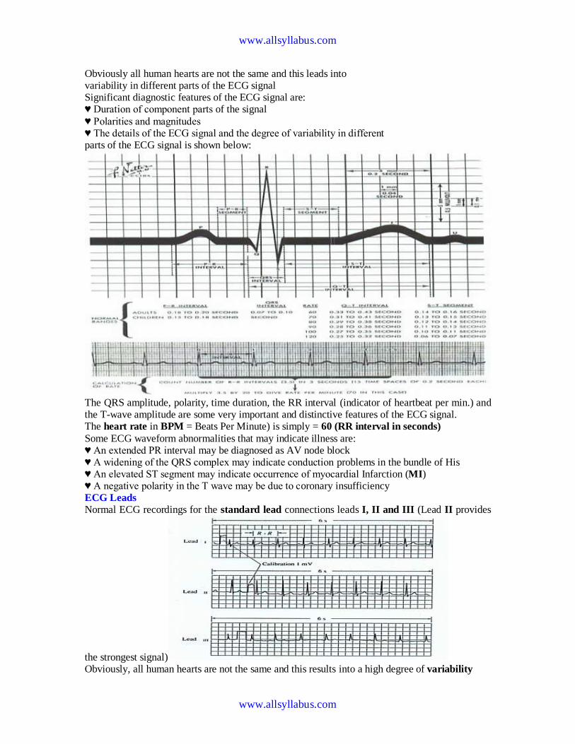
wwwallsyllabuscom
wwwallsyllabuscom
Obviously all human hearts are not the same and this leads into variability in different parts of the ECG signal Significant diagnostic features of the ECG signal are hearts Duration of component parts of the signal
hearts Polarities and magnitudes hearts The details of the ECG signal and the degree of variability in different parts of the ECG signal is shown below
The QRS amplitude polarity time duration the RR interval (indicator of heartbeat per min) and the T-wave amplitude are some very important and distinctive features of the ECG signal The heart rate in BPM = Beats Per Minute) is simply = 60 (RR interval in seconds)
Some ECG waveform abnormalities that may indicate illness are hearts An extended PR interval may be diagnosed as AV node block hearts A widening of the QRS complex may indicate conduction problems in the bundle of His hearts An elevated ST segment may indicate occurrence of myocardial Infarction (MI) hearts A negative polarity in the T wave may be due to coronary insufficiency ECG Leads
Normal ECG recordings for the standard lead connections leads I II and III (Lead II provides
the strongest signal) Obviously all human hearts are not the same and this results into a high degree of variability
wwwallsyllabuscom
wwwallsyllabuscom
hearts Note the degree of variability of different parts of the ECG Signal
Some abnormalities that may indicate illness hearts An extended P-R interval may be diagnosed as AV node block hearts Widening of the QRS complex conduction problems in the bundle of His hearts Elevated ST segment may indicate occurrence of MI
hearts Negative polarity T wave may be due to coronary insufficiency QRS amplitude polarity time domain PR interval (indicator of heat beat per min amp T-wave amplitude are some very important
distinctive features 1 Loss
2 Origin of the ECG signal
hearts We have already covered this concept extensively in the previous lectures (The Dipole filed of the heart the Eindhovenrsquos Triangle the electrical circuit model for the electrocardiographic problem etc)
hearts Standard Limb Leads (I II III)
wwwallsyllabuscom
wwwallsyllabuscom
hearts The The lead wires are color-coded according to some conventions One example is White ndashRA
(Right Arm) Black ndash LA (Left Arm) Green ndash RL (Right Leg) Red ndash LL (Left Leg) and Brown
ndash C (Chest) Note There is a CM (common mode) amplifier connected to the right leg We will discuss this in detail later Augmented Limb Leads
These leads offer a free 50 increase over leads VR VL and VF connections (unipolar leads) with respect to Wilson terminal AVR = -I ndash III2 AVL = I ndash II2 aVF = II ndash I2
wwwallsyllabuscom
wwwallsyllabuscom
Each measurement is made from the reflected limb and the average of the other two limbs Chest Leads (Precordial)
Chest Lead Anatomical Positions
V1 - 4th intercostal space ndash Right sternal margin
V2 - 4th intercostal space ndash Left sternal margin
V3 ndash Midway between V2 amp V4
V4 ndash 5th intercostal space on mid-clavicular line
V5 ndash Same as V4 on the anterior axillary line
V6 ndash Same as V5 on the mid-axillary line
12 Lead Clinical Electrocardiography
The ECG Machine
Most representative Specs
wwwallsyllabuscom
wwwallsyllabuscom
bull Zin = 10 MΩ bull Frequency response = 005 ndash100 Hz bull Strip Chart Recorder Speed = 25 mmsec bull Fast Speed = 100 mmsec
For detailed Specs Refer to the Table in your text ―Summary of performance requirements for electrocardiographs Location of the Heart bull The heart is located between the lungs behind the sternum and above the diaphragm bull It is surrounded by the pericardium bull Its size is about that of a fist and its weight is about 250-300 g bull Its center is located about 15 cm to the left of the midsagittal plane
Anatomy of the heart
bull The walls of the heart are composed of cardiac muscle called myocardium
bull It consists of four compartments ndash the right and left atria and ventricles
The Heart Valves
bull The tricuspid valve regulates blood flow between the right atrium and right
ventricle
bull The pulmonary valve controls blood flow from the right ventricle into the
pulmonary arteries
bull The mitral valve lets oxygen-rich blood from your lungs pass from the left atrium
into the left ventricle
bull The aortic valve lets oxygen-rich blood pass from the left ventricle into the aorta
then to the body
Blood circulation via heart
wwwallsyllabuscom
wwwallsyllabuscom
bull The blood returns from the systemic circulation to the right atrium and from
there goes through the tricuspid valve to the right ventricle
bull It is ejected from the right ventricle through the pulmonary valve to the lungs
bull Oxygenated blood returns from the lungs to the left atrium and from there
through the mitral valve to the left ventricle
bull Finally blood is pumped through the aortic valve to the aorta and the systemic
circulation
Electrical activation of the heart
bull In the heart muscle cell or myocyte electric activation takes place by means of
the same mechanism as in the nerve cell ie from the inflow of Na ions across
the cell membrane
bull The amplitude of the action potential is also similar being 100 mV for both nerve
and muscle
bull The duration of the cardiac impulse is however two orders of magnitude longer
than in either nerve cell or sceletal muscle cell
bull As in the nerve cell repolarization is a consequence of the outflow of K ions
bull The duration of the action impulse is about 300 ms
Mechanical contraction of Cardiac Muscle
bull Associated with the electric activation of cardiac muscle cell is its mechanical
contraction which occurs a little later
bull An important distinction between cardiac muscle tissue and skeletal muscle is
that in cardiac muscle activation can propagate from one cell to another
in any direction
bull Electrical signal begins in the sinoatrial (SA) node natural pacemaker causes
the atria to contract
bull The signal then passes through the atrioventricular (AV) node
ndash sends the signal to the ventricles via the ―bundle of His
ndash causes the ventricles to contract
The Conduction System
wwwallsyllabuscom
wwwallsyllabuscom
The Action Potential
Recording an AP requires the isolation of a single cell Microelectrodes (with tips a few μm across) are used to stimulate and record the response A typical AP is 2-4ms long with an amplitude of about 100Mv The Electroencephalogram EEG
EEG is the graphical representation of the electrical activity of the brain Very commonly used to diagnose certain neurological disorders such as epilepsy
More recently also investigated whether it can detect various forms of dementia or schizophrenia EEG is the specific recording obtained using the scalp electrodes from the surface of the skull
During surgery electrodes may also be placed directly on the cortex The resulting signal is then electrocorticogram (ECoG) Just like ECG EEG is also obtained using several different electrodes places on different regions of the head brain
The Event Related
wwwallsyllabuscom
wwwallsyllabuscom
Potentials ndash ERPs
ERPs are really EEGs obtained under a specific protocol that requires the patient to response to certain stimuli ndash hence event related potentials Also called evoked potentials these signals can be used to diagnose certain
neurological disorders such as dementia and they can also be used as a liedetector bull The oddball paradigm
bull The guilty knowledge test
Electroretinogram ERG
The ERG is the record of the retinal action currents produced by the retina in response to a light stimulus It measures the electrical responses of the light-sensitive cells (such as rods and cones)
The stimuli are often a series of light flashes or rotating patterns The ERG is recorded using contact lens electrode that the subject wears while watching the stimuli
Phonocardiogram ndash PCG
The PCG is the graphic record of the heart sounds and murmurs It is thus a mechanical audio signal rather than an electrical signal Can be easily heard using a stethoscope Or can be converted into an electrical signal using a transducer Typically used to determine the disorders related to the heart valve since their
routine opening and closing create the well-known sounds bull S1 sounds First heart sounds ndash ventricular contractions move blood into atria closing of the AV (mitral and tricuspid) valves then semilunar valves open and blood ejected out of ventricles ndash immediately follows the QRS complex bull S2 sounds Second heart sounds ndash Closure of semilunar (aortic and pulmonary) valves bull Any unexpected sound may indicate a malfunctioning valve that causes the blood flow into out of a chamber when it should not Also called heart murmurs
wwwallsyllabuscom
wwwallsyllabuscom
Define ultrasound
bull Mechanical waves in different modalities (longitudinallateral) needs medium
to be propagated (solid liquid gas)
bull gt 20 kHz
bull Continuouspulsed
bull Sphericalplanarnarrow beamsurface waveLamb-wave
Physical phenomena behind ultrasound measurements
Transmission
bull
bull reflection
bull transit time
bull differences in propagation velocities
bull returns to transit time
bull doppler-shift in frequency
bull flow velocity
bull change of acoustic impedance
bull comparing to reference
bull interference of ultrasound waves (holography)
bull interaction of ultrasound and light (photoacousticz)
bull ultrasound needs medium for propagation it doesnrsquot propagate in vacuum
bull because mechanical waves need moving massunits and spring forces between
them
bull in acoustic emission the medium creates ultrasound (for example during pressure
changes) which is received by sensors
bull pulsed mode more common than continuous
bull continuous reguires separate transducers for transmitting and receicving
bull in pulsed mode an ultrasound burst is sent to the object and the same transducer is
switched to listen echoes
bull standing wave problem
bull in us-therapy pulsed mode gives more effective care without too much heating The Doppler Equation describes the relationship of the Doppler frequency shift to target velocity
wwwallsyllabuscom
wwwallsyllabuscom
The frequency difference is equal to the reflected frequency (FR) minus the originating frequency (FT) If the resulting frequency is higher then there is a positive Doppler shift and the object is moving toward the transducer but if the resulting frequency is lower there is a negative Doppler shift and it is moving away from the transducer In its simplest form it would be calculated as if
the ultrasound was parallel to the targetrsquos direction as shown in diagram A below However this would be a rare occurrence in clinical practice because the transducer is rarely pointed head on to a blood vessel In real life the ultrasound waves would approach the target at an angle called the Doppler angle ( ) On the following page diagram B shows the Doppler
equation used in general clinical situations which includes the Doppler angle The Doppler Angle
The ultrasound beam usually approaches the moving target at an angle called the Doppler angle ( ) This reduces the frequency shift in proportion to the cosine of this
angle If this angle is known then the flow velocity can be calculated The equation used is The Doppler Equation
equiv Doppler shift frequency (the difference between the transmitted and received frequencies) equiv transmitted frequency equiv reflected frequency
V equiv velocity of the blood flow towards the transducer C equiv velocity of sound in tissue θ equiv the angle between the sound beam and the direction of moving blood Where
The Doppler angle ( ) is also known as the angle of insonation It is estimated by the sonographer by a process known as angle correction which involves aligning an indicator on the duplex image along the longitudinal axis of the vessel
There are a few considerations that affect the performance of a Doppler examination that are inherent in the Doppler equation which are ndash The cosine of 90deg is zero so if the ultrasound beam is perpendicular to the direction of blood flow there will be no Doppler shift and it will appear as if there is no flow in the vessel ndash Appropriate estimation of the angle of insonation or angle correction is essential for the accurate determination of Doppler shift and blood flow velocity The angle of insonation should also be less than 60deg at all times since the
cosine function has a steeper curve above this angle and errors in angle correction will be magnified
The simplest Doppler devices use continuous wave (CW Doppler) rather than the pulsed wave used in more complex devices CW Doppler uses two transducers (or a dual element transducer) that transmit and receive ultrasound continuously The transmit and receive beams overlap in a Doppler sample volume some distance from the transducer face as
shown in the diagram below
volume) is the region of transmitting and receiving beam overlap (shaded region) Because there is continuous transducer transmission and reception echoes from all depths within the area arrive at the transducer simultaneously So although CW Doppler can determine the direction of flow it cannot discriminate the different depths where the motion originates The usefulness of CW Doppler devices is limited but they are used clinically to confirm blood flow in superficial vessels as they are good at detecting low velocities As they are easily portable this
can be done at the bedside or in the operating room Most other clinical applications require pulsed wave Doppler Pulsed Wave Doppler (PW Doppler)
wwwallsyllabuscom
wwwallsyllabuscom
Pulsed wave Doppler (PW Doppler) uses a single-element transducer that emits brief pulses of ultrasound energy The time interval between transmitting and then receiving the echoing sound can be used to calculate the depth from where the echo arises The Doppler sample volume can be chosen as to shape depth and position in sampling the flow
data For example the depth is chosen by processing only the signals that return to the transducer in a stipulated time For this technique the ultrasound system transmits a short pulse The eceiver is opened to detect the returning echoes only after a controlled delay and only for a specific duration This time-based gating of the receiving channel allows the definition of a fixed easuring distance which is often referred to as the Sample volume or Doppler gate Then the next ultrasound wave is transmitted The number of pulses transmitted by the system within a second is referred to as the pulse repetition frequency (PRF) The upper PRF limit is given by the time interval required for the echoes to arrive from a sample volume located at a
certain depth The greater the sample-volume depth the longer the time before the echoes are returned and the longer the delay between pulse transmission The greater the samplevolume depth the lower will be the maximum PRF setting Errors in the accuracy of the information arise if the velocities exceed a certain speed The highest velocity accurately measured is called the Nyquist limit Beyond this limit the errors that occur are referred to as aliasing Volume and flow measurement
Flow ndash volume of a liquidgas passing some point over a given time Gases are compressible
Benedict Roth Spirometer
Widely used for physiological amp clinical studies
Light bell moves with the ptrsquos breathing
Movement recorded by a pen on a rotating drum
Water seal prevents leakage of gas
Small seal minimises volume of gas dissolved in water
Suitable for measuring limited gas vols (few litres)
Pneumotachograph
wwwallsyllabuscom
wwwallsyllabuscom
Cardiac Output
Defn vol of blood pumped by the heart per min
CO = SV x HR
Norm ~ 5 lmin
Cardiac index ndash corrected for body surface area
Affected by
Met Rate ndash pregnancy hyperthyroid septic
Preload contractility afterload
Clinical indicators of CO imprecise
Affected by anaesthetic agents used in everyday practice
Provides estimate of
whole body perfusion
oxygen delivery
left ventricular function
Persistently low CO assoc with poor outcome
Methods
Fick method
Dilution techniques ndash dye thermal lithium
Pulse contour analysis- LiDCO amp PiCCO
Oesophageal doppler
TOE
Transthoracic impedance plethysmography
Inert gas through flow
Non-invasive cardiac output measurement
Fick Principle measure volume displacement
1st proposed 1870
―the total uptake or release of a substance by an organ is the product of the
blood flow through that organ and the arteriovenous concentration
difference of the substance
CO = O2 consumption (mlmin) art ndash mixed
venous O2 conc (mll)
Limited by cumbersome equipment sampling errors need for invasive
monitoring and steady-state haemodynamic and metabolic conditions
Indicator dilution techniques
―An indicator mixed into a unit volume of constantly flowing blood can be used to
identify that volume of blood in time provided the indicator remains in the system
between injection and measurement and mixes completely in the blood
wwwallsyllabuscom
wwwallsyllabuscom
Dye dilution
Inert dye ndash indocyanin green
Injected into pulmonary artery and arterial conc measured using a
calibrated cuvette densitometer
Plot indicator dilution curve (see diagram)
CO derived from area under curve
Indicator Dilution Curve
Cardiac Output Measurement
wwwallsyllabuscom
wwwallsyllabuscom
Why bother to check blood pressure
For each 20 mm rise in systolic blood pressure or 10 mm rise in diastolic blood
pressure over 11575
Risk of stroke increases
Risk of heart disease doubles
Risk of renal failure increases
Systolic and diastolic blood pressure
Systolic blood pressure is the highest pressure in the arteries just after the heart
beats
Diastolic blood pressure is the lowest pressure in the arteries just before the heart
beats
Blood pressure is measured indirectly by blood pressure cuff
(sphygmomanometer)
Inflating cuff increases pressure until it cuts off arterial circulation to the arm
Deflating cuff decrease pressure by 2 to 3 mm of mercury per second until blood
first enters the artery creating turbulence this causes a sound with each heartbeat
Sounds continue with each heartbeat until pressure lowers to the lowest pressure
in the artery then turbulence stops so the sound stops
Systolic blood pressure is the cuff pressure at the first sounds diastolic is the cuff
pressure just before the sounds stop
Phase 1 sharp thuds start at systolic blood pressure
Phase 2 blowing sound may disappear entirely (the auscultatory gap )
Phase 3 crisp thud a bit quieter than phase 1
Phase 4 sounds become muffled
Phase 5 end of sounds -- ends at diastolic blood pressure
wwwallsyllabuscom
wwwallsyllabuscom
Make sure the cuff is the right size - its width should be at least 40 of the arms
circumference The cuff will overestimate blood pressure if too small and
underestimate if too large
Place the cuff snugly on patients proximal arm on skin ( not cloth) centered over
the brachial artery (most cuffs have markings)
Support the patients arm at heart level using your arm or a desk
Your patient should sit in the chair for 5 minutes before BP is measured and
should have no caffeine or nicotine for 30 minutes before (JAMA 273 p1211-
1218 1995)
With fingers palpating radial or brachial artery inflate cuff rapidly until you cant
feel the pulse then 20 mm higher
Release cuff at 2 to 3 mm Hg per second until you again feel the pulse this is the
palpable systolic pressure
Wait 30 seconds before measuring blood pressure
Measuring palpable pressure first avoids risk of seriously underestimating blood
pressure because of the auscultatory gap (mistaking Korotkoff phase 3 for phase
1) Many doctors skip this step for time reasons and instead pump cuff to 200 mm
Hg at the next step)
Phase 2 of the Korotkoff sounds can be inaudible - especially in older patients
with systolic hypertension who are at especially high risk of stroke Inflating the
cuff until you dont hear sounds can give you a reading of 14086 when the
patients actual blood pressure is 22086 Most physicians are pressed for time so
they instead inflate the cuff to 200 mm which is beyond the auscultatory gap in
most patients But palpable systolic blood pressure is according to research more
reliable
Place bell of stethoscope (diaphragm is acceptable) over brachial artery
Rapidly pump the cuff to 20 to 30 mm Hg above palpable systolic pressure
Release pressure in the cuff by 2 to 3 mm Hg per second and listen for Korotkoff
sounds including systolic (first) and diastolic (last)
Record as systolicdiastolic Check in both arms the first time you check a
patients blood pressure It may differ by 10 mm Hg or more
If the sounds continues to zero record diastolic blood pressure as the point when
sounds become muffled (phase 4) over zero eg 130700 or just as 13070
Rate
ndash Number of beats in 30 seconds x 2
Strength
ndash Bounding strong or weak (thready)
Regularity
ndash Regular or irregular
You need three readings on two occasions to diagnose hypertension unless blood
pressure is very high
Normal blood pressure in children is
wwwallsyllabuscom
wwwallsyllabuscom
ndash 10255 at 1 year 11269 at 5 years 11978 at 10 years
Blood pressures in adults (JNC VII JAMA 2892560-72 2003)
ndash Normal lt120lt80
ndash Prehypertensive 120-13980-89
ndash Stage 1 hypertension 140-15990-99
ndash Stage 2 hypertension gt160gt100
Adult 60 to 100
Newborn 120-170
1 year 80-160
3 years 80-120
6 years 75-115
10 years 70-110
How to measure observe rise and fall of chest
In infants count for 60 seconds in adults 15 or 30 seconds
Normal respiration Adults 12 to 20
Children
newborn 30-80
1 year 20-40
3 years 20-30
6 years 16-22
Rate
Number of breaths in 30 seconds x 2
Quality
Character of breathing
Rhythm
Regular or irregular
Effort
Normal or labored
Noisy respiration
Normal stridor wheezing snoring gurgling
Depth
Shallow or deep
pH electrode
wwwallsyllabuscom
wwwallsyllabuscom
Governing equation is the Nernst Equation
pCO2 Electrode
The measurement of pCO2 is based on its linear relationship with pH over the range of
10 to 90 mm Hg
The dissociation constant is given by
Taking logarithms
pH = log[HCO3-] ndash log k ndash log a ndash log pCO2
Diathermy
In the natural sciences the term diathermy means electrically induced heat and is commonly
used for muscle relaxation It is also a method of heating tissue electromagnetically or
ultrasonically for therapeutic purposes in medicineContents
[hide]
1 Heating uses 2 Surgical uses
3 Trivia
4 References
E
RT
nF
H
HH
i
ln
0
H O CO H CO H HCO2 2 2 3 3
k
H HCO
a pCO
3
2
wwwallsyllabuscom
wwwallsyllabuscom
Heating uses
Ultrasonic diathermy refers to heating of tissues by ultrasound for the purpose of
therapeutic deep heating No tissue is ordinarily damaged hence it is generally used in
biomedical applications
Electric diathermy uses high frequency alternating electric or magnetic fields sometimes
with no electrode or device contact to the skin to induce gentle deep tissue heating by
induction Again no tissue is ordinarily damaged
Surgical uses
Surgical diathermy is usually better known as electrosurgery (It is also referred to
occasionally as electrocautery but see disambiguation below) Electrosurgery and
surgical diathermy involve the use of high frequency AC electrical current in surgery as
either a cutting modality or else to cauterize small blood vessels to stop bleeding This
technique induces localized tissue burning and damage the zone of which is controlled
by the frequency and power of the device Some sources[1]
insist that electrosurgery be
applied to surgery accomplished by high frequency AC cutting and that
electrocautery be used only for the practice of cauterization with heated nichrome wires
powered by DC current as in the handheld battery-operated portable cautery tools
Trivia
Medical Diathermy devices were used to cause interference to German radio beams used
for targeting night time bombing raids in WWII during the Battle of the Beams
I Diathermy
A Therapeutic use
1 Generation of local heating by high-frequency electromagnetic waves
2 Capacitance techniquemdashbody is placed in an electric field
a Dipolesmdashstructures with positive and negative poles
b Structures with large numbers of dipoles have a greater capacitance to store an electrical charge
c Greatest heating occurs in tissues with fewer dipoles particularly fatty tissues
3 Rapid rotation of dipoles causing mechanical friction and movement of electrons results in local heating
4 Inductance techniquemdashbody is not placed in an electric field
a Magnetic waves generated by driving current through a coiled wire
b Magnetic field creates currents in tissues
wwwallsyllabuscom
wwwallsyllabuscom
c Greatest heating occurs in tissues with low impedance especially muscle
B Precautions and contraindications
1 Diathermy should not be used
a Over metal implants and cardiac pacemakersmdashmore research needed regarding its use over metallic fixations
b Near the uterus of a pregnant woman or near the abdomen or back of a woman who might be pregnant
c On individuals with infections
d On individuals with acute inflammation
e Over moist open wounds
f On patients with malignant tumors
g Over large joint effusions
C Pulsed electromagnetic fields and diathermy
1 Can be pulsed to decrease total energy transmitted to the tissues
2 Short-wave diathermy can be adjusted into a nonthermal range
a Classified as pulsed electromagnetic field (PEMF) or
b Pulsed radio frequency energy (PRFE)
3 Important reclassification as diathermy implies heating
D Efficiency of diathermy and PEMF therapy for musculoskeletal conditions
1 Current research is limited but results suggest that diathermy enhances
treatments directed at soft tissue stretching
2 Some studies suggest that PEMF may speed wound healing and promote healing of nonunion fractures
Diathermy is a modality that uses electromagnetic energy to heat deeper tissues
Diathermy is more effective than ultrasound at heating a larger area of deep tissues
The athletic trainer must identify and respect contraindications to application of ultrasound and diathermy
Pulsed ultrasound and diathermy are used to treat slow-to-heal lesions including skin ulcers and nonunion fractures and may be able to facilitate repair of other tissues including ligaments and tendons
Diathermy
Definition
In diathermy high-frequency electrical currents are used to heat deep muscular tissues The heat increases blood flow speeding up recovery Doctors also use diathermy in surgical procedures by sealing blood vessels with electrically heated probes
The term diathermy is derived from the Greek words therma meaning heat and dia meaning through Diathermy literally means heating through
Origins
The therapeutic effects of heat have long been recognized More than 2000 years ago the Romans took advantage of heat therapies by building hot-spring bathhouses Since then various
wwwallsyllabuscom
wwwallsyllabuscom
methods of using heat have evolved In the early 1890s French physiologist Arseacutene dArsonval began studying the medical application of high-frequency currents The term diathermy was coined by German physician Carl Franz Nagelschmidt who designed a prototype apparatus in 1906 Around 1925 United States doctor J W Schereschewsky began studying the physiological
effects of high-frequency electrical currents on animals It was several years however before the fundamentals of the therapy were understood and put into practice
Benefits
Diathermy can be used to treat arthritis bursitis and other conditions involving stiff painful joints It is also used to treat pelvic infections and sinusitis A benefit of diathermy is that it is a painless procedure that can be administered at a clinic Also if the treatment relieves pain then patients can discontinue pain killers and escape their high cost and side effects
Description
Diathermy involves heating deep muscular tissues When heat is applied to the painful area cellular metabolism speeds up and blood flow increases The increased metabolism and circulation accelerates tissue repair The heat helps the tissues relax and stretch thus alleviating
stiffness Heat also reduces nerve fiber sensitivity increasing the patients pain threshold
There are three methods of diathermy In each energy is delivered to the deep tissues where it is converted to heat The three methods are
Shortwave diathermy The body part to be treated is placed between two capacitor plates Heat is generated as the high-frequency waves travel through the body tissues between the plates Shortwave diathermy is most often used to treat areas like the hip which is covered with a dense tissue mass It is also used to treat pelvic infections and sinusitis The treatment reduces inflammation The Federal Communications Commission regulates the frequency allowed for short-wave diathermy treatment Most machines
function at 2733 megahertz Ultrasound diathermy In this method high-frequency acoustic vibrations are used to
generate heat in deep tissue Microwave diathermy This method uses radar waves to heat tissue This form is the
easiest to use but the microwaves cannot penetrate deep muscles
Diathermy is also used in surgical procedures Many doctors use electrically heated probes to seal blood vessels to prevent excessive bleeding This is particularly helpful in neurosurgery and eye surgery Doctors can also use diathermy to kill abnormal growths such as tumors warts and
infected tissues
Preparations
To keep patients from sweating patients are usually asked to remove clothing from the body part being treated If a patient sweats the electrical currents may pool in the area causing burns Also clothing containing metal must be removed as must earrings buttons barrettes or zippers that contain metal Watches and hearing aids should be removed because the therapy may affect their function
wwwallsyllabuscom
wwwallsyllabuscom
Practitioners of surgical diathermy should steer clear of alcohol-based solutions to prepare and cleanse the skin These preparations can create a flammable vapor and cause burns and fires
Precautions
Patients with metal implants should not undergo diathermy treatment because the metal can act as a conductor of heat and result in serious internal burns Female patients with metallic uterine implants such as an IUD should avoid treatment in the pelvic area Diathermy should not be
used in joints that have been replaced with a prosthesis or in those with sensory impairment who may not be able to tell if they are burning Furthermore pulsed shortwave diathermy should be avoided during pregnancy as it can lead to abnormal fetal development
Patients with hemophilia should avoid the treatment because the increased blood flow could cause them to hemorrhage
Side effects
Some patients may experience superficial burns Since the therapy involves creating heat care must be taken to avoid burns particularly in patients whose injuries have caused decreased sensitivity to heat Also diathermy may affect pacemaker function
Female patients who receive treatment in the lower back or pelvic area may experience an
increased menstrual flow
Research amp general acceptance
For years physiotherapists and physical therapists have used diathermy as a routine part of physical rehabilitation
Electrical Safety
Electrical safety is very important in hospitals as patients may be undergoing a diagnostic or treatment procedure where the protective effect of dry skin is reduced Also patients may be unattended unconscious or anaesthetised and may not respond normally to an electric current Further electrically conductive solutions such as blood and saline are often present in patient treatment areas and may drip or spill on electrical equipment
Electric Current Leakage Current
Extension Leads Double Adaptors Equipment Classification
Class I Class II Defibrillator-Proof
Protective Devices
Residual Current Devices (RCD) Line Isolation overload Monitors (LIMs) Equipment Earthing
Area Classification
wwwallsyllabuscom
wwwallsyllabuscom
Body Protection Area Cardiac Protected Area
Other Electrical Issues Extension Leads
Double Adapters Main Extension Devices Power Boards Installation of Additional Power Points
Electric Current
Injuries received from electric current are dependent on the magnitude of current the pathway
that it takes through the body and the time for which it flows
The nature of electricity flowing through a circuit is analogous to blood flowing through the circulatory system within the human body In this analogy the source of energy is represented by the heart and the blood flowing through arteries and veins is analagous to current flowing
through the conductors and other components of the electric circuit
The application of an electric potential to an electric circuit generates a flow of current through conductive pathways This is analogous to the changes in blood pressure caused by contraction of cardiac muscle that causes blood to flow into the circulatory system For electric current to flow there must be a continuous pathway from the source of potential through electrical components and back to the source
Leakage Current
Electrical components and systems are encased in non conducting insulation to ensure that the electric current is contained and follows the intended pathways If the insulation deteriorates or breaks down current will leak through the insulation barrier and flow to earth This may
be through the protective earth conductor or through the operator
Medical equipment and clinical areas are fitted with a number of protective devices to protect the patient and operator from harmful leakage currents
Extension Leads
Extension leads are not permitted in clinical areas of RCH organisations They may cause high
earth resistance and excessive earth leakage current An extension lead can allow equipment to be powered from areas other than the relevant protected treatment area The power from the other area may not be protected to the same level as the power in the treatment area
wwwallsyllabuscom
wwwallsyllabuscom
As the connection between the extension lead and the equipment mains cable is often on the floor there is a high danger from fluid spills tripping and damage to the mains cable by trolleys when an extension lead is used
Double Adaptors
Double adaptors must not be used in RCH organisations They may not sit securely in a wall
outlet may not be able to provide adequate earth protection and may cause overloading overheating fire or loss of electrical supply
EQUIPMENT CLASSIFICATIONS
There are several methods of providing protection for operators and patients from electrical faults
and harmful leakage current
Class I
Class I equipment is fitted with a three core mains cable containing a protective earth wire
Exposed metal parts on class I equipment are connected to this earth wire
Should a fault develop inside the equipment and the exposed metal comes into contact with the mains the earthing conductor will conduct the fault current to ground Regular testing procedures ensure that earthing conductors are intact as the integrity of the earth wire is of vital importance
Class II
Class II equipment is enclosed within a double insulated case and does not require earthing
conductors Class II equipment is usually fitted with a 2-pin mains plug An internal electrical fault is unlikely to be hazardous as the double insulation prevents any external parts from becoming alive Class II or double insulated equipment can be identified by the class II symbol on the cabinet
Class II Symbol
wwwallsyllabuscom
wwwallsyllabuscom
Defibrillator-Proof
Some medical equipment within the hospital is classified as defibrillator proof When a
defibrillator is discharged through a patient connected to defibrillator proof equipment the equipment will not be damaged by the defibrillators energy Defibrillator proof equipment can remain connected to the patient during defibrillation It is identified by one of the following
symbols
Defibrillator proof symbols
Body protected Cardiac protected
PROTECTIVE DEVICES
Most patient care areas in the hospital are fitted with protective devices These devices are
regularly tested in accordance with the relevant guidelines published by Standards Australia The level of protection provided is dependent upon the device and the area in which it is located
Residual Current Devices (RCD)
RCDs (safety switches) are used in patient treatment areas to monitor and protect the mains supply RCDs sense leakage currents flowing to earth from the equipment If a significant leakage current flows the RCD will detect it and shut off the power supplied to the equipment
within 40 milliseconds Hospital RCDs are more sensitive than those fitted in homes A hospital RCD will trip at 10 milliamperes leakage current
Power outlets supplied through an RCD have a Supply Available lamp The lamp will extinguish
when the RCD trips due to excessive leakage current
wwwallsyllabuscom
wwwallsyllabuscom
Resetting a RCD
Lamp indicates supply is no longer available Disconnect all equipment from the supply Operate the reset button or lever on the supply panel and the Supply Available lamp
should illuminate If not contact Biomedical Engineering
Connect an item of equipment If the RCD trips again then this is the faulty item and should be labelled and sent to Biomedical Engineering
If the RCD does not trip continue connecting equipment until the RCD trips The last piece of equipment connected to the supply is most likely to be faulty as it will have caused the RCD to trip Remove the faulty item from service label it and send to Biomedical Engineering as mentioned above
Line Isolation overload Monitors (LIMs)
In critical life support applications where loss of power supply cannot be tolerated special power outlets powered by isolation transformers are installed
Line Isolation Monitors are installed to continually monitor electrical leakage in the power
supply system If an electrical fault develops in a medical device connected to an isolated power outlet the LIM will detect the leakage current The LIM will alarm and indicate the level of leakage current but will not shut off the electric supply
The faulty equipment can be identified by un plugging one item of equipment at a time from the supply until the alarm stops sounding Equipment that is not faulty may be reconnected Faulty equipment should be appropriately labelled and sent to Biomedical Engineering for repair
The LIM also monitors how much power is being used by the equpiment connected to it If too much power is being used the LIM will alarm and indicate that there is an overload The power used must be reduced immediately by moving some equipment to another circuit as soon as possible until the alarm stops sounding Failure to reduce the load on the LIM will result in the circuit breaker tripping and loss of power to the circuit
wwwallsyllabuscom
wwwallsyllabuscom
Equipotential Earthing
Equipotential earthing is installed in rooms classified as Cardiac Protected electrical areas
Equipotential earthing in treatment areas used for cardiac procedures is intended to minimise any voltage differences between earthed parts of equipment and any other exposed metal in the room
This reduces the possibility of leakage currents that can cause microelectrocution when the patient comes into contact with multiple items of equipment or if the patient happens to come
into contact with metal items in the room whilr they are connected to a medical device
All conductive metal in an equipotential area is connected to a common equipotential earth point with special heavy duty cable
AREA CLASSIFICATIONS
Body Protected Area
These areas are designed for procedures in which patients are connected to equipment that lowers the natural resistance of the skin Applied parts such as electrode gels conductive fluids entering the patient metal needles and catheters provide an easy pathway for current to flow
The main occurrence of injury from Body-Type procedures is from high current levels causing electric shock A direct connection to the patients heart is not present so the risk of Microelectrocution - fibrillation from minute current levels - is reduced
Residual Current Devices (RCD) or Isolation Transformers and Line Isolation Monitors (LIMiacutes) are used in Body Protected areas to provide protection against electrocution from high leakage currents Body-Protected Areas are identified with this sign
Cardiac Protected Area
Where the procedure involves placing an electrical conductor within or near the heart protection against fibrillation induced from small leakage currents is required Electrical conductors used in these procedures include cardiac pacing electrodes intracardiac ECG electrodes and intracardiac catheters
wwwallsyllabuscom
wwwallsyllabuscom
Equipotential earthing in conjunction with RCDs or LIMs provides protection against microelectrocution in Cardiac-Type procedures
Fault currents are reduced to magnitudes that are unlikely to induce fibrillation Used in conjunction with RCDs or LIMs the magnitude and duration of any fault currents
sourced from equipment are limited Cardiac-Protected Areas are identified with this sign
Other electrical issues
This policy aims to provide guidance to those who find that they need more electrical outlets than
those available or that the existing electrical outlets are inconveniently located
As extension leads and multiple outlet power boards can introduce additional hazards into an area the following procedures should be observed
Extension leads
Approved extension leads (AS 3760 1996) may be used in some areas within the hospital but MUST NOT BE USED IN PATIENT AREAS All electrical extension leads must be tagged with an Engineering Department maintenance tag and require a yearly safety inspection and test via
the Engineering Department
Double adapters
Double adapters may cause overloading or equipment earthing problems and are not to be used in
WCH
Mains extension device
The only mains extension device that is to be used in Patient care areas is the 4-way or 8-way
portable Core Balance Unit
The Biomedical Engineering Department must approve all units prior to use These units contain a safety switch and can detect excessive leakage current and disconnect the power in the event of a hazardous situation
wwwallsyllabuscom
wwwallsyllabuscom
Care must be exercised in the use of a portable Core Balance Unit It should be located off the
floor and in a position that will protect it from physical abuse and possible entry of fluids These devices are expensive and easily damaged The device must be sent to Biomedical Engineering every 6 months for safety testing
Power boards
Approved multiple-outlet power boards can be used across RCH but must not be used in patient
care areas except areas approved by the Biomedical Engineering Department
The power boards must have overload protection be fitted with internal safety shutters that protect unused outlets and be fitted with an onoff switch for each outlet
Medical Laser Applications
The main research subjects of the group of Medical Laser Application are in the field of online
monitoring and diagnostics as well as the development of new therapheutic methods The main focus in research is based upon the use of ultrashort (fs) laser pulses
The main advantage of ultrashort laser pulses is the extrem short interaction time which suppress any unwanted side effects of the laser irradiation of the tissue Secondly the broad spectra of the fs laser pulses give the advantage to use the same laser pulses for diagnostic applications like the optical coherence tomography (OCT)
wwwallsyllabuscom
wwwallsyllabuscom
Following research subjects have special attention at the moment
Optical Coherence Tomographie (OCT)
A fs-laser light source can be used to obtain images from inside the tissue when the pulses are coherent superposed This group uses this application to obtain an inside view of the vocal fold and to measure the outline of the crystalline lens
Refractive Surgery
The principle of getting rid of glasses with the help of a laser surgery (fs-LASIK) is underlying rapid improvements concerning precision and safety New technologies were evaluated and transferred very fast in cooperation with an industrial partner
Treatment of Presbyopia
The flexibility of the crystalline lens can be increase by pricise cuts which are induce by fs laser pulse inside the lens The treatment of the presbyopia is thinkable
wwwallsyllabuscom
wwwallsyllabuscom
Ultrafast Physics
Whereas the applications of ultrashort laser pulses increase rapidly is the physics of the interaction between the laser pulses and tissue in many case not fully understood To obtain improved knowledge of the interaction for many fs-laser application this group preforms numerical simulations as well as fundamental experiments
Thermography
Thermography thermal imaging or thermal video is a type of infrared imaging Thermographic cameras detect radiation in the infrared range of the electromagnetic spectrum
(roughly 900ndash14000 nanometers or 09ndash14 microm) and produce images of that radiation Since infrared radiation is emitted by all objects based on their temperatures according to the black body radiation law thermography makes it possible to see ones environment with or without visible illumination The amount of radiation emitted by an object increases with temperature therefore thermography allows one to see variations in temperature (hence the name) When viewed by thermographic camera warm objects stand out well against cooler backgrounds humans and other warm-blooded animals become easily visible against the environment day or
night As a result thermographys extensive use can historically be ascribed to the military and security services
Thermal imaging photography finds many other uses For example firefighters use it to see through smoke find persons and localize the base of a fire With thermal imaging power lines maintenance technicians locate overheating joints and parts a telltale sign of their failure to eliminate potential hazards Where thermal insulation becomes faulty building construction technicians can see heat leaks to improve the efficiencies of cooling or heating air-conditioning Thermal imaging cameras are also installed in some luxury cars to aid the driver the first being
the 2000 Cadillac DeVille Some physiological activities particularly responses in human beings and other warm-blooded animals can also be monitored with thermographic imaging [1]
The appearance and operation of a modern thermographic camera is often similar to a camcorder Enabling the user to see in the infrared spectrum is a function so useful that ability to record their output is often optional A recording module is therefore not always built-in
Instead of CCD sensors most thermal imaging cameras use CMOS Focal Plane Array (FPA) The most common types are InSb InGaAs HgCdTe and QWIP FPA The newest technologies are using low cost and uncooled microbolometers FPA sensors Their resolution is considerably lower than of optical cameras mostly 160x120 or 320x240 pixels up to 640x512 for the most
expensive models Thermographic cameras are much more expensive than their visible-spectrum counterparts and higher-end models are often export-restricted Older bolometers or more
wwwallsyllabuscom
wwwallsyllabuscom
sensitive models as InSb require cryogenic cooling usually by a miniature Stirling cycle refrigerator or liquid nitrogen
Contents
[hide]
1 Difference between IR film and thermography 2 Advantages of Thermography
3 Limitations amp disadvantages of thermography 4 Applications 5 See also 6 External links
o 61 History of thermal imager manufacturers
[edit] Difference between IR film and thermography
Thermal imaging is going to be used on Mars to detect caves that could hold life
[edit] Advantages of Thermography
You get a visual picture so that you can compare temperatures over a large area
It is real time capable of catching moving targets Able to find deteriorating components prior to failure Measurement in areas inaccessible or hazardous for other methods It is a non-destructive test method
[edit] Limitations amp disadvantages of thermography
Quality cameras are expensive and are easily damaged Images can be hard to interpret accurately even with experience
Accurate temperature measurements are very hard to make because of emissivities Most cameras have plusmn2 or worse accuracy (not as accurate as contact) Training and staying proficient in IR scanning is time consuming Ability to only measure surface areas
[edit] Applications
Condition monitoring Medical imaging Night vision Research
Process control Non destructive testing Surveillance in security law enforcement and defense Chemical imaging
wwwallsyllabuscom
wwwallsyllabuscom
Thermal infrared imagers convert the energy in the infrared wavelength into a visible light video display All objects above 0 kelvins emit thermal infrared energy so thermal imagers can passively see all objects regardless of ambient light However most thermal imagers only see objects warmer than -50 degC
The spectrum and amount of thermal radiation depend strongly on an objects surface
temperature This makes it possible for a thermal camera to display an objects temperature However other factors also influence the radiation which limits the accuracy of this technique For example the radiation depends not only on the temperature of the object but is also a function of the emissivity of the object Also radiation also originates from the surroundings and is reflected in the object and the radiation from the object and the reflected radiation will also be influenced by the absorption of the atmosphere
Endoscopy
This page is about having an endoscopy - a test that looks at the inside of your digestive system
There is information on
What endoscopy is
Why you may have an endoscopy
Having an endoscopy
Your results
What an endoscopy is
An endoscopy is a test that looks inside the body The endoscope is a long flexible tube that can
be swallowed It has a camera and light inside it Some doctors call it a telescope
wwwallsyllabuscom
wwwallsyllabuscom
Why you may have an endoscopy
You are most likely to have an endoscopy to look at the inside of your
Gullet (oesophagus)
Stomach
Duodenum - the first part of the small bowel that attaches to the stomach
Large bowel (colon)
Your doctor may want to see inside because you have symptoms such as abnormal bleeding or
difficulty swallowing The doctor can look down the endoscope and see if there are any growths
or other abnormal looking areas Also through the endoscope the doctor can take samples
(biopsies) of any abnormal looking tissues
There is more detailed information about having a colonoscopy in the bowel cancer section of
CancerHelp UK Below is information about having other types of endoscopy
Having an endoscopy
You can have this test as an out patient Most people have a choice between having the test while
they are awake or after having a medicine to make them drowsy (a sedative) Your hospital may
not be happy for you to have a sedative if you live alone and will have no one to look after you
when you go home If you live alone but really want sedation your hospital may allow you to
stay overnight You should ask your doctor about this as early as possible as extra arrangements
will need to be made beforehand
If you dont have a sedative you will have a spray to numb the back of your throat and make it
easier for you to swallow the endoscopy tube
If you would prefer to be asleep during the test you will have an injection to make you very
drowsy just before the test You will need to take someone with you to the hospital appointment
You wont be able to drive for the rest of the day and should have someone to go home with you
You cant eat or drink for about 8 hours before the test so that your stomach and duodenum are
empty Your doctor will give you written instructions about this beforehand or they may arrive
with your appointment letter When you get to the clinic you may be asked to take your upper
clothing off and put on a hospital gown Some hospitals prefer to use gowns because your clothes
wont get messy Once you are ready you get onto the bed or X-ray couch When you are lying
comfortably you have the sedative injection to make you very drowsy Or your doctor will spray
the back of your throat to numb it
Once the sedative or throat spray has worked the doctor will pass the endoscope tube down your
wwwallsyllabuscom
wwwallsyllabuscom
throat to the area being investigated Your doctor will ask you to swallow as the tube goes down
but if youve had a sedative you wont remember that afterwards If there are any abnormalities
the doctor will take pieces of tissue from the abnormal looking area to send to the laboratory for
closer inspection under a microscope These tissue samples are called biopsies
When the test is over you will need to rest for a while If youve had a sedative you may not
remember much (if anything) about the test once you have come round You should be able to go
home the same day
The results
It can take time for test results to come through How long will depend on why you are having
the test Usually the doctor who carries out the endoscopy dictates a report straight way The
report is typed up by the department secretary and goes to your specialist who gives the results to
you If your GP has sent you for the test the results will go directly to the GP surgery
Understandably waiting for results can make you anxious It usually takes a couple of weeks for
the results to come through If your doctor needed them urgently it would have been noted on
the test request form and the results will be ready sooner than that Try to remember to ask your
doctor how long you should expect to wait for the results when you are first asked to go for the
test If it is not an emergency and you have not heard a couple of weeks after your test ring your
doctors secretary to check if they are back
Endoscopy
From Wikipedia the free encyclopedia
Jump to navigation search
Endoscopic images of a duodenal ulcer
wwwallsyllabuscom
wwwallsyllabuscom
A flexible endoscope
Endoscopy means looking inside and typically refers to looking inside the human body for medical reasons using an instrument called an endoscope Endoscopy can also refer to using a borescope in technical situations where direct line-of-sight observation is not feasible
Contents
[hide]
1 Overview 2 Components 3 Uses
4 History 5 Risks 6 After The Endoscopy 7 Recent developments 8 See also 9 References 10 Footnotes
11 External links
Overview
Endoscopy is a minimally invasive diagnostic medical procedure that is used to assess the interior surfaces of an organ by inserting a tube into the body The instrument may have a rigid or flexible tube and not only provide an image for visual inspection and photography but also enable taking
biopsies and retrieval of foreign objects Endoscopy is the vehicle for minimally invasive surgery
Many endoscopic procedures are considered to be relatively painless and at worst associated with mild discomfort for example in esophagogastroduodenoscopy most patients tolerate the procedure with only topical anaesthesia of the oropharynx using lignocaine spray[1] Complications are not common (only 5 of all operations)[citation needed] but can include perforation of the organ under inspection with the endoscope or biopsy instrument If that occurs open surgery may be required to repair the injury
wwwallsyllabuscom
wwwallsyllabuscom
Components
An endoscope can consist of
a rigid or flexible tube a light delivery system to illuminate the organ or object under inspection The light
source is normally outside the body and the light is typically directed via an optical fiber system
a lens system transmitting the image to the viewer from the fiberscope an additional channel to allow entry of medical instruments or manipulators
Uses
Endoscopy can involve
The gastrointestinal tract (GI tract) o esophagus stomach and duodenum (esophagogastroduodenoscopy) o small intestine o colon (colonoscopyproctosigmoidoscopy) o Bile duct
endoscopic retrograde cholangiopancreatography (ERCP) duodenoscope-assisted cholangiopancreatoscopy intraoperative cholangioscopy
The respiratory tract o The nose (rhinoscopy) o The lower respiratory tract (bronchoscopy)
The urinary tract (cystoscopy) The female reproductive system
o The cervix (colposcopy) o The uterus (hysteroscopy) o The Fallopian tubes (Falloscopy)
Normally closed body cavities (through a small incision) o The abdominal or pelvic cavity (laparoscopy) o The interior of a joint (arthroscopy) o Organs of the chest (thoracoscopy and mediastinoscopy)
During pregnancy o The amnion (amnioscopy) o The fetus (fetoscopy)
Plastic Surgery Panendoscopy (or triple endoscopy)
o Combines laryngoscopy esophagoscopy and bronchoscopy Non-medical uses for endoscopy
o The planning and architectural community have found the endoscope useful for
pre-visualization of scale models of proposed buildings and cities (architectural endoscopy)
o Internal inspection of complex technical systems (borescope) o Endoscopes are also a tool helpful in the examination of improvised explosive
devices by bomb disposal personnel
wwwallsyllabuscom
wwwallsyllabuscom
o The FBI uses endoscopes for conducting surveillance via tight spaces
History
The first endoscope of a kind was developed in 1806 by Philip Bozzini with his introduction of a
Lichtleiter (light conductor) for the examinations of the canals and cavities of the human body However the Vienna Medical Society disapproved of such curiosity An endoscope was first introduced into a human in 1822 by William Beaumont an army surgeon at Mackinac
Island Michigan[citation needed] The use of electric light was a major step in the improvement of endoscopy The first such lights were external Later smaller bulbs became available making internal light possible for instance in a hysteroscope by Charles David in 1908[citation needed] Hans Christian Jacobaeus has been given credit for early endoscopic explorations of the abdomen and the thorax with laparoscopy (1912) and thoracoscopy (1910)[citation needed] Laparoscopy was used in the diagnosis of liver and gallbladder disease by Heinz Kalk in the 1930s[citation needed] Hope reported in 1937 on the use of laparoscopy to diagnose ectopic pregnancy[citation needed] In 1944
Raoul Palmer placed his patients in the Trendelenburg position after gaseous distention of the abdomen and thus was able to reliably perform gynecologic laparoscopy[citation needed]
The first gastrocamera was released in 1950 by Olympus Optical Co Ltd The device took pictures on monochromatic film using a small light bulb that was triggered manually The device was of limited use however because it did not implement real-time optical capability Olympus continued its development of endoscopes by incorporating fiber optics in the early 1960s leading to the first useful endoscopes In 1964 it released a gastrocamera guided by a fiberscope[1] A few articles claim that DrBasil Hirschowitz of UnivOf MichiganAnn Arbor discussed the
endoscope in early 50s[2]
As endoscopic technology improved so did the methods of gastrointestinal endoscopy Owing primarily to the efforts of Dr Hiromi Shinya in the late 1960s GI endoscopy developed into what is more recognizable as todays colonoscopy While many doctors experimented with techniques to take advantage of the new iterations of endoscopes Dr Shinya focused on techniques that would allow for successful operation of the endoscope by an individual rejecting the common practice at the time of utilizing two people Consequently many of the fundamental methods and procedures of modern colonoscopy were developed by Dr Shinya
Dr Shinyas other great contribution was to therapeutic endoscopy in his invention of the
electrosurgical polypectomy snare with the aid of Olympus employee Hiroshi Ichikawa Shinya sketched his first plans for the device on January 8 1969 He envisioned a loop of wire attached to the end of a colonoscope that would allow for easy removal of polyps during investigation by passing a current through the wire By September of 1969 the first polypectomy using this device was performed Polypectomy has since become the most common therapeutic procedure performed with an endoscope (Sivak 2004)
By 1980 laparoscopy training was required by gynecologists to perform tubal ligation procedures and diagnostic evaluations of the pelvis The first laparoscopic cholecystectomy was performed in 1984 and the first video-laparoscopic cholecystectomy in 1987[citation needed] During the 1990s
laparoscopic surgery was extended to the appendix spleen colon stomach kidney and liver[citation needed] Wireless capsule endoscopy or Capsule Endoscopy is now approved in all the countries including Japan where government reimbusement will be available from Oct2007Capsule Endoscopy [3] increases detection of Small Bowel tumors where traditional Endoscopy is not very efficient
wwwallsyllabuscom
wwwallsyllabuscom
Risks
Infection
Punctured organs Allergic reactions due to Contrast agents or dyes (such as those used in a CT scan) Over-sedation
After The Endoscopy
After the procedure the patient will be observed and monitored by a qualified individual in the
endoscopy or a recovery area until a significant portion of the medication has worn off Occasionally a patient is left with a mild sore throat which promptly responds to saline gargles or a feeling of distention from the insufflated air that was used during the procedure Both problems are mild and fleeting When fully recovered the patient will be instructed when to resume hisher usual diet (probably within a few hours) and will be allowed to be taken home Because of the use of sedation most facilities mandate that the patient is taken home by another person and not to drive on hisher own or handle machinery for the remainder of the day
Recent developments
With the application of robotic systems telesurgery was introduced as the surgeon could operate
from a site physically removed from the patient The first transatlantic surgery has been called the Lindbergh Operation
wwwallsyllabuscom
wwwallsyllabuscom
Upper Endoscopy
Upper endoscopy enables the physician to look inside the esophagus stomach and duodenum (first part of the small intestine) The procedure might be used to discover the reason for swallowing difficulties nausea vomiting
reflux bleeding indigestion abdominal pain or chest pain Upper endoscopy is also called EGD which stands for esophagogastroduodenoscopy (eh-SAH-fuh-goh-GAS-troh-doo-AH-duh-NAH-skuh-pee)
For the procedure you will swallow a thin flexible lighted tube called an endoscope (EN-doh-skope) Right before the procedure the physician will spray your throat with a numbing agent that may help prevent gagging
You may also receive pain medicine and a sedative to help you relax during the exam The endoscope transmits an image of the inside of the esophagus stomach and duodenum so the physician can carefully examine the lining of these organs The scope also blows air into the stomach this expands the folds of tissue and makes it easier for the physician to examine the stomach
The physician can see abnormalities like inflammation or
bleeding through the endoscope that dont show up well on x rays The physician can also insert instruments into the scope to treat bleeding abnormalities or remove samples of tissue (biopsy) for further tests
Possible complications of upper endoscopy include bleeding and puncture of the stomach lining However such complications are rare Most people will probably have nothing more than a mild sore throat after the procedure
The procedure takes 20 to 30 minutes Because you will be sedated you will need to rest at the endoscopy facility for 1 to 2 hours until the medication wears off
Preparation
Your stomach and duodenum must be empty for the procedure to be thorough and safe so you will not be able to eat or drink anything for at least 6 hours beforehand Also you must
arrange for someone to take you homemdashyou will not be allowed to drive because of the sedatives Your physician may give you other special instructions
The digestive system
wwwallsyllabuscom
wwwallsyllabuscom
What is Endoscopy and Why is it Performed
Endoscopy allows physicians to peer through the bodys passageways Endoscopy is the examination and inspection of the interior of body organs joints or cavities through an endoscope An endoscope is a device that uses fiber optics and powerful lens systems to provide lighting and visualization of the interior of a joint The portion of the endoscope inserted into the body may be rigid or flexible depending upon the medical procedure
An endoscope uses two fiber optic lines A light fiber carries light into the body cavity and an image fiber carries the image of the body cavity back to the physicians viewing lens There is also a separate port to allow for administration of drugs suction and irrigation This port may also be used to introduce small folding instruments such as forceps scissors brushes snares and baskets for tissue excision (removal) sampling or other diagnostic and therapeutic work Endoscopes may be used in conjunction with a camera or video recorder to document images of the inside of the joint or chronicle an endoscopic procedure New endoscopes have digital capabilities for manipulating and enhancing the video images
This figure shows a rigid endoscope used for arthroscopy The image fiber leads from the ocular (eye piece) to the inserted end of the scope The light fiber is below and leads from the light source to the working end of the endoscope
Why Is Endoscopy Performed
Endoscopy can be used to diagnose various conditions by close examination of internal organ and body structures Endoscopy can also guide therapy and repair such as the removal of torn cartilage from the bearing surfaces of a joint Biopsy (tissue sampling for pathologic testing) may also be performed under endoscopic guidance Local or general anesthetic may be used during endoscopy depending upon the type of procedure being performed
Internal abnormalities revealed through endoscopy include abscesses biliary (liver) cirrhosis bleeding bronchitis cancer cysts degenerative disease gallbladder stones hernia inflammation metastatic cancer polyps tumors ulcers and other diseases and conditions
Endoscopy is a minimally invasive procedure and carries with it certain minor risks depending upon the type of procedure being performed However these risks are typically far outweighed by the diagnostic and therapeutic potential of the procedure
Prior to the widespread use of endoscopy and diagnostic imaging most internal conditions could only be diagnosed or treated with open surgery Until the last several decades exploratory surgery was routinely performed only when a patient was critically ill and the source of illness was not known For example in certain dire cases the patients thorax or abdomen were surgically opened and examined to try to determine the source of illness
wwwallsyllabuscom
wwwallsyllabuscom
Endoscopy can often be done on an outpatient basis Outpatient means that the procedure does not require hospital admission and acute care and observation and may be performed outside the premises of a hospital Outpatient procedures performed at hospitals or ambulatory centers allow the patient to go home or return to work within a short while after their procedure
Types of Endoscopy
Fiber optic endoscopes now have widespread use in medicine and guide a myriad of diagnostic and therapeutic procedures including
Arthroscopy examination of joints for diagnosis and treatment (arthroscopic surgery) Bronchoscopy examination of the trachea and lungs bronchial trees to reveal
abscesses bronchitis carcinoma tumors tuberculosis alveolitis infection inflammation Colonoscopy examination of the inside of the colon and large intestine to detect polyps
tumors ulceration inflammation colitis diverticula Chrohns disease and discovery and removal of foreign bodies
Colposcopy direct visualization of the vagina and cervix to detect cancer inflammation and other conditions
Cystoscopy examination of the bladder urethra urinary tract uteral orifices and
prostate (men) with insertion of the endoscope through the urethra
ERCP (endoscopic retrograde cholangio-pancreatography) uses endoscopic guidance to
place a catheter for x-ray fluorosocopy with contrast enhancement This technique is used to examine the livers biliary tree the gallbladder the pancreatic duct and other anatomy to check for stones other obstructions and disease X-ray contrast is introduced into these ducts via catheter and fluoroscopic x-ray images are taken to show any abnormality or blockage If disease is detected it can sometimes be treated at the same time or biopsy can be performed to test for cancer or other pathology ERCP can detect biliary cirrhosis
cancer of the bile ducts pancreatic cysts pseudocysts pancreatic tumors chronic pancreatitis and other conditions such as gallbladder stones
EGD (Esophogealgastroduodensoscopy) visual examination of the upper gastro-
intestinal (GI) tract (also referred to as gastroscopy) to reveal hemorrhage hiatal hernia inflammation of the esophagus gastric ulcers
Endoscopic biopsy is the removal of tissue specimens for pathologic examination and
analysis Gastroscopy examination of the lining of the esophagus stomach and duodenum
Gastroscopy is often used to diagnose ulcers and other sources of bleeding and to guide biopsy of suspect GI cancers
Laparoscopy visualization of the stomach liver and other abdominal organs including
the female reproductive organs for example the fallopian tubes Laryngoscopy examination of the larynx (voice box) Proctoscopy sigmoidoscopy proctosigmoidoscopy examination of the rectum and
sigmoid colon Thoracoscopy examination of the pleura (sac that covers the lungs) pleural spaces
mediastinum and pericardium
A Brief History of Endoscopy
In the early 1900s the first attempts to view inside the body with lighted telescopes were made These initial devices were often fully rigid In the 1930s semi-flexible endoscopes called gastroscopes were developed to view inside of the stomach Fiber-optic endoscopy was pioneered by South African-born physician Basil Hirschowitz at the University of Michigan in 1957 Widespread use of fiber optic endoscopes began in the 1960s
wwwallsyllabuscom
wwwallsyllabuscom
A fiber optic cable is simply a bundle of microscopic glass or plastic fibers that literally allows light and images to be transmitted through curved structures Fiber optic cables are also replacing metal wires as the backbone of the worlds telecommunications infrastructure This Internet page may have traveled through a fiber optic cable as a stream of digital data (bursts of light) on its way to your computer
Endoscopy Equipment
Endoscopes have many practical needs And HMB Endoscopy Products (Hollywood Florida) has been providing endoscopic equipment and educating people on the use of endoscopes for more than 17 years Be sure to Browse our Catalog for all the details on how to purchase these medical instruments that can examine any part of the body
Video
Systems
Fiber
Endoscopes
Video
Endoscopes
In the simplest terms Endoscopy equipment consists of instruments that can look at the inside of many different organs mdash these are small flexible or rigid tubes with a light or lenses on the end that can look into the esophagus stomach and colon mdash and in more general terms endoscopy equipment can help doctors look deep inside body structures and hollow organs
An endoscope and related endoscope products and equipment are usually composed of three components
An optic system that allows the doctor to look through the scope into the organ or
cavity or to attach a video camera to the scope
A fiberoptic cable to light up the bodily area
A lumen (eg the bore of a tube like a needle or catheter) to take tissue samples of the
area being viewed
The beauty of endoscopic products is that they perform dual functions mdash with both diagnostic and therapeutic capabilities For example this means that these endoscopic products and instruments can perform biopsies (eg to evaluate tissue samples) as well as provide sclerotherapy (a medical procedure used to treat varicose veins and ―spider veins) In truth these brief explanations only tell part of the story
wwwallsyllabuscom
wwwallsyllabuscom
TYPES OF ENDOSCOPES
Take a look at the different types of endoscopic equipment you can get with HMB Endoscopy Products Again in very general terms there are two main types of endoscopes
Rigid Flexible
1 rigid endoscopes ndash the majority of which use a convex (curving out like one half of a
circle) glass lens system in which the small glass lenses are separated by large air spaces
2 flexible endoscopes allow for just that mdash flexibility In the animal kingdom for example
a flexible endoscope would be perfect for examining the stomach area of a snake
The popularity of endoscopy equipment continues to grow Some of the industryrsquos household names include Olympus Pentax Funjinon and Storz mdash with HMB both selling and repairing pre-owned and completely refurbished endoscopic products from each of these major manufacturers Browse our Catalog to get the ones you need
THERMOGRAPH APPLICATION Thermographs are digital recording thermometers used to log
temperature in the
marine environment The instruments are attached to a computer to set up recording
parameters for
deployment downloading and display of data in graphic or numeric format These units
are placed in
underwater housings and attached to the bottom or suspended in the water column for
deployment
DESCRIPTION Two types of thermograph are currently available The Ryan
Tempmentor is a
reusable data logger that allows storage of a maximum of 6361 temperature
measurements in the
range of -32C to +70C with 01resolution and 03accuracy The instrument can be
programmed
to take measurements from once per second to once every other hour Instrument
dimensions are 3 x
6 x 13 and the unit weighs 11 ounces The Hobo Temp is a miniature reusable data
logger that
allows storage of 1800 measurements in the range of -20C to +70C with
03resolution and 07
wwwallsyllabuscom
wwwallsyllabuscom
accuracy Sampling intervals from 05 seconds to 48 hours are available Instrument
dimensions are
24 x 19 x 08 and the unit weighs 1 ounce Both units are battery powered with the
battery life of
the Tempmentor being approximately two years and the battery life of the Hobo Temp
being one year THERMOGRAPH APPLICATION Thermographs are digital recording thermometers used to log
temperature in the
marine environment The instruments are attached to a computer to set up recording
parameters for
deployment downloading and display of data in graphic or numeric format These units
are placed in
underwater housings and attached to the bottom or suspended in the water column for
deployment
DESCRIPTION Two types of thermograph are currently available The Ryan
Tempmentor is a
reusable data logger that allows storage of a maximum of 6361 temperature
measurements in the
range of -32C to +70C with 01resolution and 03accuracy The instrument can be
programmed
to take measurements from once per second to once every other hour Instrument
dimensions are 3 x
6 x 13 and the unit weighs 11 ounces The Hobo Temp is a miniature reusable data
logger that
allows storage of 1800 measurements in the range of -20C to +70C with
03resolution and 07
accuracy Sampling intervals from 05 seconds to 48 hours are available Instrument
dimensions are
24 x 19 x 08 and the unit weighs 1 ounce Both units are battery powered with the
battery life of
the Tempmentor being approximately two years and the battery life of the Hobo Temp
being one year THERMOGRAPH APPLICATION Thermographs are digital recording thermometers used to log
temperature in the
marine environment The instruments are attached to a computer to set up recording
parameters for
deployment downloading and display of data in graphic or numeric format These units
are placed in
underwater housings and attached to the bottom or suspended in the water column for
deployment
DESCRIPTION Two types of thermograph are currently available The Ryan
Tempmentor is a
reusable data logger that allows storage of a maximum of 6361 temperature
measurements in the
range of -32C to +70C with 01resolution and 03accuracy The instrument can be
programmed
wwwallsyllabuscom
wwwallsyllabuscom
to take measurements from once per second to once every other hour Instrument
dimensions are 3 x
6 x 13 and the unit weighs 11 ounces The Hobo Temp is a miniature reusable data
logger that
allows storage of 1800 measurements in the range of -20C to +70C with
03resolution and 07
accuracy Sampling intervals from 05 seconds to 48 hours are available Instrument
dimensions are
24 x 19 x 08 and the unit weighs 1 ounce Both units are battery powered with the
battery life of
the Tempmentor being approximately two years and the battery life of the Hobo Temp
being one year
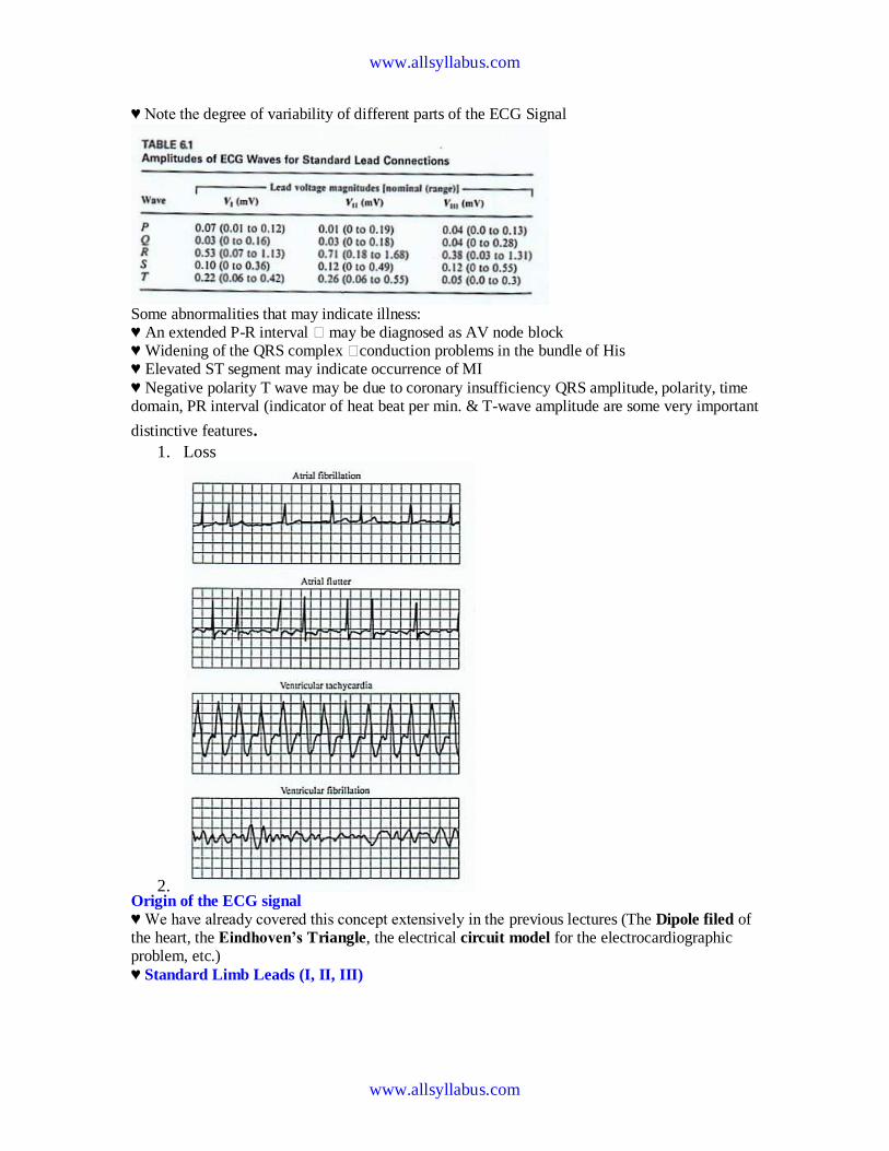
wwwallsyllabuscom
wwwallsyllabuscom
hearts The The lead wires are color-coded according to some conventions One example is White ndashRA
(Right Arm) Black ndash LA (Left Arm) Green ndash RL (Right Leg) Red ndash LL (Left Leg) and Brown
ndash C (Chest) Note There is a CM (common mode) amplifier connected to the right leg We will discuss this in detail later Augmented Limb Leads
These leads offer a free 50 increase over leads VR VL and VF connections (unipolar leads) with respect to Wilson terminal AVR = -I ndash III2 AVL = I ndash II2 aVF = II ndash I2
wwwallsyllabuscom
wwwallsyllabuscom
Each measurement is made from the reflected limb and the average of the other two limbs Chest Leads (Precordial)
Chest Lead Anatomical Positions
V1 - 4th intercostal space ndash Right sternal margin
V2 - 4th intercostal space ndash Left sternal margin
V3 ndash Midway between V2 amp V4
V4 ndash 5th intercostal space on mid-clavicular line
V5 ndash Same as V4 on the anterior axillary line
V6 ndash Same as V5 on the mid-axillary line
12 Lead Clinical Electrocardiography
The ECG Machine
Most representative Specs
wwwallsyllabuscom
wwwallsyllabuscom
bull Zin = 10 MΩ bull Frequency response = 005 ndash100 Hz bull Strip Chart Recorder Speed = 25 mmsec bull Fast Speed = 100 mmsec
For detailed Specs Refer to the Table in your text ―Summary of performance requirements for electrocardiographs Location of the Heart bull The heart is located between the lungs behind the sternum and above the diaphragm bull It is surrounded by the pericardium bull Its size is about that of a fist and its weight is about 250-300 g bull Its center is located about 15 cm to the left of the midsagittal plane
Anatomy of the heart
bull The walls of the heart are composed of cardiac muscle called myocardium
bull It consists of four compartments ndash the right and left atria and ventricles
The Heart Valves
bull The tricuspid valve regulates blood flow between the right atrium and right
ventricle
bull The pulmonary valve controls blood flow from the right ventricle into the
pulmonary arteries
bull The mitral valve lets oxygen-rich blood from your lungs pass from the left atrium
into the left ventricle
bull The aortic valve lets oxygen-rich blood pass from the left ventricle into the aorta
then to the body
Blood circulation via heart
wwwallsyllabuscom
wwwallsyllabuscom
bull The blood returns from the systemic circulation to the right atrium and from
there goes through the tricuspid valve to the right ventricle
bull It is ejected from the right ventricle through the pulmonary valve to the lungs
bull Oxygenated blood returns from the lungs to the left atrium and from there
through the mitral valve to the left ventricle
bull Finally blood is pumped through the aortic valve to the aorta and the systemic
circulation
Electrical activation of the heart
bull In the heart muscle cell or myocyte electric activation takes place by means of
the same mechanism as in the nerve cell ie from the inflow of Na ions across
the cell membrane
bull The amplitude of the action potential is also similar being 100 mV for both nerve
and muscle
bull The duration of the cardiac impulse is however two orders of magnitude longer
than in either nerve cell or sceletal muscle cell
bull As in the nerve cell repolarization is a consequence of the outflow of K ions
bull The duration of the action impulse is about 300 ms
Mechanical contraction of Cardiac Muscle
bull Associated with the electric activation of cardiac muscle cell is its mechanical
contraction which occurs a little later
bull An important distinction between cardiac muscle tissue and skeletal muscle is
that in cardiac muscle activation can propagate from one cell to another
in any direction
bull Electrical signal begins in the sinoatrial (SA) node natural pacemaker causes
the atria to contract
bull The signal then passes through the atrioventricular (AV) node
ndash sends the signal to the ventricles via the ―bundle of His
ndash causes the ventricles to contract
The Conduction System
wwwallsyllabuscom
wwwallsyllabuscom
The Action Potential
Recording an AP requires the isolation of a single cell Microelectrodes (with tips a few μm across) are used to stimulate and record the response A typical AP is 2-4ms long with an amplitude of about 100Mv The Electroencephalogram EEG
EEG is the graphical representation of the electrical activity of the brain Very commonly used to diagnose certain neurological disorders such as epilepsy
More recently also investigated whether it can detect various forms of dementia or schizophrenia EEG is the specific recording obtained using the scalp electrodes from the surface of the skull
During surgery electrodes may also be placed directly on the cortex The resulting signal is then electrocorticogram (ECoG) Just like ECG EEG is also obtained using several different electrodes places on different regions of the head brain
The Event Related
wwwallsyllabuscom
wwwallsyllabuscom
Potentials ndash ERPs
ERPs are really EEGs obtained under a specific protocol that requires the patient to response to certain stimuli ndash hence event related potentials Also called evoked potentials these signals can be used to diagnose certain
neurological disorders such as dementia and they can also be used as a liedetector bull The oddball paradigm
bull The guilty knowledge test
Electroretinogram ERG
The ERG is the record of the retinal action currents produced by the retina in response to a light stimulus It measures the electrical responses of the light-sensitive cells (such as rods and cones)
The stimuli are often a series of light flashes or rotating patterns The ERG is recorded using contact lens electrode that the subject wears while watching the stimuli
Phonocardiogram ndash PCG
The PCG is the graphic record of the heart sounds and murmurs It is thus a mechanical audio signal rather than an electrical signal Can be easily heard using a stethoscope Or can be converted into an electrical signal using a transducer Typically used to determine the disorders related to the heart valve since their
routine opening and closing create the well-known sounds bull S1 sounds First heart sounds ndash ventricular contractions move blood into atria closing of the AV (mitral and tricuspid) valves then semilunar valves open and blood ejected out of ventricles ndash immediately follows the QRS complex bull S2 sounds Second heart sounds ndash Closure of semilunar (aortic and pulmonary) valves bull Any unexpected sound may indicate a malfunctioning valve that causes the blood flow into out of a chamber when it should not Also called heart murmurs
wwwallsyllabuscom
wwwallsyllabuscom
Define ultrasound
bull Mechanical waves in different modalities (longitudinallateral) needs medium
to be propagated (solid liquid gas)
bull gt 20 kHz
bull Continuouspulsed
bull Sphericalplanarnarrow beamsurface waveLamb-wave
Physical phenomena behind ultrasound measurements
Transmission
bull
bull reflection
bull transit time
bull differences in propagation velocities
bull returns to transit time
bull doppler-shift in frequency
bull flow velocity
bull change of acoustic impedance
bull comparing to reference
bull interference of ultrasound waves (holography)
bull interaction of ultrasound and light (photoacousticz)
bull ultrasound needs medium for propagation it doesnrsquot propagate in vacuum
bull because mechanical waves need moving massunits and spring forces between
them
bull in acoustic emission the medium creates ultrasound (for example during pressure
changes) which is received by sensors
bull pulsed mode more common than continuous
bull continuous reguires separate transducers for transmitting and receicving
bull in pulsed mode an ultrasound burst is sent to the object and the same transducer is
switched to listen echoes
bull standing wave problem
bull in us-therapy pulsed mode gives more effective care without too much heating The Doppler Equation describes the relationship of the Doppler frequency shift to target velocity
wwwallsyllabuscom
wwwallsyllabuscom
The frequency difference is equal to the reflected frequency (FR) minus the originating frequency (FT) If the resulting frequency is higher then there is a positive Doppler shift and the object is moving toward the transducer but if the resulting frequency is lower there is a negative Doppler shift and it is moving away from the transducer In its simplest form it would be calculated as if
the ultrasound was parallel to the targetrsquos direction as shown in diagram A below However this would be a rare occurrence in clinical practice because the transducer is rarely pointed head on to a blood vessel In real life the ultrasound waves would approach the target at an angle called the Doppler angle ( ) On the following page diagram B shows the Doppler
equation used in general clinical situations which includes the Doppler angle The Doppler Angle
The ultrasound beam usually approaches the moving target at an angle called the Doppler angle ( ) This reduces the frequency shift in proportion to the cosine of this
angle If this angle is known then the flow velocity can be calculated The equation used is The Doppler Equation
equiv Doppler shift frequency (the difference between the transmitted and received frequencies) equiv transmitted frequency equiv reflected frequency
V equiv velocity of the blood flow towards the transducer C equiv velocity of sound in tissue θ equiv the angle between the sound beam and the direction of moving blood Where
The Doppler angle ( ) is also known as the angle of insonation It is estimated by the sonographer by a process known as angle correction which involves aligning an indicator on the duplex image along the longitudinal axis of the vessel
There are a few considerations that affect the performance of a Doppler examination that are inherent in the Doppler equation which are ndash The cosine of 90deg is zero so if the ultrasound beam is perpendicular to the direction of blood flow there will be no Doppler shift and it will appear as if there is no flow in the vessel ndash Appropriate estimation of the angle of insonation or angle correction is essential for the accurate determination of Doppler shift and blood flow velocity The angle of insonation should also be less than 60deg at all times since the
cosine function has a steeper curve above this angle and errors in angle correction will be magnified
The simplest Doppler devices use continuous wave (CW Doppler) rather than the pulsed wave used in more complex devices CW Doppler uses two transducers (or a dual element transducer) that transmit and receive ultrasound continuously The transmit and receive beams overlap in a Doppler sample volume some distance from the transducer face as
shown in the diagram below
volume) is the region of transmitting and receiving beam overlap (shaded region) Because there is continuous transducer transmission and reception echoes from all depths within the area arrive at the transducer simultaneously So although CW Doppler can determine the direction of flow it cannot discriminate the different depths where the motion originates The usefulness of CW Doppler devices is limited but they are used clinically to confirm blood flow in superficial vessels as they are good at detecting low velocities As they are easily portable this
can be done at the bedside or in the operating room Most other clinical applications require pulsed wave Doppler Pulsed Wave Doppler (PW Doppler)
wwwallsyllabuscom
wwwallsyllabuscom
Pulsed wave Doppler (PW Doppler) uses a single-element transducer that emits brief pulses of ultrasound energy The time interval between transmitting and then receiving the echoing sound can be used to calculate the depth from where the echo arises The Doppler sample volume can be chosen as to shape depth and position in sampling the flow
data For example the depth is chosen by processing only the signals that return to the transducer in a stipulated time For this technique the ultrasound system transmits a short pulse The eceiver is opened to detect the returning echoes only after a controlled delay and only for a specific duration This time-based gating of the receiving channel allows the definition of a fixed easuring distance which is often referred to as the Sample volume or Doppler gate Then the next ultrasound wave is transmitted The number of pulses transmitted by the system within a second is referred to as the pulse repetition frequency (PRF) The upper PRF limit is given by the time interval required for the echoes to arrive from a sample volume located at a
certain depth The greater the sample-volume depth the longer the time before the echoes are returned and the longer the delay between pulse transmission The greater the samplevolume depth the lower will be the maximum PRF setting Errors in the accuracy of the information arise if the velocities exceed a certain speed The highest velocity accurately measured is called the Nyquist limit Beyond this limit the errors that occur are referred to as aliasing Volume and flow measurement
Flow ndash volume of a liquidgas passing some point over a given time Gases are compressible
Benedict Roth Spirometer
Widely used for physiological amp clinical studies
Light bell moves with the ptrsquos breathing
Movement recorded by a pen on a rotating drum
Water seal prevents leakage of gas
Small seal minimises volume of gas dissolved in water
Suitable for measuring limited gas vols (few litres)
Pneumotachograph
wwwallsyllabuscom
wwwallsyllabuscom
Cardiac Output
Defn vol of blood pumped by the heart per min
CO = SV x HR
Norm ~ 5 lmin
Cardiac index ndash corrected for body surface area
Affected by
Met Rate ndash pregnancy hyperthyroid septic
Preload contractility afterload
Clinical indicators of CO imprecise
Affected by anaesthetic agents used in everyday practice
Provides estimate of
whole body perfusion
oxygen delivery
left ventricular function
Persistently low CO assoc with poor outcome
Methods
Fick method
Dilution techniques ndash dye thermal lithium
Pulse contour analysis- LiDCO amp PiCCO
Oesophageal doppler
TOE
Transthoracic impedance plethysmography
Inert gas through flow
Non-invasive cardiac output measurement
Fick Principle measure volume displacement
1st proposed 1870
―the total uptake or release of a substance by an organ is the product of the
blood flow through that organ and the arteriovenous concentration
difference of the substance
CO = O2 consumption (mlmin) art ndash mixed
venous O2 conc (mll)
Limited by cumbersome equipment sampling errors need for invasive
monitoring and steady-state haemodynamic and metabolic conditions
Indicator dilution techniques
―An indicator mixed into a unit volume of constantly flowing blood can be used to
identify that volume of blood in time provided the indicator remains in the system
between injection and measurement and mixes completely in the blood
wwwallsyllabuscom
wwwallsyllabuscom
Dye dilution
Inert dye ndash indocyanin green
Injected into pulmonary artery and arterial conc measured using a
calibrated cuvette densitometer
Plot indicator dilution curve (see diagram)
CO derived from area under curve
Indicator Dilution Curve
Cardiac Output Measurement
wwwallsyllabuscom
wwwallsyllabuscom
Why bother to check blood pressure
For each 20 mm rise in systolic blood pressure or 10 mm rise in diastolic blood
pressure over 11575
Risk of stroke increases
Risk of heart disease doubles
Risk of renal failure increases
Systolic and diastolic blood pressure
Systolic blood pressure is the highest pressure in the arteries just after the heart
beats
Diastolic blood pressure is the lowest pressure in the arteries just before the heart
beats
Blood pressure is measured indirectly by blood pressure cuff
(sphygmomanometer)
Inflating cuff increases pressure until it cuts off arterial circulation to the arm
Deflating cuff decrease pressure by 2 to 3 mm of mercury per second until blood
first enters the artery creating turbulence this causes a sound with each heartbeat
Sounds continue with each heartbeat until pressure lowers to the lowest pressure
in the artery then turbulence stops so the sound stops
Systolic blood pressure is the cuff pressure at the first sounds diastolic is the cuff
pressure just before the sounds stop
Phase 1 sharp thuds start at systolic blood pressure
Phase 2 blowing sound may disappear entirely (the auscultatory gap )
Phase 3 crisp thud a bit quieter than phase 1
Phase 4 sounds become muffled
Phase 5 end of sounds -- ends at diastolic blood pressure
wwwallsyllabuscom
wwwallsyllabuscom
Make sure the cuff is the right size - its width should be at least 40 of the arms
circumference The cuff will overestimate blood pressure if too small and
underestimate if too large
Place the cuff snugly on patients proximal arm on skin ( not cloth) centered over
the brachial artery (most cuffs have markings)
Support the patients arm at heart level using your arm or a desk
Your patient should sit in the chair for 5 minutes before BP is measured and
should have no caffeine or nicotine for 30 minutes before (JAMA 273 p1211-
1218 1995)
With fingers palpating radial or brachial artery inflate cuff rapidly until you cant
feel the pulse then 20 mm higher
Release cuff at 2 to 3 mm Hg per second until you again feel the pulse this is the
palpable systolic pressure
Wait 30 seconds before measuring blood pressure
Measuring palpable pressure first avoids risk of seriously underestimating blood
pressure because of the auscultatory gap (mistaking Korotkoff phase 3 for phase
1) Many doctors skip this step for time reasons and instead pump cuff to 200 mm
Hg at the next step)
Phase 2 of the Korotkoff sounds can be inaudible - especially in older patients
with systolic hypertension who are at especially high risk of stroke Inflating the
cuff until you dont hear sounds can give you a reading of 14086 when the
patients actual blood pressure is 22086 Most physicians are pressed for time so
they instead inflate the cuff to 200 mm which is beyond the auscultatory gap in
most patients But palpable systolic blood pressure is according to research more
reliable
Place bell of stethoscope (diaphragm is acceptable) over brachial artery
Rapidly pump the cuff to 20 to 30 mm Hg above palpable systolic pressure
Release pressure in the cuff by 2 to 3 mm Hg per second and listen for Korotkoff
sounds including systolic (first) and diastolic (last)
Record as systolicdiastolic Check in both arms the first time you check a
patients blood pressure It may differ by 10 mm Hg or more
If the sounds continues to zero record diastolic blood pressure as the point when
sounds become muffled (phase 4) over zero eg 130700 or just as 13070
Rate
ndash Number of beats in 30 seconds x 2
Strength
ndash Bounding strong or weak (thready)
Regularity
ndash Regular or irregular
You need three readings on two occasions to diagnose hypertension unless blood
pressure is very high
Normal blood pressure in children is
wwwallsyllabuscom
wwwallsyllabuscom
ndash 10255 at 1 year 11269 at 5 years 11978 at 10 years
Blood pressures in adults (JNC VII JAMA 2892560-72 2003)
ndash Normal lt120lt80
ndash Prehypertensive 120-13980-89
ndash Stage 1 hypertension 140-15990-99
ndash Stage 2 hypertension gt160gt100
Adult 60 to 100
Newborn 120-170
1 year 80-160
3 years 80-120
6 years 75-115
10 years 70-110
How to measure observe rise and fall of chest
In infants count for 60 seconds in adults 15 or 30 seconds
Normal respiration Adults 12 to 20
Children
newborn 30-80
1 year 20-40
3 years 20-30
6 years 16-22
Rate
Number of breaths in 30 seconds x 2
Quality
Character of breathing
Rhythm
Regular or irregular
Effort
Normal or labored
Noisy respiration
Normal stridor wheezing snoring gurgling
Depth
Shallow or deep
pH electrode
wwwallsyllabuscom
wwwallsyllabuscom
Governing equation is the Nernst Equation
pCO2 Electrode
The measurement of pCO2 is based on its linear relationship with pH over the range of
10 to 90 mm Hg
The dissociation constant is given by
Taking logarithms
pH = log[HCO3-] ndash log k ndash log a ndash log pCO2
Diathermy
In the natural sciences the term diathermy means electrically induced heat and is commonly
used for muscle relaxation It is also a method of heating tissue electromagnetically or
ultrasonically for therapeutic purposes in medicineContents
[hide]
1 Heating uses 2 Surgical uses
3 Trivia
4 References
E
RT
nF
H
HH
i
ln
0
H O CO H CO H HCO2 2 2 3 3
k
H HCO
a pCO
3
2
wwwallsyllabuscom
wwwallsyllabuscom
Heating uses
Ultrasonic diathermy refers to heating of tissues by ultrasound for the purpose of
therapeutic deep heating No tissue is ordinarily damaged hence it is generally used in
biomedical applications
Electric diathermy uses high frequency alternating electric or magnetic fields sometimes
with no electrode or device contact to the skin to induce gentle deep tissue heating by
induction Again no tissue is ordinarily damaged
Surgical uses
Surgical diathermy is usually better known as electrosurgery (It is also referred to
occasionally as electrocautery but see disambiguation below) Electrosurgery and
surgical diathermy involve the use of high frequency AC electrical current in surgery as
either a cutting modality or else to cauterize small blood vessels to stop bleeding This
technique induces localized tissue burning and damage the zone of which is controlled
by the frequency and power of the device Some sources[1]
insist that electrosurgery be
applied to surgery accomplished by high frequency AC cutting and that
electrocautery be used only for the practice of cauterization with heated nichrome wires
powered by DC current as in the handheld battery-operated portable cautery tools
Trivia
Medical Diathermy devices were used to cause interference to German radio beams used
for targeting night time bombing raids in WWII during the Battle of the Beams
I Diathermy
A Therapeutic use
1 Generation of local heating by high-frequency electromagnetic waves
2 Capacitance techniquemdashbody is placed in an electric field
a Dipolesmdashstructures with positive and negative poles
b Structures with large numbers of dipoles have a greater capacitance to store an electrical charge
c Greatest heating occurs in tissues with fewer dipoles particularly fatty tissues
3 Rapid rotation of dipoles causing mechanical friction and movement of electrons results in local heating
4 Inductance techniquemdashbody is not placed in an electric field
a Magnetic waves generated by driving current through a coiled wire
b Magnetic field creates currents in tissues
wwwallsyllabuscom
wwwallsyllabuscom
c Greatest heating occurs in tissues with low impedance especially muscle
B Precautions and contraindications
1 Diathermy should not be used
a Over metal implants and cardiac pacemakersmdashmore research needed regarding its use over metallic fixations
b Near the uterus of a pregnant woman or near the abdomen or back of a woman who might be pregnant
c On individuals with infections
d On individuals with acute inflammation
e Over moist open wounds
f On patients with malignant tumors
g Over large joint effusions
C Pulsed electromagnetic fields and diathermy
1 Can be pulsed to decrease total energy transmitted to the tissues
2 Short-wave diathermy can be adjusted into a nonthermal range
a Classified as pulsed electromagnetic field (PEMF) or
b Pulsed radio frequency energy (PRFE)
3 Important reclassification as diathermy implies heating
D Efficiency of diathermy and PEMF therapy for musculoskeletal conditions
1 Current research is limited but results suggest that diathermy enhances
treatments directed at soft tissue stretching
2 Some studies suggest that PEMF may speed wound healing and promote healing of nonunion fractures
Diathermy is a modality that uses electromagnetic energy to heat deeper tissues
Diathermy is more effective than ultrasound at heating a larger area of deep tissues
The athletic trainer must identify and respect contraindications to application of ultrasound and diathermy
Pulsed ultrasound and diathermy are used to treat slow-to-heal lesions including skin ulcers and nonunion fractures and may be able to facilitate repair of other tissues including ligaments and tendons
Diathermy
Definition
In diathermy high-frequency electrical currents are used to heat deep muscular tissues The heat increases blood flow speeding up recovery Doctors also use diathermy in surgical procedures by sealing blood vessels with electrically heated probes
The term diathermy is derived from the Greek words therma meaning heat and dia meaning through Diathermy literally means heating through
Origins
The therapeutic effects of heat have long been recognized More than 2000 years ago the Romans took advantage of heat therapies by building hot-spring bathhouses Since then various
wwwallsyllabuscom
wwwallsyllabuscom
methods of using heat have evolved In the early 1890s French physiologist Arseacutene dArsonval began studying the medical application of high-frequency currents The term diathermy was coined by German physician Carl Franz Nagelschmidt who designed a prototype apparatus in 1906 Around 1925 United States doctor J W Schereschewsky began studying the physiological
effects of high-frequency electrical currents on animals It was several years however before the fundamentals of the therapy were understood and put into practice
Benefits
Diathermy can be used to treat arthritis bursitis and other conditions involving stiff painful joints It is also used to treat pelvic infections and sinusitis A benefit of diathermy is that it is a painless procedure that can be administered at a clinic Also if the treatment relieves pain then patients can discontinue pain killers and escape their high cost and side effects
Description
Diathermy involves heating deep muscular tissues When heat is applied to the painful area cellular metabolism speeds up and blood flow increases The increased metabolism and circulation accelerates tissue repair The heat helps the tissues relax and stretch thus alleviating
stiffness Heat also reduces nerve fiber sensitivity increasing the patients pain threshold
There are three methods of diathermy In each energy is delivered to the deep tissues where it is converted to heat The three methods are
Shortwave diathermy The body part to be treated is placed between two capacitor plates Heat is generated as the high-frequency waves travel through the body tissues between the plates Shortwave diathermy is most often used to treat areas like the hip which is covered with a dense tissue mass It is also used to treat pelvic infections and sinusitis The treatment reduces inflammation The Federal Communications Commission regulates the frequency allowed for short-wave diathermy treatment Most machines
function at 2733 megahertz Ultrasound diathermy In this method high-frequency acoustic vibrations are used to
generate heat in deep tissue Microwave diathermy This method uses radar waves to heat tissue This form is the
easiest to use but the microwaves cannot penetrate deep muscles
Diathermy is also used in surgical procedures Many doctors use electrically heated probes to seal blood vessels to prevent excessive bleeding This is particularly helpful in neurosurgery and eye surgery Doctors can also use diathermy to kill abnormal growths such as tumors warts and
infected tissues
Preparations
To keep patients from sweating patients are usually asked to remove clothing from the body part being treated If a patient sweats the electrical currents may pool in the area causing burns Also clothing containing metal must be removed as must earrings buttons barrettes or zippers that contain metal Watches and hearing aids should be removed because the therapy may affect their function
wwwallsyllabuscom
wwwallsyllabuscom
Practitioners of surgical diathermy should steer clear of alcohol-based solutions to prepare and cleanse the skin These preparations can create a flammable vapor and cause burns and fires
Precautions
Patients with metal implants should not undergo diathermy treatment because the metal can act as a conductor of heat and result in serious internal burns Female patients with metallic uterine implants such as an IUD should avoid treatment in the pelvic area Diathermy should not be
used in joints that have been replaced with a prosthesis or in those with sensory impairment who may not be able to tell if they are burning Furthermore pulsed shortwave diathermy should be avoided during pregnancy as it can lead to abnormal fetal development
Patients with hemophilia should avoid the treatment because the increased blood flow could cause them to hemorrhage
Side effects
Some patients may experience superficial burns Since the therapy involves creating heat care must be taken to avoid burns particularly in patients whose injuries have caused decreased sensitivity to heat Also diathermy may affect pacemaker function
Female patients who receive treatment in the lower back or pelvic area may experience an
increased menstrual flow
Research amp general acceptance
For years physiotherapists and physical therapists have used diathermy as a routine part of physical rehabilitation
Electrical Safety
Electrical safety is very important in hospitals as patients may be undergoing a diagnostic or treatment procedure where the protective effect of dry skin is reduced Also patients may be unattended unconscious or anaesthetised and may not respond normally to an electric current Further electrically conductive solutions such as blood and saline are often present in patient treatment areas and may drip or spill on electrical equipment
Electric Current Leakage Current
Extension Leads Double Adaptors Equipment Classification
Class I Class II Defibrillator-Proof
Protective Devices
Residual Current Devices (RCD) Line Isolation overload Monitors (LIMs) Equipment Earthing
Area Classification
wwwallsyllabuscom
wwwallsyllabuscom
Body Protection Area Cardiac Protected Area
Other Electrical Issues Extension Leads
Double Adapters Main Extension Devices Power Boards Installation of Additional Power Points
Electric Current
Injuries received from electric current are dependent on the magnitude of current the pathway
that it takes through the body and the time for which it flows
The nature of electricity flowing through a circuit is analogous to blood flowing through the circulatory system within the human body In this analogy the source of energy is represented by the heart and the blood flowing through arteries and veins is analagous to current flowing
through the conductors and other components of the electric circuit
The application of an electric potential to an electric circuit generates a flow of current through conductive pathways This is analogous to the changes in blood pressure caused by contraction of cardiac muscle that causes blood to flow into the circulatory system For electric current to flow there must be a continuous pathway from the source of potential through electrical components and back to the source
Leakage Current
Electrical components and systems are encased in non conducting insulation to ensure that the electric current is contained and follows the intended pathways If the insulation deteriorates or breaks down current will leak through the insulation barrier and flow to earth This may
be through the protective earth conductor or through the operator
Medical equipment and clinical areas are fitted with a number of protective devices to protect the patient and operator from harmful leakage currents
Extension Leads
Extension leads are not permitted in clinical areas of RCH organisations They may cause high
earth resistance and excessive earth leakage current An extension lead can allow equipment to be powered from areas other than the relevant protected treatment area The power from the other area may not be protected to the same level as the power in the treatment area
wwwallsyllabuscom
wwwallsyllabuscom
As the connection between the extension lead and the equipment mains cable is often on the floor there is a high danger from fluid spills tripping and damage to the mains cable by trolleys when an extension lead is used
Double Adaptors
Double adaptors must not be used in RCH organisations They may not sit securely in a wall
outlet may not be able to provide adequate earth protection and may cause overloading overheating fire or loss of electrical supply
EQUIPMENT CLASSIFICATIONS
There are several methods of providing protection for operators and patients from electrical faults
and harmful leakage current
Class I
Class I equipment is fitted with a three core mains cable containing a protective earth wire
Exposed metal parts on class I equipment are connected to this earth wire
Should a fault develop inside the equipment and the exposed metal comes into contact with the mains the earthing conductor will conduct the fault current to ground Regular testing procedures ensure that earthing conductors are intact as the integrity of the earth wire is of vital importance
Class II
Class II equipment is enclosed within a double insulated case and does not require earthing
conductors Class II equipment is usually fitted with a 2-pin mains plug An internal electrical fault is unlikely to be hazardous as the double insulation prevents any external parts from becoming alive Class II or double insulated equipment can be identified by the class II symbol on the cabinet
Class II Symbol
wwwallsyllabuscom
wwwallsyllabuscom
Defibrillator-Proof
Some medical equipment within the hospital is classified as defibrillator proof When a
defibrillator is discharged through a patient connected to defibrillator proof equipment the equipment will not be damaged by the defibrillators energy Defibrillator proof equipment can remain connected to the patient during defibrillation It is identified by one of the following
symbols
Defibrillator proof symbols
Body protected Cardiac protected
PROTECTIVE DEVICES
Most patient care areas in the hospital are fitted with protective devices These devices are
regularly tested in accordance with the relevant guidelines published by Standards Australia The level of protection provided is dependent upon the device and the area in which it is located
Residual Current Devices (RCD)
RCDs (safety switches) are used in patient treatment areas to monitor and protect the mains supply RCDs sense leakage currents flowing to earth from the equipment If a significant leakage current flows the RCD will detect it and shut off the power supplied to the equipment
within 40 milliseconds Hospital RCDs are more sensitive than those fitted in homes A hospital RCD will trip at 10 milliamperes leakage current
Power outlets supplied through an RCD have a Supply Available lamp The lamp will extinguish
when the RCD trips due to excessive leakage current
wwwallsyllabuscom
wwwallsyllabuscom
Resetting a RCD
Lamp indicates supply is no longer available Disconnect all equipment from the supply Operate the reset button or lever on the supply panel and the Supply Available lamp
should illuminate If not contact Biomedical Engineering
Connect an item of equipment If the RCD trips again then this is the faulty item and should be labelled and sent to Biomedical Engineering
If the RCD does not trip continue connecting equipment until the RCD trips The last piece of equipment connected to the supply is most likely to be faulty as it will have caused the RCD to trip Remove the faulty item from service label it and send to Biomedical Engineering as mentioned above
Line Isolation overload Monitors (LIMs)
In critical life support applications where loss of power supply cannot be tolerated special power outlets powered by isolation transformers are installed
Line Isolation Monitors are installed to continually monitor electrical leakage in the power
supply system If an electrical fault develops in a medical device connected to an isolated power outlet the LIM will detect the leakage current The LIM will alarm and indicate the level of leakage current but will not shut off the electric supply
The faulty equipment can be identified by un plugging one item of equipment at a time from the supply until the alarm stops sounding Equipment that is not faulty may be reconnected Faulty equipment should be appropriately labelled and sent to Biomedical Engineering for repair
The LIM also monitors how much power is being used by the equpiment connected to it If too much power is being used the LIM will alarm and indicate that there is an overload The power used must be reduced immediately by moving some equipment to another circuit as soon as possible until the alarm stops sounding Failure to reduce the load on the LIM will result in the circuit breaker tripping and loss of power to the circuit
wwwallsyllabuscom
wwwallsyllabuscom
Equipotential Earthing
Equipotential earthing is installed in rooms classified as Cardiac Protected electrical areas
Equipotential earthing in treatment areas used for cardiac procedures is intended to minimise any voltage differences between earthed parts of equipment and any other exposed metal in the room
This reduces the possibility of leakage currents that can cause microelectrocution when the patient comes into contact with multiple items of equipment or if the patient happens to come
into contact with metal items in the room whilr they are connected to a medical device
All conductive metal in an equipotential area is connected to a common equipotential earth point with special heavy duty cable
AREA CLASSIFICATIONS
Body Protected Area
These areas are designed for procedures in which patients are connected to equipment that lowers the natural resistance of the skin Applied parts such as electrode gels conductive fluids entering the patient metal needles and catheters provide an easy pathway for current to flow
The main occurrence of injury from Body-Type procedures is from high current levels causing electric shock A direct connection to the patients heart is not present so the risk of Microelectrocution - fibrillation from minute current levels - is reduced
Residual Current Devices (RCD) or Isolation Transformers and Line Isolation Monitors (LIMiacutes) are used in Body Protected areas to provide protection against electrocution from high leakage currents Body-Protected Areas are identified with this sign
Cardiac Protected Area
Where the procedure involves placing an electrical conductor within or near the heart protection against fibrillation induced from small leakage currents is required Electrical conductors used in these procedures include cardiac pacing electrodes intracardiac ECG electrodes and intracardiac catheters
wwwallsyllabuscom
wwwallsyllabuscom
Equipotential earthing in conjunction with RCDs or LIMs provides protection against microelectrocution in Cardiac-Type procedures
Fault currents are reduced to magnitudes that are unlikely to induce fibrillation Used in conjunction with RCDs or LIMs the magnitude and duration of any fault currents
sourced from equipment are limited Cardiac-Protected Areas are identified with this sign
Other electrical issues
This policy aims to provide guidance to those who find that they need more electrical outlets than
those available or that the existing electrical outlets are inconveniently located
As extension leads and multiple outlet power boards can introduce additional hazards into an area the following procedures should be observed
Extension leads
Approved extension leads (AS 3760 1996) may be used in some areas within the hospital but MUST NOT BE USED IN PATIENT AREAS All electrical extension leads must be tagged with an Engineering Department maintenance tag and require a yearly safety inspection and test via
the Engineering Department
Double adapters
Double adapters may cause overloading or equipment earthing problems and are not to be used in
WCH
Mains extension device
The only mains extension device that is to be used in Patient care areas is the 4-way or 8-way
portable Core Balance Unit
The Biomedical Engineering Department must approve all units prior to use These units contain a safety switch and can detect excessive leakage current and disconnect the power in the event of a hazardous situation
wwwallsyllabuscom
wwwallsyllabuscom
Care must be exercised in the use of a portable Core Balance Unit It should be located off the
floor and in a position that will protect it from physical abuse and possible entry of fluids These devices are expensive and easily damaged The device must be sent to Biomedical Engineering every 6 months for safety testing
Power boards
Approved multiple-outlet power boards can be used across RCH but must not be used in patient
care areas except areas approved by the Biomedical Engineering Department
The power boards must have overload protection be fitted with internal safety shutters that protect unused outlets and be fitted with an onoff switch for each outlet
Medical Laser Applications
The main research subjects of the group of Medical Laser Application are in the field of online
monitoring and diagnostics as well as the development of new therapheutic methods The main focus in research is based upon the use of ultrashort (fs) laser pulses
The main advantage of ultrashort laser pulses is the extrem short interaction time which suppress any unwanted side effects of the laser irradiation of the tissue Secondly the broad spectra of the fs laser pulses give the advantage to use the same laser pulses for diagnostic applications like the optical coherence tomography (OCT)
wwwallsyllabuscom
wwwallsyllabuscom
Following research subjects have special attention at the moment
Optical Coherence Tomographie (OCT)
A fs-laser light source can be used to obtain images from inside the tissue when the pulses are coherent superposed This group uses this application to obtain an inside view of the vocal fold and to measure the outline of the crystalline lens
Refractive Surgery
The principle of getting rid of glasses with the help of a laser surgery (fs-LASIK) is underlying rapid improvements concerning precision and safety New technologies were evaluated and transferred very fast in cooperation with an industrial partner
Treatment of Presbyopia
The flexibility of the crystalline lens can be increase by pricise cuts which are induce by fs laser pulse inside the lens The treatment of the presbyopia is thinkable
wwwallsyllabuscom
wwwallsyllabuscom
Ultrafast Physics
Whereas the applications of ultrashort laser pulses increase rapidly is the physics of the interaction between the laser pulses and tissue in many case not fully understood To obtain improved knowledge of the interaction for many fs-laser application this group preforms numerical simulations as well as fundamental experiments
Thermography
Thermography thermal imaging or thermal video is a type of infrared imaging Thermographic cameras detect radiation in the infrared range of the electromagnetic spectrum
(roughly 900ndash14000 nanometers or 09ndash14 microm) and produce images of that radiation Since infrared radiation is emitted by all objects based on their temperatures according to the black body radiation law thermography makes it possible to see ones environment with or without visible illumination The amount of radiation emitted by an object increases with temperature therefore thermography allows one to see variations in temperature (hence the name) When viewed by thermographic camera warm objects stand out well against cooler backgrounds humans and other warm-blooded animals become easily visible against the environment day or
night As a result thermographys extensive use can historically be ascribed to the military and security services
Thermal imaging photography finds many other uses For example firefighters use it to see through smoke find persons and localize the base of a fire With thermal imaging power lines maintenance technicians locate overheating joints and parts a telltale sign of their failure to eliminate potential hazards Where thermal insulation becomes faulty building construction technicians can see heat leaks to improve the efficiencies of cooling or heating air-conditioning Thermal imaging cameras are also installed in some luxury cars to aid the driver the first being
the 2000 Cadillac DeVille Some physiological activities particularly responses in human beings and other warm-blooded animals can also be monitored with thermographic imaging [1]
The appearance and operation of a modern thermographic camera is often similar to a camcorder Enabling the user to see in the infrared spectrum is a function so useful that ability to record their output is often optional A recording module is therefore not always built-in
Instead of CCD sensors most thermal imaging cameras use CMOS Focal Plane Array (FPA) The most common types are InSb InGaAs HgCdTe and QWIP FPA The newest technologies are using low cost and uncooled microbolometers FPA sensors Their resolution is considerably lower than of optical cameras mostly 160x120 or 320x240 pixels up to 640x512 for the most
expensive models Thermographic cameras are much more expensive than their visible-spectrum counterparts and higher-end models are often export-restricted Older bolometers or more
wwwallsyllabuscom
wwwallsyllabuscom
sensitive models as InSb require cryogenic cooling usually by a miniature Stirling cycle refrigerator or liquid nitrogen
Contents
[hide]
1 Difference between IR film and thermography 2 Advantages of Thermography
3 Limitations amp disadvantages of thermography 4 Applications 5 See also 6 External links
o 61 History of thermal imager manufacturers
[edit] Difference between IR film and thermography
Thermal imaging is going to be used on Mars to detect caves that could hold life
[edit] Advantages of Thermography
You get a visual picture so that you can compare temperatures over a large area
It is real time capable of catching moving targets Able to find deteriorating components prior to failure Measurement in areas inaccessible or hazardous for other methods It is a non-destructive test method
[edit] Limitations amp disadvantages of thermography
Quality cameras are expensive and are easily damaged Images can be hard to interpret accurately even with experience
Accurate temperature measurements are very hard to make because of emissivities Most cameras have plusmn2 or worse accuracy (not as accurate as contact) Training and staying proficient in IR scanning is time consuming Ability to only measure surface areas
[edit] Applications
Condition monitoring Medical imaging Night vision Research
Process control Non destructive testing Surveillance in security law enforcement and defense Chemical imaging
wwwallsyllabuscom
wwwallsyllabuscom
Thermal infrared imagers convert the energy in the infrared wavelength into a visible light video display All objects above 0 kelvins emit thermal infrared energy so thermal imagers can passively see all objects regardless of ambient light However most thermal imagers only see objects warmer than -50 degC
The spectrum and amount of thermal radiation depend strongly on an objects surface
temperature This makes it possible for a thermal camera to display an objects temperature However other factors also influence the radiation which limits the accuracy of this technique For example the radiation depends not only on the temperature of the object but is also a function of the emissivity of the object Also radiation also originates from the surroundings and is reflected in the object and the radiation from the object and the reflected radiation will also be influenced by the absorption of the atmosphere
Endoscopy
This page is about having an endoscopy - a test that looks at the inside of your digestive system
There is information on
What endoscopy is
Why you may have an endoscopy
Having an endoscopy
Your results
What an endoscopy is
An endoscopy is a test that looks inside the body The endoscope is a long flexible tube that can
be swallowed It has a camera and light inside it Some doctors call it a telescope
wwwallsyllabuscom
wwwallsyllabuscom
Why you may have an endoscopy
You are most likely to have an endoscopy to look at the inside of your
Gullet (oesophagus)
Stomach
Duodenum - the first part of the small bowel that attaches to the stomach
Large bowel (colon)
Your doctor may want to see inside because you have symptoms such as abnormal bleeding or
difficulty swallowing The doctor can look down the endoscope and see if there are any growths
or other abnormal looking areas Also through the endoscope the doctor can take samples
(biopsies) of any abnormal looking tissues
There is more detailed information about having a colonoscopy in the bowel cancer section of
CancerHelp UK Below is information about having other types of endoscopy
Having an endoscopy
You can have this test as an out patient Most people have a choice between having the test while
they are awake or after having a medicine to make them drowsy (a sedative) Your hospital may
not be happy for you to have a sedative if you live alone and will have no one to look after you
when you go home If you live alone but really want sedation your hospital may allow you to
stay overnight You should ask your doctor about this as early as possible as extra arrangements
will need to be made beforehand
If you dont have a sedative you will have a spray to numb the back of your throat and make it
easier for you to swallow the endoscopy tube
If you would prefer to be asleep during the test you will have an injection to make you very
drowsy just before the test You will need to take someone with you to the hospital appointment
You wont be able to drive for the rest of the day and should have someone to go home with you
You cant eat or drink for about 8 hours before the test so that your stomach and duodenum are
empty Your doctor will give you written instructions about this beforehand or they may arrive
with your appointment letter When you get to the clinic you may be asked to take your upper
clothing off and put on a hospital gown Some hospitals prefer to use gowns because your clothes
wont get messy Once you are ready you get onto the bed or X-ray couch When you are lying
comfortably you have the sedative injection to make you very drowsy Or your doctor will spray
the back of your throat to numb it
Once the sedative or throat spray has worked the doctor will pass the endoscope tube down your
wwwallsyllabuscom
wwwallsyllabuscom
throat to the area being investigated Your doctor will ask you to swallow as the tube goes down
but if youve had a sedative you wont remember that afterwards If there are any abnormalities
the doctor will take pieces of tissue from the abnormal looking area to send to the laboratory for
closer inspection under a microscope These tissue samples are called biopsies
When the test is over you will need to rest for a while If youve had a sedative you may not
remember much (if anything) about the test once you have come round You should be able to go
home the same day
The results
It can take time for test results to come through How long will depend on why you are having
the test Usually the doctor who carries out the endoscopy dictates a report straight way The
report is typed up by the department secretary and goes to your specialist who gives the results to
you If your GP has sent you for the test the results will go directly to the GP surgery
Understandably waiting for results can make you anxious It usually takes a couple of weeks for
the results to come through If your doctor needed them urgently it would have been noted on
the test request form and the results will be ready sooner than that Try to remember to ask your
doctor how long you should expect to wait for the results when you are first asked to go for the
test If it is not an emergency and you have not heard a couple of weeks after your test ring your
doctors secretary to check if they are back
Endoscopy
From Wikipedia the free encyclopedia
Jump to navigation search
Endoscopic images of a duodenal ulcer
wwwallsyllabuscom
wwwallsyllabuscom
A flexible endoscope
Endoscopy means looking inside and typically refers to looking inside the human body for medical reasons using an instrument called an endoscope Endoscopy can also refer to using a borescope in technical situations where direct line-of-sight observation is not feasible
Contents
[hide]
1 Overview 2 Components 3 Uses
4 History 5 Risks 6 After The Endoscopy 7 Recent developments 8 See also 9 References 10 Footnotes
11 External links
Overview
Endoscopy is a minimally invasive diagnostic medical procedure that is used to assess the interior surfaces of an organ by inserting a tube into the body The instrument may have a rigid or flexible tube and not only provide an image for visual inspection and photography but also enable taking
biopsies and retrieval of foreign objects Endoscopy is the vehicle for minimally invasive surgery
Many endoscopic procedures are considered to be relatively painless and at worst associated with mild discomfort for example in esophagogastroduodenoscopy most patients tolerate the procedure with only topical anaesthesia of the oropharynx using lignocaine spray[1] Complications are not common (only 5 of all operations)[citation needed] but can include perforation of the organ under inspection with the endoscope or biopsy instrument If that occurs open surgery may be required to repair the injury
wwwallsyllabuscom
wwwallsyllabuscom
Components
An endoscope can consist of
a rigid or flexible tube a light delivery system to illuminate the organ or object under inspection The light
source is normally outside the body and the light is typically directed via an optical fiber system
a lens system transmitting the image to the viewer from the fiberscope an additional channel to allow entry of medical instruments or manipulators
Uses
Endoscopy can involve
The gastrointestinal tract (GI tract) o esophagus stomach and duodenum (esophagogastroduodenoscopy) o small intestine o colon (colonoscopyproctosigmoidoscopy) o Bile duct
endoscopic retrograde cholangiopancreatography (ERCP) duodenoscope-assisted cholangiopancreatoscopy intraoperative cholangioscopy
The respiratory tract o The nose (rhinoscopy) o The lower respiratory tract (bronchoscopy)
The urinary tract (cystoscopy) The female reproductive system
o The cervix (colposcopy) o The uterus (hysteroscopy) o The Fallopian tubes (Falloscopy)
Normally closed body cavities (through a small incision) o The abdominal or pelvic cavity (laparoscopy) o The interior of a joint (arthroscopy) o Organs of the chest (thoracoscopy and mediastinoscopy)
During pregnancy o The amnion (amnioscopy) o The fetus (fetoscopy)
Plastic Surgery Panendoscopy (or triple endoscopy)
o Combines laryngoscopy esophagoscopy and bronchoscopy Non-medical uses for endoscopy
o The planning and architectural community have found the endoscope useful for
pre-visualization of scale models of proposed buildings and cities (architectural endoscopy)
o Internal inspection of complex technical systems (borescope) o Endoscopes are also a tool helpful in the examination of improvised explosive
devices by bomb disposal personnel
wwwallsyllabuscom
wwwallsyllabuscom
o The FBI uses endoscopes for conducting surveillance via tight spaces
History
The first endoscope of a kind was developed in 1806 by Philip Bozzini with his introduction of a
Lichtleiter (light conductor) for the examinations of the canals and cavities of the human body However the Vienna Medical Society disapproved of such curiosity An endoscope was first introduced into a human in 1822 by William Beaumont an army surgeon at Mackinac
Island Michigan[citation needed] The use of electric light was a major step in the improvement of endoscopy The first such lights were external Later smaller bulbs became available making internal light possible for instance in a hysteroscope by Charles David in 1908[citation needed] Hans Christian Jacobaeus has been given credit for early endoscopic explorations of the abdomen and the thorax with laparoscopy (1912) and thoracoscopy (1910)[citation needed] Laparoscopy was used in the diagnosis of liver and gallbladder disease by Heinz Kalk in the 1930s[citation needed] Hope reported in 1937 on the use of laparoscopy to diagnose ectopic pregnancy[citation needed] In 1944
Raoul Palmer placed his patients in the Trendelenburg position after gaseous distention of the abdomen and thus was able to reliably perform gynecologic laparoscopy[citation needed]
The first gastrocamera was released in 1950 by Olympus Optical Co Ltd The device took pictures on monochromatic film using a small light bulb that was triggered manually The device was of limited use however because it did not implement real-time optical capability Olympus continued its development of endoscopes by incorporating fiber optics in the early 1960s leading to the first useful endoscopes In 1964 it released a gastrocamera guided by a fiberscope[1] A few articles claim that DrBasil Hirschowitz of UnivOf MichiganAnn Arbor discussed the
endoscope in early 50s[2]
As endoscopic technology improved so did the methods of gastrointestinal endoscopy Owing primarily to the efforts of Dr Hiromi Shinya in the late 1960s GI endoscopy developed into what is more recognizable as todays colonoscopy While many doctors experimented with techniques to take advantage of the new iterations of endoscopes Dr Shinya focused on techniques that would allow for successful operation of the endoscope by an individual rejecting the common practice at the time of utilizing two people Consequently many of the fundamental methods and procedures of modern colonoscopy were developed by Dr Shinya
Dr Shinyas other great contribution was to therapeutic endoscopy in his invention of the
electrosurgical polypectomy snare with the aid of Olympus employee Hiroshi Ichikawa Shinya sketched his first plans for the device on January 8 1969 He envisioned a loop of wire attached to the end of a colonoscope that would allow for easy removal of polyps during investigation by passing a current through the wire By September of 1969 the first polypectomy using this device was performed Polypectomy has since become the most common therapeutic procedure performed with an endoscope (Sivak 2004)
By 1980 laparoscopy training was required by gynecologists to perform tubal ligation procedures and diagnostic evaluations of the pelvis The first laparoscopic cholecystectomy was performed in 1984 and the first video-laparoscopic cholecystectomy in 1987[citation needed] During the 1990s
laparoscopic surgery was extended to the appendix spleen colon stomach kidney and liver[citation needed] Wireless capsule endoscopy or Capsule Endoscopy is now approved in all the countries including Japan where government reimbusement will be available from Oct2007Capsule Endoscopy [3] increases detection of Small Bowel tumors where traditional Endoscopy is not very efficient
wwwallsyllabuscom
wwwallsyllabuscom
Risks
Infection
Punctured organs Allergic reactions due to Contrast agents or dyes (such as those used in a CT scan) Over-sedation
After The Endoscopy
After the procedure the patient will be observed and monitored by a qualified individual in the
endoscopy or a recovery area until a significant portion of the medication has worn off Occasionally a patient is left with a mild sore throat which promptly responds to saline gargles or a feeling of distention from the insufflated air that was used during the procedure Both problems are mild and fleeting When fully recovered the patient will be instructed when to resume hisher usual diet (probably within a few hours) and will be allowed to be taken home Because of the use of sedation most facilities mandate that the patient is taken home by another person and not to drive on hisher own or handle machinery for the remainder of the day
Recent developments
With the application of robotic systems telesurgery was introduced as the surgeon could operate
from a site physically removed from the patient The first transatlantic surgery has been called the Lindbergh Operation
wwwallsyllabuscom
wwwallsyllabuscom
Upper Endoscopy
Upper endoscopy enables the physician to look inside the esophagus stomach and duodenum (first part of the small intestine) The procedure might be used to discover the reason for swallowing difficulties nausea vomiting
reflux bleeding indigestion abdominal pain or chest pain Upper endoscopy is also called EGD which stands for esophagogastroduodenoscopy (eh-SAH-fuh-goh-GAS-troh-doo-AH-duh-NAH-skuh-pee)
For the procedure you will swallow a thin flexible lighted tube called an endoscope (EN-doh-skope) Right before the procedure the physician will spray your throat with a numbing agent that may help prevent gagging
You may also receive pain medicine and a sedative to help you relax during the exam The endoscope transmits an image of the inside of the esophagus stomach and duodenum so the physician can carefully examine the lining of these organs The scope also blows air into the stomach this expands the folds of tissue and makes it easier for the physician to examine the stomach
The physician can see abnormalities like inflammation or
bleeding through the endoscope that dont show up well on x rays The physician can also insert instruments into the scope to treat bleeding abnormalities or remove samples of tissue (biopsy) for further tests
Possible complications of upper endoscopy include bleeding and puncture of the stomach lining However such complications are rare Most people will probably have nothing more than a mild sore throat after the procedure
The procedure takes 20 to 30 minutes Because you will be sedated you will need to rest at the endoscopy facility for 1 to 2 hours until the medication wears off
Preparation
Your stomach and duodenum must be empty for the procedure to be thorough and safe so you will not be able to eat or drink anything for at least 6 hours beforehand Also you must
arrange for someone to take you homemdashyou will not be allowed to drive because of the sedatives Your physician may give you other special instructions
The digestive system
wwwallsyllabuscom
wwwallsyllabuscom
What is Endoscopy and Why is it Performed
Endoscopy allows physicians to peer through the bodys passageways Endoscopy is the examination and inspection of the interior of body organs joints or cavities through an endoscope An endoscope is a device that uses fiber optics and powerful lens systems to provide lighting and visualization of the interior of a joint The portion of the endoscope inserted into the body may be rigid or flexible depending upon the medical procedure
An endoscope uses two fiber optic lines A light fiber carries light into the body cavity and an image fiber carries the image of the body cavity back to the physicians viewing lens There is also a separate port to allow for administration of drugs suction and irrigation This port may also be used to introduce small folding instruments such as forceps scissors brushes snares and baskets for tissue excision (removal) sampling or other diagnostic and therapeutic work Endoscopes may be used in conjunction with a camera or video recorder to document images of the inside of the joint or chronicle an endoscopic procedure New endoscopes have digital capabilities for manipulating and enhancing the video images
This figure shows a rigid endoscope used for arthroscopy The image fiber leads from the ocular (eye piece) to the inserted end of the scope The light fiber is below and leads from the light source to the working end of the endoscope
Why Is Endoscopy Performed
Endoscopy can be used to diagnose various conditions by close examination of internal organ and body structures Endoscopy can also guide therapy and repair such as the removal of torn cartilage from the bearing surfaces of a joint Biopsy (tissue sampling for pathologic testing) may also be performed under endoscopic guidance Local or general anesthetic may be used during endoscopy depending upon the type of procedure being performed
Internal abnormalities revealed through endoscopy include abscesses biliary (liver) cirrhosis bleeding bronchitis cancer cysts degenerative disease gallbladder stones hernia inflammation metastatic cancer polyps tumors ulcers and other diseases and conditions
Endoscopy is a minimally invasive procedure and carries with it certain minor risks depending upon the type of procedure being performed However these risks are typically far outweighed by the diagnostic and therapeutic potential of the procedure
Prior to the widespread use of endoscopy and diagnostic imaging most internal conditions could only be diagnosed or treated with open surgery Until the last several decades exploratory surgery was routinely performed only when a patient was critically ill and the source of illness was not known For example in certain dire cases the patients thorax or abdomen were surgically opened and examined to try to determine the source of illness
wwwallsyllabuscom
wwwallsyllabuscom
Endoscopy can often be done on an outpatient basis Outpatient means that the procedure does not require hospital admission and acute care and observation and may be performed outside the premises of a hospital Outpatient procedures performed at hospitals or ambulatory centers allow the patient to go home or return to work within a short while after their procedure
Types of Endoscopy
Fiber optic endoscopes now have widespread use in medicine and guide a myriad of diagnostic and therapeutic procedures including
Arthroscopy examination of joints for diagnosis and treatment (arthroscopic surgery) Bronchoscopy examination of the trachea and lungs bronchial trees to reveal
abscesses bronchitis carcinoma tumors tuberculosis alveolitis infection inflammation Colonoscopy examination of the inside of the colon and large intestine to detect polyps
tumors ulceration inflammation colitis diverticula Chrohns disease and discovery and removal of foreign bodies
Colposcopy direct visualization of the vagina and cervix to detect cancer inflammation and other conditions
Cystoscopy examination of the bladder urethra urinary tract uteral orifices and
prostate (men) with insertion of the endoscope through the urethra
ERCP (endoscopic retrograde cholangio-pancreatography) uses endoscopic guidance to
place a catheter for x-ray fluorosocopy with contrast enhancement This technique is used to examine the livers biliary tree the gallbladder the pancreatic duct and other anatomy to check for stones other obstructions and disease X-ray contrast is introduced into these ducts via catheter and fluoroscopic x-ray images are taken to show any abnormality or blockage If disease is detected it can sometimes be treated at the same time or biopsy can be performed to test for cancer or other pathology ERCP can detect biliary cirrhosis
cancer of the bile ducts pancreatic cysts pseudocysts pancreatic tumors chronic pancreatitis and other conditions such as gallbladder stones
EGD (Esophogealgastroduodensoscopy) visual examination of the upper gastro-
intestinal (GI) tract (also referred to as gastroscopy) to reveal hemorrhage hiatal hernia inflammation of the esophagus gastric ulcers
Endoscopic biopsy is the removal of tissue specimens for pathologic examination and
analysis Gastroscopy examination of the lining of the esophagus stomach and duodenum
Gastroscopy is often used to diagnose ulcers and other sources of bleeding and to guide biopsy of suspect GI cancers
Laparoscopy visualization of the stomach liver and other abdominal organs including
the female reproductive organs for example the fallopian tubes Laryngoscopy examination of the larynx (voice box) Proctoscopy sigmoidoscopy proctosigmoidoscopy examination of the rectum and
sigmoid colon Thoracoscopy examination of the pleura (sac that covers the lungs) pleural spaces
mediastinum and pericardium
A Brief History of Endoscopy
In the early 1900s the first attempts to view inside the body with lighted telescopes were made These initial devices were often fully rigid In the 1930s semi-flexible endoscopes called gastroscopes were developed to view inside of the stomach Fiber-optic endoscopy was pioneered by South African-born physician Basil Hirschowitz at the University of Michigan in 1957 Widespread use of fiber optic endoscopes began in the 1960s
wwwallsyllabuscom
wwwallsyllabuscom
A fiber optic cable is simply a bundle of microscopic glass or plastic fibers that literally allows light and images to be transmitted through curved structures Fiber optic cables are also replacing metal wires as the backbone of the worlds telecommunications infrastructure This Internet page may have traveled through a fiber optic cable as a stream of digital data (bursts of light) on its way to your computer
Endoscopy Equipment
Endoscopes have many practical needs And HMB Endoscopy Products (Hollywood Florida) has been providing endoscopic equipment and educating people on the use of endoscopes for more than 17 years Be sure to Browse our Catalog for all the details on how to purchase these medical instruments that can examine any part of the body
Video
Systems
Fiber
Endoscopes
Video
Endoscopes
In the simplest terms Endoscopy equipment consists of instruments that can look at the inside of many different organs mdash these are small flexible or rigid tubes with a light or lenses on the end that can look into the esophagus stomach and colon mdash and in more general terms endoscopy equipment can help doctors look deep inside body structures and hollow organs
An endoscope and related endoscope products and equipment are usually composed of three components
An optic system that allows the doctor to look through the scope into the organ or
cavity or to attach a video camera to the scope
A fiberoptic cable to light up the bodily area
A lumen (eg the bore of a tube like a needle or catheter) to take tissue samples of the
area being viewed
The beauty of endoscopic products is that they perform dual functions mdash with both diagnostic and therapeutic capabilities For example this means that these endoscopic products and instruments can perform biopsies (eg to evaluate tissue samples) as well as provide sclerotherapy (a medical procedure used to treat varicose veins and ―spider veins) In truth these brief explanations only tell part of the story
wwwallsyllabuscom
wwwallsyllabuscom
TYPES OF ENDOSCOPES
Take a look at the different types of endoscopic equipment you can get with HMB Endoscopy Products Again in very general terms there are two main types of endoscopes
Rigid Flexible
1 rigid endoscopes ndash the majority of which use a convex (curving out like one half of a
circle) glass lens system in which the small glass lenses are separated by large air spaces
2 flexible endoscopes allow for just that mdash flexibility In the animal kingdom for example
a flexible endoscope would be perfect for examining the stomach area of a snake
The popularity of endoscopy equipment continues to grow Some of the industryrsquos household names include Olympus Pentax Funjinon and Storz mdash with HMB both selling and repairing pre-owned and completely refurbished endoscopic products from each of these major manufacturers Browse our Catalog to get the ones you need
THERMOGRAPH APPLICATION Thermographs are digital recording thermometers used to log
temperature in the
marine environment The instruments are attached to a computer to set up recording
parameters for
deployment downloading and display of data in graphic or numeric format These units
are placed in
underwater housings and attached to the bottom or suspended in the water column for
deployment
DESCRIPTION Two types of thermograph are currently available The Ryan
Tempmentor is a
reusable data logger that allows storage of a maximum of 6361 temperature
measurements in the
range of -32C to +70C with 01resolution and 03accuracy The instrument can be
programmed
to take measurements from once per second to once every other hour Instrument
dimensions are 3 x
6 x 13 and the unit weighs 11 ounces The Hobo Temp is a miniature reusable data
logger that
allows storage of 1800 measurements in the range of -20C to +70C with
03resolution and 07
wwwallsyllabuscom
wwwallsyllabuscom
accuracy Sampling intervals from 05 seconds to 48 hours are available Instrument
dimensions are
24 x 19 x 08 and the unit weighs 1 ounce Both units are battery powered with the
battery life of
the Tempmentor being approximately two years and the battery life of the Hobo Temp
being one year THERMOGRAPH APPLICATION Thermographs are digital recording thermometers used to log
temperature in the
marine environment The instruments are attached to a computer to set up recording
parameters for
deployment downloading and display of data in graphic or numeric format These units
are placed in
underwater housings and attached to the bottom or suspended in the water column for
deployment
DESCRIPTION Two types of thermograph are currently available The Ryan
Tempmentor is a
reusable data logger that allows storage of a maximum of 6361 temperature
measurements in the
range of -32C to +70C with 01resolution and 03accuracy The instrument can be
programmed
to take measurements from once per second to once every other hour Instrument
dimensions are 3 x
6 x 13 and the unit weighs 11 ounces The Hobo Temp is a miniature reusable data
logger that
allows storage of 1800 measurements in the range of -20C to +70C with
03resolution and 07
accuracy Sampling intervals from 05 seconds to 48 hours are available Instrument
dimensions are
24 x 19 x 08 and the unit weighs 1 ounce Both units are battery powered with the
battery life of
the Tempmentor being approximately two years and the battery life of the Hobo Temp
being one year THERMOGRAPH APPLICATION Thermographs are digital recording thermometers used to log
temperature in the
marine environment The instruments are attached to a computer to set up recording
parameters for
deployment downloading and display of data in graphic or numeric format These units
are placed in
underwater housings and attached to the bottom or suspended in the water column for
deployment
DESCRIPTION Two types of thermograph are currently available The Ryan
Tempmentor is a
reusable data logger that allows storage of a maximum of 6361 temperature
measurements in the
range of -32C to +70C with 01resolution and 03accuracy The instrument can be
programmed
wwwallsyllabuscom
wwwallsyllabuscom
to take measurements from once per second to once every other hour Instrument
dimensions are 3 x
6 x 13 and the unit weighs 11 ounces The Hobo Temp is a miniature reusable data
logger that
allows storage of 1800 measurements in the range of -20C to +70C with
03resolution and 07
accuracy Sampling intervals from 05 seconds to 48 hours are available Instrument
dimensions are
24 x 19 x 08 and the unit weighs 1 ounce Both units are battery powered with the
battery life of
the Tempmentor being approximately two years and the battery life of the Hobo Temp
being one year
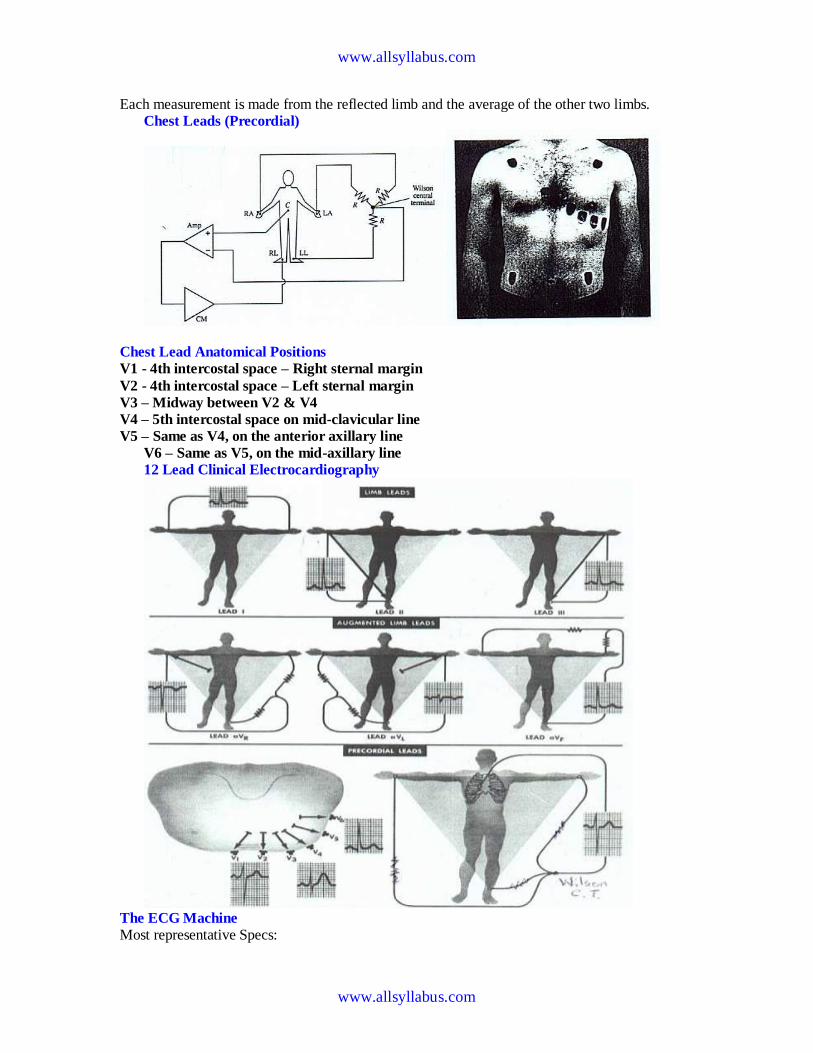
wwwallsyllabuscom
wwwallsyllabuscom
bull Zin = 10 MΩ bull Frequency response = 005 ndash100 Hz bull Strip Chart Recorder Speed = 25 mmsec bull Fast Speed = 100 mmsec
For detailed Specs Refer to the Table in your text ―Summary of performance requirements for electrocardiographs Location of the Heart bull The heart is located between the lungs behind the sternum and above the diaphragm bull It is surrounded by the pericardium bull Its size is about that of a fist and its weight is about 250-300 g bull Its center is located about 15 cm to the left of the midsagittal plane
Anatomy of the heart
bull The walls of the heart are composed of cardiac muscle called myocardium
bull It consists of four compartments ndash the right and left atria and ventricles
The Heart Valves
bull The tricuspid valve regulates blood flow between the right atrium and right
ventricle
bull The pulmonary valve controls blood flow from the right ventricle into the
pulmonary arteries
bull The mitral valve lets oxygen-rich blood from your lungs pass from the left atrium
into the left ventricle
bull The aortic valve lets oxygen-rich blood pass from the left ventricle into the aorta
then to the body
Blood circulation via heart
wwwallsyllabuscom
wwwallsyllabuscom
bull The blood returns from the systemic circulation to the right atrium and from
there goes through the tricuspid valve to the right ventricle
bull It is ejected from the right ventricle through the pulmonary valve to the lungs
bull Oxygenated blood returns from the lungs to the left atrium and from there
through the mitral valve to the left ventricle
bull Finally blood is pumped through the aortic valve to the aorta and the systemic
circulation
Electrical activation of the heart
bull In the heart muscle cell or myocyte electric activation takes place by means of
the same mechanism as in the nerve cell ie from the inflow of Na ions across
the cell membrane
bull The amplitude of the action potential is also similar being 100 mV for both nerve
and muscle
bull The duration of the cardiac impulse is however two orders of magnitude longer
than in either nerve cell or sceletal muscle cell
bull As in the nerve cell repolarization is a consequence of the outflow of K ions
bull The duration of the action impulse is about 300 ms
Mechanical contraction of Cardiac Muscle
bull Associated with the electric activation of cardiac muscle cell is its mechanical
contraction which occurs a little later
bull An important distinction between cardiac muscle tissue and skeletal muscle is
that in cardiac muscle activation can propagate from one cell to another
in any direction
bull Electrical signal begins in the sinoatrial (SA) node natural pacemaker causes
the atria to contract
bull The signal then passes through the atrioventricular (AV) node
ndash sends the signal to the ventricles via the ―bundle of His
ndash causes the ventricles to contract
The Conduction System
wwwallsyllabuscom
wwwallsyllabuscom
The Action Potential
Recording an AP requires the isolation of a single cell Microelectrodes (with tips a few μm across) are used to stimulate and record the response A typical AP is 2-4ms long with an amplitude of about 100Mv The Electroencephalogram EEG
EEG is the graphical representation of the electrical activity of the brain Very commonly used to diagnose certain neurological disorders such as epilepsy
More recently also investigated whether it can detect various forms of dementia or schizophrenia EEG is the specific recording obtained using the scalp electrodes from the surface of the skull
During surgery electrodes may also be placed directly on the cortex The resulting signal is then electrocorticogram (ECoG) Just like ECG EEG is also obtained using several different electrodes places on different regions of the head brain
The Event Related
wwwallsyllabuscom
wwwallsyllabuscom
Potentials ndash ERPs
ERPs are really EEGs obtained under a specific protocol that requires the patient to response to certain stimuli ndash hence event related potentials Also called evoked potentials these signals can be used to diagnose certain
neurological disorders such as dementia and they can also be used as a liedetector bull The oddball paradigm
bull The guilty knowledge test
Electroretinogram ERG
The ERG is the record of the retinal action currents produced by the retina in response to a light stimulus It measures the electrical responses of the light-sensitive cells (such as rods and cones)
The stimuli are often a series of light flashes or rotating patterns The ERG is recorded using contact lens electrode that the subject wears while watching the stimuli
Phonocardiogram ndash PCG
The PCG is the graphic record of the heart sounds and murmurs It is thus a mechanical audio signal rather than an electrical signal Can be easily heard using a stethoscope Or can be converted into an electrical signal using a transducer Typically used to determine the disorders related to the heart valve since their
routine opening and closing create the well-known sounds bull S1 sounds First heart sounds ndash ventricular contractions move blood into atria closing of the AV (mitral and tricuspid) valves then semilunar valves open and blood ejected out of ventricles ndash immediately follows the QRS complex bull S2 sounds Second heart sounds ndash Closure of semilunar (aortic and pulmonary) valves bull Any unexpected sound may indicate a malfunctioning valve that causes the blood flow into out of a chamber when it should not Also called heart murmurs
wwwallsyllabuscom
wwwallsyllabuscom
Define ultrasound
bull Mechanical waves in different modalities (longitudinallateral) needs medium
to be propagated (solid liquid gas)
bull gt 20 kHz
bull Continuouspulsed
bull Sphericalplanarnarrow beamsurface waveLamb-wave
Physical phenomena behind ultrasound measurements
Transmission
bull
bull reflection
bull transit time
bull differences in propagation velocities
bull returns to transit time
bull doppler-shift in frequency
bull flow velocity
bull change of acoustic impedance
bull comparing to reference
bull interference of ultrasound waves (holography)
bull interaction of ultrasound and light (photoacousticz)
bull ultrasound needs medium for propagation it doesnrsquot propagate in vacuum
bull because mechanical waves need moving massunits and spring forces between
them
bull in acoustic emission the medium creates ultrasound (for example during pressure
changes) which is received by sensors
bull pulsed mode more common than continuous
bull continuous reguires separate transducers for transmitting and receicving
bull in pulsed mode an ultrasound burst is sent to the object and the same transducer is
switched to listen echoes
bull standing wave problem
bull in us-therapy pulsed mode gives more effective care without too much heating The Doppler Equation describes the relationship of the Doppler frequency shift to target velocity
wwwallsyllabuscom
wwwallsyllabuscom
The frequency difference is equal to the reflected frequency (FR) minus the originating frequency (FT) If the resulting frequency is higher then there is a positive Doppler shift and the object is moving toward the transducer but if the resulting frequency is lower there is a negative Doppler shift and it is moving away from the transducer In its simplest form it would be calculated as if
the ultrasound was parallel to the targetrsquos direction as shown in diagram A below However this would be a rare occurrence in clinical practice because the transducer is rarely pointed head on to a blood vessel In real life the ultrasound waves would approach the target at an angle called the Doppler angle ( ) On the following page diagram B shows the Doppler
equation used in general clinical situations which includes the Doppler angle The Doppler Angle
The ultrasound beam usually approaches the moving target at an angle called the Doppler angle ( ) This reduces the frequency shift in proportion to the cosine of this
angle If this angle is known then the flow velocity can be calculated The equation used is The Doppler Equation
equiv Doppler shift frequency (the difference between the transmitted and received frequencies) equiv transmitted frequency equiv reflected frequency
V equiv velocity of the blood flow towards the transducer C equiv velocity of sound in tissue θ equiv the angle between the sound beam and the direction of moving blood Where
The Doppler angle ( ) is also known as the angle of insonation It is estimated by the sonographer by a process known as angle correction which involves aligning an indicator on the duplex image along the longitudinal axis of the vessel
There are a few considerations that affect the performance of a Doppler examination that are inherent in the Doppler equation which are ndash The cosine of 90deg is zero so if the ultrasound beam is perpendicular to the direction of blood flow there will be no Doppler shift and it will appear as if there is no flow in the vessel ndash Appropriate estimation of the angle of insonation or angle correction is essential for the accurate determination of Doppler shift and blood flow velocity The angle of insonation should also be less than 60deg at all times since the
cosine function has a steeper curve above this angle and errors in angle correction will be magnified
The simplest Doppler devices use continuous wave (CW Doppler) rather than the pulsed wave used in more complex devices CW Doppler uses two transducers (or a dual element transducer) that transmit and receive ultrasound continuously The transmit and receive beams overlap in a Doppler sample volume some distance from the transducer face as
shown in the diagram below
volume) is the region of transmitting and receiving beam overlap (shaded region) Because there is continuous transducer transmission and reception echoes from all depths within the area arrive at the transducer simultaneously So although CW Doppler can determine the direction of flow it cannot discriminate the different depths where the motion originates The usefulness of CW Doppler devices is limited but they are used clinically to confirm blood flow in superficial vessels as they are good at detecting low velocities As they are easily portable this
can be done at the bedside or in the operating room Most other clinical applications require pulsed wave Doppler Pulsed Wave Doppler (PW Doppler)
wwwallsyllabuscom
wwwallsyllabuscom
Pulsed wave Doppler (PW Doppler) uses a single-element transducer that emits brief pulses of ultrasound energy The time interval between transmitting and then receiving the echoing sound can be used to calculate the depth from where the echo arises The Doppler sample volume can be chosen as to shape depth and position in sampling the flow
data For example the depth is chosen by processing only the signals that return to the transducer in a stipulated time For this technique the ultrasound system transmits a short pulse The eceiver is opened to detect the returning echoes only after a controlled delay and only for a specific duration This time-based gating of the receiving channel allows the definition of a fixed easuring distance which is often referred to as the Sample volume or Doppler gate Then the next ultrasound wave is transmitted The number of pulses transmitted by the system within a second is referred to as the pulse repetition frequency (PRF) The upper PRF limit is given by the time interval required for the echoes to arrive from a sample volume located at a
certain depth The greater the sample-volume depth the longer the time before the echoes are returned and the longer the delay between pulse transmission The greater the samplevolume depth the lower will be the maximum PRF setting Errors in the accuracy of the information arise if the velocities exceed a certain speed The highest velocity accurately measured is called the Nyquist limit Beyond this limit the errors that occur are referred to as aliasing Volume and flow measurement
Flow ndash volume of a liquidgas passing some point over a given time Gases are compressible
Benedict Roth Spirometer
Widely used for physiological amp clinical studies
Light bell moves with the ptrsquos breathing
Movement recorded by a pen on a rotating drum
Water seal prevents leakage of gas
Small seal minimises volume of gas dissolved in water
Suitable for measuring limited gas vols (few litres)
Pneumotachograph
wwwallsyllabuscom
wwwallsyllabuscom
Cardiac Output
Defn vol of blood pumped by the heart per min
CO = SV x HR
Norm ~ 5 lmin
Cardiac index ndash corrected for body surface area
Affected by
Met Rate ndash pregnancy hyperthyroid septic
Preload contractility afterload
Clinical indicators of CO imprecise
Affected by anaesthetic agents used in everyday practice
Provides estimate of
whole body perfusion
oxygen delivery
left ventricular function
Persistently low CO assoc with poor outcome
Methods
Fick method
Dilution techniques ndash dye thermal lithium
Pulse contour analysis- LiDCO amp PiCCO
Oesophageal doppler
TOE
Transthoracic impedance plethysmography
Inert gas through flow
Non-invasive cardiac output measurement
Fick Principle measure volume displacement
1st proposed 1870
―the total uptake or release of a substance by an organ is the product of the
blood flow through that organ and the arteriovenous concentration
difference of the substance
CO = O2 consumption (mlmin) art ndash mixed
venous O2 conc (mll)
Limited by cumbersome equipment sampling errors need for invasive
monitoring and steady-state haemodynamic and metabolic conditions
Indicator dilution techniques
―An indicator mixed into a unit volume of constantly flowing blood can be used to
identify that volume of blood in time provided the indicator remains in the system
between injection and measurement and mixes completely in the blood
wwwallsyllabuscom
wwwallsyllabuscom
Dye dilution
Inert dye ndash indocyanin green
Injected into pulmonary artery and arterial conc measured using a
calibrated cuvette densitometer
Plot indicator dilution curve (see diagram)
CO derived from area under curve
Indicator Dilution Curve
Cardiac Output Measurement
wwwallsyllabuscom
wwwallsyllabuscom
Why bother to check blood pressure
For each 20 mm rise in systolic blood pressure or 10 mm rise in diastolic blood
pressure over 11575
Risk of stroke increases
Risk of heart disease doubles
Risk of renal failure increases
Systolic and diastolic blood pressure
Systolic blood pressure is the highest pressure in the arteries just after the heart
beats
Diastolic blood pressure is the lowest pressure in the arteries just before the heart
beats
Blood pressure is measured indirectly by blood pressure cuff
(sphygmomanometer)
Inflating cuff increases pressure until it cuts off arterial circulation to the arm
Deflating cuff decrease pressure by 2 to 3 mm of mercury per second until blood
first enters the artery creating turbulence this causes a sound with each heartbeat
Sounds continue with each heartbeat until pressure lowers to the lowest pressure
in the artery then turbulence stops so the sound stops
Systolic blood pressure is the cuff pressure at the first sounds diastolic is the cuff
pressure just before the sounds stop
Phase 1 sharp thuds start at systolic blood pressure
Phase 2 blowing sound may disappear entirely (the auscultatory gap )
Phase 3 crisp thud a bit quieter than phase 1
Phase 4 sounds become muffled
Phase 5 end of sounds -- ends at diastolic blood pressure
wwwallsyllabuscom
wwwallsyllabuscom
Make sure the cuff is the right size - its width should be at least 40 of the arms
circumference The cuff will overestimate blood pressure if too small and
underestimate if too large
Place the cuff snugly on patients proximal arm on skin ( not cloth) centered over
the brachial artery (most cuffs have markings)
Support the patients arm at heart level using your arm or a desk
Your patient should sit in the chair for 5 minutes before BP is measured and
should have no caffeine or nicotine for 30 minutes before (JAMA 273 p1211-
1218 1995)
With fingers palpating radial or brachial artery inflate cuff rapidly until you cant
feel the pulse then 20 mm higher
Release cuff at 2 to 3 mm Hg per second until you again feel the pulse this is the
palpable systolic pressure
Wait 30 seconds before measuring blood pressure
Measuring palpable pressure first avoids risk of seriously underestimating blood
pressure because of the auscultatory gap (mistaking Korotkoff phase 3 for phase
1) Many doctors skip this step for time reasons and instead pump cuff to 200 mm
Hg at the next step)
Phase 2 of the Korotkoff sounds can be inaudible - especially in older patients
with systolic hypertension who are at especially high risk of stroke Inflating the
cuff until you dont hear sounds can give you a reading of 14086 when the
patients actual blood pressure is 22086 Most physicians are pressed for time so
they instead inflate the cuff to 200 mm which is beyond the auscultatory gap in
most patients But palpable systolic blood pressure is according to research more
reliable
Place bell of stethoscope (diaphragm is acceptable) over brachial artery
Rapidly pump the cuff to 20 to 30 mm Hg above palpable systolic pressure
Release pressure in the cuff by 2 to 3 mm Hg per second and listen for Korotkoff
sounds including systolic (first) and diastolic (last)
Record as systolicdiastolic Check in both arms the first time you check a
patients blood pressure It may differ by 10 mm Hg or more
If the sounds continues to zero record diastolic blood pressure as the point when
sounds become muffled (phase 4) over zero eg 130700 or just as 13070
Rate
ndash Number of beats in 30 seconds x 2
Strength
ndash Bounding strong or weak (thready)
Regularity
ndash Regular or irregular
You need three readings on two occasions to diagnose hypertension unless blood
pressure is very high
Normal blood pressure in children is
wwwallsyllabuscom
wwwallsyllabuscom
ndash 10255 at 1 year 11269 at 5 years 11978 at 10 years
Blood pressures in adults (JNC VII JAMA 2892560-72 2003)
ndash Normal lt120lt80
ndash Prehypertensive 120-13980-89
ndash Stage 1 hypertension 140-15990-99
ndash Stage 2 hypertension gt160gt100
Adult 60 to 100
Newborn 120-170
1 year 80-160
3 years 80-120
6 years 75-115
10 years 70-110
How to measure observe rise and fall of chest
In infants count for 60 seconds in adults 15 or 30 seconds
Normal respiration Adults 12 to 20
Children
newborn 30-80
1 year 20-40
3 years 20-30
6 years 16-22
Rate
Number of breaths in 30 seconds x 2
Quality
Character of breathing
Rhythm
Regular or irregular
Effort
Normal or labored
Noisy respiration
Normal stridor wheezing snoring gurgling
Depth
Shallow or deep
pH electrode
wwwallsyllabuscom
wwwallsyllabuscom
Governing equation is the Nernst Equation
pCO2 Electrode
The measurement of pCO2 is based on its linear relationship with pH over the range of
10 to 90 mm Hg
The dissociation constant is given by
Taking logarithms
pH = log[HCO3-] ndash log k ndash log a ndash log pCO2
Diathermy
In the natural sciences the term diathermy means electrically induced heat and is commonly
used for muscle relaxation It is also a method of heating tissue electromagnetically or
ultrasonically for therapeutic purposes in medicineContents
[hide]
1 Heating uses 2 Surgical uses
3 Trivia
4 References
E
RT
nF
H
HH
i
ln
0
H O CO H CO H HCO2 2 2 3 3
k
H HCO
a pCO
3
2
wwwallsyllabuscom
wwwallsyllabuscom
Heating uses
Ultrasonic diathermy refers to heating of tissues by ultrasound for the purpose of
therapeutic deep heating No tissue is ordinarily damaged hence it is generally used in
biomedical applications
Electric diathermy uses high frequency alternating electric or magnetic fields sometimes
with no electrode or device contact to the skin to induce gentle deep tissue heating by
induction Again no tissue is ordinarily damaged
Surgical uses
Surgical diathermy is usually better known as electrosurgery (It is also referred to
occasionally as electrocautery but see disambiguation below) Electrosurgery and
surgical diathermy involve the use of high frequency AC electrical current in surgery as
either a cutting modality or else to cauterize small blood vessels to stop bleeding This
technique induces localized tissue burning and damage the zone of which is controlled
by the frequency and power of the device Some sources[1]
insist that electrosurgery be
applied to surgery accomplished by high frequency AC cutting and that
electrocautery be used only for the practice of cauterization with heated nichrome wires
powered by DC current as in the handheld battery-operated portable cautery tools
Trivia
Medical Diathermy devices were used to cause interference to German radio beams used
for targeting night time bombing raids in WWII during the Battle of the Beams
I Diathermy
A Therapeutic use
1 Generation of local heating by high-frequency electromagnetic waves
2 Capacitance techniquemdashbody is placed in an electric field
a Dipolesmdashstructures with positive and negative poles
b Structures with large numbers of dipoles have a greater capacitance to store an electrical charge
c Greatest heating occurs in tissues with fewer dipoles particularly fatty tissues
3 Rapid rotation of dipoles causing mechanical friction and movement of electrons results in local heating
4 Inductance techniquemdashbody is not placed in an electric field
a Magnetic waves generated by driving current through a coiled wire
b Magnetic field creates currents in tissues
wwwallsyllabuscom
wwwallsyllabuscom
c Greatest heating occurs in tissues with low impedance especially muscle
B Precautions and contraindications
1 Diathermy should not be used
a Over metal implants and cardiac pacemakersmdashmore research needed regarding its use over metallic fixations
b Near the uterus of a pregnant woman or near the abdomen or back of a woman who might be pregnant
c On individuals with infections
d On individuals with acute inflammation
e Over moist open wounds
f On patients with malignant tumors
g Over large joint effusions
C Pulsed electromagnetic fields and diathermy
1 Can be pulsed to decrease total energy transmitted to the tissues
2 Short-wave diathermy can be adjusted into a nonthermal range
a Classified as pulsed electromagnetic field (PEMF) or
b Pulsed radio frequency energy (PRFE)
3 Important reclassification as diathermy implies heating
D Efficiency of diathermy and PEMF therapy for musculoskeletal conditions
1 Current research is limited but results suggest that diathermy enhances
treatments directed at soft tissue stretching
2 Some studies suggest that PEMF may speed wound healing and promote healing of nonunion fractures
Diathermy is a modality that uses electromagnetic energy to heat deeper tissues
Diathermy is more effective than ultrasound at heating a larger area of deep tissues
The athletic trainer must identify and respect contraindications to application of ultrasound and diathermy
Pulsed ultrasound and diathermy are used to treat slow-to-heal lesions including skin ulcers and nonunion fractures and may be able to facilitate repair of other tissues including ligaments and tendons
Diathermy
Definition
In diathermy high-frequency electrical currents are used to heat deep muscular tissues The heat increases blood flow speeding up recovery Doctors also use diathermy in surgical procedures by sealing blood vessels with electrically heated probes
The term diathermy is derived from the Greek words therma meaning heat and dia meaning through Diathermy literally means heating through
Origins
The therapeutic effects of heat have long been recognized More than 2000 years ago the Romans took advantage of heat therapies by building hot-spring bathhouses Since then various
wwwallsyllabuscom
wwwallsyllabuscom
methods of using heat have evolved In the early 1890s French physiologist Arseacutene dArsonval began studying the medical application of high-frequency currents The term diathermy was coined by German physician Carl Franz Nagelschmidt who designed a prototype apparatus in 1906 Around 1925 United States doctor J W Schereschewsky began studying the physiological
effects of high-frequency electrical currents on animals It was several years however before the fundamentals of the therapy were understood and put into practice
Benefits
Diathermy can be used to treat arthritis bursitis and other conditions involving stiff painful joints It is also used to treat pelvic infections and sinusitis A benefit of diathermy is that it is a painless procedure that can be administered at a clinic Also if the treatment relieves pain then patients can discontinue pain killers and escape their high cost and side effects
Description
Diathermy involves heating deep muscular tissues When heat is applied to the painful area cellular metabolism speeds up and blood flow increases The increased metabolism and circulation accelerates tissue repair The heat helps the tissues relax and stretch thus alleviating
stiffness Heat also reduces nerve fiber sensitivity increasing the patients pain threshold
There are three methods of diathermy In each energy is delivered to the deep tissues where it is converted to heat The three methods are
Shortwave diathermy The body part to be treated is placed between two capacitor plates Heat is generated as the high-frequency waves travel through the body tissues between the plates Shortwave diathermy is most often used to treat areas like the hip which is covered with a dense tissue mass It is also used to treat pelvic infections and sinusitis The treatment reduces inflammation The Federal Communications Commission regulates the frequency allowed for short-wave diathermy treatment Most machines
function at 2733 megahertz Ultrasound diathermy In this method high-frequency acoustic vibrations are used to
generate heat in deep tissue Microwave diathermy This method uses radar waves to heat tissue This form is the
easiest to use but the microwaves cannot penetrate deep muscles
Diathermy is also used in surgical procedures Many doctors use electrically heated probes to seal blood vessels to prevent excessive bleeding This is particularly helpful in neurosurgery and eye surgery Doctors can also use diathermy to kill abnormal growths such as tumors warts and
infected tissues
Preparations
To keep patients from sweating patients are usually asked to remove clothing from the body part being treated If a patient sweats the electrical currents may pool in the area causing burns Also clothing containing metal must be removed as must earrings buttons barrettes or zippers that contain metal Watches and hearing aids should be removed because the therapy may affect their function
wwwallsyllabuscom
wwwallsyllabuscom
Practitioners of surgical diathermy should steer clear of alcohol-based solutions to prepare and cleanse the skin These preparations can create a flammable vapor and cause burns and fires
Precautions
Patients with metal implants should not undergo diathermy treatment because the metal can act as a conductor of heat and result in serious internal burns Female patients with metallic uterine implants such as an IUD should avoid treatment in the pelvic area Diathermy should not be
used in joints that have been replaced with a prosthesis or in those with sensory impairment who may not be able to tell if they are burning Furthermore pulsed shortwave diathermy should be avoided during pregnancy as it can lead to abnormal fetal development
Patients with hemophilia should avoid the treatment because the increased blood flow could cause them to hemorrhage
Side effects
Some patients may experience superficial burns Since the therapy involves creating heat care must be taken to avoid burns particularly in patients whose injuries have caused decreased sensitivity to heat Also diathermy may affect pacemaker function
Female patients who receive treatment in the lower back or pelvic area may experience an
increased menstrual flow
Research amp general acceptance
For years physiotherapists and physical therapists have used diathermy as a routine part of physical rehabilitation
Electrical Safety
Electrical safety is very important in hospitals as patients may be undergoing a diagnostic or treatment procedure where the protective effect of dry skin is reduced Also patients may be unattended unconscious or anaesthetised and may not respond normally to an electric current Further electrically conductive solutions such as blood and saline are often present in patient treatment areas and may drip or spill on electrical equipment
Electric Current Leakage Current
Extension Leads Double Adaptors Equipment Classification
Class I Class II Defibrillator-Proof
Protective Devices
Residual Current Devices (RCD) Line Isolation overload Monitors (LIMs) Equipment Earthing
Area Classification
wwwallsyllabuscom
wwwallsyllabuscom
Body Protection Area Cardiac Protected Area
Other Electrical Issues Extension Leads
Double Adapters Main Extension Devices Power Boards Installation of Additional Power Points
Electric Current
Injuries received from electric current are dependent on the magnitude of current the pathway
that it takes through the body and the time for which it flows
The nature of electricity flowing through a circuit is analogous to blood flowing through the circulatory system within the human body In this analogy the source of energy is represented by the heart and the blood flowing through arteries and veins is analagous to current flowing
through the conductors and other components of the electric circuit
The application of an electric potential to an electric circuit generates a flow of current through conductive pathways This is analogous to the changes in blood pressure caused by contraction of cardiac muscle that causes blood to flow into the circulatory system For electric current to flow there must be a continuous pathway from the source of potential through electrical components and back to the source
Leakage Current
Electrical components and systems are encased in non conducting insulation to ensure that the electric current is contained and follows the intended pathways If the insulation deteriorates or breaks down current will leak through the insulation barrier and flow to earth This may
be through the protective earth conductor or through the operator
Medical equipment and clinical areas are fitted with a number of protective devices to protect the patient and operator from harmful leakage currents
Extension Leads
Extension leads are not permitted in clinical areas of RCH organisations They may cause high
earth resistance and excessive earth leakage current An extension lead can allow equipment to be powered from areas other than the relevant protected treatment area The power from the other area may not be protected to the same level as the power in the treatment area
wwwallsyllabuscom
wwwallsyllabuscom
As the connection between the extension lead and the equipment mains cable is often on the floor there is a high danger from fluid spills tripping and damage to the mains cable by trolleys when an extension lead is used
Double Adaptors
Double adaptors must not be used in RCH organisations They may not sit securely in a wall
outlet may not be able to provide adequate earth protection and may cause overloading overheating fire or loss of electrical supply
EQUIPMENT CLASSIFICATIONS
There are several methods of providing protection for operators and patients from electrical faults
and harmful leakage current
Class I
Class I equipment is fitted with a three core mains cable containing a protective earth wire
Exposed metal parts on class I equipment are connected to this earth wire
Should a fault develop inside the equipment and the exposed metal comes into contact with the mains the earthing conductor will conduct the fault current to ground Regular testing procedures ensure that earthing conductors are intact as the integrity of the earth wire is of vital importance
Class II
Class II equipment is enclosed within a double insulated case and does not require earthing
conductors Class II equipment is usually fitted with a 2-pin mains plug An internal electrical fault is unlikely to be hazardous as the double insulation prevents any external parts from becoming alive Class II or double insulated equipment can be identified by the class II symbol on the cabinet
Class II Symbol
wwwallsyllabuscom
wwwallsyllabuscom
Defibrillator-Proof
Some medical equipment within the hospital is classified as defibrillator proof When a
defibrillator is discharged through a patient connected to defibrillator proof equipment the equipment will not be damaged by the defibrillators energy Defibrillator proof equipment can remain connected to the patient during defibrillation It is identified by one of the following
symbols
Defibrillator proof symbols
Body protected Cardiac protected
PROTECTIVE DEVICES
Most patient care areas in the hospital are fitted with protective devices These devices are
regularly tested in accordance with the relevant guidelines published by Standards Australia The level of protection provided is dependent upon the device and the area in which it is located
Residual Current Devices (RCD)
RCDs (safety switches) are used in patient treatment areas to monitor and protect the mains supply RCDs sense leakage currents flowing to earth from the equipment If a significant leakage current flows the RCD will detect it and shut off the power supplied to the equipment
within 40 milliseconds Hospital RCDs are more sensitive than those fitted in homes A hospital RCD will trip at 10 milliamperes leakage current
Power outlets supplied through an RCD have a Supply Available lamp The lamp will extinguish
when the RCD trips due to excessive leakage current
wwwallsyllabuscom
wwwallsyllabuscom
Resetting a RCD
Lamp indicates supply is no longer available Disconnect all equipment from the supply Operate the reset button or lever on the supply panel and the Supply Available lamp
should illuminate If not contact Biomedical Engineering
Connect an item of equipment If the RCD trips again then this is the faulty item and should be labelled and sent to Biomedical Engineering
If the RCD does not trip continue connecting equipment until the RCD trips The last piece of equipment connected to the supply is most likely to be faulty as it will have caused the RCD to trip Remove the faulty item from service label it and send to Biomedical Engineering as mentioned above
Line Isolation overload Monitors (LIMs)
In critical life support applications where loss of power supply cannot be tolerated special power outlets powered by isolation transformers are installed
Line Isolation Monitors are installed to continually monitor electrical leakage in the power
supply system If an electrical fault develops in a medical device connected to an isolated power outlet the LIM will detect the leakage current The LIM will alarm and indicate the level of leakage current but will not shut off the electric supply
The faulty equipment can be identified by un plugging one item of equipment at a time from the supply until the alarm stops sounding Equipment that is not faulty may be reconnected Faulty equipment should be appropriately labelled and sent to Biomedical Engineering for repair
The LIM also monitors how much power is being used by the equpiment connected to it If too much power is being used the LIM will alarm and indicate that there is an overload The power used must be reduced immediately by moving some equipment to another circuit as soon as possible until the alarm stops sounding Failure to reduce the load on the LIM will result in the circuit breaker tripping and loss of power to the circuit
wwwallsyllabuscom
wwwallsyllabuscom
Equipotential Earthing
Equipotential earthing is installed in rooms classified as Cardiac Protected electrical areas
Equipotential earthing in treatment areas used for cardiac procedures is intended to minimise any voltage differences between earthed parts of equipment and any other exposed metal in the room
This reduces the possibility of leakage currents that can cause microelectrocution when the patient comes into contact with multiple items of equipment or if the patient happens to come
into contact with metal items in the room whilr they are connected to a medical device
All conductive metal in an equipotential area is connected to a common equipotential earth point with special heavy duty cable
AREA CLASSIFICATIONS
Body Protected Area
These areas are designed for procedures in which patients are connected to equipment that lowers the natural resistance of the skin Applied parts such as electrode gels conductive fluids entering the patient metal needles and catheters provide an easy pathway for current to flow
The main occurrence of injury from Body-Type procedures is from high current levels causing electric shock A direct connection to the patients heart is not present so the risk of Microelectrocution - fibrillation from minute current levels - is reduced
Residual Current Devices (RCD) or Isolation Transformers and Line Isolation Monitors (LIMiacutes) are used in Body Protected areas to provide protection against electrocution from high leakage currents Body-Protected Areas are identified with this sign
Cardiac Protected Area
Where the procedure involves placing an electrical conductor within or near the heart protection against fibrillation induced from small leakage currents is required Electrical conductors used in these procedures include cardiac pacing electrodes intracardiac ECG electrodes and intracardiac catheters
wwwallsyllabuscom
wwwallsyllabuscom
Equipotential earthing in conjunction with RCDs or LIMs provides protection against microelectrocution in Cardiac-Type procedures
Fault currents are reduced to magnitudes that are unlikely to induce fibrillation Used in conjunction with RCDs or LIMs the magnitude and duration of any fault currents
sourced from equipment are limited Cardiac-Protected Areas are identified with this sign
Other electrical issues
This policy aims to provide guidance to those who find that they need more electrical outlets than
those available or that the existing electrical outlets are inconveniently located
As extension leads and multiple outlet power boards can introduce additional hazards into an area the following procedures should be observed
Extension leads
Approved extension leads (AS 3760 1996) may be used in some areas within the hospital but MUST NOT BE USED IN PATIENT AREAS All electrical extension leads must be tagged with an Engineering Department maintenance tag and require a yearly safety inspection and test via
the Engineering Department
Double adapters
Double adapters may cause overloading or equipment earthing problems and are not to be used in
WCH
Mains extension device
The only mains extension device that is to be used in Patient care areas is the 4-way or 8-way
portable Core Balance Unit
The Biomedical Engineering Department must approve all units prior to use These units contain a safety switch and can detect excessive leakage current and disconnect the power in the event of a hazardous situation
wwwallsyllabuscom
wwwallsyllabuscom
Care must be exercised in the use of a portable Core Balance Unit It should be located off the
floor and in a position that will protect it from physical abuse and possible entry of fluids These devices are expensive and easily damaged The device must be sent to Biomedical Engineering every 6 months for safety testing
Power boards
Approved multiple-outlet power boards can be used across RCH but must not be used in patient
care areas except areas approved by the Biomedical Engineering Department
The power boards must have overload protection be fitted with internal safety shutters that protect unused outlets and be fitted with an onoff switch for each outlet
Medical Laser Applications
The main research subjects of the group of Medical Laser Application are in the field of online
monitoring and diagnostics as well as the development of new therapheutic methods The main focus in research is based upon the use of ultrashort (fs) laser pulses
The main advantage of ultrashort laser pulses is the extrem short interaction time which suppress any unwanted side effects of the laser irradiation of the tissue Secondly the broad spectra of the fs laser pulses give the advantage to use the same laser pulses for diagnostic applications like the optical coherence tomography (OCT)
wwwallsyllabuscom
wwwallsyllabuscom
Following research subjects have special attention at the moment
Optical Coherence Tomographie (OCT)
A fs-laser light source can be used to obtain images from inside the tissue when the pulses are coherent superposed This group uses this application to obtain an inside view of the vocal fold and to measure the outline of the crystalline lens
Refractive Surgery
The principle of getting rid of glasses with the help of a laser surgery (fs-LASIK) is underlying rapid improvements concerning precision and safety New technologies were evaluated and transferred very fast in cooperation with an industrial partner
Treatment of Presbyopia
The flexibility of the crystalline lens can be increase by pricise cuts which are induce by fs laser pulse inside the lens The treatment of the presbyopia is thinkable
wwwallsyllabuscom
wwwallsyllabuscom
Ultrafast Physics
Whereas the applications of ultrashort laser pulses increase rapidly is the physics of the interaction between the laser pulses and tissue in many case not fully understood To obtain improved knowledge of the interaction for many fs-laser application this group preforms numerical simulations as well as fundamental experiments
Thermography
Thermography thermal imaging or thermal video is a type of infrared imaging Thermographic cameras detect radiation in the infrared range of the electromagnetic spectrum
(roughly 900ndash14000 nanometers or 09ndash14 microm) and produce images of that radiation Since infrared radiation is emitted by all objects based on their temperatures according to the black body radiation law thermography makes it possible to see ones environment with or without visible illumination The amount of radiation emitted by an object increases with temperature therefore thermography allows one to see variations in temperature (hence the name) When viewed by thermographic camera warm objects stand out well against cooler backgrounds humans and other warm-blooded animals become easily visible against the environment day or
night As a result thermographys extensive use can historically be ascribed to the military and security services
Thermal imaging photography finds many other uses For example firefighters use it to see through smoke find persons and localize the base of a fire With thermal imaging power lines maintenance technicians locate overheating joints and parts a telltale sign of their failure to eliminate potential hazards Where thermal insulation becomes faulty building construction technicians can see heat leaks to improve the efficiencies of cooling or heating air-conditioning Thermal imaging cameras are also installed in some luxury cars to aid the driver the first being
the 2000 Cadillac DeVille Some physiological activities particularly responses in human beings and other warm-blooded animals can also be monitored with thermographic imaging [1]
The appearance and operation of a modern thermographic camera is often similar to a camcorder Enabling the user to see in the infrared spectrum is a function so useful that ability to record their output is often optional A recording module is therefore not always built-in
Instead of CCD sensors most thermal imaging cameras use CMOS Focal Plane Array (FPA) The most common types are InSb InGaAs HgCdTe and QWIP FPA The newest technologies are using low cost and uncooled microbolometers FPA sensors Their resolution is considerably lower than of optical cameras mostly 160x120 or 320x240 pixels up to 640x512 for the most
expensive models Thermographic cameras are much more expensive than their visible-spectrum counterparts and higher-end models are often export-restricted Older bolometers or more
wwwallsyllabuscom
wwwallsyllabuscom
sensitive models as InSb require cryogenic cooling usually by a miniature Stirling cycle refrigerator or liquid nitrogen
Contents
[hide]
1 Difference between IR film and thermography 2 Advantages of Thermography
3 Limitations amp disadvantages of thermography 4 Applications 5 See also 6 External links
o 61 History of thermal imager manufacturers
[edit] Difference between IR film and thermography
Thermal imaging is going to be used on Mars to detect caves that could hold life
[edit] Advantages of Thermography
You get a visual picture so that you can compare temperatures over a large area
It is real time capable of catching moving targets Able to find deteriorating components prior to failure Measurement in areas inaccessible or hazardous for other methods It is a non-destructive test method
[edit] Limitations amp disadvantages of thermography
Quality cameras are expensive and are easily damaged Images can be hard to interpret accurately even with experience
Accurate temperature measurements are very hard to make because of emissivities Most cameras have plusmn2 or worse accuracy (not as accurate as contact) Training and staying proficient in IR scanning is time consuming Ability to only measure surface areas
[edit] Applications
Condition monitoring Medical imaging Night vision Research
Process control Non destructive testing Surveillance in security law enforcement and defense Chemical imaging
wwwallsyllabuscom
wwwallsyllabuscom
Thermal infrared imagers convert the energy in the infrared wavelength into a visible light video display All objects above 0 kelvins emit thermal infrared energy so thermal imagers can passively see all objects regardless of ambient light However most thermal imagers only see objects warmer than -50 degC
The spectrum and amount of thermal radiation depend strongly on an objects surface
temperature This makes it possible for a thermal camera to display an objects temperature However other factors also influence the radiation which limits the accuracy of this technique For example the radiation depends not only on the temperature of the object but is also a function of the emissivity of the object Also radiation also originates from the surroundings and is reflected in the object and the radiation from the object and the reflected radiation will also be influenced by the absorption of the atmosphere
Endoscopy
This page is about having an endoscopy - a test that looks at the inside of your digestive system
There is information on
What endoscopy is
Why you may have an endoscopy
Having an endoscopy
Your results
What an endoscopy is
An endoscopy is a test that looks inside the body The endoscope is a long flexible tube that can
be swallowed It has a camera and light inside it Some doctors call it a telescope
wwwallsyllabuscom
wwwallsyllabuscom
Why you may have an endoscopy
You are most likely to have an endoscopy to look at the inside of your
Gullet (oesophagus)
Stomach
Duodenum - the first part of the small bowel that attaches to the stomach
Large bowel (colon)
Your doctor may want to see inside because you have symptoms such as abnormal bleeding or
difficulty swallowing The doctor can look down the endoscope and see if there are any growths
or other abnormal looking areas Also through the endoscope the doctor can take samples
(biopsies) of any abnormal looking tissues
There is more detailed information about having a colonoscopy in the bowel cancer section of
CancerHelp UK Below is information about having other types of endoscopy
Having an endoscopy
You can have this test as an out patient Most people have a choice between having the test while
they are awake or after having a medicine to make them drowsy (a sedative) Your hospital may
not be happy for you to have a sedative if you live alone and will have no one to look after you
when you go home If you live alone but really want sedation your hospital may allow you to
stay overnight You should ask your doctor about this as early as possible as extra arrangements
will need to be made beforehand
If you dont have a sedative you will have a spray to numb the back of your throat and make it
easier for you to swallow the endoscopy tube
If you would prefer to be asleep during the test you will have an injection to make you very
drowsy just before the test You will need to take someone with you to the hospital appointment
You wont be able to drive for the rest of the day and should have someone to go home with you
You cant eat or drink for about 8 hours before the test so that your stomach and duodenum are
empty Your doctor will give you written instructions about this beforehand or they may arrive
with your appointment letter When you get to the clinic you may be asked to take your upper
clothing off and put on a hospital gown Some hospitals prefer to use gowns because your clothes
wont get messy Once you are ready you get onto the bed or X-ray couch When you are lying
comfortably you have the sedative injection to make you very drowsy Or your doctor will spray
the back of your throat to numb it
Once the sedative or throat spray has worked the doctor will pass the endoscope tube down your
wwwallsyllabuscom
wwwallsyllabuscom
throat to the area being investigated Your doctor will ask you to swallow as the tube goes down
but if youve had a sedative you wont remember that afterwards If there are any abnormalities
the doctor will take pieces of tissue from the abnormal looking area to send to the laboratory for
closer inspection under a microscope These tissue samples are called biopsies
When the test is over you will need to rest for a while If youve had a sedative you may not
remember much (if anything) about the test once you have come round You should be able to go
home the same day
The results
It can take time for test results to come through How long will depend on why you are having
the test Usually the doctor who carries out the endoscopy dictates a report straight way The
report is typed up by the department secretary and goes to your specialist who gives the results to
you If your GP has sent you for the test the results will go directly to the GP surgery
Understandably waiting for results can make you anxious It usually takes a couple of weeks for
the results to come through If your doctor needed them urgently it would have been noted on
the test request form and the results will be ready sooner than that Try to remember to ask your
doctor how long you should expect to wait for the results when you are first asked to go for the
test If it is not an emergency and you have not heard a couple of weeks after your test ring your
doctors secretary to check if they are back
Endoscopy
From Wikipedia the free encyclopedia
Jump to navigation search
Endoscopic images of a duodenal ulcer
wwwallsyllabuscom
wwwallsyllabuscom
A flexible endoscope
Endoscopy means looking inside and typically refers to looking inside the human body for medical reasons using an instrument called an endoscope Endoscopy can also refer to using a borescope in technical situations where direct line-of-sight observation is not feasible
Contents
[hide]
1 Overview 2 Components 3 Uses
4 History 5 Risks 6 After The Endoscopy 7 Recent developments 8 See also 9 References 10 Footnotes
11 External links
Overview
Endoscopy is a minimally invasive diagnostic medical procedure that is used to assess the interior surfaces of an organ by inserting a tube into the body The instrument may have a rigid or flexible tube and not only provide an image for visual inspection and photography but also enable taking
biopsies and retrieval of foreign objects Endoscopy is the vehicle for minimally invasive surgery
Many endoscopic procedures are considered to be relatively painless and at worst associated with mild discomfort for example in esophagogastroduodenoscopy most patients tolerate the procedure with only topical anaesthesia of the oropharynx using lignocaine spray[1] Complications are not common (only 5 of all operations)[citation needed] but can include perforation of the organ under inspection with the endoscope or biopsy instrument If that occurs open surgery may be required to repair the injury
wwwallsyllabuscom
wwwallsyllabuscom
Components
An endoscope can consist of
a rigid or flexible tube a light delivery system to illuminate the organ or object under inspection The light
source is normally outside the body and the light is typically directed via an optical fiber system
a lens system transmitting the image to the viewer from the fiberscope an additional channel to allow entry of medical instruments or manipulators
Uses
Endoscopy can involve
The gastrointestinal tract (GI tract) o esophagus stomach and duodenum (esophagogastroduodenoscopy) o small intestine o colon (colonoscopyproctosigmoidoscopy) o Bile duct
endoscopic retrograde cholangiopancreatography (ERCP) duodenoscope-assisted cholangiopancreatoscopy intraoperative cholangioscopy
The respiratory tract o The nose (rhinoscopy) o The lower respiratory tract (bronchoscopy)
The urinary tract (cystoscopy) The female reproductive system
o The cervix (colposcopy) o The uterus (hysteroscopy) o The Fallopian tubes (Falloscopy)
Normally closed body cavities (through a small incision) o The abdominal or pelvic cavity (laparoscopy) o The interior of a joint (arthroscopy) o Organs of the chest (thoracoscopy and mediastinoscopy)
During pregnancy o The amnion (amnioscopy) o The fetus (fetoscopy)
Plastic Surgery Panendoscopy (or triple endoscopy)
o Combines laryngoscopy esophagoscopy and bronchoscopy Non-medical uses for endoscopy
o The planning and architectural community have found the endoscope useful for
pre-visualization of scale models of proposed buildings and cities (architectural endoscopy)
o Internal inspection of complex technical systems (borescope) o Endoscopes are also a tool helpful in the examination of improvised explosive
devices by bomb disposal personnel
wwwallsyllabuscom
wwwallsyllabuscom
o The FBI uses endoscopes for conducting surveillance via tight spaces
History
The first endoscope of a kind was developed in 1806 by Philip Bozzini with his introduction of a
Lichtleiter (light conductor) for the examinations of the canals and cavities of the human body However the Vienna Medical Society disapproved of such curiosity An endoscope was first introduced into a human in 1822 by William Beaumont an army surgeon at Mackinac
Island Michigan[citation needed] The use of electric light was a major step in the improvement of endoscopy The first such lights were external Later smaller bulbs became available making internal light possible for instance in a hysteroscope by Charles David in 1908[citation needed] Hans Christian Jacobaeus has been given credit for early endoscopic explorations of the abdomen and the thorax with laparoscopy (1912) and thoracoscopy (1910)[citation needed] Laparoscopy was used in the diagnosis of liver and gallbladder disease by Heinz Kalk in the 1930s[citation needed] Hope reported in 1937 on the use of laparoscopy to diagnose ectopic pregnancy[citation needed] In 1944
Raoul Palmer placed his patients in the Trendelenburg position after gaseous distention of the abdomen and thus was able to reliably perform gynecologic laparoscopy[citation needed]
The first gastrocamera was released in 1950 by Olympus Optical Co Ltd The device took pictures on monochromatic film using a small light bulb that was triggered manually The device was of limited use however because it did not implement real-time optical capability Olympus continued its development of endoscopes by incorporating fiber optics in the early 1960s leading to the first useful endoscopes In 1964 it released a gastrocamera guided by a fiberscope[1] A few articles claim that DrBasil Hirschowitz of UnivOf MichiganAnn Arbor discussed the
endoscope in early 50s[2]
As endoscopic technology improved so did the methods of gastrointestinal endoscopy Owing primarily to the efforts of Dr Hiromi Shinya in the late 1960s GI endoscopy developed into what is more recognizable as todays colonoscopy While many doctors experimented with techniques to take advantage of the new iterations of endoscopes Dr Shinya focused on techniques that would allow for successful operation of the endoscope by an individual rejecting the common practice at the time of utilizing two people Consequently many of the fundamental methods and procedures of modern colonoscopy were developed by Dr Shinya
Dr Shinyas other great contribution was to therapeutic endoscopy in his invention of the
electrosurgical polypectomy snare with the aid of Olympus employee Hiroshi Ichikawa Shinya sketched his first plans for the device on January 8 1969 He envisioned a loop of wire attached to the end of a colonoscope that would allow for easy removal of polyps during investigation by passing a current through the wire By September of 1969 the first polypectomy using this device was performed Polypectomy has since become the most common therapeutic procedure performed with an endoscope (Sivak 2004)
By 1980 laparoscopy training was required by gynecologists to perform tubal ligation procedures and diagnostic evaluations of the pelvis The first laparoscopic cholecystectomy was performed in 1984 and the first video-laparoscopic cholecystectomy in 1987[citation needed] During the 1990s
laparoscopic surgery was extended to the appendix spleen colon stomach kidney and liver[citation needed] Wireless capsule endoscopy or Capsule Endoscopy is now approved in all the countries including Japan where government reimbusement will be available from Oct2007Capsule Endoscopy [3] increases detection of Small Bowel tumors where traditional Endoscopy is not very efficient
wwwallsyllabuscom
wwwallsyllabuscom
Risks
Infection
Punctured organs Allergic reactions due to Contrast agents or dyes (such as those used in a CT scan) Over-sedation
After The Endoscopy
After the procedure the patient will be observed and monitored by a qualified individual in the
endoscopy or a recovery area until a significant portion of the medication has worn off Occasionally a patient is left with a mild sore throat which promptly responds to saline gargles or a feeling of distention from the insufflated air that was used during the procedure Both problems are mild and fleeting When fully recovered the patient will be instructed when to resume hisher usual diet (probably within a few hours) and will be allowed to be taken home Because of the use of sedation most facilities mandate that the patient is taken home by another person and not to drive on hisher own or handle machinery for the remainder of the day
Recent developments
With the application of robotic systems telesurgery was introduced as the surgeon could operate
from a site physically removed from the patient The first transatlantic surgery has been called the Lindbergh Operation
wwwallsyllabuscom
wwwallsyllabuscom
Upper Endoscopy
Upper endoscopy enables the physician to look inside the esophagus stomach and duodenum (first part of the small intestine) The procedure might be used to discover the reason for swallowing difficulties nausea vomiting
reflux bleeding indigestion abdominal pain or chest pain Upper endoscopy is also called EGD which stands for esophagogastroduodenoscopy (eh-SAH-fuh-goh-GAS-troh-doo-AH-duh-NAH-skuh-pee)
For the procedure you will swallow a thin flexible lighted tube called an endoscope (EN-doh-skope) Right before the procedure the physician will spray your throat with a numbing agent that may help prevent gagging
You may also receive pain medicine and a sedative to help you relax during the exam The endoscope transmits an image of the inside of the esophagus stomach and duodenum so the physician can carefully examine the lining of these organs The scope also blows air into the stomach this expands the folds of tissue and makes it easier for the physician to examine the stomach
The physician can see abnormalities like inflammation or
bleeding through the endoscope that dont show up well on x rays The physician can also insert instruments into the scope to treat bleeding abnormalities or remove samples of tissue (biopsy) for further tests
Possible complications of upper endoscopy include bleeding and puncture of the stomach lining However such complications are rare Most people will probably have nothing more than a mild sore throat after the procedure
The procedure takes 20 to 30 minutes Because you will be sedated you will need to rest at the endoscopy facility for 1 to 2 hours until the medication wears off
Preparation
Your stomach and duodenum must be empty for the procedure to be thorough and safe so you will not be able to eat or drink anything for at least 6 hours beforehand Also you must
arrange for someone to take you homemdashyou will not be allowed to drive because of the sedatives Your physician may give you other special instructions
The digestive system
wwwallsyllabuscom
wwwallsyllabuscom
What is Endoscopy and Why is it Performed
Endoscopy allows physicians to peer through the bodys passageways Endoscopy is the examination and inspection of the interior of body organs joints or cavities through an endoscope An endoscope is a device that uses fiber optics and powerful lens systems to provide lighting and visualization of the interior of a joint The portion of the endoscope inserted into the body may be rigid or flexible depending upon the medical procedure
An endoscope uses two fiber optic lines A light fiber carries light into the body cavity and an image fiber carries the image of the body cavity back to the physicians viewing lens There is also a separate port to allow for administration of drugs suction and irrigation This port may also be used to introduce small folding instruments such as forceps scissors brushes snares and baskets for tissue excision (removal) sampling or other diagnostic and therapeutic work Endoscopes may be used in conjunction with a camera or video recorder to document images of the inside of the joint or chronicle an endoscopic procedure New endoscopes have digital capabilities for manipulating and enhancing the video images
This figure shows a rigid endoscope used for arthroscopy The image fiber leads from the ocular (eye piece) to the inserted end of the scope The light fiber is below and leads from the light source to the working end of the endoscope
Why Is Endoscopy Performed
Endoscopy can be used to diagnose various conditions by close examination of internal organ and body structures Endoscopy can also guide therapy and repair such as the removal of torn cartilage from the bearing surfaces of a joint Biopsy (tissue sampling for pathologic testing) may also be performed under endoscopic guidance Local or general anesthetic may be used during endoscopy depending upon the type of procedure being performed
Internal abnormalities revealed through endoscopy include abscesses biliary (liver) cirrhosis bleeding bronchitis cancer cysts degenerative disease gallbladder stones hernia inflammation metastatic cancer polyps tumors ulcers and other diseases and conditions
Endoscopy is a minimally invasive procedure and carries with it certain minor risks depending upon the type of procedure being performed However these risks are typically far outweighed by the diagnostic and therapeutic potential of the procedure
Prior to the widespread use of endoscopy and diagnostic imaging most internal conditions could only be diagnosed or treated with open surgery Until the last several decades exploratory surgery was routinely performed only when a patient was critically ill and the source of illness was not known For example in certain dire cases the patients thorax or abdomen were surgically opened and examined to try to determine the source of illness
wwwallsyllabuscom
wwwallsyllabuscom
Endoscopy can often be done on an outpatient basis Outpatient means that the procedure does not require hospital admission and acute care and observation and may be performed outside the premises of a hospital Outpatient procedures performed at hospitals or ambulatory centers allow the patient to go home or return to work within a short while after their procedure
Types of Endoscopy
Fiber optic endoscopes now have widespread use in medicine and guide a myriad of diagnostic and therapeutic procedures including
Arthroscopy examination of joints for diagnosis and treatment (arthroscopic surgery) Bronchoscopy examination of the trachea and lungs bronchial trees to reveal
abscesses bronchitis carcinoma tumors tuberculosis alveolitis infection inflammation Colonoscopy examination of the inside of the colon and large intestine to detect polyps
tumors ulceration inflammation colitis diverticula Chrohns disease and discovery and removal of foreign bodies
Colposcopy direct visualization of the vagina and cervix to detect cancer inflammation and other conditions
Cystoscopy examination of the bladder urethra urinary tract uteral orifices and
prostate (men) with insertion of the endoscope through the urethra
ERCP (endoscopic retrograde cholangio-pancreatography) uses endoscopic guidance to
place a catheter for x-ray fluorosocopy with contrast enhancement This technique is used to examine the livers biliary tree the gallbladder the pancreatic duct and other anatomy to check for stones other obstructions and disease X-ray contrast is introduced into these ducts via catheter and fluoroscopic x-ray images are taken to show any abnormality or blockage If disease is detected it can sometimes be treated at the same time or biopsy can be performed to test for cancer or other pathology ERCP can detect biliary cirrhosis
cancer of the bile ducts pancreatic cysts pseudocysts pancreatic tumors chronic pancreatitis and other conditions such as gallbladder stones
EGD (Esophogealgastroduodensoscopy) visual examination of the upper gastro-
intestinal (GI) tract (also referred to as gastroscopy) to reveal hemorrhage hiatal hernia inflammation of the esophagus gastric ulcers
Endoscopic biopsy is the removal of tissue specimens for pathologic examination and
analysis Gastroscopy examination of the lining of the esophagus stomach and duodenum
Gastroscopy is often used to diagnose ulcers and other sources of bleeding and to guide biopsy of suspect GI cancers
Laparoscopy visualization of the stomach liver and other abdominal organs including
the female reproductive organs for example the fallopian tubes Laryngoscopy examination of the larynx (voice box) Proctoscopy sigmoidoscopy proctosigmoidoscopy examination of the rectum and
sigmoid colon Thoracoscopy examination of the pleura (sac that covers the lungs) pleural spaces
mediastinum and pericardium
A Brief History of Endoscopy
In the early 1900s the first attempts to view inside the body with lighted telescopes were made These initial devices were often fully rigid In the 1930s semi-flexible endoscopes called gastroscopes were developed to view inside of the stomach Fiber-optic endoscopy was pioneered by South African-born physician Basil Hirschowitz at the University of Michigan in 1957 Widespread use of fiber optic endoscopes began in the 1960s
wwwallsyllabuscom
wwwallsyllabuscom
A fiber optic cable is simply a bundle of microscopic glass or plastic fibers that literally allows light and images to be transmitted through curved structures Fiber optic cables are also replacing metal wires as the backbone of the worlds telecommunications infrastructure This Internet page may have traveled through a fiber optic cable as a stream of digital data (bursts of light) on its way to your computer
Endoscopy Equipment
Endoscopes have many practical needs And HMB Endoscopy Products (Hollywood Florida) has been providing endoscopic equipment and educating people on the use of endoscopes for more than 17 years Be sure to Browse our Catalog for all the details on how to purchase these medical instruments that can examine any part of the body
Video
Systems
Fiber
Endoscopes
Video
Endoscopes
In the simplest terms Endoscopy equipment consists of instruments that can look at the inside of many different organs mdash these are small flexible or rigid tubes with a light or lenses on the end that can look into the esophagus stomach and colon mdash and in more general terms endoscopy equipment can help doctors look deep inside body structures and hollow organs
An endoscope and related endoscope products and equipment are usually composed of three components
An optic system that allows the doctor to look through the scope into the organ or
cavity or to attach a video camera to the scope
A fiberoptic cable to light up the bodily area
A lumen (eg the bore of a tube like a needle or catheter) to take tissue samples of the
area being viewed
The beauty of endoscopic products is that they perform dual functions mdash with both diagnostic and therapeutic capabilities For example this means that these endoscopic products and instruments can perform biopsies (eg to evaluate tissue samples) as well as provide sclerotherapy (a medical procedure used to treat varicose veins and ―spider veins) In truth these brief explanations only tell part of the story
wwwallsyllabuscom
wwwallsyllabuscom
TYPES OF ENDOSCOPES
Take a look at the different types of endoscopic equipment you can get with HMB Endoscopy Products Again in very general terms there are two main types of endoscopes
Rigid Flexible
1 rigid endoscopes ndash the majority of which use a convex (curving out like one half of a
circle) glass lens system in which the small glass lenses are separated by large air spaces
2 flexible endoscopes allow for just that mdash flexibility In the animal kingdom for example
a flexible endoscope would be perfect for examining the stomach area of a snake
The popularity of endoscopy equipment continues to grow Some of the industryrsquos household names include Olympus Pentax Funjinon and Storz mdash with HMB both selling and repairing pre-owned and completely refurbished endoscopic products from each of these major manufacturers Browse our Catalog to get the ones you need
THERMOGRAPH APPLICATION Thermographs are digital recording thermometers used to log
temperature in the
marine environment The instruments are attached to a computer to set up recording
parameters for
deployment downloading and display of data in graphic or numeric format These units
are placed in
underwater housings and attached to the bottom or suspended in the water column for
deployment
DESCRIPTION Two types of thermograph are currently available The Ryan
Tempmentor is a
reusable data logger that allows storage of a maximum of 6361 temperature
measurements in the
range of -32C to +70C with 01resolution and 03accuracy The instrument can be
programmed
to take measurements from once per second to once every other hour Instrument
dimensions are 3 x
6 x 13 and the unit weighs 11 ounces The Hobo Temp is a miniature reusable data
logger that
allows storage of 1800 measurements in the range of -20C to +70C with
03resolution and 07
wwwallsyllabuscom
wwwallsyllabuscom
accuracy Sampling intervals from 05 seconds to 48 hours are available Instrument
dimensions are
24 x 19 x 08 and the unit weighs 1 ounce Both units are battery powered with the
battery life of
the Tempmentor being approximately two years and the battery life of the Hobo Temp
being one year THERMOGRAPH APPLICATION Thermographs are digital recording thermometers used to log
temperature in the
marine environment The instruments are attached to a computer to set up recording
parameters for
deployment downloading and display of data in graphic or numeric format These units
are placed in
underwater housings and attached to the bottom or suspended in the water column for
deployment
DESCRIPTION Two types of thermograph are currently available The Ryan
Tempmentor is a
reusable data logger that allows storage of a maximum of 6361 temperature
measurements in the
range of -32C to +70C with 01resolution and 03accuracy The instrument can be
programmed
to take measurements from once per second to once every other hour Instrument
dimensions are 3 x
6 x 13 and the unit weighs 11 ounces The Hobo Temp is a miniature reusable data
logger that
allows storage of 1800 measurements in the range of -20C to +70C with
03resolution and 07
accuracy Sampling intervals from 05 seconds to 48 hours are available Instrument
dimensions are
24 x 19 x 08 and the unit weighs 1 ounce Both units are battery powered with the
battery life of
the Tempmentor being approximately two years and the battery life of the Hobo Temp
being one year THERMOGRAPH APPLICATION Thermographs are digital recording thermometers used to log
temperature in the
marine environment The instruments are attached to a computer to set up recording
parameters for
deployment downloading and display of data in graphic or numeric format These units
are placed in
underwater housings and attached to the bottom or suspended in the water column for
deployment
DESCRIPTION Two types of thermograph are currently available The Ryan
Tempmentor is a
reusable data logger that allows storage of a maximum of 6361 temperature
measurements in the
range of -32C to +70C with 01resolution and 03accuracy The instrument can be
programmed
wwwallsyllabuscom
wwwallsyllabuscom
to take measurements from once per second to once every other hour Instrument
dimensions are 3 x
6 x 13 and the unit weighs 11 ounces The Hobo Temp is a miniature reusable data
logger that
allows storage of 1800 measurements in the range of -20C to +70C with
03resolution and 07
accuracy Sampling intervals from 05 seconds to 48 hours are available Instrument
dimensions are
24 x 19 x 08 and the unit weighs 1 ounce Both units are battery powered with the
battery life of
the Tempmentor being approximately two years and the battery life of the Hobo Temp
being one year
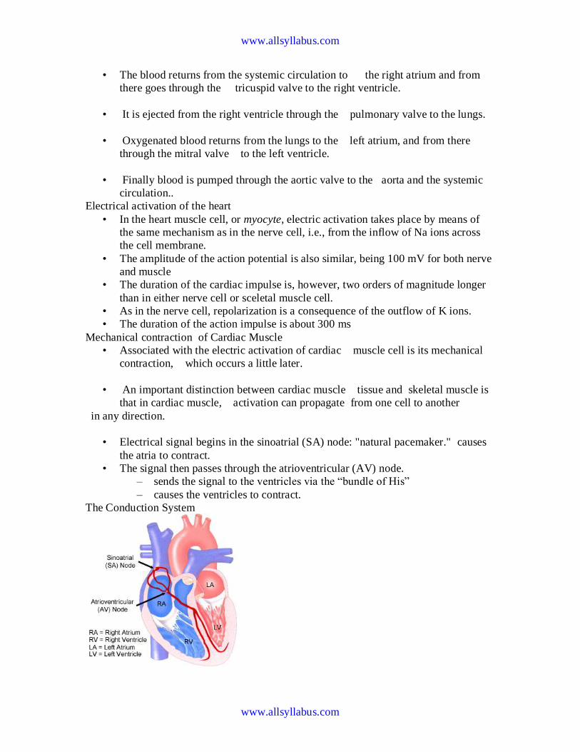
wwwallsyllabuscom
wwwallsyllabuscom
Define ultrasound
bull Mechanical waves in different modalities (longitudinallateral) needs medium
to be propagated (solid liquid gas)
bull gt 20 kHz
bull Continuouspulsed
bull Sphericalplanarnarrow beamsurface waveLamb-wave
Physical phenomena behind ultrasound measurements
Transmission
bull
bull reflection
bull transit time
bull differences in propagation velocities
bull returns to transit time
bull doppler-shift in frequency
bull flow velocity
bull change of acoustic impedance
bull comparing to reference
bull interference of ultrasound waves (holography)
bull interaction of ultrasound and light (photoacousticz)
bull ultrasound needs medium for propagation it doesnrsquot propagate in vacuum
bull because mechanical waves need moving massunits and spring forces between
them
bull in acoustic emission the medium creates ultrasound (for example during pressure
changes) which is received by sensors
bull pulsed mode more common than continuous
bull continuous reguires separate transducers for transmitting and receicving
bull in pulsed mode an ultrasound burst is sent to the object and the same transducer is
switched to listen echoes
bull standing wave problem
bull in us-therapy pulsed mode gives more effective care without too much heating The Doppler Equation describes the relationship of the Doppler frequency shift to target velocity
wwwallsyllabuscom
wwwallsyllabuscom
The frequency difference is equal to the reflected frequency (FR) minus the originating frequency (FT) If the resulting frequency is higher then there is a positive Doppler shift and the object is moving toward the transducer but if the resulting frequency is lower there is a negative Doppler shift and it is moving away from the transducer In its simplest form it would be calculated as if
the ultrasound was parallel to the targetrsquos direction as shown in diagram A below However this would be a rare occurrence in clinical practice because the transducer is rarely pointed head on to a blood vessel In real life the ultrasound waves would approach the target at an angle called the Doppler angle ( ) On the following page diagram B shows the Doppler
equation used in general clinical situations which includes the Doppler angle The Doppler Angle
The ultrasound beam usually approaches the moving target at an angle called the Doppler angle ( ) This reduces the frequency shift in proportion to the cosine of this
angle If this angle is known then the flow velocity can be calculated The equation used is The Doppler Equation
equiv Doppler shift frequency (the difference between the transmitted and received frequencies) equiv transmitted frequency equiv reflected frequency
V equiv velocity of the blood flow towards the transducer C equiv velocity of sound in tissue θ equiv the angle between the sound beam and the direction of moving blood Where
The Doppler angle ( ) is also known as the angle of insonation It is estimated by the sonographer by a process known as angle correction which involves aligning an indicator on the duplex image along the longitudinal axis of the vessel
There are a few considerations that affect the performance of a Doppler examination that are inherent in the Doppler equation which are ndash The cosine of 90deg is zero so if the ultrasound beam is perpendicular to the direction of blood flow there will be no Doppler shift and it will appear as if there is no flow in the vessel ndash Appropriate estimation of the angle of insonation or angle correction is essential for the accurate determination of Doppler shift and blood flow velocity The angle of insonation should also be less than 60deg at all times since the
cosine function has a steeper curve above this angle and errors in angle correction will be magnified
The simplest Doppler devices use continuous wave (CW Doppler) rather than the pulsed wave used in more complex devices CW Doppler uses two transducers (or a dual element transducer) that transmit and receive ultrasound continuously The transmit and receive beams overlap in a Doppler sample volume some distance from the transducer face as
shown in the diagram below
volume) is the region of transmitting and receiving beam overlap (shaded region) Because there is continuous transducer transmission and reception echoes from all depths within the area arrive at the transducer simultaneously So although CW Doppler can determine the direction of flow it cannot discriminate the different depths where the motion originates The usefulness of CW Doppler devices is limited but they are used clinically to confirm blood flow in superficial vessels as they are good at detecting low velocities As they are easily portable this
can be done at the bedside or in the operating room Most other clinical applications require pulsed wave Doppler Pulsed Wave Doppler (PW Doppler)
wwwallsyllabuscom
wwwallsyllabuscom
Pulsed wave Doppler (PW Doppler) uses a single-element transducer that emits brief pulses of ultrasound energy The time interval between transmitting and then receiving the echoing sound can be used to calculate the depth from where the echo arises The Doppler sample volume can be chosen as to shape depth and position in sampling the flow
data For example the depth is chosen by processing only the signals that return to the transducer in a stipulated time For this technique the ultrasound system transmits a short pulse The eceiver is opened to detect the returning echoes only after a controlled delay and only for a specific duration This time-based gating of the receiving channel allows the definition of a fixed easuring distance which is often referred to as the Sample volume or Doppler gate Then the next ultrasound wave is transmitted The number of pulses transmitted by the system within a second is referred to as the pulse repetition frequency (PRF) The upper PRF limit is given by the time interval required for the echoes to arrive from a sample volume located at a
certain depth The greater the sample-volume depth the longer the time before the echoes are returned and the longer the delay between pulse transmission The greater the samplevolume depth the lower will be the maximum PRF setting Errors in the accuracy of the information arise if the velocities exceed a certain speed The highest velocity accurately measured is called the Nyquist limit Beyond this limit the errors that occur are referred to as aliasing Volume and flow measurement
Flow ndash volume of a liquidgas passing some point over a given time Gases are compressible
Benedict Roth Spirometer
Widely used for physiological amp clinical studies
Light bell moves with the ptrsquos breathing
Movement recorded by a pen on a rotating drum
Water seal prevents leakage of gas
Small seal minimises volume of gas dissolved in water
Suitable for measuring limited gas vols (few litres)
Pneumotachograph
wwwallsyllabuscom
wwwallsyllabuscom
Cardiac Output
Defn vol of blood pumped by the heart per min
CO = SV x HR
Norm ~ 5 lmin
Cardiac index ndash corrected for body surface area
Affected by
Met Rate ndash pregnancy hyperthyroid septic
Preload contractility afterload
Clinical indicators of CO imprecise
Affected by anaesthetic agents used in everyday practice
Provides estimate of
whole body perfusion
oxygen delivery
left ventricular function
Persistently low CO assoc with poor outcome
Methods
Fick method
Dilution techniques ndash dye thermal lithium
Pulse contour analysis- LiDCO amp PiCCO
Oesophageal doppler
TOE
Transthoracic impedance plethysmography
Inert gas through flow
Non-invasive cardiac output measurement
Fick Principle measure volume displacement
1st proposed 1870
―the total uptake or release of a substance by an organ is the product of the
blood flow through that organ and the arteriovenous concentration
difference of the substance
CO = O2 consumption (mlmin) art ndash mixed
venous O2 conc (mll)
Limited by cumbersome equipment sampling errors need for invasive
monitoring and steady-state haemodynamic and metabolic conditions
Indicator dilution techniques
―An indicator mixed into a unit volume of constantly flowing blood can be used to
identify that volume of blood in time provided the indicator remains in the system
between injection and measurement and mixes completely in the blood
wwwallsyllabuscom
wwwallsyllabuscom
Dye dilution
Inert dye ndash indocyanin green
Injected into pulmonary artery and arterial conc measured using a
calibrated cuvette densitometer
Plot indicator dilution curve (see diagram)
CO derived from area under curve
Indicator Dilution Curve
Cardiac Output Measurement
wwwallsyllabuscom
wwwallsyllabuscom
Why bother to check blood pressure
For each 20 mm rise in systolic blood pressure or 10 mm rise in diastolic blood
pressure over 11575
Risk of stroke increases
Risk of heart disease doubles
Risk of renal failure increases
Systolic and diastolic blood pressure
Systolic blood pressure is the highest pressure in the arteries just after the heart
beats
Diastolic blood pressure is the lowest pressure in the arteries just before the heart
beats
Blood pressure is measured indirectly by blood pressure cuff
(sphygmomanometer)
Inflating cuff increases pressure until it cuts off arterial circulation to the arm
Deflating cuff decrease pressure by 2 to 3 mm of mercury per second until blood
first enters the artery creating turbulence this causes a sound with each heartbeat
Sounds continue with each heartbeat until pressure lowers to the lowest pressure
in the artery then turbulence stops so the sound stops
Systolic blood pressure is the cuff pressure at the first sounds diastolic is the cuff
pressure just before the sounds stop
Phase 1 sharp thuds start at systolic blood pressure
Phase 2 blowing sound may disappear entirely (the auscultatory gap )
Phase 3 crisp thud a bit quieter than phase 1
Phase 4 sounds become muffled
Phase 5 end of sounds -- ends at diastolic blood pressure
wwwallsyllabuscom
wwwallsyllabuscom
Make sure the cuff is the right size - its width should be at least 40 of the arms
circumference The cuff will overestimate blood pressure if too small and
underestimate if too large
Place the cuff snugly on patients proximal arm on skin ( not cloth) centered over
the brachial artery (most cuffs have markings)
Support the patients arm at heart level using your arm or a desk
Your patient should sit in the chair for 5 minutes before BP is measured and
should have no caffeine or nicotine for 30 minutes before (JAMA 273 p1211-
1218 1995)
With fingers palpating radial or brachial artery inflate cuff rapidly until you cant
feel the pulse then 20 mm higher
Release cuff at 2 to 3 mm Hg per second until you again feel the pulse this is the
palpable systolic pressure
Wait 30 seconds before measuring blood pressure
Measuring palpable pressure first avoids risk of seriously underestimating blood
pressure because of the auscultatory gap (mistaking Korotkoff phase 3 for phase
1) Many doctors skip this step for time reasons and instead pump cuff to 200 mm
Hg at the next step)
Phase 2 of the Korotkoff sounds can be inaudible - especially in older patients
with systolic hypertension who are at especially high risk of stroke Inflating the
cuff until you dont hear sounds can give you a reading of 14086 when the
patients actual blood pressure is 22086 Most physicians are pressed for time so
they instead inflate the cuff to 200 mm which is beyond the auscultatory gap in
most patients But palpable systolic blood pressure is according to research more
reliable
Place bell of stethoscope (diaphragm is acceptable) over brachial artery
Rapidly pump the cuff to 20 to 30 mm Hg above palpable systolic pressure
Release pressure in the cuff by 2 to 3 mm Hg per second and listen for Korotkoff
sounds including systolic (first) and diastolic (last)
Record as systolicdiastolic Check in both arms the first time you check a
patients blood pressure It may differ by 10 mm Hg or more
If the sounds continues to zero record diastolic blood pressure as the point when
sounds become muffled (phase 4) over zero eg 130700 or just as 13070
Rate
ndash Number of beats in 30 seconds x 2
Strength
ndash Bounding strong or weak (thready)
Regularity
ndash Regular or irregular
You need three readings on two occasions to diagnose hypertension unless blood
pressure is very high
Normal blood pressure in children is
wwwallsyllabuscom
wwwallsyllabuscom
ndash 10255 at 1 year 11269 at 5 years 11978 at 10 years
Blood pressures in adults (JNC VII JAMA 2892560-72 2003)
ndash Normal lt120lt80
ndash Prehypertensive 120-13980-89
ndash Stage 1 hypertension 140-15990-99
ndash Stage 2 hypertension gt160gt100
Adult 60 to 100
Newborn 120-170
1 year 80-160
3 years 80-120
6 years 75-115
10 years 70-110
How to measure observe rise and fall of chest
In infants count for 60 seconds in adults 15 or 30 seconds
Normal respiration Adults 12 to 20
Children
newborn 30-80
1 year 20-40
3 years 20-30
6 years 16-22
Rate
Number of breaths in 30 seconds x 2
Quality
Character of breathing
Rhythm
Regular or irregular
Effort
Normal or labored
Noisy respiration
Normal stridor wheezing snoring gurgling
Depth
Shallow or deep
pH electrode
wwwallsyllabuscom
wwwallsyllabuscom
Governing equation is the Nernst Equation
pCO2 Electrode
The measurement of pCO2 is based on its linear relationship with pH over the range of
10 to 90 mm Hg
The dissociation constant is given by
Taking logarithms
pH = log[HCO3-] ndash log k ndash log a ndash log pCO2
Diathermy
In the natural sciences the term diathermy means electrically induced heat and is commonly
used for muscle relaxation It is also a method of heating tissue electromagnetically or
ultrasonically for therapeutic purposes in medicineContents
[hide]
1 Heating uses 2 Surgical uses
3 Trivia
4 References
E
RT
nF
H
HH
i
ln
0
H O CO H CO H HCO2 2 2 3 3
k
H HCO
a pCO
3
2
wwwallsyllabuscom
wwwallsyllabuscom
Heating uses
Ultrasonic diathermy refers to heating of tissues by ultrasound for the purpose of
therapeutic deep heating No tissue is ordinarily damaged hence it is generally used in
biomedical applications
Electric diathermy uses high frequency alternating electric or magnetic fields sometimes
with no electrode or device contact to the skin to induce gentle deep tissue heating by
induction Again no tissue is ordinarily damaged
Surgical uses
Surgical diathermy is usually better known as electrosurgery (It is also referred to
occasionally as electrocautery but see disambiguation below) Electrosurgery and
surgical diathermy involve the use of high frequency AC electrical current in surgery as
either a cutting modality or else to cauterize small blood vessels to stop bleeding This
technique induces localized tissue burning and damage the zone of which is controlled
by the frequency and power of the device Some sources[1]
insist that electrosurgery be
applied to surgery accomplished by high frequency AC cutting and that
electrocautery be used only for the practice of cauterization with heated nichrome wires
powered by DC current as in the handheld battery-operated portable cautery tools
Trivia
Medical Diathermy devices were used to cause interference to German radio beams used
for targeting night time bombing raids in WWII during the Battle of the Beams
I Diathermy
A Therapeutic use
1 Generation of local heating by high-frequency electromagnetic waves
2 Capacitance techniquemdashbody is placed in an electric field
a Dipolesmdashstructures with positive and negative poles
b Structures with large numbers of dipoles have a greater capacitance to store an electrical charge
c Greatest heating occurs in tissues with fewer dipoles particularly fatty tissues
3 Rapid rotation of dipoles causing mechanical friction and movement of electrons results in local heating
4 Inductance techniquemdashbody is not placed in an electric field
a Magnetic waves generated by driving current through a coiled wire
b Magnetic field creates currents in tissues
wwwallsyllabuscom
wwwallsyllabuscom
c Greatest heating occurs in tissues with low impedance especially muscle
B Precautions and contraindications
1 Diathermy should not be used
a Over metal implants and cardiac pacemakersmdashmore research needed regarding its use over metallic fixations
b Near the uterus of a pregnant woman or near the abdomen or back of a woman who might be pregnant
c On individuals with infections
d On individuals with acute inflammation
e Over moist open wounds
f On patients with malignant tumors
g Over large joint effusions
C Pulsed electromagnetic fields and diathermy
1 Can be pulsed to decrease total energy transmitted to the tissues
2 Short-wave diathermy can be adjusted into a nonthermal range
a Classified as pulsed electromagnetic field (PEMF) or
b Pulsed radio frequency energy (PRFE)
3 Important reclassification as diathermy implies heating
D Efficiency of diathermy and PEMF therapy for musculoskeletal conditions
1 Current research is limited but results suggest that diathermy enhances
treatments directed at soft tissue stretching
2 Some studies suggest that PEMF may speed wound healing and promote healing of nonunion fractures
Diathermy is a modality that uses electromagnetic energy to heat deeper tissues
Diathermy is more effective than ultrasound at heating a larger area of deep tissues
The athletic trainer must identify and respect contraindications to application of ultrasound and diathermy
Pulsed ultrasound and diathermy are used to treat slow-to-heal lesions including skin ulcers and nonunion fractures and may be able to facilitate repair of other tissues including ligaments and tendons
Diathermy
Definition
In diathermy high-frequency electrical currents are used to heat deep muscular tissues The heat increases blood flow speeding up recovery Doctors also use diathermy in surgical procedures by sealing blood vessels with electrically heated probes
The term diathermy is derived from the Greek words therma meaning heat and dia meaning through Diathermy literally means heating through
Origins
The therapeutic effects of heat have long been recognized More than 2000 years ago the Romans took advantage of heat therapies by building hot-spring bathhouses Since then various
wwwallsyllabuscom
wwwallsyllabuscom
methods of using heat have evolved In the early 1890s French physiologist Arseacutene dArsonval began studying the medical application of high-frequency currents The term diathermy was coined by German physician Carl Franz Nagelschmidt who designed a prototype apparatus in 1906 Around 1925 United States doctor J W Schereschewsky began studying the physiological
effects of high-frequency electrical currents on animals It was several years however before the fundamentals of the therapy were understood and put into practice
Benefits
Diathermy can be used to treat arthritis bursitis and other conditions involving stiff painful joints It is also used to treat pelvic infections and sinusitis A benefit of diathermy is that it is a painless procedure that can be administered at a clinic Also if the treatment relieves pain then patients can discontinue pain killers and escape their high cost and side effects
Description
Diathermy involves heating deep muscular tissues When heat is applied to the painful area cellular metabolism speeds up and blood flow increases The increased metabolism and circulation accelerates tissue repair The heat helps the tissues relax and stretch thus alleviating
stiffness Heat also reduces nerve fiber sensitivity increasing the patients pain threshold
There are three methods of diathermy In each energy is delivered to the deep tissues where it is converted to heat The three methods are
Shortwave diathermy The body part to be treated is placed between two capacitor plates Heat is generated as the high-frequency waves travel through the body tissues between the plates Shortwave diathermy is most often used to treat areas like the hip which is covered with a dense tissue mass It is also used to treat pelvic infections and sinusitis The treatment reduces inflammation The Federal Communications Commission regulates the frequency allowed for short-wave diathermy treatment Most machines
function at 2733 megahertz Ultrasound diathermy In this method high-frequency acoustic vibrations are used to
generate heat in deep tissue Microwave diathermy This method uses radar waves to heat tissue This form is the
easiest to use but the microwaves cannot penetrate deep muscles
Diathermy is also used in surgical procedures Many doctors use electrically heated probes to seal blood vessels to prevent excessive bleeding This is particularly helpful in neurosurgery and eye surgery Doctors can also use diathermy to kill abnormal growths such as tumors warts and
infected tissues
Preparations
To keep patients from sweating patients are usually asked to remove clothing from the body part being treated If a patient sweats the electrical currents may pool in the area causing burns Also clothing containing metal must be removed as must earrings buttons barrettes or zippers that contain metal Watches and hearing aids should be removed because the therapy may affect their function
wwwallsyllabuscom
wwwallsyllabuscom
Practitioners of surgical diathermy should steer clear of alcohol-based solutions to prepare and cleanse the skin These preparations can create a flammable vapor and cause burns and fires
Precautions
Patients with metal implants should not undergo diathermy treatment because the metal can act as a conductor of heat and result in serious internal burns Female patients with metallic uterine implants such as an IUD should avoid treatment in the pelvic area Diathermy should not be
used in joints that have been replaced with a prosthesis or in those with sensory impairment who may not be able to tell if they are burning Furthermore pulsed shortwave diathermy should be avoided during pregnancy as it can lead to abnormal fetal development
Patients with hemophilia should avoid the treatment because the increased blood flow could cause them to hemorrhage
Side effects
Some patients may experience superficial burns Since the therapy involves creating heat care must be taken to avoid burns particularly in patients whose injuries have caused decreased sensitivity to heat Also diathermy may affect pacemaker function
Female patients who receive treatment in the lower back or pelvic area may experience an
increased menstrual flow
Research amp general acceptance
For years physiotherapists and physical therapists have used diathermy as a routine part of physical rehabilitation
Electrical Safety
Electrical safety is very important in hospitals as patients may be undergoing a diagnostic or treatment procedure where the protective effect of dry skin is reduced Also patients may be unattended unconscious or anaesthetised and may not respond normally to an electric current Further electrically conductive solutions such as blood and saline are often present in patient treatment areas and may drip or spill on electrical equipment
Electric Current Leakage Current
Extension Leads Double Adaptors Equipment Classification
Class I Class II Defibrillator-Proof
Protective Devices
Residual Current Devices (RCD) Line Isolation overload Monitors (LIMs) Equipment Earthing
Area Classification
wwwallsyllabuscom
wwwallsyllabuscom
Body Protection Area Cardiac Protected Area
Other Electrical Issues Extension Leads
Double Adapters Main Extension Devices Power Boards Installation of Additional Power Points
Electric Current
Injuries received from electric current are dependent on the magnitude of current the pathway
that it takes through the body and the time for which it flows
The nature of electricity flowing through a circuit is analogous to blood flowing through the circulatory system within the human body In this analogy the source of energy is represented by the heart and the blood flowing through arteries and veins is analagous to current flowing
through the conductors and other components of the electric circuit
The application of an electric potential to an electric circuit generates a flow of current through conductive pathways This is analogous to the changes in blood pressure caused by contraction of cardiac muscle that causes blood to flow into the circulatory system For electric current to flow there must be a continuous pathway from the source of potential through electrical components and back to the source
Leakage Current
Electrical components and systems are encased in non conducting insulation to ensure that the electric current is contained and follows the intended pathways If the insulation deteriorates or breaks down current will leak through the insulation barrier and flow to earth This may
be through the protective earth conductor or through the operator
Medical equipment and clinical areas are fitted with a number of protective devices to protect the patient and operator from harmful leakage currents
Extension Leads
Extension leads are not permitted in clinical areas of RCH organisations They may cause high
earth resistance and excessive earth leakage current An extension lead can allow equipment to be powered from areas other than the relevant protected treatment area The power from the other area may not be protected to the same level as the power in the treatment area
wwwallsyllabuscom
wwwallsyllabuscom
As the connection between the extension lead and the equipment mains cable is often on the floor there is a high danger from fluid spills tripping and damage to the mains cable by trolleys when an extension lead is used
Double Adaptors
Double adaptors must not be used in RCH organisations They may not sit securely in a wall
outlet may not be able to provide adequate earth protection and may cause overloading overheating fire or loss of electrical supply
EQUIPMENT CLASSIFICATIONS
There are several methods of providing protection for operators and patients from electrical faults
and harmful leakage current
Class I
Class I equipment is fitted with a three core mains cable containing a protective earth wire
Exposed metal parts on class I equipment are connected to this earth wire
Should a fault develop inside the equipment and the exposed metal comes into contact with the mains the earthing conductor will conduct the fault current to ground Regular testing procedures ensure that earthing conductors are intact as the integrity of the earth wire is of vital importance
Class II
Class II equipment is enclosed within a double insulated case and does not require earthing
conductors Class II equipment is usually fitted with a 2-pin mains plug An internal electrical fault is unlikely to be hazardous as the double insulation prevents any external parts from becoming alive Class II or double insulated equipment can be identified by the class II symbol on the cabinet
Class II Symbol
wwwallsyllabuscom
wwwallsyllabuscom
Defibrillator-Proof
Some medical equipment within the hospital is classified as defibrillator proof When a
defibrillator is discharged through a patient connected to defibrillator proof equipment the equipment will not be damaged by the defibrillators energy Defibrillator proof equipment can remain connected to the patient during defibrillation It is identified by one of the following
symbols
Defibrillator proof symbols
Body protected Cardiac protected
PROTECTIVE DEVICES
Most patient care areas in the hospital are fitted with protective devices These devices are
regularly tested in accordance with the relevant guidelines published by Standards Australia The level of protection provided is dependent upon the device and the area in which it is located
Residual Current Devices (RCD)
RCDs (safety switches) are used in patient treatment areas to monitor and protect the mains supply RCDs sense leakage currents flowing to earth from the equipment If a significant leakage current flows the RCD will detect it and shut off the power supplied to the equipment
within 40 milliseconds Hospital RCDs are more sensitive than those fitted in homes A hospital RCD will trip at 10 milliamperes leakage current
Power outlets supplied through an RCD have a Supply Available lamp The lamp will extinguish
when the RCD trips due to excessive leakage current
wwwallsyllabuscom
wwwallsyllabuscom
Resetting a RCD
Lamp indicates supply is no longer available Disconnect all equipment from the supply Operate the reset button or lever on the supply panel and the Supply Available lamp
should illuminate If not contact Biomedical Engineering
Connect an item of equipment If the RCD trips again then this is the faulty item and should be labelled and sent to Biomedical Engineering
If the RCD does not trip continue connecting equipment until the RCD trips The last piece of equipment connected to the supply is most likely to be faulty as it will have caused the RCD to trip Remove the faulty item from service label it and send to Biomedical Engineering as mentioned above
Line Isolation overload Monitors (LIMs)
In critical life support applications where loss of power supply cannot be tolerated special power outlets powered by isolation transformers are installed
Line Isolation Monitors are installed to continually monitor electrical leakage in the power
supply system If an electrical fault develops in a medical device connected to an isolated power outlet the LIM will detect the leakage current The LIM will alarm and indicate the level of leakage current but will not shut off the electric supply
The faulty equipment can be identified by un plugging one item of equipment at a time from the supply until the alarm stops sounding Equipment that is not faulty may be reconnected Faulty equipment should be appropriately labelled and sent to Biomedical Engineering for repair
The LIM also monitors how much power is being used by the equpiment connected to it If too much power is being used the LIM will alarm and indicate that there is an overload The power used must be reduced immediately by moving some equipment to another circuit as soon as possible until the alarm stops sounding Failure to reduce the load on the LIM will result in the circuit breaker tripping and loss of power to the circuit
wwwallsyllabuscom
wwwallsyllabuscom
Equipotential Earthing
Equipotential earthing is installed in rooms classified as Cardiac Protected electrical areas
Equipotential earthing in treatment areas used for cardiac procedures is intended to minimise any voltage differences between earthed parts of equipment and any other exposed metal in the room
This reduces the possibility of leakage currents that can cause microelectrocution when the patient comes into contact with multiple items of equipment or if the patient happens to come
into contact with metal items in the room whilr they are connected to a medical device
All conductive metal in an equipotential area is connected to a common equipotential earth point with special heavy duty cable
AREA CLASSIFICATIONS
Body Protected Area
These areas are designed for procedures in which patients are connected to equipment that lowers the natural resistance of the skin Applied parts such as electrode gels conductive fluids entering the patient metal needles and catheters provide an easy pathway for current to flow
The main occurrence of injury from Body-Type procedures is from high current levels causing electric shock A direct connection to the patients heart is not present so the risk of Microelectrocution - fibrillation from minute current levels - is reduced
Residual Current Devices (RCD) or Isolation Transformers and Line Isolation Monitors (LIMiacutes) are used in Body Protected areas to provide protection against electrocution from high leakage currents Body-Protected Areas are identified with this sign
Cardiac Protected Area
Where the procedure involves placing an electrical conductor within or near the heart protection against fibrillation induced from small leakage currents is required Electrical conductors used in these procedures include cardiac pacing electrodes intracardiac ECG electrodes and intracardiac catheters
wwwallsyllabuscom
wwwallsyllabuscom
Equipotential earthing in conjunction with RCDs or LIMs provides protection against microelectrocution in Cardiac-Type procedures
Fault currents are reduced to magnitudes that are unlikely to induce fibrillation Used in conjunction with RCDs or LIMs the magnitude and duration of any fault currents
sourced from equipment are limited Cardiac-Protected Areas are identified with this sign
Other electrical issues
This policy aims to provide guidance to those who find that they need more electrical outlets than
those available or that the existing electrical outlets are inconveniently located
As extension leads and multiple outlet power boards can introduce additional hazards into an area the following procedures should be observed
Extension leads
Approved extension leads (AS 3760 1996) may be used in some areas within the hospital but MUST NOT BE USED IN PATIENT AREAS All electrical extension leads must be tagged with an Engineering Department maintenance tag and require a yearly safety inspection and test via
the Engineering Department
Double adapters
Double adapters may cause overloading or equipment earthing problems and are not to be used in
WCH
Mains extension device
The only mains extension device that is to be used in Patient care areas is the 4-way or 8-way
portable Core Balance Unit
The Biomedical Engineering Department must approve all units prior to use These units contain a safety switch and can detect excessive leakage current and disconnect the power in the event of a hazardous situation
wwwallsyllabuscom
wwwallsyllabuscom
Care must be exercised in the use of a portable Core Balance Unit It should be located off the
floor and in a position that will protect it from physical abuse and possible entry of fluids These devices are expensive and easily damaged The device must be sent to Biomedical Engineering every 6 months for safety testing
Power boards
Approved multiple-outlet power boards can be used across RCH but must not be used in patient
care areas except areas approved by the Biomedical Engineering Department
The power boards must have overload protection be fitted with internal safety shutters that protect unused outlets and be fitted with an onoff switch for each outlet
Medical Laser Applications
The main research subjects of the group of Medical Laser Application are in the field of online
monitoring and diagnostics as well as the development of new therapheutic methods The main focus in research is based upon the use of ultrashort (fs) laser pulses
The main advantage of ultrashort laser pulses is the extrem short interaction time which suppress any unwanted side effects of the laser irradiation of the tissue Secondly the broad spectra of the fs laser pulses give the advantage to use the same laser pulses for diagnostic applications like the optical coherence tomography (OCT)
wwwallsyllabuscom
wwwallsyllabuscom
Following research subjects have special attention at the moment
Optical Coherence Tomographie (OCT)
A fs-laser light source can be used to obtain images from inside the tissue when the pulses are coherent superposed This group uses this application to obtain an inside view of the vocal fold and to measure the outline of the crystalline lens
Refractive Surgery
The principle of getting rid of glasses with the help of a laser surgery (fs-LASIK) is underlying rapid improvements concerning precision and safety New technologies were evaluated and transferred very fast in cooperation with an industrial partner
Treatment of Presbyopia
The flexibility of the crystalline lens can be increase by pricise cuts which are induce by fs laser pulse inside the lens The treatment of the presbyopia is thinkable
wwwallsyllabuscom
wwwallsyllabuscom
Ultrafast Physics
Whereas the applications of ultrashort laser pulses increase rapidly is the physics of the interaction between the laser pulses and tissue in many case not fully understood To obtain improved knowledge of the interaction for many fs-laser application this group preforms numerical simulations as well as fundamental experiments
Thermography
Thermography thermal imaging or thermal video is a type of infrared imaging Thermographic cameras detect radiation in the infrared range of the electromagnetic spectrum
(roughly 900ndash14000 nanometers or 09ndash14 microm) and produce images of that radiation Since infrared radiation is emitted by all objects based on their temperatures according to the black body radiation law thermography makes it possible to see ones environment with or without visible illumination The amount of radiation emitted by an object increases with temperature therefore thermography allows one to see variations in temperature (hence the name) When viewed by thermographic camera warm objects stand out well against cooler backgrounds humans and other warm-blooded animals become easily visible against the environment day or
night As a result thermographys extensive use can historically be ascribed to the military and security services
Thermal imaging photography finds many other uses For example firefighters use it to see through smoke find persons and localize the base of a fire With thermal imaging power lines maintenance technicians locate overheating joints and parts a telltale sign of their failure to eliminate potential hazards Where thermal insulation becomes faulty building construction technicians can see heat leaks to improve the efficiencies of cooling or heating air-conditioning Thermal imaging cameras are also installed in some luxury cars to aid the driver the first being
the 2000 Cadillac DeVille Some physiological activities particularly responses in human beings and other warm-blooded animals can also be monitored with thermographic imaging [1]
The appearance and operation of a modern thermographic camera is often similar to a camcorder Enabling the user to see in the infrared spectrum is a function so useful that ability to record their output is often optional A recording module is therefore not always built-in
Instead of CCD sensors most thermal imaging cameras use CMOS Focal Plane Array (FPA) The most common types are InSb InGaAs HgCdTe and QWIP FPA The newest technologies are using low cost and uncooled microbolometers FPA sensors Their resolution is considerably lower than of optical cameras mostly 160x120 or 320x240 pixels up to 640x512 for the most
expensive models Thermographic cameras are much more expensive than their visible-spectrum counterparts and higher-end models are often export-restricted Older bolometers or more
wwwallsyllabuscom
wwwallsyllabuscom
sensitive models as InSb require cryogenic cooling usually by a miniature Stirling cycle refrigerator or liquid nitrogen
Contents
[hide]
1 Difference between IR film and thermography 2 Advantages of Thermography
3 Limitations amp disadvantages of thermography 4 Applications 5 See also 6 External links
o 61 History of thermal imager manufacturers
[edit] Difference between IR film and thermography
Thermal imaging is going to be used on Mars to detect caves that could hold life
[edit] Advantages of Thermography
You get a visual picture so that you can compare temperatures over a large area
It is real time capable of catching moving targets Able to find deteriorating components prior to failure Measurement in areas inaccessible or hazardous for other methods It is a non-destructive test method
[edit] Limitations amp disadvantages of thermography
Quality cameras are expensive and are easily damaged Images can be hard to interpret accurately even with experience
Accurate temperature measurements are very hard to make because of emissivities Most cameras have plusmn2 or worse accuracy (not as accurate as contact) Training and staying proficient in IR scanning is time consuming Ability to only measure surface areas
[edit] Applications
Condition monitoring Medical imaging Night vision Research
Process control Non destructive testing Surveillance in security law enforcement and defense Chemical imaging
wwwallsyllabuscom
wwwallsyllabuscom
Thermal infrared imagers convert the energy in the infrared wavelength into a visible light video display All objects above 0 kelvins emit thermal infrared energy so thermal imagers can passively see all objects regardless of ambient light However most thermal imagers only see objects warmer than -50 degC
The spectrum and amount of thermal radiation depend strongly on an objects surface
temperature This makes it possible for a thermal camera to display an objects temperature However other factors also influence the radiation which limits the accuracy of this technique For example the radiation depends not only on the temperature of the object but is also a function of the emissivity of the object Also radiation also originates from the surroundings and is reflected in the object and the radiation from the object and the reflected radiation will also be influenced by the absorption of the atmosphere
Endoscopy
This page is about having an endoscopy - a test that looks at the inside of your digestive system
There is information on
What endoscopy is
Why you may have an endoscopy
Having an endoscopy
Your results
What an endoscopy is
An endoscopy is a test that looks inside the body The endoscope is a long flexible tube that can
be swallowed It has a camera and light inside it Some doctors call it a telescope
wwwallsyllabuscom
wwwallsyllabuscom
Why you may have an endoscopy
You are most likely to have an endoscopy to look at the inside of your
Gullet (oesophagus)
Stomach
Duodenum - the first part of the small bowel that attaches to the stomach
Large bowel (colon)
Your doctor may want to see inside because you have symptoms such as abnormal bleeding or
difficulty swallowing The doctor can look down the endoscope and see if there are any growths
or other abnormal looking areas Also through the endoscope the doctor can take samples
(biopsies) of any abnormal looking tissues
There is more detailed information about having a colonoscopy in the bowel cancer section of
CancerHelp UK Below is information about having other types of endoscopy
Having an endoscopy
You can have this test as an out patient Most people have a choice between having the test while
they are awake or after having a medicine to make them drowsy (a sedative) Your hospital may
not be happy for you to have a sedative if you live alone and will have no one to look after you
when you go home If you live alone but really want sedation your hospital may allow you to
stay overnight You should ask your doctor about this as early as possible as extra arrangements
will need to be made beforehand
If you dont have a sedative you will have a spray to numb the back of your throat and make it
easier for you to swallow the endoscopy tube
If you would prefer to be asleep during the test you will have an injection to make you very
drowsy just before the test You will need to take someone with you to the hospital appointment
You wont be able to drive for the rest of the day and should have someone to go home with you
You cant eat or drink for about 8 hours before the test so that your stomach and duodenum are
empty Your doctor will give you written instructions about this beforehand or they may arrive
with your appointment letter When you get to the clinic you may be asked to take your upper
clothing off and put on a hospital gown Some hospitals prefer to use gowns because your clothes
wont get messy Once you are ready you get onto the bed or X-ray couch When you are lying
comfortably you have the sedative injection to make you very drowsy Or your doctor will spray
the back of your throat to numb it
Once the sedative or throat spray has worked the doctor will pass the endoscope tube down your
wwwallsyllabuscom
wwwallsyllabuscom
throat to the area being investigated Your doctor will ask you to swallow as the tube goes down
but if youve had a sedative you wont remember that afterwards If there are any abnormalities
the doctor will take pieces of tissue from the abnormal looking area to send to the laboratory for
closer inspection under a microscope These tissue samples are called biopsies
When the test is over you will need to rest for a while If youve had a sedative you may not
remember much (if anything) about the test once you have come round You should be able to go
home the same day
The results
It can take time for test results to come through How long will depend on why you are having
the test Usually the doctor who carries out the endoscopy dictates a report straight way The
report is typed up by the department secretary and goes to your specialist who gives the results to
you If your GP has sent you for the test the results will go directly to the GP surgery
Understandably waiting for results can make you anxious It usually takes a couple of weeks for
the results to come through If your doctor needed them urgently it would have been noted on
the test request form and the results will be ready sooner than that Try to remember to ask your
doctor how long you should expect to wait for the results when you are first asked to go for the
test If it is not an emergency and you have not heard a couple of weeks after your test ring your
doctors secretary to check if they are back
Endoscopy
From Wikipedia the free encyclopedia
Jump to navigation search
Endoscopic images of a duodenal ulcer
wwwallsyllabuscom
wwwallsyllabuscom
A flexible endoscope
Endoscopy means looking inside and typically refers to looking inside the human body for medical reasons using an instrument called an endoscope Endoscopy can also refer to using a borescope in technical situations where direct line-of-sight observation is not feasible
Contents
[hide]
1 Overview 2 Components 3 Uses
4 History 5 Risks 6 After The Endoscopy 7 Recent developments 8 See also 9 References 10 Footnotes
11 External links
Overview
Endoscopy is a minimally invasive diagnostic medical procedure that is used to assess the interior surfaces of an organ by inserting a tube into the body The instrument may have a rigid or flexible tube and not only provide an image for visual inspection and photography but also enable taking
biopsies and retrieval of foreign objects Endoscopy is the vehicle for minimally invasive surgery
Many endoscopic procedures are considered to be relatively painless and at worst associated with mild discomfort for example in esophagogastroduodenoscopy most patients tolerate the procedure with only topical anaesthesia of the oropharynx using lignocaine spray[1] Complications are not common (only 5 of all operations)[citation needed] but can include perforation of the organ under inspection with the endoscope or biopsy instrument If that occurs open surgery may be required to repair the injury
wwwallsyllabuscom
wwwallsyllabuscom
Components
An endoscope can consist of
a rigid or flexible tube a light delivery system to illuminate the organ or object under inspection The light
source is normally outside the body and the light is typically directed via an optical fiber system
a lens system transmitting the image to the viewer from the fiberscope an additional channel to allow entry of medical instruments or manipulators
Uses
Endoscopy can involve
The gastrointestinal tract (GI tract) o esophagus stomach and duodenum (esophagogastroduodenoscopy) o small intestine o colon (colonoscopyproctosigmoidoscopy) o Bile duct
endoscopic retrograde cholangiopancreatography (ERCP) duodenoscope-assisted cholangiopancreatoscopy intraoperative cholangioscopy
The respiratory tract o The nose (rhinoscopy) o The lower respiratory tract (bronchoscopy)
The urinary tract (cystoscopy) The female reproductive system
o The cervix (colposcopy) o The uterus (hysteroscopy) o The Fallopian tubes (Falloscopy)
Normally closed body cavities (through a small incision) o The abdominal or pelvic cavity (laparoscopy) o The interior of a joint (arthroscopy) o Organs of the chest (thoracoscopy and mediastinoscopy)
During pregnancy o The amnion (amnioscopy) o The fetus (fetoscopy)
Plastic Surgery Panendoscopy (or triple endoscopy)
o Combines laryngoscopy esophagoscopy and bronchoscopy Non-medical uses for endoscopy
o The planning and architectural community have found the endoscope useful for
pre-visualization of scale models of proposed buildings and cities (architectural endoscopy)
o Internal inspection of complex technical systems (borescope) o Endoscopes are also a tool helpful in the examination of improvised explosive
devices by bomb disposal personnel
wwwallsyllabuscom
wwwallsyllabuscom
o The FBI uses endoscopes for conducting surveillance via tight spaces
History
The first endoscope of a kind was developed in 1806 by Philip Bozzini with his introduction of a
Lichtleiter (light conductor) for the examinations of the canals and cavities of the human body However the Vienna Medical Society disapproved of such curiosity An endoscope was first introduced into a human in 1822 by William Beaumont an army surgeon at Mackinac
Island Michigan[citation needed] The use of electric light was a major step in the improvement of endoscopy The first such lights were external Later smaller bulbs became available making internal light possible for instance in a hysteroscope by Charles David in 1908[citation needed] Hans Christian Jacobaeus has been given credit for early endoscopic explorations of the abdomen and the thorax with laparoscopy (1912) and thoracoscopy (1910)[citation needed] Laparoscopy was used in the diagnosis of liver and gallbladder disease by Heinz Kalk in the 1930s[citation needed] Hope reported in 1937 on the use of laparoscopy to diagnose ectopic pregnancy[citation needed] In 1944
Raoul Palmer placed his patients in the Trendelenburg position after gaseous distention of the abdomen and thus was able to reliably perform gynecologic laparoscopy[citation needed]
The first gastrocamera was released in 1950 by Olympus Optical Co Ltd The device took pictures on monochromatic film using a small light bulb that was triggered manually The device was of limited use however because it did not implement real-time optical capability Olympus continued its development of endoscopes by incorporating fiber optics in the early 1960s leading to the first useful endoscopes In 1964 it released a gastrocamera guided by a fiberscope[1] A few articles claim that DrBasil Hirschowitz of UnivOf MichiganAnn Arbor discussed the
endoscope in early 50s[2]
As endoscopic technology improved so did the methods of gastrointestinal endoscopy Owing primarily to the efforts of Dr Hiromi Shinya in the late 1960s GI endoscopy developed into what is more recognizable as todays colonoscopy While many doctors experimented with techniques to take advantage of the new iterations of endoscopes Dr Shinya focused on techniques that would allow for successful operation of the endoscope by an individual rejecting the common practice at the time of utilizing two people Consequently many of the fundamental methods and procedures of modern colonoscopy were developed by Dr Shinya
Dr Shinyas other great contribution was to therapeutic endoscopy in his invention of the
electrosurgical polypectomy snare with the aid of Olympus employee Hiroshi Ichikawa Shinya sketched his first plans for the device on January 8 1969 He envisioned a loop of wire attached to the end of a colonoscope that would allow for easy removal of polyps during investigation by passing a current through the wire By September of 1969 the first polypectomy using this device was performed Polypectomy has since become the most common therapeutic procedure performed with an endoscope (Sivak 2004)
By 1980 laparoscopy training was required by gynecologists to perform tubal ligation procedures and diagnostic evaluations of the pelvis The first laparoscopic cholecystectomy was performed in 1984 and the first video-laparoscopic cholecystectomy in 1987[citation needed] During the 1990s
laparoscopic surgery was extended to the appendix spleen colon stomach kidney and liver[citation needed] Wireless capsule endoscopy or Capsule Endoscopy is now approved in all the countries including Japan where government reimbusement will be available from Oct2007Capsule Endoscopy [3] increases detection of Small Bowel tumors where traditional Endoscopy is not very efficient
wwwallsyllabuscom
wwwallsyllabuscom
Risks
Infection
Punctured organs Allergic reactions due to Contrast agents or dyes (such as those used in a CT scan) Over-sedation
After The Endoscopy
After the procedure the patient will be observed and monitored by a qualified individual in the
endoscopy or a recovery area until a significant portion of the medication has worn off Occasionally a patient is left with a mild sore throat which promptly responds to saline gargles or a feeling of distention from the insufflated air that was used during the procedure Both problems are mild and fleeting When fully recovered the patient will be instructed when to resume hisher usual diet (probably within a few hours) and will be allowed to be taken home Because of the use of sedation most facilities mandate that the patient is taken home by another person and not to drive on hisher own or handle machinery for the remainder of the day
Recent developments
With the application of robotic systems telesurgery was introduced as the surgeon could operate
from a site physically removed from the patient The first transatlantic surgery has been called the Lindbergh Operation
wwwallsyllabuscom
wwwallsyllabuscom
Upper Endoscopy
Upper endoscopy enables the physician to look inside the esophagus stomach and duodenum (first part of the small intestine) The procedure might be used to discover the reason for swallowing difficulties nausea vomiting
reflux bleeding indigestion abdominal pain or chest pain Upper endoscopy is also called EGD which stands for esophagogastroduodenoscopy (eh-SAH-fuh-goh-GAS-troh-doo-AH-duh-NAH-skuh-pee)
For the procedure you will swallow a thin flexible lighted tube called an endoscope (EN-doh-skope) Right before the procedure the physician will spray your throat with a numbing agent that may help prevent gagging
You may also receive pain medicine and a sedative to help you relax during the exam The endoscope transmits an image of the inside of the esophagus stomach and duodenum so the physician can carefully examine the lining of these organs The scope also blows air into the stomach this expands the folds of tissue and makes it easier for the physician to examine the stomach
The physician can see abnormalities like inflammation or
bleeding through the endoscope that dont show up well on x rays The physician can also insert instruments into the scope to treat bleeding abnormalities or remove samples of tissue (biopsy) for further tests
Possible complications of upper endoscopy include bleeding and puncture of the stomach lining However such complications are rare Most people will probably have nothing more than a mild sore throat after the procedure
The procedure takes 20 to 30 minutes Because you will be sedated you will need to rest at the endoscopy facility for 1 to 2 hours until the medication wears off
Preparation
Your stomach and duodenum must be empty for the procedure to be thorough and safe so you will not be able to eat or drink anything for at least 6 hours beforehand Also you must
arrange for someone to take you homemdashyou will not be allowed to drive because of the sedatives Your physician may give you other special instructions
The digestive system
wwwallsyllabuscom
wwwallsyllabuscom
What is Endoscopy and Why is it Performed
Endoscopy allows physicians to peer through the bodys passageways Endoscopy is the examination and inspection of the interior of body organs joints or cavities through an endoscope An endoscope is a device that uses fiber optics and powerful lens systems to provide lighting and visualization of the interior of a joint The portion of the endoscope inserted into the body may be rigid or flexible depending upon the medical procedure
An endoscope uses two fiber optic lines A light fiber carries light into the body cavity and an image fiber carries the image of the body cavity back to the physicians viewing lens There is also a separate port to allow for administration of drugs suction and irrigation This port may also be used to introduce small folding instruments such as forceps scissors brushes snares and baskets for tissue excision (removal) sampling or other diagnostic and therapeutic work Endoscopes may be used in conjunction with a camera or video recorder to document images of the inside of the joint or chronicle an endoscopic procedure New endoscopes have digital capabilities for manipulating and enhancing the video images
This figure shows a rigid endoscope used for arthroscopy The image fiber leads from the ocular (eye piece) to the inserted end of the scope The light fiber is below and leads from the light source to the working end of the endoscope
Why Is Endoscopy Performed
Endoscopy can be used to diagnose various conditions by close examination of internal organ and body structures Endoscopy can also guide therapy and repair such as the removal of torn cartilage from the bearing surfaces of a joint Biopsy (tissue sampling for pathologic testing) may also be performed under endoscopic guidance Local or general anesthetic may be used during endoscopy depending upon the type of procedure being performed
Internal abnormalities revealed through endoscopy include abscesses biliary (liver) cirrhosis bleeding bronchitis cancer cysts degenerative disease gallbladder stones hernia inflammation metastatic cancer polyps tumors ulcers and other diseases and conditions
Endoscopy is a minimally invasive procedure and carries with it certain minor risks depending upon the type of procedure being performed However these risks are typically far outweighed by the diagnostic and therapeutic potential of the procedure
Prior to the widespread use of endoscopy and diagnostic imaging most internal conditions could only be diagnosed or treated with open surgery Until the last several decades exploratory surgery was routinely performed only when a patient was critically ill and the source of illness was not known For example in certain dire cases the patients thorax or abdomen were surgically opened and examined to try to determine the source of illness
wwwallsyllabuscom
wwwallsyllabuscom
Endoscopy can often be done on an outpatient basis Outpatient means that the procedure does not require hospital admission and acute care and observation and may be performed outside the premises of a hospital Outpatient procedures performed at hospitals or ambulatory centers allow the patient to go home or return to work within a short while after their procedure
Types of Endoscopy
Fiber optic endoscopes now have widespread use in medicine and guide a myriad of diagnostic and therapeutic procedures including
Arthroscopy examination of joints for diagnosis and treatment (arthroscopic surgery) Bronchoscopy examination of the trachea and lungs bronchial trees to reveal
abscesses bronchitis carcinoma tumors tuberculosis alveolitis infection inflammation Colonoscopy examination of the inside of the colon and large intestine to detect polyps
tumors ulceration inflammation colitis diverticula Chrohns disease and discovery and removal of foreign bodies
Colposcopy direct visualization of the vagina and cervix to detect cancer inflammation and other conditions
Cystoscopy examination of the bladder urethra urinary tract uteral orifices and
prostate (men) with insertion of the endoscope through the urethra
ERCP (endoscopic retrograde cholangio-pancreatography) uses endoscopic guidance to
place a catheter for x-ray fluorosocopy with contrast enhancement This technique is used to examine the livers biliary tree the gallbladder the pancreatic duct and other anatomy to check for stones other obstructions and disease X-ray contrast is introduced into these ducts via catheter and fluoroscopic x-ray images are taken to show any abnormality or blockage If disease is detected it can sometimes be treated at the same time or biopsy can be performed to test for cancer or other pathology ERCP can detect biliary cirrhosis
cancer of the bile ducts pancreatic cysts pseudocysts pancreatic tumors chronic pancreatitis and other conditions such as gallbladder stones
EGD (Esophogealgastroduodensoscopy) visual examination of the upper gastro-
intestinal (GI) tract (also referred to as gastroscopy) to reveal hemorrhage hiatal hernia inflammation of the esophagus gastric ulcers
Endoscopic biopsy is the removal of tissue specimens for pathologic examination and
analysis Gastroscopy examination of the lining of the esophagus stomach and duodenum
Gastroscopy is often used to diagnose ulcers and other sources of bleeding and to guide biopsy of suspect GI cancers
Laparoscopy visualization of the stomach liver and other abdominal organs including
the female reproductive organs for example the fallopian tubes Laryngoscopy examination of the larynx (voice box) Proctoscopy sigmoidoscopy proctosigmoidoscopy examination of the rectum and
sigmoid colon Thoracoscopy examination of the pleura (sac that covers the lungs) pleural spaces
mediastinum and pericardium
A Brief History of Endoscopy
In the early 1900s the first attempts to view inside the body with lighted telescopes were made These initial devices were often fully rigid In the 1930s semi-flexible endoscopes called gastroscopes were developed to view inside of the stomach Fiber-optic endoscopy was pioneered by South African-born physician Basil Hirschowitz at the University of Michigan in 1957 Widespread use of fiber optic endoscopes began in the 1960s
wwwallsyllabuscom
wwwallsyllabuscom
A fiber optic cable is simply a bundle of microscopic glass or plastic fibers that literally allows light and images to be transmitted through curved structures Fiber optic cables are also replacing metal wires as the backbone of the worlds telecommunications infrastructure This Internet page may have traveled through a fiber optic cable as a stream of digital data (bursts of light) on its way to your computer
Endoscopy Equipment
Endoscopes have many practical needs And HMB Endoscopy Products (Hollywood Florida) has been providing endoscopic equipment and educating people on the use of endoscopes for more than 17 years Be sure to Browse our Catalog for all the details on how to purchase these medical instruments that can examine any part of the body
Video
Systems
Fiber
Endoscopes
Video
Endoscopes
In the simplest terms Endoscopy equipment consists of instruments that can look at the inside of many different organs mdash these are small flexible or rigid tubes with a light or lenses on the end that can look into the esophagus stomach and colon mdash and in more general terms endoscopy equipment can help doctors look deep inside body structures and hollow organs
An endoscope and related endoscope products and equipment are usually composed of three components
An optic system that allows the doctor to look through the scope into the organ or
cavity or to attach a video camera to the scope
A fiberoptic cable to light up the bodily area
A lumen (eg the bore of a tube like a needle or catheter) to take tissue samples of the
area being viewed
The beauty of endoscopic products is that they perform dual functions mdash with both diagnostic and therapeutic capabilities For example this means that these endoscopic products and instruments can perform biopsies (eg to evaluate tissue samples) as well as provide sclerotherapy (a medical procedure used to treat varicose veins and ―spider veins) In truth these brief explanations only tell part of the story
wwwallsyllabuscom
wwwallsyllabuscom
TYPES OF ENDOSCOPES
Take a look at the different types of endoscopic equipment you can get with HMB Endoscopy Products Again in very general terms there are two main types of endoscopes
Rigid Flexible
1 rigid endoscopes ndash the majority of which use a convex (curving out like one half of a
circle) glass lens system in which the small glass lenses are separated by large air spaces
2 flexible endoscopes allow for just that mdash flexibility In the animal kingdom for example
a flexible endoscope would be perfect for examining the stomach area of a snake
The popularity of endoscopy equipment continues to grow Some of the industryrsquos household names include Olympus Pentax Funjinon and Storz mdash with HMB both selling and repairing pre-owned and completely refurbished endoscopic products from each of these major manufacturers Browse our Catalog to get the ones you need
THERMOGRAPH APPLICATION Thermographs are digital recording thermometers used to log
temperature in the
marine environment The instruments are attached to a computer to set up recording
parameters for
deployment downloading and display of data in graphic or numeric format These units
are placed in
underwater housings and attached to the bottom or suspended in the water column for
deployment
DESCRIPTION Two types of thermograph are currently available The Ryan
Tempmentor is a
reusable data logger that allows storage of a maximum of 6361 temperature
measurements in the
range of -32C to +70C with 01resolution and 03accuracy The instrument can be
programmed
to take measurements from once per second to once every other hour Instrument
dimensions are 3 x
6 x 13 and the unit weighs 11 ounces The Hobo Temp is a miniature reusable data
logger that
allows storage of 1800 measurements in the range of -20C to +70C with
03resolution and 07
wwwallsyllabuscom
wwwallsyllabuscom
accuracy Sampling intervals from 05 seconds to 48 hours are available Instrument
dimensions are
24 x 19 x 08 and the unit weighs 1 ounce Both units are battery powered with the
battery life of
the Tempmentor being approximately two years and the battery life of the Hobo Temp
being one year THERMOGRAPH APPLICATION Thermographs are digital recording thermometers used to log
temperature in the
marine environment The instruments are attached to a computer to set up recording
parameters for
deployment downloading and display of data in graphic or numeric format These units
are placed in
underwater housings and attached to the bottom or suspended in the water column for
deployment
DESCRIPTION Two types of thermograph are currently available The Ryan
Tempmentor is a
reusable data logger that allows storage of a maximum of 6361 temperature
measurements in the
range of -32C to +70C with 01resolution and 03accuracy The instrument can be
programmed
to take measurements from once per second to once every other hour Instrument
dimensions are 3 x
6 x 13 and the unit weighs 11 ounces The Hobo Temp is a miniature reusable data
logger that
allows storage of 1800 measurements in the range of -20C to +70C with
03resolution and 07
accuracy Sampling intervals from 05 seconds to 48 hours are available Instrument
dimensions are
24 x 19 x 08 and the unit weighs 1 ounce Both units are battery powered with the
battery life of
the Tempmentor being approximately two years and the battery life of the Hobo Temp
being one year THERMOGRAPH APPLICATION Thermographs are digital recording thermometers used to log
temperature in the
marine environment The instruments are attached to a computer to set up recording
parameters for
deployment downloading and display of data in graphic or numeric format These units
are placed in
underwater housings and attached to the bottom or suspended in the water column for
deployment
DESCRIPTION Two types of thermograph are currently available The Ryan
Tempmentor is a
reusable data logger that allows storage of a maximum of 6361 temperature
measurements in the
range of -32C to +70C with 01resolution and 03accuracy The instrument can be
programmed
wwwallsyllabuscom
wwwallsyllabuscom
to take measurements from once per second to once every other hour Instrument
dimensions are 3 x
6 x 13 and the unit weighs 11 ounces The Hobo Temp is a miniature reusable data
logger that
allows storage of 1800 measurements in the range of -20C to +70C with
03resolution and 07
accuracy Sampling intervals from 05 seconds to 48 hours are available Instrument
dimensions are
24 x 19 x 08 and the unit weighs 1 ounce Both units are battery powered with the
battery life of
the Tempmentor being approximately two years and the battery life of the Hobo Temp
being one year
wwwallsyllabuscom
wwwallsyllabuscom
The frequency difference is equal to the reflected frequency (FR) minus the originating frequency (FT) If the resulting frequency is higher then there is a positive Doppler shift and the object is moving toward the transducer but if the resulting frequency is lower there is a negative Doppler shift and it is moving away from the transducer In its simplest form it would be calculated as if
the ultrasound was parallel to the targetrsquos direction as shown in diagram A below However this would be a rare occurrence in clinical practice because the transducer is rarely pointed head on to a blood vessel In real life the ultrasound waves would approach the target at an angle called the Doppler angle ( ) On the following page diagram B shows the Doppler
equation used in general clinical situations which includes the Doppler angle The Doppler Angle
The ultrasound beam usually approaches the moving target at an angle called the Doppler angle ( ) This reduces the frequency shift in proportion to the cosine of this
angle If this angle is known then the flow velocity can be calculated The equation used is The Doppler Equation
equiv Doppler shift frequency (the difference between the transmitted and received frequencies) equiv transmitted frequency equiv reflected frequency
V equiv velocity of the blood flow towards the transducer C equiv velocity of sound in tissue θ equiv the angle between the sound beam and the direction of moving blood Where
The Doppler angle ( ) is also known as the angle of insonation It is estimated by the sonographer by a process known as angle correction which involves aligning an indicator on the duplex image along the longitudinal axis of the vessel
There are a few considerations that affect the performance of a Doppler examination that are inherent in the Doppler equation which are ndash The cosine of 90deg is zero so if the ultrasound beam is perpendicular to the direction of blood flow there will be no Doppler shift and it will appear as if there is no flow in the vessel ndash Appropriate estimation of the angle of insonation or angle correction is essential for the accurate determination of Doppler shift and blood flow velocity The angle of insonation should also be less than 60deg at all times since the
cosine function has a steeper curve above this angle and errors in angle correction will be magnified
The simplest Doppler devices use continuous wave (CW Doppler) rather than the pulsed wave used in more complex devices CW Doppler uses two transducers (or a dual element transducer) that transmit and receive ultrasound continuously The transmit and receive beams overlap in a Doppler sample volume some distance from the transducer face as
shown in the diagram below
volume) is the region of transmitting and receiving beam overlap (shaded region) Because there is continuous transducer transmission and reception echoes from all depths within the area arrive at the transducer simultaneously So although CW Doppler can determine the direction of flow it cannot discriminate the different depths where the motion originates The usefulness of CW Doppler devices is limited but they are used clinically to confirm blood flow in superficial vessels as they are good at detecting low velocities As they are easily portable this
can be done at the bedside or in the operating room Most other clinical applications require pulsed wave Doppler Pulsed Wave Doppler (PW Doppler)
wwwallsyllabuscom
wwwallsyllabuscom
Pulsed wave Doppler (PW Doppler) uses a single-element transducer that emits brief pulses of ultrasound energy The time interval between transmitting and then receiving the echoing sound can be used to calculate the depth from where the echo arises The Doppler sample volume can be chosen as to shape depth and position in sampling the flow
data For example the depth is chosen by processing only the signals that return to the transducer in a stipulated time For this technique the ultrasound system transmits a short pulse The eceiver is opened to detect the returning echoes only after a controlled delay and only for a specific duration This time-based gating of the receiving channel allows the definition of a fixed easuring distance which is often referred to as the Sample volume or Doppler gate Then the next ultrasound wave is transmitted The number of pulses transmitted by the system within a second is referred to as the pulse repetition frequency (PRF) The upper PRF limit is given by the time interval required for the echoes to arrive from a sample volume located at a
certain depth The greater the sample-volume depth the longer the time before the echoes are returned and the longer the delay between pulse transmission The greater the samplevolume depth the lower will be the maximum PRF setting Errors in the accuracy of the information arise if the velocities exceed a certain speed The highest velocity accurately measured is called the Nyquist limit Beyond this limit the errors that occur are referred to as aliasing Volume and flow measurement
Flow ndash volume of a liquidgas passing some point over a given time Gases are compressible
Benedict Roth Spirometer
Widely used for physiological amp clinical studies
Light bell moves with the ptrsquos breathing
Movement recorded by a pen on a rotating drum
Water seal prevents leakage of gas
Small seal minimises volume of gas dissolved in water
Suitable for measuring limited gas vols (few litres)
Pneumotachograph
wwwallsyllabuscom
wwwallsyllabuscom
Cardiac Output
Defn vol of blood pumped by the heart per min
CO = SV x HR
Norm ~ 5 lmin
Cardiac index ndash corrected for body surface area
Affected by
Met Rate ndash pregnancy hyperthyroid septic
Preload contractility afterload
Clinical indicators of CO imprecise
Affected by anaesthetic agents used in everyday practice
Provides estimate of
whole body perfusion
oxygen delivery
left ventricular function
Persistently low CO assoc with poor outcome
Methods
Fick method
Dilution techniques ndash dye thermal lithium
Pulse contour analysis- LiDCO amp PiCCO
Oesophageal doppler
TOE
Transthoracic impedance plethysmography
Inert gas through flow
Non-invasive cardiac output measurement
Fick Principle measure volume displacement
1st proposed 1870
―the total uptake or release of a substance by an organ is the product of the
blood flow through that organ and the arteriovenous concentration
difference of the substance
CO = O2 consumption (mlmin) art ndash mixed
venous O2 conc (mll)
Limited by cumbersome equipment sampling errors need for invasive
monitoring and steady-state haemodynamic and metabolic conditions
Indicator dilution techniques
―An indicator mixed into a unit volume of constantly flowing blood can be used to
identify that volume of blood in time provided the indicator remains in the system
between injection and measurement and mixes completely in the blood
wwwallsyllabuscom
wwwallsyllabuscom
Dye dilution
Inert dye ndash indocyanin green
Injected into pulmonary artery and arterial conc measured using a
calibrated cuvette densitometer
Plot indicator dilution curve (see diagram)
CO derived from area under curve
Indicator Dilution Curve
Cardiac Output Measurement
wwwallsyllabuscom
wwwallsyllabuscom
Why bother to check blood pressure
For each 20 mm rise in systolic blood pressure or 10 mm rise in diastolic blood
pressure over 11575
Risk of stroke increases
Risk of heart disease doubles
Risk of renal failure increases
Systolic and diastolic blood pressure
Systolic blood pressure is the highest pressure in the arteries just after the heart
beats
Diastolic blood pressure is the lowest pressure in the arteries just before the heart
beats
Blood pressure is measured indirectly by blood pressure cuff
(sphygmomanometer)
Inflating cuff increases pressure until it cuts off arterial circulation to the arm
Deflating cuff decrease pressure by 2 to 3 mm of mercury per second until blood
first enters the artery creating turbulence this causes a sound with each heartbeat
Sounds continue with each heartbeat until pressure lowers to the lowest pressure
in the artery then turbulence stops so the sound stops
Systolic blood pressure is the cuff pressure at the first sounds diastolic is the cuff
pressure just before the sounds stop
Phase 1 sharp thuds start at systolic blood pressure
Phase 2 blowing sound may disappear entirely (the auscultatory gap )
Phase 3 crisp thud a bit quieter than phase 1
Phase 4 sounds become muffled
Phase 5 end of sounds -- ends at diastolic blood pressure
wwwallsyllabuscom
wwwallsyllabuscom
Make sure the cuff is the right size - its width should be at least 40 of the arms
circumference The cuff will overestimate blood pressure if too small and
underestimate if too large
Place the cuff snugly on patients proximal arm on skin ( not cloth) centered over
the brachial artery (most cuffs have markings)
Support the patients arm at heart level using your arm or a desk
Your patient should sit in the chair for 5 minutes before BP is measured and
should have no caffeine or nicotine for 30 minutes before (JAMA 273 p1211-
1218 1995)
With fingers palpating radial or brachial artery inflate cuff rapidly until you cant
feel the pulse then 20 mm higher
Release cuff at 2 to 3 mm Hg per second until you again feel the pulse this is the
palpable systolic pressure
Wait 30 seconds before measuring blood pressure
Measuring palpable pressure first avoids risk of seriously underestimating blood
pressure because of the auscultatory gap (mistaking Korotkoff phase 3 for phase
1) Many doctors skip this step for time reasons and instead pump cuff to 200 mm
Hg at the next step)
Phase 2 of the Korotkoff sounds can be inaudible - especially in older patients
with systolic hypertension who are at especially high risk of stroke Inflating the
cuff until you dont hear sounds can give you a reading of 14086 when the
patients actual blood pressure is 22086 Most physicians are pressed for time so
they instead inflate the cuff to 200 mm which is beyond the auscultatory gap in
most patients But palpable systolic blood pressure is according to research more
reliable
Place bell of stethoscope (diaphragm is acceptable) over brachial artery
Rapidly pump the cuff to 20 to 30 mm Hg above palpable systolic pressure
Release pressure in the cuff by 2 to 3 mm Hg per second and listen for Korotkoff
sounds including systolic (first) and diastolic (last)
Record as systolicdiastolic Check in both arms the first time you check a
patients blood pressure It may differ by 10 mm Hg or more
If the sounds continues to zero record diastolic blood pressure as the point when
sounds become muffled (phase 4) over zero eg 130700 or just as 13070
Rate
ndash Number of beats in 30 seconds x 2
Strength
ndash Bounding strong or weak (thready)
Regularity
ndash Regular or irregular
You need three readings on two occasions to diagnose hypertension unless blood
pressure is very high
Normal blood pressure in children is
wwwallsyllabuscom
wwwallsyllabuscom
ndash 10255 at 1 year 11269 at 5 years 11978 at 10 years
Blood pressures in adults (JNC VII JAMA 2892560-72 2003)
ndash Normal lt120lt80
ndash Prehypertensive 120-13980-89
ndash Stage 1 hypertension 140-15990-99
ndash Stage 2 hypertension gt160gt100
Adult 60 to 100
Newborn 120-170
1 year 80-160
3 years 80-120
6 years 75-115
10 years 70-110
How to measure observe rise and fall of chest
In infants count for 60 seconds in adults 15 or 30 seconds
Normal respiration Adults 12 to 20
Children
newborn 30-80
1 year 20-40
3 years 20-30
6 years 16-22
Rate
Number of breaths in 30 seconds x 2
Quality
Character of breathing
Rhythm
Regular or irregular
Effort
Normal or labored
Noisy respiration
Normal stridor wheezing snoring gurgling
Depth
Shallow or deep
pH electrode
wwwallsyllabuscom
wwwallsyllabuscom
Governing equation is the Nernst Equation
pCO2 Electrode
The measurement of pCO2 is based on its linear relationship with pH over the range of
10 to 90 mm Hg
The dissociation constant is given by
Taking logarithms
pH = log[HCO3-] ndash log k ndash log a ndash log pCO2
Diathermy
In the natural sciences the term diathermy means electrically induced heat and is commonly
used for muscle relaxation It is also a method of heating tissue electromagnetically or
ultrasonically for therapeutic purposes in medicineContents
[hide]
1 Heating uses 2 Surgical uses
3 Trivia
4 References
E
RT
nF
H
HH
i
ln
0
H O CO H CO H HCO2 2 2 3 3
k
H HCO
a pCO
3
2
wwwallsyllabuscom
wwwallsyllabuscom
Heating uses
Ultrasonic diathermy refers to heating of tissues by ultrasound for the purpose of
therapeutic deep heating No tissue is ordinarily damaged hence it is generally used in
biomedical applications
Electric diathermy uses high frequency alternating electric or magnetic fields sometimes
with no electrode or device contact to the skin to induce gentle deep tissue heating by
induction Again no tissue is ordinarily damaged
Surgical uses
Surgical diathermy is usually better known as electrosurgery (It is also referred to
occasionally as electrocautery but see disambiguation below) Electrosurgery and
surgical diathermy involve the use of high frequency AC electrical current in surgery as
either a cutting modality or else to cauterize small blood vessels to stop bleeding This
technique induces localized tissue burning and damage the zone of which is controlled
by the frequency and power of the device Some sources[1]
insist that electrosurgery be
applied to surgery accomplished by high frequency AC cutting and that
electrocautery be used only for the practice of cauterization with heated nichrome wires
powered by DC current as in the handheld battery-operated portable cautery tools
Trivia
Medical Diathermy devices were used to cause interference to German radio beams used
for targeting night time bombing raids in WWII during the Battle of the Beams
I Diathermy
A Therapeutic use
1 Generation of local heating by high-frequency electromagnetic waves
2 Capacitance techniquemdashbody is placed in an electric field
a Dipolesmdashstructures with positive and negative poles
b Structures with large numbers of dipoles have a greater capacitance to store an electrical charge
c Greatest heating occurs in tissues with fewer dipoles particularly fatty tissues
3 Rapid rotation of dipoles causing mechanical friction and movement of electrons results in local heating
4 Inductance techniquemdashbody is not placed in an electric field
a Magnetic waves generated by driving current through a coiled wire
b Magnetic field creates currents in tissues
wwwallsyllabuscom
wwwallsyllabuscom
c Greatest heating occurs in tissues with low impedance especially muscle
B Precautions and contraindications
1 Diathermy should not be used
a Over metal implants and cardiac pacemakersmdashmore research needed regarding its use over metallic fixations
b Near the uterus of a pregnant woman or near the abdomen or back of a woman who might be pregnant
c On individuals with infections
d On individuals with acute inflammation
e Over moist open wounds
f On patients with malignant tumors
g Over large joint effusions
C Pulsed electromagnetic fields and diathermy
1 Can be pulsed to decrease total energy transmitted to the tissues
2 Short-wave diathermy can be adjusted into a nonthermal range
a Classified as pulsed electromagnetic field (PEMF) or
b Pulsed radio frequency energy (PRFE)
3 Important reclassification as diathermy implies heating
D Efficiency of diathermy and PEMF therapy for musculoskeletal conditions
1 Current research is limited but results suggest that diathermy enhances
treatments directed at soft tissue stretching
2 Some studies suggest that PEMF may speed wound healing and promote healing of nonunion fractures
Diathermy is a modality that uses electromagnetic energy to heat deeper tissues
Diathermy is more effective than ultrasound at heating a larger area of deep tissues
The athletic trainer must identify and respect contraindications to application of ultrasound and diathermy
Pulsed ultrasound and diathermy are used to treat slow-to-heal lesions including skin ulcers and nonunion fractures and may be able to facilitate repair of other tissues including ligaments and tendons
Diathermy
Definition
In diathermy high-frequency electrical currents are used to heat deep muscular tissues The heat increases blood flow speeding up recovery Doctors also use diathermy in surgical procedures by sealing blood vessels with electrically heated probes
The term diathermy is derived from the Greek words therma meaning heat and dia meaning through Diathermy literally means heating through
Origins
The therapeutic effects of heat have long been recognized More than 2000 years ago the Romans took advantage of heat therapies by building hot-spring bathhouses Since then various
wwwallsyllabuscom
wwwallsyllabuscom
methods of using heat have evolved In the early 1890s French physiologist Arseacutene dArsonval began studying the medical application of high-frequency currents The term diathermy was coined by German physician Carl Franz Nagelschmidt who designed a prototype apparatus in 1906 Around 1925 United States doctor J W Schereschewsky began studying the physiological
effects of high-frequency electrical currents on animals It was several years however before the fundamentals of the therapy were understood and put into practice
Benefits
Diathermy can be used to treat arthritis bursitis and other conditions involving stiff painful joints It is also used to treat pelvic infections and sinusitis A benefit of diathermy is that it is a painless procedure that can be administered at a clinic Also if the treatment relieves pain then patients can discontinue pain killers and escape their high cost and side effects
Description
Diathermy involves heating deep muscular tissues When heat is applied to the painful area cellular metabolism speeds up and blood flow increases The increased metabolism and circulation accelerates tissue repair The heat helps the tissues relax and stretch thus alleviating
stiffness Heat also reduces nerve fiber sensitivity increasing the patients pain threshold
There are three methods of diathermy In each energy is delivered to the deep tissues where it is converted to heat The three methods are
Shortwave diathermy The body part to be treated is placed between two capacitor plates Heat is generated as the high-frequency waves travel through the body tissues between the plates Shortwave diathermy is most often used to treat areas like the hip which is covered with a dense tissue mass It is also used to treat pelvic infections and sinusitis The treatment reduces inflammation The Federal Communications Commission regulates the frequency allowed for short-wave diathermy treatment Most machines
function at 2733 megahertz Ultrasound diathermy In this method high-frequency acoustic vibrations are used to
generate heat in deep tissue Microwave diathermy This method uses radar waves to heat tissue This form is the
easiest to use but the microwaves cannot penetrate deep muscles
Diathermy is also used in surgical procedures Many doctors use electrically heated probes to seal blood vessels to prevent excessive bleeding This is particularly helpful in neurosurgery and eye surgery Doctors can also use diathermy to kill abnormal growths such as tumors warts and
infected tissues
Preparations
To keep patients from sweating patients are usually asked to remove clothing from the body part being treated If a patient sweats the electrical currents may pool in the area causing burns Also clothing containing metal must be removed as must earrings buttons barrettes or zippers that contain metal Watches and hearing aids should be removed because the therapy may affect their function
wwwallsyllabuscom
wwwallsyllabuscom
Practitioners of surgical diathermy should steer clear of alcohol-based solutions to prepare and cleanse the skin These preparations can create a flammable vapor and cause burns and fires
Precautions
Patients with metal implants should not undergo diathermy treatment because the metal can act as a conductor of heat and result in serious internal burns Female patients with metallic uterine implants such as an IUD should avoid treatment in the pelvic area Diathermy should not be
used in joints that have been replaced with a prosthesis or in those with sensory impairment who may not be able to tell if they are burning Furthermore pulsed shortwave diathermy should be avoided during pregnancy as it can lead to abnormal fetal development
Patients with hemophilia should avoid the treatment because the increased blood flow could cause them to hemorrhage
Side effects
Some patients may experience superficial burns Since the therapy involves creating heat care must be taken to avoid burns particularly in patients whose injuries have caused decreased sensitivity to heat Also diathermy may affect pacemaker function
Female patients who receive treatment in the lower back or pelvic area may experience an
increased menstrual flow
Research amp general acceptance
For years physiotherapists and physical therapists have used diathermy as a routine part of physical rehabilitation
Electrical Safety
Electrical safety is very important in hospitals as patients may be undergoing a diagnostic or treatment procedure where the protective effect of dry skin is reduced Also patients may be unattended unconscious or anaesthetised and may not respond normally to an electric current Further electrically conductive solutions such as blood and saline are often present in patient treatment areas and may drip or spill on electrical equipment
Electric Current Leakage Current
Extension Leads Double Adaptors Equipment Classification
Class I Class II Defibrillator-Proof
Protective Devices
Residual Current Devices (RCD) Line Isolation overload Monitors (LIMs) Equipment Earthing
Area Classification
wwwallsyllabuscom
wwwallsyllabuscom
Body Protection Area Cardiac Protected Area
Other Electrical Issues Extension Leads
Double Adapters Main Extension Devices Power Boards Installation of Additional Power Points
Electric Current
Injuries received from electric current are dependent on the magnitude of current the pathway
that it takes through the body and the time for which it flows
The nature of electricity flowing through a circuit is analogous to blood flowing through the circulatory system within the human body In this analogy the source of energy is represented by the heart and the blood flowing through arteries and veins is analagous to current flowing
through the conductors and other components of the electric circuit
The application of an electric potential to an electric circuit generates a flow of current through conductive pathways This is analogous to the changes in blood pressure caused by contraction of cardiac muscle that causes blood to flow into the circulatory system For electric current to flow there must be a continuous pathway from the source of potential through electrical components and back to the source
Leakage Current
Electrical components and systems are encased in non conducting insulation to ensure that the electric current is contained and follows the intended pathways If the insulation deteriorates or breaks down current will leak through the insulation barrier and flow to earth This may
be through the protective earth conductor or through the operator
Medical equipment and clinical areas are fitted with a number of protective devices to protect the patient and operator from harmful leakage currents
Extension Leads
Extension leads are not permitted in clinical areas of RCH organisations They may cause high
earth resistance and excessive earth leakage current An extension lead can allow equipment to be powered from areas other than the relevant protected treatment area The power from the other area may not be protected to the same level as the power in the treatment area
wwwallsyllabuscom
wwwallsyllabuscom
As the connection between the extension lead and the equipment mains cable is often on the floor there is a high danger from fluid spills tripping and damage to the mains cable by trolleys when an extension lead is used
Double Adaptors
Double adaptors must not be used in RCH organisations They may not sit securely in a wall
outlet may not be able to provide adequate earth protection and may cause overloading overheating fire or loss of electrical supply
EQUIPMENT CLASSIFICATIONS
There are several methods of providing protection for operators and patients from electrical faults
and harmful leakage current
Class I
Class I equipment is fitted with a three core mains cable containing a protective earth wire
Exposed metal parts on class I equipment are connected to this earth wire
Should a fault develop inside the equipment and the exposed metal comes into contact with the mains the earthing conductor will conduct the fault current to ground Regular testing procedures ensure that earthing conductors are intact as the integrity of the earth wire is of vital importance
Class II
Class II equipment is enclosed within a double insulated case and does not require earthing
conductors Class II equipment is usually fitted with a 2-pin mains plug An internal electrical fault is unlikely to be hazardous as the double insulation prevents any external parts from becoming alive Class II or double insulated equipment can be identified by the class II symbol on the cabinet
Class II Symbol
wwwallsyllabuscom
wwwallsyllabuscom
Defibrillator-Proof
Some medical equipment within the hospital is classified as defibrillator proof When a
defibrillator is discharged through a patient connected to defibrillator proof equipment the equipment will not be damaged by the defibrillators energy Defibrillator proof equipment can remain connected to the patient during defibrillation It is identified by one of the following
symbols
Defibrillator proof symbols
Body protected Cardiac protected
PROTECTIVE DEVICES
Most patient care areas in the hospital are fitted with protective devices These devices are
regularly tested in accordance with the relevant guidelines published by Standards Australia The level of protection provided is dependent upon the device and the area in which it is located
Residual Current Devices (RCD)
RCDs (safety switches) are used in patient treatment areas to monitor and protect the mains supply RCDs sense leakage currents flowing to earth from the equipment If a significant leakage current flows the RCD will detect it and shut off the power supplied to the equipment
within 40 milliseconds Hospital RCDs are more sensitive than those fitted in homes A hospital RCD will trip at 10 milliamperes leakage current
Power outlets supplied through an RCD have a Supply Available lamp The lamp will extinguish
when the RCD trips due to excessive leakage current
wwwallsyllabuscom
wwwallsyllabuscom
Resetting a RCD
Lamp indicates supply is no longer available Disconnect all equipment from the supply Operate the reset button or lever on the supply panel and the Supply Available lamp
should illuminate If not contact Biomedical Engineering
Connect an item of equipment If the RCD trips again then this is the faulty item and should be labelled and sent to Biomedical Engineering
If the RCD does not trip continue connecting equipment until the RCD trips The last piece of equipment connected to the supply is most likely to be faulty as it will have caused the RCD to trip Remove the faulty item from service label it and send to Biomedical Engineering as mentioned above
Line Isolation overload Monitors (LIMs)
In critical life support applications where loss of power supply cannot be tolerated special power outlets powered by isolation transformers are installed
Line Isolation Monitors are installed to continually monitor electrical leakage in the power
supply system If an electrical fault develops in a medical device connected to an isolated power outlet the LIM will detect the leakage current The LIM will alarm and indicate the level of leakage current but will not shut off the electric supply
The faulty equipment can be identified by un plugging one item of equipment at a time from the supply until the alarm stops sounding Equipment that is not faulty may be reconnected Faulty equipment should be appropriately labelled and sent to Biomedical Engineering for repair
The LIM also monitors how much power is being used by the equpiment connected to it If too much power is being used the LIM will alarm and indicate that there is an overload The power used must be reduced immediately by moving some equipment to another circuit as soon as possible until the alarm stops sounding Failure to reduce the load on the LIM will result in the circuit breaker tripping and loss of power to the circuit
wwwallsyllabuscom
wwwallsyllabuscom
Equipotential Earthing
Equipotential earthing is installed in rooms classified as Cardiac Protected electrical areas
Equipotential earthing in treatment areas used for cardiac procedures is intended to minimise any voltage differences between earthed parts of equipment and any other exposed metal in the room
This reduces the possibility of leakage currents that can cause microelectrocution when the patient comes into contact with multiple items of equipment or if the patient happens to come
into contact with metal items in the room whilr they are connected to a medical device
All conductive metal in an equipotential area is connected to a common equipotential earth point with special heavy duty cable
AREA CLASSIFICATIONS
Body Protected Area
These areas are designed for procedures in which patients are connected to equipment that lowers the natural resistance of the skin Applied parts such as electrode gels conductive fluids entering the patient metal needles and catheters provide an easy pathway for current to flow
The main occurrence of injury from Body-Type procedures is from high current levels causing electric shock A direct connection to the patients heart is not present so the risk of Microelectrocution - fibrillation from minute current levels - is reduced
Residual Current Devices (RCD) or Isolation Transformers and Line Isolation Monitors (LIMiacutes) are used in Body Protected areas to provide protection against electrocution from high leakage currents Body-Protected Areas are identified with this sign
Cardiac Protected Area
Where the procedure involves placing an electrical conductor within or near the heart protection against fibrillation induced from small leakage currents is required Electrical conductors used in these procedures include cardiac pacing electrodes intracardiac ECG electrodes and intracardiac catheters
wwwallsyllabuscom
wwwallsyllabuscom
Equipotential earthing in conjunction with RCDs or LIMs provides protection against microelectrocution in Cardiac-Type procedures
Fault currents are reduced to magnitudes that are unlikely to induce fibrillation Used in conjunction with RCDs or LIMs the magnitude and duration of any fault currents
sourced from equipment are limited Cardiac-Protected Areas are identified with this sign
Other electrical issues
This policy aims to provide guidance to those who find that they need more electrical outlets than
those available or that the existing electrical outlets are inconveniently located
As extension leads and multiple outlet power boards can introduce additional hazards into an area the following procedures should be observed
Extension leads
Approved extension leads (AS 3760 1996) may be used in some areas within the hospital but MUST NOT BE USED IN PATIENT AREAS All electrical extension leads must be tagged with an Engineering Department maintenance tag and require a yearly safety inspection and test via
the Engineering Department
Double adapters
Double adapters may cause overloading or equipment earthing problems and are not to be used in
WCH
Mains extension device
The only mains extension device that is to be used in Patient care areas is the 4-way or 8-way
portable Core Balance Unit
The Biomedical Engineering Department must approve all units prior to use These units contain a safety switch and can detect excessive leakage current and disconnect the power in the event of a hazardous situation
wwwallsyllabuscom
wwwallsyllabuscom
Care must be exercised in the use of a portable Core Balance Unit It should be located off the
floor and in a position that will protect it from physical abuse and possible entry of fluids These devices are expensive and easily damaged The device must be sent to Biomedical Engineering every 6 months for safety testing
Power boards
Approved multiple-outlet power boards can be used across RCH but must not be used in patient
care areas except areas approved by the Biomedical Engineering Department
The power boards must have overload protection be fitted with internal safety shutters that protect unused outlets and be fitted with an onoff switch for each outlet
Medical Laser Applications
The main research subjects of the group of Medical Laser Application are in the field of online
monitoring and diagnostics as well as the development of new therapheutic methods The main focus in research is based upon the use of ultrashort (fs) laser pulses
The main advantage of ultrashort laser pulses is the extrem short interaction time which suppress any unwanted side effects of the laser irradiation of the tissue Secondly the broad spectra of the fs laser pulses give the advantage to use the same laser pulses for diagnostic applications like the optical coherence tomography (OCT)
wwwallsyllabuscom
wwwallsyllabuscom
Following research subjects have special attention at the moment
Optical Coherence Tomographie (OCT)
A fs-laser light source can be used to obtain images from inside the tissue when the pulses are coherent superposed This group uses this application to obtain an inside view of the vocal fold and to measure the outline of the crystalline lens
Refractive Surgery
The principle of getting rid of glasses with the help of a laser surgery (fs-LASIK) is underlying rapid improvements concerning precision and safety New technologies were evaluated and transferred very fast in cooperation with an industrial partner
Treatment of Presbyopia
The flexibility of the crystalline lens can be increase by pricise cuts which are induce by fs laser pulse inside the lens The treatment of the presbyopia is thinkable
wwwallsyllabuscom
wwwallsyllabuscom
Ultrafast Physics
Whereas the applications of ultrashort laser pulses increase rapidly is the physics of the interaction between the laser pulses and tissue in many case not fully understood To obtain improved knowledge of the interaction for many fs-laser application this group preforms numerical simulations as well as fundamental experiments
Thermography
Thermography thermal imaging or thermal video is a type of infrared imaging Thermographic cameras detect radiation in the infrared range of the electromagnetic spectrum
(roughly 900ndash14000 nanometers or 09ndash14 microm) and produce images of that radiation Since infrared radiation is emitted by all objects based on their temperatures according to the black body radiation law thermography makes it possible to see ones environment with or without visible illumination The amount of radiation emitted by an object increases with temperature therefore thermography allows one to see variations in temperature (hence the name) When viewed by thermographic camera warm objects stand out well against cooler backgrounds humans and other warm-blooded animals become easily visible against the environment day or
night As a result thermographys extensive use can historically be ascribed to the military and security services
Thermal imaging photography finds many other uses For example firefighters use it to see through smoke find persons and localize the base of a fire With thermal imaging power lines maintenance technicians locate overheating joints and parts a telltale sign of their failure to eliminate potential hazards Where thermal insulation becomes faulty building construction technicians can see heat leaks to improve the efficiencies of cooling or heating air-conditioning Thermal imaging cameras are also installed in some luxury cars to aid the driver the first being
the 2000 Cadillac DeVille Some physiological activities particularly responses in human beings and other warm-blooded animals can also be monitored with thermographic imaging [1]
The appearance and operation of a modern thermographic camera is often similar to a camcorder Enabling the user to see in the infrared spectrum is a function so useful that ability to record their output is often optional A recording module is therefore not always built-in
Instead of CCD sensors most thermal imaging cameras use CMOS Focal Plane Array (FPA) The most common types are InSb InGaAs HgCdTe and QWIP FPA The newest technologies are using low cost and uncooled microbolometers FPA sensors Their resolution is considerably lower than of optical cameras mostly 160x120 or 320x240 pixels up to 640x512 for the most
expensive models Thermographic cameras are much more expensive than their visible-spectrum counterparts and higher-end models are often export-restricted Older bolometers or more
wwwallsyllabuscom
wwwallsyllabuscom
sensitive models as InSb require cryogenic cooling usually by a miniature Stirling cycle refrigerator or liquid nitrogen
Contents
[hide]
1 Difference between IR film and thermography 2 Advantages of Thermography
3 Limitations amp disadvantages of thermography 4 Applications 5 See also 6 External links
o 61 History of thermal imager manufacturers
[edit] Difference between IR film and thermography
Thermal imaging is going to be used on Mars to detect caves that could hold life
[edit] Advantages of Thermography
You get a visual picture so that you can compare temperatures over a large area
It is real time capable of catching moving targets Able to find deteriorating components prior to failure Measurement in areas inaccessible or hazardous for other methods It is a non-destructive test method
[edit] Limitations amp disadvantages of thermography
Quality cameras are expensive and are easily damaged Images can be hard to interpret accurately even with experience
Accurate temperature measurements are very hard to make because of emissivities Most cameras have plusmn2 or worse accuracy (not as accurate as contact) Training and staying proficient in IR scanning is time consuming Ability to only measure surface areas
[edit] Applications
Condition monitoring Medical imaging Night vision Research
Process control Non destructive testing Surveillance in security law enforcement and defense Chemical imaging
wwwallsyllabuscom
wwwallsyllabuscom
Thermal infrared imagers convert the energy in the infrared wavelength into a visible light video display All objects above 0 kelvins emit thermal infrared energy so thermal imagers can passively see all objects regardless of ambient light However most thermal imagers only see objects warmer than -50 degC
The spectrum and amount of thermal radiation depend strongly on an objects surface
temperature This makes it possible for a thermal camera to display an objects temperature However other factors also influence the radiation which limits the accuracy of this technique For example the radiation depends not only on the temperature of the object but is also a function of the emissivity of the object Also radiation also originates from the surroundings and is reflected in the object and the radiation from the object and the reflected radiation will also be influenced by the absorption of the atmosphere
Endoscopy
This page is about having an endoscopy - a test that looks at the inside of your digestive system
There is information on
What endoscopy is
Why you may have an endoscopy
Having an endoscopy
Your results
What an endoscopy is
An endoscopy is a test that looks inside the body The endoscope is a long flexible tube that can
be swallowed It has a camera and light inside it Some doctors call it a telescope
wwwallsyllabuscom
wwwallsyllabuscom
Why you may have an endoscopy
You are most likely to have an endoscopy to look at the inside of your
Gullet (oesophagus)
Stomach
Duodenum - the first part of the small bowel that attaches to the stomach
Large bowel (colon)
Your doctor may want to see inside because you have symptoms such as abnormal bleeding or
difficulty swallowing The doctor can look down the endoscope and see if there are any growths
or other abnormal looking areas Also through the endoscope the doctor can take samples
(biopsies) of any abnormal looking tissues
There is more detailed information about having a colonoscopy in the bowel cancer section of
CancerHelp UK Below is information about having other types of endoscopy
Having an endoscopy
You can have this test as an out patient Most people have a choice between having the test while
they are awake or after having a medicine to make them drowsy (a sedative) Your hospital may
not be happy for you to have a sedative if you live alone and will have no one to look after you
when you go home If you live alone but really want sedation your hospital may allow you to
stay overnight You should ask your doctor about this as early as possible as extra arrangements
will need to be made beforehand
If you dont have a sedative you will have a spray to numb the back of your throat and make it
easier for you to swallow the endoscopy tube
If you would prefer to be asleep during the test you will have an injection to make you very
drowsy just before the test You will need to take someone with you to the hospital appointment
You wont be able to drive for the rest of the day and should have someone to go home with you
You cant eat or drink for about 8 hours before the test so that your stomach and duodenum are
empty Your doctor will give you written instructions about this beforehand or they may arrive
with your appointment letter When you get to the clinic you may be asked to take your upper
clothing off and put on a hospital gown Some hospitals prefer to use gowns because your clothes
wont get messy Once you are ready you get onto the bed or X-ray couch When you are lying
comfortably you have the sedative injection to make you very drowsy Or your doctor will spray
the back of your throat to numb it
Once the sedative or throat spray has worked the doctor will pass the endoscope tube down your
wwwallsyllabuscom
wwwallsyllabuscom
throat to the area being investigated Your doctor will ask you to swallow as the tube goes down
but if youve had a sedative you wont remember that afterwards If there are any abnormalities
the doctor will take pieces of tissue from the abnormal looking area to send to the laboratory for
closer inspection under a microscope These tissue samples are called biopsies
When the test is over you will need to rest for a while If youve had a sedative you may not
remember much (if anything) about the test once you have come round You should be able to go
home the same day
The results
It can take time for test results to come through How long will depend on why you are having
the test Usually the doctor who carries out the endoscopy dictates a report straight way The
report is typed up by the department secretary and goes to your specialist who gives the results to
you If your GP has sent you for the test the results will go directly to the GP surgery
Understandably waiting for results can make you anxious It usually takes a couple of weeks for
the results to come through If your doctor needed them urgently it would have been noted on
the test request form and the results will be ready sooner than that Try to remember to ask your
doctor how long you should expect to wait for the results when you are first asked to go for the
test If it is not an emergency and you have not heard a couple of weeks after your test ring your
doctors secretary to check if they are back
Endoscopy
From Wikipedia the free encyclopedia
Jump to navigation search
Endoscopic images of a duodenal ulcer
wwwallsyllabuscom
wwwallsyllabuscom
A flexible endoscope
Endoscopy means looking inside and typically refers to looking inside the human body for medical reasons using an instrument called an endoscope Endoscopy can also refer to using a borescope in technical situations where direct line-of-sight observation is not feasible
Contents
[hide]
1 Overview 2 Components 3 Uses
4 History 5 Risks 6 After The Endoscopy 7 Recent developments 8 See also 9 References 10 Footnotes
11 External links
Overview
Endoscopy is a minimally invasive diagnostic medical procedure that is used to assess the interior surfaces of an organ by inserting a tube into the body The instrument may have a rigid or flexible tube and not only provide an image for visual inspection and photography but also enable taking
biopsies and retrieval of foreign objects Endoscopy is the vehicle for minimally invasive surgery
Many endoscopic procedures are considered to be relatively painless and at worst associated with mild discomfort for example in esophagogastroduodenoscopy most patients tolerate the procedure with only topical anaesthesia of the oropharynx using lignocaine spray[1] Complications are not common (only 5 of all operations)[citation needed] but can include perforation of the organ under inspection with the endoscope or biopsy instrument If that occurs open surgery may be required to repair the injury
wwwallsyllabuscom
wwwallsyllabuscom
Components
An endoscope can consist of
a rigid or flexible tube a light delivery system to illuminate the organ or object under inspection The light
source is normally outside the body and the light is typically directed via an optical fiber system
a lens system transmitting the image to the viewer from the fiberscope an additional channel to allow entry of medical instruments or manipulators
Uses
Endoscopy can involve
The gastrointestinal tract (GI tract) o esophagus stomach and duodenum (esophagogastroduodenoscopy) o small intestine o colon (colonoscopyproctosigmoidoscopy) o Bile duct
endoscopic retrograde cholangiopancreatography (ERCP) duodenoscope-assisted cholangiopancreatoscopy intraoperative cholangioscopy
The respiratory tract o The nose (rhinoscopy) o The lower respiratory tract (bronchoscopy)
The urinary tract (cystoscopy) The female reproductive system
o The cervix (colposcopy) o The uterus (hysteroscopy) o The Fallopian tubes (Falloscopy)
Normally closed body cavities (through a small incision) o The abdominal or pelvic cavity (laparoscopy) o The interior of a joint (arthroscopy) o Organs of the chest (thoracoscopy and mediastinoscopy)
During pregnancy o The amnion (amnioscopy) o The fetus (fetoscopy)
Plastic Surgery Panendoscopy (or triple endoscopy)
o Combines laryngoscopy esophagoscopy and bronchoscopy Non-medical uses for endoscopy
o The planning and architectural community have found the endoscope useful for
pre-visualization of scale models of proposed buildings and cities (architectural endoscopy)
o Internal inspection of complex technical systems (borescope) o Endoscopes are also a tool helpful in the examination of improvised explosive
devices by bomb disposal personnel
wwwallsyllabuscom
wwwallsyllabuscom
o The FBI uses endoscopes for conducting surveillance via tight spaces
History
The first endoscope of a kind was developed in 1806 by Philip Bozzini with his introduction of a
Lichtleiter (light conductor) for the examinations of the canals and cavities of the human body However the Vienna Medical Society disapproved of such curiosity An endoscope was first introduced into a human in 1822 by William Beaumont an army surgeon at Mackinac
Island Michigan[citation needed] The use of electric light was a major step in the improvement of endoscopy The first such lights were external Later smaller bulbs became available making internal light possible for instance in a hysteroscope by Charles David in 1908[citation needed] Hans Christian Jacobaeus has been given credit for early endoscopic explorations of the abdomen and the thorax with laparoscopy (1912) and thoracoscopy (1910)[citation needed] Laparoscopy was used in the diagnosis of liver and gallbladder disease by Heinz Kalk in the 1930s[citation needed] Hope reported in 1937 on the use of laparoscopy to diagnose ectopic pregnancy[citation needed] In 1944
Raoul Palmer placed his patients in the Trendelenburg position after gaseous distention of the abdomen and thus was able to reliably perform gynecologic laparoscopy[citation needed]
The first gastrocamera was released in 1950 by Olympus Optical Co Ltd The device took pictures on monochromatic film using a small light bulb that was triggered manually The device was of limited use however because it did not implement real-time optical capability Olympus continued its development of endoscopes by incorporating fiber optics in the early 1960s leading to the first useful endoscopes In 1964 it released a gastrocamera guided by a fiberscope[1] A few articles claim that DrBasil Hirschowitz of UnivOf MichiganAnn Arbor discussed the
endoscope in early 50s[2]
As endoscopic technology improved so did the methods of gastrointestinal endoscopy Owing primarily to the efforts of Dr Hiromi Shinya in the late 1960s GI endoscopy developed into what is more recognizable as todays colonoscopy While many doctors experimented with techniques to take advantage of the new iterations of endoscopes Dr Shinya focused on techniques that would allow for successful operation of the endoscope by an individual rejecting the common practice at the time of utilizing two people Consequently many of the fundamental methods and procedures of modern colonoscopy were developed by Dr Shinya
Dr Shinyas other great contribution was to therapeutic endoscopy in his invention of the
electrosurgical polypectomy snare with the aid of Olympus employee Hiroshi Ichikawa Shinya sketched his first plans for the device on January 8 1969 He envisioned a loop of wire attached to the end of a colonoscope that would allow for easy removal of polyps during investigation by passing a current through the wire By September of 1969 the first polypectomy using this device was performed Polypectomy has since become the most common therapeutic procedure performed with an endoscope (Sivak 2004)
By 1980 laparoscopy training was required by gynecologists to perform tubal ligation procedures and diagnostic evaluations of the pelvis The first laparoscopic cholecystectomy was performed in 1984 and the first video-laparoscopic cholecystectomy in 1987[citation needed] During the 1990s
laparoscopic surgery was extended to the appendix spleen colon stomach kidney and liver[citation needed] Wireless capsule endoscopy or Capsule Endoscopy is now approved in all the countries including Japan where government reimbusement will be available from Oct2007Capsule Endoscopy [3] increases detection of Small Bowel tumors where traditional Endoscopy is not very efficient
wwwallsyllabuscom
wwwallsyllabuscom
Risks
Infection
Punctured organs Allergic reactions due to Contrast agents or dyes (such as those used in a CT scan) Over-sedation
After The Endoscopy
After the procedure the patient will be observed and monitored by a qualified individual in the
endoscopy or a recovery area until a significant portion of the medication has worn off Occasionally a patient is left with a mild sore throat which promptly responds to saline gargles or a feeling of distention from the insufflated air that was used during the procedure Both problems are mild and fleeting When fully recovered the patient will be instructed when to resume hisher usual diet (probably within a few hours) and will be allowed to be taken home Because of the use of sedation most facilities mandate that the patient is taken home by another person and not to drive on hisher own or handle machinery for the remainder of the day
Recent developments
With the application of robotic systems telesurgery was introduced as the surgeon could operate
from a site physically removed from the patient The first transatlantic surgery has been called the Lindbergh Operation
wwwallsyllabuscom
wwwallsyllabuscom
Upper Endoscopy
Upper endoscopy enables the physician to look inside the esophagus stomach and duodenum (first part of the small intestine) The procedure might be used to discover the reason for swallowing difficulties nausea vomiting
reflux bleeding indigestion abdominal pain or chest pain Upper endoscopy is also called EGD which stands for esophagogastroduodenoscopy (eh-SAH-fuh-goh-GAS-troh-doo-AH-duh-NAH-skuh-pee)
For the procedure you will swallow a thin flexible lighted tube called an endoscope (EN-doh-skope) Right before the procedure the physician will spray your throat with a numbing agent that may help prevent gagging
You may also receive pain medicine and a sedative to help you relax during the exam The endoscope transmits an image of the inside of the esophagus stomach and duodenum so the physician can carefully examine the lining of these organs The scope also blows air into the stomach this expands the folds of tissue and makes it easier for the physician to examine the stomach
The physician can see abnormalities like inflammation or
bleeding through the endoscope that dont show up well on x rays The physician can also insert instruments into the scope to treat bleeding abnormalities or remove samples of tissue (biopsy) for further tests
Possible complications of upper endoscopy include bleeding and puncture of the stomach lining However such complications are rare Most people will probably have nothing more than a mild sore throat after the procedure
The procedure takes 20 to 30 minutes Because you will be sedated you will need to rest at the endoscopy facility for 1 to 2 hours until the medication wears off
Preparation
Your stomach and duodenum must be empty for the procedure to be thorough and safe so you will not be able to eat or drink anything for at least 6 hours beforehand Also you must
arrange for someone to take you homemdashyou will not be allowed to drive because of the sedatives Your physician may give you other special instructions
The digestive system
wwwallsyllabuscom
wwwallsyllabuscom
What is Endoscopy and Why is it Performed
Endoscopy allows physicians to peer through the bodys passageways Endoscopy is the examination and inspection of the interior of body organs joints or cavities through an endoscope An endoscope is a device that uses fiber optics and powerful lens systems to provide lighting and visualization of the interior of a joint The portion of the endoscope inserted into the body may be rigid or flexible depending upon the medical procedure
An endoscope uses two fiber optic lines A light fiber carries light into the body cavity and an image fiber carries the image of the body cavity back to the physicians viewing lens There is also a separate port to allow for administration of drugs suction and irrigation This port may also be used to introduce small folding instruments such as forceps scissors brushes snares and baskets for tissue excision (removal) sampling or other diagnostic and therapeutic work Endoscopes may be used in conjunction with a camera or video recorder to document images of the inside of the joint or chronicle an endoscopic procedure New endoscopes have digital capabilities for manipulating and enhancing the video images
This figure shows a rigid endoscope used for arthroscopy The image fiber leads from the ocular (eye piece) to the inserted end of the scope The light fiber is below and leads from the light source to the working end of the endoscope
Why Is Endoscopy Performed
Endoscopy can be used to diagnose various conditions by close examination of internal organ and body structures Endoscopy can also guide therapy and repair such as the removal of torn cartilage from the bearing surfaces of a joint Biopsy (tissue sampling for pathologic testing) may also be performed under endoscopic guidance Local or general anesthetic may be used during endoscopy depending upon the type of procedure being performed
Internal abnormalities revealed through endoscopy include abscesses biliary (liver) cirrhosis bleeding bronchitis cancer cysts degenerative disease gallbladder stones hernia inflammation metastatic cancer polyps tumors ulcers and other diseases and conditions
Endoscopy is a minimally invasive procedure and carries with it certain minor risks depending upon the type of procedure being performed However these risks are typically far outweighed by the diagnostic and therapeutic potential of the procedure
Prior to the widespread use of endoscopy and diagnostic imaging most internal conditions could only be diagnosed or treated with open surgery Until the last several decades exploratory surgery was routinely performed only when a patient was critically ill and the source of illness was not known For example in certain dire cases the patients thorax or abdomen were surgically opened and examined to try to determine the source of illness
wwwallsyllabuscom
wwwallsyllabuscom
Endoscopy can often be done on an outpatient basis Outpatient means that the procedure does not require hospital admission and acute care and observation and may be performed outside the premises of a hospital Outpatient procedures performed at hospitals or ambulatory centers allow the patient to go home or return to work within a short while after their procedure
Types of Endoscopy
Fiber optic endoscopes now have widespread use in medicine and guide a myriad of diagnostic and therapeutic procedures including
Arthroscopy examination of joints for diagnosis and treatment (arthroscopic surgery) Bronchoscopy examination of the trachea and lungs bronchial trees to reveal
abscesses bronchitis carcinoma tumors tuberculosis alveolitis infection inflammation Colonoscopy examination of the inside of the colon and large intestine to detect polyps
tumors ulceration inflammation colitis diverticula Chrohns disease and discovery and removal of foreign bodies
Colposcopy direct visualization of the vagina and cervix to detect cancer inflammation and other conditions
Cystoscopy examination of the bladder urethra urinary tract uteral orifices and
prostate (men) with insertion of the endoscope through the urethra
ERCP (endoscopic retrograde cholangio-pancreatography) uses endoscopic guidance to
place a catheter for x-ray fluorosocopy with contrast enhancement This technique is used to examine the livers biliary tree the gallbladder the pancreatic duct and other anatomy to check for stones other obstructions and disease X-ray contrast is introduced into these ducts via catheter and fluoroscopic x-ray images are taken to show any abnormality or blockage If disease is detected it can sometimes be treated at the same time or biopsy can be performed to test for cancer or other pathology ERCP can detect biliary cirrhosis
cancer of the bile ducts pancreatic cysts pseudocysts pancreatic tumors chronic pancreatitis and other conditions such as gallbladder stones
EGD (Esophogealgastroduodensoscopy) visual examination of the upper gastro-
intestinal (GI) tract (also referred to as gastroscopy) to reveal hemorrhage hiatal hernia inflammation of the esophagus gastric ulcers
Endoscopic biopsy is the removal of tissue specimens for pathologic examination and
analysis Gastroscopy examination of the lining of the esophagus stomach and duodenum
Gastroscopy is often used to diagnose ulcers and other sources of bleeding and to guide biopsy of suspect GI cancers
Laparoscopy visualization of the stomach liver and other abdominal organs including
the female reproductive organs for example the fallopian tubes Laryngoscopy examination of the larynx (voice box) Proctoscopy sigmoidoscopy proctosigmoidoscopy examination of the rectum and
sigmoid colon Thoracoscopy examination of the pleura (sac that covers the lungs) pleural spaces
mediastinum and pericardium
A Brief History of Endoscopy
In the early 1900s the first attempts to view inside the body with lighted telescopes were made These initial devices were often fully rigid In the 1930s semi-flexible endoscopes called gastroscopes were developed to view inside of the stomach Fiber-optic endoscopy was pioneered by South African-born physician Basil Hirschowitz at the University of Michigan in 1957 Widespread use of fiber optic endoscopes began in the 1960s
wwwallsyllabuscom
wwwallsyllabuscom
A fiber optic cable is simply a bundle of microscopic glass or plastic fibers that literally allows light and images to be transmitted through curved structures Fiber optic cables are also replacing metal wires as the backbone of the worlds telecommunications infrastructure This Internet page may have traveled through a fiber optic cable as a stream of digital data (bursts of light) on its way to your computer
Endoscopy Equipment
Endoscopes have many practical needs And HMB Endoscopy Products (Hollywood Florida) has been providing endoscopic equipment and educating people on the use of endoscopes for more than 17 years Be sure to Browse our Catalog for all the details on how to purchase these medical instruments that can examine any part of the body
Video
Systems
Fiber
Endoscopes
Video
Endoscopes
In the simplest terms Endoscopy equipment consists of instruments that can look at the inside of many different organs mdash these are small flexible or rigid tubes with a light or lenses on the end that can look into the esophagus stomach and colon mdash and in more general terms endoscopy equipment can help doctors look deep inside body structures and hollow organs
An endoscope and related endoscope products and equipment are usually composed of three components
An optic system that allows the doctor to look through the scope into the organ or
cavity or to attach a video camera to the scope
A fiberoptic cable to light up the bodily area
A lumen (eg the bore of a tube like a needle or catheter) to take tissue samples of the
area being viewed
The beauty of endoscopic products is that they perform dual functions mdash with both diagnostic and therapeutic capabilities For example this means that these endoscopic products and instruments can perform biopsies (eg to evaluate tissue samples) as well as provide sclerotherapy (a medical procedure used to treat varicose veins and ―spider veins) In truth these brief explanations only tell part of the story
wwwallsyllabuscom
wwwallsyllabuscom
TYPES OF ENDOSCOPES
Take a look at the different types of endoscopic equipment you can get with HMB Endoscopy Products Again in very general terms there are two main types of endoscopes
Rigid Flexible
1 rigid endoscopes ndash the majority of which use a convex (curving out like one half of a
circle) glass lens system in which the small glass lenses are separated by large air spaces
2 flexible endoscopes allow for just that mdash flexibility In the animal kingdom for example
a flexible endoscope would be perfect for examining the stomach area of a snake
The popularity of endoscopy equipment continues to grow Some of the industryrsquos household names include Olympus Pentax Funjinon and Storz mdash with HMB both selling and repairing pre-owned and completely refurbished endoscopic products from each of these major manufacturers Browse our Catalog to get the ones you need
THERMOGRAPH APPLICATION Thermographs are digital recording thermometers used to log
temperature in the
marine environment The instruments are attached to a computer to set up recording
parameters for
deployment downloading and display of data in graphic or numeric format These units
are placed in
underwater housings and attached to the bottom or suspended in the water column for
deployment
DESCRIPTION Two types of thermograph are currently available The Ryan
Tempmentor is a
reusable data logger that allows storage of a maximum of 6361 temperature
measurements in the
range of -32C to +70C with 01resolution and 03accuracy The instrument can be
programmed
to take measurements from once per second to once every other hour Instrument
dimensions are 3 x
6 x 13 and the unit weighs 11 ounces The Hobo Temp is a miniature reusable data
logger that
allows storage of 1800 measurements in the range of -20C to +70C with
03resolution and 07
wwwallsyllabuscom
wwwallsyllabuscom
accuracy Sampling intervals from 05 seconds to 48 hours are available Instrument
dimensions are
24 x 19 x 08 and the unit weighs 1 ounce Both units are battery powered with the
battery life of
the Tempmentor being approximately two years and the battery life of the Hobo Temp
being one year THERMOGRAPH APPLICATION Thermographs are digital recording thermometers used to log
temperature in the
marine environment The instruments are attached to a computer to set up recording
parameters for
deployment downloading and display of data in graphic or numeric format These units
are placed in
underwater housings and attached to the bottom or suspended in the water column for
deployment
DESCRIPTION Two types of thermograph are currently available The Ryan
Tempmentor is a
reusable data logger that allows storage of a maximum of 6361 temperature
measurements in the
range of -32C to +70C with 01resolution and 03accuracy The instrument can be
programmed
to take measurements from once per second to once every other hour Instrument
dimensions are 3 x
6 x 13 and the unit weighs 11 ounces The Hobo Temp is a miniature reusable data
logger that
allows storage of 1800 measurements in the range of -20C to +70C with
03resolution and 07
accuracy Sampling intervals from 05 seconds to 48 hours are available Instrument
dimensions are
24 x 19 x 08 and the unit weighs 1 ounce Both units are battery powered with the
battery life of
the Tempmentor being approximately two years and the battery life of the Hobo Temp
being one year THERMOGRAPH APPLICATION Thermographs are digital recording thermometers used to log
temperature in the
marine environment The instruments are attached to a computer to set up recording
parameters for
deployment downloading and display of data in graphic or numeric format These units
are placed in
underwater housings and attached to the bottom or suspended in the water column for
deployment
DESCRIPTION Two types of thermograph are currently available The Ryan
Tempmentor is a
reusable data logger that allows storage of a maximum of 6361 temperature
measurements in the
range of -32C to +70C with 01resolution and 03accuracy The instrument can be
programmed
wwwallsyllabuscom
wwwallsyllabuscom
to take measurements from once per second to once every other hour Instrument
dimensions are 3 x
6 x 13 and the unit weighs 11 ounces The Hobo Temp is a miniature reusable data
logger that
allows storage of 1800 measurements in the range of -20C to +70C with
03resolution and 07
accuracy Sampling intervals from 05 seconds to 48 hours are available Instrument
dimensions are
24 x 19 x 08 and the unit weighs 1 ounce Both units are battery powered with the
battery life of
the Tempmentor being approximately two years and the battery life of the Hobo Temp
being one year

wwwallsyllabuscom
wwwallsyllabuscom
Why bother to check blood pressure
For each 20 mm rise in systolic blood pressure or 10 mm rise in diastolic blood
pressure over 11575
Risk of stroke increases
Risk of heart disease doubles
Risk of renal failure increases
Systolic and diastolic blood pressure
Systolic blood pressure is the highest pressure in the arteries just after the heart
beats
Diastolic blood pressure is the lowest pressure in the arteries just before the heart
beats
Blood pressure is measured indirectly by blood pressure cuff
(sphygmomanometer)
Inflating cuff increases pressure until it cuts off arterial circulation to the arm
Deflating cuff decrease pressure by 2 to 3 mm of mercury per second until blood
first enters the artery creating turbulence this causes a sound with each heartbeat
Sounds continue with each heartbeat until pressure lowers to the lowest pressure
in the artery then turbulence stops so the sound stops
Systolic blood pressure is the cuff pressure at the first sounds diastolic is the cuff
pressure just before the sounds stop
Phase 1 sharp thuds start at systolic blood pressure
Phase 2 blowing sound may disappear entirely (the auscultatory gap )
Phase 3 crisp thud a bit quieter than phase 1
Phase 4 sounds become muffled
Phase 5 end of sounds -- ends at diastolic blood pressure
wwwallsyllabuscom
wwwallsyllabuscom
Make sure the cuff is the right size - its width should be at least 40 of the arms
circumference The cuff will overestimate blood pressure if too small and
underestimate if too large
Place the cuff snugly on patients proximal arm on skin ( not cloth) centered over
the brachial artery (most cuffs have markings)
Support the patients arm at heart level using your arm or a desk
Your patient should sit in the chair for 5 minutes before BP is measured and
should have no caffeine or nicotine for 30 minutes before (JAMA 273 p1211-
1218 1995)
With fingers palpating radial or brachial artery inflate cuff rapidly until you cant
feel the pulse then 20 mm higher
Release cuff at 2 to 3 mm Hg per second until you again feel the pulse this is the
palpable systolic pressure
Wait 30 seconds before measuring blood pressure
Measuring palpable pressure first avoids risk of seriously underestimating blood
pressure because of the auscultatory gap (mistaking Korotkoff phase 3 for phase
1) Many doctors skip this step for time reasons and instead pump cuff to 200 mm
Hg at the next step)
Phase 2 of the Korotkoff sounds can be inaudible - especially in older patients
with systolic hypertension who are at especially high risk of stroke Inflating the
cuff until you dont hear sounds can give you a reading of 14086 when the
patients actual blood pressure is 22086 Most physicians are pressed for time so
they instead inflate the cuff to 200 mm which is beyond the auscultatory gap in
most patients But palpable systolic blood pressure is according to research more
reliable
Place bell of stethoscope (diaphragm is acceptable) over brachial artery
Rapidly pump the cuff to 20 to 30 mm Hg above palpable systolic pressure
Release pressure in the cuff by 2 to 3 mm Hg per second and listen for Korotkoff
sounds including systolic (first) and diastolic (last)
Record as systolicdiastolic Check in both arms the first time you check a
patients blood pressure It may differ by 10 mm Hg or more
If the sounds continues to zero record diastolic blood pressure as the point when
sounds become muffled (phase 4) over zero eg 130700 or just as 13070
Rate
ndash Number of beats in 30 seconds x 2
Strength
ndash Bounding strong or weak (thready)
Regularity
ndash Regular or irregular
You need three readings on two occasions to diagnose hypertension unless blood
pressure is very high
Normal blood pressure in children is
wwwallsyllabuscom
wwwallsyllabuscom
ndash 10255 at 1 year 11269 at 5 years 11978 at 10 years
Blood pressures in adults (JNC VII JAMA 2892560-72 2003)
ndash Normal lt120lt80
ndash Prehypertensive 120-13980-89
ndash Stage 1 hypertension 140-15990-99
ndash Stage 2 hypertension gt160gt100
Adult 60 to 100
Newborn 120-170
1 year 80-160
3 years 80-120
6 years 75-115
10 years 70-110
How to measure observe rise and fall of chest
In infants count for 60 seconds in adults 15 or 30 seconds
Normal respiration Adults 12 to 20
Children
newborn 30-80
1 year 20-40
3 years 20-30
6 years 16-22
Rate
Number of breaths in 30 seconds x 2
Quality
Character of breathing
Rhythm
Regular or irregular
Effort
Normal or labored
Noisy respiration
Normal stridor wheezing snoring gurgling
Depth
Shallow or deep
pH electrode
wwwallsyllabuscom
wwwallsyllabuscom
Governing equation is the Nernst Equation
pCO2 Electrode
The measurement of pCO2 is based on its linear relationship with pH over the range of
10 to 90 mm Hg
The dissociation constant is given by
Taking logarithms
pH = log[HCO3-] ndash log k ndash log a ndash log pCO2
Diathermy
In the natural sciences the term diathermy means electrically induced heat and is commonly
used for muscle relaxation It is also a method of heating tissue electromagnetically or
ultrasonically for therapeutic purposes in medicineContents
[hide]
1 Heating uses 2 Surgical uses
3 Trivia
4 References
E
RT
nF
H
HH
i
ln
0
H O CO H CO H HCO2 2 2 3 3
k
H HCO
a pCO
3
2
wwwallsyllabuscom
wwwallsyllabuscom
Heating uses
Ultrasonic diathermy refers to heating of tissues by ultrasound for the purpose of
therapeutic deep heating No tissue is ordinarily damaged hence it is generally used in
biomedical applications
Electric diathermy uses high frequency alternating electric or magnetic fields sometimes
with no electrode or device contact to the skin to induce gentle deep tissue heating by
induction Again no tissue is ordinarily damaged
Surgical uses
Surgical diathermy is usually better known as electrosurgery (It is also referred to
occasionally as electrocautery but see disambiguation below) Electrosurgery and
surgical diathermy involve the use of high frequency AC electrical current in surgery as
either a cutting modality or else to cauterize small blood vessels to stop bleeding This
technique induces localized tissue burning and damage the zone of which is controlled
by the frequency and power of the device Some sources[1]
insist that electrosurgery be
applied to surgery accomplished by high frequency AC cutting and that
electrocautery be used only for the practice of cauterization with heated nichrome wires
powered by DC current as in the handheld battery-operated portable cautery tools
Trivia
Medical Diathermy devices were used to cause interference to German radio beams used
for targeting night time bombing raids in WWII during the Battle of the Beams
I Diathermy
A Therapeutic use
1 Generation of local heating by high-frequency electromagnetic waves
2 Capacitance techniquemdashbody is placed in an electric field
a Dipolesmdashstructures with positive and negative poles
b Structures with large numbers of dipoles have a greater capacitance to store an electrical charge
c Greatest heating occurs in tissues with fewer dipoles particularly fatty tissues
3 Rapid rotation of dipoles causing mechanical friction and movement of electrons results in local heating
4 Inductance techniquemdashbody is not placed in an electric field
a Magnetic waves generated by driving current through a coiled wire
b Magnetic field creates currents in tissues
wwwallsyllabuscom
wwwallsyllabuscom
c Greatest heating occurs in tissues with low impedance especially muscle
B Precautions and contraindications
1 Diathermy should not be used
a Over metal implants and cardiac pacemakersmdashmore research needed regarding its use over metallic fixations
b Near the uterus of a pregnant woman or near the abdomen or back of a woman who might be pregnant
c On individuals with infections
d On individuals with acute inflammation
e Over moist open wounds
f On patients with malignant tumors
g Over large joint effusions
C Pulsed electromagnetic fields and diathermy
1 Can be pulsed to decrease total energy transmitted to the tissues
2 Short-wave diathermy can be adjusted into a nonthermal range
a Classified as pulsed electromagnetic field (PEMF) or
b Pulsed radio frequency energy (PRFE)
3 Important reclassification as diathermy implies heating
D Efficiency of diathermy and PEMF therapy for musculoskeletal conditions
1 Current research is limited but results suggest that diathermy enhances
treatments directed at soft tissue stretching
2 Some studies suggest that PEMF may speed wound healing and promote healing of nonunion fractures
Diathermy is a modality that uses electromagnetic energy to heat deeper tissues
Diathermy is more effective than ultrasound at heating a larger area of deep tissues
The athletic trainer must identify and respect contraindications to application of ultrasound and diathermy
Pulsed ultrasound and diathermy are used to treat slow-to-heal lesions including skin ulcers and nonunion fractures and may be able to facilitate repair of other tissues including ligaments and tendons
Diathermy
Definition
In diathermy high-frequency electrical currents are used to heat deep muscular tissues The heat increases blood flow speeding up recovery Doctors also use diathermy in surgical procedures by sealing blood vessels with electrically heated probes
The term diathermy is derived from the Greek words therma meaning heat and dia meaning through Diathermy literally means heating through
Origins
The therapeutic effects of heat have long been recognized More than 2000 years ago the Romans took advantage of heat therapies by building hot-spring bathhouses Since then various
wwwallsyllabuscom
wwwallsyllabuscom
methods of using heat have evolved In the early 1890s French physiologist Arseacutene dArsonval began studying the medical application of high-frequency currents The term diathermy was coined by German physician Carl Franz Nagelschmidt who designed a prototype apparatus in 1906 Around 1925 United States doctor J W Schereschewsky began studying the physiological
effects of high-frequency electrical currents on animals It was several years however before the fundamentals of the therapy were understood and put into practice
Benefits
Diathermy can be used to treat arthritis bursitis and other conditions involving stiff painful joints It is also used to treat pelvic infections and sinusitis A benefit of diathermy is that it is a painless procedure that can be administered at a clinic Also if the treatment relieves pain then patients can discontinue pain killers and escape their high cost and side effects
Description
Diathermy involves heating deep muscular tissues When heat is applied to the painful area cellular metabolism speeds up and blood flow increases The increased metabolism and circulation accelerates tissue repair The heat helps the tissues relax and stretch thus alleviating
stiffness Heat also reduces nerve fiber sensitivity increasing the patients pain threshold
There are three methods of diathermy In each energy is delivered to the deep tissues where it is converted to heat The three methods are
Shortwave diathermy The body part to be treated is placed between two capacitor plates Heat is generated as the high-frequency waves travel through the body tissues between the plates Shortwave diathermy is most often used to treat areas like the hip which is covered with a dense tissue mass It is also used to treat pelvic infections and sinusitis The treatment reduces inflammation The Federal Communications Commission regulates the frequency allowed for short-wave diathermy treatment Most machines
function at 2733 megahertz Ultrasound diathermy In this method high-frequency acoustic vibrations are used to
generate heat in deep tissue Microwave diathermy This method uses radar waves to heat tissue This form is the
easiest to use but the microwaves cannot penetrate deep muscles
Diathermy is also used in surgical procedures Many doctors use electrically heated probes to seal blood vessels to prevent excessive bleeding This is particularly helpful in neurosurgery and eye surgery Doctors can also use diathermy to kill abnormal growths such as tumors warts and
infected tissues
Preparations
To keep patients from sweating patients are usually asked to remove clothing from the body part being treated If a patient sweats the electrical currents may pool in the area causing burns Also clothing containing metal must be removed as must earrings buttons barrettes or zippers that contain metal Watches and hearing aids should be removed because the therapy may affect their function
wwwallsyllabuscom
wwwallsyllabuscom
Practitioners of surgical diathermy should steer clear of alcohol-based solutions to prepare and cleanse the skin These preparations can create a flammable vapor and cause burns and fires
Precautions
Patients with metal implants should not undergo diathermy treatment because the metal can act as a conductor of heat and result in serious internal burns Female patients with metallic uterine implants such as an IUD should avoid treatment in the pelvic area Diathermy should not be
used in joints that have been replaced with a prosthesis or in those with sensory impairment who may not be able to tell if they are burning Furthermore pulsed shortwave diathermy should be avoided during pregnancy as it can lead to abnormal fetal development
Patients with hemophilia should avoid the treatment because the increased blood flow could cause them to hemorrhage
Side effects
Some patients may experience superficial burns Since the therapy involves creating heat care must be taken to avoid burns particularly in patients whose injuries have caused decreased sensitivity to heat Also diathermy may affect pacemaker function
Female patients who receive treatment in the lower back or pelvic area may experience an
increased menstrual flow
Research amp general acceptance
For years physiotherapists and physical therapists have used diathermy as a routine part of physical rehabilitation
Electrical Safety
Electrical safety is very important in hospitals as patients may be undergoing a diagnostic or treatment procedure where the protective effect of dry skin is reduced Also patients may be unattended unconscious or anaesthetised and may not respond normally to an electric current Further electrically conductive solutions such as blood and saline are often present in patient treatment areas and may drip or spill on electrical equipment
Electric Current Leakage Current
Extension Leads Double Adaptors Equipment Classification
Class I Class II Defibrillator-Proof
Protective Devices
Residual Current Devices (RCD) Line Isolation overload Monitors (LIMs) Equipment Earthing
Area Classification
wwwallsyllabuscom
wwwallsyllabuscom
Body Protection Area Cardiac Protected Area
Other Electrical Issues Extension Leads
Double Adapters Main Extension Devices Power Boards Installation of Additional Power Points
Electric Current
Injuries received from electric current are dependent on the magnitude of current the pathway
that it takes through the body and the time for which it flows
The nature of electricity flowing through a circuit is analogous to blood flowing through the circulatory system within the human body In this analogy the source of energy is represented by the heart and the blood flowing through arteries and veins is analagous to current flowing
through the conductors and other components of the electric circuit
The application of an electric potential to an electric circuit generates a flow of current through conductive pathways This is analogous to the changes in blood pressure caused by contraction of cardiac muscle that causes blood to flow into the circulatory system For electric current to flow there must be a continuous pathway from the source of potential through electrical components and back to the source
Leakage Current
Electrical components and systems are encased in non conducting insulation to ensure that the electric current is contained and follows the intended pathways If the insulation deteriorates or breaks down current will leak through the insulation barrier and flow to earth This may
be through the protective earth conductor or through the operator
Medical equipment and clinical areas are fitted with a number of protective devices to protect the patient and operator from harmful leakage currents
Extension Leads
Extension leads are not permitted in clinical areas of RCH organisations They may cause high
earth resistance and excessive earth leakage current An extension lead can allow equipment to be powered from areas other than the relevant protected treatment area The power from the other area may not be protected to the same level as the power in the treatment area
wwwallsyllabuscom
wwwallsyllabuscom
As the connection between the extension lead and the equipment mains cable is often on the floor there is a high danger from fluid spills tripping and damage to the mains cable by trolleys when an extension lead is used
Double Adaptors
Double adaptors must not be used in RCH organisations They may not sit securely in a wall
outlet may not be able to provide adequate earth protection and may cause overloading overheating fire or loss of electrical supply
EQUIPMENT CLASSIFICATIONS
There are several methods of providing protection for operators and patients from electrical faults
and harmful leakage current
Class I
Class I equipment is fitted with a three core mains cable containing a protective earth wire
Exposed metal parts on class I equipment are connected to this earth wire
Should a fault develop inside the equipment and the exposed metal comes into contact with the mains the earthing conductor will conduct the fault current to ground Regular testing procedures ensure that earthing conductors are intact as the integrity of the earth wire is of vital importance
Class II
Class II equipment is enclosed within a double insulated case and does not require earthing
conductors Class II equipment is usually fitted with a 2-pin mains plug An internal electrical fault is unlikely to be hazardous as the double insulation prevents any external parts from becoming alive Class II or double insulated equipment can be identified by the class II symbol on the cabinet
Class II Symbol
wwwallsyllabuscom
wwwallsyllabuscom
Defibrillator-Proof
Some medical equipment within the hospital is classified as defibrillator proof When a
defibrillator is discharged through a patient connected to defibrillator proof equipment the equipment will not be damaged by the defibrillators energy Defibrillator proof equipment can remain connected to the patient during defibrillation It is identified by one of the following
symbols
Defibrillator proof symbols
Body protected Cardiac protected
PROTECTIVE DEVICES
Most patient care areas in the hospital are fitted with protective devices These devices are
regularly tested in accordance with the relevant guidelines published by Standards Australia The level of protection provided is dependent upon the device and the area in which it is located
Residual Current Devices (RCD)
RCDs (safety switches) are used in patient treatment areas to monitor and protect the mains supply RCDs sense leakage currents flowing to earth from the equipment If a significant leakage current flows the RCD will detect it and shut off the power supplied to the equipment
within 40 milliseconds Hospital RCDs are more sensitive than those fitted in homes A hospital RCD will trip at 10 milliamperes leakage current
Power outlets supplied through an RCD have a Supply Available lamp The lamp will extinguish
when the RCD trips due to excessive leakage current
wwwallsyllabuscom
wwwallsyllabuscom
Resetting a RCD
Lamp indicates supply is no longer available Disconnect all equipment from the supply Operate the reset button or lever on the supply panel and the Supply Available lamp
should illuminate If not contact Biomedical Engineering
Connect an item of equipment If the RCD trips again then this is the faulty item and should be labelled and sent to Biomedical Engineering
If the RCD does not trip continue connecting equipment until the RCD trips The last piece of equipment connected to the supply is most likely to be faulty as it will have caused the RCD to trip Remove the faulty item from service label it and send to Biomedical Engineering as mentioned above
Line Isolation overload Monitors (LIMs)
In critical life support applications where loss of power supply cannot be tolerated special power outlets powered by isolation transformers are installed
Line Isolation Monitors are installed to continually monitor electrical leakage in the power
supply system If an electrical fault develops in a medical device connected to an isolated power outlet the LIM will detect the leakage current The LIM will alarm and indicate the level of leakage current but will not shut off the electric supply
The faulty equipment can be identified by un plugging one item of equipment at a time from the supply until the alarm stops sounding Equipment that is not faulty may be reconnected Faulty equipment should be appropriately labelled and sent to Biomedical Engineering for repair
The LIM also monitors how much power is being used by the equpiment connected to it If too much power is being used the LIM will alarm and indicate that there is an overload The power used must be reduced immediately by moving some equipment to another circuit as soon as possible until the alarm stops sounding Failure to reduce the load on the LIM will result in the circuit breaker tripping and loss of power to the circuit
wwwallsyllabuscom
wwwallsyllabuscom
Equipotential Earthing
Equipotential earthing is installed in rooms classified as Cardiac Protected electrical areas
Equipotential earthing in treatment areas used for cardiac procedures is intended to minimise any voltage differences between earthed parts of equipment and any other exposed metal in the room
This reduces the possibility of leakage currents that can cause microelectrocution when the patient comes into contact with multiple items of equipment or if the patient happens to come
into contact with metal items in the room whilr they are connected to a medical device
All conductive metal in an equipotential area is connected to a common equipotential earth point with special heavy duty cable
AREA CLASSIFICATIONS
Body Protected Area
These areas are designed for procedures in which patients are connected to equipment that lowers the natural resistance of the skin Applied parts such as electrode gels conductive fluids entering the patient metal needles and catheters provide an easy pathway for current to flow
The main occurrence of injury from Body-Type procedures is from high current levels causing electric shock A direct connection to the patients heart is not present so the risk of Microelectrocution - fibrillation from minute current levels - is reduced
Residual Current Devices (RCD) or Isolation Transformers and Line Isolation Monitors (LIMiacutes) are used in Body Protected areas to provide protection against electrocution from high leakage currents Body-Protected Areas are identified with this sign
Cardiac Protected Area
Where the procedure involves placing an electrical conductor within or near the heart protection against fibrillation induced from small leakage currents is required Electrical conductors used in these procedures include cardiac pacing electrodes intracardiac ECG electrodes and intracardiac catheters
wwwallsyllabuscom
wwwallsyllabuscom
Equipotential earthing in conjunction with RCDs or LIMs provides protection against microelectrocution in Cardiac-Type procedures
Fault currents are reduced to magnitudes that are unlikely to induce fibrillation Used in conjunction with RCDs or LIMs the magnitude and duration of any fault currents
sourced from equipment are limited Cardiac-Protected Areas are identified with this sign
Other electrical issues
This policy aims to provide guidance to those who find that they need more electrical outlets than
those available or that the existing electrical outlets are inconveniently located
As extension leads and multiple outlet power boards can introduce additional hazards into an area the following procedures should be observed
Extension leads
Approved extension leads (AS 3760 1996) may be used in some areas within the hospital but MUST NOT BE USED IN PATIENT AREAS All electrical extension leads must be tagged with an Engineering Department maintenance tag and require a yearly safety inspection and test via
the Engineering Department
Double adapters
Double adapters may cause overloading or equipment earthing problems and are not to be used in
WCH
Mains extension device
The only mains extension device that is to be used in Patient care areas is the 4-way or 8-way
portable Core Balance Unit
The Biomedical Engineering Department must approve all units prior to use These units contain a safety switch and can detect excessive leakage current and disconnect the power in the event of a hazardous situation
wwwallsyllabuscom
wwwallsyllabuscom
Care must be exercised in the use of a portable Core Balance Unit It should be located off the
floor and in a position that will protect it from physical abuse and possible entry of fluids These devices are expensive and easily damaged The device must be sent to Biomedical Engineering every 6 months for safety testing
Power boards
Approved multiple-outlet power boards can be used across RCH but must not be used in patient
care areas except areas approved by the Biomedical Engineering Department
The power boards must have overload protection be fitted with internal safety shutters that protect unused outlets and be fitted with an onoff switch for each outlet
Medical Laser Applications
The main research subjects of the group of Medical Laser Application are in the field of online
monitoring and diagnostics as well as the development of new therapheutic methods The main focus in research is based upon the use of ultrashort (fs) laser pulses
The main advantage of ultrashort laser pulses is the extrem short interaction time which suppress any unwanted side effects of the laser irradiation of the tissue Secondly the broad spectra of the fs laser pulses give the advantage to use the same laser pulses for diagnostic applications like the optical coherence tomography (OCT)
wwwallsyllabuscom
wwwallsyllabuscom
Following research subjects have special attention at the moment
Optical Coherence Tomographie (OCT)
A fs-laser light source can be used to obtain images from inside the tissue when the pulses are coherent superposed This group uses this application to obtain an inside view of the vocal fold and to measure the outline of the crystalline lens
Refractive Surgery
The principle of getting rid of glasses with the help of a laser surgery (fs-LASIK) is underlying rapid improvements concerning precision and safety New technologies were evaluated and transferred very fast in cooperation with an industrial partner
Treatment of Presbyopia
The flexibility of the crystalline lens can be increase by pricise cuts which are induce by fs laser pulse inside the lens The treatment of the presbyopia is thinkable
wwwallsyllabuscom
wwwallsyllabuscom
Ultrafast Physics
Whereas the applications of ultrashort laser pulses increase rapidly is the physics of the interaction between the laser pulses and tissue in many case not fully understood To obtain improved knowledge of the interaction for many fs-laser application this group preforms numerical simulations as well as fundamental experiments
Thermography
Thermography thermal imaging or thermal video is a type of infrared imaging Thermographic cameras detect radiation in the infrared range of the electromagnetic spectrum
(roughly 900ndash14000 nanometers or 09ndash14 microm) and produce images of that radiation Since infrared radiation is emitted by all objects based on their temperatures according to the black body radiation law thermography makes it possible to see ones environment with or without visible illumination The amount of radiation emitted by an object increases with temperature therefore thermography allows one to see variations in temperature (hence the name) When viewed by thermographic camera warm objects stand out well against cooler backgrounds humans and other warm-blooded animals become easily visible against the environment day or
night As a result thermographys extensive use can historically be ascribed to the military and security services
Thermal imaging photography finds many other uses For example firefighters use it to see through smoke find persons and localize the base of a fire With thermal imaging power lines maintenance technicians locate overheating joints and parts a telltale sign of their failure to eliminate potential hazards Where thermal insulation becomes faulty building construction technicians can see heat leaks to improve the efficiencies of cooling or heating air-conditioning Thermal imaging cameras are also installed in some luxury cars to aid the driver the first being
the 2000 Cadillac DeVille Some physiological activities particularly responses in human beings and other warm-blooded animals can also be monitored with thermographic imaging [1]
The appearance and operation of a modern thermographic camera is often similar to a camcorder Enabling the user to see in the infrared spectrum is a function so useful that ability to record their output is often optional A recording module is therefore not always built-in
Instead of CCD sensors most thermal imaging cameras use CMOS Focal Plane Array (FPA) The most common types are InSb InGaAs HgCdTe and QWIP FPA The newest technologies are using low cost and uncooled microbolometers FPA sensors Their resolution is considerably lower than of optical cameras mostly 160x120 or 320x240 pixels up to 640x512 for the most
expensive models Thermographic cameras are much more expensive than their visible-spectrum counterparts and higher-end models are often export-restricted Older bolometers or more
wwwallsyllabuscom
wwwallsyllabuscom
sensitive models as InSb require cryogenic cooling usually by a miniature Stirling cycle refrigerator or liquid nitrogen
Contents
[hide]
1 Difference between IR film and thermography 2 Advantages of Thermography
3 Limitations amp disadvantages of thermography 4 Applications 5 See also 6 External links
o 61 History of thermal imager manufacturers
[edit] Difference between IR film and thermography
Thermal imaging is going to be used on Mars to detect caves that could hold life
[edit] Advantages of Thermography
You get a visual picture so that you can compare temperatures over a large area
It is real time capable of catching moving targets Able to find deteriorating components prior to failure Measurement in areas inaccessible or hazardous for other methods It is a non-destructive test method
[edit] Limitations amp disadvantages of thermography
Quality cameras are expensive and are easily damaged Images can be hard to interpret accurately even with experience
Accurate temperature measurements are very hard to make because of emissivities Most cameras have plusmn2 or worse accuracy (not as accurate as contact) Training and staying proficient in IR scanning is time consuming Ability to only measure surface areas
[edit] Applications
Condition monitoring Medical imaging Night vision Research
Process control Non destructive testing Surveillance in security law enforcement and defense Chemical imaging
wwwallsyllabuscom
wwwallsyllabuscom
Thermal infrared imagers convert the energy in the infrared wavelength into a visible light video display All objects above 0 kelvins emit thermal infrared energy so thermal imagers can passively see all objects regardless of ambient light However most thermal imagers only see objects warmer than -50 degC
The spectrum and amount of thermal radiation depend strongly on an objects surface
temperature This makes it possible for a thermal camera to display an objects temperature However other factors also influence the radiation which limits the accuracy of this technique For example the radiation depends not only on the temperature of the object but is also a function of the emissivity of the object Also radiation also originates from the surroundings and is reflected in the object and the radiation from the object and the reflected radiation will also be influenced by the absorption of the atmosphere
Endoscopy
This page is about having an endoscopy - a test that looks at the inside of your digestive system
There is information on
What endoscopy is
Why you may have an endoscopy
Having an endoscopy
Your results
What an endoscopy is
An endoscopy is a test that looks inside the body The endoscope is a long flexible tube that can
be swallowed It has a camera and light inside it Some doctors call it a telescope
wwwallsyllabuscom
wwwallsyllabuscom
Why you may have an endoscopy
You are most likely to have an endoscopy to look at the inside of your
Gullet (oesophagus)
Stomach
Duodenum - the first part of the small bowel that attaches to the stomach
Large bowel (colon)
Your doctor may want to see inside because you have symptoms such as abnormal bleeding or
difficulty swallowing The doctor can look down the endoscope and see if there are any growths
or other abnormal looking areas Also through the endoscope the doctor can take samples
(biopsies) of any abnormal looking tissues
There is more detailed information about having a colonoscopy in the bowel cancer section of
CancerHelp UK Below is information about having other types of endoscopy
Having an endoscopy
You can have this test as an out patient Most people have a choice between having the test while
they are awake or after having a medicine to make them drowsy (a sedative) Your hospital may
not be happy for you to have a sedative if you live alone and will have no one to look after you
when you go home If you live alone but really want sedation your hospital may allow you to
stay overnight You should ask your doctor about this as early as possible as extra arrangements
will need to be made beforehand
If you dont have a sedative you will have a spray to numb the back of your throat and make it
easier for you to swallow the endoscopy tube
If you would prefer to be asleep during the test you will have an injection to make you very
drowsy just before the test You will need to take someone with you to the hospital appointment
You wont be able to drive for the rest of the day and should have someone to go home with you
You cant eat or drink for about 8 hours before the test so that your stomach and duodenum are
empty Your doctor will give you written instructions about this beforehand or they may arrive
with your appointment letter When you get to the clinic you may be asked to take your upper
clothing off and put on a hospital gown Some hospitals prefer to use gowns because your clothes
wont get messy Once you are ready you get onto the bed or X-ray couch When you are lying
comfortably you have the sedative injection to make you very drowsy Or your doctor will spray
the back of your throat to numb it
Once the sedative or throat spray has worked the doctor will pass the endoscope tube down your
wwwallsyllabuscom
wwwallsyllabuscom
throat to the area being investigated Your doctor will ask you to swallow as the tube goes down
but if youve had a sedative you wont remember that afterwards If there are any abnormalities
the doctor will take pieces of tissue from the abnormal looking area to send to the laboratory for
closer inspection under a microscope These tissue samples are called biopsies
When the test is over you will need to rest for a while If youve had a sedative you may not
remember much (if anything) about the test once you have come round You should be able to go
home the same day
The results
It can take time for test results to come through How long will depend on why you are having
the test Usually the doctor who carries out the endoscopy dictates a report straight way The
report is typed up by the department secretary and goes to your specialist who gives the results to
you If your GP has sent you for the test the results will go directly to the GP surgery
Understandably waiting for results can make you anxious It usually takes a couple of weeks for
the results to come through If your doctor needed them urgently it would have been noted on
the test request form and the results will be ready sooner than that Try to remember to ask your
doctor how long you should expect to wait for the results when you are first asked to go for the
test If it is not an emergency and you have not heard a couple of weeks after your test ring your
doctors secretary to check if they are back
Endoscopy
From Wikipedia the free encyclopedia
Jump to navigation search
Endoscopic images of a duodenal ulcer
wwwallsyllabuscom
wwwallsyllabuscom
A flexible endoscope
Endoscopy means looking inside and typically refers to looking inside the human body for medical reasons using an instrument called an endoscope Endoscopy can also refer to using a borescope in technical situations where direct line-of-sight observation is not feasible
Contents
[hide]
1 Overview 2 Components 3 Uses
4 History 5 Risks 6 After The Endoscopy 7 Recent developments 8 See also 9 References 10 Footnotes
11 External links
Overview
Endoscopy is a minimally invasive diagnostic medical procedure that is used to assess the interior surfaces of an organ by inserting a tube into the body The instrument may have a rigid or flexible tube and not only provide an image for visual inspection and photography but also enable taking
biopsies and retrieval of foreign objects Endoscopy is the vehicle for minimally invasive surgery
Many endoscopic procedures are considered to be relatively painless and at worst associated with mild discomfort for example in esophagogastroduodenoscopy most patients tolerate the procedure with only topical anaesthesia of the oropharynx using lignocaine spray[1] Complications are not common (only 5 of all operations)[citation needed] but can include perforation of the organ under inspection with the endoscope or biopsy instrument If that occurs open surgery may be required to repair the injury
wwwallsyllabuscom
wwwallsyllabuscom
Components
An endoscope can consist of
a rigid or flexible tube a light delivery system to illuminate the organ or object under inspection The light
source is normally outside the body and the light is typically directed via an optical fiber system
a lens system transmitting the image to the viewer from the fiberscope an additional channel to allow entry of medical instruments or manipulators
Uses
Endoscopy can involve
The gastrointestinal tract (GI tract) o esophagus stomach and duodenum (esophagogastroduodenoscopy) o small intestine o colon (colonoscopyproctosigmoidoscopy) o Bile duct
endoscopic retrograde cholangiopancreatography (ERCP) duodenoscope-assisted cholangiopancreatoscopy intraoperative cholangioscopy
The respiratory tract o The nose (rhinoscopy) o The lower respiratory tract (bronchoscopy)
The urinary tract (cystoscopy) The female reproductive system
o The cervix (colposcopy) o The uterus (hysteroscopy) o The Fallopian tubes (Falloscopy)
Normally closed body cavities (through a small incision) o The abdominal or pelvic cavity (laparoscopy) o The interior of a joint (arthroscopy) o Organs of the chest (thoracoscopy and mediastinoscopy)
During pregnancy o The amnion (amnioscopy) o The fetus (fetoscopy)
Plastic Surgery Panendoscopy (or triple endoscopy)
o Combines laryngoscopy esophagoscopy and bronchoscopy Non-medical uses for endoscopy
o The planning and architectural community have found the endoscope useful for
pre-visualization of scale models of proposed buildings and cities (architectural endoscopy)
o Internal inspection of complex technical systems (borescope) o Endoscopes are also a tool helpful in the examination of improvised explosive
devices by bomb disposal personnel
wwwallsyllabuscom
wwwallsyllabuscom
o The FBI uses endoscopes for conducting surveillance via tight spaces
History
The first endoscope of a kind was developed in 1806 by Philip Bozzini with his introduction of a
Lichtleiter (light conductor) for the examinations of the canals and cavities of the human body However the Vienna Medical Society disapproved of such curiosity An endoscope was first introduced into a human in 1822 by William Beaumont an army surgeon at Mackinac
Island Michigan[citation needed] The use of electric light was a major step in the improvement of endoscopy The first such lights were external Later smaller bulbs became available making internal light possible for instance in a hysteroscope by Charles David in 1908[citation needed] Hans Christian Jacobaeus has been given credit for early endoscopic explorations of the abdomen and the thorax with laparoscopy (1912) and thoracoscopy (1910)[citation needed] Laparoscopy was used in the diagnosis of liver and gallbladder disease by Heinz Kalk in the 1930s[citation needed] Hope reported in 1937 on the use of laparoscopy to diagnose ectopic pregnancy[citation needed] In 1944
Raoul Palmer placed his patients in the Trendelenburg position after gaseous distention of the abdomen and thus was able to reliably perform gynecologic laparoscopy[citation needed]
The first gastrocamera was released in 1950 by Olympus Optical Co Ltd The device took pictures on monochromatic film using a small light bulb that was triggered manually The device was of limited use however because it did not implement real-time optical capability Olympus continued its development of endoscopes by incorporating fiber optics in the early 1960s leading to the first useful endoscopes In 1964 it released a gastrocamera guided by a fiberscope[1] A few articles claim that DrBasil Hirschowitz of UnivOf MichiganAnn Arbor discussed the
endoscope in early 50s[2]
As endoscopic technology improved so did the methods of gastrointestinal endoscopy Owing primarily to the efforts of Dr Hiromi Shinya in the late 1960s GI endoscopy developed into what is more recognizable as todays colonoscopy While many doctors experimented with techniques to take advantage of the new iterations of endoscopes Dr Shinya focused on techniques that would allow for successful operation of the endoscope by an individual rejecting the common practice at the time of utilizing two people Consequently many of the fundamental methods and procedures of modern colonoscopy were developed by Dr Shinya
Dr Shinyas other great contribution was to therapeutic endoscopy in his invention of the
electrosurgical polypectomy snare with the aid of Olympus employee Hiroshi Ichikawa Shinya sketched his first plans for the device on January 8 1969 He envisioned a loop of wire attached to the end of a colonoscope that would allow for easy removal of polyps during investigation by passing a current through the wire By September of 1969 the first polypectomy using this device was performed Polypectomy has since become the most common therapeutic procedure performed with an endoscope (Sivak 2004)
By 1980 laparoscopy training was required by gynecologists to perform tubal ligation procedures and diagnostic evaluations of the pelvis The first laparoscopic cholecystectomy was performed in 1984 and the first video-laparoscopic cholecystectomy in 1987[citation needed] During the 1990s
laparoscopic surgery was extended to the appendix spleen colon stomach kidney and liver[citation needed] Wireless capsule endoscopy or Capsule Endoscopy is now approved in all the countries including Japan where government reimbusement will be available from Oct2007Capsule Endoscopy [3] increases detection of Small Bowel tumors where traditional Endoscopy is not very efficient
wwwallsyllabuscom
wwwallsyllabuscom
Risks
Infection
Punctured organs Allergic reactions due to Contrast agents or dyes (such as those used in a CT scan) Over-sedation
After The Endoscopy
After the procedure the patient will be observed and monitored by a qualified individual in the
endoscopy or a recovery area until a significant portion of the medication has worn off Occasionally a patient is left with a mild sore throat which promptly responds to saline gargles or a feeling of distention from the insufflated air that was used during the procedure Both problems are mild and fleeting When fully recovered the patient will be instructed when to resume hisher usual diet (probably within a few hours) and will be allowed to be taken home Because of the use of sedation most facilities mandate that the patient is taken home by another person and not to drive on hisher own or handle machinery for the remainder of the day
Recent developments
With the application of robotic systems telesurgery was introduced as the surgeon could operate
from a site physically removed from the patient The first transatlantic surgery has been called the Lindbergh Operation
wwwallsyllabuscom
wwwallsyllabuscom
Upper Endoscopy
Upper endoscopy enables the physician to look inside the esophagus stomach and duodenum (first part of the small intestine) The procedure might be used to discover the reason for swallowing difficulties nausea vomiting
reflux bleeding indigestion abdominal pain or chest pain Upper endoscopy is also called EGD which stands for esophagogastroduodenoscopy (eh-SAH-fuh-goh-GAS-troh-doo-AH-duh-NAH-skuh-pee)
For the procedure you will swallow a thin flexible lighted tube called an endoscope (EN-doh-skope) Right before the procedure the physician will spray your throat with a numbing agent that may help prevent gagging
You may also receive pain medicine and a sedative to help you relax during the exam The endoscope transmits an image of the inside of the esophagus stomach and duodenum so the physician can carefully examine the lining of these organs The scope also blows air into the stomach this expands the folds of tissue and makes it easier for the physician to examine the stomach
The physician can see abnormalities like inflammation or
bleeding through the endoscope that dont show up well on x rays The physician can also insert instruments into the scope to treat bleeding abnormalities or remove samples of tissue (biopsy) for further tests
Possible complications of upper endoscopy include bleeding and puncture of the stomach lining However such complications are rare Most people will probably have nothing more than a mild sore throat after the procedure
The procedure takes 20 to 30 minutes Because you will be sedated you will need to rest at the endoscopy facility for 1 to 2 hours until the medication wears off
Preparation
Your stomach and duodenum must be empty for the procedure to be thorough and safe so you will not be able to eat or drink anything for at least 6 hours beforehand Also you must
arrange for someone to take you homemdashyou will not be allowed to drive because of the sedatives Your physician may give you other special instructions
The digestive system
wwwallsyllabuscom
wwwallsyllabuscom
What is Endoscopy and Why is it Performed
Endoscopy allows physicians to peer through the bodys passageways Endoscopy is the examination and inspection of the interior of body organs joints or cavities through an endoscope An endoscope is a device that uses fiber optics and powerful lens systems to provide lighting and visualization of the interior of a joint The portion of the endoscope inserted into the body may be rigid or flexible depending upon the medical procedure
An endoscope uses two fiber optic lines A light fiber carries light into the body cavity and an image fiber carries the image of the body cavity back to the physicians viewing lens There is also a separate port to allow for administration of drugs suction and irrigation This port may also be used to introduce small folding instruments such as forceps scissors brushes snares and baskets for tissue excision (removal) sampling or other diagnostic and therapeutic work Endoscopes may be used in conjunction with a camera or video recorder to document images of the inside of the joint or chronicle an endoscopic procedure New endoscopes have digital capabilities for manipulating and enhancing the video images
This figure shows a rigid endoscope used for arthroscopy The image fiber leads from the ocular (eye piece) to the inserted end of the scope The light fiber is below and leads from the light source to the working end of the endoscope
Why Is Endoscopy Performed
Endoscopy can be used to diagnose various conditions by close examination of internal organ and body structures Endoscopy can also guide therapy and repair such as the removal of torn cartilage from the bearing surfaces of a joint Biopsy (tissue sampling for pathologic testing) may also be performed under endoscopic guidance Local or general anesthetic may be used during endoscopy depending upon the type of procedure being performed
Internal abnormalities revealed through endoscopy include abscesses biliary (liver) cirrhosis bleeding bronchitis cancer cysts degenerative disease gallbladder stones hernia inflammation metastatic cancer polyps tumors ulcers and other diseases and conditions
Endoscopy is a minimally invasive procedure and carries with it certain minor risks depending upon the type of procedure being performed However these risks are typically far outweighed by the diagnostic and therapeutic potential of the procedure
Prior to the widespread use of endoscopy and diagnostic imaging most internal conditions could only be diagnosed or treated with open surgery Until the last several decades exploratory surgery was routinely performed only when a patient was critically ill and the source of illness was not known For example in certain dire cases the patients thorax or abdomen were surgically opened and examined to try to determine the source of illness
wwwallsyllabuscom
wwwallsyllabuscom
Endoscopy can often be done on an outpatient basis Outpatient means that the procedure does not require hospital admission and acute care and observation and may be performed outside the premises of a hospital Outpatient procedures performed at hospitals or ambulatory centers allow the patient to go home or return to work within a short while after their procedure
Types of Endoscopy
Fiber optic endoscopes now have widespread use in medicine and guide a myriad of diagnostic and therapeutic procedures including
Arthroscopy examination of joints for diagnosis and treatment (arthroscopic surgery) Bronchoscopy examination of the trachea and lungs bronchial trees to reveal
abscesses bronchitis carcinoma tumors tuberculosis alveolitis infection inflammation Colonoscopy examination of the inside of the colon and large intestine to detect polyps
tumors ulceration inflammation colitis diverticula Chrohns disease and discovery and removal of foreign bodies
Colposcopy direct visualization of the vagina and cervix to detect cancer inflammation and other conditions
Cystoscopy examination of the bladder urethra urinary tract uteral orifices and
prostate (men) with insertion of the endoscope through the urethra
ERCP (endoscopic retrograde cholangio-pancreatography) uses endoscopic guidance to
place a catheter for x-ray fluorosocopy with contrast enhancement This technique is used to examine the livers biliary tree the gallbladder the pancreatic duct and other anatomy to check for stones other obstructions and disease X-ray contrast is introduced into these ducts via catheter and fluoroscopic x-ray images are taken to show any abnormality or blockage If disease is detected it can sometimes be treated at the same time or biopsy can be performed to test for cancer or other pathology ERCP can detect biliary cirrhosis
cancer of the bile ducts pancreatic cysts pseudocysts pancreatic tumors chronic pancreatitis and other conditions such as gallbladder stones
EGD (Esophogealgastroduodensoscopy) visual examination of the upper gastro-
intestinal (GI) tract (also referred to as gastroscopy) to reveal hemorrhage hiatal hernia inflammation of the esophagus gastric ulcers
Endoscopic biopsy is the removal of tissue specimens for pathologic examination and
analysis Gastroscopy examination of the lining of the esophagus stomach and duodenum
Gastroscopy is often used to diagnose ulcers and other sources of bleeding and to guide biopsy of suspect GI cancers
Laparoscopy visualization of the stomach liver and other abdominal organs including
the female reproductive organs for example the fallopian tubes Laryngoscopy examination of the larynx (voice box) Proctoscopy sigmoidoscopy proctosigmoidoscopy examination of the rectum and
sigmoid colon Thoracoscopy examination of the pleura (sac that covers the lungs) pleural spaces
mediastinum and pericardium
A Brief History of Endoscopy
In the early 1900s the first attempts to view inside the body with lighted telescopes were made These initial devices were often fully rigid In the 1930s semi-flexible endoscopes called gastroscopes were developed to view inside of the stomach Fiber-optic endoscopy was pioneered by South African-born physician Basil Hirschowitz at the University of Michigan in 1957 Widespread use of fiber optic endoscopes began in the 1960s
wwwallsyllabuscom
wwwallsyllabuscom
A fiber optic cable is simply a bundle of microscopic glass or plastic fibers that literally allows light and images to be transmitted through curved structures Fiber optic cables are also replacing metal wires as the backbone of the worlds telecommunications infrastructure This Internet page may have traveled through a fiber optic cable as a stream of digital data (bursts of light) on its way to your computer
Endoscopy Equipment
Endoscopes have many practical needs And HMB Endoscopy Products (Hollywood Florida) has been providing endoscopic equipment and educating people on the use of endoscopes for more than 17 years Be sure to Browse our Catalog for all the details on how to purchase these medical instruments that can examine any part of the body
Video
Systems
Fiber
Endoscopes
Video
Endoscopes
In the simplest terms Endoscopy equipment consists of instruments that can look at the inside of many different organs mdash these are small flexible or rigid tubes with a light or lenses on the end that can look into the esophagus stomach and colon mdash and in more general terms endoscopy equipment can help doctors look deep inside body structures and hollow organs
An endoscope and related endoscope products and equipment are usually composed of three components
An optic system that allows the doctor to look through the scope into the organ or
cavity or to attach a video camera to the scope
A fiberoptic cable to light up the bodily area
A lumen (eg the bore of a tube like a needle or catheter) to take tissue samples of the
area being viewed
The beauty of endoscopic products is that they perform dual functions mdash with both diagnostic and therapeutic capabilities For example this means that these endoscopic products and instruments can perform biopsies (eg to evaluate tissue samples) as well as provide sclerotherapy (a medical procedure used to treat varicose veins and ―spider veins) In truth these brief explanations only tell part of the story
wwwallsyllabuscom
wwwallsyllabuscom
TYPES OF ENDOSCOPES
Take a look at the different types of endoscopic equipment you can get with HMB Endoscopy Products Again in very general terms there are two main types of endoscopes
Rigid Flexible
1 rigid endoscopes ndash the majority of which use a convex (curving out like one half of a
circle) glass lens system in which the small glass lenses are separated by large air spaces
2 flexible endoscopes allow for just that mdash flexibility In the animal kingdom for example
a flexible endoscope would be perfect for examining the stomach area of a snake
The popularity of endoscopy equipment continues to grow Some of the industryrsquos household names include Olympus Pentax Funjinon and Storz mdash with HMB both selling and repairing pre-owned and completely refurbished endoscopic products from each of these major manufacturers Browse our Catalog to get the ones you need
THERMOGRAPH APPLICATION Thermographs are digital recording thermometers used to log
temperature in the
marine environment The instruments are attached to a computer to set up recording
parameters for
deployment downloading and display of data in graphic or numeric format These units
are placed in
underwater housings and attached to the bottom or suspended in the water column for
deployment
DESCRIPTION Two types of thermograph are currently available The Ryan
Tempmentor is a
reusable data logger that allows storage of a maximum of 6361 temperature
measurements in the
range of -32C to +70C with 01resolution and 03accuracy The instrument can be
programmed
to take measurements from once per second to once every other hour Instrument
dimensions are 3 x
6 x 13 and the unit weighs 11 ounces The Hobo Temp is a miniature reusable data
logger that
allows storage of 1800 measurements in the range of -20C to +70C with
03resolution and 07
wwwallsyllabuscom
wwwallsyllabuscom
accuracy Sampling intervals from 05 seconds to 48 hours are available Instrument
dimensions are
24 x 19 x 08 and the unit weighs 1 ounce Both units are battery powered with the
battery life of
the Tempmentor being approximately two years and the battery life of the Hobo Temp
being one year THERMOGRAPH APPLICATION Thermographs are digital recording thermometers used to log
temperature in the
marine environment The instruments are attached to a computer to set up recording
parameters for
deployment downloading and display of data in graphic or numeric format These units
are placed in
underwater housings and attached to the bottom or suspended in the water column for
deployment
DESCRIPTION Two types of thermograph are currently available The Ryan
Tempmentor is a
reusable data logger that allows storage of a maximum of 6361 temperature
measurements in the
range of -32C to +70C with 01resolution and 03accuracy The instrument can be
programmed
to take measurements from once per second to once every other hour Instrument
dimensions are 3 x
6 x 13 and the unit weighs 11 ounces The Hobo Temp is a miniature reusable data
logger that
allows storage of 1800 measurements in the range of -20C to +70C with
03resolution and 07
accuracy Sampling intervals from 05 seconds to 48 hours are available Instrument
dimensions are
24 x 19 x 08 and the unit weighs 1 ounce Both units are battery powered with the
battery life of
the Tempmentor being approximately two years and the battery life of the Hobo Temp
being one year THERMOGRAPH APPLICATION Thermographs are digital recording thermometers used to log
temperature in the
marine environment The instruments are attached to a computer to set up recording
parameters for
deployment downloading and display of data in graphic or numeric format These units
are placed in
underwater housings and attached to the bottom or suspended in the water column for
deployment
DESCRIPTION Two types of thermograph are currently available The Ryan
Tempmentor is a
reusable data logger that allows storage of a maximum of 6361 temperature
measurements in the
range of -32C to +70C with 01resolution and 03accuracy The instrument can be
programmed
wwwallsyllabuscom
wwwallsyllabuscom
to take measurements from once per second to once every other hour Instrument
dimensions are 3 x
6 x 13 and the unit weighs 11 ounces The Hobo Temp is a miniature reusable data
logger that
allows storage of 1800 measurements in the range of -20C to +70C with
03resolution and 07
accuracy Sampling intervals from 05 seconds to 48 hours are available Instrument
dimensions are
24 x 19 x 08 and the unit weighs 1 ounce Both units are battery powered with the
battery life of
the Tempmentor being approximately two years and the battery life of the Hobo Temp
being one year
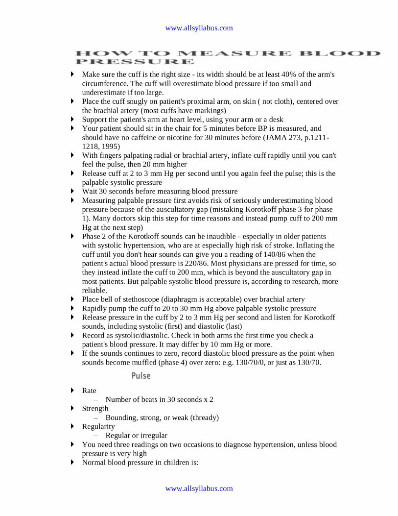
wwwallsyllabuscom
wwwallsyllabuscom
Make sure the cuff is the right size - its width should be at least 40 of the arms
circumference The cuff will overestimate blood pressure if too small and
underestimate if too large
Place the cuff snugly on patients proximal arm on skin ( not cloth) centered over
the brachial artery (most cuffs have markings)
Support the patients arm at heart level using your arm or a desk
Your patient should sit in the chair for 5 minutes before BP is measured and
should have no caffeine or nicotine for 30 minutes before (JAMA 273 p1211-
1218 1995)
With fingers palpating radial or brachial artery inflate cuff rapidly until you cant
feel the pulse then 20 mm higher
Release cuff at 2 to 3 mm Hg per second until you again feel the pulse this is the
palpable systolic pressure
Wait 30 seconds before measuring blood pressure
Measuring palpable pressure first avoids risk of seriously underestimating blood
pressure because of the auscultatory gap (mistaking Korotkoff phase 3 for phase
1) Many doctors skip this step for time reasons and instead pump cuff to 200 mm
Hg at the next step)
Phase 2 of the Korotkoff sounds can be inaudible - especially in older patients
with systolic hypertension who are at especially high risk of stroke Inflating the
cuff until you dont hear sounds can give you a reading of 14086 when the
patients actual blood pressure is 22086 Most physicians are pressed for time so
they instead inflate the cuff to 200 mm which is beyond the auscultatory gap in
most patients But palpable systolic blood pressure is according to research more
reliable
Place bell of stethoscope (diaphragm is acceptable) over brachial artery
Rapidly pump the cuff to 20 to 30 mm Hg above palpable systolic pressure
Release pressure in the cuff by 2 to 3 mm Hg per second and listen for Korotkoff
sounds including systolic (first) and diastolic (last)
Record as systolicdiastolic Check in both arms the first time you check a
patients blood pressure It may differ by 10 mm Hg or more
If the sounds continues to zero record diastolic blood pressure as the point when
sounds become muffled (phase 4) over zero eg 130700 or just as 13070
Rate
ndash Number of beats in 30 seconds x 2
Strength
ndash Bounding strong or weak (thready)
Regularity
ndash Regular or irregular
You need three readings on two occasions to diagnose hypertension unless blood
pressure is very high
Normal blood pressure in children is
wwwallsyllabuscom
wwwallsyllabuscom
ndash 10255 at 1 year 11269 at 5 years 11978 at 10 years
Blood pressures in adults (JNC VII JAMA 2892560-72 2003)
ndash Normal lt120lt80
ndash Prehypertensive 120-13980-89
ndash Stage 1 hypertension 140-15990-99
ndash Stage 2 hypertension gt160gt100
Adult 60 to 100
Newborn 120-170
1 year 80-160
3 years 80-120
6 years 75-115
10 years 70-110
How to measure observe rise and fall of chest
In infants count for 60 seconds in adults 15 or 30 seconds
Normal respiration Adults 12 to 20
Children
newborn 30-80
1 year 20-40
3 years 20-30
6 years 16-22
Rate
Number of breaths in 30 seconds x 2
Quality
Character of breathing
Rhythm
Regular or irregular
Effort
Normal or labored
Noisy respiration
Normal stridor wheezing snoring gurgling
Depth
Shallow or deep
pH electrode
wwwallsyllabuscom
wwwallsyllabuscom
Governing equation is the Nernst Equation
pCO2 Electrode
The measurement of pCO2 is based on its linear relationship with pH over the range of
10 to 90 mm Hg
The dissociation constant is given by
Taking logarithms
pH = log[HCO3-] ndash log k ndash log a ndash log pCO2
Diathermy
In the natural sciences the term diathermy means electrically induced heat and is commonly
used for muscle relaxation It is also a method of heating tissue electromagnetically or
ultrasonically for therapeutic purposes in medicineContents
[hide]
1 Heating uses 2 Surgical uses
3 Trivia
4 References
E
RT
nF
H
HH
i
ln
0
H O CO H CO H HCO2 2 2 3 3
k
H HCO
a pCO
3
2
wwwallsyllabuscom
wwwallsyllabuscom
Heating uses
Ultrasonic diathermy refers to heating of tissues by ultrasound for the purpose of
therapeutic deep heating No tissue is ordinarily damaged hence it is generally used in
biomedical applications
Electric diathermy uses high frequency alternating electric or magnetic fields sometimes
with no electrode or device contact to the skin to induce gentle deep tissue heating by
induction Again no tissue is ordinarily damaged
Surgical uses
Surgical diathermy is usually better known as electrosurgery (It is also referred to
occasionally as electrocautery but see disambiguation below) Electrosurgery and
surgical diathermy involve the use of high frequency AC electrical current in surgery as
either a cutting modality or else to cauterize small blood vessels to stop bleeding This
technique induces localized tissue burning and damage the zone of which is controlled
by the frequency and power of the device Some sources[1]
insist that electrosurgery be
applied to surgery accomplished by high frequency AC cutting and that
electrocautery be used only for the practice of cauterization with heated nichrome wires
powered by DC current as in the handheld battery-operated portable cautery tools
Trivia
Medical Diathermy devices were used to cause interference to German radio beams used
for targeting night time bombing raids in WWII during the Battle of the Beams
I Diathermy
A Therapeutic use
1 Generation of local heating by high-frequency electromagnetic waves
2 Capacitance techniquemdashbody is placed in an electric field
a Dipolesmdashstructures with positive and negative poles
b Structures with large numbers of dipoles have a greater capacitance to store an electrical charge
c Greatest heating occurs in tissues with fewer dipoles particularly fatty tissues
3 Rapid rotation of dipoles causing mechanical friction and movement of electrons results in local heating
4 Inductance techniquemdashbody is not placed in an electric field
a Magnetic waves generated by driving current through a coiled wire
b Magnetic field creates currents in tissues
wwwallsyllabuscom
wwwallsyllabuscom
c Greatest heating occurs in tissues with low impedance especially muscle
B Precautions and contraindications
1 Diathermy should not be used
a Over metal implants and cardiac pacemakersmdashmore research needed regarding its use over metallic fixations
b Near the uterus of a pregnant woman or near the abdomen or back of a woman who might be pregnant
c On individuals with infections
d On individuals with acute inflammation
e Over moist open wounds
f On patients with malignant tumors
g Over large joint effusions
C Pulsed electromagnetic fields and diathermy
1 Can be pulsed to decrease total energy transmitted to the tissues
2 Short-wave diathermy can be adjusted into a nonthermal range
a Classified as pulsed electromagnetic field (PEMF) or
b Pulsed radio frequency energy (PRFE)
3 Important reclassification as diathermy implies heating
D Efficiency of diathermy and PEMF therapy for musculoskeletal conditions
1 Current research is limited but results suggest that diathermy enhances
treatments directed at soft tissue stretching
2 Some studies suggest that PEMF may speed wound healing and promote healing of nonunion fractures
Diathermy is a modality that uses electromagnetic energy to heat deeper tissues
Diathermy is more effective than ultrasound at heating a larger area of deep tissues
The athletic trainer must identify and respect contraindications to application of ultrasound and diathermy
Pulsed ultrasound and diathermy are used to treat slow-to-heal lesions including skin ulcers and nonunion fractures and may be able to facilitate repair of other tissues including ligaments and tendons
Diathermy
Definition
In diathermy high-frequency electrical currents are used to heat deep muscular tissues The heat increases blood flow speeding up recovery Doctors also use diathermy in surgical procedures by sealing blood vessels with electrically heated probes
The term diathermy is derived from the Greek words therma meaning heat and dia meaning through Diathermy literally means heating through
Origins
The therapeutic effects of heat have long been recognized More than 2000 years ago the Romans took advantage of heat therapies by building hot-spring bathhouses Since then various
wwwallsyllabuscom
wwwallsyllabuscom
methods of using heat have evolved In the early 1890s French physiologist Arseacutene dArsonval began studying the medical application of high-frequency currents The term diathermy was coined by German physician Carl Franz Nagelschmidt who designed a prototype apparatus in 1906 Around 1925 United States doctor J W Schereschewsky began studying the physiological
effects of high-frequency electrical currents on animals It was several years however before the fundamentals of the therapy were understood and put into practice
Benefits
Diathermy can be used to treat arthritis bursitis and other conditions involving stiff painful joints It is also used to treat pelvic infections and sinusitis A benefit of diathermy is that it is a painless procedure that can be administered at a clinic Also if the treatment relieves pain then patients can discontinue pain killers and escape their high cost and side effects
Description
Diathermy involves heating deep muscular tissues When heat is applied to the painful area cellular metabolism speeds up and blood flow increases The increased metabolism and circulation accelerates tissue repair The heat helps the tissues relax and stretch thus alleviating
stiffness Heat also reduces nerve fiber sensitivity increasing the patients pain threshold
There are three methods of diathermy In each energy is delivered to the deep tissues where it is converted to heat The three methods are
Shortwave diathermy The body part to be treated is placed between two capacitor plates Heat is generated as the high-frequency waves travel through the body tissues between the plates Shortwave diathermy is most often used to treat areas like the hip which is covered with a dense tissue mass It is also used to treat pelvic infections and sinusitis The treatment reduces inflammation The Federal Communications Commission regulates the frequency allowed for short-wave diathermy treatment Most machines
function at 2733 megahertz Ultrasound diathermy In this method high-frequency acoustic vibrations are used to
generate heat in deep tissue Microwave diathermy This method uses radar waves to heat tissue This form is the
easiest to use but the microwaves cannot penetrate deep muscles
Diathermy is also used in surgical procedures Many doctors use electrically heated probes to seal blood vessels to prevent excessive bleeding This is particularly helpful in neurosurgery and eye surgery Doctors can also use diathermy to kill abnormal growths such as tumors warts and
infected tissues
Preparations
To keep patients from sweating patients are usually asked to remove clothing from the body part being treated If a patient sweats the electrical currents may pool in the area causing burns Also clothing containing metal must be removed as must earrings buttons barrettes or zippers that contain metal Watches and hearing aids should be removed because the therapy may affect their function
wwwallsyllabuscom
wwwallsyllabuscom
Practitioners of surgical diathermy should steer clear of alcohol-based solutions to prepare and cleanse the skin These preparations can create a flammable vapor and cause burns and fires
Precautions
Patients with metal implants should not undergo diathermy treatment because the metal can act as a conductor of heat and result in serious internal burns Female patients with metallic uterine implants such as an IUD should avoid treatment in the pelvic area Diathermy should not be
used in joints that have been replaced with a prosthesis or in those with sensory impairment who may not be able to tell if they are burning Furthermore pulsed shortwave diathermy should be avoided during pregnancy as it can lead to abnormal fetal development
Patients with hemophilia should avoid the treatment because the increased blood flow could cause them to hemorrhage
Side effects
Some patients may experience superficial burns Since the therapy involves creating heat care must be taken to avoid burns particularly in patients whose injuries have caused decreased sensitivity to heat Also diathermy may affect pacemaker function
Female patients who receive treatment in the lower back or pelvic area may experience an
increased menstrual flow
Research amp general acceptance
For years physiotherapists and physical therapists have used diathermy as a routine part of physical rehabilitation
Electrical Safety
Electrical safety is very important in hospitals as patients may be undergoing a diagnostic or treatment procedure where the protective effect of dry skin is reduced Also patients may be unattended unconscious or anaesthetised and may not respond normally to an electric current Further electrically conductive solutions such as blood and saline are often present in patient treatment areas and may drip or spill on electrical equipment
Electric Current Leakage Current
Extension Leads Double Adaptors Equipment Classification
Class I Class II Defibrillator-Proof
Protective Devices
Residual Current Devices (RCD) Line Isolation overload Monitors (LIMs) Equipment Earthing
Area Classification
wwwallsyllabuscom
wwwallsyllabuscom
Body Protection Area Cardiac Protected Area
Other Electrical Issues Extension Leads
Double Adapters Main Extension Devices Power Boards Installation of Additional Power Points
Electric Current
Injuries received from electric current are dependent on the magnitude of current the pathway
that it takes through the body and the time for which it flows
The nature of electricity flowing through a circuit is analogous to blood flowing through the circulatory system within the human body In this analogy the source of energy is represented by the heart and the blood flowing through arteries and veins is analagous to current flowing
through the conductors and other components of the electric circuit
The application of an electric potential to an electric circuit generates a flow of current through conductive pathways This is analogous to the changes in blood pressure caused by contraction of cardiac muscle that causes blood to flow into the circulatory system For electric current to flow there must be a continuous pathway from the source of potential through electrical components and back to the source
Leakage Current
Electrical components and systems are encased in non conducting insulation to ensure that the electric current is contained and follows the intended pathways If the insulation deteriorates or breaks down current will leak through the insulation barrier and flow to earth This may
be through the protective earth conductor or through the operator
Medical equipment and clinical areas are fitted with a number of protective devices to protect the patient and operator from harmful leakage currents
Extension Leads
Extension leads are not permitted in clinical areas of RCH organisations They may cause high
earth resistance and excessive earth leakage current An extension lead can allow equipment to be powered from areas other than the relevant protected treatment area The power from the other area may not be protected to the same level as the power in the treatment area
wwwallsyllabuscom
wwwallsyllabuscom
As the connection between the extension lead and the equipment mains cable is often on the floor there is a high danger from fluid spills tripping and damage to the mains cable by trolleys when an extension lead is used
Double Adaptors
Double adaptors must not be used in RCH organisations They may not sit securely in a wall
outlet may not be able to provide adequate earth protection and may cause overloading overheating fire or loss of electrical supply
EQUIPMENT CLASSIFICATIONS
There are several methods of providing protection for operators and patients from electrical faults
and harmful leakage current
Class I
Class I equipment is fitted with a three core mains cable containing a protective earth wire
Exposed metal parts on class I equipment are connected to this earth wire
Should a fault develop inside the equipment and the exposed metal comes into contact with the mains the earthing conductor will conduct the fault current to ground Regular testing procedures ensure that earthing conductors are intact as the integrity of the earth wire is of vital importance
Class II
Class II equipment is enclosed within a double insulated case and does not require earthing
conductors Class II equipment is usually fitted with a 2-pin mains plug An internal electrical fault is unlikely to be hazardous as the double insulation prevents any external parts from becoming alive Class II or double insulated equipment can be identified by the class II symbol on the cabinet
Class II Symbol
wwwallsyllabuscom
wwwallsyllabuscom
Defibrillator-Proof
Some medical equipment within the hospital is classified as defibrillator proof When a
defibrillator is discharged through a patient connected to defibrillator proof equipment the equipment will not be damaged by the defibrillators energy Defibrillator proof equipment can remain connected to the patient during defibrillation It is identified by one of the following
symbols
Defibrillator proof symbols
Body protected Cardiac protected
PROTECTIVE DEVICES
Most patient care areas in the hospital are fitted with protective devices These devices are
regularly tested in accordance with the relevant guidelines published by Standards Australia The level of protection provided is dependent upon the device and the area in which it is located
Residual Current Devices (RCD)
RCDs (safety switches) are used in patient treatment areas to monitor and protect the mains supply RCDs sense leakage currents flowing to earth from the equipment If a significant leakage current flows the RCD will detect it and shut off the power supplied to the equipment
within 40 milliseconds Hospital RCDs are more sensitive than those fitted in homes A hospital RCD will trip at 10 milliamperes leakage current
Power outlets supplied through an RCD have a Supply Available lamp The lamp will extinguish
when the RCD trips due to excessive leakage current
wwwallsyllabuscom
wwwallsyllabuscom
Resetting a RCD
Lamp indicates supply is no longer available Disconnect all equipment from the supply Operate the reset button or lever on the supply panel and the Supply Available lamp
should illuminate If not contact Biomedical Engineering
Connect an item of equipment If the RCD trips again then this is the faulty item and should be labelled and sent to Biomedical Engineering
If the RCD does not trip continue connecting equipment until the RCD trips The last piece of equipment connected to the supply is most likely to be faulty as it will have caused the RCD to trip Remove the faulty item from service label it and send to Biomedical Engineering as mentioned above
Line Isolation overload Monitors (LIMs)
In critical life support applications where loss of power supply cannot be tolerated special power outlets powered by isolation transformers are installed
Line Isolation Monitors are installed to continually monitor electrical leakage in the power
supply system If an electrical fault develops in a medical device connected to an isolated power outlet the LIM will detect the leakage current The LIM will alarm and indicate the level of leakage current but will not shut off the electric supply
The faulty equipment can be identified by un plugging one item of equipment at a time from the supply until the alarm stops sounding Equipment that is not faulty may be reconnected Faulty equipment should be appropriately labelled and sent to Biomedical Engineering for repair
The LIM also monitors how much power is being used by the equpiment connected to it If too much power is being used the LIM will alarm and indicate that there is an overload The power used must be reduced immediately by moving some equipment to another circuit as soon as possible until the alarm stops sounding Failure to reduce the load on the LIM will result in the circuit breaker tripping and loss of power to the circuit
wwwallsyllabuscom
wwwallsyllabuscom
Equipotential Earthing
Equipotential earthing is installed in rooms classified as Cardiac Protected electrical areas
Equipotential earthing in treatment areas used for cardiac procedures is intended to minimise any voltage differences between earthed parts of equipment and any other exposed metal in the room
This reduces the possibility of leakage currents that can cause microelectrocution when the patient comes into contact with multiple items of equipment or if the patient happens to come
into contact with metal items in the room whilr they are connected to a medical device
All conductive metal in an equipotential area is connected to a common equipotential earth point with special heavy duty cable
AREA CLASSIFICATIONS
Body Protected Area
These areas are designed for procedures in which patients are connected to equipment that lowers the natural resistance of the skin Applied parts such as electrode gels conductive fluids entering the patient metal needles and catheters provide an easy pathway for current to flow
The main occurrence of injury from Body-Type procedures is from high current levels causing electric shock A direct connection to the patients heart is not present so the risk of Microelectrocution - fibrillation from minute current levels - is reduced
Residual Current Devices (RCD) or Isolation Transformers and Line Isolation Monitors (LIMiacutes) are used in Body Protected areas to provide protection against electrocution from high leakage currents Body-Protected Areas are identified with this sign
Cardiac Protected Area
Where the procedure involves placing an electrical conductor within or near the heart protection against fibrillation induced from small leakage currents is required Electrical conductors used in these procedures include cardiac pacing electrodes intracardiac ECG electrodes and intracardiac catheters
wwwallsyllabuscom
wwwallsyllabuscom
Equipotential earthing in conjunction with RCDs or LIMs provides protection against microelectrocution in Cardiac-Type procedures
Fault currents are reduced to magnitudes that are unlikely to induce fibrillation Used in conjunction with RCDs or LIMs the magnitude and duration of any fault currents
sourced from equipment are limited Cardiac-Protected Areas are identified with this sign
Other electrical issues
This policy aims to provide guidance to those who find that they need more electrical outlets than
those available or that the existing electrical outlets are inconveniently located
As extension leads and multiple outlet power boards can introduce additional hazards into an area the following procedures should be observed
Extension leads
Approved extension leads (AS 3760 1996) may be used in some areas within the hospital but MUST NOT BE USED IN PATIENT AREAS All electrical extension leads must be tagged with an Engineering Department maintenance tag and require a yearly safety inspection and test via
the Engineering Department
Double adapters
Double adapters may cause overloading or equipment earthing problems and are not to be used in
WCH
Mains extension device
The only mains extension device that is to be used in Patient care areas is the 4-way or 8-way
portable Core Balance Unit
The Biomedical Engineering Department must approve all units prior to use These units contain a safety switch and can detect excessive leakage current and disconnect the power in the event of a hazardous situation
wwwallsyllabuscom
wwwallsyllabuscom
Care must be exercised in the use of a portable Core Balance Unit It should be located off the
floor and in a position that will protect it from physical abuse and possible entry of fluids These devices are expensive and easily damaged The device must be sent to Biomedical Engineering every 6 months for safety testing
Power boards
Approved multiple-outlet power boards can be used across RCH but must not be used in patient
care areas except areas approved by the Biomedical Engineering Department
The power boards must have overload protection be fitted with internal safety shutters that protect unused outlets and be fitted with an onoff switch for each outlet
Medical Laser Applications
The main research subjects of the group of Medical Laser Application are in the field of online
monitoring and diagnostics as well as the development of new therapheutic methods The main focus in research is based upon the use of ultrashort (fs) laser pulses
The main advantage of ultrashort laser pulses is the extrem short interaction time which suppress any unwanted side effects of the laser irradiation of the tissue Secondly the broad spectra of the fs laser pulses give the advantage to use the same laser pulses for diagnostic applications like the optical coherence tomography (OCT)
wwwallsyllabuscom
wwwallsyllabuscom
Following research subjects have special attention at the moment
Optical Coherence Tomographie (OCT)
A fs-laser light source can be used to obtain images from inside the tissue when the pulses are coherent superposed This group uses this application to obtain an inside view of the vocal fold and to measure the outline of the crystalline lens
Refractive Surgery
The principle of getting rid of glasses with the help of a laser surgery (fs-LASIK) is underlying rapid improvements concerning precision and safety New technologies were evaluated and transferred very fast in cooperation with an industrial partner
Treatment of Presbyopia
The flexibility of the crystalline lens can be increase by pricise cuts which are induce by fs laser pulse inside the lens The treatment of the presbyopia is thinkable
wwwallsyllabuscom
wwwallsyllabuscom
Ultrafast Physics
Whereas the applications of ultrashort laser pulses increase rapidly is the physics of the interaction between the laser pulses and tissue in many case not fully understood To obtain improved knowledge of the interaction for many fs-laser application this group preforms numerical simulations as well as fundamental experiments
Thermography
Thermography thermal imaging or thermal video is a type of infrared imaging Thermographic cameras detect radiation in the infrared range of the electromagnetic spectrum
(roughly 900ndash14000 nanometers or 09ndash14 microm) and produce images of that radiation Since infrared radiation is emitted by all objects based on their temperatures according to the black body radiation law thermography makes it possible to see ones environment with or without visible illumination The amount of radiation emitted by an object increases with temperature therefore thermography allows one to see variations in temperature (hence the name) When viewed by thermographic camera warm objects stand out well against cooler backgrounds humans and other warm-blooded animals become easily visible against the environment day or
night As a result thermographys extensive use can historically be ascribed to the military and security services
Thermal imaging photography finds many other uses For example firefighters use it to see through smoke find persons and localize the base of a fire With thermal imaging power lines maintenance technicians locate overheating joints and parts a telltale sign of their failure to eliminate potential hazards Where thermal insulation becomes faulty building construction technicians can see heat leaks to improve the efficiencies of cooling or heating air-conditioning Thermal imaging cameras are also installed in some luxury cars to aid the driver the first being
the 2000 Cadillac DeVille Some physiological activities particularly responses in human beings and other warm-blooded animals can also be monitored with thermographic imaging [1]
The appearance and operation of a modern thermographic camera is often similar to a camcorder Enabling the user to see in the infrared spectrum is a function so useful that ability to record their output is often optional A recording module is therefore not always built-in
Instead of CCD sensors most thermal imaging cameras use CMOS Focal Plane Array (FPA) The most common types are InSb InGaAs HgCdTe and QWIP FPA The newest technologies are using low cost and uncooled microbolometers FPA sensors Their resolution is considerably lower than of optical cameras mostly 160x120 or 320x240 pixels up to 640x512 for the most
expensive models Thermographic cameras are much more expensive than their visible-spectrum counterparts and higher-end models are often export-restricted Older bolometers or more
wwwallsyllabuscom
wwwallsyllabuscom
sensitive models as InSb require cryogenic cooling usually by a miniature Stirling cycle refrigerator or liquid nitrogen
Contents
[hide]
1 Difference between IR film and thermography 2 Advantages of Thermography
3 Limitations amp disadvantages of thermography 4 Applications 5 See also 6 External links
o 61 History of thermal imager manufacturers
[edit] Difference between IR film and thermography
Thermal imaging is going to be used on Mars to detect caves that could hold life
[edit] Advantages of Thermography
You get a visual picture so that you can compare temperatures over a large area
It is real time capable of catching moving targets Able to find deteriorating components prior to failure Measurement in areas inaccessible or hazardous for other methods It is a non-destructive test method
[edit] Limitations amp disadvantages of thermography
Quality cameras are expensive and are easily damaged Images can be hard to interpret accurately even with experience
Accurate temperature measurements are very hard to make because of emissivities Most cameras have plusmn2 or worse accuracy (not as accurate as contact) Training and staying proficient in IR scanning is time consuming Ability to only measure surface areas
[edit] Applications
Condition monitoring Medical imaging Night vision Research
Process control Non destructive testing Surveillance in security law enforcement and defense Chemical imaging
wwwallsyllabuscom
wwwallsyllabuscom
Thermal infrared imagers convert the energy in the infrared wavelength into a visible light video display All objects above 0 kelvins emit thermal infrared energy so thermal imagers can passively see all objects regardless of ambient light However most thermal imagers only see objects warmer than -50 degC
The spectrum and amount of thermal radiation depend strongly on an objects surface
temperature This makes it possible for a thermal camera to display an objects temperature However other factors also influence the radiation which limits the accuracy of this technique For example the radiation depends not only on the temperature of the object but is also a function of the emissivity of the object Also radiation also originates from the surroundings and is reflected in the object and the radiation from the object and the reflected radiation will also be influenced by the absorption of the atmosphere
Endoscopy
This page is about having an endoscopy - a test that looks at the inside of your digestive system
There is information on
What endoscopy is
Why you may have an endoscopy
Having an endoscopy
Your results
What an endoscopy is
An endoscopy is a test that looks inside the body The endoscope is a long flexible tube that can
be swallowed It has a camera and light inside it Some doctors call it a telescope
wwwallsyllabuscom
wwwallsyllabuscom
Why you may have an endoscopy
You are most likely to have an endoscopy to look at the inside of your
Gullet (oesophagus)
Stomach
Duodenum - the first part of the small bowel that attaches to the stomach
Large bowel (colon)
Your doctor may want to see inside because you have symptoms such as abnormal bleeding or
difficulty swallowing The doctor can look down the endoscope and see if there are any growths
or other abnormal looking areas Also through the endoscope the doctor can take samples
(biopsies) of any abnormal looking tissues
There is more detailed information about having a colonoscopy in the bowel cancer section of
CancerHelp UK Below is information about having other types of endoscopy
Having an endoscopy
You can have this test as an out patient Most people have a choice between having the test while
they are awake or after having a medicine to make them drowsy (a sedative) Your hospital may
not be happy for you to have a sedative if you live alone and will have no one to look after you
when you go home If you live alone but really want sedation your hospital may allow you to
stay overnight You should ask your doctor about this as early as possible as extra arrangements
will need to be made beforehand
If you dont have a sedative you will have a spray to numb the back of your throat and make it
easier for you to swallow the endoscopy tube
If you would prefer to be asleep during the test you will have an injection to make you very
drowsy just before the test You will need to take someone with you to the hospital appointment
You wont be able to drive for the rest of the day and should have someone to go home with you
You cant eat or drink for about 8 hours before the test so that your stomach and duodenum are
empty Your doctor will give you written instructions about this beforehand or they may arrive
with your appointment letter When you get to the clinic you may be asked to take your upper
clothing off and put on a hospital gown Some hospitals prefer to use gowns because your clothes
wont get messy Once you are ready you get onto the bed or X-ray couch When you are lying
comfortably you have the sedative injection to make you very drowsy Or your doctor will spray
the back of your throat to numb it
Once the sedative or throat spray has worked the doctor will pass the endoscope tube down your
wwwallsyllabuscom
wwwallsyllabuscom
throat to the area being investigated Your doctor will ask you to swallow as the tube goes down
but if youve had a sedative you wont remember that afterwards If there are any abnormalities
the doctor will take pieces of tissue from the abnormal looking area to send to the laboratory for
closer inspection under a microscope These tissue samples are called biopsies
When the test is over you will need to rest for a while If youve had a sedative you may not
remember much (if anything) about the test once you have come round You should be able to go
home the same day
The results
It can take time for test results to come through How long will depend on why you are having
the test Usually the doctor who carries out the endoscopy dictates a report straight way The
report is typed up by the department secretary and goes to your specialist who gives the results to
you If your GP has sent you for the test the results will go directly to the GP surgery
Understandably waiting for results can make you anxious It usually takes a couple of weeks for
the results to come through If your doctor needed them urgently it would have been noted on
the test request form and the results will be ready sooner than that Try to remember to ask your
doctor how long you should expect to wait for the results when you are first asked to go for the
test If it is not an emergency and you have not heard a couple of weeks after your test ring your
doctors secretary to check if they are back
Endoscopy
From Wikipedia the free encyclopedia
Jump to navigation search
Endoscopic images of a duodenal ulcer
wwwallsyllabuscom
wwwallsyllabuscom
A flexible endoscope
Endoscopy means looking inside and typically refers to looking inside the human body for medical reasons using an instrument called an endoscope Endoscopy can also refer to using a borescope in technical situations where direct line-of-sight observation is not feasible
Contents
[hide]
1 Overview 2 Components 3 Uses
4 History 5 Risks 6 After The Endoscopy 7 Recent developments 8 See also 9 References 10 Footnotes
11 External links
Overview
Endoscopy is a minimally invasive diagnostic medical procedure that is used to assess the interior surfaces of an organ by inserting a tube into the body The instrument may have a rigid or flexible tube and not only provide an image for visual inspection and photography but also enable taking
biopsies and retrieval of foreign objects Endoscopy is the vehicle for minimally invasive surgery
Many endoscopic procedures are considered to be relatively painless and at worst associated with mild discomfort for example in esophagogastroduodenoscopy most patients tolerate the procedure with only topical anaesthesia of the oropharynx using lignocaine spray[1] Complications are not common (only 5 of all operations)[citation needed] but can include perforation of the organ under inspection with the endoscope or biopsy instrument If that occurs open surgery may be required to repair the injury
wwwallsyllabuscom
wwwallsyllabuscom
Components
An endoscope can consist of
a rigid or flexible tube a light delivery system to illuminate the organ or object under inspection The light
source is normally outside the body and the light is typically directed via an optical fiber system
a lens system transmitting the image to the viewer from the fiberscope an additional channel to allow entry of medical instruments or manipulators
Uses
Endoscopy can involve
The gastrointestinal tract (GI tract) o esophagus stomach and duodenum (esophagogastroduodenoscopy) o small intestine o colon (colonoscopyproctosigmoidoscopy) o Bile duct
endoscopic retrograde cholangiopancreatography (ERCP) duodenoscope-assisted cholangiopancreatoscopy intraoperative cholangioscopy
The respiratory tract o The nose (rhinoscopy) o The lower respiratory tract (bronchoscopy)
The urinary tract (cystoscopy) The female reproductive system
o The cervix (colposcopy) o The uterus (hysteroscopy) o The Fallopian tubes (Falloscopy)
Normally closed body cavities (through a small incision) o The abdominal or pelvic cavity (laparoscopy) o The interior of a joint (arthroscopy) o Organs of the chest (thoracoscopy and mediastinoscopy)
During pregnancy o The amnion (amnioscopy) o The fetus (fetoscopy)
Plastic Surgery Panendoscopy (or triple endoscopy)
o Combines laryngoscopy esophagoscopy and bronchoscopy Non-medical uses for endoscopy
o The planning and architectural community have found the endoscope useful for
pre-visualization of scale models of proposed buildings and cities (architectural endoscopy)
o Internal inspection of complex technical systems (borescope) o Endoscopes are also a tool helpful in the examination of improvised explosive
devices by bomb disposal personnel
wwwallsyllabuscom
wwwallsyllabuscom
o The FBI uses endoscopes for conducting surveillance via tight spaces
History
The first endoscope of a kind was developed in 1806 by Philip Bozzini with his introduction of a
Lichtleiter (light conductor) for the examinations of the canals and cavities of the human body However the Vienna Medical Society disapproved of such curiosity An endoscope was first introduced into a human in 1822 by William Beaumont an army surgeon at Mackinac
Island Michigan[citation needed] The use of electric light was a major step in the improvement of endoscopy The first such lights were external Later smaller bulbs became available making internal light possible for instance in a hysteroscope by Charles David in 1908[citation needed] Hans Christian Jacobaeus has been given credit for early endoscopic explorations of the abdomen and the thorax with laparoscopy (1912) and thoracoscopy (1910)[citation needed] Laparoscopy was used in the diagnosis of liver and gallbladder disease by Heinz Kalk in the 1930s[citation needed] Hope reported in 1937 on the use of laparoscopy to diagnose ectopic pregnancy[citation needed] In 1944
Raoul Palmer placed his patients in the Trendelenburg position after gaseous distention of the abdomen and thus was able to reliably perform gynecologic laparoscopy[citation needed]
The first gastrocamera was released in 1950 by Olympus Optical Co Ltd The device took pictures on monochromatic film using a small light bulb that was triggered manually The device was of limited use however because it did not implement real-time optical capability Olympus continued its development of endoscopes by incorporating fiber optics in the early 1960s leading to the first useful endoscopes In 1964 it released a gastrocamera guided by a fiberscope[1] A few articles claim that DrBasil Hirschowitz of UnivOf MichiganAnn Arbor discussed the
endoscope in early 50s[2]
As endoscopic technology improved so did the methods of gastrointestinal endoscopy Owing primarily to the efforts of Dr Hiromi Shinya in the late 1960s GI endoscopy developed into what is more recognizable as todays colonoscopy While many doctors experimented with techniques to take advantage of the new iterations of endoscopes Dr Shinya focused on techniques that would allow for successful operation of the endoscope by an individual rejecting the common practice at the time of utilizing two people Consequently many of the fundamental methods and procedures of modern colonoscopy were developed by Dr Shinya
Dr Shinyas other great contribution was to therapeutic endoscopy in his invention of the
electrosurgical polypectomy snare with the aid of Olympus employee Hiroshi Ichikawa Shinya sketched his first plans for the device on January 8 1969 He envisioned a loop of wire attached to the end of a colonoscope that would allow for easy removal of polyps during investigation by passing a current through the wire By September of 1969 the first polypectomy using this device was performed Polypectomy has since become the most common therapeutic procedure performed with an endoscope (Sivak 2004)
By 1980 laparoscopy training was required by gynecologists to perform tubal ligation procedures and diagnostic evaluations of the pelvis The first laparoscopic cholecystectomy was performed in 1984 and the first video-laparoscopic cholecystectomy in 1987[citation needed] During the 1990s
laparoscopic surgery was extended to the appendix spleen colon stomach kidney and liver[citation needed] Wireless capsule endoscopy or Capsule Endoscopy is now approved in all the countries including Japan where government reimbusement will be available from Oct2007Capsule Endoscopy [3] increases detection of Small Bowel tumors where traditional Endoscopy is not very efficient
wwwallsyllabuscom
wwwallsyllabuscom
Risks
Infection
Punctured organs Allergic reactions due to Contrast agents or dyes (such as those used in a CT scan) Over-sedation
After The Endoscopy
After the procedure the patient will be observed and monitored by a qualified individual in the
endoscopy or a recovery area until a significant portion of the medication has worn off Occasionally a patient is left with a mild sore throat which promptly responds to saline gargles or a feeling of distention from the insufflated air that was used during the procedure Both problems are mild and fleeting When fully recovered the patient will be instructed when to resume hisher usual diet (probably within a few hours) and will be allowed to be taken home Because of the use of sedation most facilities mandate that the patient is taken home by another person and not to drive on hisher own or handle machinery for the remainder of the day
Recent developments
With the application of robotic systems telesurgery was introduced as the surgeon could operate
from a site physically removed from the patient The first transatlantic surgery has been called the Lindbergh Operation
wwwallsyllabuscom
wwwallsyllabuscom
Upper Endoscopy
Upper endoscopy enables the physician to look inside the esophagus stomach and duodenum (first part of the small intestine) The procedure might be used to discover the reason for swallowing difficulties nausea vomiting
reflux bleeding indigestion abdominal pain or chest pain Upper endoscopy is also called EGD which stands for esophagogastroduodenoscopy (eh-SAH-fuh-goh-GAS-troh-doo-AH-duh-NAH-skuh-pee)
For the procedure you will swallow a thin flexible lighted tube called an endoscope (EN-doh-skope) Right before the procedure the physician will spray your throat with a numbing agent that may help prevent gagging
You may also receive pain medicine and a sedative to help you relax during the exam The endoscope transmits an image of the inside of the esophagus stomach and duodenum so the physician can carefully examine the lining of these organs The scope also blows air into the stomach this expands the folds of tissue and makes it easier for the physician to examine the stomach
The physician can see abnormalities like inflammation or
bleeding through the endoscope that dont show up well on x rays The physician can also insert instruments into the scope to treat bleeding abnormalities or remove samples of tissue (biopsy) for further tests
Possible complications of upper endoscopy include bleeding and puncture of the stomach lining However such complications are rare Most people will probably have nothing more than a mild sore throat after the procedure
The procedure takes 20 to 30 minutes Because you will be sedated you will need to rest at the endoscopy facility for 1 to 2 hours until the medication wears off
Preparation
Your stomach and duodenum must be empty for the procedure to be thorough and safe so you will not be able to eat or drink anything for at least 6 hours beforehand Also you must
arrange for someone to take you homemdashyou will not be allowed to drive because of the sedatives Your physician may give you other special instructions
The digestive system
wwwallsyllabuscom
wwwallsyllabuscom
What is Endoscopy and Why is it Performed
Endoscopy allows physicians to peer through the bodys passageways Endoscopy is the examination and inspection of the interior of body organs joints or cavities through an endoscope An endoscope is a device that uses fiber optics and powerful lens systems to provide lighting and visualization of the interior of a joint The portion of the endoscope inserted into the body may be rigid or flexible depending upon the medical procedure
An endoscope uses two fiber optic lines A light fiber carries light into the body cavity and an image fiber carries the image of the body cavity back to the physicians viewing lens There is also a separate port to allow for administration of drugs suction and irrigation This port may also be used to introduce small folding instruments such as forceps scissors brushes snares and baskets for tissue excision (removal) sampling or other diagnostic and therapeutic work Endoscopes may be used in conjunction with a camera or video recorder to document images of the inside of the joint or chronicle an endoscopic procedure New endoscopes have digital capabilities for manipulating and enhancing the video images
This figure shows a rigid endoscope used for arthroscopy The image fiber leads from the ocular (eye piece) to the inserted end of the scope The light fiber is below and leads from the light source to the working end of the endoscope
Why Is Endoscopy Performed
Endoscopy can be used to diagnose various conditions by close examination of internal organ and body structures Endoscopy can also guide therapy and repair such as the removal of torn cartilage from the bearing surfaces of a joint Biopsy (tissue sampling for pathologic testing) may also be performed under endoscopic guidance Local or general anesthetic may be used during endoscopy depending upon the type of procedure being performed
Internal abnormalities revealed through endoscopy include abscesses biliary (liver) cirrhosis bleeding bronchitis cancer cysts degenerative disease gallbladder stones hernia inflammation metastatic cancer polyps tumors ulcers and other diseases and conditions
Endoscopy is a minimally invasive procedure and carries with it certain minor risks depending upon the type of procedure being performed However these risks are typically far outweighed by the diagnostic and therapeutic potential of the procedure
Prior to the widespread use of endoscopy and diagnostic imaging most internal conditions could only be diagnosed or treated with open surgery Until the last several decades exploratory surgery was routinely performed only when a patient was critically ill and the source of illness was not known For example in certain dire cases the patients thorax or abdomen were surgically opened and examined to try to determine the source of illness
wwwallsyllabuscom
wwwallsyllabuscom
Endoscopy can often be done on an outpatient basis Outpatient means that the procedure does not require hospital admission and acute care and observation and may be performed outside the premises of a hospital Outpatient procedures performed at hospitals or ambulatory centers allow the patient to go home or return to work within a short while after their procedure
Types of Endoscopy
Fiber optic endoscopes now have widespread use in medicine and guide a myriad of diagnostic and therapeutic procedures including
Arthroscopy examination of joints for diagnosis and treatment (arthroscopic surgery) Bronchoscopy examination of the trachea and lungs bronchial trees to reveal
abscesses bronchitis carcinoma tumors tuberculosis alveolitis infection inflammation Colonoscopy examination of the inside of the colon and large intestine to detect polyps
tumors ulceration inflammation colitis diverticula Chrohns disease and discovery and removal of foreign bodies
Colposcopy direct visualization of the vagina and cervix to detect cancer inflammation and other conditions
Cystoscopy examination of the bladder urethra urinary tract uteral orifices and
prostate (men) with insertion of the endoscope through the urethra
ERCP (endoscopic retrograde cholangio-pancreatography) uses endoscopic guidance to
place a catheter for x-ray fluorosocopy with contrast enhancement This technique is used to examine the livers biliary tree the gallbladder the pancreatic duct and other anatomy to check for stones other obstructions and disease X-ray contrast is introduced into these ducts via catheter and fluoroscopic x-ray images are taken to show any abnormality or blockage If disease is detected it can sometimes be treated at the same time or biopsy can be performed to test for cancer or other pathology ERCP can detect biliary cirrhosis
cancer of the bile ducts pancreatic cysts pseudocysts pancreatic tumors chronic pancreatitis and other conditions such as gallbladder stones
EGD (Esophogealgastroduodensoscopy) visual examination of the upper gastro-
intestinal (GI) tract (also referred to as gastroscopy) to reveal hemorrhage hiatal hernia inflammation of the esophagus gastric ulcers
Endoscopic biopsy is the removal of tissue specimens for pathologic examination and
analysis Gastroscopy examination of the lining of the esophagus stomach and duodenum
Gastroscopy is often used to diagnose ulcers and other sources of bleeding and to guide biopsy of suspect GI cancers
Laparoscopy visualization of the stomach liver and other abdominal organs including
the female reproductive organs for example the fallopian tubes Laryngoscopy examination of the larynx (voice box) Proctoscopy sigmoidoscopy proctosigmoidoscopy examination of the rectum and
sigmoid colon Thoracoscopy examination of the pleura (sac that covers the lungs) pleural spaces
mediastinum and pericardium
A Brief History of Endoscopy
In the early 1900s the first attempts to view inside the body with lighted telescopes were made These initial devices were often fully rigid In the 1930s semi-flexible endoscopes called gastroscopes were developed to view inside of the stomach Fiber-optic endoscopy was pioneered by South African-born physician Basil Hirschowitz at the University of Michigan in 1957 Widespread use of fiber optic endoscopes began in the 1960s
wwwallsyllabuscom
wwwallsyllabuscom
A fiber optic cable is simply a bundle of microscopic glass or plastic fibers that literally allows light and images to be transmitted through curved structures Fiber optic cables are also replacing metal wires as the backbone of the worlds telecommunications infrastructure This Internet page may have traveled through a fiber optic cable as a stream of digital data (bursts of light) on its way to your computer
Endoscopy Equipment
Endoscopes have many practical needs And HMB Endoscopy Products (Hollywood Florida) has been providing endoscopic equipment and educating people on the use of endoscopes for more than 17 years Be sure to Browse our Catalog for all the details on how to purchase these medical instruments that can examine any part of the body
Video
Systems
Fiber
Endoscopes
Video
Endoscopes
In the simplest terms Endoscopy equipment consists of instruments that can look at the inside of many different organs mdash these are small flexible or rigid tubes with a light or lenses on the end that can look into the esophagus stomach and colon mdash and in more general terms endoscopy equipment can help doctors look deep inside body structures and hollow organs
An endoscope and related endoscope products and equipment are usually composed of three components
An optic system that allows the doctor to look through the scope into the organ or
cavity or to attach a video camera to the scope
A fiberoptic cable to light up the bodily area
A lumen (eg the bore of a tube like a needle or catheter) to take tissue samples of the
area being viewed
The beauty of endoscopic products is that they perform dual functions mdash with both diagnostic and therapeutic capabilities For example this means that these endoscopic products and instruments can perform biopsies (eg to evaluate tissue samples) as well as provide sclerotherapy (a medical procedure used to treat varicose veins and ―spider veins) In truth these brief explanations only tell part of the story
wwwallsyllabuscom
wwwallsyllabuscom
TYPES OF ENDOSCOPES
Take a look at the different types of endoscopic equipment you can get with HMB Endoscopy Products Again in very general terms there are two main types of endoscopes
Rigid Flexible
1 rigid endoscopes ndash the majority of which use a convex (curving out like one half of a
circle) glass lens system in which the small glass lenses are separated by large air spaces
2 flexible endoscopes allow for just that mdash flexibility In the animal kingdom for example
a flexible endoscope would be perfect for examining the stomach area of a snake
The popularity of endoscopy equipment continues to grow Some of the industryrsquos household names include Olympus Pentax Funjinon and Storz mdash with HMB both selling and repairing pre-owned and completely refurbished endoscopic products from each of these major manufacturers Browse our Catalog to get the ones you need
THERMOGRAPH APPLICATION Thermographs are digital recording thermometers used to log
temperature in the
marine environment The instruments are attached to a computer to set up recording
parameters for
deployment downloading and display of data in graphic or numeric format These units
are placed in
underwater housings and attached to the bottom or suspended in the water column for
deployment
DESCRIPTION Two types of thermograph are currently available The Ryan
Tempmentor is a
reusable data logger that allows storage of a maximum of 6361 temperature
measurements in the
range of -32C to +70C with 01resolution and 03accuracy The instrument can be
programmed
to take measurements from once per second to once every other hour Instrument
dimensions are 3 x
6 x 13 and the unit weighs 11 ounces The Hobo Temp is a miniature reusable data
logger that
allows storage of 1800 measurements in the range of -20C to +70C with
03resolution and 07
wwwallsyllabuscom
wwwallsyllabuscom
accuracy Sampling intervals from 05 seconds to 48 hours are available Instrument
dimensions are
24 x 19 x 08 and the unit weighs 1 ounce Both units are battery powered with the
battery life of
the Tempmentor being approximately two years and the battery life of the Hobo Temp
being one year THERMOGRAPH APPLICATION Thermographs are digital recording thermometers used to log
temperature in the
marine environment The instruments are attached to a computer to set up recording
parameters for
deployment downloading and display of data in graphic or numeric format These units
are placed in
underwater housings and attached to the bottom or suspended in the water column for
deployment
DESCRIPTION Two types of thermograph are currently available The Ryan
Tempmentor is a
reusable data logger that allows storage of a maximum of 6361 temperature
measurements in the
range of -32C to +70C with 01resolution and 03accuracy The instrument can be
programmed
to take measurements from once per second to once every other hour Instrument
dimensions are 3 x
6 x 13 and the unit weighs 11 ounces The Hobo Temp is a miniature reusable data
logger that
allows storage of 1800 measurements in the range of -20C to +70C with
03resolution and 07
accuracy Sampling intervals from 05 seconds to 48 hours are available Instrument
dimensions are
24 x 19 x 08 and the unit weighs 1 ounce Both units are battery powered with the
battery life of
the Tempmentor being approximately two years and the battery life of the Hobo Temp
being one year THERMOGRAPH APPLICATION Thermographs are digital recording thermometers used to log
temperature in the
marine environment The instruments are attached to a computer to set up recording
parameters for
deployment downloading and display of data in graphic or numeric format These units
are placed in
underwater housings and attached to the bottom or suspended in the water column for
deployment
DESCRIPTION Two types of thermograph are currently available The Ryan
Tempmentor is a
reusable data logger that allows storage of a maximum of 6361 temperature
measurements in the
range of -32C to +70C with 01resolution and 03accuracy The instrument can be
programmed
wwwallsyllabuscom
wwwallsyllabuscom
to take measurements from once per second to once every other hour Instrument
dimensions are 3 x
6 x 13 and the unit weighs 11 ounces The Hobo Temp is a miniature reusable data
logger that
allows storage of 1800 measurements in the range of -20C to +70C with
03resolution and 07
accuracy Sampling intervals from 05 seconds to 48 hours are available Instrument
dimensions are
24 x 19 x 08 and the unit weighs 1 ounce Both units are battery powered with the
battery life of
the Tempmentor being approximately two years and the battery life of the Hobo Temp
being one year
wwwallsyllabuscom
wwwallsyllabuscom
ndash 10255 at 1 year 11269 at 5 years 11978 at 10 years
Blood pressures in adults (JNC VII JAMA 2892560-72 2003)
ndash Normal lt120lt80
ndash Prehypertensive 120-13980-89
ndash Stage 1 hypertension 140-15990-99
ndash Stage 2 hypertension gt160gt100
Adult 60 to 100
Newborn 120-170
1 year 80-160
3 years 80-120
6 years 75-115
10 years 70-110
How to measure observe rise and fall of chest
In infants count for 60 seconds in adults 15 or 30 seconds
Normal respiration Adults 12 to 20
Children
newborn 30-80
1 year 20-40
3 years 20-30
6 years 16-22
Rate
Number of breaths in 30 seconds x 2
Quality
Character of breathing
Rhythm
Regular or irregular
Effort
Normal or labored
Noisy respiration
Normal stridor wheezing snoring gurgling
Depth
Shallow or deep
pH electrode
wwwallsyllabuscom
wwwallsyllabuscom
Governing equation is the Nernst Equation
pCO2 Electrode
The measurement of pCO2 is based on its linear relationship with pH over the range of
10 to 90 mm Hg
The dissociation constant is given by
Taking logarithms
pH = log[HCO3-] ndash log k ndash log a ndash log pCO2
Diathermy
In the natural sciences the term diathermy means electrically induced heat and is commonly
used for muscle relaxation It is also a method of heating tissue electromagnetically or
ultrasonically for therapeutic purposes in medicineContents
[hide]
1 Heating uses 2 Surgical uses
3 Trivia
4 References
E
RT
nF
H
HH
i
ln
0
H O CO H CO H HCO2 2 2 3 3
k
H HCO
a pCO
3
2
wwwallsyllabuscom
wwwallsyllabuscom
Heating uses
Ultrasonic diathermy refers to heating of tissues by ultrasound for the purpose of
therapeutic deep heating No tissue is ordinarily damaged hence it is generally used in
biomedical applications
Electric diathermy uses high frequency alternating electric or magnetic fields sometimes
with no electrode or device contact to the skin to induce gentle deep tissue heating by
induction Again no tissue is ordinarily damaged
Surgical uses
Surgical diathermy is usually better known as electrosurgery (It is also referred to
occasionally as electrocautery but see disambiguation below) Electrosurgery and
surgical diathermy involve the use of high frequency AC electrical current in surgery as
either a cutting modality or else to cauterize small blood vessels to stop bleeding This
technique induces localized tissue burning and damage the zone of which is controlled
by the frequency and power of the device Some sources[1]
insist that electrosurgery be
applied to surgery accomplished by high frequency AC cutting and that
electrocautery be used only for the practice of cauterization with heated nichrome wires
powered by DC current as in the handheld battery-operated portable cautery tools
Trivia
Medical Diathermy devices were used to cause interference to German radio beams used
for targeting night time bombing raids in WWII during the Battle of the Beams
I Diathermy
A Therapeutic use
1 Generation of local heating by high-frequency electromagnetic waves
2 Capacitance techniquemdashbody is placed in an electric field
a Dipolesmdashstructures with positive and negative poles
b Structures with large numbers of dipoles have a greater capacitance to store an electrical charge
c Greatest heating occurs in tissues with fewer dipoles particularly fatty tissues
3 Rapid rotation of dipoles causing mechanical friction and movement of electrons results in local heating
4 Inductance techniquemdashbody is not placed in an electric field
a Magnetic waves generated by driving current through a coiled wire
b Magnetic field creates currents in tissues
wwwallsyllabuscom
wwwallsyllabuscom
c Greatest heating occurs in tissues with low impedance especially muscle
B Precautions and contraindications
1 Diathermy should not be used
a Over metal implants and cardiac pacemakersmdashmore research needed regarding its use over metallic fixations
b Near the uterus of a pregnant woman or near the abdomen or back of a woman who might be pregnant
c On individuals with infections
d On individuals with acute inflammation
e Over moist open wounds
f On patients with malignant tumors
g Over large joint effusions
C Pulsed electromagnetic fields and diathermy
1 Can be pulsed to decrease total energy transmitted to the tissues
2 Short-wave diathermy can be adjusted into a nonthermal range
a Classified as pulsed electromagnetic field (PEMF) or
b Pulsed radio frequency energy (PRFE)
3 Important reclassification as diathermy implies heating
D Efficiency of diathermy and PEMF therapy for musculoskeletal conditions
1 Current research is limited but results suggest that diathermy enhances
treatments directed at soft tissue stretching
2 Some studies suggest that PEMF may speed wound healing and promote healing of nonunion fractures
Diathermy is a modality that uses electromagnetic energy to heat deeper tissues
Diathermy is more effective than ultrasound at heating a larger area of deep tissues
The athletic trainer must identify and respect contraindications to application of ultrasound and diathermy
Pulsed ultrasound and diathermy are used to treat slow-to-heal lesions including skin ulcers and nonunion fractures and may be able to facilitate repair of other tissues including ligaments and tendons
Diathermy
Definition
In diathermy high-frequency electrical currents are used to heat deep muscular tissues The heat increases blood flow speeding up recovery Doctors also use diathermy in surgical procedures by sealing blood vessels with electrically heated probes
The term diathermy is derived from the Greek words therma meaning heat and dia meaning through Diathermy literally means heating through
Origins
The therapeutic effects of heat have long been recognized More than 2000 years ago the Romans took advantage of heat therapies by building hot-spring bathhouses Since then various
wwwallsyllabuscom
wwwallsyllabuscom
methods of using heat have evolved In the early 1890s French physiologist Arseacutene dArsonval began studying the medical application of high-frequency currents The term diathermy was coined by German physician Carl Franz Nagelschmidt who designed a prototype apparatus in 1906 Around 1925 United States doctor J W Schereschewsky began studying the physiological
effects of high-frequency electrical currents on animals It was several years however before the fundamentals of the therapy were understood and put into practice
Benefits
Diathermy can be used to treat arthritis bursitis and other conditions involving stiff painful joints It is also used to treat pelvic infections and sinusitis A benefit of diathermy is that it is a painless procedure that can be administered at a clinic Also if the treatment relieves pain then patients can discontinue pain killers and escape their high cost and side effects
Description
Diathermy involves heating deep muscular tissues When heat is applied to the painful area cellular metabolism speeds up and blood flow increases The increased metabolism and circulation accelerates tissue repair The heat helps the tissues relax and stretch thus alleviating
stiffness Heat also reduces nerve fiber sensitivity increasing the patients pain threshold
There are three methods of diathermy In each energy is delivered to the deep tissues where it is converted to heat The three methods are
Shortwave diathermy The body part to be treated is placed between two capacitor plates Heat is generated as the high-frequency waves travel through the body tissues between the plates Shortwave diathermy is most often used to treat areas like the hip which is covered with a dense tissue mass It is also used to treat pelvic infections and sinusitis The treatment reduces inflammation The Federal Communications Commission regulates the frequency allowed for short-wave diathermy treatment Most machines
function at 2733 megahertz Ultrasound diathermy In this method high-frequency acoustic vibrations are used to
generate heat in deep tissue Microwave diathermy This method uses radar waves to heat tissue This form is the
easiest to use but the microwaves cannot penetrate deep muscles
Diathermy is also used in surgical procedures Many doctors use electrically heated probes to seal blood vessels to prevent excessive bleeding This is particularly helpful in neurosurgery and eye surgery Doctors can also use diathermy to kill abnormal growths such as tumors warts and
infected tissues
Preparations
To keep patients from sweating patients are usually asked to remove clothing from the body part being treated If a patient sweats the electrical currents may pool in the area causing burns Also clothing containing metal must be removed as must earrings buttons barrettes or zippers that contain metal Watches and hearing aids should be removed because the therapy may affect their function
wwwallsyllabuscom
wwwallsyllabuscom
Practitioners of surgical diathermy should steer clear of alcohol-based solutions to prepare and cleanse the skin These preparations can create a flammable vapor and cause burns and fires
Precautions
Patients with metal implants should not undergo diathermy treatment because the metal can act as a conductor of heat and result in serious internal burns Female patients with metallic uterine implants such as an IUD should avoid treatment in the pelvic area Diathermy should not be
used in joints that have been replaced with a prosthesis or in those with sensory impairment who may not be able to tell if they are burning Furthermore pulsed shortwave diathermy should be avoided during pregnancy as it can lead to abnormal fetal development
Patients with hemophilia should avoid the treatment because the increased blood flow could cause them to hemorrhage
Side effects
Some patients may experience superficial burns Since the therapy involves creating heat care must be taken to avoid burns particularly in patients whose injuries have caused decreased sensitivity to heat Also diathermy may affect pacemaker function
Female patients who receive treatment in the lower back or pelvic area may experience an
increased menstrual flow
Research amp general acceptance
For years physiotherapists and physical therapists have used diathermy as a routine part of physical rehabilitation
Electrical Safety
Electrical safety is very important in hospitals as patients may be undergoing a diagnostic or treatment procedure where the protective effect of dry skin is reduced Also patients may be unattended unconscious or anaesthetised and may not respond normally to an electric current Further electrically conductive solutions such as blood and saline are often present in patient treatment areas and may drip or spill on electrical equipment
Electric Current Leakage Current
Extension Leads Double Adaptors Equipment Classification
Class I Class II Defibrillator-Proof
Protective Devices
Residual Current Devices (RCD) Line Isolation overload Monitors (LIMs) Equipment Earthing
Area Classification
wwwallsyllabuscom
wwwallsyllabuscom
Body Protection Area Cardiac Protected Area
Other Electrical Issues Extension Leads
Double Adapters Main Extension Devices Power Boards Installation of Additional Power Points
Electric Current
Injuries received from electric current are dependent on the magnitude of current the pathway
that it takes through the body and the time for which it flows
The nature of electricity flowing through a circuit is analogous to blood flowing through the circulatory system within the human body In this analogy the source of energy is represented by the heart and the blood flowing through arteries and veins is analagous to current flowing
through the conductors and other components of the electric circuit
The application of an electric potential to an electric circuit generates a flow of current through conductive pathways This is analogous to the changes in blood pressure caused by contraction of cardiac muscle that causes blood to flow into the circulatory system For electric current to flow there must be a continuous pathway from the source of potential through electrical components and back to the source
Leakage Current
Electrical components and systems are encased in non conducting insulation to ensure that the electric current is contained and follows the intended pathways If the insulation deteriorates or breaks down current will leak through the insulation barrier and flow to earth This may
be through the protective earth conductor or through the operator
Medical equipment and clinical areas are fitted with a number of protective devices to protect the patient and operator from harmful leakage currents
Extension Leads
Extension leads are not permitted in clinical areas of RCH organisations They may cause high
earth resistance and excessive earth leakage current An extension lead can allow equipment to be powered from areas other than the relevant protected treatment area The power from the other area may not be protected to the same level as the power in the treatment area
wwwallsyllabuscom
wwwallsyllabuscom
As the connection between the extension lead and the equipment mains cable is often on the floor there is a high danger from fluid spills tripping and damage to the mains cable by trolleys when an extension lead is used
Double Adaptors
Double adaptors must not be used in RCH organisations They may not sit securely in a wall
outlet may not be able to provide adequate earth protection and may cause overloading overheating fire or loss of electrical supply
EQUIPMENT CLASSIFICATIONS
There are several methods of providing protection for operators and patients from electrical faults
and harmful leakage current
Class I
Class I equipment is fitted with a three core mains cable containing a protective earth wire
Exposed metal parts on class I equipment are connected to this earth wire
Should a fault develop inside the equipment and the exposed metal comes into contact with the mains the earthing conductor will conduct the fault current to ground Regular testing procedures ensure that earthing conductors are intact as the integrity of the earth wire is of vital importance
Class II
Class II equipment is enclosed within a double insulated case and does not require earthing
conductors Class II equipment is usually fitted with a 2-pin mains plug An internal electrical fault is unlikely to be hazardous as the double insulation prevents any external parts from becoming alive Class II or double insulated equipment can be identified by the class II symbol on the cabinet
Class II Symbol
wwwallsyllabuscom
wwwallsyllabuscom
Defibrillator-Proof
Some medical equipment within the hospital is classified as defibrillator proof When a
defibrillator is discharged through a patient connected to defibrillator proof equipment the equipment will not be damaged by the defibrillators energy Defibrillator proof equipment can remain connected to the patient during defibrillation It is identified by one of the following
symbols
Defibrillator proof symbols
Body protected Cardiac protected
PROTECTIVE DEVICES
Most patient care areas in the hospital are fitted with protective devices These devices are
regularly tested in accordance with the relevant guidelines published by Standards Australia The level of protection provided is dependent upon the device and the area in which it is located
Residual Current Devices (RCD)
RCDs (safety switches) are used in patient treatment areas to monitor and protect the mains supply RCDs sense leakage currents flowing to earth from the equipment If a significant leakage current flows the RCD will detect it and shut off the power supplied to the equipment
within 40 milliseconds Hospital RCDs are more sensitive than those fitted in homes A hospital RCD will trip at 10 milliamperes leakage current
Power outlets supplied through an RCD have a Supply Available lamp The lamp will extinguish
when the RCD trips due to excessive leakage current
wwwallsyllabuscom
wwwallsyllabuscom
Resetting a RCD
Lamp indicates supply is no longer available Disconnect all equipment from the supply Operate the reset button or lever on the supply panel and the Supply Available lamp
should illuminate If not contact Biomedical Engineering
Connect an item of equipment If the RCD trips again then this is the faulty item and should be labelled and sent to Biomedical Engineering
If the RCD does not trip continue connecting equipment until the RCD trips The last piece of equipment connected to the supply is most likely to be faulty as it will have caused the RCD to trip Remove the faulty item from service label it and send to Biomedical Engineering as mentioned above
Line Isolation overload Monitors (LIMs)
In critical life support applications where loss of power supply cannot be tolerated special power outlets powered by isolation transformers are installed
Line Isolation Monitors are installed to continually monitor electrical leakage in the power
supply system If an electrical fault develops in a medical device connected to an isolated power outlet the LIM will detect the leakage current The LIM will alarm and indicate the level of leakage current but will not shut off the electric supply
The faulty equipment can be identified by un plugging one item of equipment at a time from the supply until the alarm stops sounding Equipment that is not faulty may be reconnected Faulty equipment should be appropriately labelled and sent to Biomedical Engineering for repair
The LIM also monitors how much power is being used by the equpiment connected to it If too much power is being used the LIM will alarm and indicate that there is an overload The power used must be reduced immediately by moving some equipment to another circuit as soon as possible until the alarm stops sounding Failure to reduce the load on the LIM will result in the circuit breaker tripping and loss of power to the circuit
wwwallsyllabuscom
wwwallsyllabuscom
Equipotential Earthing
Equipotential earthing is installed in rooms classified as Cardiac Protected electrical areas
Equipotential earthing in treatment areas used for cardiac procedures is intended to minimise any voltage differences between earthed parts of equipment and any other exposed metal in the room
This reduces the possibility of leakage currents that can cause microelectrocution when the patient comes into contact with multiple items of equipment or if the patient happens to come
into contact with metal items in the room whilr they are connected to a medical device
All conductive metal in an equipotential area is connected to a common equipotential earth point with special heavy duty cable
AREA CLASSIFICATIONS
Body Protected Area
These areas are designed for procedures in which patients are connected to equipment that lowers the natural resistance of the skin Applied parts such as electrode gels conductive fluids entering the patient metal needles and catheters provide an easy pathway for current to flow
The main occurrence of injury from Body-Type procedures is from high current levels causing electric shock A direct connection to the patients heart is not present so the risk of Microelectrocution - fibrillation from minute current levels - is reduced
Residual Current Devices (RCD) or Isolation Transformers and Line Isolation Monitors (LIMiacutes) are used in Body Protected areas to provide protection against electrocution from high leakage currents Body-Protected Areas are identified with this sign
Cardiac Protected Area
Where the procedure involves placing an electrical conductor within or near the heart protection against fibrillation induced from small leakage currents is required Electrical conductors used in these procedures include cardiac pacing electrodes intracardiac ECG electrodes and intracardiac catheters
wwwallsyllabuscom
wwwallsyllabuscom
Equipotential earthing in conjunction with RCDs or LIMs provides protection against microelectrocution in Cardiac-Type procedures
Fault currents are reduced to magnitudes that are unlikely to induce fibrillation Used in conjunction with RCDs or LIMs the magnitude and duration of any fault currents
sourced from equipment are limited Cardiac-Protected Areas are identified with this sign
Other electrical issues
This policy aims to provide guidance to those who find that they need more electrical outlets than
those available or that the existing electrical outlets are inconveniently located
As extension leads and multiple outlet power boards can introduce additional hazards into an area the following procedures should be observed
Extension leads
Approved extension leads (AS 3760 1996) may be used in some areas within the hospital but MUST NOT BE USED IN PATIENT AREAS All electrical extension leads must be tagged with an Engineering Department maintenance tag and require a yearly safety inspection and test via
the Engineering Department
Double adapters
Double adapters may cause overloading or equipment earthing problems and are not to be used in
WCH
Mains extension device
The only mains extension device that is to be used in Patient care areas is the 4-way or 8-way
portable Core Balance Unit
The Biomedical Engineering Department must approve all units prior to use These units contain a safety switch and can detect excessive leakage current and disconnect the power in the event of a hazardous situation
wwwallsyllabuscom
wwwallsyllabuscom
Care must be exercised in the use of a portable Core Balance Unit It should be located off the
floor and in a position that will protect it from physical abuse and possible entry of fluids These devices are expensive and easily damaged The device must be sent to Biomedical Engineering every 6 months for safety testing
Power boards
Approved multiple-outlet power boards can be used across RCH but must not be used in patient
care areas except areas approved by the Biomedical Engineering Department
The power boards must have overload protection be fitted with internal safety shutters that protect unused outlets and be fitted with an onoff switch for each outlet
Medical Laser Applications
The main research subjects of the group of Medical Laser Application are in the field of online
monitoring and diagnostics as well as the development of new therapheutic methods The main focus in research is based upon the use of ultrashort (fs) laser pulses
The main advantage of ultrashort laser pulses is the extrem short interaction time which suppress any unwanted side effects of the laser irradiation of the tissue Secondly the broad spectra of the fs laser pulses give the advantage to use the same laser pulses for diagnostic applications like the optical coherence tomography (OCT)
wwwallsyllabuscom
wwwallsyllabuscom
Following research subjects have special attention at the moment
Optical Coherence Tomographie (OCT)
A fs-laser light source can be used to obtain images from inside the tissue when the pulses are coherent superposed This group uses this application to obtain an inside view of the vocal fold and to measure the outline of the crystalline lens
Refractive Surgery
The principle of getting rid of glasses with the help of a laser surgery (fs-LASIK) is underlying rapid improvements concerning precision and safety New technologies were evaluated and transferred very fast in cooperation with an industrial partner
Treatment of Presbyopia
The flexibility of the crystalline lens can be increase by pricise cuts which are induce by fs laser pulse inside the lens The treatment of the presbyopia is thinkable
wwwallsyllabuscom
wwwallsyllabuscom
Ultrafast Physics
Whereas the applications of ultrashort laser pulses increase rapidly is the physics of the interaction between the laser pulses and tissue in many case not fully understood To obtain improved knowledge of the interaction for many fs-laser application this group preforms numerical simulations as well as fundamental experiments
Thermography
Thermography thermal imaging or thermal video is a type of infrared imaging Thermographic cameras detect radiation in the infrared range of the electromagnetic spectrum
(roughly 900ndash14000 nanometers or 09ndash14 microm) and produce images of that radiation Since infrared radiation is emitted by all objects based on their temperatures according to the black body radiation law thermography makes it possible to see ones environment with or without visible illumination The amount of radiation emitted by an object increases with temperature therefore thermography allows one to see variations in temperature (hence the name) When viewed by thermographic camera warm objects stand out well against cooler backgrounds humans and other warm-blooded animals become easily visible against the environment day or
night As a result thermographys extensive use can historically be ascribed to the military and security services
Thermal imaging photography finds many other uses For example firefighters use it to see through smoke find persons and localize the base of a fire With thermal imaging power lines maintenance technicians locate overheating joints and parts a telltale sign of their failure to eliminate potential hazards Where thermal insulation becomes faulty building construction technicians can see heat leaks to improve the efficiencies of cooling or heating air-conditioning Thermal imaging cameras are also installed in some luxury cars to aid the driver the first being
the 2000 Cadillac DeVille Some physiological activities particularly responses in human beings and other warm-blooded animals can also be monitored with thermographic imaging [1]
The appearance and operation of a modern thermographic camera is often similar to a camcorder Enabling the user to see in the infrared spectrum is a function so useful that ability to record their output is often optional A recording module is therefore not always built-in
Instead of CCD sensors most thermal imaging cameras use CMOS Focal Plane Array (FPA) The most common types are InSb InGaAs HgCdTe and QWIP FPA The newest technologies are using low cost and uncooled microbolometers FPA sensors Their resolution is considerably lower than of optical cameras mostly 160x120 or 320x240 pixels up to 640x512 for the most
expensive models Thermographic cameras are much more expensive than their visible-spectrum counterparts and higher-end models are often export-restricted Older bolometers or more
wwwallsyllabuscom
wwwallsyllabuscom
sensitive models as InSb require cryogenic cooling usually by a miniature Stirling cycle refrigerator or liquid nitrogen
Contents
[hide]
1 Difference between IR film and thermography 2 Advantages of Thermography
3 Limitations amp disadvantages of thermography 4 Applications 5 See also 6 External links
o 61 History of thermal imager manufacturers
[edit] Difference between IR film and thermography
Thermal imaging is going to be used on Mars to detect caves that could hold life
[edit] Advantages of Thermography
You get a visual picture so that you can compare temperatures over a large area
It is real time capable of catching moving targets Able to find deteriorating components prior to failure Measurement in areas inaccessible or hazardous for other methods It is a non-destructive test method
[edit] Limitations amp disadvantages of thermography
Quality cameras are expensive and are easily damaged Images can be hard to interpret accurately even with experience
Accurate temperature measurements are very hard to make because of emissivities Most cameras have plusmn2 or worse accuracy (not as accurate as contact) Training and staying proficient in IR scanning is time consuming Ability to only measure surface areas
[edit] Applications
Condition monitoring Medical imaging Night vision Research
Process control Non destructive testing Surveillance in security law enforcement and defense Chemical imaging
wwwallsyllabuscom
wwwallsyllabuscom
Thermal infrared imagers convert the energy in the infrared wavelength into a visible light video display All objects above 0 kelvins emit thermal infrared energy so thermal imagers can passively see all objects regardless of ambient light However most thermal imagers only see objects warmer than -50 degC
The spectrum and amount of thermal radiation depend strongly on an objects surface
temperature This makes it possible for a thermal camera to display an objects temperature However other factors also influence the radiation which limits the accuracy of this technique For example the radiation depends not only on the temperature of the object but is also a function of the emissivity of the object Also radiation also originates from the surroundings and is reflected in the object and the radiation from the object and the reflected radiation will also be influenced by the absorption of the atmosphere
Endoscopy
This page is about having an endoscopy - a test that looks at the inside of your digestive system
There is information on
What endoscopy is
Why you may have an endoscopy
Having an endoscopy
Your results
What an endoscopy is
An endoscopy is a test that looks inside the body The endoscope is a long flexible tube that can
be swallowed It has a camera and light inside it Some doctors call it a telescope
wwwallsyllabuscom
wwwallsyllabuscom
Why you may have an endoscopy
You are most likely to have an endoscopy to look at the inside of your
Gullet (oesophagus)
Stomach
Duodenum - the first part of the small bowel that attaches to the stomach
Large bowel (colon)
Your doctor may want to see inside because you have symptoms such as abnormal bleeding or
difficulty swallowing The doctor can look down the endoscope and see if there are any growths
or other abnormal looking areas Also through the endoscope the doctor can take samples
(biopsies) of any abnormal looking tissues
There is more detailed information about having a colonoscopy in the bowel cancer section of
CancerHelp UK Below is information about having other types of endoscopy
Having an endoscopy
You can have this test as an out patient Most people have a choice between having the test while
they are awake or after having a medicine to make them drowsy (a sedative) Your hospital may
not be happy for you to have a sedative if you live alone and will have no one to look after you
when you go home If you live alone but really want sedation your hospital may allow you to
stay overnight You should ask your doctor about this as early as possible as extra arrangements
will need to be made beforehand
If you dont have a sedative you will have a spray to numb the back of your throat and make it
easier for you to swallow the endoscopy tube
If you would prefer to be asleep during the test you will have an injection to make you very
drowsy just before the test You will need to take someone with you to the hospital appointment
You wont be able to drive for the rest of the day and should have someone to go home with you
You cant eat or drink for about 8 hours before the test so that your stomach and duodenum are
empty Your doctor will give you written instructions about this beforehand or they may arrive
with your appointment letter When you get to the clinic you may be asked to take your upper
clothing off and put on a hospital gown Some hospitals prefer to use gowns because your clothes
wont get messy Once you are ready you get onto the bed or X-ray couch When you are lying
comfortably you have the sedative injection to make you very drowsy Or your doctor will spray
the back of your throat to numb it
Once the sedative or throat spray has worked the doctor will pass the endoscope tube down your
wwwallsyllabuscom
wwwallsyllabuscom
throat to the area being investigated Your doctor will ask you to swallow as the tube goes down
but if youve had a sedative you wont remember that afterwards If there are any abnormalities
the doctor will take pieces of tissue from the abnormal looking area to send to the laboratory for
closer inspection under a microscope These tissue samples are called biopsies
When the test is over you will need to rest for a while If youve had a sedative you may not
remember much (if anything) about the test once you have come round You should be able to go
home the same day
The results
It can take time for test results to come through How long will depend on why you are having
the test Usually the doctor who carries out the endoscopy dictates a report straight way The
report is typed up by the department secretary and goes to your specialist who gives the results to
you If your GP has sent you for the test the results will go directly to the GP surgery
Understandably waiting for results can make you anxious It usually takes a couple of weeks for
the results to come through If your doctor needed them urgently it would have been noted on
the test request form and the results will be ready sooner than that Try to remember to ask your
doctor how long you should expect to wait for the results when you are first asked to go for the
test If it is not an emergency and you have not heard a couple of weeks after your test ring your
doctors secretary to check if they are back
Endoscopy
From Wikipedia the free encyclopedia
Jump to navigation search
Endoscopic images of a duodenal ulcer
wwwallsyllabuscom
wwwallsyllabuscom
A flexible endoscope
Endoscopy means looking inside and typically refers to looking inside the human body for medical reasons using an instrument called an endoscope Endoscopy can also refer to using a borescope in technical situations where direct line-of-sight observation is not feasible
Contents
[hide]
1 Overview 2 Components 3 Uses
4 History 5 Risks 6 After The Endoscopy 7 Recent developments 8 See also 9 References 10 Footnotes
11 External links
Overview
Endoscopy is a minimally invasive diagnostic medical procedure that is used to assess the interior surfaces of an organ by inserting a tube into the body The instrument may have a rigid or flexible tube and not only provide an image for visual inspection and photography but also enable taking
biopsies and retrieval of foreign objects Endoscopy is the vehicle for minimally invasive surgery
Many endoscopic procedures are considered to be relatively painless and at worst associated with mild discomfort for example in esophagogastroduodenoscopy most patients tolerate the procedure with only topical anaesthesia of the oropharynx using lignocaine spray[1] Complications are not common (only 5 of all operations)[citation needed] but can include perforation of the organ under inspection with the endoscope or biopsy instrument If that occurs open surgery may be required to repair the injury
wwwallsyllabuscom
wwwallsyllabuscom
Components
An endoscope can consist of
a rigid or flexible tube a light delivery system to illuminate the organ or object under inspection The light
source is normally outside the body and the light is typically directed via an optical fiber system
a lens system transmitting the image to the viewer from the fiberscope an additional channel to allow entry of medical instruments or manipulators
Uses
Endoscopy can involve
The gastrointestinal tract (GI tract) o esophagus stomach and duodenum (esophagogastroduodenoscopy) o small intestine o colon (colonoscopyproctosigmoidoscopy) o Bile duct
endoscopic retrograde cholangiopancreatography (ERCP) duodenoscope-assisted cholangiopancreatoscopy intraoperative cholangioscopy
The respiratory tract o The nose (rhinoscopy) o The lower respiratory tract (bronchoscopy)
The urinary tract (cystoscopy) The female reproductive system
o The cervix (colposcopy) o The uterus (hysteroscopy) o The Fallopian tubes (Falloscopy)
Normally closed body cavities (through a small incision) o The abdominal or pelvic cavity (laparoscopy) o The interior of a joint (arthroscopy) o Organs of the chest (thoracoscopy and mediastinoscopy)
During pregnancy o The amnion (amnioscopy) o The fetus (fetoscopy)
Plastic Surgery Panendoscopy (or triple endoscopy)
o Combines laryngoscopy esophagoscopy and bronchoscopy Non-medical uses for endoscopy
o The planning and architectural community have found the endoscope useful for
pre-visualization of scale models of proposed buildings and cities (architectural endoscopy)
o Internal inspection of complex technical systems (borescope) o Endoscopes are also a tool helpful in the examination of improvised explosive
devices by bomb disposal personnel
wwwallsyllabuscom
wwwallsyllabuscom
o The FBI uses endoscopes for conducting surveillance via tight spaces
History
The first endoscope of a kind was developed in 1806 by Philip Bozzini with his introduction of a
Lichtleiter (light conductor) for the examinations of the canals and cavities of the human body However the Vienna Medical Society disapproved of such curiosity An endoscope was first introduced into a human in 1822 by William Beaumont an army surgeon at Mackinac
Island Michigan[citation needed] The use of electric light was a major step in the improvement of endoscopy The first such lights were external Later smaller bulbs became available making internal light possible for instance in a hysteroscope by Charles David in 1908[citation needed] Hans Christian Jacobaeus has been given credit for early endoscopic explorations of the abdomen and the thorax with laparoscopy (1912) and thoracoscopy (1910)[citation needed] Laparoscopy was used in the diagnosis of liver and gallbladder disease by Heinz Kalk in the 1930s[citation needed] Hope reported in 1937 on the use of laparoscopy to diagnose ectopic pregnancy[citation needed] In 1944
Raoul Palmer placed his patients in the Trendelenburg position after gaseous distention of the abdomen and thus was able to reliably perform gynecologic laparoscopy[citation needed]
The first gastrocamera was released in 1950 by Olympus Optical Co Ltd The device took pictures on monochromatic film using a small light bulb that was triggered manually The device was of limited use however because it did not implement real-time optical capability Olympus continued its development of endoscopes by incorporating fiber optics in the early 1960s leading to the first useful endoscopes In 1964 it released a gastrocamera guided by a fiberscope[1] A few articles claim that DrBasil Hirschowitz of UnivOf MichiganAnn Arbor discussed the
endoscope in early 50s[2]
As endoscopic technology improved so did the methods of gastrointestinal endoscopy Owing primarily to the efforts of Dr Hiromi Shinya in the late 1960s GI endoscopy developed into what is more recognizable as todays colonoscopy While many doctors experimented with techniques to take advantage of the new iterations of endoscopes Dr Shinya focused on techniques that would allow for successful operation of the endoscope by an individual rejecting the common practice at the time of utilizing two people Consequently many of the fundamental methods and procedures of modern colonoscopy were developed by Dr Shinya
Dr Shinyas other great contribution was to therapeutic endoscopy in his invention of the
electrosurgical polypectomy snare with the aid of Olympus employee Hiroshi Ichikawa Shinya sketched his first plans for the device on January 8 1969 He envisioned a loop of wire attached to the end of a colonoscope that would allow for easy removal of polyps during investigation by passing a current through the wire By September of 1969 the first polypectomy using this device was performed Polypectomy has since become the most common therapeutic procedure performed with an endoscope (Sivak 2004)
By 1980 laparoscopy training was required by gynecologists to perform tubal ligation procedures and diagnostic evaluations of the pelvis The first laparoscopic cholecystectomy was performed in 1984 and the first video-laparoscopic cholecystectomy in 1987[citation needed] During the 1990s
laparoscopic surgery was extended to the appendix spleen colon stomach kidney and liver[citation needed] Wireless capsule endoscopy or Capsule Endoscopy is now approved in all the countries including Japan where government reimbusement will be available from Oct2007Capsule Endoscopy [3] increases detection of Small Bowel tumors where traditional Endoscopy is not very efficient
wwwallsyllabuscom
wwwallsyllabuscom
Risks
Infection
Punctured organs Allergic reactions due to Contrast agents or dyes (such as those used in a CT scan) Over-sedation
After The Endoscopy
After the procedure the patient will be observed and monitored by a qualified individual in the
endoscopy or a recovery area until a significant portion of the medication has worn off Occasionally a patient is left with a mild sore throat which promptly responds to saline gargles or a feeling of distention from the insufflated air that was used during the procedure Both problems are mild and fleeting When fully recovered the patient will be instructed when to resume hisher usual diet (probably within a few hours) and will be allowed to be taken home Because of the use of sedation most facilities mandate that the patient is taken home by another person and not to drive on hisher own or handle machinery for the remainder of the day
Recent developments
With the application of robotic systems telesurgery was introduced as the surgeon could operate
from a site physically removed from the patient The first transatlantic surgery has been called the Lindbergh Operation
wwwallsyllabuscom
wwwallsyllabuscom
Upper Endoscopy
Upper endoscopy enables the physician to look inside the esophagus stomach and duodenum (first part of the small intestine) The procedure might be used to discover the reason for swallowing difficulties nausea vomiting
reflux bleeding indigestion abdominal pain or chest pain Upper endoscopy is also called EGD which stands for esophagogastroduodenoscopy (eh-SAH-fuh-goh-GAS-troh-doo-AH-duh-NAH-skuh-pee)
For the procedure you will swallow a thin flexible lighted tube called an endoscope (EN-doh-skope) Right before the procedure the physician will spray your throat with a numbing agent that may help prevent gagging
You may also receive pain medicine and a sedative to help you relax during the exam The endoscope transmits an image of the inside of the esophagus stomach and duodenum so the physician can carefully examine the lining of these organs The scope also blows air into the stomach this expands the folds of tissue and makes it easier for the physician to examine the stomach
The physician can see abnormalities like inflammation or
bleeding through the endoscope that dont show up well on x rays The physician can also insert instruments into the scope to treat bleeding abnormalities or remove samples of tissue (biopsy) for further tests
Possible complications of upper endoscopy include bleeding and puncture of the stomach lining However such complications are rare Most people will probably have nothing more than a mild sore throat after the procedure
The procedure takes 20 to 30 minutes Because you will be sedated you will need to rest at the endoscopy facility for 1 to 2 hours until the medication wears off
Preparation
Your stomach and duodenum must be empty for the procedure to be thorough and safe so you will not be able to eat or drink anything for at least 6 hours beforehand Also you must
arrange for someone to take you homemdashyou will not be allowed to drive because of the sedatives Your physician may give you other special instructions
The digestive system
wwwallsyllabuscom
wwwallsyllabuscom
What is Endoscopy and Why is it Performed
Endoscopy allows physicians to peer through the bodys passageways Endoscopy is the examination and inspection of the interior of body organs joints or cavities through an endoscope An endoscope is a device that uses fiber optics and powerful lens systems to provide lighting and visualization of the interior of a joint The portion of the endoscope inserted into the body may be rigid or flexible depending upon the medical procedure
An endoscope uses two fiber optic lines A light fiber carries light into the body cavity and an image fiber carries the image of the body cavity back to the physicians viewing lens There is also a separate port to allow for administration of drugs suction and irrigation This port may also be used to introduce small folding instruments such as forceps scissors brushes snares and baskets for tissue excision (removal) sampling or other diagnostic and therapeutic work Endoscopes may be used in conjunction with a camera or video recorder to document images of the inside of the joint or chronicle an endoscopic procedure New endoscopes have digital capabilities for manipulating and enhancing the video images
This figure shows a rigid endoscope used for arthroscopy The image fiber leads from the ocular (eye piece) to the inserted end of the scope The light fiber is below and leads from the light source to the working end of the endoscope
Why Is Endoscopy Performed
Endoscopy can be used to diagnose various conditions by close examination of internal organ and body structures Endoscopy can also guide therapy and repair such as the removal of torn cartilage from the bearing surfaces of a joint Biopsy (tissue sampling for pathologic testing) may also be performed under endoscopic guidance Local or general anesthetic may be used during endoscopy depending upon the type of procedure being performed
Internal abnormalities revealed through endoscopy include abscesses biliary (liver) cirrhosis bleeding bronchitis cancer cysts degenerative disease gallbladder stones hernia inflammation metastatic cancer polyps tumors ulcers and other diseases and conditions
Endoscopy is a minimally invasive procedure and carries with it certain minor risks depending upon the type of procedure being performed However these risks are typically far outweighed by the diagnostic and therapeutic potential of the procedure
Prior to the widespread use of endoscopy and diagnostic imaging most internal conditions could only be diagnosed or treated with open surgery Until the last several decades exploratory surgery was routinely performed only when a patient was critically ill and the source of illness was not known For example in certain dire cases the patients thorax or abdomen were surgically opened and examined to try to determine the source of illness
wwwallsyllabuscom
wwwallsyllabuscom
Endoscopy can often be done on an outpatient basis Outpatient means that the procedure does not require hospital admission and acute care and observation and may be performed outside the premises of a hospital Outpatient procedures performed at hospitals or ambulatory centers allow the patient to go home or return to work within a short while after their procedure
Types of Endoscopy
Fiber optic endoscopes now have widespread use in medicine and guide a myriad of diagnostic and therapeutic procedures including
Arthroscopy examination of joints for diagnosis and treatment (arthroscopic surgery) Bronchoscopy examination of the trachea and lungs bronchial trees to reveal
abscesses bronchitis carcinoma tumors tuberculosis alveolitis infection inflammation Colonoscopy examination of the inside of the colon and large intestine to detect polyps
tumors ulceration inflammation colitis diverticula Chrohns disease and discovery and removal of foreign bodies
Colposcopy direct visualization of the vagina and cervix to detect cancer inflammation and other conditions
Cystoscopy examination of the bladder urethra urinary tract uteral orifices and
prostate (men) with insertion of the endoscope through the urethra
ERCP (endoscopic retrograde cholangio-pancreatography) uses endoscopic guidance to
place a catheter for x-ray fluorosocopy with contrast enhancement This technique is used to examine the livers biliary tree the gallbladder the pancreatic duct and other anatomy to check for stones other obstructions and disease X-ray contrast is introduced into these ducts via catheter and fluoroscopic x-ray images are taken to show any abnormality or blockage If disease is detected it can sometimes be treated at the same time or biopsy can be performed to test for cancer or other pathology ERCP can detect biliary cirrhosis
cancer of the bile ducts pancreatic cysts pseudocysts pancreatic tumors chronic pancreatitis and other conditions such as gallbladder stones
EGD (Esophogealgastroduodensoscopy) visual examination of the upper gastro-
intestinal (GI) tract (also referred to as gastroscopy) to reveal hemorrhage hiatal hernia inflammation of the esophagus gastric ulcers
Endoscopic biopsy is the removal of tissue specimens for pathologic examination and
analysis Gastroscopy examination of the lining of the esophagus stomach and duodenum
Gastroscopy is often used to diagnose ulcers and other sources of bleeding and to guide biopsy of suspect GI cancers
Laparoscopy visualization of the stomach liver and other abdominal organs including
the female reproductive organs for example the fallopian tubes Laryngoscopy examination of the larynx (voice box) Proctoscopy sigmoidoscopy proctosigmoidoscopy examination of the rectum and
sigmoid colon Thoracoscopy examination of the pleura (sac that covers the lungs) pleural spaces
mediastinum and pericardium
A Brief History of Endoscopy
In the early 1900s the first attempts to view inside the body with lighted telescopes were made These initial devices were often fully rigid In the 1930s semi-flexible endoscopes called gastroscopes were developed to view inside of the stomach Fiber-optic endoscopy was pioneered by South African-born physician Basil Hirschowitz at the University of Michigan in 1957 Widespread use of fiber optic endoscopes began in the 1960s
wwwallsyllabuscom
wwwallsyllabuscom
A fiber optic cable is simply a bundle of microscopic glass or plastic fibers that literally allows light and images to be transmitted through curved structures Fiber optic cables are also replacing metal wires as the backbone of the worlds telecommunications infrastructure This Internet page may have traveled through a fiber optic cable as a stream of digital data (bursts of light) on its way to your computer
Endoscopy Equipment
Endoscopes have many practical needs And HMB Endoscopy Products (Hollywood Florida) has been providing endoscopic equipment and educating people on the use of endoscopes for more than 17 years Be sure to Browse our Catalog for all the details on how to purchase these medical instruments that can examine any part of the body
Video
Systems
Fiber
Endoscopes
Video
Endoscopes
In the simplest terms Endoscopy equipment consists of instruments that can look at the inside of many different organs mdash these are small flexible or rigid tubes with a light or lenses on the end that can look into the esophagus stomach and colon mdash and in more general terms endoscopy equipment can help doctors look deep inside body structures and hollow organs
An endoscope and related endoscope products and equipment are usually composed of three components
An optic system that allows the doctor to look through the scope into the organ or
cavity or to attach a video camera to the scope
A fiberoptic cable to light up the bodily area
A lumen (eg the bore of a tube like a needle or catheter) to take tissue samples of the
area being viewed
The beauty of endoscopic products is that they perform dual functions mdash with both diagnostic and therapeutic capabilities For example this means that these endoscopic products and instruments can perform biopsies (eg to evaluate tissue samples) as well as provide sclerotherapy (a medical procedure used to treat varicose veins and ―spider veins) In truth these brief explanations only tell part of the story
wwwallsyllabuscom
wwwallsyllabuscom
TYPES OF ENDOSCOPES
Take a look at the different types of endoscopic equipment you can get with HMB Endoscopy Products Again in very general terms there are two main types of endoscopes
Rigid Flexible
1 rigid endoscopes ndash the majority of which use a convex (curving out like one half of a
circle) glass lens system in which the small glass lenses are separated by large air spaces
2 flexible endoscopes allow for just that mdash flexibility In the animal kingdom for example
a flexible endoscope would be perfect for examining the stomach area of a snake
The popularity of endoscopy equipment continues to grow Some of the industryrsquos household names include Olympus Pentax Funjinon and Storz mdash with HMB both selling and repairing pre-owned and completely refurbished endoscopic products from each of these major manufacturers Browse our Catalog to get the ones you need
THERMOGRAPH APPLICATION Thermographs are digital recording thermometers used to log
temperature in the
marine environment The instruments are attached to a computer to set up recording
parameters for
deployment downloading and display of data in graphic or numeric format These units
are placed in
underwater housings and attached to the bottom or suspended in the water column for
deployment
DESCRIPTION Two types of thermograph are currently available The Ryan
Tempmentor is a
reusable data logger that allows storage of a maximum of 6361 temperature
measurements in the
range of -32C to +70C with 01resolution and 03accuracy The instrument can be
programmed
to take measurements from once per second to once every other hour Instrument
dimensions are 3 x
6 x 13 and the unit weighs 11 ounces The Hobo Temp is a miniature reusable data
logger that
allows storage of 1800 measurements in the range of -20C to +70C with
03resolution and 07
wwwallsyllabuscom
wwwallsyllabuscom
accuracy Sampling intervals from 05 seconds to 48 hours are available Instrument
dimensions are
24 x 19 x 08 and the unit weighs 1 ounce Both units are battery powered with the
battery life of
the Tempmentor being approximately two years and the battery life of the Hobo Temp
being one year THERMOGRAPH APPLICATION Thermographs are digital recording thermometers used to log
temperature in the
marine environment The instruments are attached to a computer to set up recording
parameters for
deployment downloading and display of data in graphic or numeric format These units
are placed in
underwater housings and attached to the bottom or suspended in the water column for
deployment
DESCRIPTION Two types of thermograph are currently available The Ryan
Tempmentor is a
reusable data logger that allows storage of a maximum of 6361 temperature
measurements in the
range of -32C to +70C with 01resolution and 03accuracy The instrument can be
programmed
to take measurements from once per second to once every other hour Instrument
dimensions are 3 x
6 x 13 and the unit weighs 11 ounces The Hobo Temp is a miniature reusable data
logger that
allows storage of 1800 measurements in the range of -20C to +70C with
03resolution and 07
accuracy Sampling intervals from 05 seconds to 48 hours are available Instrument
dimensions are
24 x 19 x 08 and the unit weighs 1 ounce Both units are battery powered with the
battery life of
the Tempmentor being approximately two years and the battery life of the Hobo Temp
being one year THERMOGRAPH APPLICATION Thermographs are digital recording thermometers used to log
temperature in the
marine environment The instruments are attached to a computer to set up recording
parameters for
deployment downloading and display of data in graphic or numeric format These units
are placed in
underwater housings and attached to the bottom or suspended in the water column for
deployment
DESCRIPTION Two types of thermograph are currently available The Ryan
Tempmentor is a
reusable data logger that allows storage of a maximum of 6361 temperature
measurements in the
range of -32C to +70C with 01resolution and 03accuracy The instrument can be
programmed
wwwallsyllabuscom
wwwallsyllabuscom
to take measurements from once per second to once every other hour Instrument
dimensions are 3 x
6 x 13 and the unit weighs 11 ounces The Hobo Temp is a miniature reusable data
logger that
allows storage of 1800 measurements in the range of -20C to +70C with
03resolution and 07
accuracy Sampling intervals from 05 seconds to 48 hours are available Instrument
dimensions are
24 x 19 x 08 and the unit weighs 1 ounce Both units are battery powered with the
battery life of
the Tempmentor being approximately two years and the battery life of the Hobo Temp
being one year
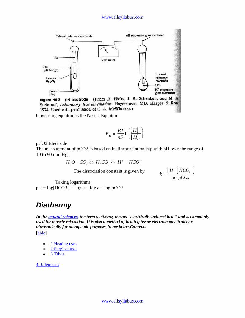
wwwallsyllabuscom
wwwallsyllabuscom
Governing equation is the Nernst Equation
pCO2 Electrode
The measurement of pCO2 is based on its linear relationship with pH over the range of
10 to 90 mm Hg
The dissociation constant is given by
Taking logarithms
pH = log[HCO3-] ndash log k ndash log a ndash log pCO2
Diathermy
In the natural sciences the term diathermy means electrically induced heat and is commonly
used for muscle relaxation It is also a method of heating tissue electromagnetically or
ultrasonically for therapeutic purposes in medicineContents
[hide]
1 Heating uses 2 Surgical uses
3 Trivia
4 References
E
RT
nF
H
HH
i
ln
0
H O CO H CO H HCO2 2 2 3 3
k
H HCO
a pCO
3
2
wwwallsyllabuscom
wwwallsyllabuscom
Heating uses
Ultrasonic diathermy refers to heating of tissues by ultrasound for the purpose of
therapeutic deep heating No tissue is ordinarily damaged hence it is generally used in
biomedical applications
Electric diathermy uses high frequency alternating electric or magnetic fields sometimes
with no electrode or device contact to the skin to induce gentle deep tissue heating by
induction Again no tissue is ordinarily damaged
Surgical uses
Surgical diathermy is usually better known as electrosurgery (It is also referred to
occasionally as electrocautery but see disambiguation below) Electrosurgery and
surgical diathermy involve the use of high frequency AC electrical current in surgery as
either a cutting modality or else to cauterize small blood vessels to stop bleeding This
technique induces localized tissue burning and damage the zone of which is controlled
by the frequency and power of the device Some sources[1]
insist that electrosurgery be
applied to surgery accomplished by high frequency AC cutting and that
electrocautery be used only for the practice of cauterization with heated nichrome wires
powered by DC current as in the handheld battery-operated portable cautery tools
Trivia
Medical Diathermy devices were used to cause interference to German radio beams used
for targeting night time bombing raids in WWII during the Battle of the Beams
I Diathermy
A Therapeutic use
1 Generation of local heating by high-frequency electromagnetic waves
2 Capacitance techniquemdashbody is placed in an electric field
a Dipolesmdashstructures with positive and negative poles
b Structures with large numbers of dipoles have a greater capacitance to store an electrical charge
c Greatest heating occurs in tissues with fewer dipoles particularly fatty tissues
3 Rapid rotation of dipoles causing mechanical friction and movement of electrons results in local heating
4 Inductance techniquemdashbody is not placed in an electric field
a Magnetic waves generated by driving current through a coiled wire
b Magnetic field creates currents in tissues
wwwallsyllabuscom
wwwallsyllabuscom
c Greatest heating occurs in tissues with low impedance especially muscle
B Precautions and contraindications
1 Diathermy should not be used
a Over metal implants and cardiac pacemakersmdashmore research needed regarding its use over metallic fixations
b Near the uterus of a pregnant woman or near the abdomen or back of a woman who might be pregnant
c On individuals with infections
d On individuals with acute inflammation
e Over moist open wounds
f On patients with malignant tumors
g Over large joint effusions
C Pulsed electromagnetic fields and diathermy
1 Can be pulsed to decrease total energy transmitted to the tissues
2 Short-wave diathermy can be adjusted into a nonthermal range
a Classified as pulsed electromagnetic field (PEMF) or
b Pulsed radio frequency energy (PRFE)
3 Important reclassification as diathermy implies heating
D Efficiency of diathermy and PEMF therapy for musculoskeletal conditions
1 Current research is limited but results suggest that diathermy enhances
treatments directed at soft tissue stretching
2 Some studies suggest that PEMF may speed wound healing and promote healing of nonunion fractures
Diathermy is a modality that uses electromagnetic energy to heat deeper tissues
Diathermy is more effective than ultrasound at heating a larger area of deep tissues
The athletic trainer must identify and respect contraindications to application of ultrasound and diathermy
Pulsed ultrasound and diathermy are used to treat slow-to-heal lesions including skin ulcers and nonunion fractures and may be able to facilitate repair of other tissues including ligaments and tendons
Diathermy
Definition
In diathermy high-frequency electrical currents are used to heat deep muscular tissues The heat increases blood flow speeding up recovery Doctors also use diathermy in surgical procedures by sealing blood vessels with electrically heated probes
The term diathermy is derived from the Greek words therma meaning heat and dia meaning through Diathermy literally means heating through
Origins
The therapeutic effects of heat have long been recognized More than 2000 years ago the Romans took advantage of heat therapies by building hot-spring bathhouses Since then various
wwwallsyllabuscom
wwwallsyllabuscom
methods of using heat have evolved In the early 1890s French physiologist Arseacutene dArsonval began studying the medical application of high-frequency currents The term diathermy was coined by German physician Carl Franz Nagelschmidt who designed a prototype apparatus in 1906 Around 1925 United States doctor J W Schereschewsky began studying the physiological
effects of high-frequency electrical currents on animals It was several years however before the fundamentals of the therapy were understood and put into practice
Benefits
Diathermy can be used to treat arthritis bursitis and other conditions involving stiff painful joints It is also used to treat pelvic infections and sinusitis A benefit of diathermy is that it is a painless procedure that can be administered at a clinic Also if the treatment relieves pain then patients can discontinue pain killers and escape their high cost and side effects
Description
Diathermy involves heating deep muscular tissues When heat is applied to the painful area cellular metabolism speeds up and blood flow increases The increased metabolism and circulation accelerates tissue repair The heat helps the tissues relax and stretch thus alleviating
stiffness Heat also reduces nerve fiber sensitivity increasing the patients pain threshold
There are three methods of diathermy In each energy is delivered to the deep tissues where it is converted to heat The three methods are
Shortwave diathermy The body part to be treated is placed between two capacitor plates Heat is generated as the high-frequency waves travel through the body tissues between the plates Shortwave diathermy is most often used to treat areas like the hip which is covered with a dense tissue mass It is also used to treat pelvic infections and sinusitis The treatment reduces inflammation The Federal Communications Commission regulates the frequency allowed for short-wave diathermy treatment Most machines
function at 2733 megahertz Ultrasound diathermy In this method high-frequency acoustic vibrations are used to
generate heat in deep tissue Microwave diathermy This method uses radar waves to heat tissue This form is the
easiest to use but the microwaves cannot penetrate deep muscles
Diathermy is also used in surgical procedures Many doctors use electrically heated probes to seal blood vessels to prevent excessive bleeding This is particularly helpful in neurosurgery and eye surgery Doctors can also use diathermy to kill abnormal growths such as tumors warts and
infected tissues
Preparations
To keep patients from sweating patients are usually asked to remove clothing from the body part being treated If a patient sweats the electrical currents may pool in the area causing burns Also clothing containing metal must be removed as must earrings buttons barrettes or zippers that contain metal Watches and hearing aids should be removed because the therapy may affect their function
wwwallsyllabuscom
wwwallsyllabuscom
Practitioners of surgical diathermy should steer clear of alcohol-based solutions to prepare and cleanse the skin These preparations can create a flammable vapor and cause burns and fires
Precautions
Patients with metal implants should not undergo diathermy treatment because the metal can act as a conductor of heat and result in serious internal burns Female patients with metallic uterine implants such as an IUD should avoid treatment in the pelvic area Diathermy should not be
used in joints that have been replaced with a prosthesis or in those with sensory impairment who may not be able to tell if they are burning Furthermore pulsed shortwave diathermy should be avoided during pregnancy as it can lead to abnormal fetal development
Patients with hemophilia should avoid the treatment because the increased blood flow could cause them to hemorrhage
Side effects
Some patients may experience superficial burns Since the therapy involves creating heat care must be taken to avoid burns particularly in patients whose injuries have caused decreased sensitivity to heat Also diathermy may affect pacemaker function
Female patients who receive treatment in the lower back or pelvic area may experience an
increased menstrual flow
Research amp general acceptance
For years physiotherapists and physical therapists have used diathermy as a routine part of physical rehabilitation
Electrical Safety
Electrical safety is very important in hospitals as patients may be undergoing a diagnostic or treatment procedure where the protective effect of dry skin is reduced Also patients may be unattended unconscious or anaesthetised and may not respond normally to an electric current Further electrically conductive solutions such as blood and saline are often present in patient treatment areas and may drip or spill on electrical equipment
Electric Current Leakage Current
Extension Leads Double Adaptors Equipment Classification
Class I Class II Defibrillator-Proof
Protective Devices
Residual Current Devices (RCD) Line Isolation overload Monitors (LIMs) Equipment Earthing
Area Classification
wwwallsyllabuscom
wwwallsyllabuscom
Body Protection Area Cardiac Protected Area
Other Electrical Issues Extension Leads
Double Adapters Main Extension Devices Power Boards Installation of Additional Power Points
Electric Current
Injuries received from electric current are dependent on the magnitude of current the pathway
that it takes through the body and the time for which it flows
The nature of electricity flowing through a circuit is analogous to blood flowing through the circulatory system within the human body In this analogy the source of energy is represented by the heart and the blood flowing through arteries and veins is analagous to current flowing
through the conductors and other components of the electric circuit
The application of an electric potential to an electric circuit generates a flow of current through conductive pathways This is analogous to the changes in blood pressure caused by contraction of cardiac muscle that causes blood to flow into the circulatory system For electric current to flow there must be a continuous pathway from the source of potential through electrical components and back to the source
Leakage Current
Electrical components and systems are encased in non conducting insulation to ensure that the electric current is contained and follows the intended pathways If the insulation deteriorates or breaks down current will leak through the insulation barrier and flow to earth This may
be through the protective earth conductor or through the operator
Medical equipment and clinical areas are fitted with a number of protective devices to protect the patient and operator from harmful leakage currents
Extension Leads
Extension leads are not permitted in clinical areas of RCH organisations They may cause high
earth resistance and excessive earth leakage current An extension lead can allow equipment to be powered from areas other than the relevant protected treatment area The power from the other area may not be protected to the same level as the power in the treatment area
wwwallsyllabuscom
wwwallsyllabuscom
As the connection between the extension lead and the equipment mains cable is often on the floor there is a high danger from fluid spills tripping and damage to the mains cable by trolleys when an extension lead is used
Double Adaptors
Double adaptors must not be used in RCH organisations They may not sit securely in a wall
outlet may not be able to provide adequate earth protection and may cause overloading overheating fire or loss of electrical supply
EQUIPMENT CLASSIFICATIONS
There are several methods of providing protection for operators and patients from electrical faults
and harmful leakage current
Class I
Class I equipment is fitted with a three core mains cable containing a protective earth wire
Exposed metal parts on class I equipment are connected to this earth wire
Should a fault develop inside the equipment and the exposed metal comes into contact with the mains the earthing conductor will conduct the fault current to ground Regular testing procedures ensure that earthing conductors are intact as the integrity of the earth wire is of vital importance
Class II
Class II equipment is enclosed within a double insulated case and does not require earthing
conductors Class II equipment is usually fitted with a 2-pin mains plug An internal electrical fault is unlikely to be hazardous as the double insulation prevents any external parts from becoming alive Class II or double insulated equipment can be identified by the class II symbol on the cabinet
Class II Symbol
wwwallsyllabuscom
wwwallsyllabuscom
Defibrillator-Proof
Some medical equipment within the hospital is classified as defibrillator proof When a
defibrillator is discharged through a patient connected to defibrillator proof equipment the equipment will not be damaged by the defibrillators energy Defibrillator proof equipment can remain connected to the patient during defibrillation It is identified by one of the following
symbols
Defibrillator proof symbols
Body protected Cardiac protected
PROTECTIVE DEVICES
Most patient care areas in the hospital are fitted with protective devices These devices are
regularly tested in accordance with the relevant guidelines published by Standards Australia The level of protection provided is dependent upon the device and the area in which it is located
Residual Current Devices (RCD)
RCDs (safety switches) are used in patient treatment areas to monitor and protect the mains supply RCDs sense leakage currents flowing to earth from the equipment If a significant leakage current flows the RCD will detect it and shut off the power supplied to the equipment
within 40 milliseconds Hospital RCDs are more sensitive than those fitted in homes A hospital RCD will trip at 10 milliamperes leakage current
Power outlets supplied through an RCD have a Supply Available lamp The lamp will extinguish
when the RCD trips due to excessive leakage current
wwwallsyllabuscom
wwwallsyllabuscom
Resetting a RCD
Lamp indicates supply is no longer available Disconnect all equipment from the supply Operate the reset button or lever on the supply panel and the Supply Available lamp
should illuminate If not contact Biomedical Engineering
Connect an item of equipment If the RCD trips again then this is the faulty item and should be labelled and sent to Biomedical Engineering
If the RCD does not trip continue connecting equipment until the RCD trips The last piece of equipment connected to the supply is most likely to be faulty as it will have caused the RCD to trip Remove the faulty item from service label it and send to Biomedical Engineering as mentioned above
Line Isolation overload Monitors (LIMs)
In critical life support applications where loss of power supply cannot be tolerated special power outlets powered by isolation transformers are installed
Line Isolation Monitors are installed to continually monitor electrical leakage in the power
supply system If an electrical fault develops in a medical device connected to an isolated power outlet the LIM will detect the leakage current The LIM will alarm and indicate the level of leakage current but will not shut off the electric supply
The faulty equipment can be identified by un plugging one item of equipment at a time from the supply until the alarm stops sounding Equipment that is not faulty may be reconnected Faulty equipment should be appropriately labelled and sent to Biomedical Engineering for repair
The LIM also monitors how much power is being used by the equpiment connected to it If too much power is being used the LIM will alarm and indicate that there is an overload The power used must be reduced immediately by moving some equipment to another circuit as soon as possible until the alarm stops sounding Failure to reduce the load on the LIM will result in the circuit breaker tripping and loss of power to the circuit
wwwallsyllabuscom
wwwallsyllabuscom
Equipotential Earthing
Equipotential earthing is installed in rooms classified as Cardiac Protected electrical areas
Equipotential earthing in treatment areas used for cardiac procedures is intended to minimise any voltage differences between earthed parts of equipment and any other exposed metal in the room
This reduces the possibility of leakage currents that can cause microelectrocution when the patient comes into contact with multiple items of equipment or if the patient happens to come
into contact with metal items in the room whilr they are connected to a medical device
All conductive metal in an equipotential area is connected to a common equipotential earth point with special heavy duty cable
AREA CLASSIFICATIONS
Body Protected Area
These areas are designed for procedures in which patients are connected to equipment that lowers the natural resistance of the skin Applied parts such as electrode gels conductive fluids entering the patient metal needles and catheters provide an easy pathway for current to flow
The main occurrence of injury from Body-Type procedures is from high current levels causing electric shock A direct connection to the patients heart is not present so the risk of Microelectrocution - fibrillation from minute current levels - is reduced
Residual Current Devices (RCD) or Isolation Transformers and Line Isolation Monitors (LIMiacutes) are used in Body Protected areas to provide protection against electrocution from high leakage currents Body-Protected Areas are identified with this sign
Cardiac Protected Area
Where the procedure involves placing an electrical conductor within or near the heart protection against fibrillation induced from small leakage currents is required Electrical conductors used in these procedures include cardiac pacing electrodes intracardiac ECG electrodes and intracardiac catheters
wwwallsyllabuscom
wwwallsyllabuscom
Equipotential earthing in conjunction with RCDs or LIMs provides protection against microelectrocution in Cardiac-Type procedures
Fault currents are reduced to magnitudes that are unlikely to induce fibrillation Used in conjunction with RCDs or LIMs the magnitude and duration of any fault currents
sourced from equipment are limited Cardiac-Protected Areas are identified with this sign
Other electrical issues
This policy aims to provide guidance to those who find that they need more electrical outlets than
those available or that the existing electrical outlets are inconveniently located
As extension leads and multiple outlet power boards can introduce additional hazards into an area the following procedures should be observed
Extension leads
Approved extension leads (AS 3760 1996) may be used in some areas within the hospital but MUST NOT BE USED IN PATIENT AREAS All electrical extension leads must be tagged with an Engineering Department maintenance tag and require a yearly safety inspection and test via
the Engineering Department
Double adapters
Double adapters may cause overloading or equipment earthing problems and are not to be used in
WCH
Mains extension device
The only mains extension device that is to be used in Patient care areas is the 4-way or 8-way
portable Core Balance Unit
The Biomedical Engineering Department must approve all units prior to use These units contain a safety switch and can detect excessive leakage current and disconnect the power in the event of a hazardous situation
wwwallsyllabuscom
wwwallsyllabuscom
Care must be exercised in the use of a portable Core Balance Unit It should be located off the
floor and in a position that will protect it from physical abuse and possible entry of fluids These devices are expensive and easily damaged The device must be sent to Biomedical Engineering every 6 months for safety testing
Power boards
Approved multiple-outlet power boards can be used across RCH but must not be used in patient
care areas except areas approved by the Biomedical Engineering Department
The power boards must have overload protection be fitted with internal safety shutters that protect unused outlets and be fitted with an onoff switch for each outlet
Medical Laser Applications
The main research subjects of the group of Medical Laser Application are in the field of online
monitoring and diagnostics as well as the development of new therapheutic methods The main focus in research is based upon the use of ultrashort (fs) laser pulses
The main advantage of ultrashort laser pulses is the extrem short interaction time which suppress any unwanted side effects of the laser irradiation of the tissue Secondly the broad spectra of the fs laser pulses give the advantage to use the same laser pulses for diagnostic applications like the optical coherence tomography (OCT)
wwwallsyllabuscom
wwwallsyllabuscom
Following research subjects have special attention at the moment
Optical Coherence Tomographie (OCT)
A fs-laser light source can be used to obtain images from inside the tissue when the pulses are coherent superposed This group uses this application to obtain an inside view of the vocal fold and to measure the outline of the crystalline lens
Refractive Surgery
The principle of getting rid of glasses with the help of a laser surgery (fs-LASIK) is underlying rapid improvements concerning precision and safety New technologies were evaluated and transferred very fast in cooperation with an industrial partner
Treatment of Presbyopia
The flexibility of the crystalline lens can be increase by pricise cuts which are induce by fs laser pulse inside the lens The treatment of the presbyopia is thinkable
wwwallsyllabuscom
wwwallsyllabuscom
Ultrafast Physics
Whereas the applications of ultrashort laser pulses increase rapidly is the physics of the interaction between the laser pulses and tissue in many case not fully understood To obtain improved knowledge of the interaction for many fs-laser application this group preforms numerical simulations as well as fundamental experiments
Thermography
Thermography thermal imaging or thermal video is a type of infrared imaging Thermographic cameras detect radiation in the infrared range of the electromagnetic spectrum
(roughly 900ndash14000 nanometers or 09ndash14 microm) and produce images of that radiation Since infrared radiation is emitted by all objects based on their temperatures according to the black body radiation law thermography makes it possible to see ones environment with or without visible illumination The amount of radiation emitted by an object increases with temperature therefore thermography allows one to see variations in temperature (hence the name) When viewed by thermographic camera warm objects stand out well against cooler backgrounds humans and other warm-blooded animals become easily visible against the environment day or
night As a result thermographys extensive use can historically be ascribed to the military and security services
Thermal imaging photography finds many other uses For example firefighters use it to see through smoke find persons and localize the base of a fire With thermal imaging power lines maintenance technicians locate overheating joints and parts a telltale sign of their failure to eliminate potential hazards Where thermal insulation becomes faulty building construction technicians can see heat leaks to improve the efficiencies of cooling or heating air-conditioning Thermal imaging cameras are also installed in some luxury cars to aid the driver the first being
the 2000 Cadillac DeVille Some physiological activities particularly responses in human beings and other warm-blooded animals can also be monitored with thermographic imaging [1]
The appearance and operation of a modern thermographic camera is often similar to a camcorder Enabling the user to see in the infrared spectrum is a function so useful that ability to record their output is often optional A recording module is therefore not always built-in
Instead of CCD sensors most thermal imaging cameras use CMOS Focal Plane Array (FPA) The most common types are InSb InGaAs HgCdTe and QWIP FPA The newest technologies are using low cost and uncooled microbolometers FPA sensors Their resolution is considerably lower than of optical cameras mostly 160x120 or 320x240 pixels up to 640x512 for the most
expensive models Thermographic cameras are much more expensive than their visible-spectrum counterparts and higher-end models are often export-restricted Older bolometers or more
wwwallsyllabuscom
wwwallsyllabuscom
sensitive models as InSb require cryogenic cooling usually by a miniature Stirling cycle refrigerator or liquid nitrogen
Contents
[hide]
1 Difference between IR film and thermography 2 Advantages of Thermography
3 Limitations amp disadvantages of thermography 4 Applications 5 See also 6 External links
o 61 History of thermal imager manufacturers
[edit] Difference between IR film and thermography
Thermal imaging is going to be used on Mars to detect caves that could hold life
[edit] Advantages of Thermography
You get a visual picture so that you can compare temperatures over a large area
It is real time capable of catching moving targets Able to find deteriorating components prior to failure Measurement in areas inaccessible or hazardous for other methods It is a non-destructive test method
[edit] Limitations amp disadvantages of thermography
Quality cameras are expensive and are easily damaged Images can be hard to interpret accurately even with experience
Accurate temperature measurements are very hard to make because of emissivities Most cameras have plusmn2 or worse accuracy (not as accurate as contact) Training and staying proficient in IR scanning is time consuming Ability to only measure surface areas
[edit] Applications
Condition monitoring Medical imaging Night vision Research
Process control Non destructive testing Surveillance in security law enforcement and defense Chemical imaging
wwwallsyllabuscom
wwwallsyllabuscom
Thermal infrared imagers convert the energy in the infrared wavelength into a visible light video display All objects above 0 kelvins emit thermal infrared energy so thermal imagers can passively see all objects regardless of ambient light However most thermal imagers only see objects warmer than -50 degC
The spectrum and amount of thermal radiation depend strongly on an objects surface
temperature This makes it possible for a thermal camera to display an objects temperature However other factors also influence the radiation which limits the accuracy of this technique For example the radiation depends not only on the temperature of the object but is also a function of the emissivity of the object Also radiation also originates from the surroundings and is reflected in the object and the radiation from the object and the reflected radiation will also be influenced by the absorption of the atmosphere
Endoscopy
This page is about having an endoscopy - a test that looks at the inside of your digestive system
There is information on
What endoscopy is
Why you may have an endoscopy
Having an endoscopy
Your results
What an endoscopy is
An endoscopy is a test that looks inside the body The endoscope is a long flexible tube that can
be swallowed It has a camera and light inside it Some doctors call it a telescope
wwwallsyllabuscom
wwwallsyllabuscom
Why you may have an endoscopy
You are most likely to have an endoscopy to look at the inside of your
Gullet (oesophagus)
Stomach
Duodenum - the first part of the small bowel that attaches to the stomach
Large bowel (colon)
Your doctor may want to see inside because you have symptoms such as abnormal bleeding or
difficulty swallowing The doctor can look down the endoscope and see if there are any growths
or other abnormal looking areas Also through the endoscope the doctor can take samples
(biopsies) of any abnormal looking tissues
There is more detailed information about having a colonoscopy in the bowel cancer section of
CancerHelp UK Below is information about having other types of endoscopy
Having an endoscopy
You can have this test as an out patient Most people have a choice between having the test while
they are awake or after having a medicine to make them drowsy (a sedative) Your hospital may
not be happy for you to have a sedative if you live alone and will have no one to look after you
when you go home If you live alone but really want sedation your hospital may allow you to
stay overnight You should ask your doctor about this as early as possible as extra arrangements
will need to be made beforehand
If you dont have a sedative you will have a spray to numb the back of your throat and make it
easier for you to swallow the endoscopy tube
If you would prefer to be asleep during the test you will have an injection to make you very
drowsy just before the test You will need to take someone with you to the hospital appointment
You wont be able to drive for the rest of the day and should have someone to go home with you
You cant eat or drink for about 8 hours before the test so that your stomach and duodenum are
empty Your doctor will give you written instructions about this beforehand or they may arrive
with your appointment letter When you get to the clinic you may be asked to take your upper
clothing off and put on a hospital gown Some hospitals prefer to use gowns because your clothes
wont get messy Once you are ready you get onto the bed or X-ray couch When you are lying
comfortably you have the sedative injection to make you very drowsy Or your doctor will spray
the back of your throat to numb it
Once the sedative or throat spray has worked the doctor will pass the endoscope tube down your
wwwallsyllabuscom
wwwallsyllabuscom
throat to the area being investigated Your doctor will ask you to swallow as the tube goes down
but if youve had a sedative you wont remember that afterwards If there are any abnormalities
the doctor will take pieces of tissue from the abnormal looking area to send to the laboratory for
closer inspection under a microscope These tissue samples are called biopsies
When the test is over you will need to rest for a while If youve had a sedative you may not
remember much (if anything) about the test once you have come round You should be able to go
home the same day
The results
It can take time for test results to come through How long will depend on why you are having
the test Usually the doctor who carries out the endoscopy dictates a report straight way The
report is typed up by the department secretary and goes to your specialist who gives the results to
you If your GP has sent you for the test the results will go directly to the GP surgery
Understandably waiting for results can make you anxious It usually takes a couple of weeks for
the results to come through If your doctor needed them urgently it would have been noted on
the test request form and the results will be ready sooner than that Try to remember to ask your
doctor how long you should expect to wait for the results when you are first asked to go for the
test If it is not an emergency and you have not heard a couple of weeks after your test ring your
doctors secretary to check if they are back
Endoscopy
From Wikipedia the free encyclopedia
Jump to navigation search
Endoscopic images of a duodenal ulcer
wwwallsyllabuscom
wwwallsyllabuscom
A flexible endoscope
Endoscopy means looking inside and typically refers to looking inside the human body for medical reasons using an instrument called an endoscope Endoscopy can also refer to using a borescope in technical situations where direct line-of-sight observation is not feasible
Contents
[hide]
1 Overview 2 Components 3 Uses
4 History 5 Risks 6 After The Endoscopy 7 Recent developments 8 See also 9 References 10 Footnotes
11 External links
Overview
Endoscopy is a minimally invasive diagnostic medical procedure that is used to assess the interior surfaces of an organ by inserting a tube into the body The instrument may have a rigid or flexible tube and not only provide an image for visual inspection and photography but also enable taking
biopsies and retrieval of foreign objects Endoscopy is the vehicle for minimally invasive surgery
Many endoscopic procedures are considered to be relatively painless and at worst associated with mild discomfort for example in esophagogastroduodenoscopy most patients tolerate the procedure with only topical anaesthesia of the oropharynx using lignocaine spray[1] Complications are not common (only 5 of all operations)[citation needed] but can include perforation of the organ under inspection with the endoscope or biopsy instrument If that occurs open surgery may be required to repair the injury
wwwallsyllabuscom
wwwallsyllabuscom
Components
An endoscope can consist of
a rigid or flexible tube a light delivery system to illuminate the organ or object under inspection The light
source is normally outside the body and the light is typically directed via an optical fiber system
a lens system transmitting the image to the viewer from the fiberscope an additional channel to allow entry of medical instruments or manipulators
Uses
Endoscopy can involve
The gastrointestinal tract (GI tract) o esophagus stomach and duodenum (esophagogastroduodenoscopy) o small intestine o colon (colonoscopyproctosigmoidoscopy) o Bile duct
endoscopic retrograde cholangiopancreatography (ERCP) duodenoscope-assisted cholangiopancreatoscopy intraoperative cholangioscopy
The respiratory tract o The nose (rhinoscopy) o The lower respiratory tract (bronchoscopy)
The urinary tract (cystoscopy) The female reproductive system
o The cervix (colposcopy) o The uterus (hysteroscopy) o The Fallopian tubes (Falloscopy)
Normally closed body cavities (through a small incision) o The abdominal or pelvic cavity (laparoscopy) o The interior of a joint (arthroscopy) o Organs of the chest (thoracoscopy and mediastinoscopy)
During pregnancy o The amnion (amnioscopy) o The fetus (fetoscopy)
Plastic Surgery Panendoscopy (or triple endoscopy)
o Combines laryngoscopy esophagoscopy and bronchoscopy Non-medical uses for endoscopy
o The planning and architectural community have found the endoscope useful for
pre-visualization of scale models of proposed buildings and cities (architectural endoscopy)
o Internal inspection of complex technical systems (borescope) o Endoscopes are also a tool helpful in the examination of improvised explosive
devices by bomb disposal personnel
wwwallsyllabuscom
wwwallsyllabuscom
o The FBI uses endoscopes for conducting surveillance via tight spaces
History
The first endoscope of a kind was developed in 1806 by Philip Bozzini with his introduction of a
Lichtleiter (light conductor) for the examinations of the canals and cavities of the human body However the Vienna Medical Society disapproved of such curiosity An endoscope was first introduced into a human in 1822 by William Beaumont an army surgeon at Mackinac
Island Michigan[citation needed] The use of electric light was a major step in the improvement of endoscopy The first such lights were external Later smaller bulbs became available making internal light possible for instance in a hysteroscope by Charles David in 1908[citation needed] Hans Christian Jacobaeus has been given credit for early endoscopic explorations of the abdomen and the thorax with laparoscopy (1912) and thoracoscopy (1910)[citation needed] Laparoscopy was used in the diagnosis of liver and gallbladder disease by Heinz Kalk in the 1930s[citation needed] Hope reported in 1937 on the use of laparoscopy to diagnose ectopic pregnancy[citation needed] In 1944
Raoul Palmer placed his patients in the Trendelenburg position after gaseous distention of the abdomen and thus was able to reliably perform gynecologic laparoscopy[citation needed]
The first gastrocamera was released in 1950 by Olympus Optical Co Ltd The device took pictures on monochromatic film using a small light bulb that was triggered manually The device was of limited use however because it did not implement real-time optical capability Olympus continued its development of endoscopes by incorporating fiber optics in the early 1960s leading to the first useful endoscopes In 1964 it released a gastrocamera guided by a fiberscope[1] A few articles claim that DrBasil Hirschowitz of UnivOf MichiganAnn Arbor discussed the
endoscope in early 50s[2]
As endoscopic technology improved so did the methods of gastrointestinal endoscopy Owing primarily to the efforts of Dr Hiromi Shinya in the late 1960s GI endoscopy developed into what is more recognizable as todays colonoscopy While many doctors experimented with techniques to take advantage of the new iterations of endoscopes Dr Shinya focused on techniques that would allow for successful operation of the endoscope by an individual rejecting the common practice at the time of utilizing two people Consequently many of the fundamental methods and procedures of modern colonoscopy were developed by Dr Shinya
Dr Shinyas other great contribution was to therapeutic endoscopy in his invention of the
electrosurgical polypectomy snare with the aid of Olympus employee Hiroshi Ichikawa Shinya sketched his first plans for the device on January 8 1969 He envisioned a loop of wire attached to the end of a colonoscope that would allow for easy removal of polyps during investigation by passing a current through the wire By September of 1969 the first polypectomy using this device was performed Polypectomy has since become the most common therapeutic procedure performed with an endoscope (Sivak 2004)
By 1980 laparoscopy training was required by gynecologists to perform tubal ligation procedures and diagnostic evaluations of the pelvis The first laparoscopic cholecystectomy was performed in 1984 and the first video-laparoscopic cholecystectomy in 1987[citation needed] During the 1990s
laparoscopic surgery was extended to the appendix spleen colon stomach kidney and liver[citation needed] Wireless capsule endoscopy or Capsule Endoscopy is now approved in all the countries including Japan where government reimbusement will be available from Oct2007Capsule Endoscopy [3] increases detection of Small Bowel tumors where traditional Endoscopy is not very efficient
wwwallsyllabuscom
wwwallsyllabuscom
Risks
Infection
Punctured organs Allergic reactions due to Contrast agents or dyes (such as those used in a CT scan) Over-sedation
After The Endoscopy
After the procedure the patient will be observed and monitored by a qualified individual in the
endoscopy or a recovery area until a significant portion of the medication has worn off Occasionally a patient is left with a mild sore throat which promptly responds to saline gargles or a feeling of distention from the insufflated air that was used during the procedure Both problems are mild and fleeting When fully recovered the patient will be instructed when to resume hisher usual diet (probably within a few hours) and will be allowed to be taken home Because of the use of sedation most facilities mandate that the patient is taken home by another person and not to drive on hisher own or handle machinery for the remainder of the day
Recent developments
With the application of robotic systems telesurgery was introduced as the surgeon could operate
from a site physically removed from the patient The first transatlantic surgery has been called the Lindbergh Operation
wwwallsyllabuscom
wwwallsyllabuscom
Upper Endoscopy
Upper endoscopy enables the physician to look inside the esophagus stomach and duodenum (first part of the small intestine) The procedure might be used to discover the reason for swallowing difficulties nausea vomiting
reflux bleeding indigestion abdominal pain or chest pain Upper endoscopy is also called EGD which stands for esophagogastroduodenoscopy (eh-SAH-fuh-goh-GAS-troh-doo-AH-duh-NAH-skuh-pee)
For the procedure you will swallow a thin flexible lighted tube called an endoscope (EN-doh-skope) Right before the procedure the physician will spray your throat with a numbing agent that may help prevent gagging
You may also receive pain medicine and a sedative to help you relax during the exam The endoscope transmits an image of the inside of the esophagus stomach and duodenum so the physician can carefully examine the lining of these organs The scope also blows air into the stomach this expands the folds of tissue and makes it easier for the physician to examine the stomach
The physician can see abnormalities like inflammation or
bleeding through the endoscope that dont show up well on x rays The physician can also insert instruments into the scope to treat bleeding abnormalities or remove samples of tissue (biopsy) for further tests
Possible complications of upper endoscopy include bleeding and puncture of the stomach lining However such complications are rare Most people will probably have nothing more than a mild sore throat after the procedure
The procedure takes 20 to 30 minutes Because you will be sedated you will need to rest at the endoscopy facility for 1 to 2 hours until the medication wears off
Preparation
Your stomach and duodenum must be empty for the procedure to be thorough and safe so you will not be able to eat or drink anything for at least 6 hours beforehand Also you must
arrange for someone to take you homemdashyou will not be allowed to drive because of the sedatives Your physician may give you other special instructions
The digestive system
wwwallsyllabuscom
wwwallsyllabuscom
What is Endoscopy and Why is it Performed
Endoscopy allows physicians to peer through the bodys passageways Endoscopy is the examination and inspection of the interior of body organs joints or cavities through an endoscope An endoscope is a device that uses fiber optics and powerful lens systems to provide lighting and visualization of the interior of a joint The portion of the endoscope inserted into the body may be rigid or flexible depending upon the medical procedure
An endoscope uses two fiber optic lines A light fiber carries light into the body cavity and an image fiber carries the image of the body cavity back to the physicians viewing lens There is also a separate port to allow for administration of drugs suction and irrigation This port may also be used to introduce small folding instruments such as forceps scissors brushes snares and baskets for tissue excision (removal) sampling or other diagnostic and therapeutic work Endoscopes may be used in conjunction with a camera or video recorder to document images of the inside of the joint or chronicle an endoscopic procedure New endoscopes have digital capabilities for manipulating and enhancing the video images
This figure shows a rigid endoscope used for arthroscopy The image fiber leads from the ocular (eye piece) to the inserted end of the scope The light fiber is below and leads from the light source to the working end of the endoscope
Why Is Endoscopy Performed
Endoscopy can be used to diagnose various conditions by close examination of internal organ and body structures Endoscopy can also guide therapy and repair such as the removal of torn cartilage from the bearing surfaces of a joint Biopsy (tissue sampling for pathologic testing) may also be performed under endoscopic guidance Local or general anesthetic may be used during endoscopy depending upon the type of procedure being performed
Internal abnormalities revealed through endoscopy include abscesses biliary (liver) cirrhosis bleeding bronchitis cancer cysts degenerative disease gallbladder stones hernia inflammation metastatic cancer polyps tumors ulcers and other diseases and conditions
Endoscopy is a minimally invasive procedure and carries with it certain minor risks depending upon the type of procedure being performed However these risks are typically far outweighed by the diagnostic and therapeutic potential of the procedure
Prior to the widespread use of endoscopy and diagnostic imaging most internal conditions could only be diagnosed or treated with open surgery Until the last several decades exploratory surgery was routinely performed only when a patient was critically ill and the source of illness was not known For example in certain dire cases the patients thorax or abdomen were surgically opened and examined to try to determine the source of illness
wwwallsyllabuscom
wwwallsyllabuscom
Endoscopy can often be done on an outpatient basis Outpatient means that the procedure does not require hospital admission and acute care and observation and may be performed outside the premises of a hospital Outpatient procedures performed at hospitals or ambulatory centers allow the patient to go home or return to work within a short while after their procedure
Types of Endoscopy
Fiber optic endoscopes now have widespread use in medicine and guide a myriad of diagnostic and therapeutic procedures including
Arthroscopy examination of joints for diagnosis and treatment (arthroscopic surgery) Bronchoscopy examination of the trachea and lungs bronchial trees to reveal
abscesses bronchitis carcinoma tumors tuberculosis alveolitis infection inflammation Colonoscopy examination of the inside of the colon and large intestine to detect polyps
tumors ulceration inflammation colitis diverticula Chrohns disease and discovery and removal of foreign bodies
Colposcopy direct visualization of the vagina and cervix to detect cancer inflammation and other conditions
Cystoscopy examination of the bladder urethra urinary tract uteral orifices and
prostate (men) with insertion of the endoscope through the urethra
ERCP (endoscopic retrograde cholangio-pancreatography) uses endoscopic guidance to
place a catheter for x-ray fluorosocopy with contrast enhancement This technique is used to examine the livers biliary tree the gallbladder the pancreatic duct and other anatomy to check for stones other obstructions and disease X-ray contrast is introduced into these ducts via catheter and fluoroscopic x-ray images are taken to show any abnormality or blockage If disease is detected it can sometimes be treated at the same time or biopsy can be performed to test for cancer or other pathology ERCP can detect biliary cirrhosis
cancer of the bile ducts pancreatic cysts pseudocysts pancreatic tumors chronic pancreatitis and other conditions such as gallbladder stones
EGD (Esophogealgastroduodensoscopy) visual examination of the upper gastro-
intestinal (GI) tract (also referred to as gastroscopy) to reveal hemorrhage hiatal hernia inflammation of the esophagus gastric ulcers
Endoscopic biopsy is the removal of tissue specimens for pathologic examination and
analysis Gastroscopy examination of the lining of the esophagus stomach and duodenum
Gastroscopy is often used to diagnose ulcers and other sources of bleeding and to guide biopsy of suspect GI cancers
Laparoscopy visualization of the stomach liver and other abdominal organs including
the female reproductive organs for example the fallopian tubes Laryngoscopy examination of the larynx (voice box) Proctoscopy sigmoidoscopy proctosigmoidoscopy examination of the rectum and
sigmoid colon Thoracoscopy examination of the pleura (sac that covers the lungs) pleural spaces
mediastinum and pericardium
A Brief History of Endoscopy
In the early 1900s the first attempts to view inside the body with lighted telescopes were made These initial devices were often fully rigid In the 1930s semi-flexible endoscopes called gastroscopes were developed to view inside of the stomach Fiber-optic endoscopy was pioneered by South African-born physician Basil Hirschowitz at the University of Michigan in 1957 Widespread use of fiber optic endoscopes began in the 1960s
wwwallsyllabuscom
wwwallsyllabuscom
A fiber optic cable is simply a bundle of microscopic glass or plastic fibers that literally allows light and images to be transmitted through curved structures Fiber optic cables are also replacing metal wires as the backbone of the worlds telecommunications infrastructure This Internet page may have traveled through a fiber optic cable as a stream of digital data (bursts of light) on its way to your computer
Endoscopy Equipment
Endoscopes have many practical needs And HMB Endoscopy Products (Hollywood Florida) has been providing endoscopic equipment and educating people on the use of endoscopes for more than 17 years Be sure to Browse our Catalog for all the details on how to purchase these medical instruments that can examine any part of the body
Video
Systems
Fiber
Endoscopes
Video
Endoscopes
In the simplest terms Endoscopy equipment consists of instruments that can look at the inside of many different organs mdash these are small flexible or rigid tubes with a light or lenses on the end that can look into the esophagus stomach and colon mdash and in more general terms endoscopy equipment can help doctors look deep inside body structures and hollow organs
An endoscope and related endoscope products and equipment are usually composed of three components
An optic system that allows the doctor to look through the scope into the organ or
cavity or to attach a video camera to the scope
A fiberoptic cable to light up the bodily area
A lumen (eg the bore of a tube like a needle or catheter) to take tissue samples of the
area being viewed
The beauty of endoscopic products is that they perform dual functions mdash with both diagnostic and therapeutic capabilities For example this means that these endoscopic products and instruments can perform biopsies (eg to evaluate tissue samples) as well as provide sclerotherapy (a medical procedure used to treat varicose veins and ―spider veins) In truth these brief explanations only tell part of the story
wwwallsyllabuscom
wwwallsyllabuscom
TYPES OF ENDOSCOPES
Take a look at the different types of endoscopic equipment you can get with HMB Endoscopy Products Again in very general terms there are two main types of endoscopes
Rigid Flexible
1 rigid endoscopes ndash the majority of which use a convex (curving out like one half of a
circle) glass lens system in which the small glass lenses are separated by large air spaces
2 flexible endoscopes allow for just that mdash flexibility In the animal kingdom for example
a flexible endoscope would be perfect for examining the stomach area of a snake
The popularity of endoscopy equipment continues to grow Some of the industryrsquos household names include Olympus Pentax Funjinon and Storz mdash with HMB both selling and repairing pre-owned and completely refurbished endoscopic products from each of these major manufacturers Browse our Catalog to get the ones you need
THERMOGRAPH APPLICATION Thermographs are digital recording thermometers used to log
temperature in the
marine environment The instruments are attached to a computer to set up recording
parameters for
deployment downloading and display of data in graphic or numeric format These units
are placed in
underwater housings and attached to the bottom or suspended in the water column for
deployment
DESCRIPTION Two types of thermograph are currently available The Ryan
Tempmentor is a
reusable data logger that allows storage of a maximum of 6361 temperature
measurements in the
range of -32C to +70C with 01resolution and 03accuracy The instrument can be
programmed
to take measurements from once per second to once every other hour Instrument
dimensions are 3 x
6 x 13 and the unit weighs 11 ounces The Hobo Temp is a miniature reusable data
logger that
allows storage of 1800 measurements in the range of -20C to +70C with
03resolution and 07
wwwallsyllabuscom
wwwallsyllabuscom
accuracy Sampling intervals from 05 seconds to 48 hours are available Instrument
dimensions are
24 x 19 x 08 and the unit weighs 1 ounce Both units are battery powered with the
battery life of
the Tempmentor being approximately two years and the battery life of the Hobo Temp
being one year THERMOGRAPH APPLICATION Thermographs are digital recording thermometers used to log
temperature in the
marine environment The instruments are attached to a computer to set up recording
parameters for
deployment downloading and display of data in graphic or numeric format These units
are placed in
underwater housings and attached to the bottom or suspended in the water column for
deployment
DESCRIPTION Two types of thermograph are currently available The Ryan
Tempmentor is a
reusable data logger that allows storage of a maximum of 6361 temperature
measurements in the
range of -32C to +70C with 01resolution and 03accuracy The instrument can be
programmed
to take measurements from once per second to once every other hour Instrument
dimensions are 3 x
6 x 13 and the unit weighs 11 ounces The Hobo Temp is a miniature reusable data
logger that
allows storage of 1800 measurements in the range of -20C to +70C with
03resolution and 07
accuracy Sampling intervals from 05 seconds to 48 hours are available Instrument
dimensions are
24 x 19 x 08 and the unit weighs 1 ounce Both units are battery powered with the
battery life of
the Tempmentor being approximately two years and the battery life of the Hobo Temp
being one year THERMOGRAPH APPLICATION Thermographs are digital recording thermometers used to log
temperature in the
marine environment The instruments are attached to a computer to set up recording
parameters for
deployment downloading and display of data in graphic or numeric format These units
are placed in
underwater housings and attached to the bottom or suspended in the water column for
deployment
DESCRIPTION Two types of thermograph are currently available The Ryan
Tempmentor is a
reusable data logger that allows storage of a maximum of 6361 temperature
measurements in the
range of -32C to +70C with 01resolution and 03accuracy The instrument can be
programmed
wwwallsyllabuscom
wwwallsyllabuscom
to take measurements from once per second to once every other hour Instrument
dimensions are 3 x
6 x 13 and the unit weighs 11 ounces The Hobo Temp is a miniature reusable data
logger that
allows storage of 1800 measurements in the range of -20C to +70C with
03resolution and 07
accuracy Sampling intervals from 05 seconds to 48 hours are available Instrument
dimensions are
24 x 19 x 08 and the unit weighs 1 ounce Both units are battery powered with the
battery life of
the Tempmentor being approximately two years and the battery life of the Hobo Temp
being one year
wwwallsyllabuscom
wwwallsyllabuscom
c Greatest heating occurs in tissues with low impedance especially muscle
B Precautions and contraindications
1 Diathermy should not be used
a Over metal implants and cardiac pacemakersmdashmore research needed regarding its use over metallic fixations
b Near the uterus of a pregnant woman or near the abdomen or back of a woman who might be pregnant
c On individuals with infections
d On individuals with acute inflammation
e Over moist open wounds
f On patients with malignant tumors
g Over large joint effusions
C Pulsed electromagnetic fields and diathermy
1 Can be pulsed to decrease total energy transmitted to the tissues
2 Short-wave diathermy can be adjusted into a nonthermal range
a Classified as pulsed electromagnetic field (PEMF) or
b Pulsed radio frequency energy (PRFE)
3 Important reclassification as diathermy implies heating
D Efficiency of diathermy and PEMF therapy for musculoskeletal conditions
1 Current research is limited but results suggest that diathermy enhances
treatments directed at soft tissue stretching
2 Some studies suggest that PEMF may speed wound healing and promote healing of nonunion fractures
Diathermy is a modality that uses electromagnetic energy to heat deeper tissues
Diathermy is more effective than ultrasound at heating a larger area of deep tissues
The athletic trainer must identify and respect contraindications to application of ultrasound and diathermy
Pulsed ultrasound and diathermy are used to treat slow-to-heal lesions including skin ulcers and nonunion fractures and may be able to facilitate repair of other tissues including ligaments and tendons
Diathermy
Definition
In diathermy high-frequency electrical currents are used to heat deep muscular tissues The heat increases blood flow speeding up recovery Doctors also use diathermy in surgical procedures by sealing blood vessels with electrically heated probes
The term diathermy is derived from the Greek words therma meaning heat and dia meaning through Diathermy literally means heating through
Origins
The therapeutic effects of heat have long been recognized More than 2000 years ago the Romans took advantage of heat therapies by building hot-spring bathhouses Since then various
wwwallsyllabuscom
wwwallsyllabuscom
methods of using heat have evolved In the early 1890s French physiologist Arseacutene dArsonval began studying the medical application of high-frequency currents The term diathermy was coined by German physician Carl Franz Nagelschmidt who designed a prototype apparatus in 1906 Around 1925 United States doctor J W Schereschewsky began studying the physiological
effects of high-frequency electrical currents on animals It was several years however before the fundamentals of the therapy were understood and put into practice
Benefits
Diathermy can be used to treat arthritis bursitis and other conditions involving stiff painful joints It is also used to treat pelvic infections and sinusitis A benefit of diathermy is that it is a painless procedure that can be administered at a clinic Also if the treatment relieves pain then patients can discontinue pain killers and escape their high cost and side effects
Description
Diathermy involves heating deep muscular tissues When heat is applied to the painful area cellular metabolism speeds up and blood flow increases The increased metabolism and circulation accelerates tissue repair The heat helps the tissues relax and stretch thus alleviating
stiffness Heat also reduces nerve fiber sensitivity increasing the patients pain threshold
There are three methods of diathermy In each energy is delivered to the deep tissues where it is converted to heat The three methods are
Shortwave diathermy The body part to be treated is placed between two capacitor plates Heat is generated as the high-frequency waves travel through the body tissues between the plates Shortwave diathermy is most often used to treat areas like the hip which is covered with a dense tissue mass It is also used to treat pelvic infections and sinusitis The treatment reduces inflammation The Federal Communications Commission regulates the frequency allowed for short-wave diathermy treatment Most machines
function at 2733 megahertz Ultrasound diathermy In this method high-frequency acoustic vibrations are used to
generate heat in deep tissue Microwave diathermy This method uses radar waves to heat tissue This form is the
easiest to use but the microwaves cannot penetrate deep muscles
Diathermy is also used in surgical procedures Many doctors use electrically heated probes to seal blood vessels to prevent excessive bleeding This is particularly helpful in neurosurgery and eye surgery Doctors can also use diathermy to kill abnormal growths such as tumors warts and
infected tissues
Preparations
To keep patients from sweating patients are usually asked to remove clothing from the body part being treated If a patient sweats the electrical currents may pool in the area causing burns Also clothing containing metal must be removed as must earrings buttons barrettes or zippers that contain metal Watches and hearing aids should be removed because the therapy may affect their function
wwwallsyllabuscom
wwwallsyllabuscom
Practitioners of surgical diathermy should steer clear of alcohol-based solutions to prepare and cleanse the skin These preparations can create a flammable vapor and cause burns and fires
Precautions
Patients with metal implants should not undergo diathermy treatment because the metal can act as a conductor of heat and result in serious internal burns Female patients with metallic uterine implants such as an IUD should avoid treatment in the pelvic area Diathermy should not be
used in joints that have been replaced with a prosthesis or in those with sensory impairment who may not be able to tell if they are burning Furthermore pulsed shortwave diathermy should be avoided during pregnancy as it can lead to abnormal fetal development
Patients with hemophilia should avoid the treatment because the increased blood flow could cause them to hemorrhage
Side effects
Some patients may experience superficial burns Since the therapy involves creating heat care must be taken to avoid burns particularly in patients whose injuries have caused decreased sensitivity to heat Also diathermy may affect pacemaker function
Female patients who receive treatment in the lower back or pelvic area may experience an
increased menstrual flow
Research amp general acceptance
For years physiotherapists and physical therapists have used diathermy as a routine part of physical rehabilitation
Electrical Safety
Electrical safety is very important in hospitals as patients may be undergoing a diagnostic or treatment procedure where the protective effect of dry skin is reduced Also patients may be unattended unconscious or anaesthetised and may not respond normally to an electric current Further electrically conductive solutions such as blood and saline are often present in patient treatment areas and may drip or spill on electrical equipment
Electric Current Leakage Current
Extension Leads Double Adaptors Equipment Classification
Class I Class II Defibrillator-Proof
Protective Devices
Residual Current Devices (RCD) Line Isolation overload Monitors (LIMs) Equipment Earthing
Area Classification
wwwallsyllabuscom
wwwallsyllabuscom
Body Protection Area Cardiac Protected Area
Other Electrical Issues Extension Leads
Double Adapters Main Extension Devices Power Boards Installation of Additional Power Points
Electric Current
Injuries received from electric current are dependent on the magnitude of current the pathway
that it takes through the body and the time for which it flows
The nature of electricity flowing through a circuit is analogous to blood flowing through the circulatory system within the human body In this analogy the source of energy is represented by the heart and the blood flowing through arteries and veins is analagous to current flowing
through the conductors and other components of the electric circuit
The application of an electric potential to an electric circuit generates a flow of current through conductive pathways This is analogous to the changes in blood pressure caused by contraction of cardiac muscle that causes blood to flow into the circulatory system For electric current to flow there must be a continuous pathway from the source of potential through electrical components and back to the source
Leakage Current
Electrical components and systems are encased in non conducting insulation to ensure that the electric current is contained and follows the intended pathways If the insulation deteriorates or breaks down current will leak through the insulation barrier and flow to earth This may
be through the protective earth conductor or through the operator
Medical equipment and clinical areas are fitted with a number of protective devices to protect the patient and operator from harmful leakage currents
Extension Leads
Extension leads are not permitted in clinical areas of RCH organisations They may cause high
earth resistance and excessive earth leakage current An extension lead can allow equipment to be powered from areas other than the relevant protected treatment area The power from the other area may not be protected to the same level as the power in the treatment area
wwwallsyllabuscom
wwwallsyllabuscom
As the connection between the extension lead and the equipment mains cable is often on the floor there is a high danger from fluid spills tripping and damage to the mains cable by trolleys when an extension lead is used
Double Adaptors
Double adaptors must not be used in RCH organisations They may not sit securely in a wall
outlet may not be able to provide adequate earth protection and may cause overloading overheating fire or loss of electrical supply
EQUIPMENT CLASSIFICATIONS
There are several methods of providing protection for operators and patients from electrical faults
and harmful leakage current
Class I
Class I equipment is fitted with a three core mains cable containing a protective earth wire
Exposed metal parts on class I equipment are connected to this earth wire
Should a fault develop inside the equipment and the exposed metal comes into contact with the mains the earthing conductor will conduct the fault current to ground Regular testing procedures ensure that earthing conductors are intact as the integrity of the earth wire is of vital importance
Class II
Class II equipment is enclosed within a double insulated case and does not require earthing
conductors Class II equipment is usually fitted with a 2-pin mains plug An internal electrical fault is unlikely to be hazardous as the double insulation prevents any external parts from becoming alive Class II or double insulated equipment can be identified by the class II symbol on the cabinet
Class II Symbol
wwwallsyllabuscom
wwwallsyllabuscom
Defibrillator-Proof
Some medical equipment within the hospital is classified as defibrillator proof When a
defibrillator is discharged through a patient connected to defibrillator proof equipment the equipment will not be damaged by the defibrillators energy Defibrillator proof equipment can remain connected to the patient during defibrillation It is identified by one of the following
symbols
Defibrillator proof symbols
Body protected Cardiac protected
PROTECTIVE DEVICES
Most patient care areas in the hospital are fitted with protective devices These devices are
regularly tested in accordance with the relevant guidelines published by Standards Australia The level of protection provided is dependent upon the device and the area in which it is located
Residual Current Devices (RCD)
RCDs (safety switches) are used in patient treatment areas to monitor and protect the mains supply RCDs sense leakage currents flowing to earth from the equipment If a significant leakage current flows the RCD will detect it and shut off the power supplied to the equipment
within 40 milliseconds Hospital RCDs are more sensitive than those fitted in homes A hospital RCD will trip at 10 milliamperes leakage current
Power outlets supplied through an RCD have a Supply Available lamp The lamp will extinguish
when the RCD trips due to excessive leakage current
wwwallsyllabuscom
wwwallsyllabuscom
Resetting a RCD
Lamp indicates supply is no longer available Disconnect all equipment from the supply Operate the reset button or lever on the supply panel and the Supply Available lamp
should illuminate If not contact Biomedical Engineering
Connect an item of equipment If the RCD trips again then this is the faulty item and should be labelled and sent to Biomedical Engineering
If the RCD does not trip continue connecting equipment until the RCD trips The last piece of equipment connected to the supply is most likely to be faulty as it will have caused the RCD to trip Remove the faulty item from service label it and send to Biomedical Engineering as mentioned above
Line Isolation overload Monitors (LIMs)
In critical life support applications where loss of power supply cannot be tolerated special power outlets powered by isolation transformers are installed
Line Isolation Monitors are installed to continually monitor electrical leakage in the power
supply system If an electrical fault develops in a medical device connected to an isolated power outlet the LIM will detect the leakage current The LIM will alarm and indicate the level of leakage current but will not shut off the electric supply
The faulty equipment can be identified by un plugging one item of equipment at a time from the supply until the alarm stops sounding Equipment that is not faulty may be reconnected Faulty equipment should be appropriately labelled and sent to Biomedical Engineering for repair
The LIM also monitors how much power is being used by the equpiment connected to it If too much power is being used the LIM will alarm and indicate that there is an overload The power used must be reduced immediately by moving some equipment to another circuit as soon as possible until the alarm stops sounding Failure to reduce the load on the LIM will result in the circuit breaker tripping and loss of power to the circuit
wwwallsyllabuscom
wwwallsyllabuscom
Equipotential Earthing
Equipotential earthing is installed in rooms classified as Cardiac Protected electrical areas
Equipotential earthing in treatment areas used for cardiac procedures is intended to minimise any voltage differences between earthed parts of equipment and any other exposed metal in the room
This reduces the possibility of leakage currents that can cause microelectrocution when the patient comes into contact with multiple items of equipment or if the patient happens to come
into contact with metal items in the room whilr they are connected to a medical device
All conductive metal in an equipotential area is connected to a common equipotential earth point with special heavy duty cable
AREA CLASSIFICATIONS
Body Protected Area
These areas are designed for procedures in which patients are connected to equipment that lowers the natural resistance of the skin Applied parts such as electrode gels conductive fluids entering the patient metal needles and catheters provide an easy pathway for current to flow
The main occurrence of injury from Body-Type procedures is from high current levels causing electric shock A direct connection to the patients heart is not present so the risk of Microelectrocution - fibrillation from minute current levels - is reduced
Residual Current Devices (RCD) or Isolation Transformers and Line Isolation Monitors (LIMiacutes) are used in Body Protected areas to provide protection against electrocution from high leakage currents Body-Protected Areas are identified with this sign
Cardiac Protected Area
Where the procedure involves placing an electrical conductor within or near the heart protection against fibrillation induced from small leakage currents is required Electrical conductors used in these procedures include cardiac pacing electrodes intracardiac ECG electrodes and intracardiac catheters
wwwallsyllabuscom
wwwallsyllabuscom
Equipotential earthing in conjunction with RCDs or LIMs provides protection against microelectrocution in Cardiac-Type procedures
Fault currents are reduced to magnitudes that are unlikely to induce fibrillation Used in conjunction with RCDs or LIMs the magnitude and duration of any fault currents
sourced from equipment are limited Cardiac-Protected Areas are identified with this sign
Other electrical issues
This policy aims to provide guidance to those who find that they need more electrical outlets than
those available or that the existing electrical outlets are inconveniently located
As extension leads and multiple outlet power boards can introduce additional hazards into an area the following procedures should be observed
Extension leads
Approved extension leads (AS 3760 1996) may be used in some areas within the hospital but MUST NOT BE USED IN PATIENT AREAS All electrical extension leads must be tagged with an Engineering Department maintenance tag and require a yearly safety inspection and test via
the Engineering Department
Double adapters
Double adapters may cause overloading or equipment earthing problems and are not to be used in
WCH
Mains extension device
The only mains extension device that is to be used in Patient care areas is the 4-way or 8-way
portable Core Balance Unit
The Biomedical Engineering Department must approve all units prior to use These units contain a safety switch and can detect excessive leakage current and disconnect the power in the event of a hazardous situation
wwwallsyllabuscom
wwwallsyllabuscom
Care must be exercised in the use of a portable Core Balance Unit It should be located off the
floor and in a position that will protect it from physical abuse and possible entry of fluids These devices are expensive and easily damaged The device must be sent to Biomedical Engineering every 6 months for safety testing
Power boards
Approved multiple-outlet power boards can be used across RCH but must not be used in patient
care areas except areas approved by the Biomedical Engineering Department
The power boards must have overload protection be fitted with internal safety shutters that protect unused outlets and be fitted with an onoff switch for each outlet
Medical Laser Applications
The main research subjects of the group of Medical Laser Application are in the field of online
monitoring and diagnostics as well as the development of new therapheutic methods The main focus in research is based upon the use of ultrashort (fs) laser pulses
The main advantage of ultrashort laser pulses is the extrem short interaction time which suppress any unwanted side effects of the laser irradiation of the tissue Secondly the broad spectra of the fs laser pulses give the advantage to use the same laser pulses for diagnostic applications like the optical coherence tomography (OCT)
wwwallsyllabuscom
wwwallsyllabuscom
Following research subjects have special attention at the moment
Optical Coherence Tomographie (OCT)
A fs-laser light source can be used to obtain images from inside the tissue when the pulses are coherent superposed This group uses this application to obtain an inside view of the vocal fold and to measure the outline of the crystalline lens
Refractive Surgery
The principle of getting rid of glasses with the help of a laser surgery (fs-LASIK) is underlying rapid improvements concerning precision and safety New technologies were evaluated and transferred very fast in cooperation with an industrial partner
Treatment of Presbyopia
The flexibility of the crystalline lens can be increase by pricise cuts which are induce by fs laser pulse inside the lens The treatment of the presbyopia is thinkable
wwwallsyllabuscom
wwwallsyllabuscom
Ultrafast Physics
Whereas the applications of ultrashort laser pulses increase rapidly is the physics of the interaction between the laser pulses and tissue in many case not fully understood To obtain improved knowledge of the interaction for many fs-laser application this group preforms numerical simulations as well as fundamental experiments
Thermography
Thermography thermal imaging or thermal video is a type of infrared imaging Thermographic cameras detect radiation in the infrared range of the electromagnetic spectrum
(roughly 900ndash14000 nanometers or 09ndash14 microm) and produce images of that radiation Since infrared radiation is emitted by all objects based on their temperatures according to the black body radiation law thermography makes it possible to see ones environment with or without visible illumination The amount of radiation emitted by an object increases with temperature therefore thermography allows one to see variations in temperature (hence the name) When viewed by thermographic camera warm objects stand out well against cooler backgrounds humans and other warm-blooded animals become easily visible against the environment day or
night As a result thermographys extensive use can historically be ascribed to the military and security services
Thermal imaging photography finds many other uses For example firefighters use it to see through smoke find persons and localize the base of a fire With thermal imaging power lines maintenance technicians locate overheating joints and parts a telltale sign of their failure to eliminate potential hazards Where thermal insulation becomes faulty building construction technicians can see heat leaks to improve the efficiencies of cooling or heating air-conditioning Thermal imaging cameras are also installed in some luxury cars to aid the driver the first being
the 2000 Cadillac DeVille Some physiological activities particularly responses in human beings and other warm-blooded animals can also be monitored with thermographic imaging [1]
The appearance and operation of a modern thermographic camera is often similar to a camcorder Enabling the user to see in the infrared spectrum is a function so useful that ability to record their output is often optional A recording module is therefore not always built-in
Instead of CCD sensors most thermal imaging cameras use CMOS Focal Plane Array (FPA) The most common types are InSb InGaAs HgCdTe and QWIP FPA The newest technologies are using low cost and uncooled microbolometers FPA sensors Their resolution is considerably lower than of optical cameras mostly 160x120 or 320x240 pixels up to 640x512 for the most
expensive models Thermographic cameras are much more expensive than their visible-spectrum counterparts and higher-end models are often export-restricted Older bolometers or more
wwwallsyllabuscom
wwwallsyllabuscom
sensitive models as InSb require cryogenic cooling usually by a miniature Stirling cycle refrigerator or liquid nitrogen
Contents
[hide]
1 Difference between IR film and thermography 2 Advantages of Thermography
3 Limitations amp disadvantages of thermography 4 Applications 5 See also 6 External links
o 61 History of thermal imager manufacturers
[edit] Difference between IR film and thermography
Thermal imaging is going to be used on Mars to detect caves that could hold life
[edit] Advantages of Thermography
You get a visual picture so that you can compare temperatures over a large area
It is real time capable of catching moving targets Able to find deteriorating components prior to failure Measurement in areas inaccessible or hazardous for other methods It is a non-destructive test method
[edit] Limitations amp disadvantages of thermography
Quality cameras are expensive and are easily damaged Images can be hard to interpret accurately even with experience
Accurate temperature measurements are very hard to make because of emissivities Most cameras have plusmn2 or worse accuracy (not as accurate as contact) Training and staying proficient in IR scanning is time consuming Ability to only measure surface areas
[edit] Applications
Condition monitoring Medical imaging Night vision Research
Process control Non destructive testing Surveillance in security law enforcement and defense Chemical imaging
wwwallsyllabuscom
wwwallsyllabuscom
Thermal infrared imagers convert the energy in the infrared wavelength into a visible light video display All objects above 0 kelvins emit thermal infrared energy so thermal imagers can passively see all objects regardless of ambient light However most thermal imagers only see objects warmer than -50 degC
The spectrum and amount of thermal radiation depend strongly on an objects surface
temperature This makes it possible for a thermal camera to display an objects temperature However other factors also influence the radiation which limits the accuracy of this technique For example the radiation depends not only on the temperature of the object but is also a function of the emissivity of the object Also radiation also originates from the surroundings and is reflected in the object and the radiation from the object and the reflected radiation will also be influenced by the absorption of the atmosphere
Endoscopy
This page is about having an endoscopy - a test that looks at the inside of your digestive system
There is information on
What endoscopy is
Why you may have an endoscopy
Having an endoscopy
Your results
What an endoscopy is
An endoscopy is a test that looks inside the body The endoscope is a long flexible tube that can
be swallowed It has a camera and light inside it Some doctors call it a telescope
wwwallsyllabuscom
wwwallsyllabuscom
Why you may have an endoscopy
You are most likely to have an endoscopy to look at the inside of your
Gullet (oesophagus)
Stomach
Duodenum - the first part of the small bowel that attaches to the stomach
Large bowel (colon)
Your doctor may want to see inside because you have symptoms such as abnormal bleeding or
difficulty swallowing The doctor can look down the endoscope and see if there are any growths
or other abnormal looking areas Also through the endoscope the doctor can take samples
(biopsies) of any abnormal looking tissues
There is more detailed information about having a colonoscopy in the bowel cancer section of
CancerHelp UK Below is information about having other types of endoscopy
Having an endoscopy
You can have this test as an out patient Most people have a choice between having the test while
they are awake or after having a medicine to make them drowsy (a sedative) Your hospital may
not be happy for you to have a sedative if you live alone and will have no one to look after you
when you go home If you live alone but really want sedation your hospital may allow you to
stay overnight You should ask your doctor about this as early as possible as extra arrangements
will need to be made beforehand
If you dont have a sedative you will have a spray to numb the back of your throat and make it
easier for you to swallow the endoscopy tube
If you would prefer to be asleep during the test you will have an injection to make you very
drowsy just before the test You will need to take someone with you to the hospital appointment
You wont be able to drive for the rest of the day and should have someone to go home with you
You cant eat or drink for about 8 hours before the test so that your stomach and duodenum are
empty Your doctor will give you written instructions about this beforehand or they may arrive
with your appointment letter When you get to the clinic you may be asked to take your upper
clothing off and put on a hospital gown Some hospitals prefer to use gowns because your clothes
wont get messy Once you are ready you get onto the bed or X-ray couch When you are lying
comfortably you have the sedative injection to make you very drowsy Or your doctor will spray
the back of your throat to numb it
Once the sedative or throat spray has worked the doctor will pass the endoscope tube down your
wwwallsyllabuscom
wwwallsyllabuscom
throat to the area being investigated Your doctor will ask you to swallow as the tube goes down
but if youve had a sedative you wont remember that afterwards If there are any abnormalities
the doctor will take pieces of tissue from the abnormal looking area to send to the laboratory for
closer inspection under a microscope These tissue samples are called biopsies
When the test is over you will need to rest for a while If youve had a sedative you may not
remember much (if anything) about the test once you have come round You should be able to go
home the same day
The results
It can take time for test results to come through How long will depend on why you are having
the test Usually the doctor who carries out the endoscopy dictates a report straight way The
report is typed up by the department secretary and goes to your specialist who gives the results to
you If your GP has sent you for the test the results will go directly to the GP surgery
Understandably waiting for results can make you anxious It usually takes a couple of weeks for
the results to come through If your doctor needed them urgently it would have been noted on
the test request form and the results will be ready sooner than that Try to remember to ask your
doctor how long you should expect to wait for the results when you are first asked to go for the
test If it is not an emergency and you have not heard a couple of weeks after your test ring your
doctors secretary to check if they are back
Endoscopy
From Wikipedia the free encyclopedia
Jump to navigation search
Endoscopic images of a duodenal ulcer
wwwallsyllabuscom
wwwallsyllabuscom
A flexible endoscope
Endoscopy means looking inside and typically refers to looking inside the human body for medical reasons using an instrument called an endoscope Endoscopy can also refer to using a borescope in technical situations where direct line-of-sight observation is not feasible
Contents
[hide]
1 Overview 2 Components 3 Uses
4 History 5 Risks 6 After The Endoscopy 7 Recent developments 8 See also 9 References 10 Footnotes
11 External links
Overview
Endoscopy is a minimally invasive diagnostic medical procedure that is used to assess the interior surfaces of an organ by inserting a tube into the body The instrument may have a rigid or flexible tube and not only provide an image for visual inspection and photography but also enable taking
biopsies and retrieval of foreign objects Endoscopy is the vehicle for minimally invasive surgery
Many endoscopic procedures are considered to be relatively painless and at worst associated with mild discomfort for example in esophagogastroduodenoscopy most patients tolerate the procedure with only topical anaesthesia of the oropharynx using lignocaine spray[1] Complications are not common (only 5 of all operations)[citation needed] but can include perforation of the organ under inspection with the endoscope or biopsy instrument If that occurs open surgery may be required to repair the injury
wwwallsyllabuscom
wwwallsyllabuscom
Components
An endoscope can consist of
a rigid or flexible tube a light delivery system to illuminate the organ or object under inspection The light
source is normally outside the body and the light is typically directed via an optical fiber system
a lens system transmitting the image to the viewer from the fiberscope an additional channel to allow entry of medical instruments or manipulators
Uses
Endoscopy can involve
The gastrointestinal tract (GI tract) o esophagus stomach and duodenum (esophagogastroduodenoscopy) o small intestine o colon (colonoscopyproctosigmoidoscopy) o Bile duct
endoscopic retrograde cholangiopancreatography (ERCP) duodenoscope-assisted cholangiopancreatoscopy intraoperative cholangioscopy
The respiratory tract o The nose (rhinoscopy) o The lower respiratory tract (bronchoscopy)
The urinary tract (cystoscopy) The female reproductive system
o The cervix (colposcopy) o The uterus (hysteroscopy) o The Fallopian tubes (Falloscopy)
Normally closed body cavities (through a small incision) o The abdominal or pelvic cavity (laparoscopy) o The interior of a joint (arthroscopy) o Organs of the chest (thoracoscopy and mediastinoscopy)
During pregnancy o The amnion (amnioscopy) o The fetus (fetoscopy)
Plastic Surgery Panendoscopy (or triple endoscopy)
o Combines laryngoscopy esophagoscopy and bronchoscopy Non-medical uses for endoscopy
o The planning and architectural community have found the endoscope useful for
pre-visualization of scale models of proposed buildings and cities (architectural endoscopy)
o Internal inspection of complex technical systems (borescope) o Endoscopes are also a tool helpful in the examination of improvised explosive
devices by bomb disposal personnel
wwwallsyllabuscom
wwwallsyllabuscom
o The FBI uses endoscopes for conducting surveillance via tight spaces
History
The first endoscope of a kind was developed in 1806 by Philip Bozzini with his introduction of a
Lichtleiter (light conductor) for the examinations of the canals and cavities of the human body However the Vienna Medical Society disapproved of such curiosity An endoscope was first introduced into a human in 1822 by William Beaumont an army surgeon at Mackinac
Island Michigan[citation needed] The use of electric light was a major step in the improvement of endoscopy The first such lights were external Later smaller bulbs became available making internal light possible for instance in a hysteroscope by Charles David in 1908[citation needed] Hans Christian Jacobaeus has been given credit for early endoscopic explorations of the abdomen and the thorax with laparoscopy (1912) and thoracoscopy (1910)[citation needed] Laparoscopy was used in the diagnosis of liver and gallbladder disease by Heinz Kalk in the 1930s[citation needed] Hope reported in 1937 on the use of laparoscopy to diagnose ectopic pregnancy[citation needed] In 1944
Raoul Palmer placed his patients in the Trendelenburg position after gaseous distention of the abdomen and thus was able to reliably perform gynecologic laparoscopy[citation needed]
The first gastrocamera was released in 1950 by Olympus Optical Co Ltd The device took pictures on monochromatic film using a small light bulb that was triggered manually The device was of limited use however because it did not implement real-time optical capability Olympus continued its development of endoscopes by incorporating fiber optics in the early 1960s leading to the first useful endoscopes In 1964 it released a gastrocamera guided by a fiberscope[1] A few articles claim that DrBasil Hirschowitz of UnivOf MichiganAnn Arbor discussed the
endoscope in early 50s[2]
As endoscopic technology improved so did the methods of gastrointestinal endoscopy Owing primarily to the efforts of Dr Hiromi Shinya in the late 1960s GI endoscopy developed into what is more recognizable as todays colonoscopy While many doctors experimented with techniques to take advantage of the new iterations of endoscopes Dr Shinya focused on techniques that would allow for successful operation of the endoscope by an individual rejecting the common practice at the time of utilizing two people Consequently many of the fundamental methods and procedures of modern colonoscopy were developed by Dr Shinya
Dr Shinyas other great contribution was to therapeutic endoscopy in his invention of the
electrosurgical polypectomy snare with the aid of Olympus employee Hiroshi Ichikawa Shinya sketched his first plans for the device on January 8 1969 He envisioned a loop of wire attached to the end of a colonoscope that would allow for easy removal of polyps during investigation by passing a current through the wire By September of 1969 the first polypectomy using this device was performed Polypectomy has since become the most common therapeutic procedure performed with an endoscope (Sivak 2004)
By 1980 laparoscopy training was required by gynecologists to perform tubal ligation procedures and diagnostic evaluations of the pelvis The first laparoscopic cholecystectomy was performed in 1984 and the first video-laparoscopic cholecystectomy in 1987[citation needed] During the 1990s
laparoscopic surgery was extended to the appendix spleen colon stomach kidney and liver[citation needed] Wireless capsule endoscopy or Capsule Endoscopy is now approved in all the countries including Japan where government reimbusement will be available from Oct2007Capsule Endoscopy [3] increases detection of Small Bowel tumors where traditional Endoscopy is not very efficient
wwwallsyllabuscom
wwwallsyllabuscom
Risks
Infection
Punctured organs Allergic reactions due to Contrast agents or dyes (such as those used in a CT scan) Over-sedation
After The Endoscopy
After the procedure the patient will be observed and monitored by a qualified individual in the
endoscopy or a recovery area until a significant portion of the medication has worn off Occasionally a patient is left with a mild sore throat which promptly responds to saline gargles or a feeling of distention from the insufflated air that was used during the procedure Both problems are mild and fleeting When fully recovered the patient will be instructed when to resume hisher usual diet (probably within a few hours) and will be allowed to be taken home Because of the use of sedation most facilities mandate that the patient is taken home by another person and not to drive on hisher own or handle machinery for the remainder of the day
Recent developments
With the application of robotic systems telesurgery was introduced as the surgeon could operate
from a site physically removed from the patient The first transatlantic surgery has been called the Lindbergh Operation
wwwallsyllabuscom
wwwallsyllabuscom
Upper Endoscopy
Upper endoscopy enables the physician to look inside the esophagus stomach and duodenum (first part of the small intestine) The procedure might be used to discover the reason for swallowing difficulties nausea vomiting
reflux bleeding indigestion abdominal pain or chest pain Upper endoscopy is also called EGD which stands for esophagogastroduodenoscopy (eh-SAH-fuh-goh-GAS-troh-doo-AH-duh-NAH-skuh-pee)
For the procedure you will swallow a thin flexible lighted tube called an endoscope (EN-doh-skope) Right before the procedure the physician will spray your throat with a numbing agent that may help prevent gagging
You may also receive pain medicine and a sedative to help you relax during the exam The endoscope transmits an image of the inside of the esophagus stomach and duodenum so the physician can carefully examine the lining of these organs The scope also blows air into the stomach this expands the folds of tissue and makes it easier for the physician to examine the stomach
The physician can see abnormalities like inflammation or
bleeding through the endoscope that dont show up well on x rays The physician can also insert instruments into the scope to treat bleeding abnormalities or remove samples of tissue (biopsy) for further tests
Possible complications of upper endoscopy include bleeding and puncture of the stomach lining However such complications are rare Most people will probably have nothing more than a mild sore throat after the procedure
The procedure takes 20 to 30 minutes Because you will be sedated you will need to rest at the endoscopy facility for 1 to 2 hours until the medication wears off
Preparation
Your stomach and duodenum must be empty for the procedure to be thorough and safe so you will not be able to eat or drink anything for at least 6 hours beforehand Also you must
arrange for someone to take you homemdashyou will not be allowed to drive because of the sedatives Your physician may give you other special instructions
The digestive system
wwwallsyllabuscom
wwwallsyllabuscom
What is Endoscopy and Why is it Performed
Endoscopy allows physicians to peer through the bodys passageways Endoscopy is the examination and inspection of the interior of body organs joints or cavities through an endoscope An endoscope is a device that uses fiber optics and powerful lens systems to provide lighting and visualization of the interior of a joint The portion of the endoscope inserted into the body may be rigid or flexible depending upon the medical procedure
An endoscope uses two fiber optic lines A light fiber carries light into the body cavity and an image fiber carries the image of the body cavity back to the physicians viewing lens There is also a separate port to allow for administration of drugs suction and irrigation This port may also be used to introduce small folding instruments such as forceps scissors brushes snares and baskets for tissue excision (removal) sampling or other diagnostic and therapeutic work Endoscopes may be used in conjunction with a camera or video recorder to document images of the inside of the joint or chronicle an endoscopic procedure New endoscopes have digital capabilities for manipulating and enhancing the video images
This figure shows a rigid endoscope used for arthroscopy The image fiber leads from the ocular (eye piece) to the inserted end of the scope The light fiber is below and leads from the light source to the working end of the endoscope
Why Is Endoscopy Performed
Endoscopy can be used to diagnose various conditions by close examination of internal organ and body structures Endoscopy can also guide therapy and repair such as the removal of torn cartilage from the bearing surfaces of a joint Biopsy (tissue sampling for pathologic testing) may also be performed under endoscopic guidance Local or general anesthetic may be used during endoscopy depending upon the type of procedure being performed
Internal abnormalities revealed through endoscopy include abscesses biliary (liver) cirrhosis bleeding bronchitis cancer cysts degenerative disease gallbladder stones hernia inflammation metastatic cancer polyps tumors ulcers and other diseases and conditions
Endoscopy is a minimally invasive procedure and carries with it certain minor risks depending upon the type of procedure being performed However these risks are typically far outweighed by the diagnostic and therapeutic potential of the procedure
Prior to the widespread use of endoscopy and diagnostic imaging most internal conditions could only be diagnosed or treated with open surgery Until the last several decades exploratory surgery was routinely performed only when a patient was critically ill and the source of illness was not known For example in certain dire cases the patients thorax or abdomen were surgically opened and examined to try to determine the source of illness
wwwallsyllabuscom
wwwallsyllabuscom
Endoscopy can often be done on an outpatient basis Outpatient means that the procedure does not require hospital admission and acute care and observation and may be performed outside the premises of a hospital Outpatient procedures performed at hospitals or ambulatory centers allow the patient to go home or return to work within a short while after their procedure
Types of Endoscopy
Fiber optic endoscopes now have widespread use in medicine and guide a myriad of diagnostic and therapeutic procedures including
Arthroscopy examination of joints for diagnosis and treatment (arthroscopic surgery) Bronchoscopy examination of the trachea and lungs bronchial trees to reveal
abscesses bronchitis carcinoma tumors tuberculosis alveolitis infection inflammation Colonoscopy examination of the inside of the colon and large intestine to detect polyps
tumors ulceration inflammation colitis diverticula Chrohns disease and discovery and removal of foreign bodies
Colposcopy direct visualization of the vagina and cervix to detect cancer inflammation and other conditions
Cystoscopy examination of the bladder urethra urinary tract uteral orifices and
prostate (men) with insertion of the endoscope through the urethra
ERCP (endoscopic retrograde cholangio-pancreatography) uses endoscopic guidance to
place a catheter for x-ray fluorosocopy with contrast enhancement This technique is used to examine the livers biliary tree the gallbladder the pancreatic duct and other anatomy to check for stones other obstructions and disease X-ray contrast is introduced into these ducts via catheter and fluoroscopic x-ray images are taken to show any abnormality or blockage If disease is detected it can sometimes be treated at the same time or biopsy can be performed to test for cancer or other pathology ERCP can detect biliary cirrhosis
cancer of the bile ducts pancreatic cysts pseudocysts pancreatic tumors chronic pancreatitis and other conditions such as gallbladder stones
EGD (Esophogealgastroduodensoscopy) visual examination of the upper gastro-
intestinal (GI) tract (also referred to as gastroscopy) to reveal hemorrhage hiatal hernia inflammation of the esophagus gastric ulcers
Endoscopic biopsy is the removal of tissue specimens for pathologic examination and
analysis Gastroscopy examination of the lining of the esophagus stomach and duodenum
Gastroscopy is often used to diagnose ulcers and other sources of bleeding and to guide biopsy of suspect GI cancers
Laparoscopy visualization of the stomach liver and other abdominal organs including
the female reproductive organs for example the fallopian tubes Laryngoscopy examination of the larynx (voice box) Proctoscopy sigmoidoscopy proctosigmoidoscopy examination of the rectum and
sigmoid colon Thoracoscopy examination of the pleura (sac that covers the lungs) pleural spaces
mediastinum and pericardium
A Brief History of Endoscopy
In the early 1900s the first attempts to view inside the body with lighted telescopes were made These initial devices were often fully rigid In the 1930s semi-flexible endoscopes called gastroscopes were developed to view inside of the stomach Fiber-optic endoscopy was pioneered by South African-born physician Basil Hirschowitz at the University of Michigan in 1957 Widespread use of fiber optic endoscopes began in the 1960s
wwwallsyllabuscom
wwwallsyllabuscom
A fiber optic cable is simply a bundle of microscopic glass or plastic fibers that literally allows light and images to be transmitted through curved structures Fiber optic cables are also replacing metal wires as the backbone of the worlds telecommunications infrastructure This Internet page may have traveled through a fiber optic cable as a stream of digital data (bursts of light) on its way to your computer
Endoscopy Equipment
Endoscopes have many practical needs And HMB Endoscopy Products (Hollywood Florida) has been providing endoscopic equipment and educating people on the use of endoscopes for more than 17 years Be sure to Browse our Catalog for all the details on how to purchase these medical instruments that can examine any part of the body
Video
Systems
Fiber
Endoscopes
Video
Endoscopes
In the simplest terms Endoscopy equipment consists of instruments that can look at the inside of many different organs mdash these are small flexible or rigid tubes with a light or lenses on the end that can look into the esophagus stomach and colon mdash and in more general terms endoscopy equipment can help doctors look deep inside body structures and hollow organs
An endoscope and related endoscope products and equipment are usually composed of three components
An optic system that allows the doctor to look through the scope into the organ or
cavity or to attach a video camera to the scope
A fiberoptic cable to light up the bodily area
A lumen (eg the bore of a tube like a needle or catheter) to take tissue samples of the
area being viewed
The beauty of endoscopic products is that they perform dual functions mdash with both diagnostic and therapeutic capabilities For example this means that these endoscopic products and instruments can perform biopsies (eg to evaluate tissue samples) as well as provide sclerotherapy (a medical procedure used to treat varicose veins and ―spider veins) In truth these brief explanations only tell part of the story
wwwallsyllabuscom
wwwallsyllabuscom
TYPES OF ENDOSCOPES
Take a look at the different types of endoscopic equipment you can get with HMB Endoscopy Products Again in very general terms there are two main types of endoscopes
Rigid Flexible
1 rigid endoscopes ndash the majority of which use a convex (curving out like one half of a
circle) glass lens system in which the small glass lenses are separated by large air spaces
2 flexible endoscopes allow for just that mdash flexibility In the animal kingdom for example
a flexible endoscope would be perfect for examining the stomach area of a snake
The popularity of endoscopy equipment continues to grow Some of the industryrsquos household names include Olympus Pentax Funjinon and Storz mdash with HMB both selling and repairing pre-owned and completely refurbished endoscopic products from each of these major manufacturers Browse our Catalog to get the ones you need
THERMOGRAPH APPLICATION Thermographs are digital recording thermometers used to log
temperature in the
marine environment The instruments are attached to a computer to set up recording
parameters for
deployment downloading and display of data in graphic or numeric format These units
are placed in
underwater housings and attached to the bottom or suspended in the water column for
deployment
DESCRIPTION Two types of thermograph are currently available The Ryan
Tempmentor is a
reusable data logger that allows storage of a maximum of 6361 temperature
measurements in the
range of -32C to +70C with 01resolution and 03accuracy The instrument can be
programmed
to take measurements from once per second to once every other hour Instrument
dimensions are 3 x
6 x 13 and the unit weighs 11 ounces The Hobo Temp is a miniature reusable data
logger that
allows storage of 1800 measurements in the range of -20C to +70C with
03resolution and 07
wwwallsyllabuscom
wwwallsyllabuscom
accuracy Sampling intervals from 05 seconds to 48 hours are available Instrument
dimensions are
24 x 19 x 08 and the unit weighs 1 ounce Both units are battery powered with the
battery life of
the Tempmentor being approximately two years and the battery life of the Hobo Temp
being one year THERMOGRAPH APPLICATION Thermographs are digital recording thermometers used to log
temperature in the
marine environment The instruments are attached to a computer to set up recording
parameters for
deployment downloading and display of data in graphic or numeric format These units
are placed in
underwater housings and attached to the bottom or suspended in the water column for
deployment
DESCRIPTION Two types of thermograph are currently available The Ryan
Tempmentor is a
reusable data logger that allows storage of a maximum of 6361 temperature
measurements in the
range of -32C to +70C with 01resolution and 03accuracy The instrument can be
programmed
to take measurements from once per second to once every other hour Instrument
dimensions are 3 x
6 x 13 and the unit weighs 11 ounces The Hobo Temp is a miniature reusable data
logger that
allows storage of 1800 measurements in the range of -20C to +70C with
03resolution and 07
accuracy Sampling intervals from 05 seconds to 48 hours are available Instrument
dimensions are
24 x 19 x 08 and the unit weighs 1 ounce Both units are battery powered with the
battery life of
the Tempmentor being approximately two years and the battery life of the Hobo Temp
being one year THERMOGRAPH APPLICATION Thermographs are digital recording thermometers used to log
temperature in the
marine environment The instruments are attached to a computer to set up recording
parameters for
deployment downloading and display of data in graphic or numeric format These units
are placed in
underwater housings and attached to the bottom or suspended in the water column for
deployment
DESCRIPTION Two types of thermograph are currently available The Ryan
Tempmentor is a
reusable data logger that allows storage of a maximum of 6361 temperature
measurements in the
range of -32C to +70C with 01resolution and 03accuracy The instrument can be
programmed
wwwallsyllabuscom
wwwallsyllabuscom
to take measurements from once per second to once every other hour Instrument
dimensions are 3 x
6 x 13 and the unit weighs 11 ounces The Hobo Temp is a miniature reusable data
logger that
allows storage of 1800 measurements in the range of -20C to +70C with
03resolution and 07
accuracy Sampling intervals from 05 seconds to 48 hours are available Instrument
dimensions are
24 x 19 x 08 and the unit weighs 1 ounce Both units are battery powered with the
battery life of
the Tempmentor being approximately two years and the battery life of the Hobo Temp
being one year

wwwallsyllabuscom
wwwallsyllabuscom
Practitioners of surgical diathermy should steer clear of alcohol-based solutions to prepare and cleanse the skin These preparations can create a flammable vapor and cause burns and fires
Precautions
Patients with metal implants should not undergo diathermy treatment because the metal can act as a conductor of heat and result in serious internal burns Female patients with metallic uterine implants such as an IUD should avoid treatment in the pelvic area Diathermy should not be
used in joints that have been replaced with a prosthesis or in those with sensory impairment who may not be able to tell if they are burning Furthermore pulsed shortwave diathermy should be avoided during pregnancy as it can lead to abnormal fetal development
Patients with hemophilia should avoid the treatment because the increased blood flow could cause them to hemorrhage
Side effects
Some patients may experience superficial burns Since the therapy involves creating heat care must be taken to avoid burns particularly in patients whose injuries have caused decreased sensitivity to heat Also diathermy may affect pacemaker function
Female patients who receive treatment in the lower back or pelvic area may experience an
increased menstrual flow
Research amp general acceptance
For years physiotherapists and physical therapists have used diathermy as a routine part of physical rehabilitation
Electrical Safety
Electrical safety is very important in hospitals as patients may be undergoing a diagnostic or treatment procedure where the protective effect of dry skin is reduced Also patients may be unattended unconscious or anaesthetised and may not respond normally to an electric current Further electrically conductive solutions such as blood and saline are often present in patient treatment areas and may drip or spill on electrical equipment
Electric Current Leakage Current
Extension Leads Double Adaptors Equipment Classification
Class I Class II Defibrillator-Proof
Protective Devices
Residual Current Devices (RCD) Line Isolation overload Monitors (LIMs) Equipment Earthing
Area Classification
wwwallsyllabuscom
wwwallsyllabuscom
Body Protection Area Cardiac Protected Area
Other Electrical Issues Extension Leads
Double Adapters Main Extension Devices Power Boards Installation of Additional Power Points
Electric Current
Injuries received from electric current are dependent on the magnitude of current the pathway
that it takes through the body and the time for which it flows
The nature of electricity flowing through a circuit is analogous to blood flowing through the circulatory system within the human body In this analogy the source of energy is represented by the heart and the blood flowing through arteries and veins is analagous to current flowing
through the conductors and other components of the electric circuit
The application of an electric potential to an electric circuit generates a flow of current through conductive pathways This is analogous to the changes in blood pressure caused by contraction of cardiac muscle that causes blood to flow into the circulatory system For electric current to flow there must be a continuous pathway from the source of potential through electrical components and back to the source
Leakage Current
Electrical components and systems are encased in non conducting insulation to ensure that the electric current is contained and follows the intended pathways If the insulation deteriorates or breaks down current will leak through the insulation barrier and flow to earth This may
be through the protective earth conductor or through the operator
Medical equipment and clinical areas are fitted with a number of protective devices to protect the patient and operator from harmful leakage currents
Extension Leads
Extension leads are not permitted in clinical areas of RCH organisations They may cause high
earth resistance and excessive earth leakage current An extension lead can allow equipment to be powered from areas other than the relevant protected treatment area The power from the other area may not be protected to the same level as the power in the treatment area
wwwallsyllabuscom
wwwallsyllabuscom
As the connection between the extension lead and the equipment mains cable is often on the floor there is a high danger from fluid spills tripping and damage to the mains cable by trolleys when an extension lead is used
Double Adaptors
Double adaptors must not be used in RCH organisations They may not sit securely in a wall
outlet may not be able to provide adequate earth protection and may cause overloading overheating fire or loss of electrical supply
EQUIPMENT CLASSIFICATIONS
There are several methods of providing protection for operators and patients from electrical faults
and harmful leakage current
Class I
Class I equipment is fitted with a three core mains cable containing a protective earth wire
Exposed metal parts on class I equipment are connected to this earth wire
Should a fault develop inside the equipment and the exposed metal comes into contact with the mains the earthing conductor will conduct the fault current to ground Regular testing procedures ensure that earthing conductors are intact as the integrity of the earth wire is of vital importance
Class II
Class II equipment is enclosed within a double insulated case and does not require earthing
conductors Class II equipment is usually fitted with a 2-pin mains plug An internal electrical fault is unlikely to be hazardous as the double insulation prevents any external parts from becoming alive Class II or double insulated equipment can be identified by the class II symbol on the cabinet
Class II Symbol
wwwallsyllabuscom
wwwallsyllabuscom
Defibrillator-Proof
Some medical equipment within the hospital is classified as defibrillator proof When a
defibrillator is discharged through a patient connected to defibrillator proof equipment the equipment will not be damaged by the defibrillators energy Defibrillator proof equipment can remain connected to the patient during defibrillation It is identified by one of the following
symbols
Defibrillator proof symbols
Body protected Cardiac protected
PROTECTIVE DEVICES
Most patient care areas in the hospital are fitted with protective devices These devices are
regularly tested in accordance with the relevant guidelines published by Standards Australia The level of protection provided is dependent upon the device and the area in which it is located
Residual Current Devices (RCD)
RCDs (safety switches) are used in patient treatment areas to monitor and protect the mains supply RCDs sense leakage currents flowing to earth from the equipment If a significant leakage current flows the RCD will detect it and shut off the power supplied to the equipment
within 40 milliseconds Hospital RCDs are more sensitive than those fitted in homes A hospital RCD will trip at 10 milliamperes leakage current
Power outlets supplied through an RCD have a Supply Available lamp The lamp will extinguish
when the RCD trips due to excessive leakage current
wwwallsyllabuscom
wwwallsyllabuscom
Resetting a RCD
Lamp indicates supply is no longer available Disconnect all equipment from the supply Operate the reset button or lever on the supply panel and the Supply Available lamp
should illuminate If not contact Biomedical Engineering
Connect an item of equipment If the RCD trips again then this is the faulty item and should be labelled and sent to Biomedical Engineering
If the RCD does not trip continue connecting equipment until the RCD trips The last piece of equipment connected to the supply is most likely to be faulty as it will have caused the RCD to trip Remove the faulty item from service label it and send to Biomedical Engineering as mentioned above
Line Isolation overload Monitors (LIMs)
In critical life support applications where loss of power supply cannot be tolerated special power outlets powered by isolation transformers are installed
Line Isolation Monitors are installed to continually monitor electrical leakage in the power
supply system If an electrical fault develops in a medical device connected to an isolated power outlet the LIM will detect the leakage current The LIM will alarm and indicate the level of leakage current but will not shut off the electric supply
The faulty equipment can be identified by un plugging one item of equipment at a time from the supply until the alarm stops sounding Equipment that is not faulty may be reconnected Faulty equipment should be appropriately labelled and sent to Biomedical Engineering for repair
The LIM also monitors how much power is being used by the equpiment connected to it If too much power is being used the LIM will alarm and indicate that there is an overload The power used must be reduced immediately by moving some equipment to another circuit as soon as possible until the alarm stops sounding Failure to reduce the load on the LIM will result in the circuit breaker tripping and loss of power to the circuit
wwwallsyllabuscom
wwwallsyllabuscom
Equipotential Earthing
Equipotential earthing is installed in rooms classified as Cardiac Protected electrical areas
Equipotential earthing in treatment areas used for cardiac procedures is intended to minimise any voltage differences between earthed parts of equipment and any other exposed metal in the room
This reduces the possibility of leakage currents that can cause microelectrocution when the patient comes into contact with multiple items of equipment or if the patient happens to come
into contact with metal items in the room whilr they are connected to a medical device
All conductive metal in an equipotential area is connected to a common equipotential earth point with special heavy duty cable
AREA CLASSIFICATIONS
Body Protected Area
These areas are designed for procedures in which patients are connected to equipment that lowers the natural resistance of the skin Applied parts such as electrode gels conductive fluids entering the patient metal needles and catheters provide an easy pathway for current to flow
The main occurrence of injury from Body-Type procedures is from high current levels causing electric shock A direct connection to the patients heart is not present so the risk of Microelectrocution - fibrillation from minute current levels - is reduced
Residual Current Devices (RCD) or Isolation Transformers and Line Isolation Monitors (LIMiacutes) are used in Body Protected areas to provide protection against electrocution from high leakage currents Body-Protected Areas are identified with this sign
Cardiac Protected Area
Where the procedure involves placing an electrical conductor within or near the heart protection against fibrillation induced from small leakage currents is required Electrical conductors used in these procedures include cardiac pacing electrodes intracardiac ECG electrodes and intracardiac catheters
wwwallsyllabuscom
wwwallsyllabuscom
Equipotential earthing in conjunction with RCDs or LIMs provides protection against microelectrocution in Cardiac-Type procedures
Fault currents are reduced to magnitudes that are unlikely to induce fibrillation Used in conjunction with RCDs or LIMs the magnitude and duration of any fault currents
sourced from equipment are limited Cardiac-Protected Areas are identified with this sign
Other electrical issues
This policy aims to provide guidance to those who find that they need more electrical outlets than
those available or that the existing electrical outlets are inconveniently located
As extension leads and multiple outlet power boards can introduce additional hazards into an area the following procedures should be observed
Extension leads
Approved extension leads (AS 3760 1996) may be used in some areas within the hospital but MUST NOT BE USED IN PATIENT AREAS All electrical extension leads must be tagged with an Engineering Department maintenance tag and require a yearly safety inspection and test via
the Engineering Department
Double adapters
Double adapters may cause overloading or equipment earthing problems and are not to be used in
WCH
Mains extension device
The only mains extension device that is to be used in Patient care areas is the 4-way or 8-way
portable Core Balance Unit
The Biomedical Engineering Department must approve all units prior to use These units contain a safety switch and can detect excessive leakage current and disconnect the power in the event of a hazardous situation
wwwallsyllabuscom
wwwallsyllabuscom
Care must be exercised in the use of a portable Core Balance Unit It should be located off the
floor and in a position that will protect it from physical abuse and possible entry of fluids These devices are expensive and easily damaged The device must be sent to Biomedical Engineering every 6 months for safety testing
Power boards
Approved multiple-outlet power boards can be used across RCH but must not be used in patient
care areas except areas approved by the Biomedical Engineering Department
The power boards must have overload protection be fitted with internal safety shutters that protect unused outlets and be fitted with an onoff switch for each outlet
Medical Laser Applications
The main research subjects of the group of Medical Laser Application are in the field of online
monitoring and diagnostics as well as the development of new therapheutic methods The main focus in research is based upon the use of ultrashort (fs) laser pulses
The main advantage of ultrashort laser pulses is the extrem short interaction time which suppress any unwanted side effects of the laser irradiation of the tissue Secondly the broad spectra of the fs laser pulses give the advantage to use the same laser pulses for diagnostic applications like the optical coherence tomography (OCT)
wwwallsyllabuscom
wwwallsyllabuscom
Following research subjects have special attention at the moment
Optical Coherence Tomographie (OCT)
A fs-laser light source can be used to obtain images from inside the tissue when the pulses are coherent superposed This group uses this application to obtain an inside view of the vocal fold and to measure the outline of the crystalline lens
Refractive Surgery
The principle of getting rid of glasses with the help of a laser surgery (fs-LASIK) is underlying rapid improvements concerning precision and safety New technologies were evaluated and transferred very fast in cooperation with an industrial partner
Treatment of Presbyopia
The flexibility of the crystalline lens can be increase by pricise cuts which are induce by fs laser pulse inside the lens The treatment of the presbyopia is thinkable
wwwallsyllabuscom
wwwallsyllabuscom
Ultrafast Physics
Whereas the applications of ultrashort laser pulses increase rapidly is the physics of the interaction between the laser pulses and tissue in many case not fully understood To obtain improved knowledge of the interaction for many fs-laser application this group preforms numerical simulations as well as fundamental experiments
Thermography
Thermography thermal imaging or thermal video is a type of infrared imaging Thermographic cameras detect radiation in the infrared range of the electromagnetic spectrum
(roughly 900ndash14000 nanometers or 09ndash14 microm) and produce images of that radiation Since infrared radiation is emitted by all objects based on their temperatures according to the black body radiation law thermography makes it possible to see ones environment with or without visible illumination The amount of radiation emitted by an object increases with temperature therefore thermography allows one to see variations in temperature (hence the name) When viewed by thermographic camera warm objects stand out well against cooler backgrounds humans and other warm-blooded animals become easily visible against the environment day or
night As a result thermographys extensive use can historically be ascribed to the military and security services
Thermal imaging photography finds many other uses For example firefighters use it to see through smoke find persons and localize the base of a fire With thermal imaging power lines maintenance technicians locate overheating joints and parts a telltale sign of their failure to eliminate potential hazards Where thermal insulation becomes faulty building construction technicians can see heat leaks to improve the efficiencies of cooling or heating air-conditioning Thermal imaging cameras are also installed in some luxury cars to aid the driver the first being
the 2000 Cadillac DeVille Some physiological activities particularly responses in human beings and other warm-blooded animals can also be monitored with thermographic imaging [1]
The appearance and operation of a modern thermographic camera is often similar to a camcorder Enabling the user to see in the infrared spectrum is a function so useful that ability to record their output is often optional A recording module is therefore not always built-in
Instead of CCD sensors most thermal imaging cameras use CMOS Focal Plane Array (FPA) The most common types are InSb InGaAs HgCdTe and QWIP FPA The newest technologies are using low cost and uncooled microbolometers FPA sensors Their resolution is considerably lower than of optical cameras mostly 160x120 or 320x240 pixels up to 640x512 for the most
expensive models Thermographic cameras are much more expensive than their visible-spectrum counterparts and higher-end models are often export-restricted Older bolometers or more
wwwallsyllabuscom
wwwallsyllabuscom
sensitive models as InSb require cryogenic cooling usually by a miniature Stirling cycle refrigerator or liquid nitrogen
Contents
[hide]
1 Difference between IR film and thermography 2 Advantages of Thermography
3 Limitations amp disadvantages of thermography 4 Applications 5 See also 6 External links
o 61 History of thermal imager manufacturers
[edit] Difference between IR film and thermography
Thermal imaging is going to be used on Mars to detect caves that could hold life
[edit] Advantages of Thermography
You get a visual picture so that you can compare temperatures over a large area
It is real time capable of catching moving targets Able to find deteriorating components prior to failure Measurement in areas inaccessible or hazardous for other methods It is a non-destructive test method
[edit] Limitations amp disadvantages of thermography
Quality cameras are expensive and are easily damaged Images can be hard to interpret accurately even with experience
Accurate temperature measurements are very hard to make because of emissivities Most cameras have plusmn2 or worse accuracy (not as accurate as contact) Training and staying proficient in IR scanning is time consuming Ability to only measure surface areas
[edit] Applications
Condition monitoring Medical imaging Night vision Research
Process control Non destructive testing Surveillance in security law enforcement and defense Chemical imaging
wwwallsyllabuscom
wwwallsyllabuscom
Thermal infrared imagers convert the energy in the infrared wavelength into a visible light video display All objects above 0 kelvins emit thermal infrared energy so thermal imagers can passively see all objects regardless of ambient light However most thermal imagers only see objects warmer than -50 degC
The spectrum and amount of thermal radiation depend strongly on an objects surface
temperature This makes it possible for a thermal camera to display an objects temperature However other factors also influence the radiation which limits the accuracy of this technique For example the radiation depends not only on the temperature of the object but is also a function of the emissivity of the object Also radiation also originates from the surroundings and is reflected in the object and the radiation from the object and the reflected radiation will also be influenced by the absorption of the atmosphere
Endoscopy
This page is about having an endoscopy - a test that looks at the inside of your digestive system
There is information on
What endoscopy is
Why you may have an endoscopy
Having an endoscopy
Your results
What an endoscopy is
An endoscopy is a test that looks inside the body The endoscope is a long flexible tube that can
be swallowed It has a camera and light inside it Some doctors call it a telescope
wwwallsyllabuscom
wwwallsyllabuscom
Why you may have an endoscopy
You are most likely to have an endoscopy to look at the inside of your
Gullet (oesophagus)
Stomach
Duodenum - the first part of the small bowel that attaches to the stomach
Large bowel (colon)
Your doctor may want to see inside because you have symptoms such as abnormal bleeding or
difficulty swallowing The doctor can look down the endoscope and see if there are any growths
or other abnormal looking areas Also through the endoscope the doctor can take samples
(biopsies) of any abnormal looking tissues
There is more detailed information about having a colonoscopy in the bowel cancer section of
CancerHelp UK Below is information about having other types of endoscopy
Having an endoscopy
You can have this test as an out patient Most people have a choice between having the test while
they are awake or after having a medicine to make them drowsy (a sedative) Your hospital may
not be happy for you to have a sedative if you live alone and will have no one to look after you
when you go home If you live alone but really want sedation your hospital may allow you to
stay overnight You should ask your doctor about this as early as possible as extra arrangements
will need to be made beforehand
If you dont have a sedative you will have a spray to numb the back of your throat and make it
easier for you to swallow the endoscopy tube
If you would prefer to be asleep during the test you will have an injection to make you very
drowsy just before the test You will need to take someone with you to the hospital appointment
You wont be able to drive for the rest of the day and should have someone to go home with you
You cant eat or drink for about 8 hours before the test so that your stomach and duodenum are
empty Your doctor will give you written instructions about this beforehand or they may arrive
with your appointment letter When you get to the clinic you may be asked to take your upper
clothing off and put on a hospital gown Some hospitals prefer to use gowns because your clothes
wont get messy Once you are ready you get onto the bed or X-ray couch When you are lying
comfortably you have the sedative injection to make you very drowsy Or your doctor will spray
the back of your throat to numb it
Once the sedative or throat spray has worked the doctor will pass the endoscope tube down your
wwwallsyllabuscom
wwwallsyllabuscom
throat to the area being investigated Your doctor will ask you to swallow as the tube goes down
but if youve had a sedative you wont remember that afterwards If there are any abnormalities
the doctor will take pieces of tissue from the abnormal looking area to send to the laboratory for
closer inspection under a microscope These tissue samples are called biopsies
When the test is over you will need to rest for a while If youve had a sedative you may not
remember much (if anything) about the test once you have come round You should be able to go
home the same day
The results
It can take time for test results to come through How long will depend on why you are having
the test Usually the doctor who carries out the endoscopy dictates a report straight way The
report is typed up by the department secretary and goes to your specialist who gives the results to
you If your GP has sent you for the test the results will go directly to the GP surgery
Understandably waiting for results can make you anxious It usually takes a couple of weeks for
the results to come through If your doctor needed them urgently it would have been noted on
the test request form and the results will be ready sooner than that Try to remember to ask your
doctor how long you should expect to wait for the results when you are first asked to go for the
test If it is not an emergency and you have not heard a couple of weeks after your test ring your
doctors secretary to check if they are back
Endoscopy
From Wikipedia the free encyclopedia
Jump to navigation search
Endoscopic images of a duodenal ulcer
wwwallsyllabuscom
wwwallsyllabuscom
A flexible endoscope
Endoscopy means looking inside and typically refers to looking inside the human body for medical reasons using an instrument called an endoscope Endoscopy can also refer to using a borescope in technical situations where direct line-of-sight observation is not feasible
Contents
[hide]
1 Overview 2 Components 3 Uses
4 History 5 Risks 6 After The Endoscopy 7 Recent developments 8 See also 9 References 10 Footnotes
11 External links
Overview
Endoscopy is a minimally invasive diagnostic medical procedure that is used to assess the interior surfaces of an organ by inserting a tube into the body The instrument may have a rigid or flexible tube and not only provide an image for visual inspection and photography but also enable taking
biopsies and retrieval of foreign objects Endoscopy is the vehicle for minimally invasive surgery
Many endoscopic procedures are considered to be relatively painless and at worst associated with mild discomfort for example in esophagogastroduodenoscopy most patients tolerate the procedure with only topical anaesthesia of the oropharynx using lignocaine spray[1] Complications are not common (only 5 of all operations)[citation needed] but can include perforation of the organ under inspection with the endoscope or biopsy instrument If that occurs open surgery may be required to repair the injury
wwwallsyllabuscom
wwwallsyllabuscom
Components
An endoscope can consist of
a rigid or flexible tube a light delivery system to illuminate the organ or object under inspection The light
source is normally outside the body and the light is typically directed via an optical fiber system
a lens system transmitting the image to the viewer from the fiberscope an additional channel to allow entry of medical instruments or manipulators
Uses
Endoscopy can involve
The gastrointestinal tract (GI tract) o esophagus stomach and duodenum (esophagogastroduodenoscopy) o small intestine o colon (colonoscopyproctosigmoidoscopy) o Bile duct
endoscopic retrograde cholangiopancreatography (ERCP) duodenoscope-assisted cholangiopancreatoscopy intraoperative cholangioscopy
The respiratory tract o The nose (rhinoscopy) o The lower respiratory tract (bronchoscopy)
The urinary tract (cystoscopy) The female reproductive system
o The cervix (colposcopy) o The uterus (hysteroscopy) o The Fallopian tubes (Falloscopy)
Normally closed body cavities (through a small incision) o The abdominal or pelvic cavity (laparoscopy) o The interior of a joint (arthroscopy) o Organs of the chest (thoracoscopy and mediastinoscopy)
During pregnancy o The amnion (amnioscopy) o The fetus (fetoscopy)
Plastic Surgery Panendoscopy (or triple endoscopy)
o Combines laryngoscopy esophagoscopy and bronchoscopy Non-medical uses for endoscopy
o The planning and architectural community have found the endoscope useful for
pre-visualization of scale models of proposed buildings and cities (architectural endoscopy)
o Internal inspection of complex technical systems (borescope) o Endoscopes are also a tool helpful in the examination of improvised explosive
devices by bomb disposal personnel
wwwallsyllabuscom
wwwallsyllabuscom
o The FBI uses endoscopes for conducting surveillance via tight spaces
History
The first endoscope of a kind was developed in 1806 by Philip Bozzini with his introduction of a
Lichtleiter (light conductor) for the examinations of the canals and cavities of the human body However the Vienna Medical Society disapproved of such curiosity An endoscope was first introduced into a human in 1822 by William Beaumont an army surgeon at Mackinac
Island Michigan[citation needed] The use of electric light was a major step in the improvement of endoscopy The first such lights were external Later smaller bulbs became available making internal light possible for instance in a hysteroscope by Charles David in 1908[citation needed] Hans Christian Jacobaeus has been given credit for early endoscopic explorations of the abdomen and the thorax with laparoscopy (1912) and thoracoscopy (1910)[citation needed] Laparoscopy was used in the diagnosis of liver and gallbladder disease by Heinz Kalk in the 1930s[citation needed] Hope reported in 1937 on the use of laparoscopy to diagnose ectopic pregnancy[citation needed] In 1944
Raoul Palmer placed his patients in the Trendelenburg position after gaseous distention of the abdomen and thus was able to reliably perform gynecologic laparoscopy[citation needed]
The first gastrocamera was released in 1950 by Olympus Optical Co Ltd The device took pictures on monochromatic film using a small light bulb that was triggered manually The device was of limited use however because it did not implement real-time optical capability Olympus continued its development of endoscopes by incorporating fiber optics in the early 1960s leading to the first useful endoscopes In 1964 it released a gastrocamera guided by a fiberscope[1] A few articles claim that DrBasil Hirschowitz of UnivOf MichiganAnn Arbor discussed the
endoscope in early 50s[2]
As endoscopic technology improved so did the methods of gastrointestinal endoscopy Owing primarily to the efforts of Dr Hiromi Shinya in the late 1960s GI endoscopy developed into what is more recognizable as todays colonoscopy While many doctors experimented with techniques to take advantage of the new iterations of endoscopes Dr Shinya focused on techniques that would allow for successful operation of the endoscope by an individual rejecting the common practice at the time of utilizing two people Consequently many of the fundamental methods and procedures of modern colonoscopy were developed by Dr Shinya
Dr Shinyas other great contribution was to therapeutic endoscopy in his invention of the
electrosurgical polypectomy snare with the aid of Olympus employee Hiroshi Ichikawa Shinya sketched his first plans for the device on January 8 1969 He envisioned a loop of wire attached to the end of a colonoscope that would allow for easy removal of polyps during investigation by passing a current through the wire By September of 1969 the first polypectomy using this device was performed Polypectomy has since become the most common therapeutic procedure performed with an endoscope (Sivak 2004)
By 1980 laparoscopy training was required by gynecologists to perform tubal ligation procedures and diagnostic evaluations of the pelvis The first laparoscopic cholecystectomy was performed in 1984 and the first video-laparoscopic cholecystectomy in 1987[citation needed] During the 1990s
laparoscopic surgery was extended to the appendix spleen colon stomach kidney and liver[citation needed] Wireless capsule endoscopy or Capsule Endoscopy is now approved in all the countries including Japan where government reimbusement will be available from Oct2007Capsule Endoscopy [3] increases detection of Small Bowel tumors where traditional Endoscopy is not very efficient
wwwallsyllabuscom
wwwallsyllabuscom
Risks
Infection
Punctured organs Allergic reactions due to Contrast agents or dyes (such as those used in a CT scan) Over-sedation
After The Endoscopy
After the procedure the patient will be observed and monitored by a qualified individual in the
endoscopy or a recovery area until a significant portion of the medication has worn off Occasionally a patient is left with a mild sore throat which promptly responds to saline gargles or a feeling of distention from the insufflated air that was used during the procedure Both problems are mild and fleeting When fully recovered the patient will be instructed when to resume hisher usual diet (probably within a few hours) and will be allowed to be taken home Because of the use of sedation most facilities mandate that the patient is taken home by another person and not to drive on hisher own or handle machinery for the remainder of the day
Recent developments
With the application of robotic systems telesurgery was introduced as the surgeon could operate
from a site physically removed from the patient The first transatlantic surgery has been called the Lindbergh Operation
wwwallsyllabuscom
wwwallsyllabuscom
Upper Endoscopy
Upper endoscopy enables the physician to look inside the esophagus stomach and duodenum (first part of the small intestine) The procedure might be used to discover the reason for swallowing difficulties nausea vomiting
reflux bleeding indigestion abdominal pain or chest pain Upper endoscopy is also called EGD which stands for esophagogastroduodenoscopy (eh-SAH-fuh-goh-GAS-troh-doo-AH-duh-NAH-skuh-pee)
For the procedure you will swallow a thin flexible lighted tube called an endoscope (EN-doh-skope) Right before the procedure the physician will spray your throat with a numbing agent that may help prevent gagging
You may also receive pain medicine and a sedative to help you relax during the exam The endoscope transmits an image of the inside of the esophagus stomach and duodenum so the physician can carefully examine the lining of these organs The scope also blows air into the stomach this expands the folds of tissue and makes it easier for the physician to examine the stomach
The physician can see abnormalities like inflammation or
bleeding through the endoscope that dont show up well on x rays The physician can also insert instruments into the scope to treat bleeding abnormalities or remove samples of tissue (biopsy) for further tests
Possible complications of upper endoscopy include bleeding and puncture of the stomach lining However such complications are rare Most people will probably have nothing more than a mild sore throat after the procedure
The procedure takes 20 to 30 minutes Because you will be sedated you will need to rest at the endoscopy facility for 1 to 2 hours until the medication wears off
Preparation
Your stomach and duodenum must be empty for the procedure to be thorough and safe so you will not be able to eat or drink anything for at least 6 hours beforehand Also you must
arrange for someone to take you homemdashyou will not be allowed to drive because of the sedatives Your physician may give you other special instructions
The digestive system
wwwallsyllabuscom
wwwallsyllabuscom
What is Endoscopy and Why is it Performed
Endoscopy allows physicians to peer through the bodys passageways Endoscopy is the examination and inspection of the interior of body organs joints or cavities through an endoscope An endoscope is a device that uses fiber optics and powerful lens systems to provide lighting and visualization of the interior of a joint The portion of the endoscope inserted into the body may be rigid or flexible depending upon the medical procedure
An endoscope uses two fiber optic lines A light fiber carries light into the body cavity and an image fiber carries the image of the body cavity back to the physicians viewing lens There is also a separate port to allow for administration of drugs suction and irrigation This port may also be used to introduce small folding instruments such as forceps scissors brushes snares and baskets for tissue excision (removal) sampling or other diagnostic and therapeutic work Endoscopes may be used in conjunction with a camera or video recorder to document images of the inside of the joint or chronicle an endoscopic procedure New endoscopes have digital capabilities for manipulating and enhancing the video images
This figure shows a rigid endoscope used for arthroscopy The image fiber leads from the ocular (eye piece) to the inserted end of the scope The light fiber is below and leads from the light source to the working end of the endoscope
Why Is Endoscopy Performed
Endoscopy can be used to diagnose various conditions by close examination of internal organ and body structures Endoscopy can also guide therapy and repair such as the removal of torn cartilage from the bearing surfaces of a joint Biopsy (tissue sampling for pathologic testing) may also be performed under endoscopic guidance Local or general anesthetic may be used during endoscopy depending upon the type of procedure being performed
Internal abnormalities revealed through endoscopy include abscesses biliary (liver) cirrhosis bleeding bronchitis cancer cysts degenerative disease gallbladder stones hernia inflammation metastatic cancer polyps tumors ulcers and other diseases and conditions
Endoscopy is a minimally invasive procedure and carries with it certain minor risks depending upon the type of procedure being performed However these risks are typically far outweighed by the diagnostic and therapeutic potential of the procedure
Prior to the widespread use of endoscopy and diagnostic imaging most internal conditions could only be diagnosed or treated with open surgery Until the last several decades exploratory surgery was routinely performed only when a patient was critically ill and the source of illness was not known For example in certain dire cases the patients thorax or abdomen were surgically opened and examined to try to determine the source of illness
wwwallsyllabuscom
wwwallsyllabuscom
Endoscopy can often be done on an outpatient basis Outpatient means that the procedure does not require hospital admission and acute care and observation and may be performed outside the premises of a hospital Outpatient procedures performed at hospitals or ambulatory centers allow the patient to go home or return to work within a short while after their procedure
Types of Endoscopy
Fiber optic endoscopes now have widespread use in medicine and guide a myriad of diagnostic and therapeutic procedures including
Arthroscopy examination of joints for diagnosis and treatment (arthroscopic surgery) Bronchoscopy examination of the trachea and lungs bronchial trees to reveal
abscesses bronchitis carcinoma tumors tuberculosis alveolitis infection inflammation Colonoscopy examination of the inside of the colon and large intestine to detect polyps
tumors ulceration inflammation colitis diverticula Chrohns disease and discovery and removal of foreign bodies
Colposcopy direct visualization of the vagina and cervix to detect cancer inflammation and other conditions
Cystoscopy examination of the bladder urethra urinary tract uteral orifices and
prostate (men) with insertion of the endoscope through the urethra
ERCP (endoscopic retrograde cholangio-pancreatography) uses endoscopic guidance to
place a catheter for x-ray fluorosocopy with contrast enhancement This technique is used to examine the livers biliary tree the gallbladder the pancreatic duct and other anatomy to check for stones other obstructions and disease X-ray contrast is introduced into these ducts via catheter and fluoroscopic x-ray images are taken to show any abnormality or blockage If disease is detected it can sometimes be treated at the same time or biopsy can be performed to test for cancer or other pathology ERCP can detect biliary cirrhosis
cancer of the bile ducts pancreatic cysts pseudocysts pancreatic tumors chronic pancreatitis and other conditions such as gallbladder stones
EGD (Esophogealgastroduodensoscopy) visual examination of the upper gastro-
intestinal (GI) tract (also referred to as gastroscopy) to reveal hemorrhage hiatal hernia inflammation of the esophagus gastric ulcers
Endoscopic biopsy is the removal of tissue specimens for pathologic examination and
analysis Gastroscopy examination of the lining of the esophagus stomach and duodenum
Gastroscopy is often used to diagnose ulcers and other sources of bleeding and to guide biopsy of suspect GI cancers
Laparoscopy visualization of the stomach liver and other abdominal organs including
the female reproductive organs for example the fallopian tubes Laryngoscopy examination of the larynx (voice box) Proctoscopy sigmoidoscopy proctosigmoidoscopy examination of the rectum and
sigmoid colon Thoracoscopy examination of the pleura (sac that covers the lungs) pleural spaces
mediastinum and pericardium
A Brief History of Endoscopy
In the early 1900s the first attempts to view inside the body with lighted telescopes were made These initial devices were often fully rigid In the 1930s semi-flexible endoscopes called gastroscopes were developed to view inside of the stomach Fiber-optic endoscopy was pioneered by South African-born physician Basil Hirschowitz at the University of Michigan in 1957 Widespread use of fiber optic endoscopes began in the 1960s
wwwallsyllabuscom
wwwallsyllabuscom
A fiber optic cable is simply a bundle of microscopic glass or plastic fibers that literally allows light and images to be transmitted through curved structures Fiber optic cables are also replacing metal wires as the backbone of the worlds telecommunications infrastructure This Internet page may have traveled through a fiber optic cable as a stream of digital data (bursts of light) on its way to your computer
Endoscopy Equipment
Endoscopes have many practical needs And HMB Endoscopy Products (Hollywood Florida) has been providing endoscopic equipment and educating people on the use of endoscopes for more than 17 years Be sure to Browse our Catalog for all the details on how to purchase these medical instruments that can examine any part of the body
Video
Systems
Fiber
Endoscopes
Video
Endoscopes
In the simplest terms Endoscopy equipment consists of instruments that can look at the inside of many different organs mdash these are small flexible or rigid tubes with a light or lenses on the end that can look into the esophagus stomach and colon mdash and in more general terms endoscopy equipment can help doctors look deep inside body structures and hollow organs
An endoscope and related endoscope products and equipment are usually composed of three components
An optic system that allows the doctor to look through the scope into the organ or
cavity or to attach a video camera to the scope
A fiberoptic cable to light up the bodily area
A lumen (eg the bore of a tube like a needle or catheter) to take tissue samples of the
area being viewed
The beauty of endoscopic products is that they perform dual functions mdash with both diagnostic and therapeutic capabilities For example this means that these endoscopic products and instruments can perform biopsies (eg to evaluate tissue samples) as well as provide sclerotherapy (a medical procedure used to treat varicose veins and ―spider veins) In truth these brief explanations only tell part of the story
wwwallsyllabuscom
wwwallsyllabuscom
TYPES OF ENDOSCOPES
Take a look at the different types of endoscopic equipment you can get with HMB Endoscopy Products Again in very general terms there are two main types of endoscopes
Rigid Flexible
1 rigid endoscopes ndash the majority of which use a convex (curving out like one half of a
circle) glass lens system in which the small glass lenses are separated by large air spaces
2 flexible endoscopes allow for just that mdash flexibility In the animal kingdom for example
a flexible endoscope would be perfect for examining the stomach area of a snake
The popularity of endoscopy equipment continues to grow Some of the industryrsquos household names include Olympus Pentax Funjinon and Storz mdash with HMB both selling and repairing pre-owned and completely refurbished endoscopic products from each of these major manufacturers Browse our Catalog to get the ones you need
THERMOGRAPH APPLICATION Thermographs are digital recording thermometers used to log
temperature in the
marine environment The instruments are attached to a computer to set up recording
parameters for
deployment downloading and display of data in graphic or numeric format These units
are placed in
underwater housings and attached to the bottom or suspended in the water column for
deployment
DESCRIPTION Two types of thermograph are currently available The Ryan
Tempmentor is a
reusable data logger that allows storage of a maximum of 6361 temperature
measurements in the
range of -32C to +70C with 01resolution and 03accuracy The instrument can be
programmed
to take measurements from once per second to once every other hour Instrument
dimensions are 3 x
6 x 13 and the unit weighs 11 ounces The Hobo Temp is a miniature reusable data
logger that
allows storage of 1800 measurements in the range of -20C to +70C with
03resolution and 07
wwwallsyllabuscom
wwwallsyllabuscom
accuracy Sampling intervals from 05 seconds to 48 hours are available Instrument
dimensions are
24 x 19 x 08 and the unit weighs 1 ounce Both units are battery powered with the
battery life of
the Tempmentor being approximately two years and the battery life of the Hobo Temp
being one year THERMOGRAPH APPLICATION Thermographs are digital recording thermometers used to log
temperature in the
marine environment The instruments are attached to a computer to set up recording
parameters for
deployment downloading and display of data in graphic or numeric format These units
are placed in
underwater housings and attached to the bottom or suspended in the water column for
deployment
DESCRIPTION Two types of thermograph are currently available The Ryan
Tempmentor is a
reusable data logger that allows storage of a maximum of 6361 temperature
measurements in the
range of -32C to +70C with 01resolution and 03accuracy The instrument can be
programmed
to take measurements from once per second to once every other hour Instrument
dimensions are 3 x
6 x 13 and the unit weighs 11 ounces The Hobo Temp is a miniature reusable data
logger that
allows storage of 1800 measurements in the range of -20C to +70C with
03resolution and 07
accuracy Sampling intervals from 05 seconds to 48 hours are available Instrument
dimensions are
24 x 19 x 08 and the unit weighs 1 ounce Both units are battery powered with the
battery life of
the Tempmentor being approximately two years and the battery life of the Hobo Temp
being one year THERMOGRAPH APPLICATION Thermographs are digital recording thermometers used to log
temperature in the
marine environment The instruments are attached to a computer to set up recording
parameters for
deployment downloading and display of data in graphic or numeric format These units
are placed in
underwater housings and attached to the bottom or suspended in the water column for
deployment
DESCRIPTION Two types of thermograph are currently available The Ryan
Tempmentor is a
reusable data logger that allows storage of a maximum of 6361 temperature
measurements in the
range of -32C to +70C with 01resolution and 03accuracy The instrument can be
programmed
wwwallsyllabuscom
wwwallsyllabuscom
to take measurements from once per second to once every other hour Instrument
dimensions are 3 x
6 x 13 and the unit weighs 11 ounces The Hobo Temp is a miniature reusable data
logger that
allows storage of 1800 measurements in the range of -20C to +70C with
03resolution and 07
accuracy Sampling intervals from 05 seconds to 48 hours are available Instrument
dimensions are
24 x 19 x 08 and the unit weighs 1 ounce Both units are battery powered with the
battery life of
the Tempmentor being approximately two years and the battery life of the Hobo Temp
being one year
wwwallsyllabuscom
wwwallsyllabuscom
Body Protection Area Cardiac Protected Area
Other Electrical Issues Extension Leads
Double Adapters Main Extension Devices Power Boards Installation of Additional Power Points
Electric Current
Injuries received from electric current are dependent on the magnitude of current the pathway
that it takes through the body and the time for which it flows
The nature of electricity flowing through a circuit is analogous to blood flowing through the circulatory system within the human body In this analogy the source of energy is represented by the heart and the blood flowing through arteries and veins is analagous to current flowing
through the conductors and other components of the electric circuit
The application of an electric potential to an electric circuit generates a flow of current through conductive pathways This is analogous to the changes in blood pressure caused by contraction of cardiac muscle that causes blood to flow into the circulatory system For electric current to flow there must be a continuous pathway from the source of potential through electrical components and back to the source
Leakage Current
Electrical components and systems are encased in non conducting insulation to ensure that the electric current is contained and follows the intended pathways If the insulation deteriorates or breaks down current will leak through the insulation barrier and flow to earth This may
be through the protective earth conductor or through the operator
Medical equipment and clinical areas are fitted with a number of protective devices to protect the patient and operator from harmful leakage currents
Extension Leads
Extension leads are not permitted in clinical areas of RCH organisations They may cause high
earth resistance and excessive earth leakage current An extension lead can allow equipment to be powered from areas other than the relevant protected treatment area The power from the other area may not be protected to the same level as the power in the treatment area
wwwallsyllabuscom
wwwallsyllabuscom
As the connection between the extension lead and the equipment mains cable is often on the floor there is a high danger from fluid spills tripping and damage to the mains cable by trolleys when an extension lead is used
Double Adaptors
Double adaptors must not be used in RCH organisations They may not sit securely in a wall
outlet may not be able to provide adequate earth protection and may cause overloading overheating fire or loss of electrical supply
EQUIPMENT CLASSIFICATIONS
There are several methods of providing protection for operators and patients from electrical faults
and harmful leakage current
Class I
Class I equipment is fitted with a three core mains cable containing a protective earth wire
Exposed metal parts on class I equipment are connected to this earth wire
Should a fault develop inside the equipment and the exposed metal comes into contact with the mains the earthing conductor will conduct the fault current to ground Regular testing procedures ensure that earthing conductors are intact as the integrity of the earth wire is of vital importance
Class II
Class II equipment is enclosed within a double insulated case and does not require earthing
conductors Class II equipment is usually fitted with a 2-pin mains plug An internal electrical fault is unlikely to be hazardous as the double insulation prevents any external parts from becoming alive Class II or double insulated equipment can be identified by the class II symbol on the cabinet
Class II Symbol
wwwallsyllabuscom
wwwallsyllabuscom
Defibrillator-Proof
Some medical equipment within the hospital is classified as defibrillator proof When a
defibrillator is discharged through a patient connected to defibrillator proof equipment the equipment will not be damaged by the defibrillators energy Defibrillator proof equipment can remain connected to the patient during defibrillation It is identified by one of the following
symbols
Defibrillator proof symbols
Body protected Cardiac protected
PROTECTIVE DEVICES
Most patient care areas in the hospital are fitted with protective devices These devices are
regularly tested in accordance with the relevant guidelines published by Standards Australia The level of protection provided is dependent upon the device and the area in which it is located
Residual Current Devices (RCD)
RCDs (safety switches) are used in patient treatment areas to monitor and protect the mains supply RCDs sense leakage currents flowing to earth from the equipment If a significant leakage current flows the RCD will detect it and shut off the power supplied to the equipment
within 40 milliseconds Hospital RCDs are more sensitive than those fitted in homes A hospital RCD will trip at 10 milliamperes leakage current
Power outlets supplied through an RCD have a Supply Available lamp The lamp will extinguish
when the RCD trips due to excessive leakage current
wwwallsyllabuscom
wwwallsyllabuscom
Resetting a RCD
Lamp indicates supply is no longer available Disconnect all equipment from the supply Operate the reset button or lever on the supply panel and the Supply Available lamp
should illuminate If not contact Biomedical Engineering
Connect an item of equipment If the RCD trips again then this is the faulty item and should be labelled and sent to Biomedical Engineering
If the RCD does not trip continue connecting equipment until the RCD trips The last piece of equipment connected to the supply is most likely to be faulty as it will have caused the RCD to trip Remove the faulty item from service label it and send to Biomedical Engineering as mentioned above
Line Isolation overload Monitors (LIMs)
In critical life support applications where loss of power supply cannot be tolerated special power outlets powered by isolation transformers are installed
Line Isolation Monitors are installed to continually monitor electrical leakage in the power
supply system If an electrical fault develops in a medical device connected to an isolated power outlet the LIM will detect the leakage current The LIM will alarm and indicate the level of leakage current but will not shut off the electric supply
The faulty equipment can be identified by un plugging one item of equipment at a time from the supply until the alarm stops sounding Equipment that is not faulty may be reconnected Faulty equipment should be appropriately labelled and sent to Biomedical Engineering for repair
The LIM also monitors how much power is being used by the equpiment connected to it If too much power is being used the LIM will alarm and indicate that there is an overload The power used must be reduced immediately by moving some equipment to another circuit as soon as possible until the alarm stops sounding Failure to reduce the load on the LIM will result in the circuit breaker tripping and loss of power to the circuit
wwwallsyllabuscom
wwwallsyllabuscom
Equipotential Earthing
Equipotential earthing is installed in rooms classified as Cardiac Protected electrical areas
Equipotential earthing in treatment areas used for cardiac procedures is intended to minimise any voltage differences between earthed parts of equipment and any other exposed metal in the room
This reduces the possibility of leakage currents that can cause microelectrocution when the patient comes into contact with multiple items of equipment or if the patient happens to come
into contact with metal items in the room whilr they are connected to a medical device
All conductive metal in an equipotential area is connected to a common equipotential earth point with special heavy duty cable
AREA CLASSIFICATIONS
Body Protected Area
These areas are designed for procedures in which patients are connected to equipment that lowers the natural resistance of the skin Applied parts such as electrode gels conductive fluids entering the patient metal needles and catheters provide an easy pathway for current to flow
The main occurrence of injury from Body-Type procedures is from high current levels causing electric shock A direct connection to the patients heart is not present so the risk of Microelectrocution - fibrillation from minute current levels - is reduced
Residual Current Devices (RCD) or Isolation Transformers and Line Isolation Monitors (LIMiacutes) are used in Body Protected areas to provide protection against electrocution from high leakage currents Body-Protected Areas are identified with this sign
Cardiac Protected Area
Where the procedure involves placing an electrical conductor within or near the heart protection against fibrillation induced from small leakage currents is required Electrical conductors used in these procedures include cardiac pacing electrodes intracardiac ECG electrodes and intracardiac catheters
wwwallsyllabuscom
wwwallsyllabuscom
Equipotential earthing in conjunction with RCDs or LIMs provides protection against microelectrocution in Cardiac-Type procedures
Fault currents are reduced to magnitudes that are unlikely to induce fibrillation Used in conjunction with RCDs or LIMs the magnitude and duration of any fault currents
sourced from equipment are limited Cardiac-Protected Areas are identified with this sign
Other electrical issues
This policy aims to provide guidance to those who find that they need more electrical outlets than
those available or that the existing electrical outlets are inconveniently located
As extension leads and multiple outlet power boards can introduce additional hazards into an area the following procedures should be observed
Extension leads
Approved extension leads (AS 3760 1996) may be used in some areas within the hospital but MUST NOT BE USED IN PATIENT AREAS All electrical extension leads must be tagged with an Engineering Department maintenance tag and require a yearly safety inspection and test via
the Engineering Department
Double adapters
Double adapters may cause overloading or equipment earthing problems and are not to be used in
WCH
Mains extension device
The only mains extension device that is to be used in Patient care areas is the 4-way or 8-way
portable Core Balance Unit
The Biomedical Engineering Department must approve all units prior to use These units contain a safety switch and can detect excessive leakage current and disconnect the power in the event of a hazardous situation
wwwallsyllabuscom
wwwallsyllabuscom
Care must be exercised in the use of a portable Core Balance Unit It should be located off the
floor and in a position that will protect it from physical abuse and possible entry of fluids These devices are expensive and easily damaged The device must be sent to Biomedical Engineering every 6 months for safety testing
Power boards
Approved multiple-outlet power boards can be used across RCH but must not be used in patient
care areas except areas approved by the Biomedical Engineering Department
The power boards must have overload protection be fitted with internal safety shutters that protect unused outlets and be fitted with an onoff switch for each outlet
Medical Laser Applications
The main research subjects of the group of Medical Laser Application are in the field of online
monitoring and diagnostics as well as the development of new therapheutic methods The main focus in research is based upon the use of ultrashort (fs) laser pulses
The main advantage of ultrashort laser pulses is the extrem short interaction time which suppress any unwanted side effects of the laser irradiation of the tissue Secondly the broad spectra of the fs laser pulses give the advantage to use the same laser pulses for diagnostic applications like the optical coherence tomography (OCT)
wwwallsyllabuscom
wwwallsyllabuscom
Following research subjects have special attention at the moment
Optical Coherence Tomographie (OCT)
A fs-laser light source can be used to obtain images from inside the tissue when the pulses are coherent superposed This group uses this application to obtain an inside view of the vocal fold and to measure the outline of the crystalline lens
Refractive Surgery
The principle of getting rid of glasses with the help of a laser surgery (fs-LASIK) is underlying rapid improvements concerning precision and safety New technologies were evaluated and transferred very fast in cooperation with an industrial partner
Treatment of Presbyopia
The flexibility of the crystalline lens can be increase by pricise cuts which are induce by fs laser pulse inside the lens The treatment of the presbyopia is thinkable
wwwallsyllabuscom
wwwallsyllabuscom
Ultrafast Physics
Whereas the applications of ultrashort laser pulses increase rapidly is the physics of the interaction between the laser pulses and tissue in many case not fully understood To obtain improved knowledge of the interaction for many fs-laser application this group preforms numerical simulations as well as fundamental experiments
Thermography
Thermography thermal imaging or thermal video is a type of infrared imaging Thermographic cameras detect radiation in the infrared range of the electromagnetic spectrum
(roughly 900ndash14000 nanometers or 09ndash14 microm) and produce images of that radiation Since infrared radiation is emitted by all objects based on their temperatures according to the black body radiation law thermography makes it possible to see ones environment with or without visible illumination The amount of radiation emitted by an object increases with temperature therefore thermography allows one to see variations in temperature (hence the name) When viewed by thermographic camera warm objects stand out well against cooler backgrounds humans and other warm-blooded animals become easily visible against the environment day or
night As a result thermographys extensive use can historically be ascribed to the military and security services
Thermal imaging photography finds many other uses For example firefighters use it to see through smoke find persons and localize the base of a fire With thermal imaging power lines maintenance technicians locate overheating joints and parts a telltale sign of their failure to eliminate potential hazards Where thermal insulation becomes faulty building construction technicians can see heat leaks to improve the efficiencies of cooling or heating air-conditioning Thermal imaging cameras are also installed in some luxury cars to aid the driver the first being
the 2000 Cadillac DeVille Some physiological activities particularly responses in human beings and other warm-blooded animals can also be monitored with thermographic imaging [1]
The appearance and operation of a modern thermographic camera is often similar to a camcorder Enabling the user to see in the infrared spectrum is a function so useful that ability to record their output is often optional A recording module is therefore not always built-in
Instead of CCD sensors most thermal imaging cameras use CMOS Focal Plane Array (FPA) The most common types are InSb InGaAs HgCdTe and QWIP FPA The newest technologies are using low cost and uncooled microbolometers FPA sensors Their resolution is considerably lower than of optical cameras mostly 160x120 or 320x240 pixels up to 640x512 for the most
expensive models Thermographic cameras are much more expensive than their visible-spectrum counterparts and higher-end models are often export-restricted Older bolometers or more
wwwallsyllabuscom
wwwallsyllabuscom
sensitive models as InSb require cryogenic cooling usually by a miniature Stirling cycle refrigerator or liquid nitrogen
Contents
[hide]
1 Difference between IR film and thermography 2 Advantages of Thermography
3 Limitations amp disadvantages of thermography 4 Applications 5 See also 6 External links
o 61 History of thermal imager manufacturers
[edit] Difference between IR film and thermography
Thermal imaging is going to be used on Mars to detect caves that could hold life
[edit] Advantages of Thermography
You get a visual picture so that you can compare temperatures over a large area
It is real time capable of catching moving targets Able to find deteriorating components prior to failure Measurement in areas inaccessible or hazardous for other methods It is a non-destructive test method
[edit] Limitations amp disadvantages of thermography
Quality cameras are expensive and are easily damaged Images can be hard to interpret accurately even with experience
Accurate temperature measurements are very hard to make because of emissivities Most cameras have plusmn2 or worse accuracy (not as accurate as contact) Training and staying proficient in IR scanning is time consuming Ability to only measure surface areas
[edit] Applications
Condition monitoring Medical imaging Night vision Research
Process control Non destructive testing Surveillance in security law enforcement and defense Chemical imaging
wwwallsyllabuscom
wwwallsyllabuscom
Thermal infrared imagers convert the energy in the infrared wavelength into a visible light video display All objects above 0 kelvins emit thermal infrared energy so thermal imagers can passively see all objects regardless of ambient light However most thermal imagers only see objects warmer than -50 degC
The spectrum and amount of thermal radiation depend strongly on an objects surface
temperature This makes it possible for a thermal camera to display an objects temperature However other factors also influence the radiation which limits the accuracy of this technique For example the radiation depends not only on the temperature of the object but is also a function of the emissivity of the object Also radiation also originates from the surroundings and is reflected in the object and the radiation from the object and the reflected radiation will also be influenced by the absorption of the atmosphere
Endoscopy
This page is about having an endoscopy - a test that looks at the inside of your digestive system
There is information on
What endoscopy is
Why you may have an endoscopy
Having an endoscopy
Your results
What an endoscopy is
An endoscopy is a test that looks inside the body The endoscope is a long flexible tube that can
be swallowed It has a camera and light inside it Some doctors call it a telescope
wwwallsyllabuscom
wwwallsyllabuscom
Why you may have an endoscopy
You are most likely to have an endoscopy to look at the inside of your
Gullet (oesophagus)
Stomach
Duodenum - the first part of the small bowel that attaches to the stomach
Large bowel (colon)
Your doctor may want to see inside because you have symptoms such as abnormal bleeding or
difficulty swallowing The doctor can look down the endoscope and see if there are any growths
or other abnormal looking areas Also through the endoscope the doctor can take samples
(biopsies) of any abnormal looking tissues
There is more detailed information about having a colonoscopy in the bowel cancer section of
CancerHelp UK Below is information about having other types of endoscopy
Having an endoscopy
You can have this test as an out patient Most people have a choice between having the test while
they are awake or after having a medicine to make them drowsy (a sedative) Your hospital may
not be happy for you to have a sedative if you live alone and will have no one to look after you
when you go home If you live alone but really want sedation your hospital may allow you to
stay overnight You should ask your doctor about this as early as possible as extra arrangements
will need to be made beforehand
If you dont have a sedative you will have a spray to numb the back of your throat and make it
easier for you to swallow the endoscopy tube
If you would prefer to be asleep during the test you will have an injection to make you very
drowsy just before the test You will need to take someone with you to the hospital appointment
You wont be able to drive for the rest of the day and should have someone to go home with you
You cant eat or drink for about 8 hours before the test so that your stomach and duodenum are
empty Your doctor will give you written instructions about this beforehand or they may arrive
with your appointment letter When you get to the clinic you may be asked to take your upper
clothing off and put on a hospital gown Some hospitals prefer to use gowns because your clothes
wont get messy Once you are ready you get onto the bed or X-ray couch When you are lying
comfortably you have the sedative injection to make you very drowsy Or your doctor will spray
the back of your throat to numb it
Once the sedative or throat spray has worked the doctor will pass the endoscope tube down your
wwwallsyllabuscom
wwwallsyllabuscom
throat to the area being investigated Your doctor will ask you to swallow as the tube goes down
but if youve had a sedative you wont remember that afterwards If there are any abnormalities
the doctor will take pieces of tissue from the abnormal looking area to send to the laboratory for
closer inspection under a microscope These tissue samples are called biopsies
When the test is over you will need to rest for a while If youve had a sedative you may not
remember much (if anything) about the test once you have come round You should be able to go
home the same day
The results
It can take time for test results to come through How long will depend on why you are having
the test Usually the doctor who carries out the endoscopy dictates a report straight way The
report is typed up by the department secretary and goes to your specialist who gives the results to
you If your GP has sent you for the test the results will go directly to the GP surgery
Understandably waiting for results can make you anxious It usually takes a couple of weeks for
the results to come through If your doctor needed them urgently it would have been noted on
the test request form and the results will be ready sooner than that Try to remember to ask your
doctor how long you should expect to wait for the results when you are first asked to go for the
test If it is not an emergency and you have not heard a couple of weeks after your test ring your
doctors secretary to check if they are back
Endoscopy
From Wikipedia the free encyclopedia
Jump to navigation search
Endoscopic images of a duodenal ulcer
wwwallsyllabuscom
wwwallsyllabuscom
A flexible endoscope
Endoscopy means looking inside and typically refers to looking inside the human body for medical reasons using an instrument called an endoscope Endoscopy can also refer to using a borescope in technical situations where direct line-of-sight observation is not feasible
Contents
[hide]
1 Overview 2 Components 3 Uses
4 History 5 Risks 6 After The Endoscopy 7 Recent developments 8 See also 9 References 10 Footnotes
11 External links
Overview
Endoscopy is a minimally invasive diagnostic medical procedure that is used to assess the interior surfaces of an organ by inserting a tube into the body The instrument may have a rigid or flexible tube and not only provide an image for visual inspection and photography but also enable taking
biopsies and retrieval of foreign objects Endoscopy is the vehicle for minimally invasive surgery
Many endoscopic procedures are considered to be relatively painless and at worst associated with mild discomfort for example in esophagogastroduodenoscopy most patients tolerate the procedure with only topical anaesthesia of the oropharynx using lignocaine spray[1] Complications are not common (only 5 of all operations)[citation needed] but can include perforation of the organ under inspection with the endoscope or biopsy instrument If that occurs open surgery may be required to repair the injury
wwwallsyllabuscom
wwwallsyllabuscom
Components
An endoscope can consist of
a rigid or flexible tube a light delivery system to illuminate the organ or object under inspection The light
source is normally outside the body and the light is typically directed via an optical fiber system
a lens system transmitting the image to the viewer from the fiberscope an additional channel to allow entry of medical instruments or manipulators
Uses
Endoscopy can involve
The gastrointestinal tract (GI tract) o esophagus stomach and duodenum (esophagogastroduodenoscopy) o small intestine o colon (colonoscopyproctosigmoidoscopy) o Bile duct
endoscopic retrograde cholangiopancreatography (ERCP) duodenoscope-assisted cholangiopancreatoscopy intraoperative cholangioscopy
The respiratory tract o The nose (rhinoscopy) o The lower respiratory tract (bronchoscopy)
The urinary tract (cystoscopy) The female reproductive system
o The cervix (colposcopy) o The uterus (hysteroscopy) o The Fallopian tubes (Falloscopy)
Normally closed body cavities (through a small incision) o The abdominal or pelvic cavity (laparoscopy) o The interior of a joint (arthroscopy) o Organs of the chest (thoracoscopy and mediastinoscopy)
During pregnancy o The amnion (amnioscopy) o The fetus (fetoscopy)
Plastic Surgery Panendoscopy (or triple endoscopy)
o Combines laryngoscopy esophagoscopy and bronchoscopy Non-medical uses for endoscopy
o The planning and architectural community have found the endoscope useful for
pre-visualization of scale models of proposed buildings and cities (architectural endoscopy)
o Internal inspection of complex technical systems (borescope) o Endoscopes are also a tool helpful in the examination of improvised explosive
devices by bomb disposal personnel
wwwallsyllabuscom
wwwallsyllabuscom
o The FBI uses endoscopes for conducting surveillance via tight spaces
History
The first endoscope of a kind was developed in 1806 by Philip Bozzini with his introduction of a
Lichtleiter (light conductor) for the examinations of the canals and cavities of the human body However the Vienna Medical Society disapproved of such curiosity An endoscope was first introduced into a human in 1822 by William Beaumont an army surgeon at Mackinac
Island Michigan[citation needed] The use of electric light was a major step in the improvement of endoscopy The first such lights were external Later smaller bulbs became available making internal light possible for instance in a hysteroscope by Charles David in 1908[citation needed] Hans Christian Jacobaeus has been given credit for early endoscopic explorations of the abdomen and the thorax with laparoscopy (1912) and thoracoscopy (1910)[citation needed] Laparoscopy was used in the diagnosis of liver and gallbladder disease by Heinz Kalk in the 1930s[citation needed] Hope reported in 1937 on the use of laparoscopy to diagnose ectopic pregnancy[citation needed] In 1944
Raoul Palmer placed his patients in the Trendelenburg position after gaseous distention of the abdomen and thus was able to reliably perform gynecologic laparoscopy[citation needed]
The first gastrocamera was released in 1950 by Olympus Optical Co Ltd The device took pictures on monochromatic film using a small light bulb that was triggered manually The device was of limited use however because it did not implement real-time optical capability Olympus continued its development of endoscopes by incorporating fiber optics in the early 1960s leading to the first useful endoscopes In 1964 it released a gastrocamera guided by a fiberscope[1] A few articles claim that DrBasil Hirschowitz of UnivOf MichiganAnn Arbor discussed the
endoscope in early 50s[2]
As endoscopic technology improved so did the methods of gastrointestinal endoscopy Owing primarily to the efforts of Dr Hiromi Shinya in the late 1960s GI endoscopy developed into what is more recognizable as todays colonoscopy While many doctors experimented with techniques to take advantage of the new iterations of endoscopes Dr Shinya focused on techniques that would allow for successful operation of the endoscope by an individual rejecting the common practice at the time of utilizing two people Consequently many of the fundamental methods and procedures of modern colonoscopy were developed by Dr Shinya
Dr Shinyas other great contribution was to therapeutic endoscopy in his invention of the
electrosurgical polypectomy snare with the aid of Olympus employee Hiroshi Ichikawa Shinya sketched his first plans for the device on January 8 1969 He envisioned a loop of wire attached to the end of a colonoscope that would allow for easy removal of polyps during investigation by passing a current through the wire By September of 1969 the first polypectomy using this device was performed Polypectomy has since become the most common therapeutic procedure performed with an endoscope (Sivak 2004)
By 1980 laparoscopy training was required by gynecologists to perform tubal ligation procedures and diagnostic evaluations of the pelvis The first laparoscopic cholecystectomy was performed in 1984 and the first video-laparoscopic cholecystectomy in 1987[citation needed] During the 1990s
laparoscopic surgery was extended to the appendix spleen colon stomach kidney and liver[citation needed] Wireless capsule endoscopy or Capsule Endoscopy is now approved in all the countries including Japan where government reimbusement will be available from Oct2007Capsule Endoscopy [3] increases detection of Small Bowel tumors where traditional Endoscopy is not very efficient
wwwallsyllabuscom
wwwallsyllabuscom
Risks
Infection
Punctured organs Allergic reactions due to Contrast agents or dyes (such as those used in a CT scan) Over-sedation
After The Endoscopy
After the procedure the patient will be observed and monitored by a qualified individual in the
endoscopy or a recovery area until a significant portion of the medication has worn off Occasionally a patient is left with a mild sore throat which promptly responds to saline gargles or a feeling of distention from the insufflated air that was used during the procedure Both problems are mild and fleeting When fully recovered the patient will be instructed when to resume hisher usual diet (probably within a few hours) and will be allowed to be taken home Because of the use of sedation most facilities mandate that the patient is taken home by another person and not to drive on hisher own or handle machinery for the remainder of the day
Recent developments
With the application of robotic systems telesurgery was introduced as the surgeon could operate
from a site physically removed from the patient The first transatlantic surgery has been called the Lindbergh Operation
wwwallsyllabuscom
wwwallsyllabuscom
Upper Endoscopy
Upper endoscopy enables the physician to look inside the esophagus stomach and duodenum (first part of the small intestine) The procedure might be used to discover the reason for swallowing difficulties nausea vomiting
reflux bleeding indigestion abdominal pain or chest pain Upper endoscopy is also called EGD which stands for esophagogastroduodenoscopy (eh-SAH-fuh-goh-GAS-troh-doo-AH-duh-NAH-skuh-pee)
For the procedure you will swallow a thin flexible lighted tube called an endoscope (EN-doh-skope) Right before the procedure the physician will spray your throat with a numbing agent that may help prevent gagging
You may also receive pain medicine and a sedative to help you relax during the exam The endoscope transmits an image of the inside of the esophagus stomach and duodenum so the physician can carefully examine the lining of these organs The scope also blows air into the stomach this expands the folds of tissue and makes it easier for the physician to examine the stomach
The physician can see abnormalities like inflammation or
bleeding through the endoscope that dont show up well on x rays The physician can also insert instruments into the scope to treat bleeding abnormalities or remove samples of tissue (biopsy) for further tests
Possible complications of upper endoscopy include bleeding and puncture of the stomach lining However such complications are rare Most people will probably have nothing more than a mild sore throat after the procedure
The procedure takes 20 to 30 minutes Because you will be sedated you will need to rest at the endoscopy facility for 1 to 2 hours until the medication wears off
Preparation
Your stomach and duodenum must be empty for the procedure to be thorough and safe so you will not be able to eat or drink anything for at least 6 hours beforehand Also you must
arrange for someone to take you homemdashyou will not be allowed to drive because of the sedatives Your physician may give you other special instructions
The digestive system
wwwallsyllabuscom
wwwallsyllabuscom
What is Endoscopy and Why is it Performed
Endoscopy allows physicians to peer through the bodys passageways Endoscopy is the examination and inspection of the interior of body organs joints or cavities through an endoscope An endoscope is a device that uses fiber optics and powerful lens systems to provide lighting and visualization of the interior of a joint The portion of the endoscope inserted into the body may be rigid or flexible depending upon the medical procedure
An endoscope uses two fiber optic lines A light fiber carries light into the body cavity and an image fiber carries the image of the body cavity back to the physicians viewing lens There is also a separate port to allow for administration of drugs suction and irrigation This port may also be used to introduce small folding instruments such as forceps scissors brushes snares and baskets for tissue excision (removal) sampling or other diagnostic and therapeutic work Endoscopes may be used in conjunction with a camera or video recorder to document images of the inside of the joint or chronicle an endoscopic procedure New endoscopes have digital capabilities for manipulating and enhancing the video images
This figure shows a rigid endoscope used for arthroscopy The image fiber leads from the ocular (eye piece) to the inserted end of the scope The light fiber is below and leads from the light source to the working end of the endoscope
Why Is Endoscopy Performed
Endoscopy can be used to diagnose various conditions by close examination of internal organ and body structures Endoscopy can also guide therapy and repair such as the removal of torn cartilage from the bearing surfaces of a joint Biopsy (tissue sampling for pathologic testing) may also be performed under endoscopic guidance Local or general anesthetic may be used during endoscopy depending upon the type of procedure being performed
Internal abnormalities revealed through endoscopy include abscesses biliary (liver) cirrhosis bleeding bronchitis cancer cysts degenerative disease gallbladder stones hernia inflammation metastatic cancer polyps tumors ulcers and other diseases and conditions
Endoscopy is a minimally invasive procedure and carries with it certain minor risks depending upon the type of procedure being performed However these risks are typically far outweighed by the diagnostic and therapeutic potential of the procedure
Prior to the widespread use of endoscopy and diagnostic imaging most internal conditions could only be diagnosed or treated with open surgery Until the last several decades exploratory surgery was routinely performed only when a patient was critically ill and the source of illness was not known For example in certain dire cases the patients thorax or abdomen were surgically opened and examined to try to determine the source of illness
wwwallsyllabuscom
wwwallsyllabuscom
Endoscopy can often be done on an outpatient basis Outpatient means that the procedure does not require hospital admission and acute care and observation and may be performed outside the premises of a hospital Outpatient procedures performed at hospitals or ambulatory centers allow the patient to go home or return to work within a short while after their procedure
Types of Endoscopy
Fiber optic endoscopes now have widespread use in medicine and guide a myriad of diagnostic and therapeutic procedures including
Arthroscopy examination of joints for diagnosis and treatment (arthroscopic surgery) Bronchoscopy examination of the trachea and lungs bronchial trees to reveal
abscesses bronchitis carcinoma tumors tuberculosis alveolitis infection inflammation Colonoscopy examination of the inside of the colon and large intestine to detect polyps
tumors ulceration inflammation colitis diverticula Chrohns disease and discovery and removal of foreign bodies
Colposcopy direct visualization of the vagina and cervix to detect cancer inflammation and other conditions
Cystoscopy examination of the bladder urethra urinary tract uteral orifices and
prostate (men) with insertion of the endoscope through the urethra
ERCP (endoscopic retrograde cholangio-pancreatography) uses endoscopic guidance to
place a catheter for x-ray fluorosocopy with contrast enhancement This technique is used to examine the livers biliary tree the gallbladder the pancreatic duct and other anatomy to check for stones other obstructions and disease X-ray contrast is introduced into these ducts via catheter and fluoroscopic x-ray images are taken to show any abnormality or blockage If disease is detected it can sometimes be treated at the same time or biopsy can be performed to test for cancer or other pathology ERCP can detect biliary cirrhosis
cancer of the bile ducts pancreatic cysts pseudocysts pancreatic tumors chronic pancreatitis and other conditions such as gallbladder stones
EGD (Esophogealgastroduodensoscopy) visual examination of the upper gastro-
intestinal (GI) tract (also referred to as gastroscopy) to reveal hemorrhage hiatal hernia inflammation of the esophagus gastric ulcers
Endoscopic biopsy is the removal of tissue specimens for pathologic examination and
analysis Gastroscopy examination of the lining of the esophagus stomach and duodenum
Gastroscopy is often used to diagnose ulcers and other sources of bleeding and to guide biopsy of suspect GI cancers
Laparoscopy visualization of the stomach liver and other abdominal organs including
the female reproductive organs for example the fallopian tubes Laryngoscopy examination of the larynx (voice box) Proctoscopy sigmoidoscopy proctosigmoidoscopy examination of the rectum and
sigmoid colon Thoracoscopy examination of the pleura (sac that covers the lungs) pleural spaces
mediastinum and pericardium
A Brief History of Endoscopy
In the early 1900s the first attempts to view inside the body with lighted telescopes were made These initial devices were often fully rigid In the 1930s semi-flexible endoscopes called gastroscopes were developed to view inside of the stomach Fiber-optic endoscopy was pioneered by South African-born physician Basil Hirschowitz at the University of Michigan in 1957 Widespread use of fiber optic endoscopes began in the 1960s
wwwallsyllabuscom
wwwallsyllabuscom
A fiber optic cable is simply a bundle of microscopic glass or plastic fibers that literally allows light and images to be transmitted through curved structures Fiber optic cables are also replacing metal wires as the backbone of the worlds telecommunications infrastructure This Internet page may have traveled through a fiber optic cable as a stream of digital data (bursts of light) on its way to your computer
Endoscopy Equipment
Endoscopes have many practical needs And HMB Endoscopy Products (Hollywood Florida) has been providing endoscopic equipment and educating people on the use of endoscopes for more than 17 years Be sure to Browse our Catalog for all the details on how to purchase these medical instruments that can examine any part of the body
Video
Systems
Fiber
Endoscopes
Video
Endoscopes
In the simplest terms Endoscopy equipment consists of instruments that can look at the inside of many different organs mdash these are small flexible or rigid tubes with a light or lenses on the end that can look into the esophagus stomach and colon mdash and in more general terms endoscopy equipment can help doctors look deep inside body structures and hollow organs
An endoscope and related endoscope products and equipment are usually composed of three components
An optic system that allows the doctor to look through the scope into the organ or
cavity or to attach a video camera to the scope
A fiberoptic cable to light up the bodily area
A lumen (eg the bore of a tube like a needle or catheter) to take tissue samples of the
area being viewed
The beauty of endoscopic products is that they perform dual functions mdash with both diagnostic and therapeutic capabilities For example this means that these endoscopic products and instruments can perform biopsies (eg to evaluate tissue samples) as well as provide sclerotherapy (a medical procedure used to treat varicose veins and ―spider veins) In truth these brief explanations only tell part of the story
wwwallsyllabuscom
wwwallsyllabuscom
TYPES OF ENDOSCOPES
Take a look at the different types of endoscopic equipment you can get with HMB Endoscopy Products Again in very general terms there are two main types of endoscopes
Rigid Flexible
1 rigid endoscopes ndash the majority of which use a convex (curving out like one half of a
circle) glass lens system in which the small glass lenses are separated by large air spaces
2 flexible endoscopes allow for just that mdash flexibility In the animal kingdom for example
a flexible endoscope would be perfect for examining the stomach area of a snake
The popularity of endoscopy equipment continues to grow Some of the industryrsquos household names include Olympus Pentax Funjinon and Storz mdash with HMB both selling and repairing pre-owned and completely refurbished endoscopic products from each of these major manufacturers Browse our Catalog to get the ones you need
THERMOGRAPH APPLICATION Thermographs are digital recording thermometers used to log
temperature in the
marine environment The instruments are attached to a computer to set up recording
parameters for
deployment downloading and display of data in graphic or numeric format These units
are placed in
underwater housings and attached to the bottom or suspended in the water column for
deployment
DESCRIPTION Two types of thermograph are currently available The Ryan
Tempmentor is a
reusable data logger that allows storage of a maximum of 6361 temperature
measurements in the
range of -32C to +70C with 01resolution and 03accuracy The instrument can be
programmed
to take measurements from once per second to once every other hour Instrument
dimensions are 3 x
6 x 13 and the unit weighs 11 ounces The Hobo Temp is a miniature reusable data
logger that
allows storage of 1800 measurements in the range of -20C to +70C with
03resolution and 07
wwwallsyllabuscom
wwwallsyllabuscom
accuracy Sampling intervals from 05 seconds to 48 hours are available Instrument
dimensions are
24 x 19 x 08 and the unit weighs 1 ounce Both units are battery powered with the
battery life of
the Tempmentor being approximately two years and the battery life of the Hobo Temp
being one year THERMOGRAPH APPLICATION Thermographs are digital recording thermometers used to log
temperature in the
marine environment The instruments are attached to a computer to set up recording
parameters for
deployment downloading and display of data in graphic or numeric format These units
are placed in
underwater housings and attached to the bottom or suspended in the water column for
deployment
DESCRIPTION Two types of thermograph are currently available The Ryan
Tempmentor is a
reusable data logger that allows storage of a maximum of 6361 temperature
measurements in the
range of -32C to +70C with 01resolution and 03accuracy The instrument can be
programmed
to take measurements from once per second to once every other hour Instrument
dimensions are 3 x
6 x 13 and the unit weighs 11 ounces The Hobo Temp is a miniature reusable data
logger that
allows storage of 1800 measurements in the range of -20C to +70C with
03resolution and 07
accuracy Sampling intervals from 05 seconds to 48 hours are available Instrument
dimensions are
24 x 19 x 08 and the unit weighs 1 ounce Both units are battery powered with the
battery life of
the Tempmentor being approximately two years and the battery life of the Hobo Temp
being one year THERMOGRAPH APPLICATION Thermographs are digital recording thermometers used to log
temperature in the
marine environment The instruments are attached to a computer to set up recording
parameters for
deployment downloading and display of data in graphic or numeric format These units
are placed in
underwater housings and attached to the bottom or suspended in the water column for
deployment
DESCRIPTION Two types of thermograph are currently available The Ryan
Tempmentor is a
reusable data logger that allows storage of a maximum of 6361 temperature
measurements in the
range of -32C to +70C with 01resolution and 03accuracy The instrument can be
programmed
wwwallsyllabuscom
wwwallsyllabuscom
to take measurements from once per second to once every other hour Instrument
dimensions are 3 x
6 x 13 and the unit weighs 11 ounces The Hobo Temp is a miniature reusable data
logger that
allows storage of 1800 measurements in the range of -20C to +70C with
03resolution and 07
accuracy Sampling intervals from 05 seconds to 48 hours are available Instrument
dimensions are
24 x 19 x 08 and the unit weighs 1 ounce Both units are battery powered with the
battery life of
the Tempmentor being approximately two years and the battery life of the Hobo Temp
being one year
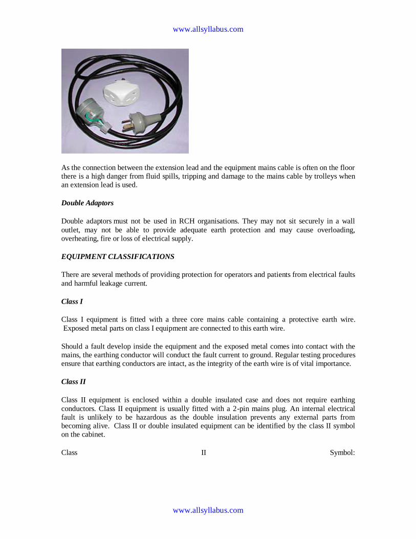
wwwallsyllabuscom
wwwallsyllabuscom
As the connection between the extension lead and the equipment mains cable is often on the floor there is a high danger from fluid spills tripping and damage to the mains cable by trolleys when an extension lead is used
Double Adaptors
Double adaptors must not be used in RCH organisations They may not sit securely in a wall
outlet may not be able to provide adequate earth protection and may cause overloading overheating fire or loss of electrical supply
EQUIPMENT CLASSIFICATIONS
There are several methods of providing protection for operators and patients from electrical faults
and harmful leakage current
Class I
Class I equipment is fitted with a three core mains cable containing a protective earth wire
Exposed metal parts on class I equipment are connected to this earth wire
Should a fault develop inside the equipment and the exposed metal comes into contact with the mains the earthing conductor will conduct the fault current to ground Regular testing procedures ensure that earthing conductors are intact as the integrity of the earth wire is of vital importance
Class II
Class II equipment is enclosed within a double insulated case and does not require earthing
conductors Class II equipment is usually fitted with a 2-pin mains plug An internal electrical fault is unlikely to be hazardous as the double insulation prevents any external parts from becoming alive Class II or double insulated equipment can be identified by the class II symbol on the cabinet
Class II Symbol
wwwallsyllabuscom
wwwallsyllabuscom
Defibrillator-Proof
Some medical equipment within the hospital is classified as defibrillator proof When a
defibrillator is discharged through a patient connected to defibrillator proof equipment the equipment will not be damaged by the defibrillators energy Defibrillator proof equipment can remain connected to the patient during defibrillation It is identified by one of the following
symbols
Defibrillator proof symbols
Body protected Cardiac protected
PROTECTIVE DEVICES
Most patient care areas in the hospital are fitted with protective devices These devices are
regularly tested in accordance with the relevant guidelines published by Standards Australia The level of protection provided is dependent upon the device and the area in which it is located
Residual Current Devices (RCD)
RCDs (safety switches) are used in patient treatment areas to monitor and protect the mains supply RCDs sense leakage currents flowing to earth from the equipment If a significant leakage current flows the RCD will detect it and shut off the power supplied to the equipment
within 40 milliseconds Hospital RCDs are more sensitive than those fitted in homes A hospital RCD will trip at 10 milliamperes leakage current
Power outlets supplied through an RCD have a Supply Available lamp The lamp will extinguish
when the RCD trips due to excessive leakage current
wwwallsyllabuscom
wwwallsyllabuscom
Resetting a RCD
Lamp indicates supply is no longer available Disconnect all equipment from the supply Operate the reset button or lever on the supply panel and the Supply Available lamp
should illuminate If not contact Biomedical Engineering
Connect an item of equipment If the RCD trips again then this is the faulty item and should be labelled and sent to Biomedical Engineering
If the RCD does not trip continue connecting equipment until the RCD trips The last piece of equipment connected to the supply is most likely to be faulty as it will have caused the RCD to trip Remove the faulty item from service label it and send to Biomedical Engineering as mentioned above
Line Isolation overload Monitors (LIMs)
In critical life support applications where loss of power supply cannot be tolerated special power outlets powered by isolation transformers are installed
Line Isolation Monitors are installed to continually monitor electrical leakage in the power
supply system If an electrical fault develops in a medical device connected to an isolated power outlet the LIM will detect the leakage current The LIM will alarm and indicate the level of leakage current but will not shut off the electric supply
The faulty equipment can be identified by un plugging one item of equipment at a time from the supply until the alarm stops sounding Equipment that is not faulty may be reconnected Faulty equipment should be appropriately labelled and sent to Biomedical Engineering for repair
The LIM also monitors how much power is being used by the equpiment connected to it If too much power is being used the LIM will alarm and indicate that there is an overload The power used must be reduced immediately by moving some equipment to another circuit as soon as possible until the alarm stops sounding Failure to reduce the load on the LIM will result in the circuit breaker tripping and loss of power to the circuit
wwwallsyllabuscom
wwwallsyllabuscom
Equipotential Earthing
Equipotential earthing is installed in rooms classified as Cardiac Protected electrical areas
Equipotential earthing in treatment areas used for cardiac procedures is intended to minimise any voltage differences between earthed parts of equipment and any other exposed metal in the room
This reduces the possibility of leakage currents that can cause microelectrocution when the patient comes into contact with multiple items of equipment or if the patient happens to come
into contact with metal items in the room whilr they are connected to a medical device
All conductive metal in an equipotential area is connected to a common equipotential earth point with special heavy duty cable
AREA CLASSIFICATIONS
Body Protected Area
These areas are designed for procedures in which patients are connected to equipment that lowers the natural resistance of the skin Applied parts such as electrode gels conductive fluids entering the patient metal needles and catheters provide an easy pathway for current to flow
The main occurrence of injury from Body-Type procedures is from high current levels causing electric shock A direct connection to the patients heart is not present so the risk of Microelectrocution - fibrillation from minute current levels - is reduced
Residual Current Devices (RCD) or Isolation Transformers and Line Isolation Monitors (LIMiacutes) are used in Body Protected areas to provide protection against electrocution from high leakage currents Body-Protected Areas are identified with this sign
Cardiac Protected Area
Where the procedure involves placing an electrical conductor within or near the heart protection against fibrillation induced from small leakage currents is required Electrical conductors used in these procedures include cardiac pacing electrodes intracardiac ECG electrodes and intracardiac catheters
wwwallsyllabuscom
wwwallsyllabuscom
Equipotential earthing in conjunction with RCDs or LIMs provides protection against microelectrocution in Cardiac-Type procedures
Fault currents are reduced to magnitudes that are unlikely to induce fibrillation Used in conjunction with RCDs or LIMs the magnitude and duration of any fault currents
sourced from equipment are limited Cardiac-Protected Areas are identified with this sign
Other electrical issues
This policy aims to provide guidance to those who find that they need more electrical outlets than
those available or that the existing electrical outlets are inconveniently located
As extension leads and multiple outlet power boards can introduce additional hazards into an area the following procedures should be observed
Extension leads
Approved extension leads (AS 3760 1996) may be used in some areas within the hospital but MUST NOT BE USED IN PATIENT AREAS All electrical extension leads must be tagged with an Engineering Department maintenance tag and require a yearly safety inspection and test via
the Engineering Department
Double adapters
Double adapters may cause overloading or equipment earthing problems and are not to be used in
WCH
Mains extension device
The only mains extension device that is to be used in Patient care areas is the 4-way or 8-way
portable Core Balance Unit
The Biomedical Engineering Department must approve all units prior to use These units contain a safety switch and can detect excessive leakage current and disconnect the power in the event of a hazardous situation
wwwallsyllabuscom
wwwallsyllabuscom
Care must be exercised in the use of a portable Core Balance Unit It should be located off the
floor and in a position that will protect it from physical abuse and possible entry of fluids These devices are expensive and easily damaged The device must be sent to Biomedical Engineering every 6 months for safety testing
Power boards
Approved multiple-outlet power boards can be used across RCH but must not be used in patient
care areas except areas approved by the Biomedical Engineering Department
The power boards must have overload protection be fitted with internal safety shutters that protect unused outlets and be fitted with an onoff switch for each outlet
Medical Laser Applications
The main research subjects of the group of Medical Laser Application are in the field of online
monitoring and diagnostics as well as the development of new therapheutic methods The main focus in research is based upon the use of ultrashort (fs) laser pulses
The main advantage of ultrashort laser pulses is the extrem short interaction time which suppress any unwanted side effects of the laser irradiation of the tissue Secondly the broad spectra of the fs laser pulses give the advantage to use the same laser pulses for diagnostic applications like the optical coherence tomography (OCT)
wwwallsyllabuscom
wwwallsyllabuscom
Following research subjects have special attention at the moment
Optical Coherence Tomographie (OCT)
A fs-laser light source can be used to obtain images from inside the tissue when the pulses are coherent superposed This group uses this application to obtain an inside view of the vocal fold and to measure the outline of the crystalline lens
Refractive Surgery
The principle of getting rid of glasses with the help of a laser surgery (fs-LASIK) is underlying rapid improvements concerning precision and safety New technologies were evaluated and transferred very fast in cooperation with an industrial partner
Treatment of Presbyopia
The flexibility of the crystalline lens can be increase by pricise cuts which are induce by fs laser pulse inside the lens The treatment of the presbyopia is thinkable
wwwallsyllabuscom
wwwallsyllabuscom
Ultrafast Physics
Whereas the applications of ultrashort laser pulses increase rapidly is the physics of the interaction between the laser pulses and tissue in many case not fully understood To obtain improved knowledge of the interaction for many fs-laser application this group preforms numerical simulations as well as fundamental experiments
Thermography
Thermography thermal imaging or thermal video is a type of infrared imaging Thermographic cameras detect radiation in the infrared range of the electromagnetic spectrum
(roughly 900ndash14000 nanometers or 09ndash14 microm) and produce images of that radiation Since infrared radiation is emitted by all objects based on their temperatures according to the black body radiation law thermography makes it possible to see ones environment with or without visible illumination The amount of radiation emitted by an object increases with temperature therefore thermography allows one to see variations in temperature (hence the name) When viewed by thermographic camera warm objects stand out well against cooler backgrounds humans and other warm-blooded animals become easily visible against the environment day or
night As a result thermographys extensive use can historically be ascribed to the military and security services
Thermal imaging photography finds many other uses For example firefighters use it to see through smoke find persons and localize the base of a fire With thermal imaging power lines maintenance technicians locate overheating joints and parts a telltale sign of their failure to eliminate potential hazards Where thermal insulation becomes faulty building construction technicians can see heat leaks to improve the efficiencies of cooling or heating air-conditioning Thermal imaging cameras are also installed in some luxury cars to aid the driver the first being
the 2000 Cadillac DeVille Some physiological activities particularly responses in human beings and other warm-blooded animals can also be monitored with thermographic imaging [1]
The appearance and operation of a modern thermographic camera is often similar to a camcorder Enabling the user to see in the infrared spectrum is a function so useful that ability to record their output is often optional A recording module is therefore not always built-in
Instead of CCD sensors most thermal imaging cameras use CMOS Focal Plane Array (FPA) The most common types are InSb InGaAs HgCdTe and QWIP FPA The newest technologies are using low cost and uncooled microbolometers FPA sensors Their resolution is considerably lower than of optical cameras mostly 160x120 or 320x240 pixels up to 640x512 for the most
expensive models Thermographic cameras are much more expensive than their visible-spectrum counterparts and higher-end models are often export-restricted Older bolometers or more
wwwallsyllabuscom
wwwallsyllabuscom
sensitive models as InSb require cryogenic cooling usually by a miniature Stirling cycle refrigerator or liquid nitrogen
Contents
[hide]
1 Difference between IR film and thermography 2 Advantages of Thermography
3 Limitations amp disadvantages of thermography 4 Applications 5 See also 6 External links
o 61 History of thermal imager manufacturers
[edit] Difference between IR film and thermography
Thermal imaging is going to be used on Mars to detect caves that could hold life
[edit] Advantages of Thermography
You get a visual picture so that you can compare temperatures over a large area
It is real time capable of catching moving targets Able to find deteriorating components prior to failure Measurement in areas inaccessible or hazardous for other methods It is a non-destructive test method
[edit] Limitations amp disadvantages of thermography
Quality cameras are expensive and are easily damaged Images can be hard to interpret accurately even with experience
Accurate temperature measurements are very hard to make because of emissivities Most cameras have plusmn2 or worse accuracy (not as accurate as contact) Training and staying proficient in IR scanning is time consuming Ability to only measure surface areas
[edit] Applications
Condition monitoring Medical imaging Night vision Research
Process control Non destructive testing Surveillance in security law enforcement and defense Chemical imaging
wwwallsyllabuscom
wwwallsyllabuscom
Thermal infrared imagers convert the energy in the infrared wavelength into a visible light video display All objects above 0 kelvins emit thermal infrared energy so thermal imagers can passively see all objects regardless of ambient light However most thermal imagers only see objects warmer than -50 degC
The spectrum and amount of thermal radiation depend strongly on an objects surface
temperature This makes it possible for a thermal camera to display an objects temperature However other factors also influence the radiation which limits the accuracy of this technique For example the radiation depends not only on the temperature of the object but is also a function of the emissivity of the object Also radiation also originates from the surroundings and is reflected in the object and the radiation from the object and the reflected radiation will also be influenced by the absorption of the atmosphere
Endoscopy
This page is about having an endoscopy - a test that looks at the inside of your digestive system
There is information on
What endoscopy is
Why you may have an endoscopy
Having an endoscopy
Your results
What an endoscopy is
An endoscopy is a test that looks inside the body The endoscope is a long flexible tube that can
be swallowed It has a camera and light inside it Some doctors call it a telescope
wwwallsyllabuscom
wwwallsyllabuscom
Why you may have an endoscopy
You are most likely to have an endoscopy to look at the inside of your
Gullet (oesophagus)
Stomach
Duodenum - the first part of the small bowel that attaches to the stomach
Large bowel (colon)
Your doctor may want to see inside because you have symptoms such as abnormal bleeding or
difficulty swallowing The doctor can look down the endoscope and see if there are any growths
or other abnormal looking areas Also through the endoscope the doctor can take samples
(biopsies) of any abnormal looking tissues
There is more detailed information about having a colonoscopy in the bowel cancer section of
CancerHelp UK Below is information about having other types of endoscopy
Having an endoscopy
You can have this test as an out patient Most people have a choice between having the test while
they are awake or after having a medicine to make them drowsy (a sedative) Your hospital may
not be happy for you to have a sedative if you live alone and will have no one to look after you
when you go home If you live alone but really want sedation your hospital may allow you to
stay overnight You should ask your doctor about this as early as possible as extra arrangements
will need to be made beforehand
If you dont have a sedative you will have a spray to numb the back of your throat and make it
easier for you to swallow the endoscopy tube
If you would prefer to be asleep during the test you will have an injection to make you very
drowsy just before the test You will need to take someone with you to the hospital appointment
You wont be able to drive for the rest of the day and should have someone to go home with you
You cant eat or drink for about 8 hours before the test so that your stomach and duodenum are
empty Your doctor will give you written instructions about this beforehand or they may arrive
with your appointment letter When you get to the clinic you may be asked to take your upper
clothing off and put on a hospital gown Some hospitals prefer to use gowns because your clothes
wont get messy Once you are ready you get onto the bed or X-ray couch When you are lying
comfortably you have the sedative injection to make you very drowsy Or your doctor will spray
the back of your throat to numb it
Once the sedative or throat spray has worked the doctor will pass the endoscope tube down your
wwwallsyllabuscom
wwwallsyllabuscom
throat to the area being investigated Your doctor will ask you to swallow as the tube goes down
but if youve had a sedative you wont remember that afterwards If there are any abnormalities
the doctor will take pieces of tissue from the abnormal looking area to send to the laboratory for
closer inspection under a microscope These tissue samples are called biopsies
When the test is over you will need to rest for a while If youve had a sedative you may not
remember much (if anything) about the test once you have come round You should be able to go
home the same day
The results
It can take time for test results to come through How long will depend on why you are having
the test Usually the doctor who carries out the endoscopy dictates a report straight way The
report is typed up by the department secretary and goes to your specialist who gives the results to
you If your GP has sent you for the test the results will go directly to the GP surgery
Understandably waiting for results can make you anxious It usually takes a couple of weeks for
the results to come through If your doctor needed them urgently it would have been noted on
the test request form and the results will be ready sooner than that Try to remember to ask your
doctor how long you should expect to wait for the results when you are first asked to go for the
test If it is not an emergency and you have not heard a couple of weeks after your test ring your
doctors secretary to check if they are back
Endoscopy
From Wikipedia the free encyclopedia
Jump to navigation search
Endoscopic images of a duodenal ulcer
wwwallsyllabuscom
wwwallsyllabuscom
A flexible endoscope
Endoscopy means looking inside and typically refers to looking inside the human body for medical reasons using an instrument called an endoscope Endoscopy can also refer to using a borescope in technical situations where direct line-of-sight observation is not feasible
Contents
[hide]
1 Overview 2 Components 3 Uses
4 History 5 Risks 6 After The Endoscopy 7 Recent developments 8 See also 9 References 10 Footnotes
11 External links
Overview
Endoscopy is a minimally invasive diagnostic medical procedure that is used to assess the interior surfaces of an organ by inserting a tube into the body The instrument may have a rigid or flexible tube and not only provide an image for visual inspection and photography but also enable taking
biopsies and retrieval of foreign objects Endoscopy is the vehicle for minimally invasive surgery
Many endoscopic procedures are considered to be relatively painless and at worst associated with mild discomfort for example in esophagogastroduodenoscopy most patients tolerate the procedure with only topical anaesthesia of the oropharynx using lignocaine spray[1] Complications are not common (only 5 of all operations)[citation needed] but can include perforation of the organ under inspection with the endoscope or biopsy instrument If that occurs open surgery may be required to repair the injury
wwwallsyllabuscom
wwwallsyllabuscom
Components
An endoscope can consist of
a rigid or flexible tube a light delivery system to illuminate the organ or object under inspection The light
source is normally outside the body and the light is typically directed via an optical fiber system
a lens system transmitting the image to the viewer from the fiberscope an additional channel to allow entry of medical instruments or manipulators
Uses
Endoscopy can involve
The gastrointestinal tract (GI tract) o esophagus stomach and duodenum (esophagogastroduodenoscopy) o small intestine o colon (colonoscopyproctosigmoidoscopy) o Bile duct
endoscopic retrograde cholangiopancreatography (ERCP) duodenoscope-assisted cholangiopancreatoscopy intraoperative cholangioscopy
The respiratory tract o The nose (rhinoscopy) o The lower respiratory tract (bronchoscopy)
The urinary tract (cystoscopy) The female reproductive system
o The cervix (colposcopy) o The uterus (hysteroscopy) o The Fallopian tubes (Falloscopy)
Normally closed body cavities (through a small incision) o The abdominal or pelvic cavity (laparoscopy) o The interior of a joint (arthroscopy) o Organs of the chest (thoracoscopy and mediastinoscopy)
During pregnancy o The amnion (amnioscopy) o The fetus (fetoscopy)
Plastic Surgery Panendoscopy (or triple endoscopy)
o Combines laryngoscopy esophagoscopy and bronchoscopy Non-medical uses for endoscopy
o The planning and architectural community have found the endoscope useful for
pre-visualization of scale models of proposed buildings and cities (architectural endoscopy)
o Internal inspection of complex technical systems (borescope) o Endoscopes are also a tool helpful in the examination of improvised explosive
devices by bomb disposal personnel
wwwallsyllabuscom
wwwallsyllabuscom
o The FBI uses endoscopes for conducting surveillance via tight spaces
History
The first endoscope of a kind was developed in 1806 by Philip Bozzini with his introduction of a
Lichtleiter (light conductor) for the examinations of the canals and cavities of the human body However the Vienna Medical Society disapproved of such curiosity An endoscope was first introduced into a human in 1822 by William Beaumont an army surgeon at Mackinac
Island Michigan[citation needed] The use of electric light was a major step in the improvement of endoscopy The first such lights were external Later smaller bulbs became available making internal light possible for instance in a hysteroscope by Charles David in 1908[citation needed] Hans Christian Jacobaeus has been given credit for early endoscopic explorations of the abdomen and the thorax with laparoscopy (1912) and thoracoscopy (1910)[citation needed] Laparoscopy was used in the diagnosis of liver and gallbladder disease by Heinz Kalk in the 1930s[citation needed] Hope reported in 1937 on the use of laparoscopy to diagnose ectopic pregnancy[citation needed] In 1944
Raoul Palmer placed his patients in the Trendelenburg position after gaseous distention of the abdomen and thus was able to reliably perform gynecologic laparoscopy[citation needed]
The first gastrocamera was released in 1950 by Olympus Optical Co Ltd The device took pictures on monochromatic film using a small light bulb that was triggered manually The device was of limited use however because it did not implement real-time optical capability Olympus continued its development of endoscopes by incorporating fiber optics in the early 1960s leading to the first useful endoscopes In 1964 it released a gastrocamera guided by a fiberscope[1] A few articles claim that DrBasil Hirschowitz of UnivOf MichiganAnn Arbor discussed the
endoscope in early 50s[2]
As endoscopic technology improved so did the methods of gastrointestinal endoscopy Owing primarily to the efforts of Dr Hiromi Shinya in the late 1960s GI endoscopy developed into what is more recognizable as todays colonoscopy While many doctors experimented with techniques to take advantage of the new iterations of endoscopes Dr Shinya focused on techniques that would allow for successful operation of the endoscope by an individual rejecting the common practice at the time of utilizing two people Consequently many of the fundamental methods and procedures of modern colonoscopy were developed by Dr Shinya
Dr Shinyas other great contribution was to therapeutic endoscopy in his invention of the
electrosurgical polypectomy snare with the aid of Olympus employee Hiroshi Ichikawa Shinya sketched his first plans for the device on January 8 1969 He envisioned a loop of wire attached to the end of a colonoscope that would allow for easy removal of polyps during investigation by passing a current through the wire By September of 1969 the first polypectomy using this device was performed Polypectomy has since become the most common therapeutic procedure performed with an endoscope (Sivak 2004)
By 1980 laparoscopy training was required by gynecologists to perform tubal ligation procedures and diagnostic evaluations of the pelvis The first laparoscopic cholecystectomy was performed in 1984 and the first video-laparoscopic cholecystectomy in 1987[citation needed] During the 1990s
laparoscopic surgery was extended to the appendix spleen colon stomach kidney and liver[citation needed] Wireless capsule endoscopy or Capsule Endoscopy is now approved in all the countries including Japan where government reimbusement will be available from Oct2007Capsule Endoscopy [3] increases detection of Small Bowel tumors where traditional Endoscopy is not very efficient
wwwallsyllabuscom
wwwallsyllabuscom
Risks
Infection
Punctured organs Allergic reactions due to Contrast agents or dyes (such as those used in a CT scan) Over-sedation
After The Endoscopy
After the procedure the patient will be observed and monitored by a qualified individual in the
endoscopy or a recovery area until a significant portion of the medication has worn off Occasionally a patient is left with a mild sore throat which promptly responds to saline gargles or a feeling of distention from the insufflated air that was used during the procedure Both problems are mild and fleeting When fully recovered the patient will be instructed when to resume hisher usual diet (probably within a few hours) and will be allowed to be taken home Because of the use of sedation most facilities mandate that the patient is taken home by another person and not to drive on hisher own or handle machinery for the remainder of the day
Recent developments
With the application of robotic systems telesurgery was introduced as the surgeon could operate
from a site physically removed from the patient The first transatlantic surgery has been called the Lindbergh Operation
wwwallsyllabuscom
wwwallsyllabuscom
Upper Endoscopy
Upper endoscopy enables the physician to look inside the esophagus stomach and duodenum (first part of the small intestine) The procedure might be used to discover the reason for swallowing difficulties nausea vomiting
reflux bleeding indigestion abdominal pain or chest pain Upper endoscopy is also called EGD which stands for esophagogastroduodenoscopy (eh-SAH-fuh-goh-GAS-troh-doo-AH-duh-NAH-skuh-pee)
For the procedure you will swallow a thin flexible lighted tube called an endoscope (EN-doh-skope) Right before the procedure the physician will spray your throat with a numbing agent that may help prevent gagging
You may also receive pain medicine and a sedative to help you relax during the exam The endoscope transmits an image of the inside of the esophagus stomach and duodenum so the physician can carefully examine the lining of these organs The scope also blows air into the stomach this expands the folds of tissue and makes it easier for the physician to examine the stomach
The physician can see abnormalities like inflammation or
bleeding through the endoscope that dont show up well on x rays The physician can also insert instruments into the scope to treat bleeding abnormalities or remove samples of tissue (biopsy) for further tests
Possible complications of upper endoscopy include bleeding and puncture of the stomach lining However such complications are rare Most people will probably have nothing more than a mild sore throat after the procedure
The procedure takes 20 to 30 minutes Because you will be sedated you will need to rest at the endoscopy facility for 1 to 2 hours until the medication wears off
Preparation
Your stomach and duodenum must be empty for the procedure to be thorough and safe so you will not be able to eat or drink anything for at least 6 hours beforehand Also you must
arrange for someone to take you homemdashyou will not be allowed to drive because of the sedatives Your physician may give you other special instructions
The digestive system
wwwallsyllabuscom
wwwallsyllabuscom
What is Endoscopy and Why is it Performed
Endoscopy allows physicians to peer through the bodys passageways Endoscopy is the examination and inspection of the interior of body organs joints or cavities through an endoscope An endoscope is a device that uses fiber optics and powerful lens systems to provide lighting and visualization of the interior of a joint The portion of the endoscope inserted into the body may be rigid or flexible depending upon the medical procedure
An endoscope uses two fiber optic lines A light fiber carries light into the body cavity and an image fiber carries the image of the body cavity back to the physicians viewing lens There is also a separate port to allow for administration of drugs suction and irrigation This port may also be used to introduce small folding instruments such as forceps scissors brushes snares and baskets for tissue excision (removal) sampling or other diagnostic and therapeutic work Endoscopes may be used in conjunction with a camera or video recorder to document images of the inside of the joint or chronicle an endoscopic procedure New endoscopes have digital capabilities for manipulating and enhancing the video images
This figure shows a rigid endoscope used for arthroscopy The image fiber leads from the ocular (eye piece) to the inserted end of the scope The light fiber is below and leads from the light source to the working end of the endoscope
Why Is Endoscopy Performed
Endoscopy can be used to diagnose various conditions by close examination of internal organ and body structures Endoscopy can also guide therapy and repair such as the removal of torn cartilage from the bearing surfaces of a joint Biopsy (tissue sampling for pathologic testing) may also be performed under endoscopic guidance Local or general anesthetic may be used during endoscopy depending upon the type of procedure being performed
Internal abnormalities revealed through endoscopy include abscesses biliary (liver) cirrhosis bleeding bronchitis cancer cysts degenerative disease gallbladder stones hernia inflammation metastatic cancer polyps tumors ulcers and other diseases and conditions
Endoscopy is a minimally invasive procedure and carries with it certain minor risks depending upon the type of procedure being performed However these risks are typically far outweighed by the diagnostic and therapeutic potential of the procedure
Prior to the widespread use of endoscopy and diagnostic imaging most internal conditions could only be diagnosed or treated with open surgery Until the last several decades exploratory surgery was routinely performed only when a patient was critically ill and the source of illness was not known For example in certain dire cases the patients thorax or abdomen were surgically opened and examined to try to determine the source of illness
wwwallsyllabuscom
wwwallsyllabuscom
Endoscopy can often be done on an outpatient basis Outpatient means that the procedure does not require hospital admission and acute care and observation and may be performed outside the premises of a hospital Outpatient procedures performed at hospitals or ambulatory centers allow the patient to go home or return to work within a short while after their procedure
Types of Endoscopy
Fiber optic endoscopes now have widespread use in medicine and guide a myriad of diagnostic and therapeutic procedures including
Arthroscopy examination of joints for diagnosis and treatment (arthroscopic surgery) Bronchoscopy examination of the trachea and lungs bronchial trees to reveal
abscesses bronchitis carcinoma tumors tuberculosis alveolitis infection inflammation Colonoscopy examination of the inside of the colon and large intestine to detect polyps
tumors ulceration inflammation colitis diverticula Chrohns disease and discovery and removal of foreign bodies
Colposcopy direct visualization of the vagina and cervix to detect cancer inflammation and other conditions
Cystoscopy examination of the bladder urethra urinary tract uteral orifices and
prostate (men) with insertion of the endoscope through the urethra
ERCP (endoscopic retrograde cholangio-pancreatography) uses endoscopic guidance to
place a catheter for x-ray fluorosocopy with contrast enhancement This technique is used to examine the livers biliary tree the gallbladder the pancreatic duct and other anatomy to check for stones other obstructions and disease X-ray contrast is introduced into these ducts via catheter and fluoroscopic x-ray images are taken to show any abnormality or blockage If disease is detected it can sometimes be treated at the same time or biopsy can be performed to test for cancer or other pathology ERCP can detect biliary cirrhosis
cancer of the bile ducts pancreatic cysts pseudocysts pancreatic tumors chronic pancreatitis and other conditions such as gallbladder stones
EGD (Esophogealgastroduodensoscopy) visual examination of the upper gastro-
intestinal (GI) tract (also referred to as gastroscopy) to reveal hemorrhage hiatal hernia inflammation of the esophagus gastric ulcers
Endoscopic biopsy is the removal of tissue specimens for pathologic examination and
analysis Gastroscopy examination of the lining of the esophagus stomach and duodenum
Gastroscopy is often used to diagnose ulcers and other sources of bleeding and to guide biopsy of suspect GI cancers
Laparoscopy visualization of the stomach liver and other abdominal organs including
the female reproductive organs for example the fallopian tubes Laryngoscopy examination of the larynx (voice box) Proctoscopy sigmoidoscopy proctosigmoidoscopy examination of the rectum and
sigmoid colon Thoracoscopy examination of the pleura (sac that covers the lungs) pleural spaces
mediastinum and pericardium
A Brief History of Endoscopy
In the early 1900s the first attempts to view inside the body with lighted telescopes were made These initial devices were often fully rigid In the 1930s semi-flexible endoscopes called gastroscopes were developed to view inside of the stomach Fiber-optic endoscopy was pioneered by South African-born physician Basil Hirschowitz at the University of Michigan in 1957 Widespread use of fiber optic endoscopes began in the 1960s
wwwallsyllabuscom
wwwallsyllabuscom
A fiber optic cable is simply a bundle of microscopic glass or plastic fibers that literally allows light and images to be transmitted through curved structures Fiber optic cables are also replacing metal wires as the backbone of the worlds telecommunications infrastructure This Internet page may have traveled through a fiber optic cable as a stream of digital data (bursts of light) on its way to your computer
Endoscopy Equipment
Endoscopes have many practical needs And HMB Endoscopy Products (Hollywood Florida) has been providing endoscopic equipment and educating people on the use of endoscopes for more than 17 years Be sure to Browse our Catalog for all the details on how to purchase these medical instruments that can examine any part of the body
Video
Systems
Fiber
Endoscopes
Video
Endoscopes
In the simplest terms Endoscopy equipment consists of instruments that can look at the inside of many different organs mdash these are small flexible or rigid tubes with a light or lenses on the end that can look into the esophagus stomach and colon mdash and in more general terms endoscopy equipment can help doctors look deep inside body structures and hollow organs
An endoscope and related endoscope products and equipment are usually composed of three components
An optic system that allows the doctor to look through the scope into the organ or
cavity or to attach a video camera to the scope
A fiberoptic cable to light up the bodily area
A lumen (eg the bore of a tube like a needle or catheter) to take tissue samples of the
area being viewed
The beauty of endoscopic products is that they perform dual functions mdash with both diagnostic and therapeutic capabilities For example this means that these endoscopic products and instruments can perform biopsies (eg to evaluate tissue samples) as well as provide sclerotherapy (a medical procedure used to treat varicose veins and ―spider veins) In truth these brief explanations only tell part of the story
wwwallsyllabuscom
wwwallsyllabuscom
TYPES OF ENDOSCOPES
Take a look at the different types of endoscopic equipment you can get with HMB Endoscopy Products Again in very general terms there are two main types of endoscopes
Rigid Flexible
1 rigid endoscopes ndash the majority of which use a convex (curving out like one half of a
circle) glass lens system in which the small glass lenses are separated by large air spaces
2 flexible endoscopes allow for just that mdash flexibility In the animal kingdom for example
a flexible endoscope would be perfect for examining the stomach area of a snake
The popularity of endoscopy equipment continues to grow Some of the industryrsquos household names include Olympus Pentax Funjinon and Storz mdash with HMB both selling and repairing pre-owned and completely refurbished endoscopic products from each of these major manufacturers Browse our Catalog to get the ones you need
THERMOGRAPH APPLICATION Thermographs are digital recording thermometers used to log
temperature in the
marine environment The instruments are attached to a computer to set up recording
parameters for
deployment downloading and display of data in graphic or numeric format These units
are placed in
underwater housings and attached to the bottom or suspended in the water column for
deployment
DESCRIPTION Two types of thermograph are currently available The Ryan
Tempmentor is a
reusable data logger that allows storage of a maximum of 6361 temperature
measurements in the
range of -32C to +70C with 01resolution and 03accuracy The instrument can be
programmed
to take measurements from once per second to once every other hour Instrument
dimensions are 3 x
6 x 13 and the unit weighs 11 ounces The Hobo Temp is a miniature reusable data
logger that
allows storage of 1800 measurements in the range of -20C to +70C with
03resolution and 07
wwwallsyllabuscom
wwwallsyllabuscom
accuracy Sampling intervals from 05 seconds to 48 hours are available Instrument
dimensions are
24 x 19 x 08 and the unit weighs 1 ounce Both units are battery powered with the
battery life of
the Tempmentor being approximately two years and the battery life of the Hobo Temp
being one year THERMOGRAPH APPLICATION Thermographs are digital recording thermometers used to log
temperature in the
marine environment The instruments are attached to a computer to set up recording
parameters for
deployment downloading and display of data in graphic or numeric format These units
are placed in
underwater housings and attached to the bottom or suspended in the water column for
deployment
DESCRIPTION Two types of thermograph are currently available The Ryan
Tempmentor is a
reusable data logger that allows storage of a maximum of 6361 temperature
measurements in the
range of -32C to +70C with 01resolution and 03accuracy The instrument can be
programmed
to take measurements from once per second to once every other hour Instrument
dimensions are 3 x
6 x 13 and the unit weighs 11 ounces The Hobo Temp is a miniature reusable data
logger that
allows storage of 1800 measurements in the range of -20C to +70C with
03resolution and 07
accuracy Sampling intervals from 05 seconds to 48 hours are available Instrument
dimensions are
24 x 19 x 08 and the unit weighs 1 ounce Both units are battery powered with the
battery life of
the Tempmentor being approximately two years and the battery life of the Hobo Temp
being one year THERMOGRAPH APPLICATION Thermographs are digital recording thermometers used to log
temperature in the
marine environment The instruments are attached to a computer to set up recording
parameters for
deployment downloading and display of data in graphic or numeric format These units
are placed in
underwater housings and attached to the bottom or suspended in the water column for
deployment
DESCRIPTION Two types of thermograph are currently available The Ryan
Tempmentor is a
reusable data logger that allows storage of a maximum of 6361 temperature
measurements in the
range of -32C to +70C with 01resolution and 03accuracy The instrument can be
programmed
wwwallsyllabuscom
wwwallsyllabuscom
to take measurements from once per second to once every other hour Instrument
dimensions are 3 x
6 x 13 and the unit weighs 11 ounces The Hobo Temp is a miniature reusable data
logger that
allows storage of 1800 measurements in the range of -20C to +70C with
03resolution and 07
accuracy Sampling intervals from 05 seconds to 48 hours are available Instrument
dimensions are
24 x 19 x 08 and the unit weighs 1 ounce Both units are battery powered with the
battery life of
the Tempmentor being approximately two years and the battery life of the Hobo Temp
being one year
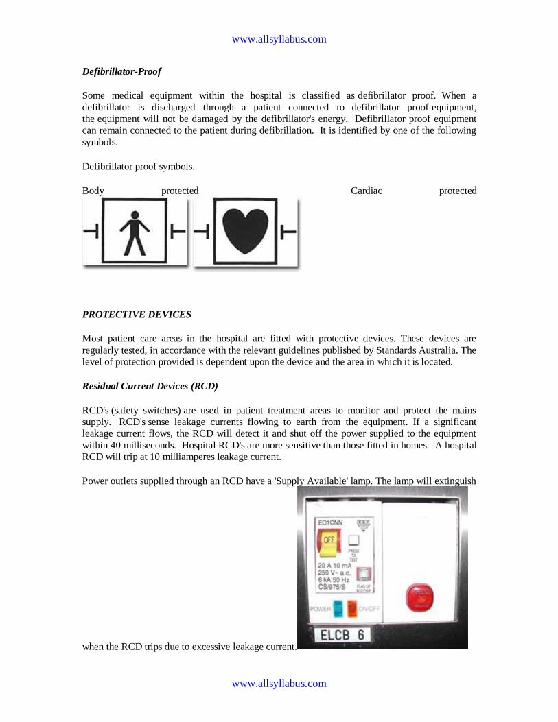
wwwallsyllabuscom
wwwallsyllabuscom
Defibrillator-Proof
Some medical equipment within the hospital is classified as defibrillator proof When a
defibrillator is discharged through a patient connected to defibrillator proof equipment the equipment will not be damaged by the defibrillators energy Defibrillator proof equipment can remain connected to the patient during defibrillation It is identified by one of the following
symbols
Defibrillator proof symbols
Body protected Cardiac protected
PROTECTIVE DEVICES
Most patient care areas in the hospital are fitted with protective devices These devices are
regularly tested in accordance with the relevant guidelines published by Standards Australia The level of protection provided is dependent upon the device and the area in which it is located
Residual Current Devices (RCD)
RCDs (safety switches) are used in patient treatment areas to monitor and protect the mains supply RCDs sense leakage currents flowing to earth from the equipment If a significant leakage current flows the RCD will detect it and shut off the power supplied to the equipment
within 40 milliseconds Hospital RCDs are more sensitive than those fitted in homes A hospital RCD will trip at 10 milliamperes leakage current
Power outlets supplied through an RCD have a Supply Available lamp The lamp will extinguish
when the RCD trips due to excessive leakage current
wwwallsyllabuscom
wwwallsyllabuscom
Resetting a RCD
Lamp indicates supply is no longer available Disconnect all equipment from the supply Operate the reset button or lever on the supply panel and the Supply Available lamp
should illuminate If not contact Biomedical Engineering
Connect an item of equipment If the RCD trips again then this is the faulty item and should be labelled and sent to Biomedical Engineering
If the RCD does not trip continue connecting equipment until the RCD trips The last piece of equipment connected to the supply is most likely to be faulty as it will have caused the RCD to trip Remove the faulty item from service label it and send to Biomedical Engineering as mentioned above
Line Isolation overload Monitors (LIMs)
In critical life support applications where loss of power supply cannot be tolerated special power outlets powered by isolation transformers are installed
Line Isolation Monitors are installed to continually monitor electrical leakage in the power
supply system If an electrical fault develops in a medical device connected to an isolated power outlet the LIM will detect the leakage current The LIM will alarm and indicate the level of leakage current but will not shut off the electric supply
The faulty equipment can be identified by un plugging one item of equipment at a time from the supply until the alarm stops sounding Equipment that is not faulty may be reconnected Faulty equipment should be appropriately labelled and sent to Biomedical Engineering for repair
The LIM also monitors how much power is being used by the equpiment connected to it If too much power is being used the LIM will alarm and indicate that there is an overload The power used must be reduced immediately by moving some equipment to another circuit as soon as possible until the alarm stops sounding Failure to reduce the load on the LIM will result in the circuit breaker tripping and loss of power to the circuit
wwwallsyllabuscom
wwwallsyllabuscom
Equipotential Earthing
Equipotential earthing is installed in rooms classified as Cardiac Protected electrical areas
Equipotential earthing in treatment areas used for cardiac procedures is intended to minimise any voltage differences between earthed parts of equipment and any other exposed metal in the room
This reduces the possibility of leakage currents that can cause microelectrocution when the patient comes into contact with multiple items of equipment or if the patient happens to come
into contact with metal items in the room whilr they are connected to a medical device
All conductive metal in an equipotential area is connected to a common equipotential earth point with special heavy duty cable
AREA CLASSIFICATIONS
Body Protected Area
These areas are designed for procedures in which patients are connected to equipment that lowers the natural resistance of the skin Applied parts such as electrode gels conductive fluids entering the patient metal needles and catheters provide an easy pathway for current to flow
The main occurrence of injury from Body-Type procedures is from high current levels causing electric shock A direct connection to the patients heart is not present so the risk of Microelectrocution - fibrillation from minute current levels - is reduced
Residual Current Devices (RCD) or Isolation Transformers and Line Isolation Monitors (LIMiacutes) are used in Body Protected areas to provide protection against electrocution from high leakage currents Body-Protected Areas are identified with this sign
Cardiac Protected Area
Where the procedure involves placing an electrical conductor within or near the heart protection against fibrillation induced from small leakage currents is required Electrical conductors used in these procedures include cardiac pacing electrodes intracardiac ECG electrodes and intracardiac catheters
wwwallsyllabuscom
wwwallsyllabuscom
Equipotential earthing in conjunction with RCDs or LIMs provides protection against microelectrocution in Cardiac-Type procedures
Fault currents are reduced to magnitudes that are unlikely to induce fibrillation Used in conjunction with RCDs or LIMs the magnitude and duration of any fault currents
sourced from equipment are limited Cardiac-Protected Areas are identified with this sign
Other electrical issues
This policy aims to provide guidance to those who find that they need more electrical outlets than
those available or that the existing electrical outlets are inconveniently located
As extension leads and multiple outlet power boards can introduce additional hazards into an area the following procedures should be observed
Extension leads
Approved extension leads (AS 3760 1996) may be used in some areas within the hospital but MUST NOT BE USED IN PATIENT AREAS All electrical extension leads must be tagged with an Engineering Department maintenance tag and require a yearly safety inspection and test via
the Engineering Department
Double adapters
Double adapters may cause overloading or equipment earthing problems and are not to be used in
WCH
Mains extension device
The only mains extension device that is to be used in Patient care areas is the 4-way or 8-way
portable Core Balance Unit
The Biomedical Engineering Department must approve all units prior to use These units contain a safety switch and can detect excessive leakage current and disconnect the power in the event of a hazardous situation
wwwallsyllabuscom
wwwallsyllabuscom
Care must be exercised in the use of a portable Core Balance Unit It should be located off the
floor and in a position that will protect it from physical abuse and possible entry of fluids These devices are expensive and easily damaged The device must be sent to Biomedical Engineering every 6 months for safety testing
Power boards
Approved multiple-outlet power boards can be used across RCH but must not be used in patient
care areas except areas approved by the Biomedical Engineering Department
The power boards must have overload protection be fitted with internal safety shutters that protect unused outlets and be fitted with an onoff switch for each outlet
Medical Laser Applications
The main research subjects of the group of Medical Laser Application are in the field of online
monitoring and diagnostics as well as the development of new therapheutic methods The main focus in research is based upon the use of ultrashort (fs) laser pulses
The main advantage of ultrashort laser pulses is the extrem short interaction time which suppress any unwanted side effects of the laser irradiation of the tissue Secondly the broad spectra of the fs laser pulses give the advantage to use the same laser pulses for diagnostic applications like the optical coherence tomography (OCT)
wwwallsyllabuscom
wwwallsyllabuscom
Following research subjects have special attention at the moment
Optical Coherence Tomographie (OCT)
A fs-laser light source can be used to obtain images from inside the tissue when the pulses are coherent superposed This group uses this application to obtain an inside view of the vocal fold and to measure the outline of the crystalline lens
Refractive Surgery
The principle of getting rid of glasses with the help of a laser surgery (fs-LASIK) is underlying rapid improvements concerning precision and safety New technologies were evaluated and transferred very fast in cooperation with an industrial partner
Treatment of Presbyopia
The flexibility of the crystalline lens can be increase by pricise cuts which are induce by fs laser pulse inside the lens The treatment of the presbyopia is thinkable
wwwallsyllabuscom
wwwallsyllabuscom
Ultrafast Physics
Whereas the applications of ultrashort laser pulses increase rapidly is the physics of the interaction between the laser pulses and tissue in many case not fully understood To obtain improved knowledge of the interaction for many fs-laser application this group preforms numerical simulations as well as fundamental experiments
Thermography
Thermography thermal imaging or thermal video is a type of infrared imaging Thermographic cameras detect radiation in the infrared range of the electromagnetic spectrum
(roughly 900ndash14000 nanometers or 09ndash14 microm) and produce images of that radiation Since infrared radiation is emitted by all objects based on their temperatures according to the black body radiation law thermography makes it possible to see ones environment with or without visible illumination The amount of radiation emitted by an object increases with temperature therefore thermography allows one to see variations in temperature (hence the name) When viewed by thermographic camera warm objects stand out well against cooler backgrounds humans and other warm-blooded animals become easily visible against the environment day or
night As a result thermographys extensive use can historically be ascribed to the military and security services
Thermal imaging photography finds many other uses For example firefighters use it to see through smoke find persons and localize the base of a fire With thermal imaging power lines maintenance technicians locate overheating joints and parts a telltale sign of their failure to eliminate potential hazards Where thermal insulation becomes faulty building construction technicians can see heat leaks to improve the efficiencies of cooling or heating air-conditioning Thermal imaging cameras are also installed in some luxury cars to aid the driver the first being
the 2000 Cadillac DeVille Some physiological activities particularly responses in human beings and other warm-blooded animals can also be monitored with thermographic imaging [1]
The appearance and operation of a modern thermographic camera is often similar to a camcorder Enabling the user to see in the infrared spectrum is a function so useful that ability to record their output is often optional A recording module is therefore not always built-in
Instead of CCD sensors most thermal imaging cameras use CMOS Focal Plane Array (FPA) The most common types are InSb InGaAs HgCdTe and QWIP FPA The newest technologies are using low cost and uncooled microbolometers FPA sensors Their resolution is considerably lower than of optical cameras mostly 160x120 or 320x240 pixels up to 640x512 for the most
expensive models Thermographic cameras are much more expensive than their visible-spectrum counterparts and higher-end models are often export-restricted Older bolometers or more
wwwallsyllabuscom
wwwallsyllabuscom
sensitive models as InSb require cryogenic cooling usually by a miniature Stirling cycle refrigerator or liquid nitrogen
Contents
[hide]
1 Difference between IR film and thermography 2 Advantages of Thermography
3 Limitations amp disadvantages of thermography 4 Applications 5 See also 6 External links
o 61 History of thermal imager manufacturers
[edit] Difference between IR film and thermography
Thermal imaging is going to be used on Mars to detect caves that could hold life
[edit] Advantages of Thermography
You get a visual picture so that you can compare temperatures over a large area
It is real time capable of catching moving targets Able to find deteriorating components prior to failure Measurement in areas inaccessible or hazardous for other methods It is a non-destructive test method
[edit] Limitations amp disadvantages of thermography
Quality cameras are expensive and are easily damaged Images can be hard to interpret accurately even with experience
Accurate temperature measurements are very hard to make because of emissivities Most cameras have plusmn2 or worse accuracy (not as accurate as contact) Training and staying proficient in IR scanning is time consuming Ability to only measure surface areas
[edit] Applications
Condition monitoring Medical imaging Night vision Research
Process control Non destructive testing Surveillance in security law enforcement and defense Chemical imaging
wwwallsyllabuscom
wwwallsyllabuscom
Thermal infrared imagers convert the energy in the infrared wavelength into a visible light video display All objects above 0 kelvins emit thermal infrared energy so thermal imagers can passively see all objects regardless of ambient light However most thermal imagers only see objects warmer than -50 degC
The spectrum and amount of thermal radiation depend strongly on an objects surface
temperature This makes it possible for a thermal camera to display an objects temperature However other factors also influence the radiation which limits the accuracy of this technique For example the radiation depends not only on the temperature of the object but is also a function of the emissivity of the object Also radiation also originates from the surroundings and is reflected in the object and the radiation from the object and the reflected radiation will also be influenced by the absorption of the atmosphere
Endoscopy
This page is about having an endoscopy - a test that looks at the inside of your digestive system
There is information on
What endoscopy is
Why you may have an endoscopy
Having an endoscopy
Your results
What an endoscopy is
An endoscopy is a test that looks inside the body The endoscope is a long flexible tube that can
be swallowed It has a camera and light inside it Some doctors call it a telescope
wwwallsyllabuscom
wwwallsyllabuscom
Why you may have an endoscopy
You are most likely to have an endoscopy to look at the inside of your
Gullet (oesophagus)
Stomach
Duodenum - the first part of the small bowel that attaches to the stomach
Large bowel (colon)
Your doctor may want to see inside because you have symptoms such as abnormal bleeding or
difficulty swallowing The doctor can look down the endoscope and see if there are any growths
or other abnormal looking areas Also through the endoscope the doctor can take samples
(biopsies) of any abnormal looking tissues
There is more detailed information about having a colonoscopy in the bowel cancer section of
CancerHelp UK Below is information about having other types of endoscopy
Having an endoscopy
You can have this test as an out patient Most people have a choice between having the test while
they are awake or after having a medicine to make them drowsy (a sedative) Your hospital may
not be happy for you to have a sedative if you live alone and will have no one to look after you
when you go home If you live alone but really want sedation your hospital may allow you to
stay overnight You should ask your doctor about this as early as possible as extra arrangements
will need to be made beforehand
If you dont have a sedative you will have a spray to numb the back of your throat and make it
easier for you to swallow the endoscopy tube
If you would prefer to be asleep during the test you will have an injection to make you very
drowsy just before the test You will need to take someone with you to the hospital appointment
You wont be able to drive for the rest of the day and should have someone to go home with you
You cant eat or drink for about 8 hours before the test so that your stomach and duodenum are
empty Your doctor will give you written instructions about this beforehand or they may arrive
with your appointment letter When you get to the clinic you may be asked to take your upper
clothing off and put on a hospital gown Some hospitals prefer to use gowns because your clothes
wont get messy Once you are ready you get onto the bed or X-ray couch When you are lying
comfortably you have the sedative injection to make you very drowsy Or your doctor will spray
the back of your throat to numb it
Once the sedative or throat spray has worked the doctor will pass the endoscope tube down your
wwwallsyllabuscom
wwwallsyllabuscom
throat to the area being investigated Your doctor will ask you to swallow as the tube goes down
but if youve had a sedative you wont remember that afterwards If there are any abnormalities
the doctor will take pieces of tissue from the abnormal looking area to send to the laboratory for
closer inspection under a microscope These tissue samples are called biopsies
When the test is over you will need to rest for a while If youve had a sedative you may not
remember much (if anything) about the test once you have come round You should be able to go
home the same day
The results
It can take time for test results to come through How long will depend on why you are having
the test Usually the doctor who carries out the endoscopy dictates a report straight way The
report is typed up by the department secretary and goes to your specialist who gives the results to
you If your GP has sent you for the test the results will go directly to the GP surgery
Understandably waiting for results can make you anxious It usually takes a couple of weeks for
the results to come through If your doctor needed them urgently it would have been noted on
the test request form and the results will be ready sooner than that Try to remember to ask your
doctor how long you should expect to wait for the results when you are first asked to go for the
test If it is not an emergency and you have not heard a couple of weeks after your test ring your
doctors secretary to check if they are back
Endoscopy
From Wikipedia the free encyclopedia
Jump to navigation search
Endoscopic images of a duodenal ulcer
wwwallsyllabuscom
wwwallsyllabuscom
A flexible endoscope
Endoscopy means looking inside and typically refers to looking inside the human body for medical reasons using an instrument called an endoscope Endoscopy can also refer to using a borescope in technical situations where direct line-of-sight observation is not feasible
Contents
[hide]
1 Overview 2 Components 3 Uses
4 History 5 Risks 6 After The Endoscopy 7 Recent developments 8 See also 9 References 10 Footnotes
11 External links
Overview
Endoscopy is a minimally invasive diagnostic medical procedure that is used to assess the interior surfaces of an organ by inserting a tube into the body The instrument may have a rigid or flexible tube and not only provide an image for visual inspection and photography but also enable taking
biopsies and retrieval of foreign objects Endoscopy is the vehicle for minimally invasive surgery
Many endoscopic procedures are considered to be relatively painless and at worst associated with mild discomfort for example in esophagogastroduodenoscopy most patients tolerate the procedure with only topical anaesthesia of the oropharynx using lignocaine spray[1] Complications are not common (only 5 of all operations)[citation needed] but can include perforation of the organ under inspection with the endoscope or biopsy instrument If that occurs open surgery may be required to repair the injury
wwwallsyllabuscom
wwwallsyllabuscom
Components
An endoscope can consist of
a rigid or flexible tube a light delivery system to illuminate the organ or object under inspection The light
source is normally outside the body and the light is typically directed via an optical fiber system
a lens system transmitting the image to the viewer from the fiberscope an additional channel to allow entry of medical instruments or manipulators
Uses
Endoscopy can involve
The gastrointestinal tract (GI tract) o esophagus stomach and duodenum (esophagogastroduodenoscopy) o small intestine o colon (colonoscopyproctosigmoidoscopy) o Bile duct
endoscopic retrograde cholangiopancreatography (ERCP) duodenoscope-assisted cholangiopancreatoscopy intraoperative cholangioscopy
The respiratory tract o The nose (rhinoscopy) o The lower respiratory tract (bronchoscopy)
The urinary tract (cystoscopy) The female reproductive system
o The cervix (colposcopy) o The uterus (hysteroscopy) o The Fallopian tubes (Falloscopy)
Normally closed body cavities (through a small incision) o The abdominal or pelvic cavity (laparoscopy) o The interior of a joint (arthroscopy) o Organs of the chest (thoracoscopy and mediastinoscopy)
During pregnancy o The amnion (amnioscopy) o The fetus (fetoscopy)
Plastic Surgery Panendoscopy (or triple endoscopy)
o Combines laryngoscopy esophagoscopy and bronchoscopy Non-medical uses for endoscopy
o The planning and architectural community have found the endoscope useful for
pre-visualization of scale models of proposed buildings and cities (architectural endoscopy)
o Internal inspection of complex technical systems (borescope) o Endoscopes are also a tool helpful in the examination of improvised explosive
devices by bomb disposal personnel
wwwallsyllabuscom
wwwallsyllabuscom
o The FBI uses endoscopes for conducting surveillance via tight spaces
History
The first endoscope of a kind was developed in 1806 by Philip Bozzini with his introduction of a
Lichtleiter (light conductor) for the examinations of the canals and cavities of the human body However the Vienna Medical Society disapproved of such curiosity An endoscope was first introduced into a human in 1822 by William Beaumont an army surgeon at Mackinac
Island Michigan[citation needed] The use of electric light was a major step in the improvement of endoscopy The first such lights were external Later smaller bulbs became available making internal light possible for instance in a hysteroscope by Charles David in 1908[citation needed] Hans Christian Jacobaeus has been given credit for early endoscopic explorations of the abdomen and the thorax with laparoscopy (1912) and thoracoscopy (1910)[citation needed] Laparoscopy was used in the diagnosis of liver and gallbladder disease by Heinz Kalk in the 1930s[citation needed] Hope reported in 1937 on the use of laparoscopy to diagnose ectopic pregnancy[citation needed] In 1944
Raoul Palmer placed his patients in the Trendelenburg position after gaseous distention of the abdomen and thus was able to reliably perform gynecologic laparoscopy[citation needed]
The first gastrocamera was released in 1950 by Olympus Optical Co Ltd The device took pictures on monochromatic film using a small light bulb that was triggered manually The device was of limited use however because it did not implement real-time optical capability Olympus continued its development of endoscopes by incorporating fiber optics in the early 1960s leading to the first useful endoscopes In 1964 it released a gastrocamera guided by a fiberscope[1] A few articles claim that DrBasil Hirschowitz of UnivOf MichiganAnn Arbor discussed the
endoscope in early 50s[2]
As endoscopic technology improved so did the methods of gastrointestinal endoscopy Owing primarily to the efforts of Dr Hiromi Shinya in the late 1960s GI endoscopy developed into what is more recognizable as todays colonoscopy While many doctors experimented with techniques to take advantage of the new iterations of endoscopes Dr Shinya focused on techniques that would allow for successful operation of the endoscope by an individual rejecting the common practice at the time of utilizing two people Consequently many of the fundamental methods and procedures of modern colonoscopy were developed by Dr Shinya
Dr Shinyas other great contribution was to therapeutic endoscopy in his invention of the
electrosurgical polypectomy snare with the aid of Olympus employee Hiroshi Ichikawa Shinya sketched his first plans for the device on January 8 1969 He envisioned a loop of wire attached to the end of a colonoscope that would allow for easy removal of polyps during investigation by passing a current through the wire By September of 1969 the first polypectomy using this device was performed Polypectomy has since become the most common therapeutic procedure performed with an endoscope (Sivak 2004)
By 1980 laparoscopy training was required by gynecologists to perform tubal ligation procedures and diagnostic evaluations of the pelvis The first laparoscopic cholecystectomy was performed in 1984 and the first video-laparoscopic cholecystectomy in 1987[citation needed] During the 1990s
laparoscopic surgery was extended to the appendix spleen colon stomach kidney and liver[citation needed] Wireless capsule endoscopy or Capsule Endoscopy is now approved in all the countries including Japan where government reimbusement will be available from Oct2007Capsule Endoscopy [3] increases detection of Small Bowel tumors where traditional Endoscopy is not very efficient
wwwallsyllabuscom
wwwallsyllabuscom
Risks
Infection
Punctured organs Allergic reactions due to Contrast agents or dyes (such as those used in a CT scan) Over-sedation
After The Endoscopy
After the procedure the patient will be observed and monitored by a qualified individual in the
endoscopy or a recovery area until a significant portion of the medication has worn off Occasionally a patient is left with a mild sore throat which promptly responds to saline gargles or a feeling of distention from the insufflated air that was used during the procedure Both problems are mild and fleeting When fully recovered the patient will be instructed when to resume hisher usual diet (probably within a few hours) and will be allowed to be taken home Because of the use of sedation most facilities mandate that the patient is taken home by another person and not to drive on hisher own or handle machinery for the remainder of the day
Recent developments
With the application of robotic systems telesurgery was introduced as the surgeon could operate
from a site physically removed from the patient The first transatlantic surgery has been called the Lindbergh Operation
wwwallsyllabuscom
wwwallsyllabuscom
Upper Endoscopy
Upper endoscopy enables the physician to look inside the esophagus stomach and duodenum (first part of the small intestine) The procedure might be used to discover the reason for swallowing difficulties nausea vomiting
reflux bleeding indigestion abdominal pain or chest pain Upper endoscopy is also called EGD which stands for esophagogastroduodenoscopy (eh-SAH-fuh-goh-GAS-troh-doo-AH-duh-NAH-skuh-pee)
For the procedure you will swallow a thin flexible lighted tube called an endoscope (EN-doh-skope) Right before the procedure the physician will spray your throat with a numbing agent that may help prevent gagging
You may also receive pain medicine and a sedative to help you relax during the exam The endoscope transmits an image of the inside of the esophagus stomach and duodenum so the physician can carefully examine the lining of these organs The scope also blows air into the stomach this expands the folds of tissue and makes it easier for the physician to examine the stomach
The physician can see abnormalities like inflammation or
bleeding through the endoscope that dont show up well on x rays The physician can also insert instruments into the scope to treat bleeding abnormalities or remove samples of tissue (biopsy) for further tests
Possible complications of upper endoscopy include bleeding and puncture of the stomach lining However such complications are rare Most people will probably have nothing more than a mild sore throat after the procedure
The procedure takes 20 to 30 minutes Because you will be sedated you will need to rest at the endoscopy facility for 1 to 2 hours until the medication wears off
Preparation
Your stomach and duodenum must be empty for the procedure to be thorough and safe so you will not be able to eat or drink anything for at least 6 hours beforehand Also you must
arrange for someone to take you homemdashyou will not be allowed to drive because of the sedatives Your physician may give you other special instructions
The digestive system
wwwallsyllabuscom
wwwallsyllabuscom
What is Endoscopy and Why is it Performed
Endoscopy allows physicians to peer through the bodys passageways Endoscopy is the examination and inspection of the interior of body organs joints or cavities through an endoscope An endoscope is a device that uses fiber optics and powerful lens systems to provide lighting and visualization of the interior of a joint The portion of the endoscope inserted into the body may be rigid or flexible depending upon the medical procedure
An endoscope uses two fiber optic lines A light fiber carries light into the body cavity and an image fiber carries the image of the body cavity back to the physicians viewing lens There is also a separate port to allow for administration of drugs suction and irrigation This port may also be used to introduce small folding instruments such as forceps scissors brushes snares and baskets for tissue excision (removal) sampling or other diagnostic and therapeutic work Endoscopes may be used in conjunction with a camera or video recorder to document images of the inside of the joint or chronicle an endoscopic procedure New endoscopes have digital capabilities for manipulating and enhancing the video images
This figure shows a rigid endoscope used for arthroscopy The image fiber leads from the ocular (eye piece) to the inserted end of the scope The light fiber is below and leads from the light source to the working end of the endoscope
Why Is Endoscopy Performed
Endoscopy can be used to diagnose various conditions by close examination of internal organ and body structures Endoscopy can also guide therapy and repair such as the removal of torn cartilage from the bearing surfaces of a joint Biopsy (tissue sampling for pathologic testing) may also be performed under endoscopic guidance Local or general anesthetic may be used during endoscopy depending upon the type of procedure being performed
Internal abnormalities revealed through endoscopy include abscesses biliary (liver) cirrhosis bleeding bronchitis cancer cysts degenerative disease gallbladder stones hernia inflammation metastatic cancer polyps tumors ulcers and other diseases and conditions
Endoscopy is a minimally invasive procedure and carries with it certain minor risks depending upon the type of procedure being performed However these risks are typically far outweighed by the diagnostic and therapeutic potential of the procedure
Prior to the widespread use of endoscopy and diagnostic imaging most internal conditions could only be diagnosed or treated with open surgery Until the last several decades exploratory surgery was routinely performed only when a patient was critically ill and the source of illness was not known For example in certain dire cases the patients thorax or abdomen were surgically opened and examined to try to determine the source of illness
wwwallsyllabuscom
wwwallsyllabuscom
Endoscopy can often be done on an outpatient basis Outpatient means that the procedure does not require hospital admission and acute care and observation and may be performed outside the premises of a hospital Outpatient procedures performed at hospitals or ambulatory centers allow the patient to go home or return to work within a short while after their procedure
Types of Endoscopy
Fiber optic endoscopes now have widespread use in medicine and guide a myriad of diagnostic and therapeutic procedures including
Arthroscopy examination of joints for diagnosis and treatment (arthroscopic surgery) Bronchoscopy examination of the trachea and lungs bronchial trees to reveal
abscesses bronchitis carcinoma tumors tuberculosis alveolitis infection inflammation Colonoscopy examination of the inside of the colon and large intestine to detect polyps
tumors ulceration inflammation colitis diverticula Chrohns disease and discovery and removal of foreign bodies
Colposcopy direct visualization of the vagina and cervix to detect cancer inflammation and other conditions
Cystoscopy examination of the bladder urethra urinary tract uteral orifices and
prostate (men) with insertion of the endoscope through the urethra
ERCP (endoscopic retrograde cholangio-pancreatography) uses endoscopic guidance to
place a catheter for x-ray fluorosocopy with contrast enhancement This technique is used to examine the livers biliary tree the gallbladder the pancreatic duct and other anatomy to check for stones other obstructions and disease X-ray contrast is introduced into these ducts via catheter and fluoroscopic x-ray images are taken to show any abnormality or blockage If disease is detected it can sometimes be treated at the same time or biopsy can be performed to test for cancer or other pathology ERCP can detect biliary cirrhosis
cancer of the bile ducts pancreatic cysts pseudocysts pancreatic tumors chronic pancreatitis and other conditions such as gallbladder stones
EGD (Esophogealgastroduodensoscopy) visual examination of the upper gastro-
intestinal (GI) tract (also referred to as gastroscopy) to reveal hemorrhage hiatal hernia inflammation of the esophagus gastric ulcers
Endoscopic biopsy is the removal of tissue specimens for pathologic examination and
analysis Gastroscopy examination of the lining of the esophagus stomach and duodenum
Gastroscopy is often used to diagnose ulcers and other sources of bleeding and to guide biopsy of suspect GI cancers
Laparoscopy visualization of the stomach liver and other abdominal organs including
the female reproductive organs for example the fallopian tubes Laryngoscopy examination of the larynx (voice box) Proctoscopy sigmoidoscopy proctosigmoidoscopy examination of the rectum and
sigmoid colon Thoracoscopy examination of the pleura (sac that covers the lungs) pleural spaces
mediastinum and pericardium
A Brief History of Endoscopy
In the early 1900s the first attempts to view inside the body with lighted telescopes were made These initial devices were often fully rigid In the 1930s semi-flexible endoscopes called gastroscopes were developed to view inside of the stomach Fiber-optic endoscopy was pioneered by South African-born physician Basil Hirschowitz at the University of Michigan in 1957 Widespread use of fiber optic endoscopes began in the 1960s
wwwallsyllabuscom
wwwallsyllabuscom
A fiber optic cable is simply a bundle of microscopic glass or plastic fibers that literally allows light and images to be transmitted through curved structures Fiber optic cables are also replacing metal wires as the backbone of the worlds telecommunications infrastructure This Internet page may have traveled through a fiber optic cable as a stream of digital data (bursts of light) on its way to your computer
Endoscopy Equipment
Endoscopes have many practical needs And HMB Endoscopy Products (Hollywood Florida) has been providing endoscopic equipment and educating people on the use of endoscopes for more than 17 years Be sure to Browse our Catalog for all the details on how to purchase these medical instruments that can examine any part of the body
Video
Systems
Fiber
Endoscopes
Video
Endoscopes
In the simplest terms Endoscopy equipment consists of instruments that can look at the inside of many different organs mdash these are small flexible or rigid tubes with a light or lenses on the end that can look into the esophagus stomach and colon mdash and in more general terms endoscopy equipment can help doctors look deep inside body structures and hollow organs
An endoscope and related endoscope products and equipment are usually composed of three components
An optic system that allows the doctor to look through the scope into the organ or
cavity or to attach a video camera to the scope
A fiberoptic cable to light up the bodily area
A lumen (eg the bore of a tube like a needle or catheter) to take tissue samples of the
area being viewed
The beauty of endoscopic products is that they perform dual functions mdash with both diagnostic and therapeutic capabilities For example this means that these endoscopic products and instruments can perform biopsies (eg to evaluate tissue samples) as well as provide sclerotherapy (a medical procedure used to treat varicose veins and ―spider veins) In truth these brief explanations only tell part of the story
wwwallsyllabuscom
wwwallsyllabuscom
TYPES OF ENDOSCOPES
Take a look at the different types of endoscopic equipment you can get with HMB Endoscopy Products Again in very general terms there are two main types of endoscopes
Rigid Flexible
1 rigid endoscopes ndash the majority of which use a convex (curving out like one half of a
circle) glass lens system in which the small glass lenses are separated by large air spaces
2 flexible endoscopes allow for just that mdash flexibility In the animal kingdom for example
a flexible endoscope would be perfect for examining the stomach area of a snake
The popularity of endoscopy equipment continues to grow Some of the industryrsquos household names include Olympus Pentax Funjinon and Storz mdash with HMB both selling and repairing pre-owned and completely refurbished endoscopic products from each of these major manufacturers Browse our Catalog to get the ones you need
THERMOGRAPH APPLICATION Thermographs are digital recording thermometers used to log
temperature in the
marine environment The instruments are attached to a computer to set up recording
parameters for
deployment downloading and display of data in graphic or numeric format These units
are placed in
underwater housings and attached to the bottom or suspended in the water column for
deployment
DESCRIPTION Two types of thermograph are currently available The Ryan
Tempmentor is a
reusable data logger that allows storage of a maximum of 6361 temperature
measurements in the
range of -32C to +70C with 01resolution and 03accuracy The instrument can be
programmed
to take measurements from once per second to once every other hour Instrument
dimensions are 3 x
6 x 13 and the unit weighs 11 ounces The Hobo Temp is a miniature reusable data
logger that
allows storage of 1800 measurements in the range of -20C to +70C with
03resolution and 07
wwwallsyllabuscom
wwwallsyllabuscom
accuracy Sampling intervals from 05 seconds to 48 hours are available Instrument
dimensions are
24 x 19 x 08 and the unit weighs 1 ounce Both units are battery powered with the
battery life of
the Tempmentor being approximately two years and the battery life of the Hobo Temp
being one year THERMOGRAPH APPLICATION Thermographs are digital recording thermometers used to log
temperature in the
marine environment The instruments are attached to a computer to set up recording
parameters for
deployment downloading and display of data in graphic or numeric format These units
are placed in
underwater housings and attached to the bottom or suspended in the water column for
deployment
DESCRIPTION Two types of thermograph are currently available The Ryan
Tempmentor is a
reusable data logger that allows storage of a maximum of 6361 temperature
measurements in the
range of -32C to +70C with 01resolution and 03accuracy The instrument can be
programmed
to take measurements from once per second to once every other hour Instrument
dimensions are 3 x
6 x 13 and the unit weighs 11 ounces The Hobo Temp is a miniature reusable data
logger that
allows storage of 1800 measurements in the range of -20C to +70C with
03resolution and 07
accuracy Sampling intervals from 05 seconds to 48 hours are available Instrument
dimensions are
24 x 19 x 08 and the unit weighs 1 ounce Both units are battery powered with the
battery life of
the Tempmentor being approximately two years and the battery life of the Hobo Temp
being one year THERMOGRAPH APPLICATION Thermographs are digital recording thermometers used to log
temperature in the
marine environment The instruments are attached to a computer to set up recording
parameters for
deployment downloading and display of data in graphic or numeric format These units
are placed in
underwater housings and attached to the bottom or suspended in the water column for
deployment
DESCRIPTION Two types of thermograph are currently available The Ryan
Tempmentor is a
reusable data logger that allows storage of a maximum of 6361 temperature
measurements in the
range of -32C to +70C with 01resolution and 03accuracy The instrument can be
programmed
wwwallsyllabuscom
wwwallsyllabuscom
to take measurements from once per second to once every other hour Instrument
dimensions are 3 x
6 x 13 and the unit weighs 11 ounces The Hobo Temp is a miniature reusable data
logger that
allows storage of 1800 measurements in the range of -20C to +70C with
03resolution and 07
accuracy Sampling intervals from 05 seconds to 48 hours are available Instrument
dimensions are
24 x 19 x 08 and the unit weighs 1 ounce Both units are battery powered with the
battery life of
the Tempmentor being approximately two years and the battery life of the Hobo Temp
being one year
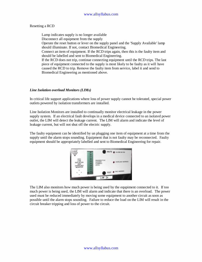
wwwallsyllabuscom
wwwallsyllabuscom
Resetting a RCD
Lamp indicates supply is no longer available Disconnect all equipment from the supply Operate the reset button or lever on the supply panel and the Supply Available lamp
should illuminate If not contact Biomedical Engineering
Connect an item of equipment If the RCD trips again then this is the faulty item and should be labelled and sent to Biomedical Engineering
If the RCD does not trip continue connecting equipment until the RCD trips The last piece of equipment connected to the supply is most likely to be faulty as it will have caused the RCD to trip Remove the faulty item from service label it and send to Biomedical Engineering as mentioned above
Line Isolation overload Monitors (LIMs)
In critical life support applications where loss of power supply cannot be tolerated special power outlets powered by isolation transformers are installed
Line Isolation Monitors are installed to continually monitor electrical leakage in the power
supply system If an electrical fault develops in a medical device connected to an isolated power outlet the LIM will detect the leakage current The LIM will alarm and indicate the level of leakage current but will not shut off the electric supply
The faulty equipment can be identified by un plugging one item of equipment at a time from the supply until the alarm stops sounding Equipment that is not faulty may be reconnected Faulty equipment should be appropriately labelled and sent to Biomedical Engineering for repair
The LIM also monitors how much power is being used by the equpiment connected to it If too much power is being used the LIM will alarm and indicate that there is an overload The power used must be reduced immediately by moving some equipment to another circuit as soon as possible until the alarm stops sounding Failure to reduce the load on the LIM will result in the circuit breaker tripping and loss of power to the circuit
wwwallsyllabuscom
wwwallsyllabuscom
Equipotential Earthing
Equipotential earthing is installed in rooms classified as Cardiac Protected electrical areas
Equipotential earthing in treatment areas used for cardiac procedures is intended to minimise any voltage differences between earthed parts of equipment and any other exposed metal in the room
This reduces the possibility of leakage currents that can cause microelectrocution when the patient comes into contact with multiple items of equipment or if the patient happens to come
into contact with metal items in the room whilr they are connected to a medical device
All conductive metal in an equipotential area is connected to a common equipotential earth point with special heavy duty cable
AREA CLASSIFICATIONS
Body Protected Area
These areas are designed for procedures in which patients are connected to equipment that lowers the natural resistance of the skin Applied parts such as electrode gels conductive fluids entering the patient metal needles and catheters provide an easy pathway for current to flow
The main occurrence of injury from Body-Type procedures is from high current levels causing electric shock A direct connection to the patients heart is not present so the risk of Microelectrocution - fibrillation from minute current levels - is reduced
Residual Current Devices (RCD) or Isolation Transformers and Line Isolation Monitors (LIMiacutes) are used in Body Protected areas to provide protection against electrocution from high leakage currents Body-Protected Areas are identified with this sign
Cardiac Protected Area
Where the procedure involves placing an electrical conductor within or near the heart protection against fibrillation induced from small leakage currents is required Electrical conductors used in these procedures include cardiac pacing electrodes intracardiac ECG electrodes and intracardiac catheters
wwwallsyllabuscom
wwwallsyllabuscom
Equipotential earthing in conjunction with RCDs or LIMs provides protection against microelectrocution in Cardiac-Type procedures
Fault currents are reduced to magnitudes that are unlikely to induce fibrillation Used in conjunction with RCDs or LIMs the magnitude and duration of any fault currents
sourced from equipment are limited Cardiac-Protected Areas are identified with this sign
Other electrical issues
This policy aims to provide guidance to those who find that they need more electrical outlets than
those available or that the existing electrical outlets are inconveniently located
As extension leads and multiple outlet power boards can introduce additional hazards into an area the following procedures should be observed
Extension leads
Approved extension leads (AS 3760 1996) may be used in some areas within the hospital but MUST NOT BE USED IN PATIENT AREAS All electrical extension leads must be tagged with an Engineering Department maintenance tag and require a yearly safety inspection and test via
the Engineering Department
Double adapters
Double adapters may cause overloading or equipment earthing problems and are not to be used in
WCH
Mains extension device
The only mains extension device that is to be used in Patient care areas is the 4-way or 8-way
portable Core Balance Unit
The Biomedical Engineering Department must approve all units prior to use These units contain a safety switch and can detect excessive leakage current and disconnect the power in the event of a hazardous situation
wwwallsyllabuscom
wwwallsyllabuscom
Care must be exercised in the use of a portable Core Balance Unit It should be located off the
floor and in a position that will protect it from physical abuse and possible entry of fluids These devices are expensive and easily damaged The device must be sent to Biomedical Engineering every 6 months for safety testing
Power boards
Approved multiple-outlet power boards can be used across RCH but must not be used in patient
care areas except areas approved by the Biomedical Engineering Department
The power boards must have overload protection be fitted with internal safety shutters that protect unused outlets and be fitted with an onoff switch for each outlet
Medical Laser Applications
The main research subjects of the group of Medical Laser Application are in the field of online
monitoring and diagnostics as well as the development of new therapheutic methods The main focus in research is based upon the use of ultrashort (fs) laser pulses
The main advantage of ultrashort laser pulses is the extrem short interaction time which suppress any unwanted side effects of the laser irradiation of the tissue Secondly the broad spectra of the fs laser pulses give the advantage to use the same laser pulses for diagnostic applications like the optical coherence tomography (OCT)
wwwallsyllabuscom
wwwallsyllabuscom
Following research subjects have special attention at the moment
Optical Coherence Tomographie (OCT)
A fs-laser light source can be used to obtain images from inside the tissue when the pulses are coherent superposed This group uses this application to obtain an inside view of the vocal fold and to measure the outline of the crystalline lens
Refractive Surgery
The principle of getting rid of glasses with the help of a laser surgery (fs-LASIK) is underlying rapid improvements concerning precision and safety New technologies were evaluated and transferred very fast in cooperation with an industrial partner
Treatment of Presbyopia
The flexibility of the crystalline lens can be increase by pricise cuts which are induce by fs laser pulse inside the lens The treatment of the presbyopia is thinkable
wwwallsyllabuscom
wwwallsyllabuscom
Ultrafast Physics
Whereas the applications of ultrashort laser pulses increase rapidly is the physics of the interaction between the laser pulses and tissue in many case not fully understood To obtain improved knowledge of the interaction for many fs-laser application this group preforms numerical simulations as well as fundamental experiments
Thermography
Thermography thermal imaging or thermal video is a type of infrared imaging Thermographic cameras detect radiation in the infrared range of the electromagnetic spectrum
(roughly 900ndash14000 nanometers or 09ndash14 microm) and produce images of that radiation Since infrared radiation is emitted by all objects based on their temperatures according to the black body radiation law thermography makes it possible to see ones environment with or without visible illumination The amount of radiation emitted by an object increases with temperature therefore thermography allows one to see variations in temperature (hence the name) When viewed by thermographic camera warm objects stand out well against cooler backgrounds humans and other warm-blooded animals become easily visible against the environment day or
night As a result thermographys extensive use can historically be ascribed to the military and security services
Thermal imaging photography finds many other uses For example firefighters use it to see through smoke find persons and localize the base of a fire With thermal imaging power lines maintenance technicians locate overheating joints and parts a telltale sign of their failure to eliminate potential hazards Where thermal insulation becomes faulty building construction technicians can see heat leaks to improve the efficiencies of cooling or heating air-conditioning Thermal imaging cameras are also installed in some luxury cars to aid the driver the first being
the 2000 Cadillac DeVille Some physiological activities particularly responses in human beings and other warm-blooded animals can also be monitored with thermographic imaging [1]
The appearance and operation of a modern thermographic camera is often similar to a camcorder Enabling the user to see in the infrared spectrum is a function so useful that ability to record their output is often optional A recording module is therefore not always built-in
Instead of CCD sensors most thermal imaging cameras use CMOS Focal Plane Array (FPA) The most common types are InSb InGaAs HgCdTe and QWIP FPA The newest technologies are using low cost and uncooled microbolometers FPA sensors Their resolution is considerably lower than of optical cameras mostly 160x120 or 320x240 pixels up to 640x512 for the most
expensive models Thermographic cameras are much more expensive than their visible-spectrum counterparts and higher-end models are often export-restricted Older bolometers or more
wwwallsyllabuscom
wwwallsyllabuscom
sensitive models as InSb require cryogenic cooling usually by a miniature Stirling cycle refrigerator or liquid nitrogen
Contents
[hide]
1 Difference between IR film and thermography 2 Advantages of Thermography
3 Limitations amp disadvantages of thermography 4 Applications 5 See also 6 External links
o 61 History of thermal imager manufacturers
[edit] Difference between IR film and thermography
Thermal imaging is going to be used on Mars to detect caves that could hold life
[edit] Advantages of Thermography
You get a visual picture so that you can compare temperatures over a large area
It is real time capable of catching moving targets Able to find deteriorating components prior to failure Measurement in areas inaccessible or hazardous for other methods It is a non-destructive test method
[edit] Limitations amp disadvantages of thermography
Quality cameras are expensive and are easily damaged Images can be hard to interpret accurately even with experience
Accurate temperature measurements are very hard to make because of emissivities Most cameras have plusmn2 or worse accuracy (not as accurate as contact) Training and staying proficient in IR scanning is time consuming Ability to only measure surface areas
[edit] Applications
Condition monitoring Medical imaging Night vision Research
Process control Non destructive testing Surveillance in security law enforcement and defense Chemical imaging
wwwallsyllabuscom
wwwallsyllabuscom
Thermal infrared imagers convert the energy in the infrared wavelength into a visible light video display All objects above 0 kelvins emit thermal infrared energy so thermal imagers can passively see all objects regardless of ambient light However most thermal imagers only see objects warmer than -50 degC
The spectrum and amount of thermal radiation depend strongly on an objects surface
temperature This makes it possible for a thermal camera to display an objects temperature However other factors also influence the radiation which limits the accuracy of this technique For example the radiation depends not only on the temperature of the object but is also a function of the emissivity of the object Also radiation also originates from the surroundings and is reflected in the object and the radiation from the object and the reflected radiation will also be influenced by the absorption of the atmosphere
Endoscopy
This page is about having an endoscopy - a test that looks at the inside of your digestive system
There is information on
What endoscopy is
Why you may have an endoscopy
Having an endoscopy
Your results
What an endoscopy is
An endoscopy is a test that looks inside the body The endoscope is a long flexible tube that can
be swallowed It has a camera and light inside it Some doctors call it a telescope
wwwallsyllabuscom
wwwallsyllabuscom
Why you may have an endoscopy
You are most likely to have an endoscopy to look at the inside of your
Gullet (oesophagus)
Stomach
Duodenum - the first part of the small bowel that attaches to the stomach
Large bowel (colon)
Your doctor may want to see inside because you have symptoms such as abnormal bleeding or
difficulty swallowing The doctor can look down the endoscope and see if there are any growths
or other abnormal looking areas Also through the endoscope the doctor can take samples
(biopsies) of any abnormal looking tissues
There is more detailed information about having a colonoscopy in the bowel cancer section of
CancerHelp UK Below is information about having other types of endoscopy
Having an endoscopy
You can have this test as an out patient Most people have a choice between having the test while
they are awake or after having a medicine to make them drowsy (a sedative) Your hospital may
not be happy for you to have a sedative if you live alone and will have no one to look after you
when you go home If you live alone but really want sedation your hospital may allow you to
stay overnight You should ask your doctor about this as early as possible as extra arrangements
will need to be made beforehand
If you dont have a sedative you will have a spray to numb the back of your throat and make it
easier for you to swallow the endoscopy tube
If you would prefer to be asleep during the test you will have an injection to make you very
drowsy just before the test You will need to take someone with you to the hospital appointment
You wont be able to drive for the rest of the day and should have someone to go home with you
You cant eat or drink for about 8 hours before the test so that your stomach and duodenum are
empty Your doctor will give you written instructions about this beforehand or they may arrive
with your appointment letter When you get to the clinic you may be asked to take your upper
clothing off and put on a hospital gown Some hospitals prefer to use gowns because your clothes
wont get messy Once you are ready you get onto the bed or X-ray couch When you are lying
comfortably you have the sedative injection to make you very drowsy Or your doctor will spray
the back of your throat to numb it
Once the sedative or throat spray has worked the doctor will pass the endoscope tube down your
wwwallsyllabuscom
wwwallsyllabuscom
throat to the area being investigated Your doctor will ask you to swallow as the tube goes down
but if youve had a sedative you wont remember that afterwards If there are any abnormalities
the doctor will take pieces of tissue from the abnormal looking area to send to the laboratory for
closer inspection under a microscope These tissue samples are called biopsies
When the test is over you will need to rest for a while If youve had a sedative you may not
remember much (if anything) about the test once you have come round You should be able to go
home the same day
The results
It can take time for test results to come through How long will depend on why you are having
the test Usually the doctor who carries out the endoscopy dictates a report straight way The
report is typed up by the department secretary and goes to your specialist who gives the results to
you If your GP has sent you for the test the results will go directly to the GP surgery
Understandably waiting for results can make you anxious It usually takes a couple of weeks for
the results to come through If your doctor needed them urgently it would have been noted on
the test request form and the results will be ready sooner than that Try to remember to ask your
doctor how long you should expect to wait for the results when you are first asked to go for the
test If it is not an emergency and you have not heard a couple of weeks after your test ring your
doctors secretary to check if they are back
Endoscopy
From Wikipedia the free encyclopedia
Jump to navigation search
Endoscopic images of a duodenal ulcer
wwwallsyllabuscom
wwwallsyllabuscom
A flexible endoscope
Endoscopy means looking inside and typically refers to looking inside the human body for medical reasons using an instrument called an endoscope Endoscopy can also refer to using a borescope in technical situations where direct line-of-sight observation is not feasible
Contents
[hide]
1 Overview 2 Components 3 Uses
4 History 5 Risks 6 After The Endoscopy 7 Recent developments 8 See also 9 References 10 Footnotes
11 External links
Overview
Endoscopy is a minimally invasive diagnostic medical procedure that is used to assess the interior surfaces of an organ by inserting a tube into the body The instrument may have a rigid or flexible tube and not only provide an image for visual inspection and photography but also enable taking
biopsies and retrieval of foreign objects Endoscopy is the vehicle for minimally invasive surgery
Many endoscopic procedures are considered to be relatively painless and at worst associated with mild discomfort for example in esophagogastroduodenoscopy most patients tolerate the procedure with only topical anaesthesia of the oropharynx using lignocaine spray[1] Complications are not common (only 5 of all operations)[citation needed] but can include perforation of the organ under inspection with the endoscope or biopsy instrument If that occurs open surgery may be required to repair the injury
wwwallsyllabuscom
wwwallsyllabuscom
Components
An endoscope can consist of
a rigid or flexible tube a light delivery system to illuminate the organ or object under inspection The light
source is normally outside the body and the light is typically directed via an optical fiber system
a lens system transmitting the image to the viewer from the fiberscope an additional channel to allow entry of medical instruments or manipulators
Uses
Endoscopy can involve
The gastrointestinal tract (GI tract) o esophagus stomach and duodenum (esophagogastroduodenoscopy) o small intestine o colon (colonoscopyproctosigmoidoscopy) o Bile duct
endoscopic retrograde cholangiopancreatography (ERCP) duodenoscope-assisted cholangiopancreatoscopy intraoperative cholangioscopy
The respiratory tract o The nose (rhinoscopy) o The lower respiratory tract (bronchoscopy)
The urinary tract (cystoscopy) The female reproductive system
o The cervix (colposcopy) o The uterus (hysteroscopy) o The Fallopian tubes (Falloscopy)
Normally closed body cavities (through a small incision) o The abdominal or pelvic cavity (laparoscopy) o The interior of a joint (arthroscopy) o Organs of the chest (thoracoscopy and mediastinoscopy)
During pregnancy o The amnion (amnioscopy) o The fetus (fetoscopy)
Plastic Surgery Panendoscopy (or triple endoscopy)
o Combines laryngoscopy esophagoscopy and bronchoscopy Non-medical uses for endoscopy
o The planning and architectural community have found the endoscope useful for
pre-visualization of scale models of proposed buildings and cities (architectural endoscopy)
o Internal inspection of complex technical systems (borescope) o Endoscopes are also a tool helpful in the examination of improvised explosive
devices by bomb disposal personnel
wwwallsyllabuscom
wwwallsyllabuscom
o The FBI uses endoscopes for conducting surveillance via tight spaces
History
The first endoscope of a kind was developed in 1806 by Philip Bozzini with his introduction of a
Lichtleiter (light conductor) for the examinations of the canals and cavities of the human body However the Vienna Medical Society disapproved of such curiosity An endoscope was first introduced into a human in 1822 by William Beaumont an army surgeon at Mackinac
Island Michigan[citation needed] The use of electric light was a major step in the improvement of endoscopy The first such lights were external Later smaller bulbs became available making internal light possible for instance in a hysteroscope by Charles David in 1908[citation needed] Hans Christian Jacobaeus has been given credit for early endoscopic explorations of the abdomen and the thorax with laparoscopy (1912) and thoracoscopy (1910)[citation needed] Laparoscopy was used in the diagnosis of liver and gallbladder disease by Heinz Kalk in the 1930s[citation needed] Hope reported in 1937 on the use of laparoscopy to diagnose ectopic pregnancy[citation needed] In 1944
Raoul Palmer placed his patients in the Trendelenburg position after gaseous distention of the abdomen and thus was able to reliably perform gynecologic laparoscopy[citation needed]
The first gastrocamera was released in 1950 by Olympus Optical Co Ltd The device took pictures on monochromatic film using a small light bulb that was triggered manually The device was of limited use however because it did not implement real-time optical capability Olympus continued its development of endoscopes by incorporating fiber optics in the early 1960s leading to the first useful endoscopes In 1964 it released a gastrocamera guided by a fiberscope[1] A few articles claim that DrBasil Hirschowitz of UnivOf MichiganAnn Arbor discussed the
endoscope in early 50s[2]
As endoscopic technology improved so did the methods of gastrointestinal endoscopy Owing primarily to the efforts of Dr Hiromi Shinya in the late 1960s GI endoscopy developed into what is more recognizable as todays colonoscopy While many doctors experimented with techniques to take advantage of the new iterations of endoscopes Dr Shinya focused on techniques that would allow for successful operation of the endoscope by an individual rejecting the common practice at the time of utilizing two people Consequently many of the fundamental methods and procedures of modern colonoscopy were developed by Dr Shinya
Dr Shinyas other great contribution was to therapeutic endoscopy in his invention of the
electrosurgical polypectomy snare with the aid of Olympus employee Hiroshi Ichikawa Shinya sketched his first plans for the device on January 8 1969 He envisioned a loop of wire attached to the end of a colonoscope that would allow for easy removal of polyps during investigation by passing a current through the wire By September of 1969 the first polypectomy using this device was performed Polypectomy has since become the most common therapeutic procedure performed with an endoscope (Sivak 2004)
By 1980 laparoscopy training was required by gynecologists to perform tubal ligation procedures and diagnostic evaluations of the pelvis The first laparoscopic cholecystectomy was performed in 1984 and the first video-laparoscopic cholecystectomy in 1987[citation needed] During the 1990s
laparoscopic surgery was extended to the appendix spleen colon stomach kidney and liver[citation needed] Wireless capsule endoscopy or Capsule Endoscopy is now approved in all the countries including Japan where government reimbusement will be available from Oct2007Capsule Endoscopy [3] increases detection of Small Bowel tumors where traditional Endoscopy is not very efficient
wwwallsyllabuscom
wwwallsyllabuscom
Risks
Infection
Punctured organs Allergic reactions due to Contrast agents or dyes (such as those used in a CT scan) Over-sedation
After The Endoscopy
After the procedure the patient will be observed and monitored by a qualified individual in the
endoscopy or a recovery area until a significant portion of the medication has worn off Occasionally a patient is left with a mild sore throat which promptly responds to saline gargles or a feeling of distention from the insufflated air that was used during the procedure Both problems are mild and fleeting When fully recovered the patient will be instructed when to resume hisher usual diet (probably within a few hours) and will be allowed to be taken home Because of the use of sedation most facilities mandate that the patient is taken home by another person and not to drive on hisher own or handle machinery for the remainder of the day
Recent developments
With the application of robotic systems telesurgery was introduced as the surgeon could operate
from a site physically removed from the patient The first transatlantic surgery has been called the Lindbergh Operation
wwwallsyllabuscom
wwwallsyllabuscom
Upper Endoscopy
Upper endoscopy enables the physician to look inside the esophagus stomach and duodenum (first part of the small intestine) The procedure might be used to discover the reason for swallowing difficulties nausea vomiting
reflux bleeding indigestion abdominal pain or chest pain Upper endoscopy is also called EGD which stands for esophagogastroduodenoscopy (eh-SAH-fuh-goh-GAS-troh-doo-AH-duh-NAH-skuh-pee)
For the procedure you will swallow a thin flexible lighted tube called an endoscope (EN-doh-skope) Right before the procedure the physician will spray your throat with a numbing agent that may help prevent gagging
You may also receive pain medicine and a sedative to help you relax during the exam The endoscope transmits an image of the inside of the esophagus stomach and duodenum so the physician can carefully examine the lining of these organs The scope also blows air into the stomach this expands the folds of tissue and makes it easier for the physician to examine the stomach
The physician can see abnormalities like inflammation or
bleeding through the endoscope that dont show up well on x rays The physician can also insert instruments into the scope to treat bleeding abnormalities or remove samples of tissue (biopsy) for further tests
Possible complications of upper endoscopy include bleeding and puncture of the stomach lining However such complications are rare Most people will probably have nothing more than a mild sore throat after the procedure
The procedure takes 20 to 30 minutes Because you will be sedated you will need to rest at the endoscopy facility for 1 to 2 hours until the medication wears off
Preparation
Your stomach and duodenum must be empty for the procedure to be thorough and safe so you will not be able to eat or drink anything for at least 6 hours beforehand Also you must
arrange for someone to take you homemdashyou will not be allowed to drive because of the sedatives Your physician may give you other special instructions
The digestive system
wwwallsyllabuscom
wwwallsyllabuscom
What is Endoscopy and Why is it Performed
Endoscopy allows physicians to peer through the bodys passageways Endoscopy is the examination and inspection of the interior of body organs joints or cavities through an endoscope An endoscope is a device that uses fiber optics and powerful lens systems to provide lighting and visualization of the interior of a joint The portion of the endoscope inserted into the body may be rigid or flexible depending upon the medical procedure
An endoscope uses two fiber optic lines A light fiber carries light into the body cavity and an image fiber carries the image of the body cavity back to the physicians viewing lens There is also a separate port to allow for administration of drugs suction and irrigation This port may also be used to introduce small folding instruments such as forceps scissors brushes snares and baskets for tissue excision (removal) sampling or other diagnostic and therapeutic work Endoscopes may be used in conjunction with a camera or video recorder to document images of the inside of the joint or chronicle an endoscopic procedure New endoscopes have digital capabilities for manipulating and enhancing the video images
This figure shows a rigid endoscope used for arthroscopy The image fiber leads from the ocular (eye piece) to the inserted end of the scope The light fiber is below and leads from the light source to the working end of the endoscope
Why Is Endoscopy Performed
Endoscopy can be used to diagnose various conditions by close examination of internal organ and body structures Endoscopy can also guide therapy and repair such as the removal of torn cartilage from the bearing surfaces of a joint Biopsy (tissue sampling for pathologic testing) may also be performed under endoscopic guidance Local or general anesthetic may be used during endoscopy depending upon the type of procedure being performed
Internal abnormalities revealed through endoscopy include abscesses biliary (liver) cirrhosis bleeding bronchitis cancer cysts degenerative disease gallbladder stones hernia inflammation metastatic cancer polyps tumors ulcers and other diseases and conditions
Endoscopy is a minimally invasive procedure and carries with it certain minor risks depending upon the type of procedure being performed However these risks are typically far outweighed by the diagnostic and therapeutic potential of the procedure
Prior to the widespread use of endoscopy and diagnostic imaging most internal conditions could only be diagnosed or treated with open surgery Until the last several decades exploratory surgery was routinely performed only when a patient was critically ill and the source of illness was not known For example in certain dire cases the patients thorax or abdomen were surgically opened and examined to try to determine the source of illness
wwwallsyllabuscom
wwwallsyllabuscom
Endoscopy can often be done on an outpatient basis Outpatient means that the procedure does not require hospital admission and acute care and observation and may be performed outside the premises of a hospital Outpatient procedures performed at hospitals or ambulatory centers allow the patient to go home or return to work within a short while after their procedure
Types of Endoscopy
Fiber optic endoscopes now have widespread use in medicine and guide a myriad of diagnostic and therapeutic procedures including
Arthroscopy examination of joints for diagnosis and treatment (arthroscopic surgery) Bronchoscopy examination of the trachea and lungs bronchial trees to reveal
abscesses bronchitis carcinoma tumors tuberculosis alveolitis infection inflammation Colonoscopy examination of the inside of the colon and large intestine to detect polyps
tumors ulceration inflammation colitis diverticula Chrohns disease and discovery and removal of foreign bodies
Colposcopy direct visualization of the vagina and cervix to detect cancer inflammation and other conditions
Cystoscopy examination of the bladder urethra urinary tract uteral orifices and
prostate (men) with insertion of the endoscope through the urethra
ERCP (endoscopic retrograde cholangio-pancreatography) uses endoscopic guidance to
place a catheter for x-ray fluorosocopy with contrast enhancement This technique is used to examine the livers biliary tree the gallbladder the pancreatic duct and other anatomy to check for stones other obstructions and disease X-ray contrast is introduced into these ducts via catheter and fluoroscopic x-ray images are taken to show any abnormality or blockage If disease is detected it can sometimes be treated at the same time or biopsy can be performed to test for cancer or other pathology ERCP can detect biliary cirrhosis
cancer of the bile ducts pancreatic cysts pseudocysts pancreatic tumors chronic pancreatitis and other conditions such as gallbladder stones
EGD (Esophogealgastroduodensoscopy) visual examination of the upper gastro-
intestinal (GI) tract (also referred to as gastroscopy) to reveal hemorrhage hiatal hernia inflammation of the esophagus gastric ulcers
Endoscopic biopsy is the removal of tissue specimens for pathologic examination and
analysis Gastroscopy examination of the lining of the esophagus stomach and duodenum
Gastroscopy is often used to diagnose ulcers and other sources of bleeding and to guide biopsy of suspect GI cancers
Laparoscopy visualization of the stomach liver and other abdominal organs including
the female reproductive organs for example the fallopian tubes Laryngoscopy examination of the larynx (voice box) Proctoscopy sigmoidoscopy proctosigmoidoscopy examination of the rectum and
sigmoid colon Thoracoscopy examination of the pleura (sac that covers the lungs) pleural spaces
mediastinum and pericardium
A Brief History of Endoscopy
In the early 1900s the first attempts to view inside the body with lighted telescopes were made These initial devices were often fully rigid In the 1930s semi-flexible endoscopes called gastroscopes were developed to view inside of the stomach Fiber-optic endoscopy was pioneered by South African-born physician Basil Hirschowitz at the University of Michigan in 1957 Widespread use of fiber optic endoscopes began in the 1960s
wwwallsyllabuscom
wwwallsyllabuscom
A fiber optic cable is simply a bundle of microscopic glass or plastic fibers that literally allows light and images to be transmitted through curved structures Fiber optic cables are also replacing metal wires as the backbone of the worlds telecommunications infrastructure This Internet page may have traveled through a fiber optic cable as a stream of digital data (bursts of light) on its way to your computer
Endoscopy Equipment
Endoscopes have many practical needs And HMB Endoscopy Products (Hollywood Florida) has been providing endoscopic equipment and educating people on the use of endoscopes for more than 17 years Be sure to Browse our Catalog for all the details on how to purchase these medical instruments that can examine any part of the body
Video
Systems
Fiber
Endoscopes
Video
Endoscopes
In the simplest terms Endoscopy equipment consists of instruments that can look at the inside of many different organs mdash these are small flexible or rigid tubes with a light or lenses on the end that can look into the esophagus stomach and colon mdash and in more general terms endoscopy equipment can help doctors look deep inside body structures and hollow organs
An endoscope and related endoscope products and equipment are usually composed of three components
An optic system that allows the doctor to look through the scope into the organ or
cavity or to attach a video camera to the scope
A fiberoptic cable to light up the bodily area
A lumen (eg the bore of a tube like a needle or catheter) to take tissue samples of the
area being viewed
The beauty of endoscopic products is that they perform dual functions mdash with both diagnostic and therapeutic capabilities For example this means that these endoscopic products and instruments can perform biopsies (eg to evaluate tissue samples) as well as provide sclerotherapy (a medical procedure used to treat varicose veins and ―spider veins) In truth these brief explanations only tell part of the story
wwwallsyllabuscom
wwwallsyllabuscom
TYPES OF ENDOSCOPES
Take a look at the different types of endoscopic equipment you can get with HMB Endoscopy Products Again in very general terms there are two main types of endoscopes
Rigid Flexible
1 rigid endoscopes ndash the majority of which use a convex (curving out like one half of a
circle) glass lens system in which the small glass lenses are separated by large air spaces
2 flexible endoscopes allow for just that mdash flexibility In the animal kingdom for example
a flexible endoscope would be perfect for examining the stomach area of a snake
The popularity of endoscopy equipment continues to grow Some of the industryrsquos household names include Olympus Pentax Funjinon and Storz mdash with HMB both selling and repairing pre-owned and completely refurbished endoscopic products from each of these major manufacturers Browse our Catalog to get the ones you need
THERMOGRAPH APPLICATION Thermographs are digital recording thermometers used to log
temperature in the
marine environment The instruments are attached to a computer to set up recording
parameters for
deployment downloading and display of data in graphic or numeric format These units
are placed in
underwater housings and attached to the bottom or suspended in the water column for
deployment
DESCRIPTION Two types of thermograph are currently available The Ryan
Tempmentor is a
reusable data logger that allows storage of a maximum of 6361 temperature
measurements in the
range of -32C to +70C with 01resolution and 03accuracy The instrument can be
programmed
to take measurements from once per second to once every other hour Instrument
dimensions are 3 x
6 x 13 and the unit weighs 11 ounces The Hobo Temp is a miniature reusable data
logger that
allows storage of 1800 measurements in the range of -20C to +70C with
03resolution and 07
wwwallsyllabuscom
wwwallsyllabuscom
accuracy Sampling intervals from 05 seconds to 48 hours are available Instrument
dimensions are
24 x 19 x 08 and the unit weighs 1 ounce Both units are battery powered with the
battery life of
the Tempmentor being approximately two years and the battery life of the Hobo Temp
being one year THERMOGRAPH APPLICATION Thermographs are digital recording thermometers used to log
temperature in the
marine environment The instruments are attached to a computer to set up recording
parameters for
deployment downloading and display of data in graphic or numeric format These units
are placed in
underwater housings and attached to the bottom or suspended in the water column for
deployment
DESCRIPTION Two types of thermograph are currently available The Ryan
Tempmentor is a
reusable data logger that allows storage of a maximum of 6361 temperature
measurements in the
range of -32C to +70C with 01resolution and 03accuracy The instrument can be
programmed
to take measurements from once per second to once every other hour Instrument
dimensions are 3 x
6 x 13 and the unit weighs 11 ounces The Hobo Temp is a miniature reusable data
logger that
allows storage of 1800 measurements in the range of -20C to +70C with
03resolution and 07
accuracy Sampling intervals from 05 seconds to 48 hours are available Instrument
dimensions are
24 x 19 x 08 and the unit weighs 1 ounce Both units are battery powered with the
battery life of
the Tempmentor being approximately two years and the battery life of the Hobo Temp
being one year THERMOGRAPH APPLICATION Thermographs are digital recording thermometers used to log
temperature in the
marine environment The instruments are attached to a computer to set up recording
parameters for
deployment downloading and display of data in graphic or numeric format These units
are placed in
underwater housings and attached to the bottom or suspended in the water column for
deployment
DESCRIPTION Two types of thermograph are currently available The Ryan
Tempmentor is a
reusable data logger that allows storage of a maximum of 6361 temperature
measurements in the
range of -32C to +70C with 01resolution and 03accuracy The instrument can be
programmed
wwwallsyllabuscom
wwwallsyllabuscom
to take measurements from once per second to once every other hour Instrument
dimensions are 3 x
6 x 13 and the unit weighs 11 ounces The Hobo Temp is a miniature reusable data
logger that
allows storage of 1800 measurements in the range of -20C to +70C with
03resolution and 07
accuracy Sampling intervals from 05 seconds to 48 hours are available Instrument
dimensions are
24 x 19 x 08 and the unit weighs 1 ounce Both units are battery powered with the
battery life of
the Tempmentor being approximately two years and the battery life of the Hobo Temp
being one year
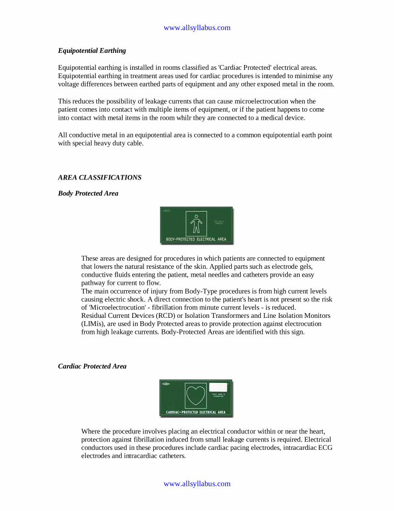
wwwallsyllabuscom
wwwallsyllabuscom
Equipotential earthing in conjunction with RCDs or LIMs provides protection against microelectrocution in Cardiac-Type procedures
Fault currents are reduced to magnitudes that are unlikely to induce fibrillation Used in conjunction with RCDs or LIMs the magnitude and duration of any fault currents
sourced from equipment are limited Cardiac-Protected Areas are identified with this sign
Other electrical issues
This policy aims to provide guidance to those who find that they need more electrical outlets than
those available or that the existing electrical outlets are inconveniently located
As extension leads and multiple outlet power boards can introduce additional hazards into an area the following procedures should be observed
Extension leads
Approved extension leads (AS 3760 1996) may be used in some areas within the hospital but MUST NOT BE USED IN PATIENT AREAS All electrical extension leads must be tagged with an Engineering Department maintenance tag and require a yearly safety inspection and test via
the Engineering Department
Double adapters
Double adapters may cause overloading or equipment earthing problems and are not to be used in
WCH
Mains extension device
The only mains extension device that is to be used in Patient care areas is the 4-way or 8-way
portable Core Balance Unit
The Biomedical Engineering Department must approve all units prior to use These units contain a safety switch and can detect excessive leakage current and disconnect the power in the event of a hazardous situation
wwwallsyllabuscom
wwwallsyllabuscom
Care must be exercised in the use of a portable Core Balance Unit It should be located off the
floor and in a position that will protect it from physical abuse and possible entry of fluids These devices are expensive and easily damaged The device must be sent to Biomedical Engineering every 6 months for safety testing
Power boards
Approved multiple-outlet power boards can be used across RCH but must not be used in patient
care areas except areas approved by the Biomedical Engineering Department
The power boards must have overload protection be fitted with internal safety shutters that protect unused outlets and be fitted with an onoff switch for each outlet
Medical Laser Applications
The main research subjects of the group of Medical Laser Application are in the field of online
monitoring and diagnostics as well as the development of new therapheutic methods The main focus in research is based upon the use of ultrashort (fs) laser pulses
The main advantage of ultrashort laser pulses is the extrem short interaction time which suppress any unwanted side effects of the laser irradiation of the tissue Secondly the broad spectra of the fs laser pulses give the advantage to use the same laser pulses for diagnostic applications like the optical coherence tomography (OCT)
wwwallsyllabuscom
wwwallsyllabuscom
Following research subjects have special attention at the moment
Optical Coherence Tomographie (OCT)
A fs-laser light source can be used to obtain images from inside the tissue when the pulses are coherent superposed This group uses this application to obtain an inside view of the vocal fold and to measure the outline of the crystalline lens
Refractive Surgery
The principle of getting rid of glasses with the help of a laser surgery (fs-LASIK) is underlying rapid improvements concerning precision and safety New technologies were evaluated and transferred very fast in cooperation with an industrial partner
Treatment of Presbyopia
The flexibility of the crystalline lens can be increase by pricise cuts which are induce by fs laser pulse inside the lens The treatment of the presbyopia is thinkable
wwwallsyllabuscom
wwwallsyllabuscom
Ultrafast Physics
Whereas the applications of ultrashort laser pulses increase rapidly is the physics of the interaction between the laser pulses and tissue in many case not fully understood To obtain improved knowledge of the interaction for many fs-laser application this group preforms numerical simulations as well as fundamental experiments
Thermography
Thermography thermal imaging or thermal video is a type of infrared imaging Thermographic cameras detect radiation in the infrared range of the electromagnetic spectrum
(roughly 900ndash14000 nanometers or 09ndash14 microm) and produce images of that radiation Since infrared radiation is emitted by all objects based on their temperatures according to the black body radiation law thermography makes it possible to see ones environment with or without visible illumination The amount of radiation emitted by an object increases with temperature therefore thermography allows one to see variations in temperature (hence the name) When viewed by thermographic camera warm objects stand out well against cooler backgrounds humans and other warm-blooded animals become easily visible against the environment day or
night As a result thermographys extensive use can historically be ascribed to the military and security services
Thermal imaging photography finds many other uses For example firefighters use it to see through smoke find persons and localize the base of a fire With thermal imaging power lines maintenance technicians locate overheating joints and parts a telltale sign of their failure to eliminate potential hazards Where thermal insulation becomes faulty building construction technicians can see heat leaks to improve the efficiencies of cooling or heating air-conditioning Thermal imaging cameras are also installed in some luxury cars to aid the driver the first being
the 2000 Cadillac DeVille Some physiological activities particularly responses in human beings and other warm-blooded animals can also be monitored with thermographic imaging [1]
The appearance and operation of a modern thermographic camera is often similar to a camcorder Enabling the user to see in the infrared spectrum is a function so useful that ability to record their output is often optional A recording module is therefore not always built-in
Instead of CCD sensors most thermal imaging cameras use CMOS Focal Plane Array (FPA) The most common types are InSb InGaAs HgCdTe and QWIP FPA The newest technologies are using low cost and uncooled microbolometers FPA sensors Their resolution is considerably lower than of optical cameras mostly 160x120 or 320x240 pixels up to 640x512 for the most
expensive models Thermographic cameras are much more expensive than their visible-spectrum counterparts and higher-end models are often export-restricted Older bolometers or more
wwwallsyllabuscom
wwwallsyllabuscom
sensitive models as InSb require cryogenic cooling usually by a miniature Stirling cycle refrigerator or liquid nitrogen
Contents
[hide]
1 Difference between IR film and thermography 2 Advantages of Thermography
3 Limitations amp disadvantages of thermography 4 Applications 5 See also 6 External links
o 61 History of thermal imager manufacturers
[edit] Difference between IR film and thermography
Thermal imaging is going to be used on Mars to detect caves that could hold life
[edit] Advantages of Thermography
You get a visual picture so that you can compare temperatures over a large area
It is real time capable of catching moving targets Able to find deteriorating components prior to failure Measurement in areas inaccessible or hazardous for other methods It is a non-destructive test method
[edit] Limitations amp disadvantages of thermography
Quality cameras are expensive and are easily damaged Images can be hard to interpret accurately even with experience
Accurate temperature measurements are very hard to make because of emissivities Most cameras have plusmn2 or worse accuracy (not as accurate as contact) Training and staying proficient in IR scanning is time consuming Ability to only measure surface areas
[edit] Applications
Condition monitoring Medical imaging Night vision Research
Process control Non destructive testing Surveillance in security law enforcement and defense Chemical imaging
wwwallsyllabuscom
wwwallsyllabuscom
Thermal infrared imagers convert the energy in the infrared wavelength into a visible light video display All objects above 0 kelvins emit thermal infrared energy so thermal imagers can passively see all objects regardless of ambient light However most thermal imagers only see objects warmer than -50 degC
The spectrum and amount of thermal radiation depend strongly on an objects surface
temperature This makes it possible for a thermal camera to display an objects temperature However other factors also influence the radiation which limits the accuracy of this technique For example the radiation depends not only on the temperature of the object but is also a function of the emissivity of the object Also radiation also originates from the surroundings and is reflected in the object and the radiation from the object and the reflected radiation will also be influenced by the absorption of the atmosphere
Endoscopy
This page is about having an endoscopy - a test that looks at the inside of your digestive system
There is information on
What endoscopy is
Why you may have an endoscopy
Having an endoscopy
Your results
What an endoscopy is
An endoscopy is a test that looks inside the body The endoscope is a long flexible tube that can
be swallowed It has a camera and light inside it Some doctors call it a telescope
wwwallsyllabuscom
wwwallsyllabuscom
Why you may have an endoscopy
You are most likely to have an endoscopy to look at the inside of your
Gullet (oesophagus)
Stomach
Duodenum - the first part of the small bowel that attaches to the stomach
Large bowel (colon)
Your doctor may want to see inside because you have symptoms such as abnormal bleeding or
difficulty swallowing The doctor can look down the endoscope and see if there are any growths
or other abnormal looking areas Also through the endoscope the doctor can take samples
(biopsies) of any abnormal looking tissues
There is more detailed information about having a colonoscopy in the bowel cancer section of
CancerHelp UK Below is information about having other types of endoscopy
Having an endoscopy
You can have this test as an out patient Most people have a choice between having the test while
they are awake or after having a medicine to make them drowsy (a sedative) Your hospital may
not be happy for you to have a sedative if you live alone and will have no one to look after you
when you go home If you live alone but really want sedation your hospital may allow you to
stay overnight You should ask your doctor about this as early as possible as extra arrangements
will need to be made beforehand
If you dont have a sedative you will have a spray to numb the back of your throat and make it
easier for you to swallow the endoscopy tube
If you would prefer to be asleep during the test you will have an injection to make you very
drowsy just before the test You will need to take someone with you to the hospital appointment
You wont be able to drive for the rest of the day and should have someone to go home with you
You cant eat or drink for about 8 hours before the test so that your stomach and duodenum are
empty Your doctor will give you written instructions about this beforehand or they may arrive
with your appointment letter When you get to the clinic you may be asked to take your upper
clothing off and put on a hospital gown Some hospitals prefer to use gowns because your clothes
wont get messy Once you are ready you get onto the bed or X-ray couch When you are lying
comfortably you have the sedative injection to make you very drowsy Or your doctor will spray
the back of your throat to numb it
Once the sedative or throat spray has worked the doctor will pass the endoscope tube down your
wwwallsyllabuscom
wwwallsyllabuscom
throat to the area being investigated Your doctor will ask you to swallow as the tube goes down
but if youve had a sedative you wont remember that afterwards If there are any abnormalities
the doctor will take pieces of tissue from the abnormal looking area to send to the laboratory for
closer inspection under a microscope These tissue samples are called biopsies
When the test is over you will need to rest for a while If youve had a sedative you may not
remember much (if anything) about the test once you have come round You should be able to go
home the same day
The results
It can take time for test results to come through How long will depend on why you are having
the test Usually the doctor who carries out the endoscopy dictates a report straight way The
report is typed up by the department secretary and goes to your specialist who gives the results to
you If your GP has sent you for the test the results will go directly to the GP surgery
Understandably waiting for results can make you anxious It usually takes a couple of weeks for
the results to come through If your doctor needed them urgently it would have been noted on
the test request form and the results will be ready sooner than that Try to remember to ask your
doctor how long you should expect to wait for the results when you are first asked to go for the
test If it is not an emergency and you have not heard a couple of weeks after your test ring your
doctors secretary to check if they are back
Endoscopy
From Wikipedia the free encyclopedia
Jump to navigation search
Endoscopic images of a duodenal ulcer
wwwallsyllabuscom
wwwallsyllabuscom
A flexible endoscope
Endoscopy means looking inside and typically refers to looking inside the human body for medical reasons using an instrument called an endoscope Endoscopy can also refer to using a borescope in technical situations where direct line-of-sight observation is not feasible
Contents
[hide]
1 Overview 2 Components 3 Uses
4 History 5 Risks 6 After The Endoscopy 7 Recent developments 8 See also 9 References 10 Footnotes
11 External links
Overview
Endoscopy is a minimally invasive diagnostic medical procedure that is used to assess the interior surfaces of an organ by inserting a tube into the body The instrument may have a rigid or flexible tube and not only provide an image for visual inspection and photography but also enable taking
biopsies and retrieval of foreign objects Endoscopy is the vehicle for minimally invasive surgery
Many endoscopic procedures are considered to be relatively painless and at worst associated with mild discomfort for example in esophagogastroduodenoscopy most patients tolerate the procedure with only topical anaesthesia of the oropharynx using lignocaine spray[1] Complications are not common (only 5 of all operations)[citation needed] but can include perforation of the organ under inspection with the endoscope or biopsy instrument If that occurs open surgery may be required to repair the injury
wwwallsyllabuscom
wwwallsyllabuscom
Components
An endoscope can consist of
a rigid or flexible tube a light delivery system to illuminate the organ or object under inspection The light
source is normally outside the body and the light is typically directed via an optical fiber system
a lens system transmitting the image to the viewer from the fiberscope an additional channel to allow entry of medical instruments or manipulators
Uses
Endoscopy can involve
The gastrointestinal tract (GI tract) o esophagus stomach and duodenum (esophagogastroduodenoscopy) o small intestine o colon (colonoscopyproctosigmoidoscopy) o Bile duct
endoscopic retrograde cholangiopancreatography (ERCP) duodenoscope-assisted cholangiopancreatoscopy intraoperative cholangioscopy
The respiratory tract o The nose (rhinoscopy) o The lower respiratory tract (bronchoscopy)
The urinary tract (cystoscopy) The female reproductive system
o The cervix (colposcopy) o The uterus (hysteroscopy) o The Fallopian tubes (Falloscopy)
Normally closed body cavities (through a small incision) o The abdominal or pelvic cavity (laparoscopy) o The interior of a joint (arthroscopy) o Organs of the chest (thoracoscopy and mediastinoscopy)
During pregnancy o The amnion (amnioscopy) o The fetus (fetoscopy)
Plastic Surgery Panendoscopy (or triple endoscopy)
o Combines laryngoscopy esophagoscopy and bronchoscopy Non-medical uses for endoscopy
o The planning and architectural community have found the endoscope useful for
pre-visualization of scale models of proposed buildings and cities (architectural endoscopy)
o Internal inspection of complex technical systems (borescope) o Endoscopes are also a tool helpful in the examination of improvised explosive
devices by bomb disposal personnel
wwwallsyllabuscom
wwwallsyllabuscom
o The FBI uses endoscopes for conducting surveillance via tight spaces
History
The first endoscope of a kind was developed in 1806 by Philip Bozzini with his introduction of a
Lichtleiter (light conductor) for the examinations of the canals and cavities of the human body However the Vienna Medical Society disapproved of such curiosity An endoscope was first introduced into a human in 1822 by William Beaumont an army surgeon at Mackinac
Island Michigan[citation needed] The use of electric light was a major step in the improvement of endoscopy The first such lights were external Later smaller bulbs became available making internal light possible for instance in a hysteroscope by Charles David in 1908[citation needed] Hans Christian Jacobaeus has been given credit for early endoscopic explorations of the abdomen and the thorax with laparoscopy (1912) and thoracoscopy (1910)[citation needed] Laparoscopy was used in the diagnosis of liver and gallbladder disease by Heinz Kalk in the 1930s[citation needed] Hope reported in 1937 on the use of laparoscopy to diagnose ectopic pregnancy[citation needed] In 1944
Raoul Palmer placed his patients in the Trendelenburg position after gaseous distention of the abdomen and thus was able to reliably perform gynecologic laparoscopy[citation needed]
The first gastrocamera was released in 1950 by Olympus Optical Co Ltd The device took pictures on monochromatic film using a small light bulb that was triggered manually The device was of limited use however because it did not implement real-time optical capability Olympus continued its development of endoscopes by incorporating fiber optics in the early 1960s leading to the first useful endoscopes In 1964 it released a gastrocamera guided by a fiberscope[1] A few articles claim that DrBasil Hirschowitz of UnivOf MichiganAnn Arbor discussed the
endoscope in early 50s[2]
As endoscopic technology improved so did the methods of gastrointestinal endoscopy Owing primarily to the efforts of Dr Hiromi Shinya in the late 1960s GI endoscopy developed into what is more recognizable as todays colonoscopy While many doctors experimented with techniques to take advantage of the new iterations of endoscopes Dr Shinya focused on techniques that would allow for successful operation of the endoscope by an individual rejecting the common practice at the time of utilizing two people Consequently many of the fundamental methods and procedures of modern colonoscopy were developed by Dr Shinya
Dr Shinyas other great contribution was to therapeutic endoscopy in his invention of the
electrosurgical polypectomy snare with the aid of Olympus employee Hiroshi Ichikawa Shinya sketched his first plans for the device on January 8 1969 He envisioned a loop of wire attached to the end of a colonoscope that would allow for easy removal of polyps during investigation by passing a current through the wire By September of 1969 the first polypectomy using this device was performed Polypectomy has since become the most common therapeutic procedure performed with an endoscope (Sivak 2004)
By 1980 laparoscopy training was required by gynecologists to perform tubal ligation procedures and diagnostic evaluations of the pelvis The first laparoscopic cholecystectomy was performed in 1984 and the first video-laparoscopic cholecystectomy in 1987[citation needed] During the 1990s
laparoscopic surgery was extended to the appendix spleen colon stomach kidney and liver[citation needed] Wireless capsule endoscopy or Capsule Endoscopy is now approved in all the countries including Japan where government reimbusement will be available from Oct2007Capsule Endoscopy [3] increases detection of Small Bowel tumors where traditional Endoscopy is not very efficient
wwwallsyllabuscom
wwwallsyllabuscom
Risks
Infection
Punctured organs Allergic reactions due to Contrast agents or dyes (such as those used in a CT scan) Over-sedation
After The Endoscopy
After the procedure the patient will be observed and monitored by a qualified individual in the
endoscopy or a recovery area until a significant portion of the medication has worn off Occasionally a patient is left with a mild sore throat which promptly responds to saline gargles or a feeling of distention from the insufflated air that was used during the procedure Both problems are mild and fleeting When fully recovered the patient will be instructed when to resume hisher usual diet (probably within a few hours) and will be allowed to be taken home Because of the use of sedation most facilities mandate that the patient is taken home by another person and not to drive on hisher own or handle machinery for the remainder of the day
Recent developments
With the application of robotic systems telesurgery was introduced as the surgeon could operate
from a site physically removed from the patient The first transatlantic surgery has been called the Lindbergh Operation
wwwallsyllabuscom
wwwallsyllabuscom
Upper Endoscopy
Upper endoscopy enables the physician to look inside the esophagus stomach and duodenum (first part of the small intestine) The procedure might be used to discover the reason for swallowing difficulties nausea vomiting
reflux bleeding indigestion abdominal pain or chest pain Upper endoscopy is also called EGD which stands for esophagogastroduodenoscopy (eh-SAH-fuh-goh-GAS-troh-doo-AH-duh-NAH-skuh-pee)
For the procedure you will swallow a thin flexible lighted tube called an endoscope (EN-doh-skope) Right before the procedure the physician will spray your throat with a numbing agent that may help prevent gagging
You may also receive pain medicine and a sedative to help you relax during the exam The endoscope transmits an image of the inside of the esophagus stomach and duodenum so the physician can carefully examine the lining of these organs The scope also blows air into the stomach this expands the folds of tissue and makes it easier for the physician to examine the stomach
The physician can see abnormalities like inflammation or
bleeding through the endoscope that dont show up well on x rays The physician can also insert instruments into the scope to treat bleeding abnormalities or remove samples of tissue (biopsy) for further tests
Possible complications of upper endoscopy include bleeding and puncture of the stomach lining However such complications are rare Most people will probably have nothing more than a mild sore throat after the procedure
The procedure takes 20 to 30 minutes Because you will be sedated you will need to rest at the endoscopy facility for 1 to 2 hours until the medication wears off
Preparation
Your stomach and duodenum must be empty for the procedure to be thorough and safe so you will not be able to eat or drink anything for at least 6 hours beforehand Also you must
arrange for someone to take you homemdashyou will not be allowed to drive because of the sedatives Your physician may give you other special instructions
The digestive system
wwwallsyllabuscom
wwwallsyllabuscom
What is Endoscopy and Why is it Performed
Endoscopy allows physicians to peer through the bodys passageways Endoscopy is the examination and inspection of the interior of body organs joints or cavities through an endoscope An endoscope is a device that uses fiber optics and powerful lens systems to provide lighting and visualization of the interior of a joint The portion of the endoscope inserted into the body may be rigid or flexible depending upon the medical procedure
An endoscope uses two fiber optic lines A light fiber carries light into the body cavity and an image fiber carries the image of the body cavity back to the physicians viewing lens There is also a separate port to allow for administration of drugs suction and irrigation This port may also be used to introduce small folding instruments such as forceps scissors brushes snares and baskets for tissue excision (removal) sampling or other diagnostic and therapeutic work Endoscopes may be used in conjunction with a camera or video recorder to document images of the inside of the joint or chronicle an endoscopic procedure New endoscopes have digital capabilities for manipulating and enhancing the video images
This figure shows a rigid endoscope used for arthroscopy The image fiber leads from the ocular (eye piece) to the inserted end of the scope The light fiber is below and leads from the light source to the working end of the endoscope
Why Is Endoscopy Performed
Endoscopy can be used to diagnose various conditions by close examination of internal organ and body structures Endoscopy can also guide therapy and repair such as the removal of torn cartilage from the bearing surfaces of a joint Biopsy (tissue sampling for pathologic testing) may also be performed under endoscopic guidance Local or general anesthetic may be used during endoscopy depending upon the type of procedure being performed
Internal abnormalities revealed through endoscopy include abscesses biliary (liver) cirrhosis bleeding bronchitis cancer cysts degenerative disease gallbladder stones hernia inflammation metastatic cancer polyps tumors ulcers and other diseases and conditions
Endoscopy is a minimally invasive procedure and carries with it certain minor risks depending upon the type of procedure being performed However these risks are typically far outweighed by the diagnostic and therapeutic potential of the procedure
Prior to the widespread use of endoscopy and diagnostic imaging most internal conditions could only be diagnosed or treated with open surgery Until the last several decades exploratory surgery was routinely performed only when a patient was critically ill and the source of illness was not known For example in certain dire cases the patients thorax or abdomen were surgically opened and examined to try to determine the source of illness
wwwallsyllabuscom
wwwallsyllabuscom
Endoscopy can often be done on an outpatient basis Outpatient means that the procedure does not require hospital admission and acute care and observation and may be performed outside the premises of a hospital Outpatient procedures performed at hospitals or ambulatory centers allow the patient to go home or return to work within a short while after their procedure
Types of Endoscopy
Fiber optic endoscopes now have widespread use in medicine and guide a myriad of diagnostic and therapeutic procedures including
Arthroscopy examination of joints for diagnosis and treatment (arthroscopic surgery) Bronchoscopy examination of the trachea and lungs bronchial trees to reveal
abscesses bronchitis carcinoma tumors tuberculosis alveolitis infection inflammation Colonoscopy examination of the inside of the colon and large intestine to detect polyps
tumors ulceration inflammation colitis diverticula Chrohns disease and discovery and removal of foreign bodies
Colposcopy direct visualization of the vagina and cervix to detect cancer inflammation and other conditions
Cystoscopy examination of the bladder urethra urinary tract uteral orifices and
prostate (men) with insertion of the endoscope through the urethra
ERCP (endoscopic retrograde cholangio-pancreatography) uses endoscopic guidance to
place a catheter for x-ray fluorosocopy with contrast enhancement This technique is used to examine the livers biliary tree the gallbladder the pancreatic duct and other anatomy to check for stones other obstructions and disease X-ray contrast is introduced into these ducts via catheter and fluoroscopic x-ray images are taken to show any abnormality or blockage If disease is detected it can sometimes be treated at the same time or biopsy can be performed to test for cancer or other pathology ERCP can detect biliary cirrhosis
cancer of the bile ducts pancreatic cysts pseudocysts pancreatic tumors chronic pancreatitis and other conditions such as gallbladder stones
EGD (Esophogealgastroduodensoscopy) visual examination of the upper gastro-
intestinal (GI) tract (also referred to as gastroscopy) to reveal hemorrhage hiatal hernia inflammation of the esophagus gastric ulcers
Endoscopic biopsy is the removal of tissue specimens for pathologic examination and
analysis Gastroscopy examination of the lining of the esophagus stomach and duodenum
Gastroscopy is often used to diagnose ulcers and other sources of bleeding and to guide biopsy of suspect GI cancers
Laparoscopy visualization of the stomach liver and other abdominal organs including
the female reproductive organs for example the fallopian tubes Laryngoscopy examination of the larynx (voice box) Proctoscopy sigmoidoscopy proctosigmoidoscopy examination of the rectum and
sigmoid colon Thoracoscopy examination of the pleura (sac that covers the lungs) pleural spaces
mediastinum and pericardium
A Brief History of Endoscopy
In the early 1900s the first attempts to view inside the body with lighted telescopes were made These initial devices were often fully rigid In the 1930s semi-flexible endoscopes called gastroscopes were developed to view inside of the stomach Fiber-optic endoscopy was pioneered by South African-born physician Basil Hirschowitz at the University of Michigan in 1957 Widespread use of fiber optic endoscopes began in the 1960s
wwwallsyllabuscom
wwwallsyllabuscom
A fiber optic cable is simply a bundle of microscopic glass or plastic fibers that literally allows light and images to be transmitted through curved structures Fiber optic cables are also replacing metal wires as the backbone of the worlds telecommunications infrastructure This Internet page may have traveled through a fiber optic cable as a stream of digital data (bursts of light) on its way to your computer
Endoscopy Equipment
Endoscopes have many practical needs And HMB Endoscopy Products (Hollywood Florida) has been providing endoscopic equipment and educating people on the use of endoscopes for more than 17 years Be sure to Browse our Catalog for all the details on how to purchase these medical instruments that can examine any part of the body
Video
Systems
Fiber
Endoscopes
Video
Endoscopes
In the simplest terms Endoscopy equipment consists of instruments that can look at the inside of many different organs mdash these are small flexible or rigid tubes with a light or lenses on the end that can look into the esophagus stomach and colon mdash and in more general terms endoscopy equipment can help doctors look deep inside body structures and hollow organs
An endoscope and related endoscope products and equipment are usually composed of three components
An optic system that allows the doctor to look through the scope into the organ or
cavity or to attach a video camera to the scope
A fiberoptic cable to light up the bodily area
A lumen (eg the bore of a tube like a needle or catheter) to take tissue samples of the
area being viewed
The beauty of endoscopic products is that they perform dual functions mdash with both diagnostic and therapeutic capabilities For example this means that these endoscopic products and instruments can perform biopsies (eg to evaluate tissue samples) as well as provide sclerotherapy (a medical procedure used to treat varicose veins and ―spider veins) In truth these brief explanations only tell part of the story
wwwallsyllabuscom
wwwallsyllabuscom
TYPES OF ENDOSCOPES
Take a look at the different types of endoscopic equipment you can get with HMB Endoscopy Products Again in very general terms there are two main types of endoscopes
Rigid Flexible
1 rigid endoscopes ndash the majority of which use a convex (curving out like one half of a
circle) glass lens system in which the small glass lenses are separated by large air spaces
2 flexible endoscopes allow for just that mdash flexibility In the animal kingdom for example
a flexible endoscope would be perfect for examining the stomach area of a snake
The popularity of endoscopy equipment continues to grow Some of the industryrsquos household names include Olympus Pentax Funjinon and Storz mdash with HMB both selling and repairing pre-owned and completely refurbished endoscopic products from each of these major manufacturers Browse our Catalog to get the ones you need
THERMOGRAPH APPLICATION Thermographs are digital recording thermometers used to log
temperature in the
marine environment The instruments are attached to a computer to set up recording
parameters for
deployment downloading and display of data in graphic or numeric format These units
are placed in
underwater housings and attached to the bottom or suspended in the water column for
deployment
DESCRIPTION Two types of thermograph are currently available The Ryan
Tempmentor is a
reusable data logger that allows storage of a maximum of 6361 temperature
measurements in the
range of -32C to +70C with 01resolution and 03accuracy The instrument can be
programmed
to take measurements from once per second to once every other hour Instrument
dimensions are 3 x
6 x 13 and the unit weighs 11 ounces The Hobo Temp is a miniature reusable data
logger that
allows storage of 1800 measurements in the range of -20C to +70C with
03resolution and 07
wwwallsyllabuscom
wwwallsyllabuscom
accuracy Sampling intervals from 05 seconds to 48 hours are available Instrument
dimensions are
24 x 19 x 08 and the unit weighs 1 ounce Both units are battery powered with the
battery life of
the Tempmentor being approximately two years and the battery life of the Hobo Temp
being one year THERMOGRAPH APPLICATION Thermographs are digital recording thermometers used to log
temperature in the
marine environment The instruments are attached to a computer to set up recording
parameters for
deployment downloading and display of data in graphic or numeric format These units
are placed in
underwater housings and attached to the bottom or suspended in the water column for
deployment
DESCRIPTION Two types of thermograph are currently available The Ryan
Tempmentor is a
reusable data logger that allows storage of a maximum of 6361 temperature
measurements in the
range of -32C to +70C with 01resolution and 03accuracy The instrument can be
programmed
to take measurements from once per second to once every other hour Instrument
dimensions are 3 x
6 x 13 and the unit weighs 11 ounces The Hobo Temp is a miniature reusable data
logger that
allows storage of 1800 measurements in the range of -20C to +70C with
03resolution and 07
accuracy Sampling intervals from 05 seconds to 48 hours are available Instrument
dimensions are
24 x 19 x 08 and the unit weighs 1 ounce Both units are battery powered with the
battery life of
the Tempmentor being approximately two years and the battery life of the Hobo Temp
being one year THERMOGRAPH APPLICATION Thermographs are digital recording thermometers used to log
temperature in the
marine environment The instruments are attached to a computer to set up recording
parameters for
deployment downloading and display of data in graphic or numeric format These units
are placed in
underwater housings and attached to the bottom or suspended in the water column for
deployment
DESCRIPTION Two types of thermograph are currently available The Ryan
Tempmentor is a
reusable data logger that allows storage of a maximum of 6361 temperature
measurements in the
range of -32C to +70C with 01resolution and 03accuracy The instrument can be
programmed
wwwallsyllabuscom
wwwallsyllabuscom
to take measurements from once per second to once every other hour Instrument
dimensions are 3 x
6 x 13 and the unit weighs 11 ounces The Hobo Temp is a miniature reusable data
logger that
allows storage of 1800 measurements in the range of -20C to +70C with
03resolution and 07
accuracy Sampling intervals from 05 seconds to 48 hours are available Instrument
dimensions are
24 x 19 x 08 and the unit weighs 1 ounce Both units are battery powered with the
battery life of
the Tempmentor being approximately two years and the battery life of the Hobo Temp
being one year
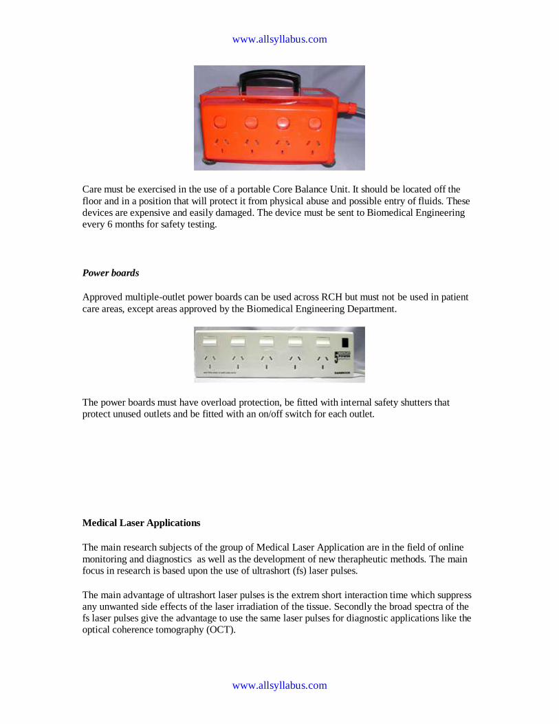
wwwallsyllabuscom
wwwallsyllabuscom
Care must be exercised in the use of a portable Core Balance Unit It should be located off the
floor and in a position that will protect it from physical abuse and possible entry of fluids These devices are expensive and easily damaged The device must be sent to Biomedical Engineering every 6 months for safety testing
Power boards
Approved multiple-outlet power boards can be used across RCH but must not be used in patient
care areas except areas approved by the Biomedical Engineering Department
The power boards must have overload protection be fitted with internal safety shutters that protect unused outlets and be fitted with an onoff switch for each outlet
Medical Laser Applications
The main research subjects of the group of Medical Laser Application are in the field of online
monitoring and diagnostics as well as the development of new therapheutic methods The main focus in research is based upon the use of ultrashort (fs) laser pulses
The main advantage of ultrashort laser pulses is the extrem short interaction time which suppress any unwanted side effects of the laser irradiation of the tissue Secondly the broad spectra of the fs laser pulses give the advantage to use the same laser pulses for diagnostic applications like the optical coherence tomography (OCT)
wwwallsyllabuscom
wwwallsyllabuscom
Following research subjects have special attention at the moment
Optical Coherence Tomographie (OCT)
A fs-laser light source can be used to obtain images from inside the tissue when the pulses are coherent superposed This group uses this application to obtain an inside view of the vocal fold and to measure the outline of the crystalline lens
Refractive Surgery
The principle of getting rid of glasses with the help of a laser surgery (fs-LASIK) is underlying rapid improvements concerning precision and safety New technologies were evaluated and transferred very fast in cooperation with an industrial partner
Treatment of Presbyopia
The flexibility of the crystalline lens can be increase by pricise cuts which are induce by fs laser pulse inside the lens The treatment of the presbyopia is thinkable
wwwallsyllabuscom
wwwallsyllabuscom
Ultrafast Physics
Whereas the applications of ultrashort laser pulses increase rapidly is the physics of the interaction between the laser pulses and tissue in many case not fully understood To obtain improved knowledge of the interaction for many fs-laser application this group preforms numerical simulations as well as fundamental experiments
Thermography
Thermography thermal imaging or thermal video is a type of infrared imaging Thermographic cameras detect radiation in the infrared range of the electromagnetic spectrum
(roughly 900ndash14000 nanometers or 09ndash14 microm) and produce images of that radiation Since infrared radiation is emitted by all objects based on their temperatures according to the black body radiation law thermography makes it possible to see ones environment with or without visible illumination The amount of radiation emitted by an object increases with temperature therefore thermography allows one to see variations in temperature (hence the name) When viewed by thermographic camera warm objects stand out well against cooler backgrounds humans and other warm-blooded animals become easily visible against the environment day or
night As a result thermographys extensive use can historically be ascribed to the military and security services
Thermal imaging photography finds many other uses For example firefighters use it to see through smoke find persons and localize the base of a fire With thermal imaging power lines maintenance technicians locate overheating joints and parts a telltale sign of their failure to eliminate potential hazards Where thermal insulation becomes faulty building construction technicians can see heat leaks to improve the efficiencies of cooling or heating air-conditioning Thermal imaging cameras are also installed in some luxury cars to aid the driver the first being
the 2000 Cadillac DeVille Some physiological activities particularly responses in human beings and other warm-blooded animals can also be monitored with thermographic imaging [1]
The appearance and operation of a modern thermographic camera is often similar to a camcorder Enabling the user to see in the infrared spectrum is a function so useful that ability to record their output is often optional A recording module is therefore not always built-in
Instead of CCD sensors most thermal imaging cameras use CMOS Focal Plane Array (FPA) The most common types are InSb InGaAs HgCdTe and QWIP FPA The newest technologies are using low cost and uncooled microbolometers FPA sensors Their resolution is considerably lower than of optical cameras mostly 160x120 or 320x240 pixels up to 640x512 for the most
expensive models Thermographic cameras are much more expensive than their visible-spectrum counterparts and higher-end models are often export-restricted Older bolometers or more
wwwallsyllabuscom
wwwallsyllabuscom
sensitive models as InSb require cryogenic cooling usually by a miniature Stirling cycle refrigerator or liquid nitrogen
Contents
[hide]
1 Difference between IR film and thermography 2 Advantages of Thermography
3 Limitations amp disadvantages of thermography 4 Applications 5 See also 6 External links
o 61 History of thermal imager manufacturers
[edit] Difference between IR film and thermography
Thermal imaging is going to be used on Mars to detect caves that could hold life
[edit] Advantages of Thermography
You get a visual picture so that you can compare temperatures over a large area
It is real time capable of catching moving targets Able to find deteriorating components prior to failure Measurement in areas inaccessible or hazardous for other methods It is a non-destructive test method
[edit] Limitations amp disadvantages of thermography
Quality cameras are expensive and are easily damaged Images can be hard to interpret accurately even with experience
Accurate temperature measurements are very hard to make because of emissivities Most cameras have plusmn2 or worse accuracy (not as accurate as contact) Training and staying proficient in IR scanning is time consuming Ability to only measure surface areas
[edit] Applications
Condition monitoring Medical imaging Night vision Research
Process control Non destructive testing Surveillance in security law enforcement and defense Chemical imaging
wwwallsyllabuscom
wwwallsyllabuscom
Thermal infrared imagers convert the energy in the infrared wavelength into a visible light video display All objects above 0 kelvins emit thermal infrared energy so thermal imagers can passively see all objects regardless of ambient light However most thermal imagers only see objects warmer than -50 degC
The spectrum and amount of thermal radiation depend strongly on an objects surface
temperature This makes it possible for a thermal camera to display an objects temperature However other factors also influence the radiation which limits the accuracy of this technique For example the radiation depends not only on the temperature of the object but is also a function of the emissivity of the object Also radiation also originates from the surroundings and is reflected in the object and the radiation from the object and the reflected radiation will also be influenced by the absorption of the atmosphere
Endoscopy
This page is about having an endoscopy - a test that looks at the inside of your digestive system
There is information on
What endoscopy is
Why you may have an endoscopy
Having an endoscopy
Your results
What an endoscopy is
An endoscopy is a test that looks inside the body The endoscope is a long flexible tube that can
be swallowed It has a camera and light inside it Some doctors call it a telescope
wwwallsyllabuscom
wwwallsyllabuscom
Why you may have an endoscopy
You are most likely to have an endoscopy to look at the inside of your
Gullet (oesophagus)
Stomach
Duodenum - the first part of the small bowel that attaches to the stomach
Large bowel (colon)
Your doctor may want to see inside because you have symptoms such as abnormal bleeding or
difficulty swallowing The doctor can look down the endoscope and see if there are any growths
or other abnormal looking areas Also through the endoscope the doctor can take samples
(biopsies) of any abnormal looking tissues
There is more detailed information about having a colonoscopy in the bowel cancer section of
CancerHelp UK Below is information about having other types of endoscopy
Having an endoscopy
You can have this test as an out patient Most people have a choice between having the test while
they are awake or after having a medicine to make them drowsy (a sedative) Your hospital may
not be happy for you to have a sedative if you live alone and will have no one to look after you
when you go home If you live alone but really want sedation your hospital may allow you to
stay overnight You should ask your doctor about this as early as possible as extra arrangements
will need to be made beforehand
If you dont have a sedative you will have a spray to numb the back of your throat and make it
easier for you to swallow the endoscopy tube
If you would prefer to be asleep during the test you will have an injection to make you very
drowsy just before the test You will need to take someone with you to the hospital appointment
You wont be able to drive for the rest of the day and should have someone to go home with you
You cant eat or drink for about 8 hours before the test so that your stomach and duodenum are
empty Your doctor will give you written instructions about this beforehand or they may arrive
with your appointment letter When you get to the clinic you may be asked to take your upper
clothing off and put on a hospital gown Some hospitals prefer to use gowns because your clothes
wont get messy Once you are ready you get onto the bed or X-ray couch When you are lying
comfortably you have the sedative injection to make you very drowsy Or your doctor will spray
the back of your throat to numb it
Once the sedative or throat spray has worked the doctor will pass the endoscope tube down your
wwwallsyllabuscom
wwwallsyllabuscom
throat to the area being investigated Your doctor will ask you to swallow as the tube goes down
but if youve had a sedative you wont remember that afterwards If there are any abnormalities
the doctor will take pieces of tissue from the abnormal looking area to send to the laboratory for
closer inspection under a microscope These tissue samples are called biopsies
When the test is over you will need to rest for a while If youve had a sedative you may not
remember much (if anything) about the test once you have come round You should be able to go
home the same day
The results
It can take time for test results to come through How long will depend on why you are having
the test Usually the doctor who carries out the endoscopy dictates a report straight way The
report is typed up by the department secretary and goes to your specialist who gives the results to
you If your GP has sent you for the test the results will go directly to the GP surgery
Understandably waiting for results can make you anxious It usually takes a couple of weeks for
the results to come through If your doctor needed them urgently it would have been noted on
the test request form and the results will be ready sooner than that Try to remember to ask your
doctor how long you should expect to wait for the results when you are first asked to go for the
test If it is not an emergency and you have not heard a couple of weeks after your test ring your
doctors secretary to check if they are back
Endoscopy
From Wikipedia the free encyclopedia
Jump to navigation search
Endoscopic images of a duodenal ulcer
wwwallsyllabuscom
wwwallsyllabuscom
A flexible endoscope
Endoscopy means looking inside and typically refers to looking inside the human body for medical reasons using an instrument called an endoscope Endoscopy can also refer to using a borescope in technical situations where direct line-of-sight observation is not feasible
Contents
[hide]
1 Overview 2 Components 3 Uses
4 History 5 Risks 6 After The Endoscopy 7 Recent developments 8 See also 9 References 10 Footnotes
11 External links
Overview
Endoscopy is a minimally invasive diagnostic medical procedure that is used to assess the interior surfaces of an organ by inserting a tube into the body The instrument may have a rigid or flexible tube and not only provide an image for visual inspection and photography but also enable taking
biopsies and retrieval of foreign objects Endoscopy is the vehicle for minimally invasive surgery
Many endoscopic procedures are considered to be relatively painless and at worst associated with mild discomfort for example in esophagogastroduodenoscopy most patients tolerate the procedure with only topical anaesthesia of the oropharynx using lignocaine spray[1] Complications are not common (only 5 of all operations)[citation needed] but can include perforation of the organ under inspection with the endoscope or biopsy instrument If that occurs open surgery may be required to repair the injury
wwwallsyllabuscom
wwwallsyllabuscom
Components
An endoscope can consist of
a rigid or flexible tube a light delivery system to illuminate the organ or object under inspection The light
source is normally outside the body and the light is typically directed via an optical fiber system
a lens system transmitting the image to the viewer from the fiberscope an additional channel to allow entry of medical instruments or manipulators
Uses
Endoscopy can involve
The gastrointestinal tract (GI tract) o esophagus stomach and duodenum (esophagogastroduodenoscopy) o small intestine o colon (colonoscopyproctosigmoidoscopy) o Bile duct
endoscopic retrograde cholangiopancreatography (ERCP) duodenoscope-assisted cholangiopancreatoscopy intraoperative cholangioscopy
The respiratory tract o The nose (rhinoscopy) o The lower respiratory tract (bronchoscopy)
The urinary tract (cystoscopy) The female reproductive system
o The cervix (colposcopy) o The uterus (hysteroscopy) o The Fallopian tubes (Falloscopy)
Normally closed body cavities (through a small incision) o The abdominal or pelvic cavity (laparoscopy) o The interior of a joint (arthroscopy) o Organs of the chest (thoracoscopy and mediastinoscopy)
During pregnancy o The amnion (amnioscopy) o The fetus (fetoscopy)
Plastic Surgery Panendoscopy (or triple endoscopy)
o Combines laryngoscopy esophagoscopy and bronchoscopy Non-medical uses for endoscopy
o The planning and architectural community have found the endoscope useful for
pre-visualization of scale models of proposed buildings and cities (architectural endoscopy)
o Internal inspection of complex technical systems (borescope) o Endoscopes are also a tool helpful in the examination of improvised explosive
devices by bomb disposal personnel
wwwallsyllabuscom
wwwallsyllabuscom
o The FBI uses endoscopes for conducting surveillance via tight spaces
History
The first endoscope of a kind was developed in 1806 by Philip Bozzini with his introduction of a
Lichtleiter (light conductor) for the examinations of the canals and cavities of the human body However the Vienna Medical Society disapproved of such curiosity An endoscope was first introduced into a human in 1822 by William Beaumont an army surgeon at Mackinac
Island Michigan[citation needed] The use of electric light was a major step in the improvement of endoscopy The first such lights were external Later smaller bulbs became available making internal light possible for instance in a hysteroscope by Charles David in 1908[citation needed] Hans Christian Jacobaeus has been given credit for early endoscopic explorations of the abdomen and the thorax with laparoscopy (1912) and thoracoscopy (1910)[citation needed] Laparoscopy was used in the diagnosis of liver and gallbladder disease by Heinz Kalk in the 1930s[citation needed] Hope reported in 1937 on the use of laparoscopy to diagnose ectopic pregnancy[citation needed] In 1944
Raoul Palmer placed his patients in the Trendelenburg position after gaseous distention of the abdomen and thus was able to reliably perform gynecologic laparoscopy[citation needed]
The first gastrocamera was released in 1950 by Olympus Optical Co Ltd The device took pictures on monochromatic film using a small light bulb that was triggered manually The device was of limited use however because it did not implement real-time optical capability Olympus continued its development of endoscopes by incorporating fiber optics in the early 1960s leading to the first useful endoscopes In 1964 it released a gastrocamera guided by a fiberscope[1] A few articles claim that DrBasil Hirschowitz of UnivOf MichiganAnn Arbor discussed the
endoscope in early 50s[2]
As endoscopic technology improved so did the methods of gastrointestinal endoscopy Owing primarily to the efforts of Dr Hiromi Shinya in the late 1960s GI endoscopy developed into what is more recognizable as todays colonoscopy While many doctors experimented with techniques to take advantage of the new iterations of endoscopes Dr Shinya focused on techniques that would allow for successful operation of the endoscope by an individual rejecting the common practice at the time of utilizing two people Consequently many of the fundamental methods and procedures of modern colonoscopy were developed by Dr Shinya
Dr Shinyas other great contribution was to therapeutic endoscopy in his invention of the
electrosurgical polypectomy snare with the aid of Olympus employee Hiroshi Ichikawa Shinya sketched his first plans for the device on January 8 1969 He envisioned a loop of wire attached to the end of a colonoscope that would allow for easy removal of polyps during investigation by passing a current through the wire By September of 1969 the first polypectomy using this device was performed Polypectomy has since become the most common therapeutic procedure performed with an endoscope (Sivak 2004)
By 1980 laparoscopy training was required by gynecologists to perform tubal ligation procedures and diagnostic evaluations of the pelvis The first laparoscopic cholecystectomy was performed in 1984 and the first video-laparoscopic cholecystectomy in 1987[citation needed] During the 1990s
laparoscopic surgery was extended to the appendix spleen colon stomach kidney and liver[citation needed] Wireless capsule endoscopy or Capsule Endoscopy is now approved in all the countries including Japan where government reimbusement will be available from Oct2007Capsule Endoscopy [3] increases detection of Small Bowel tumors where traditional Endoscopy is not very efficient
wwwallsyllabuscom
wwwallsyllabuscom
Risks
Infection
Punctured organs Allergic reactions due to Contrast agents or dyes (such as those used in a CT scan) Over-sedation
After The Endoscopy
After the procedure the patient will be observed and monitored by a qualified individual in the
endoscopy or a recovery area until a significant portion of the medication has worn off Occasionally a patient is left with a mild sore throat which promptly responds to saline gargles or a feeling of distention from the insufflated air that was used during the procedure Both problems are mild and fleeting When fully recovered the patient will be instructed when to resume hisher usual diet (probably within a few hours) and will be allowed to be taken home Because of the use of sedation most facilities mandate that the patient is taken home by another person and not to drive on hisher own or handle machinery for the remainder of the day
Recent developments
With the application of robotic systems telesurgery was introduced as the surgeon could operate
from a site physically removed from the patient The first transatlantic surgery has been called the Lindbergh Operation
wwwallsyllabuscom
wwwallsyllabuscom
Upper Endoscopy
Upper endoscopy enables the physician to look inside the esophagus stomach and duodenum (first part of the small intestine) The procedure might be used to discover the reason for swallowing difficulties nausea vomiting
reflux bleeding indigestion abdominal pain or chest pain Upper endoscopy is also called EGD which stands for esophagogastroduodenoscopy (eh-SAH-fuh-goh-GAS-troh-doo-AH-duh-NAH-skuh-pee)
For the procedure you will swallow a thin flexible lighted tube called an endoscope (EN-doh-skope) Right before the procedure the physician will spray your throat with a numbing agent that may help prevent gagging
You may also receive pain medicine and a sedative to help you relax during the exam The endoscope transmits an image of the inside of the esophagus stomach and duodenum so the physician can carefully examine the lining of these organs The scope also blows air into the stomach this expands the folds of tissue and makes it easier for the physician to examine the stomach
The physician can see abnormalities like inflammation or
bleeding through the endoscope that dont show up well on x rays The physician can also insert instruments into the scope to treat bleeding abnormalities or remove samples of tissue (biopsy) for further tests
Possible complications of upper endoscopy include bleeding and puncture of the stomach lining However such complications are rare Most people will probably have nothing more than a mild sore throat after the procedure
The procedure takes 20 to 30 minutes Because you will be sedated you will need to rest at the endoscopy facility for 1 to 2 hours until the medication wears off
Preparation
Your stomach and duodenum must be empty for the procedure to be thorough and safe so you will not be able to eat or drink anything for at least 6 hours beforehand Also you must
arrange for someone to take you homemdashyou will not be allowed to drive because of the sedatives Your physician may give you other special instructions
The digestive system
wwwallsyllabuscom
wwwallsyllabuscom
What is Endoscopy and Why is it Performed
Endoscopy allows physicians to peer through the bodys passageways Endoscopy is the examination and inspection of the interior of body organs joints or cavities through an endoscope An endoscope is a device that uses fiber optics and powerful lens systems to provide lighting and visualization of the interior of a joint The portion of the endoscope inserted into the body may be rigid or flexible depending upon the medical procedure
An endoscope uses two fiber optic lines A light fiber carries light into the body cavity and an image fiber carries the image of the body cavity back to the physicians viewing lens There is also a separate port to allow for administration of drugs suction and irrigation This port may also be used to introduce small folding instruments such as forceps scissors brushes snares and baskets for tissue excision (removal) sampling or other diagnostic and therapeutic work Endoscopes may be used in conjunction with a camera or video recorder to document images of the inside of the joint or chronicle an endoscopic procedure New endoscopes have digital capabilities for manipulating and enhancing the video images
This figure shows a rigid endoscope used for arthroscopy The image fiber leads from the ocular (eye piece) to the inserted end of the scope The light fiber is below and leads from the light source to the working end of the endoscope
Why Is Endoscopy Performed
Endoscopy can be used to diagnose various conditions by close examination of internal organ and body structures Endoscopy can also guide therapy and repair such as the removal of torn cartilage from the bearing surfaces of a joint Biopsy (tissue sampling for pathologic testing) may also be performed under endoscopic guidance Local or general anesthetic may be used during endoscopy depending upon the type of procedure being performed
Internal abnormalities revealed through endoscopy include abscesses biliary (liver) cirrhosis bleeding bronchitis cancer cysts degenerative disease gallbladder stones hernia inflammation metastatic cancer polyps tumors ulcers and other diseases and conditions
Endoscopy is a minimally invasive procedure and carries with it certain minor risks depending upon the type of procedure being performed However these risks are typically far outweighed by the diagnostic and therapeutic potential of the procedure
Prior to the widespread use of endoscopy and diagnostic imaging most internal conditions could only be diagnosed or treated with open surgery Until the last several decades exploratory surgery was routinely performed only when a patient was critically ill and the source of illness was not known For example in certain dire cases the patients thorax or abdomen were surgically opened and examined to try to determine the source of illness
wwwallsyllabuscom
wwwallsyllabuscom
Endoscopy can often be done on an outpatient basis Outpatient means that the procedure does not require hospital admission and acute care and observation and may be performed outside the premises of a hospital Outpatient procedures performed at hospitals or ambulatory centers allow the patient to go home or return to work within a short while after their procedure
Types of Endoscopy
Fiber optic endoscopes now have widespread use in medicine and guide a myriad of diagnostic and therapeutic procedures including
Arthroscopy examination of joints for diagnosis and treatment (arthroscopic surgery) Bronchoscopy examination of the trachea and lungs bronchial trees to reveal
abscesses bronchitis carcinoma tumors tuberculosis alveolitis infection inflammation Colonoscopy examination of the inside of the colon and large intestine to detect polyps
tumors ulceration inflammation colitis diverticula Chrohns disease and discovery and removal of foreign bodies
Colposcopy direct visualization of the vagina and cervix to detect cancer inflammation and other conditions
Cystoscopy examination of the bladder urethra urinary tract uteral orifices and
prostate (men) with insertion of the endoscope through the urethra
ERCP (endoscopic retrograde cholangio-pancreatography) uses endoscopic guidance to
place a catheter for x-ray fluorosocopy with contrast enhancement This technique is used to examine the livers biliary tree the gallbladder the pancreatic duct and other anatomy to check for stones other obstructions and disease X-ray contrast is introduced into these ducts via catheter and fluoroscopic x-ray images are taken to show any abnormality or blockage If disease is detected it can sometimes be treated at the same time or biopsy can be performed to test for cancer or other pathology ERCP can detect biliary cirrhosis
cancer of the bile ducts pancreatic cysts pseudocysts pancreatic tumors chronic pancreatitis and other conditions such as gallbladder stones
EGD (Esophogealgastroduodensoscopy) visual examination of the upper gastro-
intestinal (GI) tract (also referred to as gastroscopy) to reveal hemorrhage hiatal hernia inflammation of the esophagus gastric ulcers
Endoscopic biopsy is the removal of tissue specimens for pathologic examination and
analysis Gastroscopy examination of the lining of the esophagus stomach and duodenum
Gastroscopy is often used to diagnose ulcers and other sources of bleeding and to guide biopsy of suspect GI cancers
Laparoscopy visualization of the stomach liver and other abdominal organs including
the female reproductive organs for example the fallopian tubes Laryngoscopy examination of the larynx (voice box) Proctoscopy sigmoidoscopy proctosigmoidoscopy examination of the rectum and
sigmoid colon Thoracoscopy examination of the pleura (sac that covers the lungs) pleural spaces
mediastinum and pericardium
A Brief History of Endoscopy
In the early 1900s the first attempts to view inside the body with lighted telescopes were made These initial devices were often fully rigid In the 1930s semi-flexible endoscopes called gastroscopes were developed to view inside of the stomach Fiber-optic endoscopy was pioneered by South African-born physician Basil Hirschowitz at the University of Michigan in 1957 Widespread use of fiber optic endoscopes began in the 1960s
wwwallsyllabuscom
wwwallsyllabuscom
A fiber optic cable is simply a bundle of microscopic glass or plastic fibers that literally allows light and images to be transmitted through curved structures Fiber optic cables are also replacing metal wires as the backbone of the worlds telecommunications infrastructure This Internet page may have traveled through a fiber optic cable as a stream of digital data (bursts of light) on its way to your computer
Endoscopy Equipment
Endoscopes have many practical needs And HMB Endoscopy Products (Hollywood Florida) has been providing endoscopic equipment and educating people on the use of endoscopes for more than 17 years Be sure to Browse our Catalog for all the details on how to purchase these medical instruments that can examine any part of the body
Video
Systems
Fiber
Endoscopes
Video
Endoscopes
In the simplest terms Endoscopy equipment consists of instruments that can look at the inside of many different organs mdash these are small flexible or rigid tubes with a light or lenses on the end that can look into the esophagus stomach and colon mdash and in more general terms endoscopy equipment can help doctors look deep inside body structures and hollow organs
An endoscope and related endoscope products and equipment are usually composed of three components
An optic system that allows the doctor to look through the scope into the organ or
cavity or to attach a video camera to the scope
A fiberoptic cable to light up the bodily area
A lumen (eg the bore of a tube like a needle or catheter) to take tissue samples of the
area being viewed
The beauty of endoscopic products is that they perform dual functions mdash with both diagnostic and therapeutic capabilities For example this means that these endoscopic products and instruments can perform biopsies (eg to evaluate tissue samples) as well as provide sclerotherapy (a medical procedure used to treat varicose veins and ―spider veins) In truth these brief explanations only tell part of the story
wwwallsyllabuscom
wwwallsyllabuscom
TYPES OF ENDOSCOPES
Take a look at the different types of endoscopic equipment you can get with HMB Endoscopy Products Again in very general terms there are two main types of endoscopes
Rigid Flexible
1 rigid endoscopes ndash the majority of which use a convex (curving out like one half of a
circle) glass lens system in which the small glass lenses are separated by large air spaces
2 flexible endoscopes allow for just that mdash flexibility In the animal kingdom for example
a flexible endoscope would be perfect for examining the stomach area of a snake
The popularity of endoscopy equipment continues to grow Some of the industryrsquos household names include Olympus Pentax Funjinon and Storz mdash with HMB both selling and repairing pre-owned and completely refurbished endoscopic products from each of these major manufacturers Browse our Catalog to get the ones you need
THERMOGRAPH APPLICATION Thermographs are digital recording thermometers used to log
temperature in the
marine environment The instruments are attached to a computer to set up recording
parameters for
deployment downloading and display of data in graphic or numeric format These units
are placed in
underwater housings and attached to the bottom or suspended in the water column for
deployment
DESCRIPTION Two types of thermograph are currently available The Ryan
Tempmentor is a
reusable data logger that allows storage of a maximum of 6361 temperature
measurements in the
range of -32C to +70C with 01resolution and 03accuracy The instrument can be
programmed
to take measurements from once per second to once every other hour Instrument
dimensions are 3 x
6 x 13 and the unit weighs 11 ounces The Hobo Temp is a miniature reusable data
logger that
allows storage of 1800 measurements in the range of -20C to +70C with
03resolution and 07
wwwallsyllabuscom
wwwallsyllabuscom
accuracy Sampling intervals from 05 seconds to 48 hours are available Instrument
dimensions are
24 x 19 x 08 and the unit weighs 1 ounce Both units are battery powered with the
battery life of
the Tempmentor being approximately two years and the battery life of the Hobo Temp
being one year THERMOGRAPH APPLICATION Thermographs are digital recording thermometers used to log
temperature in the
marine environment The instruments are attached to a computer to set up recording
parameters for
deployment downloading and display of data in graphic or numeric format These units
are placed in
underwater housings and attached to the bottom or suspended in the water column for
deployment
DESCRIPTION Two types of thermograph are currently available The Ryan
Tempmentor is a
reusable data logger that allows storage of a maximum of 6361 temperature
measurements in the
range of -32C to +70C with 01resolution and 03accuracy The instrument can be
programmed
to take measurements from once per second to once every other hour Instrument
dimensions are 3 x
6 x 13 and the unit weighs 11 ounces The Hobo Temp is a miniature reusable data
logger that
allows storage of 1800 measurements in the range of -20C to +70C with
03resolution and 07
accuracy Sampling intervals from 05 seconds to 48 hours are available Instrument
dimensions are
24 x 19 x 08 and the unit weighs 1 ounce Both units are battery powered with the
battery life of
the Tempmentor being approximately two years and the battery life of the Hobo Temp
being one year THERMOGRAPH APPLICATION Thermographs are digital recording thermometers used to log
temperature in the
marine environment The instruments are attached to a computer to set up recording
parameters for
deployment downloading and display of data in graphic or numeric format These units
are placed in
underwater housings and attached to the bottom or suspended in the water column for
deployment
DESCRIPTION Two types of thermograph are currently available The Ryan
Tempmentor is a
reusable data logger that allows storage of a maximum of 6361 temperature
measurements in the
range of -32C to +70C with 01resolution and 03accuracy The instrument can be
programmed
wwwallsyllabuscom
wwwallsyllabuscom
to take measurements from once per second to once every other hour Instrument
dimensions are 3 x
6 x 13 and the unit weighs 11 ounces The Hobo Temp is a miniature reusable data
logger that
allows storage of 1800 measurements in the range of -20C to +70C with
03resolution and 07
accuracy Sampling intervals from 05 seconds to 48 hours are available Instrument
dimensions are
24 x 19 x 08 and the unit weighs 1 ounce Both units are battery powered with the
battery life of
the Tempmentor being approximately two years and the battery life of the Hobo Temp
being one year
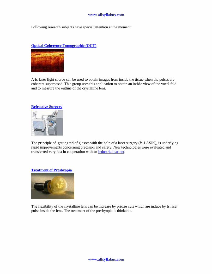
wwwallsyllabuscom
wwwallsyllabuscom
Following research subjects have special attention at the moment
Optical Coherence Tomographie (OCT)
A fs-laser light source can be used to obtain images from inside the tissue when the pulses are coherent superposed This group uses this application to obtain an inside view of the vocal fold and to measure the outline of the crystalline lens
Refractive Surgery
The principle of getting rid of glasses with the help of a laser surgery (fs-LASIK) is underlying rapid improvements concerning precision and safety New technologies were evaluated and transferred very fast in cooperation with an industrial partner
Treatment of Presbyopia
The flexibility of the crystalline lens can be increase by pricise cuts which are induce by fs laser pulse inside the lens The treatment of the presbyopia is thinkable
wwwallsyllabuscom
wwwallsyllabuscom
Ultrafast Physics
Whereas the applications of ultrashort laser pulses increase rapidly is the physics of the interaction between the laser pulses and tissue in many case not fully understood To obtain improved knowledge of the interaction for many fs-laser application this group preforms numerical simulations as well as fundamental experiments
Thermography
Thermography thermal imaging or thermal video is a type of infrared imaging Thermographic cameras detect radiation in the infrared range of the electromagnetic spectrum
(roughly 900ndash14000 nanometers or 09ndash14 microm) and produce images of that radiation Since infrared radiation is emitted by all objects based on their temperatures according to the black body radiation law thermography makes it possible to see ones environment with or without visible illumination The amount of radiation emitted by an object increases with temperature therefore thermography allows one to see variations in temperature (hence the name) When viewed by thermographic camera warm objects stand out well against cooler backgrounds humans and other warm-blooded animals become easily visible against the environment day or
night As a result thermographys extensive use can historically be ascribed to the military and security services
Thermal imaging photography finds many other uses For example firefighters use it to see through smoke find persons and localize the base of a fire With thermal imaging power lines maintenance technicians locate overheating joints and parts a telltale sign of their failure to eliminate potential hazards Where thermal insulation becomes faulty building construction technicians can see heat leaks to improve the efficiencies of cooling or heating air-conditioning Thermal imaging cameras are also installed in some luxury cars to aid the driver the first being
the 2000 Cadillac DeVille Some physiological activities particularly responses in human beings and other warm-blooded animals can also be monitored with thermographic imaging [1]
The appearance and operation of a modern thermographic camera is often similar to a camcorder Enabling the user to see in the infrared spectrum is a function so useful that ability to record their output is often optional A recording module is therefore not always built-in
Instead of CCD sensors most thermal imaging cameras use CMOS Focal Plane Array (FPA) The most common types are InSb InGaAs HgCdTe and QWIP FPA The newest technologies are using low cost and uncooled microbolometers FPA sensors Their resolution is considerably lower than of optical cameras mostly 160x120 or 320x240 pixels up to 640x512 for the most
expensive models Thermographic cameras are much more expensive than their visible-spectrum counterparts and higher-end models are often export-restricted Older bolometers or more
wwwallsyllabuscom
wwwallsyllabuscom
sensitive models as InSb require cryogenic cooling usually by a miniature Stirling cycle refrigerator or liquid nitrogen
Contents
[hide]
1 Difference between IR film and thermography 2 Advantages of Thermography
3 Limitations amp disadvantages of thermography 4 Applications 5 See also 6 External links
o 61 History of thermal imager manufacturers
[edit] Difference between IR film and thermography
Thermal imaging is going to be used on Mars to detect caves that could hold life
[edit] Advantages of Thermography
You get a visual picture so that you can compare temperatures over a large area
It is real time capable of catching moving targets Able to find deteriorating components prior to failure Measurement in areas inaccessible or hazardous for other methods It is a non-destructive test method
[edit] Limitations amp disadvantages of thermography
Quality cameras are expensive and are easily damaged Images can be hard to interpret accurately even with experience
Accurate temperature measurements are very hard to make because of emissivities Most cameras have plusmn2 or worse accuracy (not as accurate as contact) Training and staying proficient in IR scanning is time consuming Ability to only measure surface areas
[edit] Applications
Condition monitoring Medical imaging Night vision Research
Process control Non destructive testing Surveillance in security law enforcement and defense Chemical imaging
wwwallsyllabuscom
wwwallsyllabuscom
Thermal infrared imagers convert the energy in the infrared wavelength into a visible light video display All objects above 0 kelvins emit thermal infrared energy so thermal imagers can passively see all objects regardless of ambient light However most thermal imagers only see objects warmer than -50 degC
The spectrum and amount of thermal radiation depend strongly on an objects surface
temperature This makes it possible for a thermal camera to display an objects temperature However other factors also influence the radiation which limits the accuracy of this technique For example the radiation depends not only on the temperature of the object but is also a function of the emissivity of the object Also radiation also originates from the surroundings and is reflected in the object and the radiation from the object and the reflected radiation will also be influenced by the absorption of the atmosphere
Endoscopy
This page is about having an endoscopy - a test that looks at the inside of your digestive system
There is information on
What endoscopy is
Why you may have an endoscopy
Having an endoscopy
Your results
What an endoscopy is
An endoscopy is a test that looks inside the body The endoscope is a long flexible tube that can
be swallowed It has a camera and light inside it Some doctors call it a telescope
wwwallsyllabuscom
wwwallsyllabuscom
Why you may have an endoscopy
You are most likely to have an endoscopy to look at the inside of your
Gullet (oesophagus)
Stomach
Duodenum - the first part of the small bowel that attaches to the stomach
Large bowel (colon)
Your doctor may want to see inside because you have symptoms such as abnormal bleeding or
difficulty swallowing The doctor can look down the endoscope and see if there are any growths
or other abnormal looking areas Also through the endoscope the doctor can take samples
(biopsies) of any abnormal looking tissues
There is more detailed information about having a colonoscopy in the bowel cancer section of
CancerHelp UK Below is information about having other types of endoscopy
Having an endoscopy
You can have this test as an out patient Most people have a choice between having the test while
they are awake or after having a medicine to make them drowsy (a sedative) Your hospital may
not be happy for you to have a sedative if you live alone and will have no one to look after you
when you go home If you live alone but really want sedation your hospital may allow you to
stay overnight You should ask your doctor about this as early as possible as extra arrangements
will need to be made beforehand
If you dont have a sedative you will have a spray to numb the back of your throat and make it
easier for you to swallow the endoscopy tube
If you would prefer to be asleep during the test you will have an injection to make you very
drowsy just before the test You will need to take someone with you to the hospital appointment
You wont be able to drive for the rest of the day and should have someone to go home with you
You cant eat or drink for about 8 hours before the test so that your stomach and duodenum are
empty Your doctor will give you written instructions about this beforehand or they may arrive
with your appointment letter When you get to the clinic you may be asked to take your upper
clothing off and put on a hospital gown Some hospitals prefer to use gowns because your clothes
wont get messy Once you are ready you get onto the bed or X-ray couch When you are lying
comfortably you have the sedative injection to make you very drowsy Or your doctor will spray
the back of your throat to numb it
Once the sedative or throat spray has worked the doctor will pass the endoscope tube down your
wwwallsyllabuscom
wwwallsyllabuscom
throat to the area being investigated Your doctor will ask you to swallow as the tube goes down
but if youve had a sedative you wont remember that afterwards If there are any abnormalities
the doctor will take pieces of tissue from the abnormal looking area to send to the laboratory for
closer inspection under a microscope These tissue samples are called biopsies
When the test is over you will need to rest for a while If youve had a sedative you may not
remember much (if anything) about the test once you have come round You should be able to go
home the same day
The results
It can take time for test results to come through How long will depend on why you are having
the test Usually the doctor who carries out the endoscopy dictates a report straight way The
report is typed up by the department secretary and goes to your specialist who gives the results to
you If your GP has sent you for the test the results will go directly to the GP surgery
Understandably waiting for results can make you anxious It usually takes a couple of weeks for
the results to come through If your doctor needed them urgently it would have been noted on
the test request form and the results will be ready sooner than that Try to remember to ask your
doctor how long you should expect to wait for the results when you are first asked to go for the
test If it is not an emergency and you have not heard a couple of weeks after your test ring your
doctors secretary to check if they are back
Endoscopy
From Wikipedia the free encyclopedia
Jump to navigation search
Endoscopic images of a duodenal ulcer
wwwallsyllabuscom
wwwallsyllabuscom
A flexible endoscope
Endoscopy means looking inside and typically refers to looking inside the human body for medical reasons using an instrument called an endoscope Endoscopy can also refer to using a borescope in technical situations where direct line-of-sight observation is not feasible
Contents
[hide]
1 Overview 2 Components 3 Uses
4 History 5 Risks 6 After The Endoscopy 7 Recent developments 8 See also 9 References 10 Footnotes
11 External links
Overview
Endoscopy is a minimally invasive diagnostic medical procedure that is used to assess the interior surfaces of an organ by inserting a tube into the body The instrument may have a rigid or flexible tube and not only provide an image for visual inspection and photography but also enable taking
biopsies and retrieval of foreign objects Endoscopy is the vehicle for minimally invasive surgery
Many endoscopic procedures are considered to be relatively painless and at worst associated with mild discomfort for example in esophagogastroduodenoscopy most patients tolerate the procedure with only topical anaesthesia of the oropharynx using lignocaine spray[1] Complications are not common (only 5 of all operations)[citation needed] but can include perforation of the organ under inspection with the endoscope or biopsy instrument If that occurs open surgery may be required to repair the injury
wwwallsyllabuscom
wwwallsyllabuscom
Components
An endoscope can consist of
a rigid or flexible tube a light delivery system to illuminate the organ or object under inspection The light
source is normally outside the body and the light is typically directed via an optical fiber system
a lens system transmitting the image to the viewer from the fiberscope an additional channel to allow entry of medical instruments or manipulators
Uses
Endoscopy can involve
The gastrointestinal tract (GI tract) o esophagus stomach and duodenum (esophagogastroduodenoscopy) o small intestine o colon (colonoscopyproctosigmoidoscopy) o Bile duct
endoscopic retrograde cholangiopancreatography (ERCP) duodenoscope-assisted cholangiopancreatoscopy intraoperative cholangioscopy
The respiratory tract o The nose (rhinoscopy) o The lower respiratory tract (bronchoscopy)
The urinary tract (cystoscopy) The female reproductive system
o The cervix (colposcopy) o The uterus (hysteroscopy) o The Fallopian tubes (Falloscopy)
Normally closed body cavities (through a small incision) o The abdominal or pelvic cavity (laparoscopy) o The interior of a joint (arthroscopy) o Organs of the chest (thoracoscopy and mediastinoscopy)
During pregnancy o The amnion (amnioscopy) o The fetus (fetoscopy)
Plastic Surgery Panendoscopy (or triple endoscopy)
o Combines laryngoscopy esophagoscopy and bronchoscopy Non-medical uses for endoscopy
o The planning and architectural community have found the endoscope useful for
pre-visualization of scale models of proposed buildings and cities (architectural endoscopy)
o Internal inspection of complex technical systems (borescope) o Endoscopes are also a tool helpful in the examination of improvised explosive
devices by bomb disposal personnel
wwwallsyllabuscom
wwwallsyllabuscom
o The FBI uses endoscopes for conducting surveillance via tight spaces
History
The first endoscope of a kind was developed in 1806 by Philip Bozzini with his introduction of a
Lichtleiter (light conductor) for the examinations of the canals and cavities of the human body However the Vienna Medical Society disapproved of such curiosity An endoscope was first introduced into a human in 1822 by William Beaumont an army surgeon at Mackinac
Island Michigan[citation needed] The use of electric light was a major step in the improvement of endoscopy The first such lights were external Later smaller bulbs became available making internal light possible for instance in a hysteroscope by Charles David in 1908[citation needed] Hans Christian Jacobaeus has been given credit for early endoscopic explorations of the abdomen and the thorax with laparoscopy (1912) and thoracoscopy (1910)[citation needed] Laparoscopy was used in the diagnosis of liver and gallbladder disease by Heinz Kalk in the 1930s[citation needed] Hope reported in 1937 on the use of laparoscopy to diagnose ectopic pregnancy[citation needed] In 1944
Raoul Palmer placed his patients in the Trendelenburg position after gaseous distention of the abdomen and thus was able to reliably perform gynecologic laparoscopy[citation needed]
The first gastrocamera was released in 1950 by Olympus Optical Co Ltd The device took pictures on monochromatic film using a small light bulb that was triggered manually The device was of limited use however because it did not implement real-time optical capability Olympus continued its development of endoscopes by incorporating fiber optics in the early 1960s leading to the first useful endoscopes In 1964 it released a gastrocamera guided by a fiberscope[1] A few articles claim that DrBasil Hirschowitz of UnivOf MichiganAnn Arbor discussed the
endoscope in early 50s[2]
As endoscopic technology improved so did the methods of gastrointestinal endoscopy Owing primarily to the efforts of Dr Hiromi Shinya in the late 1960s GI endoscopy developed into what is more recognizable as todays colonoscopy While many doctors experimented with techniques to take advantage of the new iterations of endoscopes Dr Shinya focused on techniques that would allow for successful operation of the endoscope by an individual rejecting the common practice at the time of utilizing two people Consequently many of the fundamental methods and procedures of modern colonoscopy were developed by Dr Shinya
Dr Shinyas other great contribution was to therapeutic endoscopy in his invention of the
electrosurgical polypectomy snare with the aid of Olympus employee Hiroshi Ichikawa Shinya sketched his first plans for the device on January 8 1969 He envisioned a loop of wire attached to the end of a colonoscope that would allow for easy removal of polyps during investigation by passing a current through the wire By September of 1969 the first polypectomy using this device was performed Polypectomy has since become the most common therapeutic procedure performed with an endoscope (Sivak 2004)
By 1980 laparoscopy training was required by gynecologists to perform tubal ligation procedures and diagnostic evaluations of the pelvis The first laparoscopic cholecystectomy was performed in 1984 and the first video-laparoscopic cholecystectomy in 1987[citation needed] During the 1990s
laparoscopic surgery was extended to the appendix spleen colon stomach kidney and liver[citation needed] Wireless capsule endoscopy or Capsule Endoscopy is now approved in all the countries including Japan where government reimbusement will be available from Oct2007Capsule Endoscopy [3] increases detection of Small Bowel tumors where traditional Endoscopy is not very efficient
wwwallsyllabuscom
wwwallsyllabuscom
Risks
Infection
Punctured organs Allergic reactions due to Contrast agents or dyes (such as those used in a CT scan) Over-sedation
After The Endoscopy
After the procedure the patient will be observed and monitored by a qualified individual in the
endoscopy or a recovery area until a significant portion of the medication has worn off Occasionally a patient is left with a mild sore throat which promptly responds to saline gargles or a feeling of distention from the insufflated air that was used during the procedure Both problems are mild and fleeting When fully recovered the patient will be instructed when to resume hisher usual diet (probably within a few hours) and will be allowed to be taken home Because of the use of sedation most facilities mandate that the patient is taken home by another person and not to drive on hisher own or handle machinery for the remainder of the day
Recent developments
With the application of robotic systems telesurgery was introduced as the surgeon could operate
from a site physically removed from the patient The first transatlantic surgery has been called the Lindbergh Operation
wwwallsyllabuscom
wwwallsyllabuscom
Upper Endoscopy
Upper endoscopy enables the physician to look inside the esophagus stomach and duodenum (first part of the small intestine) The procedure might be used to discover the reason for swallowing difficulties nausea vomiting
reflux bleeding indigestion abdominal pain or chest pain Upper endoscopy is also called EGD which stands for esophagogastroduodenoscopy (eh-SAH-fuh-goh-GAS-troh-doo-AH-duh-NAH-skuh-pee)
For the procedure you will swallow a thin flexible lighted tube called an endoscope (EN-doh-skope) Right before the procedure the physician will spray your throat with a numbing agent that may help prevent gagging
You may also receive pain medicine and a sedative to help you relax during the exam The endoscope transmits an image of the inside of the esophagus stomach and duodenum so the physician can carefully examine the lining of these organs The scope also blows air into the stomach this expands the folds of tissue and makes it easier for the physician to examine the stomach
The physician can see abnormalities like inflammation or
bleeding through the endoscope that dont show up well on x rays The physician can also insert instruments into the scope to treat bleeding abnormalities or remove samples of tissue (biopsy) for further tests
Possible complications of upper endoscopy include bleeding and puncture of the stomach lining However such complications are rare Most people will probably have nothing more than a mild sore throat after the procedure
The procedure takes 20 to 30 minutes Because you will be sedated you will need to rest at the endoscopy facility for 1 to 2 hours until the medication wears off
Preparation
Your stomach and duodenum must be empty for the procedure to be thorough and safe so you will not be able to eat or drink anything for at least 6 hours beforehand Also you must
arrange for someone to take you homemdashyou will not be allowed to drive because of the sedatives Your physician may give you other special instructions
The digestive system
wwwallsyllabuscom
wwwallsyllabuscom
What is Endoscopy and Why is it Performed
Endoscopy allows physicians to peer through the bodys passageways Endoscopy is the examination and inspection of the interior of body organs joints or cavities through an endoscope An endoscope is a device that uses fiber optics and powerful lens systems to provide lighting and visualization of the interior of a joint The portion of the endoscope inserted into the body may be rigid or flexible depending upon the medical procedure
An endoscope uses two fiber optic lines A light fiber carries light into the body cavity and an image fiber carries the image of the body cavity back to the physicians viewing lens There is also a separate port to allow for administration of drugs suction and irrigation This port may also be used to introduce small folding instruments such as forceps scissors brushes snares and baskets for tissue excision (removal) sampling or other diagnostic and therapeutic work Endoscopes may be used in conjunction with a camera or video recorder to document images of the inside of the joint or chronicle an endoscopic procedure New endoscopes have digital capabilities for manipulating and enhancing the video images
This figure shows a rigid endoscope used for arthroscopy The image fiber leads from the ocular (eye piece) to the inserted end of the scope The light fiber is below and leads from the light source to the working end of the endoscope
Why Is Endoscopy Performed
Endoscopy can be used to diagnose various conditions by close examination of internal organ and body structures Endoscopy can also guide therapy and repair such as the removal of torn cartilage from the bearing surfaces of a joint Biopsy (tissue sampling for pathologic testing) may also be performed under endoscopic guidance Local or general anesthetic may be used during endoscopy depending upon the type of procedure being performed
Internal abnormalities revealed through endoscopy include abscesses biliary (liver) cirrhosis bleeding bronchitis cancer cysts degenerative disease gallbladder stones hernia inflammation metastatic cancer polyps tumors ulcers and other diseases and conditions
Endoscopy is a minimally invasive procedure and carries with it certain minor risks depending upon the type of procedure being performed However these risks are typically far outweighed by the diagnostic and therapeutic potential of the procedure
Prior to the widespread use of endoscopy and diagnostic imaging most internal conditions could only be diagnosed or treated with open surgery Until the last several decades exploratory surgery was routinely performed only when a patient was critically ill and the source of illness was not known For example in certain dire cases the patients thorax or abdomen were surgically opened and examined to try to determine the source of illness
wwwallsyllabuscom
wwwallsyllabuscom
Endoscopy can often be done on an outpatient basis Outpatient means that the procedure does not require hospital admission and acute care and observation and may be performed outside the premises of a hospital Outpatient procedures performed at hospitals or ambulatory centers allow the patient to go home or return to work within a short while after their procedure
Types of Endoscopy
Fiber optic endoscopes now have widespread use in medicine and guide a myriad of diagnostic and therapeutic procedures including
Arthroscopy examination of joints for diagnosis and treatment (arthroscopic surgery) Bronchoscopy examination of the trachea and lungs bronchial trees to reveal
abscesses bronchitis carcinoma tumors tuberculosis alveolitis infection inflammation Colonoscopy examination of the inside of the colon and large intestine to detect polyps
tumors ulceration inflammation colitis diverticula Chrohns disease and discovery and removal of foreign bodies
Colposcopy direct visualization of the vagina and cervix to detect cancer inflammation and other conditions
Cystoscopy examination of the bladder urethra urinary tract uteral orifices and
prostate (men) with insertion of the endoscope through the urethra
ERCP (endoscopic retrograde cholangio-pancreatography) uses endoscopic guidance to
place a catheter for x-ray fluorosocopy with contrast enhancement This technique is used to examine the livers biliary tree the gallbladder the pancreatic duct and other anatomy to check for stones other obstructions and disease X-ray contrast is introduced into these ducts via catheter and fluoroscopic x-ray images are taken to show any abnormality or blockage If disease is detected it can sometimes be treated at the same time or biopsy can be performed to test for cancer or other pathology ERCP can detect biliary cirrhosis
cancer of the bile ducts pancreatic cysts pseudocysts pancreatic tumors chronic pancreatitis and other conditions such as gallbladder stones
EGD (Esophogealgastroduodensoscopy) visual examination of the upper gastro-
intestinal (GI) tract (also referred to as gastroscopy) to reveal hemorrhage hiatal hernia inflammation of the esophagus gastric ulcers
Endoscopic biopsy is the removal of tissue specimens for pathologic examination and
analysis Gastroscopy examination of the lining of the esophagus stomach and duodenum
Gastroscopy is often used to diagnose ulcers and other sources of bleeding and to guide biopsy of suspect GI cancers
Laparoscopy visualization of the stomach liver and other abdominal organs including
the female reproductive organs for example the fallopian tubes Laryngoscopy examination of the larynx (voice box) Proctoscopy sigmoidoscopy proctosigmoidoscopy examination of the rectum and
sigmoid colon Thoracoscopy examination of the pleura (sac that covers the lungs) pleural spaces
mediastinum and pericardium
A Brief History of Endoscopy
In the early 1900s the first attempts to view inside the body with lighted telescopes were made These initial devices were often fully rigid In the 1930s semi-flexible endoscopes called gastroscopes were developed to view inside of the stomach Fiber-optic endoscopy was pioneered by South African-born physician Basil Hirschowitz at the University of Michigan in 1957 Widespread use of fiber optic endoscopes began in the 1960s
wwwallsyllabuscom
wwwallsyllabuscom
A fiber optic cable is simply a bundle of microscopic glass or plastic fibers that literally allows light and images to be transmitted through curved structures Fiber optic cables are also replacing metal wires as the backbone of the worlds telecommunications infrastructure This Internet page may have traveled through a fiber optic cable as a stream of digital data (bursts of light) on its way to your computer
Endoscopy Equipment
Endoscopes have many practical needs And HMB Endoscopy Products (Hollywood Florida) has been providing endoscopic equipment and educating people on the use of endoscopes for more than 17 years Be sure to Browse our Catalog for all the details on how to purchase these medical instruments that can examine any part of the body
Video
Systems
Fiber
Endoscopes
Video
Endoscopes
In the simplest terms Endoscopy equipment consists of instruments that can look at the inside of many different organs mdash these are small flexible or rigid tubes with a light or lenses on the end that can look into the esophagus stomach and colon mdash and in more general terms endoscopy equipment can help doctors look deep inside body structures and hollow organs
An endoscope and related endoscope products and equipment are usually composed of three components
An optic system that allows the doctor to look through the scope into the organ or
cavity or to attach a video camera to the scope
A fiberoptic cable to light up the bodily area
A lumen (eg the bore of a tube like a needle or catheter) to take tissue samples of the
area being viewed
The beauty of endoscopic products is that they perform dual functions mdash with both diagnostic and therapeutic capabilities For example this means that these endoscopic products and instruments can perform biopsies (eg to evaluate tissue samples) as well as provide sclerotherapy (a medical procedure used to treat varicose veins and ―spider veins) In truth these brief explanations only tell part of the story
wwwallsyllabuscom
wwwallsyllabuscom
TYPES OF ENDOSCOPES
Take a look at the different types of endoscopic equipment you can get with HMB Endoscopy Products Again in very general terms there are two main types of endoscopes
Rigid Flexible
1 rigid endoscopes ndash the majority of which use a convex (curving out like one half of a
circle) glass lens system in which the small glass lenses are separated by large air spaces
2 flexible endoscopes allow for just that mdash flexibility In the animal kingdom for example
a flexible endoscope would be perfect for examining the stomach area of a snake
The popularity of endoscopy equipment continues to grow Some of the industryrsquos household names include Olympus Pentax Funjinon and Storz mdash with HMB both selling and repairing pre-owned and completely refurbished endoscopic products from each of these major manufacturers Browse our Catalog to get the ones you need
THERMOGRAPH APPLICATION Thermographs are digital recording thermometers used to log
temperature in the
marine environment The instruments are attached to a computer to set up recording
parameters for
deployment downloading and display of data in graphic or numeric format These units
are placed in
underwater housings and attached to the bottom or suspended in the water column for
deployment
DESCRIPTION Two types of thermograph are currently available The Ryan
Tempmentor is a
reusable data logger that allows storage of a maximum of 6361 temperature
measurements in the
range of -32C to +70C with 01resolution and 03accuracy The instrument can be
programmed
to take measurements from once per second to once every other hour Instrument
dimensions are 3 x
6 x 13 and the unit weighs 11 ounces The Hobo Temp is a miniature reusable data
logger that
allows storage of 1800 measurements in the range of -20C to +70C with
03resolution and 07
wwwallsyllabuscom
wwwallsyllabuscom
accuracy Sampling intervals from 05 seconds to 48 hours are available Instrument
dimensions are
24 x 19 x 08 and the unit weighs 1 ounce Both units are battery powered with the
battery life of
the Tempmentor being approximately two years and the battery life of the Hobo Temp
being one year THERMOGRAPH APPLICATION Thermographs are digital recording thermometers used to log
temperature in the
marine environment The instruments are attached to a computer to set up recording
parameters for
deployment downloading and display of data in graphic or numeric format These units
are placed in
underwater housings and attached to the bottom or suspended in the water column for
deployment
DESCRIPTION Two types of thermograph are currently available The Ryan
Tempmentor is a
reusable data logger that allows storage of a maximum of 6361 temperature
measurements in the
range of -32C to +70C with 01resolution and 03accuracy The instrument can be
programmed
to take measurements from once per second to once every other hour Instrument
dimensions are 3 x
6 x 13 and the unit weighs 11 ounces The Hobo Temp is a miniature reusable data
logger that
allows storage of 1800 measurements in the range of -20C to +70C with
03resolution and 07
accuracy Sampling intervals from 05 seconds to 48 hours are available Instrument
dimensions are
24 x 19 x 08 and the unit weighs 1 ounce Both units are battery powered with the
battery life of
the Tempmentor being approximately two years and the battery life of the Hobo Temp
being one year THERMOGRAPH APPLICATION Thermographs are digital recording thermometers used to log
temperature in the
marine environment The instruments are attached to a computer to set up recording
parameters for
deployment downloading and display of data in graphic or numeric format These units
are placed in
underwater housings and attached to the bottom or suspended in the water column for
deployment
DESCRIPTION Two types of thermograph are currently available The Ryan
Tempmentor is a
reusable data logger that allows storage of a maximum of 6361 temperature
measurements in the
range of -32C to +70C with 01resolution and 03accuracy The instrument can be
programmed
wwwallsyllabuscom
wwwallsyllabuscom
to take measurements from once per second to once every other hour Instrument
dimensions are 3 x
6 x 13 and the unit weighs 11 ounces The Hobo Temp is a miniature reusable data
logger that
allows storage of 1800 measurements in the range of -20C to +70C with
03resolution and 07
accuracy Sampling intervals from 05 seconds to 48 hours are available Instrument
dimensions are
24 x 19 x 08 and the unit weighs 1 ounce Both units are battery powered with the
battery life of
the Tempmentor being approximately two years and the battery life of the Hobo Temp
being one year
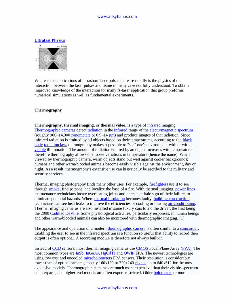
wwwallsyllabuscom
wwwallsyllabuscom
sensitive models as InSb require cryogenic cooling usually by a miniature Stirling cycle refrigerator or liquid nitrogen
Contents
[hide]
1 Difference between IR film and thermography 2 Advantages of Thermography
3 Limitations amp disadvantages of thermography 4 Applications 5 See also 6 External links
o 61 History of thermal imager manufacturers
[edit] Difference between IR film and thermography
Thermal imaging is going to be used on Mars to detect caves that could hold life
[edit] Advantages of Thermography
You get a visual picture so that you can compare temperatures over a large area
It is real time capable of catching moving targets Able to find deteriorating components prior to failure Measurement in areas inaccessible or hazardous for other methods It is a non-destructive test method
[edit] Limitations amp disadvantages of thermography
Quality cameras are expensive and are easily damaged Images can be hard to interpret accurately even with experience
Accurate temperature measurements are very hard to make because of emissivities Most cameras have plusmn2 or worse accuracy (not as accurate as contact) Training and staying proficient in IR scanning is time consuming Ability to only measure surface areas
[edit] Applications
Condition monitoring Medical imaging Night vision Research
Process control Non destructive testing Surveillance in security law enforcement and defense Chemical imaging
wwwallsyllabuscom
wwwallsyllabuscom
Thermal infrared imagers convert the energy in the infrared wavelength into a visible light video display All objects above 0 kelvins emit thermal infrared energy so thermal imagers can passively see all objects regardless of ambient light However most thermal imagers only see objects warmer than -50 degC
The spectrum and amount of thermal radiation depend strongly on an objects surface
temperature This makes it possible for a thermal camera to display an objects temperature However other factors also influence the radiation which limits the accuracy of this technique For example the radiation depends not only on the temperature of the object but is also a function of the emissivity of the object Also radiation also originates from the surroundings and is reflected in the object and the radiation from the object and the reflected radiation will also be influenced by the absorption of the atmosphere
Endoscopy
This page is about having an endoscopy - a test that looks at the inside of your digestive system
There is information on
What endoscopy is
Why you may have an endoscopy
Having an endoscopy
Your results
What an endoscopy is
An endoscopy is a test that looks inside the body The endoscope is a long flexible tube that can
be swallowed It has a camera and light inside it Some doctors call it a telescope
wwwallsyllabuscom
wwwallsyllabuscom
Why you may have an endoscopy
You are most likely to have an endoscopy to look at the inside of your
Gullet (oesophagus)
Stomach
Duodenum - the first part of the small bowel that attaches to the stomach
Large bowel (colon)
Your doctor may want to see inside because you have symptoms such as abnormal bleeding or
difficulty swallowing The doctor can look down the endoscope and see if there are any growths
or other abnormal looking areas Also through the endoscope the doctor can take samples
(biopsies) of any abnormal looking tissues
There is more detailed information about having a colonoscopy in the bowel cancer section of
CancerHelp UK Below is information about having other types of endoscopy
Having an endoscopy
You can have this test as an out patient Most people have a choice between having the test while
they are awake or after having a medicine to make them drowsy (a sedative) Your hospital may
not be happy for you to have a sedative if you live alone and will have no one to look after you
when you go home If you live alone but really want sedation your hospital may allow you to
stay overnight You should ask your doctor about this as early as possible as extra arrangements
will need to be made beforehand
If you dont have a sedative you will have a spray to numb the back of your throat and make it
easier for you to swallow the endoscopy tube
If you would prefer to be asleep during the test you will have an injection to make you very
drowsy just before the test You will need to take someone with you to the hospital appointment
You wont be able to drive for the rest of the day and should have someone to go home with you
You cant eat or drink for about 8 hours before the test so that your stomach and duodenum are
empty Your doctor will give you written instructions about this beforehand or they may arrive
with your appointment letter When you get to the clinic you may be asked to take your upper
clothing off and put on a hospital gown Some hospitals prefer to use gowns because your clothes
wont get messy Once you are ready you get onto the bed or X-ray couch When you are lying
comfortably you have the sedative injection to make you very drowsy Or your doctor will spray
the back of your throat to numb it
Once the sedative or throat spray has worked the doctor will pass the endoscope tube down your
wwwallsyllabuscom
wwwallsyllabuscom
throat to the area being investigated Your doctor will ask you to swallow as the tube goes down
but if youve had a sedative you wont remember that afterwards If there are any abnormalities
the doctor will take pieces of tissue from the abnormal looking area to send to the laboratory for
closer inspection under a microscope These tissue samples are called biopsies
When the test is over you will need to rest for a while If youve had a sedative you may not
remember much (if anything) about the test once you have come round You should be able to go
home the same day
The results
It can take time for test results to come through How long will depend on why you are having
the test Usually the doctor who carries out the endoscopy dictates a report straight way The
report is typed up by the department secretary and goes to your specialist who gives the results to
you If your GP has sent you for the test the results will go directly to the GP surgery
Understandably waiting for results can make you anxious It usually takes a couple of weeks for
the results to come through If your doctor needed them urgently it would have been noted on
the test request form and the results will be ready sooner than that Try to remember to ask your
doctor how long you should expect to wait for the results when you are first asked to go for the
test If it is not an emergency and you have not heard a couple of weeks after your test ring your
doctors secretary to check if they are back
Endoscopy
From Wikipedia the free encyclopedia
Jump to navigation search
Endoscopic images of a duodenal ulcer
wwwallsyllabuscom
wwwallsyllabuscom
A flexible endoscope
Endoscopy means looking inside and typically refers to looking inside the human body for medical reasons using an instrument called an endoscope Endoscopy can also refer to using a borescope in technical situations where direct line-of-sight observation is not feasible
Contents
[hide]
1 Overview 2 Components 3 Uses
4 History 5 Risks 6 After The Endoscopy 7 Recent developments 8 See also 9 References 10 Footnotes
11 External links
Overview
Endoscopy is a minimally invasive diagnostic medical procedure that is used to assess the interior surfaces of an organ by inserting a tube into the body The instrument may have a rigid or flexible tube and not only provide an image for visual inspection and photography but also enable taking
biopsies and retrieval of foreign objects Endoscopy is the vehicle for minimally invasive surgery
Many endoscopic procedures are considered to be relatively painless and at worst associated with mild discomfort for example in esophagogastroduodenoscopy most patients tolerate the procedure with only topical anaesthesia of the oropharynx using lignocaine spray[1] Complications are not common (only 5 of all operations)[citation needed] but can include perforation of the organ under inspection with the endoscope or biopsy instrument If that occurs open surgery may be required to repair the injury
wwwallsyllabuscom
wwwallsyllabuscom
Components
An endoscope can consist of
a rigid or flexible tube a light delivery system to illuminate the organ or object under inspection The light
source is normally outside the body and the light is typically directed via an optical fiber system
a lens system transmitting the image to the viewer from the fiberscope an additional channel to allow entry of medical instruments or manipulators
Uses
Endoscopy can involve
The gastrointestinal tract (GI tract) o esophagus stomach and duodenum (esophagogastroduodenoscopy) o small intestine o colon (colonoscopyproctosigmoidoscopy) o Bile duct
endoscopic retrograde cholangiopancreatography (ERCP) duodenoscope-assisted cholangiopancreatoscopy intraoperative cholangioscopy
The respiratory tract o The nose (rhinoscopy) o The lower respiratory tract (bronchoscopy)
The urinary tract (cystoscopy) The female reproductive system
o The cervix (colposcopy) o The uterus (hysteroscopy) o The Fallopian tubes (Falloscopy)
Normally closed body cavities (through a small incision) o The abdominal or pelvic cavity (laparoscopy) o The interior of a joint (arthroscopy) o Organs of the chest (thoracoscopy and mediastinoscopy)
During pregnancy o The amnion (amnioscopy) o The fetus (fetoscopy)
Plastic Surgery Panendoscopy (or triple endoscopy)
o Combines laryngoscopy esophagoscopy and bronchoscopy Non-medical uses for endoscopy
o The planning and architectural community have found the endoscope useful for
pre-visualization of scale models of proposed buildings and cities (architectural endoscopy)
o Internal inspection of complex technical systems (borescope) o Endoscopes are also a tool helpful in the examination of improvised explosive
devices by bomb disposal personnel
wwwallsyllabuscom
wwwallsyllabuscom
o The FBI uses endoscopes for conducting surveillance via tight spaces
History
The first endoscope of a kind was developed in 1806 by Philip Bozzini with his introduction of a
Lichtleiter (light conductor) for the examinations of the canals and cavities of the human body However the Vienna Medical Society disapproved of such curiosity An endoscope was first introduced into a human in 1822 by William Beaumont an army surgeon at Mackinac
Island Michigan[citation needed] The use of electric light was a major step in the improvement of endoscopy The first such lights were external Later smaller bulbs became available making internal light possible for instance in a hysteroscope by Charles David in 1908[citation needed] Hans Christian Jacobaeus has been given credit for early endoscopic explorations of the abdomen and the thorax with laparoscopy (1912) and thoracoscopy (1910)[citation needed] Laparoscopy was used in the diagnosis of liver and gallbladder disease by Heinz Kalk in the 1930s[citation needed] Hope reported in 1937 on the use of laparoscopy to diagnose ectopic pregnancy[citation needed] In 1944
Raoul Palmer placed his patients in the Trendelenburg position after gaseous distention of the abdomen and thus was able to reliably perform gynecologic laparoscopy[citation needed]
The first gastrocamera was released in 1950 by Olympus Optical Co Ltd The device took pictures on monochromatic film using a small light bulb that was triggered manually The device was of limited use however because it did not implement real-time optical capability Olympus continued its development of endoscopes by incorporating fiber optics in the early 1960s leading to the first useful endoscopes In 1964 it released a gastrocamera guided by a fiberscope[1] A few articles claim that DrBasil Hirschowitz of UnivOf MichiganAnn Arbor discussed the
endoscope in early 50s[2]
As endoscopic technology improved so did the methods of gastrointestinal endoscopy Owing primarily to the efforts of Dr Hiromi Shinya in the late 1960s GI endoscopy developed into what is more recognizable as todays colonoscopy While many doctors experimented with techniques to take advantage of the new iterations of endoscopes Dr Shinya focused on techniques that would allow for successful operation of the endoscope by an individual rejecting the common practice at the time of utilizing two people Consequently many of the fundamental methods and procedures of modern colonoscopy were developed by Dr Shinya
Dr Shinyas other great contribution was to therapeutic endoscopy in his invention of the
electrosurgical polypectomy snare with the aid of Olympus employee Hiroshi Ichikawa Shinya sketched his first plans for the device on January 8 1969 He envisioned a loop of wire attached to the end of a colonoscope that would allow for easy removal of polyps during investigation by passing a current through the wire By September of 1969 the first polypectomy using this device was performed Polypectomy has since become the most common therapeutic procedure performed with an endoscope (Sivak 2004)
By 1980 laparoscopy training was required by gynecologists to perform tubal ligation procedures and diagnostic evaluations of the pelvis The first laparoscopic cholecystectomy was performed in 1984 and the first video-laparoscopic cholecystectomy in 1987[citation needed] During the 1990s
laparoscopic surgery was extended to the appendix spleen colon stomach kidney and liver[citation needed] Wireless capsule endoscopy or Capsule Endoscopy is now approved in all the countries including Japan where government reimbusement will be available from Oct2007Capsule Endoscopy [3] increases detection of Small Bowel tumors where traditional Endoscopy is not very efficient
wwwallsyllabuscom
wwwallsyllabuscom
Risks
Infection
Punctured organs Allergic reactions due to Contrast agents or dyes (such as those used in a CT scan) Over-sedation
After The Endoscopy
After the procedure the patient will be observed and monitored by a qualified individual in the
endoscopy or a recovery area until a significant portion of the medication has worn off Occasionally a patient is left with a mild sore throat which promptly responds to saline gargles or a feeling of distention from the insufflated air that was used during the procedure Both problems are mild and fleeting When fully recovered the patient will be instructed when to resume hisher usual diet (probably within a few hours) and will be allowed to be taken home Because of the use of sedation most facilities mandate that the patient is taken home by another person and not to drive on hisher own or handle machinery for the remainder of the day
Recent developments
With the application of robotic systems telesurgery was introduced as the surgeon could operate
from a site physically removed from the patient The first transatlantic surgery has been called the Lindbergh Operation
wwwallsyllabuscom
wwwallsyllabuscom
Upper Endoscopy
Upper endoscopy enables the physician to look inside the esophagus stomach and duodenum (first part of the small intestine) The procedure might be used to discover the reason for swallowing difficulties nausea vomiting
reflux bleeding indigestion abdominal pain or chest pain Upper endoscopy is also called EGD which stands for esophagogastroduodenoscopy (eh-SAH-fuh-goh-GAS-troh-doo-AH-duh-NAH-skuh-pee)
For the procedure you will swallow a thin flexible lighted tube called an endoscope (EN-doh-skope) Right before the procedure the physician will spray your throat with a numbing agent that may help prevent gagging
You may also receive pain medicine and a sedative to help you relax during the exam The endoscope transmits an image of the inside of the esophagus stomach and duodenum so the physician can carefully examine the lining of these organs The scope also blows air into the stomach this expands the folds of tissue and makes it easier for the physician to examine the stomach
The physician can see abnormalities like inflammation or
bleeding through the endoscope that dont show up well on x rays The physician can also insert instruments into the scope to treat bleeding abnormalities or remove samples of tissue (biopsy) for further tests
Possible complications of upper endoscopy include bleeding and puncture of the stomach lining However such complications are rare Most people will probably have nothing more than a mild sore throat after the procedure
The procedure takes 20 to 30 minutes Because you will be sedated you will need to rest at the endoscopy facility for 1 to 2 hours until the medication wears off
Preparation
Your stomach and duodenum must be empty for the procedure to be thorough and safe so you will not be able to eat or drink anything for at least 6 hours beforehand Also you must
arrange for someone to take you homemdashyou will not be allowed to drive because of the sedatives Your physician may give you other special instructions
The digestive system
wwwallsyllabuscom
wwwallsyllabuscom
What is Endoscopy and Why is it Performed
Endoscopy allows physicians to peer through the bodys passageways Endoscopy is the examination and inspection of the interior of body organs joints or cavities through an endoscope An endoscope is a device that uses fiber optics and powerful lens systems to provide lighting and visualization of the interior of a joint The portion of the endoscope inserted into the body may be rigid or flexible depending upon the medical procedure
An endoscope uses two fiber optic lines A light fiber carries light into the body cavity and an image fiber carries the image of the body cavity back to the physicians viewing lens There is also a separate port to allow for administration of drugs suction and irrigation This port may also be used to introduce small folding instruments such as forceps scissors brushes snares and baskets for tissue excision (removal) sampling or other diagnostic and therapeutic work Endoscopes may be used in conjunction with a camera or video recorder to document images of the inside of the joint or chronicle an endoscopic procedure New endoscopes have digital capabilities for manipulating and enhancing the video images
This figure shows a rigid endoscope used for arthroscopy The image fiber leads from the ocular (eye piece) to the inserted end of the scope The light fiber is below and leads from the light source to the working end of the endoscope
Why Is Endoscopy Performed
Endoscopy can be used to diagnose various conditions by close examination of internal organ and body structures Endoscopy can also guide therapy and repair such as the removal of torn cartilage from the bearing surfaces of a joint Biopsy (tissue sampling for pathologic testing) may also be performed under endoscopic guidance Local or general anesthetic may be used during endoscopy depending upon the type of procedure being performed
Internal abnormalities revealed through endoscopy include abscesses biliary (liver) cirrhosis bleeding bronchitis cancer cysts degenerative disease gallbladder stones hernia inflammation metastatic cancer polyps tumors ulcers and other diseases and conditions
Endoscopy is a minimally invasive procedure and carries with it certain minor risks depending upon the type of procedure being performed However these risks are typically far outweighed by the diagnostic and therapeutic potential of the procedure
Prior to the widespread use of endoscopy and diagnostic imaging most internal conditions could only be diagnosed or treated with open surgery Until the last several decades exploratory surgery was routinely performed only when a patient was critically ill and the source of illness was not known For example in certain dire cases the patients thorax or abdomen were surgically opened and examined to try to determine the source of illness
wwwallsyllabuscom
wwwallsyllabuscom
Endoscopy can often be done on an outpatient basis Outpatient means that the procedure does not require hospital admission and acute care and observation and may be performed outside the premises of a hospital Outpatient procedures performed at hospitals or ambulatory centers allow the patient to go home or return to work within a short while after their procedure
Types of Endoscopy
Fiber optic endoscopes now have widespread use in medicine and guide a myriad of diagnostic and therapeutic procedures including
Arthroscopy examination of joints for diagnosis and treatment (arthroscopic surgery) Bronchoscopy examination of the trachea and lungs bronchial trees to reveal
abscesses bronchitis carcinoma tumors tuberculosis alveolitis infection inflammation Colonoscopy examination of the inside of the colon and large intestine to detect polyps
tumors ulceration inflammation colitis diverticula Chrohns disease and discovery and removal of foreign bodies
Colposcopy direct visualization of the vagina and cervix to detect cancer inflammation and other conditions
Cystoscopy examination of the bladder urethra urinary tract uteral orifices and
prostate (men) with insertion of the endoscope through the urethra
ERCP (endoscopic retrograde cholangio-pancreatography) uses endoscopic guidance to
place a catheter for x-ray fluorosocopy with contrast enhancement This technique is used to examine the livers biliary tree the gallbladder the pancreatic duct and other anatomy to check for stones other obstructions and disease X-ray contrast is introduced into these ducts via catheter and fluoroscopic x-ray images are taken to show any abnormality or blockage If disease is detected it can sometimes be treated at the same time or biopsy can be performed to test for cancer or other pathology ERCP can detect biliary cirrhosis
cancer of the bile ducts pancreatic cysts pseudocysts pancreatic tumors chronic pancreatitis and other conditions such as gallbladder stones
EGD (Esophogealgastroduodensoscopy) visual examination of the upper gastro-
intestinal (GI) tract (also referred to as gastroscopy) to reveal hemorrhage hiatal hernia inflammation of the esophagus gastric ulcers
Endoscopic biopsy is the removal of tissue specimens for pathologic examination and
analysis Gastroscopy examination of the lining of the esophagus stomach and duodenum
Gastroscopy is often used to diagnose ulcers and other sources of bleeding and to guide biopsy of suspect GI cancers
Laparoscopy visualization of the stomach liver and other abdominal organs including
the female reproductive organs for example the fallopian tubes Laryngoscopy examination of the larynx (voice box) Proctoscopy sigmoidoscopy proctosigmoidoscopy examination of the rectum and
sigmoid colon Thoracoscopy examination of the pleura (sac that covers the lungs) pleural spaces
mediastinum and pericardium
A Brief History of Endoscopy
In the early 1900s the first attempts to view inside the body with lighted telescopes were made These initial devices were often fully rigid In the 1930s semi-flexible endoscopes called gastroscopes were developed to view inside of the stomach Fiber-optic endoscopy was pioneered by South African-born physician Basil Hirschowitz at the University of Michigan in 1957 Widespread use of fiber optic endoscopes began in the 1960s
wwwallsyllabuscom
wwwallsyllabuscom
A fiber optic cable is simply a bundle of microscopic glass or plastic fibers that literally allows light and images to be transmitted through curved structures Fiber optic cables are also replacing metal wires as the backbone of the worlds telecommunications infrastructure This Internet page may have traveled through a fiber optic cable as a stream of digital data (bursts of light) on its way to your computer
Endoscopy Equipment
Endoscopes have many practical needs And HMB Endoscopy Products (Hollywood Florida) has been providing endoscopic equipment and educating people on the use of endoscopes for more than 17 years Be sure to Browse our Catalog for all the details on how to purchase these medical instruments that can examine any part of the body
Video
Systems
Fiber
Endoscopes
Video
Endoscopes
In the simplest terms Endoscopy equipment consists of instruments that can look at the inside of many different organs mdash these are small flexible or rigid tubes with a light or lenses on the end that can look into the esophagus stomach and colon mdash and in more general terms endoscopy equipment can help doctors look deep inside body structures and hollow organs
An endoscope and related endoscope products and equipment are usually composed of three components
An optic system that allows the doctor to look through the scope into the organ or
cavity or to attach a video camera to the scope
A fiberoptic cable to light up the bodily area
A lumen (eg the bore of a tube like a needle or catheter) to take tissue samples of the
area being viewed
The beauty of endoscopic products is that they perform dual functions mdash with both diagnostic and therapeutic capabilities For example this means that these endoscopic products and instruments can perform biopsies (eg to evaluate tissue samples) as well as provide sclerotherapy (a medical procedure used to treat varicose veins and ―spider veins) In truth these brief explanations only tell part of the story
wwwallsyllabuscom
wwwallsyllabuscom
TYPES OF ENDOSCOPES
Take a look at the different types of endoscopic equipment you can get with HMB Endoscopy Products Again in very general terms there are two main types of endoscopes
Rigid Flexible
1 rigid endoscopes ndash the majority of which use a convex (curving out like one half of a
circle) glass lens system in which the small glass lenses are separated by large air spaces
2 flexible endoscopes allow for just that mdash flexibility In the animal kingdom for example
a flexible endoscope would be perfect for examining the stomach area of a snake
The popularity of endoscopy equipment continues to grow Some of the industryrsquos household names include Olympus Pentax Funjinon and Storz mdash with HMB both selling and repairing pre-owned and completely refurbished endoscopic products from each of these major manufacturers Browse our Catalog to get the ones you need
THERMOGRAPH APPLICATION Thermographs are digital recording thermometers used to log
temperature in the
marine environment The instruments are attached to a computer to set up recording
parameters for
deployment downloading and display of data in graphic or numeric format These units
are placed in
underwater housings and attached to the bottom or suspended in the water column for
deployment
DESCRIPTION Two types of thermograph are currently available The Ryan
Tempmentor is a
reusable data logger that allows storage of a maximum of 6361 temperature
measurements in the
range of -32C to +70C with 01resolution and 03accuracy The instrument can be
programmed
to take measurements from once per second to once every other hour Instrument
dimensions are 3 x
6 x 13 and the unit weighs 11 ounces The Hobo Temp is a miniature reusable data
logger that
allows storage of 1800 measurements in the range of -20C to +70C with
03resolution and 07
wwwallsyllabuscom
wwwallsyllabuscom
accuracy Sampling intervals from 05 seconds to 48 hours are available Instrument
dimensions are
24 x 19 x 08 and the unit weighs 1 ounce Both units are battery powered with the
battery life of
the Tempmentor being approximately two years and the battery life of the Hobo Temp
being one year THERMOGRAPH APPLICATION Thermographs are digital recording thermometers used to log
temperature in the
marine environment The instruments are attached to a computer to set up recording
parameters for
deployment downloading and display of data in graphic or numeric format These units
are placed in
underwater housings and attached to the bottom or suspended in the water column for
deployment
DESCRIPTION Two types of thermograph are currently available The Ryan
Tempmentor is a
reusable data logger that allows storage of a maximum of 6361 temperature
measurements in the
range of -32C to +70C with 01resolution and 03accuracy The instrument can be
programmed
to take measurements from once per second to once every other hour Instrument
dimensions are 3 x
6 x 13 and the unit weighs 11 ounces The Hobo Temp is a miniature reusable data
logger that
allows storage of 1800 measurements in the range of -20C to +70C with
03resolution and 07
accuracy Sampling intervals from 05 seconds to 48 hours are available Instrument
dimensions are
24 x 19 x 08 and the unit weighs 1 ounce Both units are battery powered with the
battery life of
the Tempmentor being approximately two years and the battery life of the Hobo Temp
being one year THERMOGRAPH APPLICATION Thermographs are digital recording thermometers used to log
temperature in the
marine environment The instruments are attached to a computer to set up recording
parameters for
deployment downloading and display of data in graphic or numeric format These units
are placed in
underwater housings and attached to the bottom or suspended in the water column for
deployment
DESCRIPTION Two types of thermograph are currently available The Ryan
Tempmentor is a
reusable data logger that allows storage of a maximum of 6361 temperature
measurements in the
range of -32C to +70C with 01resolution and 03accuracy The instrument can be
programmed
wwwallsyllabuscom
wwwallsyllabuscom
to take measurements from once per second to once every other hour Instrument
dimensions are 3 x
6 x 13 and the unit weighs 11 ounces The Hobo Temp is a miniature reusable data
logger that
allows storage of 1800 measurements in the range of -20C to +70C with
03resolution and 07
accuracy Sampling intervals from 05 seconds to 48 hours are available Instrument
dimensions are
24 x 19 x 08 and the unit weighs 1 ounce Both units are battery powered with the
battery life of
the Tempmentor being approximately two years and the battery life of the Hobo Temp
being one year
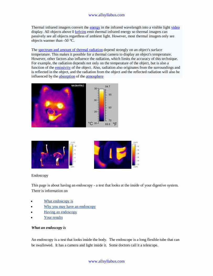
wwwallsyllabuscom
wwwallsyllabuscom
Thermal infrared imagers convert the energy in the infrared wavelength into a visible light video display All objects above 0 kelvins emit thermal infrared energy so thermal imagers can passively see all objects regardless of ambient light However most thermal imagers only see objects warmer than -50 degC
The spectrum and amount of thermal radiation depend strongly on an objects surface
temperature This makes it possible for a thermal camera to display an objects temperature However other factors also influence the radiation which limits the accuracy of this technique For example the radiation depends not only on the temperature of the object but is also a function of the emissivity of the object Also radiation also originates from the surroundings and is reflected in the object and the radiation from the object and the reflected radiation will also be influenced by the absorption of the atmosphere
Endoscopy
This page is about having an endoscopy - a test that looks at the inside of your digestive system
There is information on
What endoscopy is
Why you may have an endoscopy
Having an endoscopy
Your results
What an endoscopy is
An endoscopy is a test that looks inside the body The endoscope is a long flexible tube that can
be swallowed It has a camera and light inside it Some doctors call it a telescope
wwwallsyllabuscom
wwwallsyllabuscom
Why you may have an endoscopy
You are most likely to have an endoscopy to look at the inside of your
Gullet (oesophagus)
Stomach
Duodenum - the first part of the small bowel that attaches to the stomach
Large bowel (colon)
Your doctor may want to see inside because you have symptoms such as abnormal bleeding or
difficulty swallowing The doctor can look down the endoscope and see if there are any growths
or other abnormal looking areas Also through the endoscope the doctor can take samples
(biopsies) of any abnormal looking tissues
There is more detailed information about having a colonoscopy in the bowel cancer section of
CancerHelp UK Below is information about having other types of endoscopy
Having an endoscopy
You can have this test as an out patient Most people have a choice between having the test while
they are awake or after having a medicine to make them drowsy (a sedative) Your hospital may
not be happy for you to have a sedative if you live alone and will have no one to look after you
when you go home If you live alone but really want sedation your hospital may allow you to
stay overnight You should ask your doctor about this as early as possible as extra arrangements
will need to be made beforehand
If you dont have a sedative you will have a spray to numb the back of your throat and make it
easier for you to swallow the endoscopy tube
If you would prefer to be asleep during the test you will have an injection to make you very
drowsy just before the test You will need to take someone with you to the hospital appointment
You wont be able to drive for the rest of the day and should have someone to go home with you
You cant eat or drink for about 8 hours before the test so that your stomach and duodenum are
empty Your doctor will give you written instructions about this beforehand or they may arrive
with your appointment letter When you get to the clinic you may be asked to take your upper
clothing off and put on a hospital gown Some hospitals prefer to use gowns because your clothes
wont get messy Once you are ready you get onto the bed or X-ray couch When you are lying
comfortably you have the sedative injection to make you very drowsy Or your doctor will spray
the back of your throat to numb it
Once the sedative or throat spray has worked the doctor will pass the endoscope tube down your
wwwallsyllabuscom
wwwallsyllabuscom
throat to the area being investigated Your doctor will ask you to swallow as the tube goes down
but if youve had a sedative you wont remember that afterwards If there are any abnormalities
the doctor will take pieces of tissue from the abnormal looking area to send to the laboratory for
closer inspection under a microscope These tissue samples are called biopsies
When the test is over you will need to rest for a while If youve had a sedative you may not
remember much (if anything) about the test once you have come round You should be able to go
home the same day
The results
It can take time for test results to come through How long will depend on why you are having
the test Usually the doctor who carries out the endoscopy dictates a report straight way The
report is typed up by the department secretary and goes to your specialist who gives the results to
you If your GP has sent you for the test the results will go directly to the GP surgery
Understandably waiting for results can make you anxious It usually takes a couple of weeks for
the results to come through If your doctor needed them urgently it would have been noted on
the test request form and the results will be ready sooner than that Try to remember to ask your
doctor how long you should expect to wait for the results when you are first asked to go for the
test If it is not an emergency and you have not heard a couple of weeks after your test ring your
doctors secretary to check if they are back
Endoscopy
From Wikipedia the free encyclopedia
Jump to navigation search
Endoscopic images of a duodenal ulcer
wwwallsyllabuscom
wwwallsyllabuscom
A flexible endoscope
Endoscopy means looking inside and typically refers to looking inside the human body for medical reasons using an instrument called an endoscope Endoscopy can also refer to using a borescope in technical situations where direct line-of-sight observation is not feasible
Contents
[hide]
1 Overview 2 Components 3 Uses
4 History 5 Risks 6 After The Endoscopy 7 Recent developments 8 See also 9 References 10 Footnotes
11 External links
Overview
Endoscopy is a minimally invasive diagnostic medical procedure that is used to assess the interior surfaces of an organ by inserting a tube into the body The instrument may have a rigid or flexible tube and not only provide an image for visual inspection and photography but also enable taking
biopsies and retrieval of foreign objects Endoscopy is the vehicle for minimally invasive surgery
Many endoscopic procedures are considered to be relatively painless and at worst associated with mild discomfort for example in esophagogastroduodenoscopy most patients tolerate the procedure with only topical anaesthesia of the oropharynx using lignocaine spray[1] Complications are not common (only 5 of all operations)[citation needed] but can include perforation of the organ under inspection with the endoscope or biopsy instrument If that occurs open surgery may be required to repair the injury
wwwallsyllabuscom
wwwallsyllabuscom
Components
An endoscope can consist of
a rigid or flexible tube a light delivery system to illuminate the organ or object under inspection The light
source is normally outside the body and the light is typically directed via an optical fiber system
a lens system transmitting the image to the viewer from the fiberscope an additional channel to allow entry of medical instruments or manipulators
Uses
Endoscopy can involve
The gastrointestinal tract (GI tract) o esophagus stomach and duodenum (esophagogastroduodenoscopy) o small intestine o colon (colonoscopyproctosigmoidoscopy) o Bile duct
endoscopic retrograde cholangiopancreatography (ERCP) duodenoscope-assisted cholangiopancreatoscopy intraoperative cholangioscopy
The respiratory tract o The nose (rhinoscopy) o The lower respiratory tract (bronchoscopy)
The urinary tract (cystoscopy) The female reproductive system
o The cervix (colposcopy) o The uterus (hysteroscopy) o The Fallopian tubes (Falloscopy)
Normally closed body cavities (through a small incision) o The abdominal or pelvic cavity (laparoscopy) o The interior of a joint (arthroscopy) o Organs of the chest (thoracoscopy and mediastinoscopy)
During pregnancy o The amnion (amnioscopy) o The fetus (fetoscopy)
Plastic Surgery Panendoscopy (or triple endoscopy)
o Combines laryngoscopy esophagoscopy and bronchoscopy Non-medical uses for endoscopy
o The planning and architectural community have found the endoscope useful for
pre-visualization of scale models of proposed buildings and cities (architectural endoscopy)
o Internal inspection of complex technical systems (borescope) o Endoscopes are also a tool helpful in the examination of improvised explosive
devices by bomb disposal personnel
wwwallsyllabuscom
wwwallsyllabuscom
o The FBI uses endoscopes for conducting surveillance via tight spaces
History
The first endoscope of a kind was developed in 1806 by Philip Bozzini with his introduction of a
Lichtleiter (light conductor) for the examinations of the canals and cavities of the human body However the Vienna Medical Society disapproved of such curiosity An endoscope was first introduced into a human in 1822 by William Beaumont an army surgeon at Mackinac
Island Michigan[citation needed] The use of electric light was a major step in the improvement of endoscopy The first such lights were external Later smaller bulbs became available making internal light possible for instance in a hysteroscope by Charles David in 1908[citation needed] Hans Christian Jacobaeus has been given credit for early endoscopic explorations of the abdomen and the thorax with laparoscopy (1912) and thoracoscopy (1910)[citation needed] Laparoscopy was used in the diagnosis of liver and gallbladder disease by Heinz Kalk in the 1930s[citation needed] Hope reported in 1937 on the use of laparoscopy to diagnose ectopic pregnancy[citation needed] In 1944
Raoul Palmer placed his patients in the Trendelenburg position after gaseous distention of the abdomen and thus was able to reliably perform gynecologic laparoscopy[citation needed]
The first gastrocamera was released in 1950 by Olympus Optical Co Ltd The device took pictures on monochromatic film using a small light bulb that was triggered manually The device was of limited use however because it did not implement real-time optical capability Olympus continued its development of endoscopes by incorporating fiber optics in the early 1960s leading to the first useful endoscopes In 1964 it released a gastrocamera guided by a fiberscope[1] A few articles claim that DrBasil Hirschowitz of UnivOf MichiganAnn Arbor discussed the
endoscope in early 50s[2]
As endoscopic technology improved so did the methods of gastrointestinal endoscopy Owing primarily to the efforts of Dr Hiromi Shinya in the late 1960s GI endoscopy developed into what is more recognizable as todays colonoscopy While many doctors experimented with techniques to take advantage of the new iterations of endoscopes Dr Shinya focused on techniques that would allow for successful operation of the endoscope by an individual rejecting the common practice at the time of utilizing two people Consequently many of the fundamental methods and procedures of modern colonoscopy were developed by Dr Shinya
Dr Shinyas other great contribution was to therapeutic endoscopy in his invention of the
electrosurgical polypectomy snare with the aid of Olympus employee Hiroshi Ichikawa Shinya sketched his first plans for the device on January 8 1969 He envisioned a loop of wire attached to the end of a colonoscope that would allow for easy removal of polyps during investigation by passing a current through the wire By September of 1969 the first polypectomy using this device was performed Polypectomy has since become the most common therapeutic procedure performed with an endoscope (Sivak 2004)
By 1980 laparoscopy training was required by gynecologists to perform tubal ligation procedures and diagnostic evaluations of the pelvis The first laparoscopic cholecystectomy was performed in 1984 and the first video-laparoscopic cholecystectomy in 1987[citation needed] During the 1990s
laparoscopic surgery was extended to the appendix spleen colon stomach kidney and liver[citation needed] Wireless capsule endoscopy or Capsule Endoscopy is now approved in all the countries including Japan where government reimbusement will be available from Oct2007Capsule Endoscopy [3] increases detection of Small Bowel tumors where traditional Endoscopy is not very efficient
wwwallsyllabuscom
wwwallsyllabuscom
Risks
Infection
Punctured organs Allergic reactions due to Contrast agents or dyes (such as those used in a CT scan) Over-sedation
After The Endoscopy
After the procedure the patient will be observed and monitored by a qualified individual in the
endoscopy or a recovery area until a significant portion of the medication has worn off Occasionally a patient is left with a mild sore throat which promptly responds to saline gargles or a feeling of distention from the insufflated air that was used during the procedure Both problems are mild and fleeting When fully recovered the patient will be instructed when to resume hisher usual diet (probably within a few hours) and will be allowed to be taken home Because of the use of sedation most facilities mandate that the patient is taken home by another person and not to drive on hisher own or handle machinery for the remainder of the day
Recent developments
With the application of robotic systems telesurgery was introduced as the surgeon could operate
from a site physically removed from the patient The first transatlantic surgery has been called the Lindbergh Operation
wwwallsyllabuscom
wwwallsyllabuscom
Upper Endoscopy
Upper endoscopy enables the physician to look inside the esophagus stomach and duodenum (first part of the small intestine) The procedure might be used to discover the reason for swallowing difficulties nausea vomiting
reflux bleeding indigestion abdominal pain or chest pain Upper endoscopy is also called EGD which stands for esophagogastroduodenoscopy (eh-SAH-fuh-goh-GAS-troh-doo-AH-duh-NAH-skuh-pee)
For the procedure you will swallow a thin flexible lighted tube called an endoscope (EN-doh-skope) Right before the procedure the physician will spray your throat with a numbing agent that may help prevent gagging
You may also receive pain medicine and a sedative to help you relax during the exam The endoscope transmits an image of the inside of the esophagus stomach and duodenum so the physician can carefully examine the lining of these organs The scope also blows air into the stomach this expands the folds of tissue and makes it easier for the physician to examine the stomach
The physician can see abnormalities like inflammation or
bleeding through the endoscope that dont show up well on x rays The physician can also insert instruments into the scope to treat bleeding abnormalities or remove samples of tissue (biopsy) for further tests
Possible complications of upper endoscopy include bleeding and puncture of the stomach lining However such complications are rare Most people will probably have nothing more than a mild sore throat after the procedure
The procedure takes 20 to 30 minutes Because you will be sedated you will need to rest at the endoscopy facility for 1 to 2 hours until the medication wears off
Preparation
Your stomach and duodenum must be empty for the procedure to be thorough and safe so you will not be able to eat or drink anything for at least 6 hours beforehand Also you must
arrange for someone to take you homemdashyou will not be allowed to drive because of the sedatives Your physician may give you other special instructions
The digestive system
wwwallsyllabuscom
wwwallsyllabuscom
What is Endoscopy and Why is it Performed
Endoscopy allows physicians to peer through the bodys passageways Endoscopy is the examination and inspection of the interior of body organs joints or cavities through an endoscope An endoscope is a device that uses fiber optics and powerful lens systems to provide lighting and visualization of the interior of a joint The portion of the endoscope inserted into the body may be rigid or flexible depending upon the medical procedure
An endoscope uses two fiber optic lines A light fiber carries light into the body cavity and an image fiber carries the image of the body cavity back to the physicians viewing lens There is also a separate port to allow for administration of drugs suction and irrigation This port may also be used to introduce small folding instruments such as forceps scissors brushes snares and baskets for tissue excision (removal) sampling or other diagnostic and therapeutic work Endoscopes may be used in conjunction with a camera or video recorder to document images of the inside of the joint or chronicle an endoscopic procedure New endoscopes have digital capabilities for manipulating and enhancing the video images
This figure shows a rigid endoscope used for arthroscopy The image fiber leads from the ocular (eye piece) to the inserted end of the scope The light fiber is below and leads from the light source to the working end of the endoscope
Why Is Endoscopy Performed
Endoscopy can be used to diagnose various conditions by close examination of internal organ and body structures Endoscopy can also guide therapy and repair such as the removal of torn cartilage from the bearing surfaces of a joint Biopsy (tissue sampling for pathologic testing) may also be performed under endoscopic guidance Local or general anesthetic may be used during endoscopy depending upon the type of procedure being performed
Internal abnormalities revealed through endoscopy include abscesses biliary (liver) cirrhosis bleeding bronchitis cancer cysts degenerative disease gallbladder stones hernia inflammation metastatic cancer polyps tumors ulcers and other diseases and conditions
Endoscopy is a minimally invasive procedure and carries with it certain minor risks depending upon the type of procedure being performed However these risks are typically far outweighed by the diagnostic and therapeutic potential of the procedure
Prior to the widespread use of endoscopy and diagnostic imaging most internal conditions could only be diagnosed or treated with open surgery Until the last several decades exploratory surgery was routinely performed only when a patient was critically ill and the source of illness was not known For example in certain dire cases the patients thorax or abdomen were surgically opened and examined to try to determine the source of illness
wwwallsyllabuscom
wwwallsyllabuscom
Endoscopy can often be done on an outpatient basis Outpatient means that the procedure does not require hospital admission and acute care and observation and may be performed outside the premises of a hospital Outpatient procedures performed at hospitals or ambulatory centers allow the patient to go home or return to work within a short while after their procedure
Types of Endoscopy
Fiber optic endoscopes now have widespread use in medicine and guide a myriad of diagnostic and therapeutic procedures including
Arthroscopy examination of joints for diagnosis and treatment (arthroscopic surgery) Bronchoscopy examination of the trachea and lungs bronchial trees to reveal
abscesses bronchitis carcinoma tumors tuberculosis alveolitis infection inflammation Colonoscopy examination of the inside of the colon and large intestine to detect polyps
tumors ulceration inflammation colitis diverticula Chrohns disease and discovery and removal of foreign bodies
Colposcopy direct visualization of the vagina and cervix to detect cancer inflammation and other conditions
Cystoscopy examination of the bladder urethra urinary tract uteral orifices and
prostate (men) with insertion of the endoscope through the urethra
ERCP (endoscopic retrograde cholangio-pancreatography) uses endoscopic guidance to
place a catheter for x-ray fluorosocopy with contrast enhancement This technique is used to examine the livers biliary tree the gallbladder the pancreatic duct and other anatomy to check for stones other obstructions and disease X-ray contrast is introduced into these ducts via catheter and fluoroscopic x-ray images are taken to show any abnormality or blockage If disease is detected it can sometimes be treated at the same time or biopsy can be performed to test for cancer or other pathology ERCP can detect biliary cirrhosis
cancer of the bile ducts pancreatic cysts pseudocysts pancreatic tumors chronic pancreatitis and other conditions such as gallbladder stones
EGD (Esophogealgastroduodensoscopy) visual examination of the upper gastro-
intestinal (GI) tract (also referred to as gastroscopy) to reveal hemorrhage hiatal hernia inflammation of the esophagus gastric ulcers
Endoscopic biopsy is the removal of tissue specimens for pathologic examination and
analysis Gastroscopy examination of the lining of the esophagus stomach and duodenum
Gastroscopy is often used to diagnose ulcers and other sources of bleeding and to guide biopsy of suspect GI cancers
Laparoscopy visualization of the stomach liver and other abdominal organs including
the female reproductive organs for example the fallopian tubes Laryngoscopy examination of the larynx (voice box) Proctoscopy sigmoidoscopy proctosigmoidoscopy examination of the rectum and
sigmoid colon Thoracoscopy examination of the pleura (sac that covers the lungs) pleural spaces
mediastinum and pericardium
A Brief History of Endoscopy
In the early 1900s the first attempts to view inside the body with lighted telescopes were made These initial devices were often fully rigid In the 1930s semi-flexible endoscopes called gastroscopes were developed to view inside of the stomach Fiber-optic endoscopy was pioneered by South African-born physician Basil Hirschowitz at the University of Michigan in 1957 Widespread use of fiber optic endoscopes began in the 1960s
wwwallsyllabuscom
wwwallsyllabuscom
A fiber optic cable is simply a bundle of microscopic glass or plastic fibers that literally allows light and images to be transmitted through curved structures Fiber optic cables are also replacing metal wires as the backbone of the worlds telecommunications infrastructure This Internet page may have traveled through a fiber optic cable as a stream of digital data (bursts of light) on its way to your computer
Endoscopy Equipment
Endoscopes have many practical needs And HMB Endoscopy Products (Hollywood Florida) has been providing endoscopic equipment and educating people on the use of endoscopes for more than 17 years Be sure to Browse our Catalog for all the details on how to purchase these medical instruments that can examine any part of the body
Video
Systems
Fiber
Endoscopes
Video
Endoscopes
In the simplest terms Endoscopy equipment consists of instruments that can look at the inside of many different organs mdash these are small flexible or rigid tubes with a light or lenses on the end that can look into the esophagus stomach and colon mdash and in more general terms endoscopy equipment can help doctors look deep inside body structures and hollow organs
An endoscope and related endoscope products and equipment are usually composed of three components
An optic system that allows the doctor to look through the scope into the organ or
cavity or to attach a video camera to the scope
A fiberoptic cable to light up the bodily area
A lumen (eg the bore of a tube like a needle or catheter) to take tissue samples of the
area being viewed
The beauty of endoscopic products is that they perform dual functions mdash with both diagnostic and therapeutic capabilities For example this means that these endoscopic products and instruments can perform biopsies (eg to evaluate tissue samples) as well as provide sclerotherapy (a medical procedure used to treat varicose veins and ―spider veins) In truth these brief explanations only tell part of the story
wwwallsyllabuscom
wwwallsyllabuscom
TYPES OF ENDOSCOPES
Take a look at the different types of endoscopic equipment you can get with HMB Endoscopy Products Again in very general terms there are two main types of endoscopes
Rigid Flexible
1 rigid endoscopes ndash the majority of which use a convex (curving out like one half of a
circle) glass lens system in which the small glass lenses are separated by large air spaces
2 flexible endoscopes allow for just that mdash flexibility In the animal kingdom for example
a flexible endoscope would be perfect for examining the stomach area of a snake
The popularity of endoscopy equipment continues to grow Some of the industryrsquos household names include Olympus Pentax Funjinon and Storz mdash with HMB both selling and repairing pre-owned and completely refurbished endoscopic products from each of these major manufacturers Browse our Catalog to get the ones you need
THERMOGRAPH APPLICATION Thermographs are digital recording thermometers used to log
temperature in the
marine environment The instruments are attached to a computer to set up recording
parameters for
deployment downloading and display of data in graphic or numeric format These units
are placed in
underwater housings and attached to the bottom or suspended in the water column for
deployment
DESCRIPTION Two types of thermograph are currently available The Ryan
Tempmentor is a
reusable data logger that allows storage of a maximum of 6361 temperature
measurements in the
range of -32C to +70C with 01resolution and 03accuracy The instrument can be
programmed
to take measurements from once per second to once every other hour Instrument
dimensions are 3 x
6 x 13 and the unit weighs 11 ounces The Hobo Temp is a miniature reusable data
logger that
allows storage of 1800 measurements in the range of -20C to +70C with
03resolution and 07
wwwallsyllabuscom
wwwallsyllabuscom
accuracy Sampling intervals from 05 seconds to 48 hours are available Instrument
dimensions are
24 x 19 x 08 and the unit weighs 1 ounce Both units are battery powered with the
battery life of
the Tempmentor being approximately two years and the battery life of the Hobo Temp
being one year THERMOGRAPH APPLICATION Thermographs are digital recording thermometers used to log
temperature in the
marine environment The instruments are attached to a computer to set up recording
parameters for
deployment downloading and display of data in graphic or numeric format These units
are placed in
underwater housings and attached to the bottom or suspended in the water column for
deployment
DESCRIPTION Two types of thermograph are currently available The Ryan
Tempmentor is a
reusable data logger that allows storage of a maximum of 6361 temperature
measurements in the
range of -32C to +70C with 01resolution and 03accuracy The instrument can be
programmed
to take measurements from once per second to once every other hour Instrument
dimensions are 3 x
6 x 13 and the unit weighs 11 ounces The Hobo Temp is a miniature reusable data
logger that
allows storage of 1800 measurements in the range of -20C to +70C with
03resolution and 07
accuracy Sampling intervals from 05 seconds to 48 hours are available Instrument
dimensions are
24 x 19 x 08 and the unit weighs 1 ounce Both units are battery powered with the
battery life of
the Tempmentor being approximately two years and the battery life of the Hobo Temp
being one year THERMOGRAPH APPLICATION Thermographs are digital recording thermometers used to log
temperature in the
marine environment The instruments are attached to a computer to set up recording
parameters for
deployment downloading and display of data in graphic or numeric format These units
are placed in
underwater housings and attached to the bottom or suspended in the water column for
deployment
DESCRIPTION Two types of thermograph are currently available The Ryan
Tempmentor is a
reusable data logger that allows storage of a maximum of 6361 temperature
measurements in the
range of -32C to +70C with 01resolution and 03accuracy The instrument can be
programmed
wwwallsyllabuscom
wwwallsyllabuscom
to take measurements from once per second to once every other hour Instrument
dimensions are 3 x
6 x 13 and the unit weighs 11 ounces The Hobo Temp is a miniature reusable data
logger that
allows storage of 1800 measurements in the range of -20C to +70C with
03resolution and 07
accuracy Sampling intervals from 05 seconds to 48 hours are available Instrument
dimensions are
24 x 19 x 08 and the unit weighs 1 ounce Both units are battery powered with the
battery life of
the Tempmentor being approximately two years and the battery life of the Hobo Temp
being one year
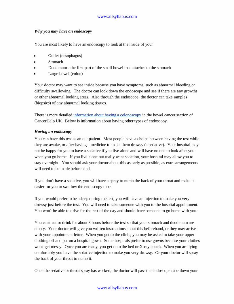
wwwallsyllabuscom
wwwallsyllabuscom
Why you may have an endoscopy
You are most likely to have an endoscopy to look at the inside of your
Gullet (oesophagus)
Stomach
Duodenum - the first part of the small bowel that attaches to the stomach
Large bowel (colon)
Your doctor may want to see inside because you have symptoms such as abnormal bleeding or
difficulty swallowing The doctor can look down the endoscope and see if there are any growths
or other abnormal looking areas Also through the endoscope the doctor can take samples
(biopsies) of any abnormal looking tissues
There is more detailed information about having a colonoscopy in the bowel cancer section of
CancerHelp UK Below is information about having other types of endoscopy
Having an endoscopy
You can have this test as an out patient Most people have a choice between having the test while
they are awake or after having a medicine to make them drowsy (a sedative) Your hospital may
not be happy for you to have a sedative if you live alone and will have no one to look after you
when you go home If you live alone but really want sedation your hospital may allow you to
stay overnight You should ask your doctor about this as early as possible as extra arrangements
will need to be made beforehand
If you dont have a sedative you will have a spray to numb the back of your throat and make it
easier for you to swallow the endoscopy tube
If you would prefer to be asleep during the test you will have an injection to make you very
drowsy just before the test You will need to take someone with you to the hospital appointment
You wont be able to drive for the rest of the day and should have someone to go home with you
You cant eat or drink for about 8 hours before the test so that your stomach and duodenum are
empty Your doctor will give you written instructions about this beforehand or they may arrive
with your appointment letter When you get to the clinic you may be asked to take your upper
clothing off and put on a hospital gown Some hospitals prefer to use gowns because your clothes
wont get messy Once you are ready you get onto the bed or X-ray couch When you are lying
comfortably you have the sedative injection to make you very drowsy Or your doctor will spray
the back of your throat to numb it
Once the sedative or throat spray has worked the doctor will pass the endoscope tube down your
wwwallsyllabuscom
wwwallsyllabuscom
throat to the area being investigated Your doctor will ask you to swallow as the tube goes down
but if youve had a sedative you wont remember that afterwards If there are any abnormalities
the doctor will take pieces of tissue from the abnormal looking area to send to the laboratory for
closer inspection under a microscope These tissue samples are called biopsies
When the test is over you will need to rest for a while If youve had a sedative you may not
remember much (if anything) about the test once you have come round You should be able to go
home the same day
The results
It can take time for test results to come through How long will depend on why you are having
the test Usually the doctor who carries out the endoscopy dictates a report straight way The
report is typed up by the department secretary and goes to your specialist who gives the results to
you If your GP has sent you for the test the results will go directly to the GP surgery
Understandably waiting for results can make you anxious It usually takes a couple of weeks for
the results to come through If your doctor needed them urgently it would have been noted on
the test request form and the results will be ready sooner than that Try to remember to ask your
doctor how long you should expect to wait for the results when you are first asked to go for the
test If it is not an emergency and you have not heard a couple of weeks after your test ring your
doctors secretary to check if they are back
Endoscopy
From Wikipedia the free encyclopedia
Jump to navigation search
Endoscopic images of a duodenal ulcer
wwwallsyllabuscom
wwwallsyllabuscom
A flexible endoscope
Endoscopy means looking inside and typically refers to looking inside the human body for medical reasons using an instrument called an endoscope Endoscopy can also refer to using a borescope in technical situations where direct line-of-sight observation is not feasible
Contents
[hide]
1 Overview 2 Components 3 Uses
4 History 5 Risks 6 After The Endoscopy 7 Recent developments 8 See also 9 References 10 Footnotes
11 External links
Overview
Endoscopy is a minimally invasive diagnostic medical procedure that is used to assess the interior surfaces of an organ by inserting a tube into the body The instrument may have a rigid or flexible tube and not only provide an image for visual inspection and photography but also enable taking
biopsies and retrieval of foreign objects Endoscopy is the vehicle for minimally invasive surgery
Many endoscopic procedures are considered to be relatively painless and at worst associated with mild discomfort for example in esophagogastroduodenoscopy most patients tolerate the procedure with only topical anaesthesia of the oropharynx using lignocaine spray[1] Complications are not common (only 5 of all operations)[citation needed] but can include perforation of the organ under inspection with the endoscope or biopsy instrument If that occurs open surgery may be required to repair the injury
wwwallsyllabuscom
wwwallsyllabuscom
Components
An endoscope can consist of
a rigid or flexible tube a light delivery system to illuminate the organ or object under inspection The light
source is normally outside the body and the light is typically directed via an optical fiber system
a lens system transmitting the image to the viewer from the fiberscope an additional channel to allow entry of medical instruments or manipulators
Uses
Endoscopy can involve
The gastrointestinal tract (GI tract) o esophagus stomach and duodenum (esophagogastroduodenoscopy) o small intestine o colon (colonoscopyproctosigmoidoscopy) o Bile duct
endoscopic retrograde cholangiopancreatography (ERCP) duodenoscope-assisted cholangiopancreatoscopy intraoperative cholangioscopy
The respiratory tract o The nose (rhinoscopy) o The lower respiratory tract (bronchoscopy)
The urinary tract (cystoscopy) The female reproductive system
o The cervix (colposcopy) o The uterus (hysteroscopy) o The Fallopian tubes (Falloscopy)
Normally closed body cavities (through a small incision) o The abdominal or pelvic cavity (laparoscopy) o The interior of a joint (arthroscopy) o Organs of the chest (thoracoscopy and mediastinoscopy)
During pregnancy o The amnion (amnioscopy) o The fetus (fetoscopy)
Plastic Surgery Panendoscopy (or triple endoscopy)
o Combines laryngoscopy esophagoscopy and bronchoscopy Non-medical uses for endoscopy
o The planning and architectural community have found the endoscope useful for
pre-visualization of scale models of proposed buildings and cities (architectural endoscopy)
o Internal inspection of complex technical systems (borescope) o Endoscopes are also a tool helpful in the examination of improvised explosive
devices by bomb disposal personnel
wwwallsyllabuscom
wwwallsyllabuscom
o The FBI uses endoscopes for conducting surveillance via tight spaces
History
The first endoscope of a kind was developed in 1806 by Philip Bozzini with his introduction of a
Lichtleiter (light conductor) for the examinations of the canals and cavities of the human body However the Vienna Medical Society disapproved of such curiosity An endoscope was first introduced into a human in 1822 by William Beaumont an army surgeon at Mackinac
Island Michigan[citation needed] The use of electric light was a major step in the improvement of endoscopy The first such lights were external Later smaller bulbs became available making internal light possible for instance in a hysteroscope by Charles David in 1908[citation needed] Hans Christian Jacobaeus has been given credit for early endoscopic explorations of the abdomen and the thorax with laparoscopy (1912) and thoracoscopy (1910)[citation needed] Laparoscopy was used in the diagnosis of liver and gallbladder disease by Heinz Kalk in the 1930s[citation needed] Hope reported in 1937 on the use of laparoscopy to diagnose ectopic pregnancy[citation needed] In 1944
Raoul Palmer placed his patients in the Trendelenburg position after gaseous distention of the abdomen and thus was able to reliably perform gynecologic laparoscopy[citation needed]
The first gastrocamera was released in 1950 by Olympus Optical Co Ltd The device took pictures on monochromatic film using a small light bulb that was triggered manually The device was of limited use however because it did not implement real-time optical capability Olympus continued its development of endoscopes by incorporating fiber optics in the early 1960s leading to the first useful endoscopes In 1964 it released a gastrocamera guided by a fiberscope[1] A few articles claim that DrBasil Hirschowitz of UnivOf MichiganAnn Arbor discussed the
endoscope in early 50s[2]
As endoscopic technology improved so did the methods of gastrointestinal endoscopy Owing primarily to the efforts of Dr Hiromi Shinya in the late 1960s GI endoscopy developed into what is more recognizable as todays colonoscopy While many doctors experimented with techniques to take advantage of the new iterations of endoscopes Dr Shinya focused on techniques that would allow for successful operation of the endoscope by an individual rejecting the common practice at the time of utilizing two people Consequently many of the fundamental methods and procedures of modern colonoscopy were developed by Dr Shinya
Dr Shinyas other great contribution was to therapeutic endoscopy in his invention of the
electrosurgical polypectomy snare with the aid of Olympus employee Hiroshi Ichikawa Shinya sketched his first plans for the device on January 8 1969 He envisioned a loop of wire attached to the end of a colonoscope that would allow for easy removal of polyps during investigation by passing a current through the wire By September of 1969 the first polypectomy using this device was performed Polypectomy has since become the most common therapeutic procedure performed with an endoscope (Sivak 2004)
By 1980 laparoscopy training was required by gynecologists to perform tubal ligation procedures and diagnostic evaluations of the pelvis The first laparoscopic cholecystectomy was performed in 1984 and the first video-laparoscopic cholecystectomy in 1987[citation needed] During the 1990s
laparoscopic surgery was extended to the appendix spleen colon stomach kidney and liver[citation needed] Wireless capsule endoscopy or Capsule Endoscopy is now approved in all the countries including Japan where government reimbusement will be available from Oct2007Capsule Endoscopy [3] increases detection of Small Bowel tumors where traditional Endoscopy is not very efficient
wwwallsyllabuscom
wwwallsyllabuscom
Risks
Infection
Punctured organs Allergic reactions due to Contrast agents or dyes (such as those used in a CT scan) Over-sedation
After The Endoscopy
After the procedure the patient will be observed and monitored by a qualified individual in the
endoscopy or a recovery area until a significant portion of the medication has worn off Occasionally a patient is left with a mild sore throat which promptly responds to saline gargles or a feeling of distention from the insufflated air that was used during the procedure Both problems are mild and fleeting When fully recovered the patient will be instructed when to resume hisher usual diet (probably within a few hours) and will be allowed to be taken home Because of the use of sedation most facilities mandate that the patient is taken home by another person and not to drive on hisher own or handle machinery for the remainder of the day
Recent developments
With the application of robotic systems telesurgery was introduced as the surgeon could operate
from a site physically removed from the patient The first transatlantic surgery has been called the Lindbergh Operation
wwwallsyllabuscom
wwwallsyllabuscom
Upper Endoscopy
Upper endoscopy enables the physician to look inside the esophagus stomach and duodenum (first part of the small intestine) The procedure might be used to discover the reason for swallowing difficulties nausea vomiting
reflux bleeding indigestion abdominal pain or chest pain Upper endoscopy is also called EGD which stands for esophagogastroduodenoscopy (eh-SAH-fuh-goh-GAS-troh-doo-AH-duh-NAH-skuh-pee)
For the procedure you will swallow a thin flexible lighted tube called an endoscope (EN-doh-skope) Right before the procedure the physician will spray your throat with a numbing agent that may help prevent gagging
You may also receive pain medicine and a sedative to help you relax during the exam The endoscope transmits an image of the inside of the esophagus stomach and duodenum so the physician can carefully examine the lining of these organs The scope also blows air into the stomach this expands the folds of tissue and makes it easier for the physician to examine the stomach
The physician can see abnormalities like inflammation or
bleeding through the endoscope that dont show up well on x rays The physician can also insert instruments into the scope to treat bleeding abnormalities or remove samples of tissue (biopsy) for further tests
Possible complications of upper endoscopy include bleeding and puncture of the stomach lining However such complications are rare Most people will probably have nothing more than a mild sore throat after the procedure
The procedure takes 20 to 30 minutes Because you will be sedated you will need to rest at the endoscopy facility for 1 to 2 hours until the medication wears off
Preparation
Your stomach and duodenum must be empty for the procedure to be thorough and safe so you will not be able to eat or drink anything for at least 6 hours beforehand Also you must
arrange for someone to take you homemdashyou will not be allowed to drive because of the sedatives Your physician may give you other special instructions
The digestive system
wwwallsyllabuscom
wwwallsyllabuscom
What is Endoscopy and Why is it Performed
Endoscopy allows physicians to peer through the bodys passageways Endoscopy is the examination and inspection of the interior of body organs joints or cavities through an endoscope An endoscope is a device that uses fiber optics and powerful lens systems to provide lighting and visualization of the interior of a joint The portion of the endoscope inserted into the body may be rigid or flexible depending upon the medical procedure
An endoscope uses two fiber optic lines A light fiber carries light into the body cavity and an image fiber carries the image of the body cavity back to the physicians viewing lens There is also a separate port to allow for administration of drugs suction and irrigation This port may also be used to introduce small folding instruments such as forceps scissors brushes snares and baskets for tissue excision (removal) sampling or other diagnostic and therapeutic work Endoscopes may be used in conjunction with a camera or video recorder to document images of the inside of the joint or chronicle an endoscopic procedure New endoscopes have digital capabilities for manipulating and enhancing the video images
This figure shows a rigid endoscope used for arthroscopy The image fiber leads from the ocular (eye piece) to the inserted end of the scope The light fiber is below and leads from the light source to the working end of the endoscope
Why Is Endoscopy Performed
Endoscopy can be used to diagnose various conditions by close examination of internal organ and body structures Endoscopy can also guide therapy and repair such as the removal of torn cartilage from the bearing surfaces of a joint Biopsy (tissue sampling for pathologic testing) may also be performed under endoscopic guidance Local or general anesthetic may be used during endoscopy depending upon the type of procedure being performed
Internal abnormalities revealed through endoscopy include abscesses biliary (liver) cirrhosis bleeding bronchitis cancer cysts degenerative disease gallbladder stones hernia inflammation metastatic cancer polyps tumors ulcers and other diseases and conditions
Endoscopy is a minimally invasive procedure and carries with it certain minor risks depending upon the type of procedure being performed However these risks are typically far outweighed by the diagnostic and therapeutic potential of the procedure
Prior to the widespread use of endoscopy and diagnostic imaging most internal conditions could only be diagnosed or treated with open surgery Until the last several decades exploratory surgery was routinely performed only when a patient was critically ill and the source of illness was not known For example in certain dire cases the patients thorax or abdomen were surgically opened and examined to try to determine the source of illness
wwwallsyllabuscom
wwwallsyllabuscom
Endoscopy can often be done on an outpatient basis Outpatient means that the procedure does not require hospital admission and acute care and observation and may be performed outside the premises of a hospital Outpatient procedures performed at hospitals or ambulatory centers allow the patient to go home or return to work within a short while after their procedure
Types of Endoscopy
Fiber optic endoscopes now have widespread use in medicine and guide a myriad of diagnostic and therapeutic procedures including
Arthroscopy examination of joints for diagnosis and treatment (arthroscopic surgery) Bronchoscopy examination of the trachea and lungs bronchial trees to reveal
abscesses bronchitis carcinoma tumors tuberculosis alveolitis infection inflammation Colonoscopy examination of the inside of the colon and large intestine to detect polyps
tumors ulceration inflammation colitis diverticula Chrohns disease and discovery and removal of foreign bodies
Colposcopy direct visualization of the vagina and cervix to detect cancer inflammation and other conditions
Cystoscopy examination of the bladder urethra urinary tract uteral orifices and
prostate (men) with insertion of the endoscope through the urethra
ERCP (endoscopic retrograde cholangio-pancreatography) uses endoscopic guidance to
place a catheter for x-ray fluorosocopy with contrast enhancement This technique is used to examine the livers biliary tree the gallbladder the pancreatic duct and other anatomy to check for stones other obstructions and disease X-ray contrast is introduced into these ducts via catheter and fluoroscopic x-ray images are taken to show any abnormality or blockage If disease is detected it can sometimes be treated at the same time or biopsy can be performed to test for cancer or other pathology ERCP can detect biliary cirrhosis
cancer of the bile ducts pancreatic cysts pseudocysts pancreatic tumors chronic pancreatitis and other conditions such as gallbladder stones
EGD (Esophogealgastroduodensoscopy) visual examination of the upper gastro-
intestinal (GI) tract (also referred to as gastroscopy) to reveal hemorrhage hiatal hernia inflammation of the esophagus gastric ulcers
Endoscopic biopsy is the removal of tissue specimens for pathologic examination and
analysis Gastroscopy examination of the lining of the esophagus stomach and duodenum
Gastroscopy is often used to diagnose ulcers and other sources of bleeding and to guide biopsy of suspect GI cancers
Laparoscopy visualization of the stomach liver and other abdominal organs including
the female reproductive organs for example the fallopian tubes Laryngoscopy examination of the larynx (voice box) Proctoscopy sigmoidoscopy proctosigmoidoscopy examination of the rectum and
sigmoid colon Thoracoscopy examination of the pleura (sac that covers the lungs) pleural spaces
mediastinum and pericardium
A Brief History of Endoscopy
In the early 1900s the first attempts to view inside the body with lighted telescopes were made These initial devices were often fully rigid In the 1930s semi-flexible endoscopes called gastroscopes were developed to view inside of the stomach Fiber-optic endoscopy was pioneered by South African-born physician Basil Hirschowitz at the University of Michigan in 1957 Widespread use of fiber optic endoscopes began in the 1960s
wwwallsyllabuscom
wwwallsyllabuscom
A fiber optic cable is simply a bundle of microscopic glass or plastic fibers that literally allows light and images to be transmitted through curved structures Fiber optic cables are also replacing metal wires as the backbone of the worlds telecommunications infrastructure This Internet page may have traveled through a fiber optic cable as a stream of digital data (bursts of light) on its way to your computer
Endoscopy Equipment
Endoscopes have many practical needs And HMB Endoscopy Products (Hollywood Florida) has been providing endoscopic equipment and educating people on the use of endoscopes for more than 17 years Be sure to Browse our Catalog for all the details on how to purchase these medical instruments that can examine any part of the body
Video
Systems
Fiber
Endoscopes
Video
Endoscopes
In the simplest terms Endoscopy equipment consists of instruments that can look at the inside of many different organs mdash these are small flexible or rigid tubes with a light or lenses on the end that can look into the esophagus stomach and colon mdash and in more general terms endoscopy equipment can help doctors look deep inside body structures and hollow organs
An endoscope and related endoscope products and equipment are usually composed of three components
An optic system that allows the doctor to look through the scope into the organ or
cavity or to attach a video camera to the scope
A fiberoptic cable to light up the bodily area
A lumen (eg the bore of a tube like a needle or catheter) to take tissue samples of the
area being viewed
The beauty of endoscopic products is that they perform dual functions mdash with both diagnostic and therapeutic capabilities For example this means that these endoscopic products and instruments can perform biopsies (eg to evaluate tissue samples) as well as provide sclerotherapy (a medical procedure used to treat varicose veins and ―spider veins) In truth these brief explanations only tell part of the story
wwwallsyllabuscom
wwwallsyllabuscom
TYPES OF ENDOSCOPES
Take a look at the different types of endoscopic equipment you can get with HMB Endoscopy Products Again in very general terms there are two main types of endoscopes
Rigid Flexible
1 rigid endoscopes ndash the majority of which use a convex (curving out like one half of a
circle) glass lens system in which the small glass lenses are separated by large air spaces
2 flexible endoscopes allow for just that mdash flexibility In the animal kingdom for example
a flexible endoscope would be perfect for examining the stomach area of a snake
The popularity of endoscopy equipment continues to grow Some of the industryrsquos household names include Olympus Pentax Funjinon and Storz mdash with HMB both selling and repairing pre-owned and completely refurbished endoscopic products from each of these major manufacturers Browse our Catalog to get the ones you need
THERMOGRAPH APPLICATION Thermographs are digital recording thermometers used to log
temperature in the
marine environment The instruments are attached to a computer to set up recording
parameters for
deployment downloading and display of data in graphic or numeric format These units
are placed in
underwater housings and attached to the bottom or suspended in the water column for
deployment
DESCRIPTION Two types of thermograph are currently available The Ryan
Tempmentor is a
reusable data logger that allows storage of a maximum of 6361 temperature
measurements in the
range of -32C to +70C with 01resolution and 03accuracy The instrument can be
programmed
to take measurements from once per second to once every other hour Instrument
dimensions are 3 x
6 x 13 and the unit weighs 11 ounces The Hobo Temp is a miniature reusable data
logger that
allows storage of 1800 measurements in the range of -20C to +70C with
03resolution and 07
wwwallsyllabuscom
wwwallsyllabuscom
accuracy Sampling intervals from 05 seconds to 48 hours are available Instrument
dimensions are
24 x 19 x 08 and the unit weighs 1 ounce Both units are battery powered with the
battery life of
the Tempmentor being approximately two years and the battery life of the Hobo Temp
being one year THERMOGRAPH APPLICATION Thermographs are digital recording thermometers used to log
temperature in the
marine environment The instruments are attached to a computer to set up recording
parameters for
deployment downloading and display of data in graphic or numeric format These units
are placed in
underwater housings and attached to the bottom or suspended in the water column for
deployment
DESCRIPTION Two types of thermograph are currently available The Ryan
Tempmentor is a
reusable data logger that allows storage of a maximum of 6361 temperature
measurements in the
range of -32C to +70C with 01resolution and 03accuracy The instrument can be
programmed
to take measurements from once per second to once every other hour Instrument
dimensions are 3 x
6 x 13 and the unit weighs 11 ounces The Hobo Temp is a miniature reusable data
logger that
allows storage of 1800 measurements in the range of -20C to +70C with
03resolution and 07
accuracy Sampling intervals from 05 seconds to 48 hours are available Instrument
dimensions are
24 x 19 x 08 and the unit weighs 1 ounce Both units are battery powered with the
battery life of
the Tempmentor being approximately two years and the battery life of the Hobo Temp
being one year THERMOGRAPH APPLICATION Thermographs are digital recording thermometers used to log
temperature in the
marine environment The instruments are attached to a computer to set up recording
parameters for
deployment downloading and display of data in graphic or numeric format These units
are placed in
underwater housings and attached to the bottom or suspended in the water column for
deployment
DESCRIPTION Two types of thermograph are currently available The Ryan
Tempmentor is a
reusable data logger that allows storage of a maximum of 6361 temperature
measurements in the
range of -32C to +70C with 01resolution and 03accuracy The instrument can be
programmed
wwwallsyllabuscom
wwwallsyllabuscom
to take measurements from once per second to once every other hour Instrument
dimensions are 3 x
6 x 13 and the unit weighs 11 ounces The Hobo Temp is a miniature reusable data
logger that
allows storage of 1800 measurements in the range of -20C to +70C with
03resolution and 07
accuracy Sampling intervals from 05 seconds to 48 hours are available Instrument
dimensions are
24 x 19 x 08 and the unit weighs 1 ounce Both units are battery powered with the
battery life of
the Tempmentor being approximately two years and the battery life of the Hobo Temp
being one year
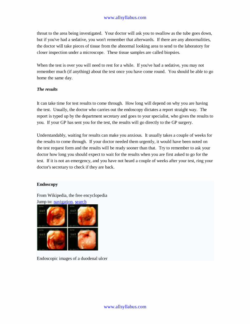
wwwallsyllabuscom
wwwallsyllabuscom
throat to the area being investigated Your doctor will ask you to swallow as the tube goes down
but if youve had a sedative you wont remember that afterwards If there are any abnormalities
the doctor will take pieces of tissue from the abnormal looking area to send to the laboratory for
closer inspection under a microscope These tissue samples are called biopsies
When the test is over you will need to rest for a while If youve had a sedative you may not
remember much (if anything) about the test once you have come round You should be able to go
home the same day
The results
It can take time for test results to come through How long will depend on why you are having
the test Usually the doctor who carries out the endoscopy dictates a report straight way The
report is typed up by the department secretary and goes to your specialist who gives the results to
you If your GP has sent you for the test the results will go directly to the GP surgery
Understandably waiting for results can make you anxious It usually takes a couple of weeks for
the results to come through If your doctor needed them urgently it would have been noted on
the test request form and the results will be ready sooner than that Try to remember to ask your
doctor how long you should expect to wait for the results when you are first asked to go for the
test If it is not an emergency and you have not heard a couple of weeks after your test ring your
doctors secretary to check if they are back
Endoscopy
From Wikipedia the free encyclopedia
Jump to navigation search
Endoscopic images of a duodenal ulcer
wwwallsyllabuscom
wwwallsyllabuscom
A flexible endoscope
Endoscopy means looking inside and typically refers to looking inside the human body for medical reasons using an instrument called an endoscope Endoscopy can also refer to using a borescope in technical situations where direct line-of-sight observation is not feasible
Contents
[hide]
1 Overview 2 Components 3 Uses
4 History 5 Risks 6 After The Endoscopy 7 Recent developments 8 See also 9 References 10 Footnotes
11 External links
Overview
Endoscopy is a minimally invasive diagnostic medical procedure that is used to assess the interior surfaces of an organ by inserting a tube into the body The instrument may have a rigid or flexible tube and not only provide an image for visual inspection and photography but also enable taking
biopsies and retrieval of foreign objects Endoscopy is the vehicle for minimally invasive surgery
Many endoscopic procedures are considered to be relatively painless and at worst associated with mild discomfort for example in esophagogastroduodenoscopy most patients tolerate the procedure with only topical anaesthesia of the oropharynx using lignocaine spray[1] Complications are not common (only 5 of all operations)[citation needed] but can include perforation of the organ under inspection with the endoscope or biopsy instrument If that occurs open surgery may be required to repair the injury
wwwallsyllabuscom
wwwallsyllabuscom
Components
An endoscope can consist of
a rigid or flexible tube a light delivery system to illuminate the organ or object under inspection The light
source is normally outside the body and the light is typically directed via an optical fiber system
a lens system transmitting the image to the viewer from the fiberscope an additional channel to allow entry of medical instruments or manipulators
Uses
Endoscopy can involve
The gastrointestinal tract (GI tract) o esophagus stomach and duodenum (esophagogastroduodenoscopy) o small intestine o colon (colonoscopyproctosigmoidoscopy) o Bile duct
endoscopic retrograde cholangiopancreatography (ERCP) duodenoscope-assisted cholangiopancreatoscopy intraoperative cholangioscopy
The respiratory tract o The nose (rhinoscopy) o The lower respiratory tract (bronchoscopy)
The urinary tract (cystoscopy) The female reproductive system
o The cervix (colposcopy) o The uterus (hysteroscopy) o The Fallopian tubes (Falloscopy)
Normally closed body cavities (through a small incision) o The abdominal or pelvic cavity (laparoscopy) o The interior of a joint (arthroscopy) o Organs of the chest (thoracoscopy and mediastinoscopy)
During pregnancy o The amnion (amnioscopy) o The fetus (fetoscopy)
Plastic Surgery Panendoscopy (or triple endoscopy)
o Combines laryngoscopy esophagoscopy and bronchoscopy Non-medical uses for endoscopy
o The planning and architectural community have found the endoscope useful for
pre-visualization of scale models of proposed buildings and cities (architectural endoscopy)
o Internal inspection of complex technical systems (borescope) o Endoscopes are also a tool helpful in the examination of improvised explosive
devices by bomb disposal personnel
wwwallsyllabuscom
wwwallsyllabuscom
o The FBI uses endoscopes for conducting surveillance via tight spaces
History
The first endoscope of a kind was developed in 1806 by Philip Bozzini with his introduction of a
Lichtleiter (light conductor) for the examinations of the canals and cavities of the human body However the Vienna Medical Society disapproved of such curiosity An endoscope was first introduced into a human in 1822 by William Beaumont an army surgeon at Mackinac
Island Michigan[citation needed] The use of electric light was a major step in the improvement of endoscopy The first such lights were external Later smaller bulbs became available making internal light possible for instance in a hysteroscope by Charles David in 1908[citation needed] Hans Christian Jacobaeus has been given credit for early endoscopic explorations of the abdomen and the thorax with laparoscopy (1912) and thoracoscopy (1910)[citation needed] Laparoscopy was used in the diagnosis of liver and gallbladder disease by Heinz Kalk in the 1930s[citation needed] Hope reported in 1937 on the use of laparoscopy to diagnose ectopic pregnancy[citation needed] In 1944
Raoul Palmer placed his patients in the Trendelenburg position after gaseous distention of the abdomen and thus was able to reliably perform gynecologic laparoscopy[citation needed]
The first gastrocamera was released in 1950 by Olympus Optical Co Ltd The device took pictures on monochromatic film using a small light bulb that was triggered manually The device was of limited use however because it did not implement real-time optical capability Olympus continued its development of endoscopes by incorporating fiber optics in the early 1960s leading to the first useful endoscopes In 1964 it released a gastrocamera guided by a fiberscope[1] A few articles claim that DrBasil Hirschowitz of UnivOf MichiganAnn Arbor discussed the
endoscope in early 50s[2]
As endoscopic technology improved so did the methods of gastrointestinal endoscopy Owing primarily to the efforts of Dr Hiromi Shinya in the late 1960s GI endoscopy developed into what is more recognizable as todays colonoscopy While many doctors experimented with techniques to take advantage of the new iterations of endoscopes Dr Shinya focused on techniques that would allow for successful operation of the endoscope by an individual rejecting the common practice at the time of utilizing two people Consequently many of the fundamental methods and procedures of modern colonoscopy were developed by Dr Shinya
Dr Shinyas other great contribution was to therapeutic endoscopy in his invention of the
electrosurgical polypectomy snare with the aid of Olympus employee Hiroshi Ichikawa Shinya sketched his first plans for the device on January 8 1969 He envisioned a loop of wire attached to the end of a colonoscope that would allow for easy removal of polyps during investigation by passing a current through the wire By September of 1969 the first polypectomy using this device was performed Polypectomy has since become the most common therapeutic procedure performed with an endoscope (Sivak 2004)
By 1980 laparoscopy training was required by gynecologists to perform tubal ligation procedures and diagnostic evaluations of the pelvis The first laparoscopic cholecystectomy was performed in 1984 and the first video-laparoscopic cholecystectomy in 1987[citation needed] During the 1990s
laparoscopic surgery was extended to the appendix spleen colon stomach kidney and liver[citation needed] Wireless capsule endoscopy or Capsule Endoscopy is now approved in all the countries including Japan where government reimbusement will be available from Oct2007Capsule Endoscopy [3] increases detection of Small Bowel tumors where traditional Endoscopy is not very efficient
wwwallsyllabuscom
wwwallsyllabuscom
Risks
Infection
Punctured organs Allergic reactions due to Contrast agents or dyes (such as those used in a CT scan) Over-sedation
After The Endoscopy
After the procedure the patient will be observed and monitored by a qualified individual in the
endoscopy or a recovery area until a significant portion of the medication has worn off Occasionally a patient is left with a mild sore throat which promptly responds to saline gargles or a feeling of distention from the insufflated air that was used during the procedure Both problems are mild and fleeting When fully recovered the patient will be instructed when to resume hisher usual diet (probably within a few hours) and will be allowed to be taken home Because of the use of sedation most facilities mandate that the patient is taken home by another person and not to drive on hisher own or handle machinery for the remainder of the day
Recent developments
With the application of robotic systems telesurgery was introduced as the surgeon could operate
from a site physically removed from the patient The first transatlantic surgery has been called the Lindbergh Operation
wwwallsyllabuscom
wwwallsyllabuscom
Upper Endoscopy
Upper endoscopy enables the physician to look inside the esophagus stomach and duodenum (first part of the small intestine) The procedure might be used to discover the reason for swallowing difficulties nausea vomiting
reflux bleeding indigestion abdominal pain or chest pain Upper endoscopy is also called EGD which stands for esophagogastroduodenoscopy (eh-SAH-fuh-goh-GAS-troh-doo-AH-duh-NAH-skuh-pee)
For the procedure you will swallow a thin flexible lighted tube called an endoscope (EN-doh-skope) Right before the procedure the physician will spray your throat with a numbing agent that may help prevent gagging
You may also receive pain medicine and a sedative to help you relax during the exam The endoscope transmits an image of the inside of the esophagus stomach and duodenum so the physician can carefully examine the lining of these organs The scope also blows air into the stomach this expands the folds of tissue and makes it easier for the physician to examine the stomach
The physician can see abnormalities like inflammation or
bleeding through the endoscope that dont show up well on x rays The physician can also insert instruments into the scope to treat bleeding abnormalities or remove samples of tissue (biopsy) for further tests
Possible complications of upper endoscopy include bleeding and puncture of the stomach lining However such complications are rare Most people will probably have nothing more than a mild sore throat after the procedure
The procedure takes 20 to 30 minutes Because you will be sedated you will need to rest at the endoscopy facility for 1 to 2 hours until the medication wears off
Preparation
Your stomach and duodenum must be empty for the procedure to be thorough and safe so you will not be able to eat or drink anything for at least 6 hours beforehand Also you must
arrange for someone to take you homemdashyou will not be allowed to drive because of the sedatives Your physician may give you other special instructions
The digestive system
wwwallsyllabuscom
wwwallsyllabuscom
What is Endoscopy and Why is it Performed
Endoscopy allows physicians to peer through the bodys passageways Endoscopy is the examination and inspection of the interior of body organs joints or cavities through an endoscope An endoscope is a device that uses fiber optics and powerful lens systems to provide lighting and visualization of the interior of a joint The portion of the endoscope inserted into the body may be rigid or flexible depending upon the medical procedure
An endoscope uses two fiber optic lines A light fiber carries light into the body cavity and an image fiber carries the image of the body cavity back to the physicians viewing lens There is also a separate port to allow for administration of drugs suction and irrigation This port may also be used to introduce small folding instruments such as forceps scissors brushes snares and baskets for tissue excision (removal) sampling or other diagnostic and therapeutic work Endoscopes may be used in conjunction with a camera or video recorder to document images of the inside of the joint or chronicle an endoscopic procedure New endoscopes have digital capabilities for manipulating and enhancing the video images
This figure shows a rigid endoscope used for arthroscopy The image fiber leads from the ocular (eye piece) to the inserted end of the scope The light fiber is below and leads from the light source to the working end of the endoscope
Why Is Endoscopy Performed
Endoscopy can be used to diagnose various conditions by close examination of internal organ and body structures Endoscopy can also guide therapy and repair such as the removal of torn cartilage from the bearing surfaces of a joint Biopsy (tissue sampling for pathologic testing) may also be performed under endoscopic guidance Local or general anesthetic may be used during endoscopy depending upon the type of procedure being performed
Internal abnormalities revealed through endoscopy include abscesses biliary (liver) cirrhosis bleeding bronchitis cancer cysts degenerative disease gallbladder stones hernia inflammation metastatic cancer polyps tumors ulcers and other diseases and conditions
Endoscopy is a minimally invasive procedure and carries with it certain minor risks depending upon the type of procedure being performed However these risks are typically far outweighed by the diagnostic and therapeutic potential of the procedure
Prior to the widespread use of endoscopy and diagnostic imaging most internal conditions could only be diagnosed or treated with open surgery Until the last several decades exploratory surgery was routinely performed only when a patient was critically ill and the source of illness was not known For example in certain dire cases the patients thorax or abdomen were surgically opened and examined to try to determine the source of illness
wwwallsyllabuscom
wwwallsyllabuscom
Endoscopy can often be done on an outpatient basis Outpatient means that the procedure does not require hospital admission and acute care and observation and may be performed outside the premises of a hospital Outpatient procedures performed at hospitals or ambulatory centers allow the patient to go home or return to work within a short while after their procedure
Types of Endoscopy
Fiber optic endoscopes now have widespread use in medicine and guide a myriad of diagnostic and therapeutic procedures including
Arthroscopy examination of joints for diagnosis and treatment (arthroscopic surgery) Bronchoscopy examination of the trachea and lungs bronchial trees to reveal
abscesses bronchitis carcinoma tumors tuberculosis alveolitis infection inflammation Colonoscopy examination of the inside of the colon and large intestine to detect polyps
tumors ulceration inflammation colitis diverticula Chrohns disease and discovery and removal of foreign bodies
Colposcopy direct visualization of the vagina and cervix to detect cancer inflammation and other conditions
Cystoscopy examination of the bladder urethra urinary tract uteral orifices and
prostate (men) with insertion of the endoscope through the urethra
ERCP (endoscopic retrograde cholangio-pancreatography) uses endoscopic guidance to
place a catheter for x-ray fluorosocopy with contrast enhancement This technique is used to examine the livers biliary tree the gallbladder the pancreatic duct and other anatomy to check for stones other obstructions and disease X-ray contrast is introduced into these ducts via catheter and fluoroscopic x-ray images are taken to show any abnormality or blockage If disease is detected it can sometimes be treated at the same time or biopsy can be performed to test for cancer or other pathology ERCP can detect biliary cirrhosis
cancer of the bile ducts pancreatic cysts pseudocysts pancreatic tumors chronic pancreatitis and other conditions such as gallbladder stones
EGD (Esophogealgastroduodensoscopy) visual examination of the upper gastro-
intestinal (GI) tract (also referred to as gastroscopy) to reveal hemorrhage hiatal hernia inflammation of the esophagus gastric ulcers
Endoscopic biopsy is the removal of tissue specimens for pathologic examination and
analysis Gastroscopy examination of the lining of the esophagus stomach and duodenum
Gastroscopy is often used to diagnose ulcers and other sources of bleeding and to guide biopsy of suspect GI cancers
Laparoscopy visualization of the stomach liver and other abdominal organs including
the female reproductive organs for example the fallopian tubes Laryngoscopy examination of the larynx (voice box) Proctoscopy sigmoidoscopy proctosigmoidoscopy examination of the rectum and
sigmoid colon Thoracoscopy examination of the pleura (sac that covers the lungs) pleural spaces
mediastinum and pericardium
A Brief History of Endoscopy
In the early 1900s the first attempts to view inside the body with lighted telescopes were made These initial devices were often fully rigid In the 1930s semi-flexible endoscopes called gastroscopes were developed to view inside of the stomach Fiber-optic endoscopy was pioneered by South African-born physician Basil Hirschowitz at the University of Michigan in 1957 Widespread use of fiber optic endoscopes began in the 1960s
wwwallsyllabuscom
wwwallsyllabuscom
A fiber optic cable is simply a bundle of microscopic glass or plastic fibers that literally allows light and images to be transmitted through curved structures Fiber optic cables are also replacing metal wires as the backbone of the worlds telecommunications infrastructure This Internet page may have traveled through a fiber optic cable as a stream of digital data (bursts of light) on its way to your computer
Endoscopy Equipment
Endoscopes have many practical needs And HMB Endoscopy Products (Hollywood Florida) has been providing endoscopic equipment and educating people on the use of endoscopes for more than 17 years Be sure to Browse our Catalog for all the details on how to purchase these medical instruments that can examine any part of the body
Video
Systems
Fiber
Endoscopes
Video
Endoscopes
In the simplest terms Endoscopy equipment consists of instruments that can look at the inside of many different organs mdash these are small flexible or rigid tubes with a light or lenses on the end that can look into the esophagus stomach and colon mdash and in more general terms endoscopy equipment can help doctors look deep inside body structures and hollow organs
An endoscope and related endoscope products and equipment are usually composed of three components
An optic system that allows the doctor to look through the scope into the organ or
cavity or to attach a video camera to the scope
A fiberoptic cable to light up the bodily area
A lumen (eg the bore of a tube like a needle or catheter) to take tissue samples of the
area being viewed
The beauty of endoscopic products is that they perform dual functions mdash with both diagnostic and therapeutic capabilities For example this means that these endoscopic products and instruments can perform biopsies (eg to evaluate tissue samples) as well as provide sclerotherapy (a medical procedure used to treat varicose veins and ―spider veins) In truth these brief explanations only tell part of the story
wwwallsyllabuscom
wwwallsyllabuscom
TYPES OF ENDOSCOPES
Take a look at the different types of endoscopic equipment you can get with HMB Endoscopy Products Again in very general terms there are two main types of endoscopes
Rigid Flexible
1 rigid endoscopes ndash the majority of which use a convex (curving out like one half of a
circle) glass lens system in which the small glass lenses are separated by large air spaces
2 flexible endoscopes allow for just that mdash flexibility In the animal kingdom for example
a flexible endoscope would be perfect for examining the stomach area of a snake
The popularity of endoscopy equipment continues to grow Some of the industryrsquos household names include Olympus Pentax Funjinon and Storz mdash with HMB both selling and repairing pre-owned and completely refurbished endoscopic products from each of these major manufacturers Browse our Catalog to get the ones you need
THERMOGRAPH APPLICATION Thermographs are digital recording thermometers used to log
temperature in the
marine environment The instruments are attached to a computer to set up recording
parameters for
deployment downloading and display of data in graphic or numeric format These units
are placed in
underwater housings and attached to the bottom or suspended in the water column for
deployment
DESCRIPTION Two types of thermograph are currently available The Ryan
Tempmentor is a
reusable data logger that allows storage of a maximum of 6361 temperature
measurements in the
range of -32C to +70C with 01resolution and 03accuracy The instrument can be
programmed
to take measurements from once per second to once every other hour Instrument
dimensions are 3 x
6 x 13 and the unit weighs 11 ounces The Hobo Temp is a miniature reusable data
logger that
allows storage of 1800 measurements in the range of -20C to +70C with
03resolution and 07
wwwallsyllabuscom
wwwallsyllabuscom
accuracy Sampling intervals from 05 seconds to 48 hours are available Instrument
dimensions are
24 x 19 x 08 and the unit weighs 1 ounce Both units are battery powered with the
battery life of
the Tempmentor being approximately two years and the battery life of the Hobo Temp
being one year THERMOGRAPH APPLICATION Thermographs are digital recording thermometers used to log
temperature in the
marine environment The instruments are attached to a computer to set up recording
parameters for
deployment downloading and display of data in graphic or numeric format These units
are placed in
underwater housings and attached to the bottom or suspended in the water column for
deployment
DESCRIPTION Two types of thermograph are currently available The Ryan
Tempmentor is a
reusable data logger that allows storage of a maximum of 6361 temperature
measurements in the
range of -32C to +70C with 01resolution and 03accuracy The instrument can be
programmed
to take measurements from once per second to once every other hour Instrument
dimensions are 3 x
6 x 13 and the unit weighs 11 ounces The Hobo Temp is a miniature reusable data
logger that
allows storage of 1800 measurements in the range of -20C to +70C with
03resolution and 07
accuracy Sampling intervals from 05 seconds to 48 hours are available Instrument
dimensions are
24 x 19 x 08 and the unit weighs 1 ounce Both units are battery powered with the
battery life of
the Tempmentor being approximately two years and the battery life of the Hobo Temp
being one year THERMOGRAPH APPLICATION Thermographs are digital recording thermometers used to log
temperature in the
marine environment The instruments are attached to a computer to set up recording
parameters for
deployment downloading and display of data in graphic or numeric format These units
are placed in
underwater housings and attached to the bottom or suspended in the water column for
deployment
DESCRIPTION Two types of thermograph are currently available The Ryan
Tempmentor is a
reusable data logger that allows storage of a maximum of 6361 temperature
measurements in the
range of -32C to +70C with 01resolution and 03accuracy The instrument can be
programmed
wwwallsyllabuscom
wwwallsyllabuscom
to take measurements from once per second to once every other hour Instrument
dimensions are 3 x
6 x 13 and the unit weighs 11 ounces The Hobo Temp is a miniature reusable data
logger that
allows storage of 1800 measurements in the range of -20C to +70C with
03resolution and 07
accuracy Sampling intervals from 05 seconds to 48 hours are available Instrument
dimensions are
24 x 19 x 08 and the unit weighs 1 ounce Both units are battery powered with the
battery life of
the Tempmentor being approximately two years and the battery life of the Hobo Temp
being one year
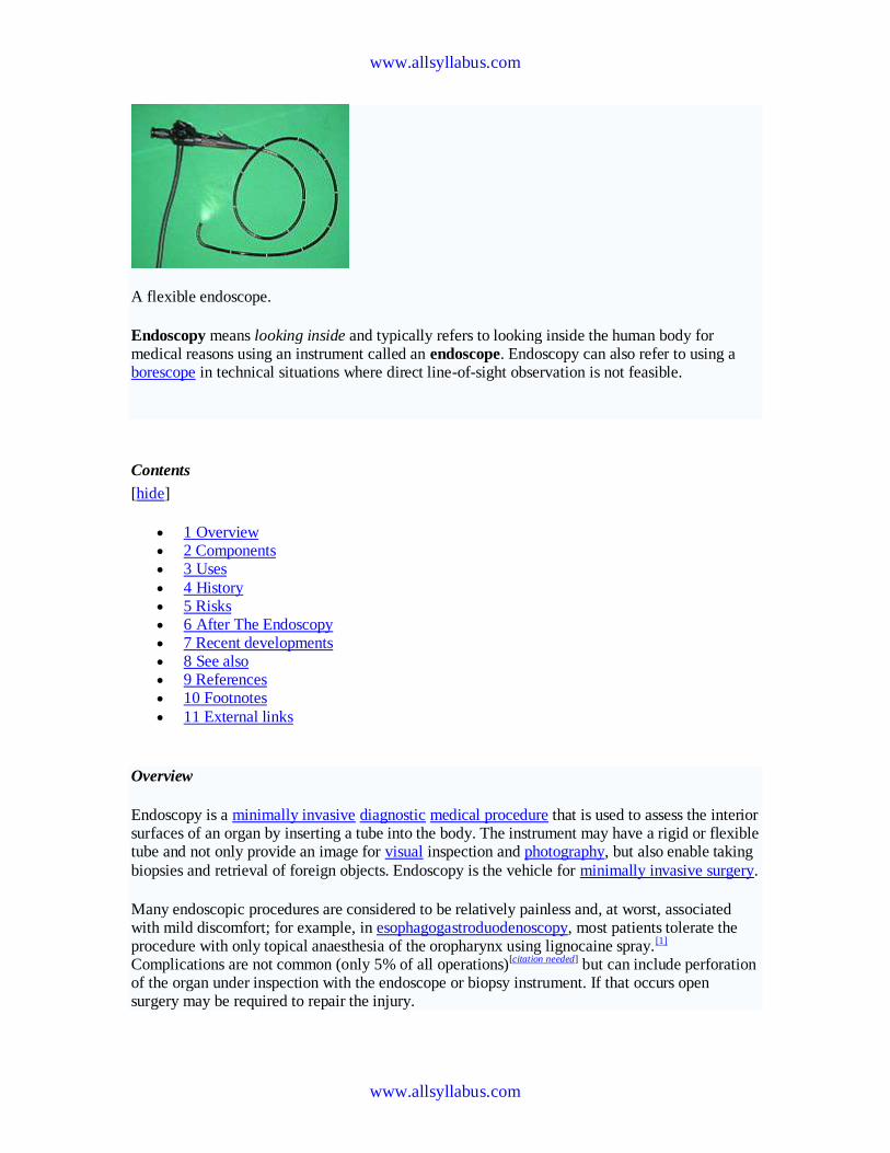
wwwallsyllabuscom
wwwallsyllabuscom
Components
An endoscope can consist of
a rigid or flexible tube a light delivery system to illuminate the organ or object under inspection The light
source is normally outside the body and the light is typically directed via an optical fiber system
a lens system transmitting the image to the viewer from the fiberscope an additional channel to allow entry of medical instruments or manipulators
Uses
Endoscopy can involve
The gastrointestinal tract (GI tract) o esophagus stomach and duodenum (esophagogastroduodenoscopy) o small intestine o colon (colonoscopyproctosigmoidoscopy) o Bile duct
endoscopic retrograde cholangiopancreatography (ERCP) duodenoscope-assisted cholangiopancreatoscopy intraoperative cholangioscopy
The respiratory tract o The nose (rhinoscopy) o The lower respiratory tract (bronchoscopy)
The urinary tract (cystoscopy) The female reproductive system
o The cervix (colposcopy) o The uterus (hysteroscopy) o The Fallopian tubes (Falloscopy)
Normally closed body cavities (through a small incision) o The abdominal or pelvic cavity (laparoscopy) o The interior of a joint (arthroscopy) o Organs of the chest (thoracoscopy and mediastinoscopy)
During pregnancy o The amnion (amnioscopy) o The fetus (fetoscopy)
Plastic Surgery Panendoscopy (or triple endoscopy)
o Combines laryngoscopy esophagoscopy and bronchoscopy Non-medical uses for endoscopy
o The planning and architectural community have found the endoscope useful for
pre-visualization of scale models of proposed buildings and cities (architectural endoscopy)
o Internal inspection of complex technical systems (borescope) o Endoscopes are also a tool helpful in the examination of improvised explosive
devices by bomb disposal personnel
wwwallsyllabuscom
wwwallsyllabuscom
o The FBI uses endoscopes for conducting surveillance via tight spaces
History
The first endoscope of a kind was developed in 1806 by Philip Bozzini with his introduction of a
Lichtleiter (light conductor) for the examinations of the canals and cavities of the human body However the Vienna Medical Society disapproved of such curiosity An endoscope was first introduced into a human in 1822 by William Beaumont an army surgeon at Mackinac
Island Michigan[citation needed] The use of electric light was a major step in the improvement of endoscopy The first such lights were external Later smaller bulbs became available making internal light possible for instance in a hysteroscope by Charles David in 1908[citation needed] Hans Christian Jacobaeus has been given credit for early endoscopic explorations of the abdomen and the thorax with laparoscopy (1912) and thoracoscopy (1910)[citation needed] Laparoscopy was used in the diagnosis of liver and gallbladder disease by Heinz Kalk in the 1930s[citation needed] Hope reported in 1937 on the use of laparoscopy to diagnose ectopic pregnancy[citation needed] In 1944
Raoul Palmer placed his patients in the Trendelenburg position after gaseous distention of the abdomen and thus was able to reliably perform gynecologic laparoscopy[citation needed]
The first gastrocamera was released in 1950 by Olympus Optical Co Ltd The device took pictures on monochromatic film using a small light bulb that was triggered manually The device was of limited use however because it did not implement real-time optical capability Olympus continued its development of endoscopes by incorporating fiber optics in the early 1960s leading to the first useful endoscopes In 1964 it released a gastrocamera guided by a fiberscope[1] A few articles claim that DrBasil Hirschowitz of UnivOf MichiganAnn Arbor discussed the
endoscope in early 50s[2]
As endoscopic technology improved so did the methods of gastrointestinal endoscopy Owing primarily to the efforts of Dr Hiromi Shinya in the late 1960s GI endoscopy developed into what is more recognizable as todays colonoscopy While many doctors experimented with techniques to take advantage of the new iterations of endoscopes Dr Shinya focused on techniques that would allow for successful operation of the endoscope by an individual rejecting the common practice at the time of utilizing two people Consequently many of the fundamental methods and procedures of modern colonoscopy were developed by Dr Shinya
Dr Shinyas other great contribution was to therapeutic endoscopy in his invention of the
electrosurgical polypectomy snare with the aid of Olympus employee Hiroshi Ichikawa Shinya sketched his first plans for the device on January 8 1969 He envisioned a loop of wire attached to the end of a colonoscope that would allow for easy removal of polyps during investigation by passing a current through the wire By September of 1969 the first polypectomy using this device was performed Polypectomy has since become the most common therapeutic procedure performed with an endoscope (Sivak 2004)
By 1980 laparoscopy training was required by gynecologists to perform tubal ligation procedures and diagnostic evaluations of the pelvis The first laparoscopic cholecystectomy was performed in 1984 and the first video-laparoscopic cholecystectomy in 1987[citation needed] During the 1990s
laparoscopic surgery was extended to the appendix spleen colon stomach kidney and liver[citation needed] Wireless capsule endoscopy or Capsule Endoscopy is now approved in all the countries including Japan where government reimbusement will be available from Oct2007Capsule Endoscopy [3] increases detection of Small Bowel tumors where traditional Endoscopy is not very efficient
wwwallsyllabuscom
wwwallsyllabuscom
Risks
Infection
Punctured organs Allergic reactions due to Contrast agents or dyes (such as those used in a CT scan) Over-sedation
After The Endoscopy
After the procedure the patient will be observed and monitored by a qualified individual in the
endoscopy or a recovery area until a significant portion of the medication has worn off Occasionally a patient is left with a mild sore throat which promptly responds to saline gargles or a feeling of distention from the insufflated air that was used during the procedure Both problems are mild and fleeting When fully recovered the patient will be instructed when to resume hisher usual diet (probably within a few hours) and will be allowed to be taken home Because of the use of sedation most facilities mandate that the patient is taken home by another person and not to drive on hisher own or handle machinery for the remainder of the day
Recent developments
With the application of robotic systems telesurgery was introduced as the surgeon could operate
from a site physically removed from the patient The first transatlantic surgery has been called the Lindbergh Operation
wwwallsyllabuscom
wwwallsyllabuscom
Upper Endoscopy
Upper endoscopy enables the physician to look inside the esophagus stomach and duodenum (first part of the small intestine) The procedure might be used to discover the reason for swallowing difficulties nausea vomiting
reflux bleeding indigestion abdominal pain or chest pain Upper endoscopy is also called EGD which stands for esophagogastroduodenoscopy (eh-SAH-fuh-goh-GAS-troh-doo-AH-duh-NAH-skuh-pee)
For the procedure you will swallow a thin flexible lighted tube called an endoscope (EN-doh-skope) Right before the procedure the physician will spray your throat with a numbing agent that may help prevent gagging
You may also receive pain medicine and a sedative to help you relax during the exam The endoscope transmits an image of the inside of the esophagus stomach and duodenum so the physician can carefully examine the lining of these organs The scope also blows air into the stomach this expands the folds of tissue and makes it easier for the physician to examine the stomach
The physician can see abnormalities like inflammation or
bleeding through the endoscope that dont show up well on x rays The physician can also insert instruments into the scope to treat bleeding abnormalities or remove samples of tissue (biopsy) for further tests
Possible complications of upper endoscopy include bleeding and puncture of the stomach lining However such complications are rare Most people will probably have nothing more than a mild sore throat after the procedure
The procedure takes 20 to 30 minutes Because you will be sedated you will need to rest at the endoscopy facility for 1 to 2 hours until the medication wears off
Preparation
Your stomach and duodenum must be empty for the procedure to be thorough and safe so you will not be able to eat or drink anything for at least 6 hours beforehand Also you must
arrange for someone to take you homemdashyou will not be allowed to drive because of the sedatives Your physician may give you other special instructions
The digestive system
wwwallsyllabuscom
wwwallsyllabuscom
What is Endoscopy and Why is it Performed
Endoscopy allows physicians to peer through the bodys passageways Endoscopy is the examination and inspection of the interior of body organs joints or cavities through an endoscope An endoscope is a device that uses fiber optics and powerful lens systems to provide lighting and visualization of the interior of a joint The portion of the endoscope inserted into the body may be rigid or flexible depending upon the medical procedure
An endoscope uses two fiber optic lines A light fiber carries light into the body cavity and an image fiber carries the image of the body cavity back to the physicians viewing lens There is also a separate port to allow for administration of drugs suction and irrigation This port may also be used to introduce small folding instruments such as forceps scissors brushes snares and baskets for tissue excision (removal) sampling or other diagnostic and therapeutic work Endoscopes may be used in conjunction with a camera or video recorder to document images of the inside of the joint or chronicle an endoscopic procedure New endoscopes have digital capabilities for manipulating and enhancing the video images
This figure shows a rigid endoscope used for arthroscopy The image fiber leads from the ocular (eye piece) to the inserted end of the scope The light fiber is below and leads from the light source to the working end of the endoscope
Why Is Endoscopy Performed
Endoscopy can be used to diagnose various conditions by close examination of internal organ and body structures Endoscopy can also guide therapy and repair such as the removal of torn cartilage from the bearing surfaces of a joint Biopsy (tissue sampling for pathologic testing) may also be performed under endoscopic guidance Local or general anesthetic may be used during endoscopy depending upon the type of procedure being performed
Internal abnormalities revealed through endoscopy include abscesses biliary (liver) cirrhosis bleeding bronchitis cancer cysts degenerative disease gallbladder stones hernia inflammation metastatic cancer polyps tumors ulcers and other diseases and conditions
Endoscopy is a minimally invasive procedure and carries with it certain minor risks depending upon the type of procedure being performed However these risks are typically far outweighed by the diagnostic and therapeutic potential of the procedure
Prior to the widespread use of endoscopy and diagnostic imaging most internal conditions could only be diagnosed or treated with open surgery Until the last several decades exploratory surgery was routinely performed only when a patient was critically ill and the source of illness was not known For example in certain dire cases the patients thorax or abdomen were surgically opened and examined to try to determine the source of illness
wwwallsyllabuscom
wwwallsyllabuscom
Endoscopy can often be done on an outpatient basis Outpatient means that the procedure does not require hospital admission and acute care and observation and may be performed outside the premises of a hospital Outpatient procedures performed at hospitals or ambulatory centers allow the patient to go home or return to work within a short while after their procedure
Types of Endoscopy
Fiber optic endoscopes now have widespread use in medicine and guide a myriad of diagnostic and therapeutic procedures including
Arthroscopy examination of joints for diagnosis and treatment (arthroscopic surgery) Bronchoscopy examination of the trachea and lungs bronchial trees to reveal
abscesses bronchitis carcinoma tumors tuberculosis alveolitis infection inflammation Colonoscopy examination of the inside of the colon and large intestine to detect polyps
tumors ulceration inflammation colitis diverticula Chrohns disease and discovery and removal of foreign bodies
Colposcopy direct visualization of the vagina and cervix to detect cancer inflammation and other conditions
Cystoscopy examination of the bladder urethra urinary tract uteral orifices and
prostate (men) with insertion of the endoscope through the urethra
ERCP (endoscopic retrograde cholangio-pancreatography) uses endoscopic guidance to
place a catheter for x-ray fluorosocopy with contrast enhancement This technique is used to examine the livers biliary tree the gallbladder the pancreatic duct and other anatomy to check for stones other obstructions and disease X-ray contrast is introduced into these ducts via catheter and fluoroscopic x-ray images are taken to show any abnormality or blockage If disease is detected it can sometimes be treated at the same time or biopsy can be performed to test for cancer or other pathology ERCP can detect biliary cirrhosis
cancer of the bile ducts pancreatic cysts pseudocysts pancreatic tumors chronic pancreatitis and other conditions such as gallbladder stones
EGD (Esophogealgastroduodensoscopy) visual examination of the upper gastro-
intestinal (GI) tract (also referred to as gastroscopy) to reveal hemorrhage hiatal hernia inflammation of the esophagus gastric ulcers
Endoscopic biopsy is the removal of tissue specimens for pathologic examination and
analysis Gastroscopy examination of the lining of the esophagus stomach and duodenum
Gastroscopy is often used to diagnose ulcers and other sources of bleeding and to guide biopsy of suspect GI cancers
Laparoscopy visualization of the stomach liver and other abdominal organs including
the female reproductive organs for example the fallopian tubes Laryngoscopy examination of the larynx (voice box) Proctoscopy sigmoidoscopy proctosigmoidoscopy examination of the rectum and
sigmoid colon Thoracoscopy examination of the pleura (sac that covers the lungs) pleural spaces
mediastinum and pericardium
A Brief History of Endoscopy
In the early 1900s the first attempts to view inside the body with lighted telescopes were made These initial devices were often fully rigid In the 1930s semi-flexible endoscopes called gastroscopes were developed to view inside of the stomach Fiber-optic endoscopy was pioneered by South African-born physician Basil Hirschowitz at the University of Michigan in 1957 Widespread use of fiber optic endoscopes began in the 1960s
wwwallsyllabuscom
wwwallsyllabuscom
A fiber optic cable is simply a bundle of microscopic glass or plastic fibers that literally allows light and images to be transmitted through curved structures Fiber optic cables are also replacing metal wires as the backbone of the worlds telecommunications infrastructure This Internet page may have traveled through a fiber optic cable as a stream of digital data (bursts of light) on its way to your computer
Endoscopy Equipment
Endoscopes have many practical needs And HMB Endoscopy Products (Hollywood Florida) has been providing endoscopic equipment and educating people on the use of endoscopes for more than 17 years Be sure to Browse our Catalog for all the details on how to purchase these medical instruments that can examine any part of the body
Video
Systems
Fiber
Endoscopes
Video
Endoscopes
In the simplest terms Endoscopy equipment consists of instruments that can look at the inside of many different organs mdash these are small flexible or rigid tubes with a light or lenses on the end that can look into the esophagus stomach and colon mdash and in more general terms endoscopy equipment can help doctors look deep inside body structures and hollow organs
An endoscope and related endoscope products and equipment are usually composed of three components
An optic system that allows the doctor to look through the scope into the organ or
cavity or to attach a video camera to the scope
A fiberoptic cable to light up the bodily area
A lumen (eg the bore of a tube like a needle or catheter) to take tissue samples of the
area being viewed
The beauty of endoscopic products is that they perform dual functions mdash with both diagnostic and therapeutic capabilities For example this means that these endoscopic products and instruments can perform biopsies (eg to evaluate tissue samples) as well as provide sclerotherapy (a medical procedure used to treat varicose veins and ―spider veins) In truth these brief explanations only tell part of the story
wwwallsyllabuscom
wwwallsyllabuscom
TYPES OF ENDOSCOPES
Take a look at the different types of endoscopic equipment you can get with HMB Endoscopy Products Again in very general terms there are two main types of endoscopes
Rigid Flexible
1 rigid endoscopes ndash the majority of which use a convex (curving out like one half of a
circle) glass lens system in which the small glass lenses are separated by large air spaces
2 flexible endoscopes allow for just that mdash flexibility In the animal kingdom for example
a flexible endoscope would be perfect for examining the stomach area of a snake
The popularity of endoscopy equipment continues to grow Some of the industryrsquos household names include Olympus Pentax Funjinon and Storz mdash with HMB both selling and repairing pre-owned and completely refurbished endoscopic products from each of these major manufacturers Browse our Catalog to get the ones you need
THERMOGRAPH APPLICATION Thermographs are digital recording thermometers used to log
temperature in the
marine environment The instruments are attached to a computer to set up recording
parameters for
deployment downloading and display of data in graphic or numeric format These units
are placed in
underwater housings and attached to the bottom or suspended in the water column for
deployment
DESCRIPTION Two types of thermograph are currently available The Ryan
Tempmentor is a
reusable data logger that allows storage of a maximum of 6361 temperature
measurements in the
range of -32C to +70C with 01resolution and 03accuracy The instrument can be
programmed
to take measurements from once per second to once every other hour Instrument
dimensions are 3 x
6 x 13 and the unit weighs 11 ounces The Hobo Temp is a miniature reusable data
logger that
allows storage of 1800 measurements in the range of -20C to +70C with
03resolution and 07
wwwallsyllabuscom
wwwallsyllabuscom
accuracy Sampling intervals from 05 seconds to 48 hours are available Instrument
dimensions are
24 x 19 x 08 and the unit weighs 1 ounce Both units are battery powered with the
battery life of
the Tempmentor being approximately two years and the battery life of the Hobo Temp
being one year THERMOGRAPH APPLICATION Thermographs are digital recording thermometers used to log
temperature in the
marine environment The instruments are attached to a computer to set up recording
parameters for
deployment downloading and display of data in graphic or numeric format These units
are placed in
underwater housings and attached to the bottom or suspended in the water column for
deployment
DESCRIPTION Two types of thermograph are currently available The Ryan
Tempmentor is a
reusable data logger that allows storage of a maximum of 6361 temperature
measurements in the
range of -32C to +70C with 01resolution and 03accuracy The instrument can be
programmed
to take measurements from once per second to once every other hour Instrument
dimensions are 3 x
6 x 13 and the unit weighs 11 ounces The Hobo Temp is a miniature reusable data
logger that
allows storage of 1800 measurements in the range of -20C to +70C with
03resolution and 07
accuracy Sampling intervals from 05 seconds to 48 hours are available Instrument
dimensions are
24 x 19 x 08 and the unit weighs 1 ounce Both units are battery powered with the
battery life of
the Tempmentor being approximately two years and the battery life of the Hobo Temp
being one year THERMOGRAPH APPLICATION Thermographs are digital recording thermometers used to log
temperature in the
marine environment The instruments are attached to a computer to set up recording
parameters for
deployment downloading and display of data in graphic or numeric format These units
are placed in
underwater housings and attached to the bottom or suspended in the water column for
deployment
DESCRIPTION Two types of thermograph are currently available The Ryan
Tempmentor is a
reusable data logger that allows storage of a maximum of 6361 temperature
measurements in the
range of -32C to +70C with 01resolution and 03accuracy The instrument can be
programmed
wwwallsyllabuscom
wwwallsyllabuscom
to take measurements from once per second to once every other hour Instrument
dimensions are 3 x
6 x 13 and the unit weighs 11 ounces The Hobo Temp is a miniature reusable data
logger that
allows storage of 1800 measurements in the range of -20C to +70C with
03resolution and 07
accuracy Sampling intervals from 05 seconds to 48 hours are available Instrument
dimensions are
24 x 19 x 08 and the unit weighs 1 ounce Both units are battery powered with the
battery life of
the Tempmentor being approximately two years and the battery life of the Hobo Temp
being one year
wwwallsyllabuscom
wwwallsyllabuscom
Risks
Infection
Punctured organs Allergic reactions due to Contrast agents or dyes (such as those used in a CT scan) Over-sedation
After The Endoscopy
After the procedure the patient will be observed and monitored by a qualified individual in the
endoscopy or a recovery area until a significant portion of the medication has worn off Occasionally a patient is left with a mild sore throat which promptly responds to saline gargles or a feeling of distention from the insufflated air that was used during the procedure Both problems are mild and fleeting When fully recovered the patient will be instructed when to resume hisher usual diet (probably within a few hours) and will be allowed to be taken home Because of the use of sedation most facilities mandate that the patient is taken home by another person and not to drive on hisher own or handle machinery for the remainder of the day
Recent developments
With the application of robotic systems telesurgery was introduced as the surgeon could operate
from a site physically removed from the patient The first transatlantic surgery has been called the Lindbergh Operation
wwwallsyllabuscom
wwwallsyllabuscom
Upper Endoscopy
Upper endoscopy enables the physician to look inside the esophagus stomach and duodenum (first part of the small intestine) The procedure might be used to discover the reason for swallowing difficulties nausea vomiting
reflux bleeding indigestion abdominal pain or chest pain Upper endoscopy is also called EGD which stands for esophagogastroduodenoscopy (eh-SAH-fuh-goh-GAS-troh-doo-AH-duh-NAH-skuh-pee)
For the procedure you will swallow a thin flexible lighted tube called an endoscope (EN-doh-skope) Right before the procedure the physician will spray your throat with a numbing agent that may help prevent gagging
You may also receive pain medicine and a sedative to help you relax during the exam The endoscope transmits an image of the inside of the esophagus stomach and duodenum so the physician can carefully examine the lining of these organs The scope also blows air into the stomach this expands the folds of tissue and makes it easier for the physician to examine the stomach
The physician can see abnormalities like inflammation or
bleeding through the endoscope that dont show up well on x rays The physician can also insert instruments into the scope to treat bleeding abnormalities or remove samples of tissue (biopsy) for further tests
Possible complications of upper endoscopy include bleeding and puncture of the stomach lining However such complications are rare Most people will probably have nothing more than a mild sore throat after the procedure
The procedure takes 20 to 30 minutes Because you will be sedated you will need to rest at the endoscopy facility for 1 to 2 hours until the medication wears off
Preparation
Your stomach and duodenum must be empty for the procedure to be thorough and safe so you will not be able to eat or drink anything for at least 6 hours beforehand Also you must
arrange for someone to take you homemdashyou will not be allowed to drive because of the sedatives Your physician may give you other special instructions
The digestive system
wwwallsyllabuscom
wwwallsyllabuscom
What is Endoscopy and Why is it Performed
Endoscopy allows physicians to peer through the bodys passageways Endoscopy is the examination and inspection of the interior of body organs joints or cavities through an endoscope An endoscope is a device that uses fiber optics and powerful lens systems to provide lighting and visualization of the interior of a joint The portion of the endoscope inserted into the body may be rigid or flexible depending upon the medical procedure
An endoscope uses two fiber optic lines A light fiber carries light into the body cavity and an image fiber carries the image of the body cavity back to the physicians viewing lens There is also a separate port to allow for administration of drugs suction and irrigation This port may also be used to introduce small folding instruments such as forceps scissors brushes snares and baskets for tissue excision (removal) sampling or other diagnostic and therapeutic work Endoscopes may be used in conjunction with a camera or video recorder to document images of the inside of the joint or chronicle an endoscopic procedure New endoscopes have digital capabilities for manipulating and enhancing the video images
This figure shows a rigid endoscope used for arthroscopy The image fiber leads from the ocular (eye piece) to the inserted end of the scope The light fiber is below and leads from the light source to the working end of the endoscope
Why Is Endoscopy Performed
Endoscopy can be used to diagnose various conditions by close examination of internal organ and body structures Endoscopy can also guide therapy and repair such as the removal of torn cartilage from the bearing surfaces of a joint Biopsy (tissue sampling for pathologic testing) may also be performed under endoscopic guidance Local or general anesthetic may be used during endoscopy depending upon the type of procedure being performed
Internal abnormalities revealed through endoscopy include abscesses biliary (liver) cirrhosis bleeding bronchitis cancer cysts degenerative disease gallbladder stones hernia inflammation metastatic cancer polyps tumors ulcers and other diseases and conditions
Endoscopy is a minimally invasive procedure and carries with it certain minor risks depending upon the type of procedure being performed However these risks are typically far outweighed by the diagnostic and therapeutic potential of the procedure
Prior to the widespread use of endoscopy and diagnostic imaging most internal conditions could only be diagnosed or treated with open surgery Until the last several decades exploratory surgery was routinely performed only when a patient was critically ill and the source of illness was not known For example in certain dire cases the patients thorax or abdomen were surgically opened and examined to try to determine the source of illness
wwwallsyllabuscom
wwwallsyllabuscom
Endoscopy can often be done on an outpatient basis Outpatient means that the procedure does not require hospital admission and acute care and observation and may be performed outside the premises of a hospital Outpatient procedures performed at hospitals or ambulatory centers allow the patient to go home or return to work within a short while after their procedure
Types of Endoscopy
Fiber optic endoscopes now have widespread use in medicine and guide a myriad of diagnostic and therapeutic procedures including
Arthroscopy examination of joints for diagnosis and treatment (arthroscopic surgery) Bronchoscopy examination of the trachea and lungs bronchial trees to reveal
abscesses bronchitis carcinoma tumors tuberculosis alveolitis infection inflammation Colonoscopy examination of the inside of the colon and large intestine to detect polyps
tumors ulceration inflammation colitis diverticula Chrohns disease and discovery and removal of foreign bodies
Colposcopy direct visualization of the vagina and cervix to detect cancer inflammation and other conditions
Cystoscopy examination of the bladder urethra urinary tract uteral orifices and
prostate (men) with insertion of the endoscope through the urethra
ERCP (endoscopic retrograde cholangio-pancreatography) uses endoscopic guidance to
place a catheter for x-ray fluorosocopy with contrast enhancement This technique is used to examine the livers biliary tree the gallbladder the pancreatic duct and other anatomy to check for stones other obstructions and disease X-ray contrast is introduced into these ducts via catheter and fluoroscopic x-ray images are taken to show any abnormality or blockage If disease is detected it can sometimes be treated at the same time or biopsy can be performed to test for cancer or other pathology ERCP can detect biliary cirrhosis
cancer of the bile ducts pancreatic cysts pseudocysts pancreatic tumors chronic pancreatitis and other conditions such as gallbladder stones
EGD (Esophogealgastroduodensoscopy) visual examination of the upper gastro-
intestinal (GI) tract (also referred to as gastroscopy) to reveal hemorrhage hiatal hernia inflammation of the esophagus gastric ulcers
Endoscopic biopsy is the removal of tissue specimens for pathologic examination and
analysis Gastroscopy examination of the lining of the esophagus stomach and duodenum
Gastroscopy is often used to diagnose ulcers and other sources of bleeding and to guide biopsy of suspect GI cancers
Laparoscopy visualization of the stomach liver and other abdominal organs including
the female reproductive organs for example the fallopian tubes Laryngoscopy examination of the larynx (voice box) Proctoscopy sigmoidoscopy proctosigmoidoscopy examination of the rectum and
sigmoid colon Thoracoscopy examination of the pleura (sac that covers the lungs) pleural spaces
mediastinum and pericardium
A Brief History of Endoscopy
In the early 1900s the first attempts to view inside the body with lighted telescopes were made These initial devices were often fully rigid In the 1930s semi-flexible endoscopes called gastroscopes were developed to view inside of the stomach Fiber-optic endoscopy was pioneered by South African-born physician Basil Hirschowitz at the University of Michigan in 1957 Widespread use of fiber optic endoscopes began in the 1960s
wwwallsyllabuscom
wwwallsyllabuscom
A fiber optic cable is simply a bundle of microscopic glass or plastic fibers that literally allows light and images to be transmitted through curved structures Fiber optic cables are also replacing metal wires as the backbone of the worlds telecommunications infrastructure This Internet page may have traveled through a fiber optic cable as a stream of digital data (bursts of light) on its way to your computer
Endoscopy Equipment
Endoscopes have many practical needs And HMB Endoscopy Products (Hollywood Florida) has been providing endoscopic equipment and educating people on the use of endoscopes for more than 17 years Be sure to Browse our Catalog for all the details on how to purchase these medical instruments that can examine any part of the body
Video
Systems
Fiber
Endoscopes
Video
Endoscopes
In the simplest terms Endoscopy equipment consists of instruments that can look at the inside of many different organs mdash these are small flexible or rigid tubes with a light or lenses on the end that can look into the esophagus stomach and colon mdash and in more general terms endoscopy equipment can help doctors look deep inside body structures and hollow organs
An endoscope and related endoscope products and equipment are usually composed of three components
An optic system that allows the doctor to look through the scope into the organ or
cavity or to attach a video camera to the scope
A fiberoptic cable to light up the bodily area
A lumen (eg the bore of a tube like a needle or catheter) to take tissue samples of the
area being viewed
The beauty of endoscopic products is that they perform dual functions mdash with both diagnostic and therapeutic capabilities For example this means that these endoscopic products and instruments can perform biopsies (eg to evaluate tissue samples) as well as provide sclerotherapy (a medical procedure used to treat varicose veins and ―spider veins) In truth these brief explanations only tell part of the story
wwwallsyllabuscom
wwwallsyllabuscom
TYPES OF ENDOSCOPES
Take a look at the different types of endoscopic equipment you can get with HMB Endoscopy Products Again in very general terms there are two main types of endoscopes
Rigid Flexible
1 rigid endoscopes ndash the majority of which use a convex (curving out like one half of a
circle) glass lens system in which the small glass lenses are separated by large air spaces
2 flexible endoscopes allow for just that mdash flexibility In the animal kingdom for example
a flexible endoscope would be perfect for examining the stomach area of a snake
The popularity of endoscopy equipment continues to grow Some of the industryrsquos household names include Olympus Pentax Funjinon and Storz mdash with HMB both selling and repairing pre-owned and completely refurbished endoscopic products from each of these major manufacturers Browse our Catalog to get the ones you need
THERMOGRAPH APPLICATION Thermographs are digital recording thermometers used to log
temperature in the
marine environment The instruments are attached to a computer to set up recording
parameters for
deployment downloading and display of data in graphic or numeric format These units
are placed in
underwater housings and attached to the bottom or suspended in the water column for
deployment
DESCRIPTION Two types of thermograph are currently available The Ryan
Tempmentor is a
reusable data logger that allows storage of a maximum of 6361 temperature
measurements in the
range of -32C to +70C with 01resolution and 03accuracy The instrument can be
programmed
to take measurements from once per second to once every other hour Instrument
dimensions are 3 x
6 x 13 and the unit weighs 11 ounces The Hobo Temp is a miniature reusable data
logger that
allows storage of 1800 measurements in the range of -20C to +70C with
03resolution and 07
wwwallsyllabuscom
wwwallsyllabuscom
accuracy Sampling intervals from 05 seconds to 48 hours are available Instrument
dimensions are
24 x 19 x 08 and the unit weighs 1 ounce Both units are battery powered with the
battery life of
the Tempmentor being approximately two years and the battery life of the Hobo Temp
being one year THERMOGRAPH APPLICATION Thermographs are digital recording thermometers used to log
temperature in the
marine environment The instruments are attached to a computer to set up recording
parameters for
deployment downloading and display of data in graphic or numeric format These units
are placed in
underwater housings and attached to the bottom or suspended in the water column for
deployment
DESCRIPTION Two types of thermograph are currently available The Ryan
Tempmentor is a
reusable data logger that allows storage of a maximum of 6361 temperature
measurements in the
range of -32C to +70C with 01resolution and 03accuracy The instrument can be
programmed
to take measurements from once per second to once every other hour Instrument
dimensions are 3 x
6 x 13 and the unit weighs 11 ounces The Hobo Temp is a miniature reusable data
logger that
allows storage of 1800 measurements in the range of -20C to +70C with
03resolution and 07
accuracy Sampling intervals from 05 seconds to 48 hours are available Instrument
dimensions are
24 x 19 x 08 and the unit weighs 1 ounce Both units are battery powered with the
battery life of
the Tempmentor being approximately two years and the battery life of the Hobo Temp
being one year THERMOGRAPH APPLICATION Thermographs are digital recording thermometers used to log
temperature in the
marine environment The instruments are attached to a computer to set up recording
parameters for
deployment downloading and display of data in graphic or numeric format These units
are placed in
underwater housings and attached to the bottom or suspended in the water column for
deployment
DESCRIPTION Two types of thermograph are currently available The Ryan
Tempmentor is a
reusable data logger that allows storage of a maximum of 6361 temperature
measurements in the
range of -32C to +70C with 01resolution and 03accuracy The instrument can be
programmed
wwwallsyllabuscom
wwwallsyllabuscom
to take measurements from once per second to once every other hour Instrument
dimensions are 3 x
6 x 13 and the unit weighs 11 ounces The Hobo Temp is a miniature reusable data
logger that
allows storage of 1800 measurements in the range of -20C to +70C with
03resolution and 07
accuracy Sampling intervals from 05 seconds to 48 hours are available Instrument
dimensions are
24 x 19 x 08 and the unit weighs 1 ounce Both units are battery powered with the
battery life of
the Tempmentor being approximately two years and the battery life of the Hobo Temp
being one year
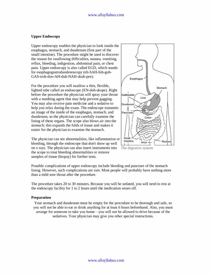
wwwallsyllabuscom
wwwallsyllabuscom
Upper Endoscopy
Upper endoscopy enables the physician to look inside the esophagus stomach and duodenum (first part of the small intestine) The procedure might be used to discover the reason for swallowing difficulties nausea vomiting
reflux bleeding indigestion abdominal pain or chest pain Upper endoscopy is also called EGD which stands for esophagogastroduodenoscopy (eh-SAH-fuh-goh-GAS-troh-doo-AH-duh-NAH-skuh-pee)
For the procedure you will swallow a thin flexible lighted tube called an endoscope (EN-doh-skope) Right before the procedure the physician will spray your throat with a numbing agent that may help prevent gagging
You may also receive pain medicine and a sedative to help you relax during the exam The endoscope transmits an image of the inside of the esophagus stomach and duodenum so the physician can carefully examine the lining of these organs The scope also blows air into the stomach this expands the folds of tissue and makes it easier for the physician to examine the stomach
The physician can see abnormalities like inflammation or
bleeding through the endoscope that dont show up well on x rays The physician can also insert instruments into the scope to treat bleeding abnormalities or remove samples of tissue (biopsy) for further tests
Possible complications of upper endoscopy include bleeding and puncture of the stomach lining However such complications are rare Most people will probably have nothing more than a mild sore throat after the procedure
The procedure takes 20 to 30 minutes Because you will be sedated you will need to rest at the endoscopy facility for 1 to 2 hours until the medication wears off
Preparation
Your stomach and duodenum must be empty for the procedure to be thorough and safe so you will not be able to eat or drink anything for at least 6 hours beforehand Also you must
arrange for someone to take you homemdashyou will not be allowed to drive because of the sedatives Your physician may give you other special instructions
The digestive system
wwwallsyllabuscom
wwwallsyllabuscom
What is Endoscopy and Why is it Performed
Endoscopy allows physicians to peer through the bodys passageways Endoscopy is the examination and inspection of the interior of body organs joints or cavities through an endoscope An endoscope is a device that uses fiber optics and powerful lens systems to provide lighting and visualization of the interior of a joint The portion of the endoscope inserted into the body may be rigid or flexible depending upon the medical procedure
An endoscope uses two fiber optic lines A light fiber carries light into the body cavity and an image fiber carries the image of the body cavity back to the physicians viewing lens There is also a separate port to allow for administration of drugs suction and irrigation This port may also be used to introduce small folding instruments such as forceps scissors brushes snares and baskets for tissue excision (removal) sampling or other diagnostic and therapeutic work Endoscopes may be used in conjunction with a camera or video recorder to document images of the inside of the joint or chronicle an endoscopic procedure New endoscopes have digital capabilities for manipulating and enhancing the video images
This figure shows a rigid endoscope used for arthroscopy The image fiber leads from the ocular (eye piece) to the inserted end of the scope The light fiber is below and leads from the light source to the working end of the endoscope
Why Is Endoscopy Performed
Endoscopy can be used to diagnose various conditions by close examination of internal organ and body structures Endoscopy can also guide therapy and repair such as the removal of torn cartilage from the bearing surfaces of a joint Biopsy (tissue sampling for pathologic testing) may also be performed under endoscopic guidance Local or general anesthetic may be used during endoscopy depending upon the type of procedure being performed
Internal abnormalities revealed through endoscopy include abscesses biliary (liver) cirrhosis bleeding bronchitis cancer cysts degenerative disease gallbladder stones hernia inflammation metastatic cancer polyps tumors ulcers and other diseases and conditions
Endoscopy is a minimally invasive procedure and carries with it certain minor risks depending upon the type of procedure being performed However these risks are typically far outweighed by the diagnostic and therapeutic potential of the procedure
Prior to the widespread use of endoscopy and diagnostic imaging most internal conditions could only be diagnosed or treated with open surgery Until the last several decades exploratory surgery was routinely performed only when a patient was critically ill and the source of illness was not known For example in certain dire cases the patients thorax or abdomen were surgically opened and examined to try to determine the source of illness
wwwallsyllabuscom
wwwallsyllabuscom
Endoscopy can often be done on an outpatient basis Outpatient means that the procedure does not require hospital admission and acute care and observation and may be performed outside the premises of a hospital Outpatient procedures performed at hospitals or ambulatory centers allow the patient to go home or return to work within a short while after their procedure
Types of Endoscopy
Fiber optic endoscopes now have widespread use in medicine and guide a myriad of diagnostic and therapeutic procedures including
Arthroscopy examination of joints for diagnosis and treatment (arthroscopic surgery) Bronchoscopy examination of the trachea and lungs bronchial trees to reveal
abscesses bronchitis carcinoma tumors tuberculosis alveolitis infection inflammation Colonoscopy examination of the inside of the colon and large intestine to detect polyps
tumors ulceration inflammation colitis diverticula Chrohns disease and discovery and removal of foreign bodies
Colposcopy direct visualization of the vagina and cervix to detect cancer inflammation and other conditions
Cystoscopy examination of the bladder urethra urinary tract uteral orifices and
prostate (men) with insertion of the endoscope through the urethra
ERCP (endoscopic retrograde cholangio-pancreatography) uses endoscopic guidance to
place a catheter for x-ray fluorosocopy with contrast enhancement This technique is used to examine the livers biliary tree the gallbladder the pancreatic duct and other anatomy to check for stones other obstructions and disease X-ray contrast is introduced into these ducts via catheter and fluoroscopic x-ray images are taken to show any abnormality or blockage If disease is detected it can sometimes be treated at the same time or biopsy can be performed to test for cancer or other pathology ERCP can detect biliary cirrhosis
cancer of the bile ducts pancreatic cysts pseudocysts pancreatic tumors chronic pancreatitis and other conditions such as gallbladder stones
EGD (Esophogealgastroduodensoscopy) visual examination of the upper gastro-
intestinal (GI) tract (also referred to as gastroscopy) to reveal hemorrhage hiatal hernia inflammation of the esophagus gastric ulcers
Endoscopic biopsy is the removal of tissue specimens for pathologic examination and
analysis Gastroscopy examination of the lining of the esophagus stomach and duodenum
Gastroscopy is often used to diagnose ulcers and other sources of bleeding and to guide biopsy of suspect GI cancers
Laparoscopy visualization of the stomach liver and other abdominal organs including
the female reproductive organs for example the fallopian tubes Laryngoscopy examination of the larynx (voice box) Proctoscopy sigmoidoscopy proctosigmoidoscopy examination of the rectum and
sigmoid colon Thoracoscopy examination of the pleura (sac that covers the lungs) pleural spaces
mediastinum and pericardium
A Brief History of Endoscopy
In the early 1900s the first attempts to view inside the body with lighted telescopes were made These initial devices were often fully rigid In the 1930s semi-flexible endoscopes called gastroscopes were developed to view inside of the stomach Fiber-optic endoscopy was pioneered by South African-born physician Basil Hirschowitz at the University of Michigan in 1957 Widespread use of fiber optic endoscopes began in the 1960s
wwwallsyllabuscom
wwwallsyllabuscom
A fiber optic cable is simply a bundle of microscopic glass or plastic fibers that literally allows light and images to be transmitted through curved structures Fiber optic cables are also replacing metal wires as the backbone of the worlds telecommunications infrastructure This Internet page may have traveled through a fiber optic cable as a stream of digital data (bursts of light) on its way to your computer
Endoscopy Equipment
Endoscopes have many practical needs And HMB Endoscopy Products (Hollywood Florida) has been providing endoscopic equipment and educating people on the use of endoscopes for more than 17 years Be sure to Browse our Catalog for all the details on how to purchase these medical instruments that can examine any part of the body
Video
Systems
Fiber
Endoscopes
Video
Endoscopes
In the simplest terms Endoscopy equipment consists of instruments that can look at the inside of many different organs mdash these are small flexible or rigid tubes with a light or lenses on the end that can look into the esophagus stomach and colon mdash and in more general terms endoscopy equipment can help doctors look deep inside body structures and hollow organs
An endoscope and related endoscope products and equipment are usually composed of three components
An optic system that allows the doctor to look through the scope into the organ or
cavity or to attach a video camera to the scope
A fiberoptic cable to light up the bodily area
A lumen (eg the bore of a tube like a needle or catheter) to take tissue samples of the
area being viewed
The beauty of endoscopic products is that they perform dual functions mdash with both diagnostic and therapeutic capabilities For example this means that these endoscopic products and instruments can perform biopsies (eg to evaluate tissue samples) as well as provide sclerotherapy (a medical procedure used to treat varicose veins and ―spider veins) In truth these brief explanations only tell part of the story
wwwallsyllabuscom
wwwallsyllabuscom
TYPES OF ENDOSCOPES
Take a look at the different types of endoscopic equipment you can get with HMB Endoscopy Products Again in very general terms there are two main types of endoscopes
Rigid Flexible
1 rigid endoscopes ndash the majority of which use a convex (curving out like one half of a
circle) glass lens system in which the small glass lenses are separated by large air spaces
2 flexible endoscopes allow for just that mdash flexibility In the animal kingdom for example
a flexible endoscope would be perfect for examining the stomach area of a snake
The popularity of endoscopy equipment continues to grow Some of the industryrsquos household names include Olympus Pentax Funjinon and Storz mdash with HMB both selling and repairing pre-owned and completely refurbished endoscopic products from each of these major manufacturers Browse our Catalog to get the ones you need
THERMOGRAPH APPLICATION Thermographs are digital recording thermometers used to log
temperature in the
marine environment The instruments are attached to a computer to set up recording
parameters for
deployment downloading and display of data in graphic or numeric format These units
are placed in
underwater housings and attached to the bottom or suspended in the water column for
deployment
DESCRIPTION Two types of thermograph are currently available The Ryan
Tempmentor is a
reusable data logger that allows storage of a maximum of 6361 temperature
measurements in the
range of -32C to +70C with 01resolution and 03accuracy The instrument can be
programmed
to take measurements from once per second to once every other hour Instrument
dimensions are 3 x
6 x 13 and the unit weighs 11 ounces The Hobo Temp is a miniature reusable data
logger that
allows storage of 1800 measurements in the range of -20C to +70C with
03resolution and 07
wwwallsyllabuscom
wwwallsyllabuscom
accuracy Sampling intervals from 05 seconds to 48 hours are available Instrument
dimensions are
24 x 19 x 08 and the unit weighs 1 ounce Both units are battery powered with the
battery life of
the Tempmentor being approximately two years and the battery life of the Hobo Temp
being one year THERMOGRAPH APPLICATION Thermographs are digital recording thermometers used to log
temperature in the
marine environment The instruments are attached to a computer to set up recording
parameters for
deployment downloading and display of data in graphic or numeric format These units
are placed in
underwater housings and attached to the bottom or suspended in the water column for
deployment
DESCRIPTION Two types of thermograph are currently available The Ryan
Tempmentor is a
reusable data logger that allows storage of a maximum of 6361 temperature
measurements in the
range of -32C to +70C with 01resolution and 03accuracy The instrument can be
programmed
to take measurements from once per second to once every other hour Instrument
dimensions are 3 x
6 x 13 and the unit weighs 11 ounces The Hobo Temp is a miniature reusable data
logger that
allows storage of 1800 measurements in the range of -20C to +70C with
03resolution and 07
accuracy Sampling intervals from 05 seconds to 48 hours are available Instrument
dimensions are
24 x 19 x 08 and the unit weighs 1 ounce Both units are battery powered with the
battery life of
the Tempmentor being approximately two years and the battery life of the Hobo Temp
being one year THERMOGRAPH APPLICATION Thermographs are digital recording thermometers used to log
temperature in the
marine environment The instruments are attached to a computer to set up recording
parameters for
deployment downloading and display of data in graphic or numeric format These units
are placed in
underwater housings and attached to the bottom or suspended in the water column for
deployment
DESCRIPTION Two types of thermograph are currently available The Ryan
Tempmentor is a
reusable data logger that allows storage of a maximum of 6361 temperature
measurements in the
range of -32C to +70C with 01resolution and 03accuracy The instrument can be
programmed
wwwallsyllabuscom
wwwallsyllabuscom
to take measurements from once per second to once every other hour Instrument
dimensions are 3 x
6 x 13 and the unit weighs 11 ounces The Hobo Temp is a miniature reusable data
logger that
allows storage of 1800 measurements in the range of -20C to +70C with
03resolution and 07
accuracy Sampling intervals from 05 seconds to 48 hours are available Instrument
dimensions are
24 x 19 x 08 and the unit weighs 1 ounce Both units are battery powered with the
battery life of
the Tempmentor being approximately two years and the battery life of the Hobo Temp
being one year
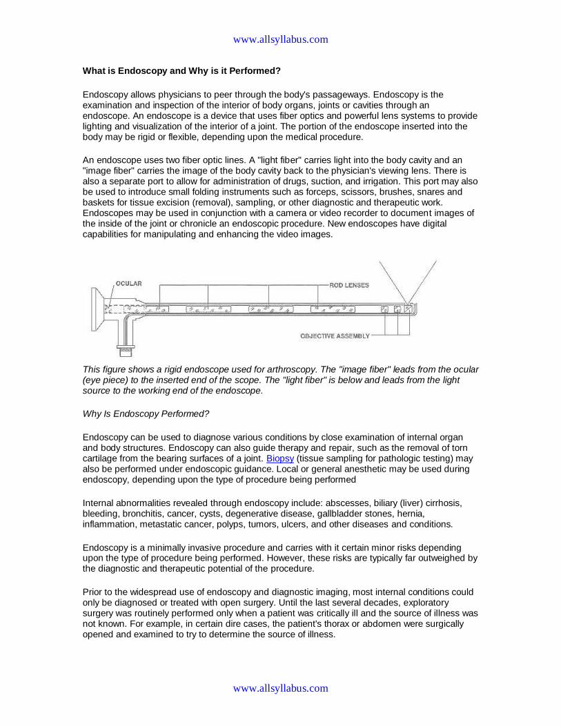
wwwallsyllabuscom
wwwallsyllabuscom
What is Endoscopy and Why is it Performed
Endoscopy allows physicians to peer through the bodys passageways Endoscopy is the examination and inspection of the interior of body organs joints or cavities through an endoscope An endoscope is a device that uses fiber optics and powerful lens systems to provide lighting and visualization of the interior of a joint The portion of the endoscope inserted into the body may be rigid or flexible depending upon the medical procedure
An endoscope uses two fiber optic lines A light fiber carries light into the body cavity and an image fiber carries the image of the body cavity back to the physicians viewing lens There is also a separate port to allow for administration of drugs suction and irrigation This port may also be used to introduce small folding instruments such as forceps scissors brushes snares and baskets for tissue excision (removal) sampling or other diagnostic and therapeutic work Endoscopes may be used in conjunction with a camera or video recorder to document images of the inside of the joint or chronicle an endoscopic procedure New endoscopes have digital capabilities for manipulating and enhancing the video images
This figure shows a rigid endoscope used for arthroscopy The image fiber leads from the ocular (eye piece) to the inserted end of the scope The light fiber is below and leads from the light source to the working end of the endoscope
Why Is Endoscopy Performed
Endoscopy can be used to diagnose various conditions by close examination of internal organ and body structures Endoscopy can also guide therapy and repair such as the removal of torn cartilage from the bearing surfaces of a joint Biopsy (tissue sampling for pathologic testing) may also be performed under endoscopic guidance Local or general anesthetic may be used during endoscopy depending upon the type of procedure being performed
Internal abnormalities revealed through endoscopy include abscesses biliary (liver) cirrhosis bleeding bronchitis cancer cysts degenerative disease gallbladder stones hernia inflammation metastatic cancer polyps tumors ulcers and other diseases and conditions
Endoscopy is a minimally invasive procedure and carries with it certain minor risks depending upon the type of procedure being performed However these risks are typically far outweighed by the diagnostic and therapeutic potential of the procedure
Prior to the widespread use of endoscopy and diagnostic imaging most internal conditions could only be diagnosed or treated with open surgery Until the last several decades exploratory surgery was routinely performed only when a patient was critically ill and the source of illness was not known For example in certain dire cases the patients thorax or abdomen were surgically opened and examined to try to determine the source of illness
wwwallsyllabuscom
wwwallsyllabuscom
Endoscopy can often be done on an outpatient basis Outpatient means that the procedure does not require hospital admission and acute care and observation and may be performed outside the premises of a hospital Outpatient procedures performed at hospitals or ambulatory centers allow the patient to go home or return to work within a short while after their procedure
Types of Endoscopy
Fiber optic endoscopes now have widespread use in medicine and guide a myriad of diagnostic and therapeutic procedures including
Arthroscopy examination of joints for diagnosis and treatment (arthroscopic surgery) Bronchoscopy examination of the trachea and lungs bronchial trees to reveal
abscesses bronchitis carcinoma tumors tuberculosis alveolitis infection inflammation Colonoscopy examination of the inside of the colon and large intestine to detect polyps
tumors ulceration inflammation colitis diverticula Chrohns disease and discovery and removal of foreign bodies
Colposcopy direct visualization of the vagina and cervix to detect cancer inflammation and other conditions
Cystoscopy examination of the bladder urethra urinary tract uteral orifices and
prostate (men) with insertion of the endoscope through the urethra
ERCP (endoscopic retrograde cholangio-pancreatography) uses endoscopic guidance to
place a catheter for x-ray fluorosocopy with contrast enhancement This technique is used to examine the livers biliary tree the gallbladder the pancreatic duct and other anatomy to check for stones other obstructions and disease X-ray contrast is introduced into these ducts via catheter and fluoroscopic x-ray images are taken to show any abnormality or blockage If disease is detected it can sometimes be treated at the same time or biopsy can be performed to test for cancer or other pathology ERCP can detect biliary cirrhosis
cancer of the bile ducts pancreatic cysts pseudocysts pancreatic tumors chronic pancreatitis and other conditions such as gallbladder stones
EGD (Esophogealgastroduodensoscopy) visual examination of the upper gastro-
intestinal (GI) tract (also referred to as gastroscopy) to reveal hemorrhage hiatal hernia inflammation of the esophagus gastric ulcers
Endoscopic biopsy is the removal of tissue specimens for pathologic examination and
analysis Gastroscopy examination of the lining of the esophagus stomach and duodenum
Gastroscopy is often used to diagnose ulcers and other sources of bleeding and to guide biopsy of suspect GI cancers
Laparoscopy visualization of the stomach liver and other abdominal organs including
the female reproductive organs for example the fallopian tubes Laryngoscopy examination of the larynx (voice box) Proctoscopy sigmoidoscopy proctosigmoidoscopy examination of the rectum and
sigmoid colon Thoracoscopy examination of the pleura (sac that covers the lungs) pleural spaces
mediastinum and pericardium
A Brief History of Endoscopy
In the early 1900s the first attempts to view inside the body with lighted telescopes were made These initial devices were often fully rigid In the 1930s semi-flexible endoscopes called gastroscopes were developed to view inside of the stomach Fiber-optic endoscopy was pioneered by South African-born physician Basil Hirschowitz at the University of Michigan in 1957 Widespread use of fiber optic endoscopes began in the 1960s
wwwallsyllabuscom
wwwallsyllabuscom
A fiber optic cable is simply a bundle of microscopic glass or plastic fibers that literally allows light and images to be transmitted through curved structures Fiber optic cables are also replacing metal wires as the backbone of the worlds telecommunications infrastructure This Internet page may have traveled through a fiber optic cable as a stream of digital data (bursts of light) on its way to your computer
Endoscopy Equipment
Endoscopes have many practical needs And HMB Endoscopy Products (Hollywood Florida) has been providing endoscopic equipment and educating people on the use of endoscopes for more than 17 years Be sure to Browse our Catalog for all the details on how to purchase these medical instruments that can examine any part of the body
Video
Systems
Fiber
Endoscopes
Video
Endoscopes
In the simplest terms Endoscopy equipment consists of instruments that can look at the inside of many different organs mdash these are small flexible or rigid tubes with a light or lenses on the end that can look into the esophagus stomach and colon mdash and in more general terms endoscopy equipment can help doctors look deep inside body structures and hollow organs
An endoscope and related endoscope products and equipment are usually composed of three components
An optic system that allows the doctor to look through the scope into the organ or
cavity or to attach a video camera to the scope
A fiberoptic cable to light up the bodily area
A lumen (eg the bore of a tube like a needle or catheter) to take tissue samples of the
area being viewed
The beauty of endoscopic products is that they perform dual functions mdash with both diagnostic and therapeutic capabilities For example this means that these endoscopic products and instruments can perform biopsies (eg to evaluate tissue samples) as well as provide sclerotherapy (a medical procedure used to treat varicose veins and ―spider veins) In truth these brief explanations only tell part of the story
wwwallsyllabuscom
wwwallsyllabuscom
TYPES OF ENDOSCOPES
Take a look at the different types of endoscopic equipment you can get with HMB Endoscopy Products Again in very general terms there are two main types of endoscopes
Rigid Flexible
1 rigid endoscopes ndash the majority of which use a convex (curving out like one half of a
circle) glass lens system in which the small glass lenses are separated by large air spaces
2 flexible endoscopes allow for just that mdash flexibility In the animal kingdom for example
a flexible endoscope would be perfect for examining the stomach area of a snake
The popularity of endoscopy equipment continues to grow Some of the industryrsquos household names include Olympus Pentax Funjinon and Storz mdash with HMB both selling and repairing pre-owned and completely refurbished endoscopic products from each of these major manufacturers Browse our Catalog to get the ones you need
THERMOGRAPH APPLICATION Thermographs are digital recording thermometers used to log
temperature in the
marine environment The instruments are attached to a computer to set up recording
parameters for
deployment downloading and display of data in graphic or numeric format These units
are placed in
underwater housings and attached to the bottom or suspended in the water column for
deployment
DESCRIPTION Two types of thermograph are currently available The Ryan
Tempmentor is a
reusable data logger that allows storage of a maximum of 6361 temperature
measurements in the
range of -32C to +70C with 01resolution and 03accuracy The instrument can be
programmed
to take measurements from once per second to once every other hour Instrument
dimensions are 3 x
6 x 13 and the unit weighs 11 ounces The Hobo Temp is a miniature reusable data
logger that
allows storage of 1800 measurements in the range of -20C to +70C with
03resolution and 07
wwwallsyllabuscom
wwwallsyllabuscom
accuracy Sampling intervals from 05 seconds to 48 hours are available Instrument
dimensions are
24 x 19 x 08 and the unit weighs 1 ounce Both units are battery powered with the
battery life of
the Tempmentor being approximately two years and the battery life of the Hobo Temp
being one year THERMOGRAPH APPLICATION Thermographs are digital recording thermometers used to log
temperature in the
marine environment The instruments are attached to a computer to set up recording
parameters for
deployment downloading and display of data in graphic or numeric format These units
are placed in
underwater housings and attached to the bottom or suspended in the water column for
deployment
DESCRIPTION Two types of thermograph are currently available The Ryan
Tempmentor is a
reusable data logger that allows storage of a maximum of 6361 temperature
measurements in the
range of -32C to +70C with 01resolution and 03accuracy The instrument can be
programmed
to take measurements from once per second to once every other hour Instrument
dimensions are 3 x
6 x 13 and the unit weighs 11 ounces The Hobo Temp is a miniature reusable data
logger that
allows storage of 1800 measurements in the range of -20C to +70C with
03resolution and 07
accuracy Sampling intervals from 05 seconds to 48 hours are available Instrument
dimensions are
24 x 19 x 08 and the unit weighs 1 ounce Both units are battery powered with the
battery life of
the Tempmentor being approximately two years and the battery life of the Hobo Temp
being one year THERMOGRAPH APPLICATION Thermographs are digital recording thermometers used to log
temperature in the
marine environment The instruments are attached to a computer to set up recording
parameters for
deployment downloading and display of data in graphic or numeric format These units
are placed in
underwater housings and attached to the bottom or suspended in the water column for
deployment
DESCRIPTION Two types of thermograph are currently available The Ryan
Tempmentor is a
reusable data logger that allows storage of a maximum of 6361 temperature
measurements in the
range of -32C to +70C with 01resolution and 03accuracy The instrument can be
programmed
wwwallsyllabuscom
wwwallsyllabuscom
to take measurements from once per second to once every other hour Instrument
dimensions are 3 x
6 x 13 and the unit weighs 11 ounces The Hobo Temp is a miniature reusable data
logger that
allows storage of 1800 measurements in the range of -20C to +70C with
03resolution and 07
accuracy Sampling intervals from 05 seconds to 48 hours are available Instrument
dimensions are
24 x 19 x 08 and the unit weighs 1 ounce Both units are battery powered with the
battery life of
the Tempmentor being approximately two years and the battery life of the Hobo Temp
being one year
wwwallsyllabuscom
wwwallsyllabuscom
Endoscopy can often be done on an outpatient basis Outpatient means that the procedure does not require hospital admission and acute care and observation and may be performed outside the premises of a hospital Outpatient procedures performed at hospitals or ambulatory centers allow the patient to go home or return to work within a short while after their procedure
Types of Endoscopy
Fiber optic endoscopes now have widespread use in medicine and guide a myriad of diagnostic and therapeutic procedures including
Arthroscopy examination of joints for diagnosis and treatment (arthroscopic surgery) Bronchoscopy examination of the trachea and lungs bronchial trees to reveal
abscesses bronchitis carcinoma tumors tuberculosis alveolitis infection inflammation Colonoscopy examination of the inside of the colon and large intestine to detect polyps
tumors ulceration inflammation colitis diverticula Chrohns disease and discovery and removal of foreign bodies
Colposcopy direct visualization of the vagina and cervix to detect cancer inflammation and other conditions
Cystoscopy examination of the bladder urethra urinary tract uteral orifices and
prostate (men) with insertion of the endoscope through the urethra
ERCP (endoscopic retrograde cholangio-pancreatography) uses endoscopic guidance to
place a catheter for x-ray fluorosocopy with contrast enhancement This technique is used to examine the livers biliary tree the gallbladder the pancreatic duct and other anatomy to check for stones other obstructions and disease X-ray contrast is introduced into these ducts via catheter and fluoroscopic x-ray images are taken to show any abnormality or blockage If disease is detected it can sometimes be treated at the same time or biopsy can be performed to test for cancer or other pathology ERCP can detect biliary cirrhosis
cancer of the bile ducts pancreatic cysts pseudocysts pancreatic tumors chronic pancreatitis and other conditions such as gallbladder stones
EGD (Esophogealgastroduodensoscopy) visual examination of the upper gastro-
intestinal (GI) tract (also referred to as gastroscopy) to reveal hemorrhage hiatal hernia inflammation of the esophagus gastric ulcers
Endoscopic biopsy is the removal of tissue specimens for pathologic examination and
analysis Gastroscopy examination of the lining of the esophagus stomach and duodenum
Gastroscopy is often used to diagnose ulcers and other sources of bleeding and to guide biopsy of suspect GI cancers
Laparoscopy visualization of the stomach liver and other abdominal organs including
the female reproductive organs for example the fallopian tubes Laryngoscopy examination of the larynx (voice box) Proctoscopy sigmoidoscopy proctosigmoidoscopy examination of the rectum and
sigmoid colon Thoracoscopy examination of the pleura (sac that covers the lungs) pleural spaces
mediastinum and pericardium
A Brief History of Endoscopy
In the early 1900s the first attempts to view inside the body with lighted telescopes were made These initial devices were often fully rigid In the 1930s semi-flexible endoscopes called gastroscopes were developed to view inside of the stomach Fiber-optic endoscopy was pioneered by South African-born physician Basil Hirschowitz at the University of Michigan in 1957 Widespread use of fiber optic endoscopes began in the 1960s
wwwallsyllabuscom
wwwallsyllabuscom
A fiber optic cable is simply a bundle of microscopic glass or plastic fibers that literally allows light and images to be transmitted through curved structures Fiber optic cables are also replacing metal wires as the backbone of the worlds telecommunications infrastructure This Internet page may have traveled through a fiber optic cable as a stream of digital data (bursts of light) on its way to your computer
Endoscopy Equipment
Endoscopes have many practical needs And HMB Endoscopy Products (Hollywood Florida) has been providing endoscopic equipment and educating people on the use of endoscopes for more than 17 years Be sure to Browse our Catalog for all the details on how to purchase these medical instruments that can examine any part of the body
Video
Systems
Fiber
Endoscopes
Video
Endoscopes
In the simplest terms Endoscopy equipment consists of instruments that can look at the inside of many different organs mdash these are small flexible or rigid tubes with a light or lenses on the end that can look into the esophagus stomach and colon mdash and in more general terms endoscopy equipment can help doctors look deep inside body structures and hollow organs
An endoscope and related endoscope products and equipment are usually composed of three components
An optic system that allows the doctor to look through the scope into the organ or
cavity or to attach a video camera to the scope
A fiberoptic cable to light up the bodily area
A lumen (eg the bore of a tube like a needle or catheter) to take tissue samples of the
area being viewed
The beauty of endoscopic products is that they perform dual functions mdash with both diagnostic and therapeutic capabilities For example this means that these endoscopic products and instruments can perform biopsies (eg to evaluate tissue samples) as well as provide sclerotherapy (a medical procedure used to treat varicose veins and ―spider veins) In truth these brief explanations only tell part of the story
wwwallsyllabuscom
wwwallsyllabuscom
TYPES OF ENDOSCOPES
Take a look at the different types of endoscopic equipment you can get with HMB Endoscopy Products Again in very general terms there are two main types of endoscopes
Rigid Flexible
1 rigid endoscopes ndash the majority of which use a convex (curving out like one half of a
circle) glass lens system in which the small glass lenses are separated by large air spaces
2 flexible endoscopes allow for just that mdash flexibility In the animal kingdom for example
a flexible endoscope would be perfect for examining the stomach area of a snake
The popularity of endoscopy equipment continues to grow Some of the industryrsquos household names include Olympus Pentax Funjinon and Storz mdash with HMB both selling and repairing pre-owned and completely refurbished endoscopic products from each of these major manufacturers Browse our Catalog to get the ones you need
THERMOGRAPH APPLICATION Thermographs are digital recording thermometers used to log
temperature in the
marine environment The instruments are attached to a computer to set up recording
parameters for
deployment downloading and display of data in graphic or numeric format These units
are placed in
underwater housings and attached to the bottom or suspended in the water column for
deployment
DESCRIPTION Two types of thermograph are currently available The Ryan
Tempmentor is a
reusable data logger that allows storage of a maximum of 6361 temperature
measurements in the
range of -32C to +70C with 01resolution and 03accuracy The instrument can be
programmed
to take measurements from once per second to once every other hour Instrument
dimensions are 3 x
6 x 13 and the unit weighs 11 ounces The Hobo Temp is a miniature reusable data
logger that
allows storage of 1800 measurements in the range of -20C to +70C with
03resolution and 07
wwwallsyllabuscom
wwwallsyllabuscom
accuracy Sampling intervals from 05 seconds to 48 hours are available Instrument
dimensions are
24 x 19 x 08 and the unit weighs 1 ounce Both units are battery powered with the
battery life of
the Tempmentor being approximately two years and the battery life of the Hobo Temp
being one year THERMOGRAPH APPLICATION Thermographs are digital recording thermometers used to log
temperature in the
marine environment The instruments are attached to a computer to set up recording
parameters for
deployment downloading and display of data in graphic or numeric format These units
are placed in
underwater housings and attached to the bottom or suspended in the water column for
deployment
DESCRIPTION Two types of thermograph are currently available The Ryan
Tempmentor is a
reusable data logger that allows storage of a maximum of 6361 temperature
measurements in the
range of -32C to +70C with 01resolution and 03accuracy The instrument can be
programmed
to take measurements from once per second to once every other hour Instrument
dimensions are 3 x
6 x 13 and the unit weighs 11 ounces The Hobo Temp is a miniature reusable data
logger that
allows storage of 1800 measurements in the range of -20C to +70C with
03resolution and 07
accuracy Sampling intervals from 05 seconds to 48 hours are available Instrument
dimensions are
24 x 19 x 08 and the unit weighs 1 ounce Both units are battery powered with the
battery life of
the Tempmentor being approximately two years and the battery life of the Hobo Temp
being one year THERMOGRAPH APPLICATION Thermographs are digital recording thermometers used to log
temperature in the
marine environment The instruments are attached to a computer to set up recording
parameters for
deployment downloading and display of data in graphic or numeric format These units
are placed in
underwater housings and attached to the bottom or suspended in the water column for
deployment
DESCRIPTION Two types of thermograph are currently available The Ryan
Tempmentor is a
reusable data logger that allows storage of a maximum of 6361 temperature
measurements in the
range of -32C to +70C with 01resolution and 03accuracy The instrument can be
programmed
wwwallsyllabuscom
wwwallsyllabuscom
to take measurements from once per second to once every other hour Instrument
dimensions are 3 x
6 x 13 and the unit weighs 11 ounces The Hobo Temp is a miniature reusable data
logger that
allows storage of 1800 measurements in the range of -20C to +70C with
03resolution and 07
accuracy Sampling intervals from 05 seconds to 48 hours are available Instrument
dimensions are
24 x 19 x 08 and the unit weighs 1 ounce Both units are battery powered with the
battery life of
the Tempmentor being approximately two years and the battery life of the Hobo Temp
being one year
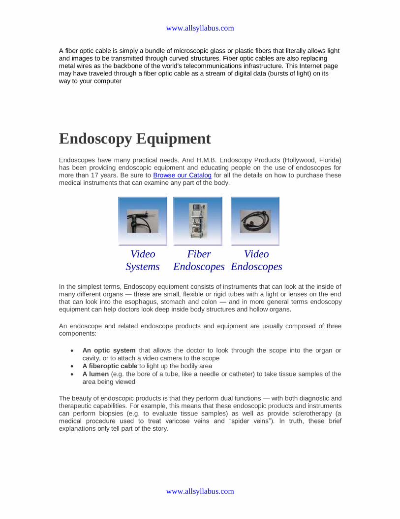
wwwallsyllabuscom
wwwallsyllabuscom
TYPES OF ENDOSCOPES
Take a look at the different types of endoscopic equipment you can get with HMB Endoscopy Products Again in very general terms there are two main types of endoscopes
Rigid Flexible
1 rigid endoscopes ndash the majority of which use a convex (curving out like one half of a
circle) glass lens system in which the small glass lenses are separated by large air spaces
2 flexible endoscopes allow for just that mdash flexibility In the animal kingdom for example
a flexible endoscope would be perfect for examining the stomach area of a snake
The popularity of endoscopy equipment continues to grow Some of the industryrsquos household names include Olympus Pentax Funjinon and Storz mdash with HMB both selling and repairing pre-owned and completely refurbished endoscopic products from each of these major manufacturers Browse our Catalog to get the ones you need
THERMOGRAPH APPLICATION Thermographs are digital recording thermometers used to log
temperature in the
marine environment The instruments are attached to a computer to set up recording
parameters for
deployment downloading and display of data in graphic or numeric format These units
are placed in
underwater housings and attached to the bottom or suspended in the water column for
deployment
DESCRIPTION Two types of thermograph are currently available The Ryan
Tempmentor is a
reusable data logger that allows storage of a maximum of 6361 temperature
measurements in the
range of -32C to +70C with 01resolution and 03accuracy The instrument can be
programmed
to take measurements from once per second to once every other hour Instrument
dimensions are 3 x
6 x 13 and the unit weighs 11 ounces The Hobo Temp is a miniature reusable data
logger that
allows storage of 1800 measurements in the range of -20C to +70C with
03resolution and 07
wwwallsyllabuscom
wwwallsyllabuscom
accuracy Sampling intervals from 05 seconds to 48 hours are available Instrument
dimensions are
24 x 19 x 08 and the unit weighs 1 ounce Both units are battery powered with the
battery life of
the Tempmentor being approximately two years and the battery life of the Hobo Temp
being one year THERMOGRAPH APPLICATION Thermographs are digital recording thermometers used to log
temperature in the
marine environment The instruments are attached to a computer to set up recording
parameters for
deployment downloading and display of data in graphic or numeric format These units
are placed in
underwater housings and attached to the bottom or suspended in the water column for
deployment
DESCRIPTION Two types of thermograph are currently available The Ryan
Tempmentor is a
reusable data logger that allows storage of a maximum of 6361 temperature
measurements in the
range of -32C to +70C with 01resolution and 03accuracy The instrument can be
programmed
to take measurements from once per second to once every other hour Instrument
dimensions are 3 x
6 x 13 and the unit weighs 11 ounces The Hobo Temp is a miniature reusable data
logger that
allows storage of 1800 measurements in the range of -20C to +70C with
03resolution and 07
accuracy Sampling intervals from 05 seconds to 48 hours are available Instrument
dimensions are
24 x 19 x 08 and the unit weighs 1 ounce Both units are battery powered with the
battery life of
the Tempmentor being approximately two years and the battery life of the Hobo Temp
being one year THERMOGRAPH APPLICATION Thermographs are digital recording thermometers used to log
temperature in the
marine environment The instruments are attached to a computer to set up recording
parameters for
deployment downloading and display of data in graphic or numeric format These units
are placed in
underwater housings and attached to the bottom or suspended in the water column for
deployment
DESCRIPTION Two types of thermograph are currently available The Ryan
Tempmentor is a
reusable data logger that allows storage of a maximum of 6361 temperature
measurements in the
range of -32C to +70C with 01resolution and 03accuracy The instrument can be
programmed
wwwallsyllabuscom
wwwallsyllabuscom
to take measurements from once per second to once every other hour Instrument
dimensions are 3 x
6 x 13 and the unit weighs 11 ounces The Hobo Temp is a miniature reusable data
logger that
allows storage of 1800 measurements in the range of -20C to +70C with
03resolution and 07
accuracy Sampling intervals from 05 seconds to 48 hours are available Instrument
dimensions are
24 x 19 x 08 and the unit weighs 1 ounce Both units are battery powered with the
battery life of
the Tempmentor being approximately two years and the battery life of the Hobo Temp
being one year
wwwallsyllabuscom
wwwallsyllabuscom
accuracy Sampling intervals from 05 seconds to 48 hours are available Instrument
dimensions are
24 x 19 x 08 and the unit weighs 1 ounce Both units are battery powered with the
battery life of
the Tempmentor being approximately two years and the battery life of the Hobo Temp
being one year THERMOGRAPH APPLICATION Thermographs are digital recording thermometers used to log
temperature in the
marine environment The instruments are attached to a computer to set up recording
parameters for
deployment downloading and display of data in graphic or numeric format These units
are placed in
underwater housings and attached to the bottom or suspended in the water column for
deployment
DESCRIPTION Two types of thermograph are currently available The Ryan
Tempmentor is a
reusable data logger that allows storage of a maximum of 6361 temperature
measurements in the
range of -32C to +70C with 01resolution and 03accuracy The instrument can be
programmed
to take measurements from once per second to once every other hour Instrument
dimensions are 3 x
6 x 13 and the unit weighs 11 ounces The Hobo Temp is a miniature reusable data
logger that
allows storage of 1800 measurements in the range of -20C to +70C with
03resolution and 07
accuracy Sampling intervals from 05 seconds to 48 hours are available Instrument
dimensions are
24 x 19 x 08 and the unit weighs 1 ounce Both units are battery powered with the
battery life of
the Tempmentor being approximately two years and the battery life of the Hobo Temp
being one year THERMOGRAPH APPLICATION Thermographs are digital recording thermometers used to log
temperature in the
marine environment The instruments are attached to a computer to set up recording
parameters for
deployment downloading and display of data in graphic or numeric format These units
are placed in
underwater housings and attached to the bottom or suspended in the water column for
deployment
DESCRIPTION Two types of thermograph are currently available The Ryan
Tempmentor is a
reusable data logger that allows storage of a maximum of 6361 temperature
measurements in the
range of -32C to +70C with 01resolution and 03accuracy The instrument can be
programmed
wwwallsyllabuscom
wwwallsyllabuscom
to take measurements from once per second to once every other hour Instrument
dimensions are 3 x
6 x 13 and the unit weighs 11 ounces The Hobo Temp is a miniature reusable data
logger that
allows storage of 1800 measurements in the range of -20C to +70C with
03resolution and 07
accuracy Sampling intervals from 05 seconds to 48 hours are available Instrument
dimensions are
24 x 19 x 08 and the unit weighs 1 ounce Both units are battery powered with the
battery life of
the Tempmentor being approximately two years and the battery life of the Hobo Temp
being one year
wwwallsyllabuscom
wwwallsyllabuscom
to take measurements from once per second to once every other hour Instrument
dimensions are 3 x
6 x 13 and the unit weighs 11 ounces The Hobo Temp is a miniature reusable data
logger that
allows storage of 1800 measurements in the range of -20C to +70C with
03resolution and 07
accuracy Sampling intervals from 05 seconds to 48 hours are available Instrument
dimensions are
24 x 19 x 08 and the unit weighs 1 ounce Both units are battery powered with the
battery life of
the Tempmentor being approximately two years and the battery life of the Hobo Temp
being one year