Embed Size (px)
Citation preview

GENERAL PRACTICAL (I) INTRODUCTORY
LECTURE
Steven Odongo [email protected] /[email protected]
CMIM, Building E, Floor 8, Room E8.07
14/11/2011 Steven Odongo, Dep't of Structural Biology, CMIM
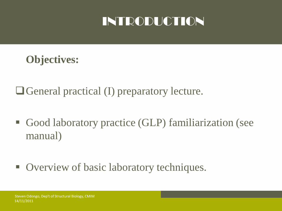
INTRODUCTION
Objectives:
General practical (I) preparatory lecture.
Good laboratory practice (GLP) familiarization (see
manual)
Overview of basic laboratory techniques.
14/11/2011 Steven Odongo, Dep't of Structural Biology, CMIM
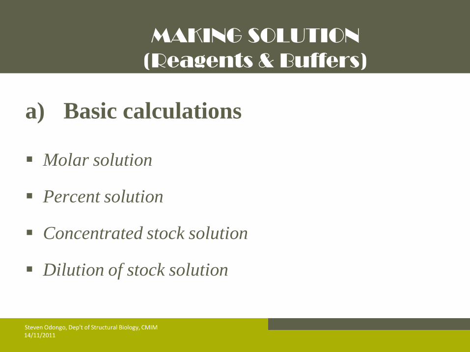
MAKING SOLUTION
(Reagents & Buffers)
a) Basic calculations
Molar solution
Percent solution
Concentrated stock solution
Dilution of stock solution
14/11/2011 Steven Odongo, Dep't of Structural Biology, CMIM
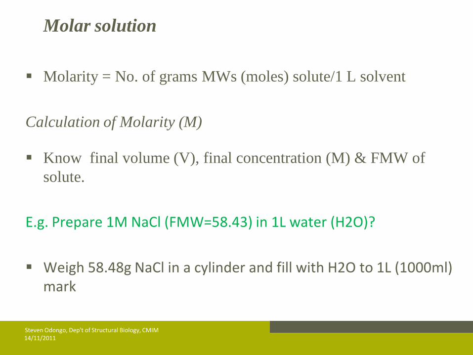
Molar solution
Molarity = No. of grams MWs (moles) solute/1 L solvent
Calculation of Molarity (M)
Know final volume (V), final concentration (M) & FMW of
solute.
E.g. Prepare 1M NaCl (FMW=58.43) in 1L water (H2O)?
Weigh 58.48g NaCl in a cylinder and fill with H2O to 1L (1000ml) mark
14/11/2011 Steven Odongo, Dep't of Structural Biology, CMIM
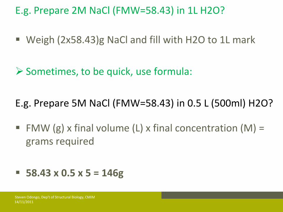
E.g. Prepare 2M NaCl (FMW=58.43) in 1L H2O?
Weigh (2x58.43)g NaCl and fill with H2O to 1L mark
Sometimes, to be quick, use formula:
E.g. Prepare 5M NaCl (FMW=58.43) in 0.5 L (500ml) H2O?
FMW (g) x final volume (L) x final concentration (M) = grams required
58.43 x 0.5 x 5 = 146g
14/11/2011 Steven Odongo, Dep't of Structural Biology, CMIM
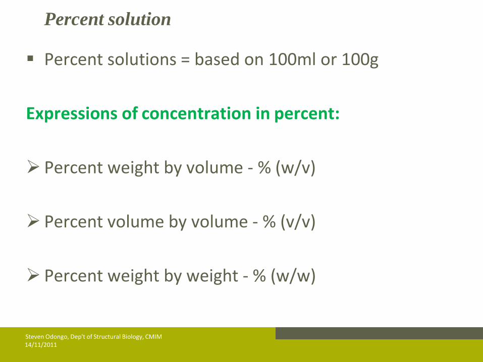
Percent solution
Percent solutions = based on 100ml or 100g
Expressions of concentration in percent:
Percent weight by volume - % (w/v)
Percent volume by volume - % (v/v)
Percent weight by weight - % (w/w)
14/11/2011 Steven Odongo, Dep't of Structural Biology, CMIM
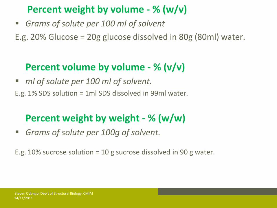
Percent weight by volume - % (w/v)
Grams of solute per 100 ml of solvent
E.g. 20% Glucose = 20g glucose dissolved in 80g (80ml) water.
Percent volume by volume - % (v/v)
ml of solute per 100 ml of solvent. E.g. 1% SDS solution = 1ml SDS dissolved in 99ml water.
Percent weight by weight - % (w/w)
Grams of solute per 100g of solvent.
E.g. 10% sucrose solution = 10 g sucrose dissolved in 90 g water.
14/11/2011 Steven Odongo, Dep't of Structural Biology, CMIM
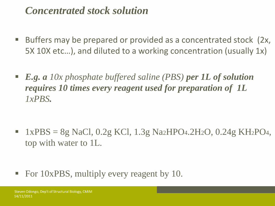
Concentrated stock solution
Buffers may be prepared or provided as a concentrated stock (2x, 5X 10X etc…), and diluted to a working concentration (usually 1x)
E.g. a 10x phosphate buffered saline (PBS) per 1L of solution
requires 10 times every reagent used for preparation of 1L
1xPBS.
1xPBS = 8g NaCl, 0.2g KCl, 1.3g Na2HPO4.2H2O, 0.24g KH2PO4,
top with water to 1L.
For 10xPBS, multiply every reagent by 10.
14/11/2011 Steven Odongo, Dep't of Structural Biology, CMIM
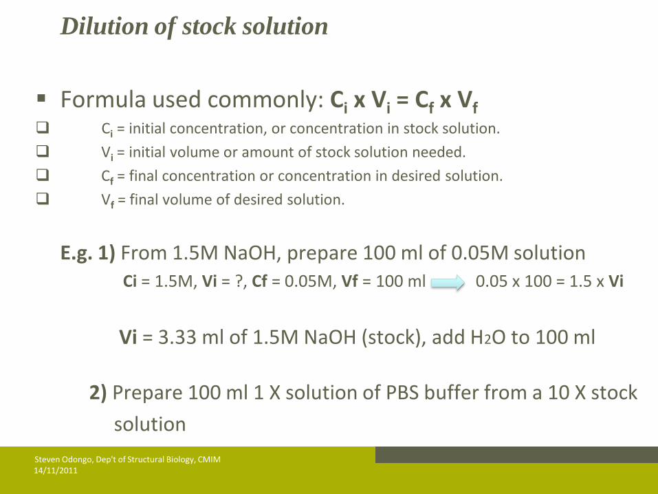
Dilution of stock solution
Formula used commonly: Ci x Vi = Cf x Vf Ci = initial concentration, or concentration in stock solution.
Vi = initial volume or amount of stock solution needed.
Cf = final concentration or concentration in desired solution.
Vf = final volume of desired solution.
E.g. 1) From 1.5M NaOH, prepare 100 ml of 0.05M solution Ci = 1.5M, Vi = ?, Cf = 0.05M, Vf = 100 ml 0.05 x 100 = 1.5 x Vi
Vi = 3.33 ml of 1.5M NaOH (stock), add H2O to 100 ml
2) Prepare 100 ml 1 X solution of PBS buffer from a 10 X stock
solution
14/11/2011 Steven Odongo, Dep't of Structural Biology, CMIM
14/11/2011 Steven Odongo, Dep't of Structural Biology, CMIM
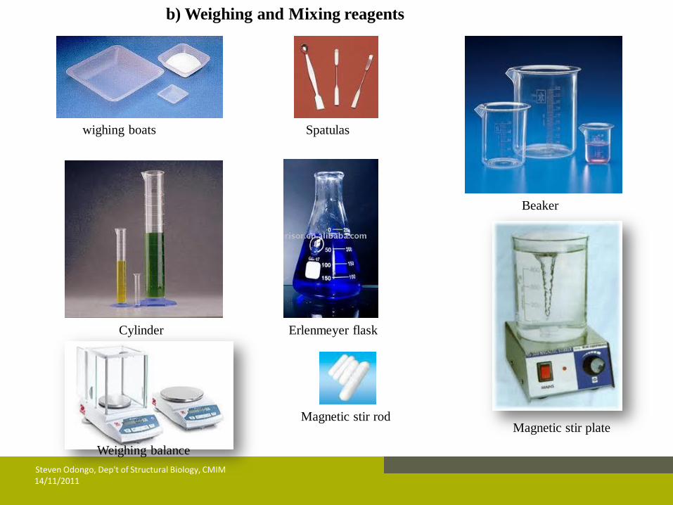
b) Weighing and Mixing reagents
wighing boats Spatulas
Cylinder
Beaker
Erlenmeyer flask
Magnetic stir plate Magnetic stir rod
Weighing balance

Weighing:
Put on nitril/latex gloves
Read instruction on the bottle (bse may require wearing mask)
Turn on the balance & tare the weigh boat
Weigh your material & pour it into the beaker or flask ( may
beaker or cover flask immediately)
Close the lid of the stock jar & put in the shelf immediately
Clean the balance when finished
14/11/2011 Steven Odongo, Dep't of Structural Biology, CMIM
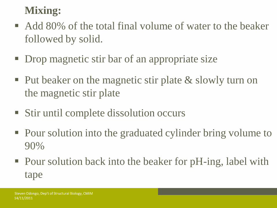
Mixing:
Add 80% of the total final volume of water to the beaker
followed by solid.
Drop magnetic stir bar of an appropriate size
Put beaker on the magnetic stir plate & slowly turn on
the magnetic stir plate
Stir until complete dissolution occurs
Pour solution into the graduated cylinder bring volume to
90%
Pour solution back into the beaker for pH-ing, label with
tape
14/11/2011 Steven Odongo, Dep't of Structural Biology, CMIM

14/11/2011 Steven Odongo, Dep't of Structural Biology, CMIM
C) pH measurements and adjustments
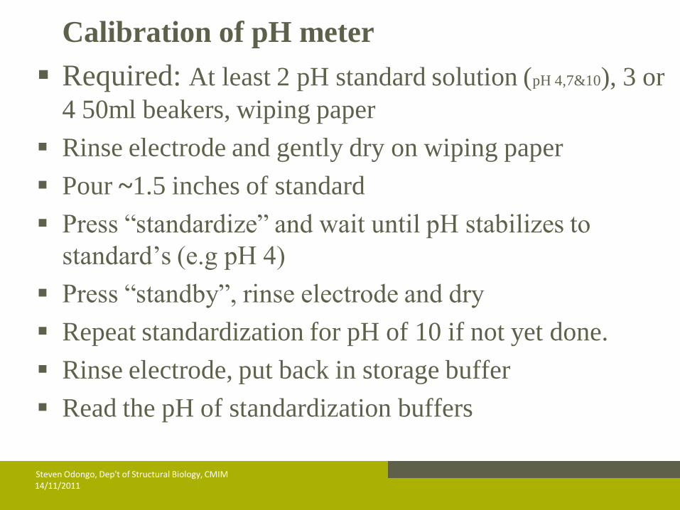
Calibration of pH meter
Required: At least 2 pH standard solution (pH 4,7&10), 3 or
4 50ml beakers, wiping paper
Rinse electrode and gently dry on wiping paper
Pour ~1.5 inches of standard
Press “standardize” and wait until pH stabilizes to
standard’s (e.g pH 4)
Press “standby”, rinse electrode and dry
Repeat standardization for pH of 10 if not yet done.
Rinse electrode, put back in storage buffer
Read the pH of standardization buffers
14/11/2011 Steven Odongo, Dep't of Structural Biology, CMIM
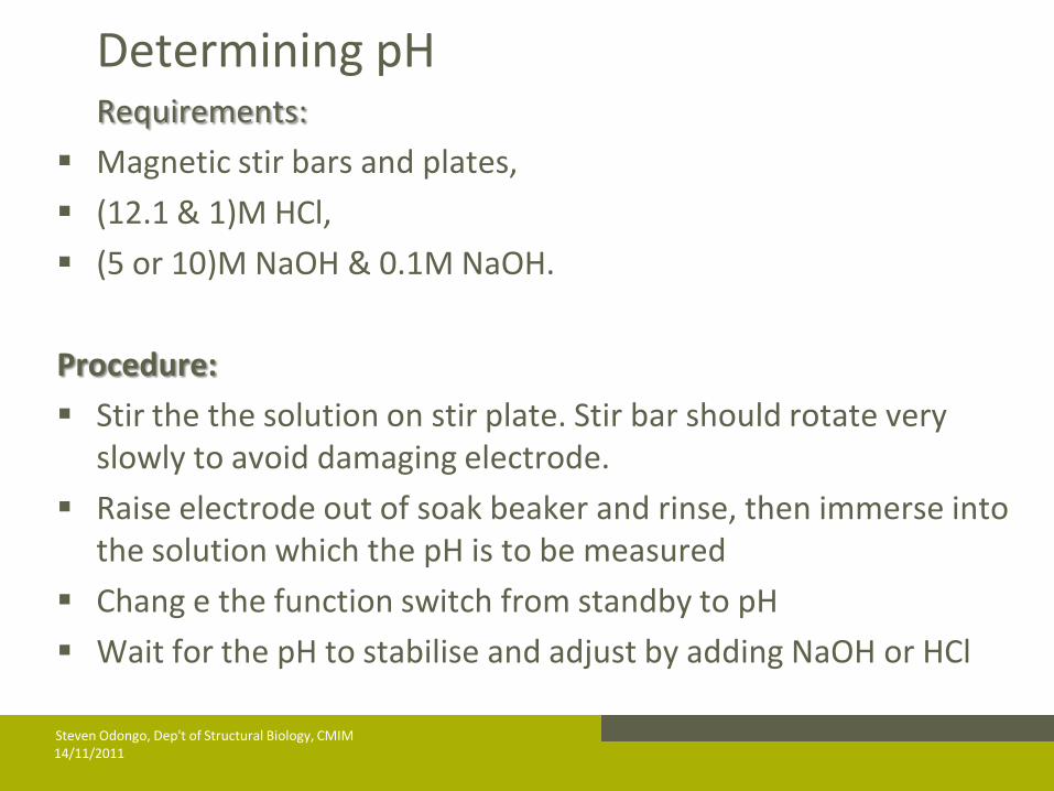
Determining pH Requirements:
Magnetic stir bars and plates,
(12.1 & 1)M HCl,
(5 or 10)M NaOH & 0.1M NaOH.
Procedure:
Stir the the solution on stir plate. Stir bar should rotate very slowly to avoid damaging electrode.
Raise electrode out of soak beaker and rinse, then immerse into the solution which the pH is to be measured
Chang e the function switch from standby to pH
Wait for the pH to stabilise and adjust by adding NaOH or HCl
14/11/2011 Steven Odongo, Dep't of Structural Biology, CMIM
Add a drop of (NaOH or HCl) at a time, stir, then measure
If the pH is off by a unit, use more conc acid (12.1M HCl) or base (5 or 10M NaOH).
Don’t use the same pasteur pipette for acid and base addition
You will need more drops near pH at which the solution is buffered.
14/11/2011 Steven Odongo, Dep't of Structural Biology, CMIM
Transfer the solution into a measuring cylinder, bring the volume to 100%
Pour the solution into glass or plastic bottle with cap
Label bottle:- date, components, concentration, pH and your initials.
Put apiece of autoclave tape. Dark strips appear after autoclaving.
If not autoclaving immediately keep at 4°C
14/11/2011 Steven Odongo, Dep't of Structural Biology, CMIM

D) Sterilizing solution:
1) Autoclave:
Most buffers
Undefined bacterial and yeast media
But don’t autoclave:
Corrosives (acids, bases & phenols), solvents or volatiles (the Ohs, Chloroform), or radioactive materials, liquids containing beach (sodium hypochlorite), formalin, or glutaraldehyde, heat labile ingredients (serum, antibiotics, proteins & vit), mammalian, plant, and insect media, HEPES containing solution, DTT or BME
solutions, buffers with detergent such as 10%SDS.
14/11/2011 Steven Odongo, Dep't of Structural Biology, CMIM

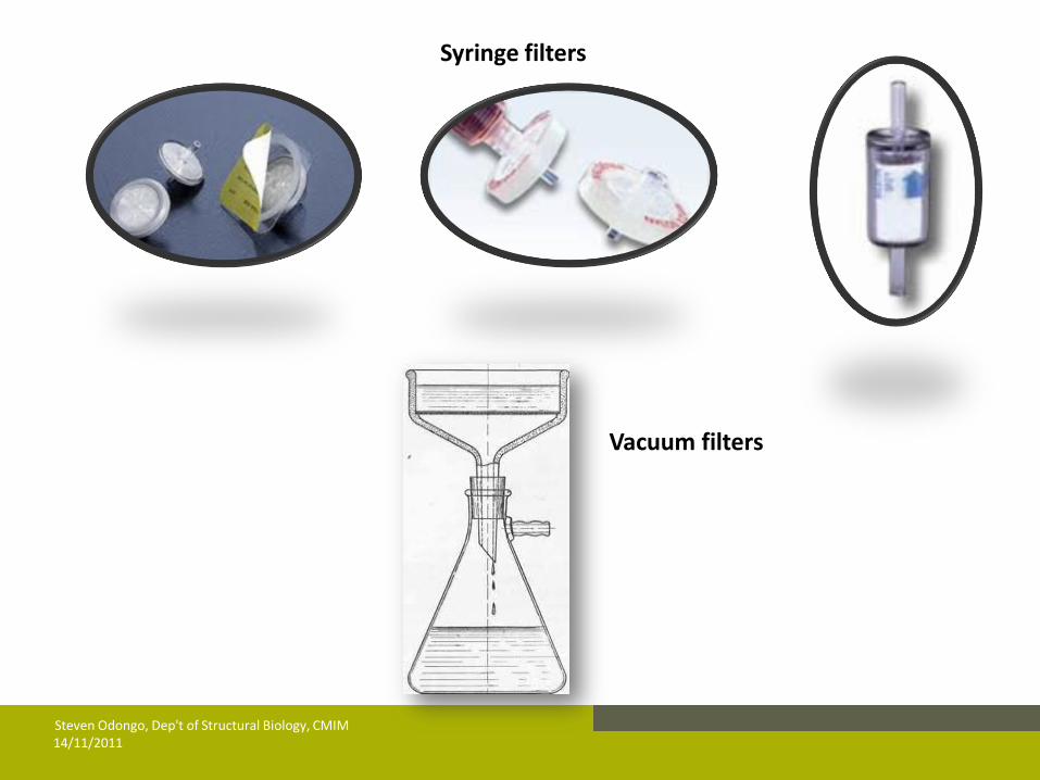
2) Filtration
Heat labile or volatile or solution less than 20ml.
Viscous solution is pre-filtered through 0.4µm
pore. a 0.2 µm (most media and buffers) or 0.1µm
(tissue culture media)
Note that virus will pass through filter hence can
be removed by filtration
14/11/2011 Steven Odongo, Dep't of Structural Biology, CMIM
14/11/2011 Steven Odongo, Dep't of Structural Biology, CMIM
Syringe filters
Vacuum filters
e) Storage of buffers and solutions
Store media at 4°C
Buffers stored at room temp or 4°C
Concentrated solutions at room temperature
Light sensitive reagents store at appropriate temperature
and kept in a brown bottle or cover with foil or in a box.
f)Discard buffers
Whenever discoloration occurs
Contamination appears
Precipitate persists after warming in water bath at 37°C
for 20min (or check for salt cyrstals under microscope
X100). Salt crytals are large while contaminants are uniform and small
14/11/2011 Steven Odongo, Dep't of Structural Biology, CMIM
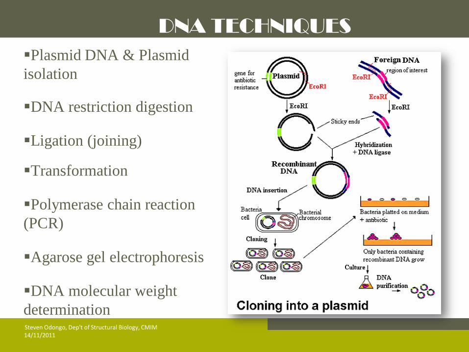
DNA TECHNIQUES
Plasmid DNA & Plasmid
isolation
DNA restriction digestion
Ligation (joining)
Transformation
Polymerase chain reaction
(PCR)
Agarose gel electrophoresis
DNA molecular weight
determination
14/11/2011 Steven Odongo, Dep't of Structural Biology, CMIM
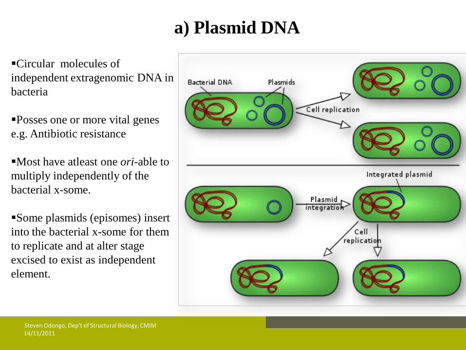
a) Plasmid DNA
Circular molecules of
independent extragenomic DNA in
bacteria
Posses one or more vital genes
e.g. Antibiotic resistance
Most have atleast one ori-able to
multiply independently of the
bacterial x-some.
Some plasmids (episomes) insert
into the bacterial x-some for them
to replicate and at alter stage
excised to exist as independent
element.
14/11/2011 Steven Odongo, Dep't of Structural Biology, CMIM

14/11/2011 Steven Odongo, Dep't of Structural Biology, CMIM
Artificially modified Plasmid DNA
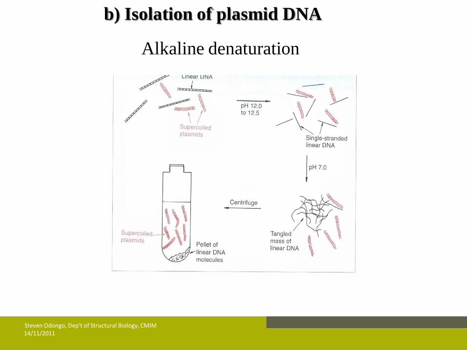
b) Isolation of plasmid DNA
Alkaline denaturation
14/11/2011 Steven Odongo, Dep't of Structural Biology, CMIM

14/11/2011 Steven Odongo, Dep't of Structural Biology, CMIM
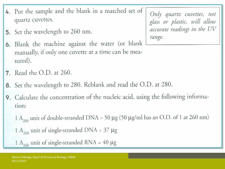
c) Determining Nucleic Acid Concentration
& Purity by UV spectroscopy
14/11/2011 Steven Odongo, Dep't of Structural Biology, CMIM
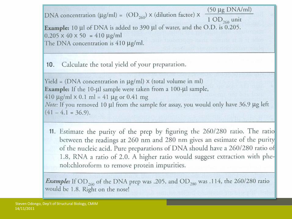
14/11/2011 Steven Odongo, Dep't of Structural Biology, CMIM
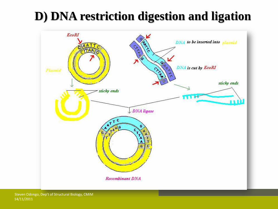
D) DNA restriction digestion and ligation
14/11/2011 Steven Odongo, Dep't of Structural Biology, CMIM
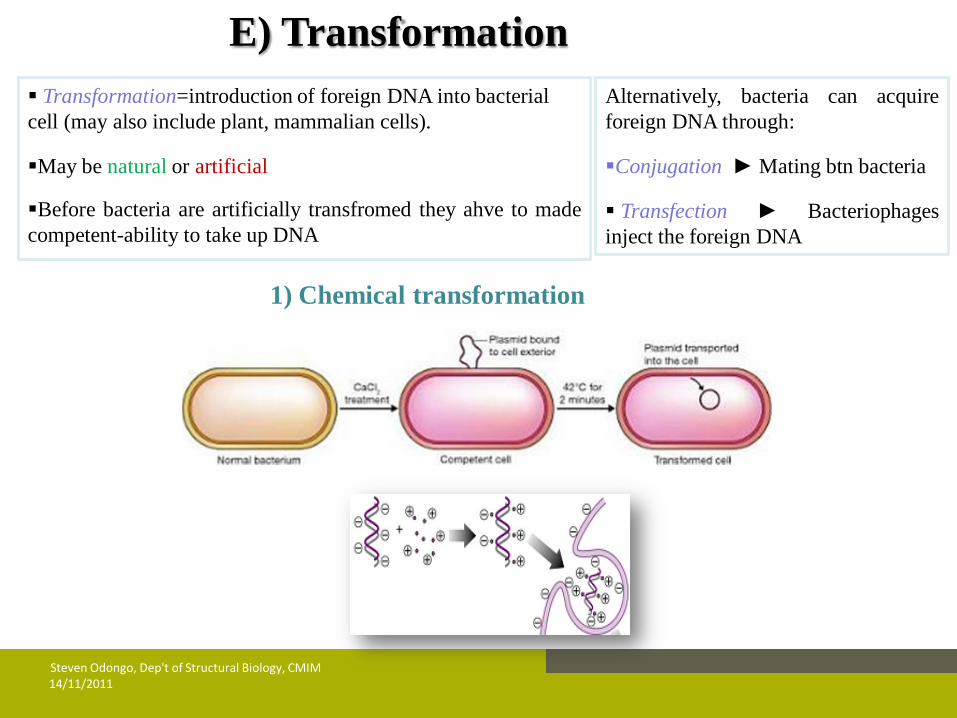
E) Transformation
Transformation=introduction of foreign DNA into bacterial
cell (may also include plant, mammalian cells).
May be natural or artificial
Before bacteria are artificially transfromed they ahve to made
competent-ability to take up DNA
1) Chemical transformation
14/11/2011 Steven Odongo, Dep't of Structural Biology, CMIM
Alternatively, bacteria can acquire
foreign DNA through:
Conjugation ► Mating btn bacteria
Transfection ► Bacteriophages
inject the foreign DNA

14/11/2011 Steven Odongo, Dep't of Structural Biology, CMIM
2) Electroporation
14/11/2011 Steven Odongo, Dep't of Structural Biology, CMIM
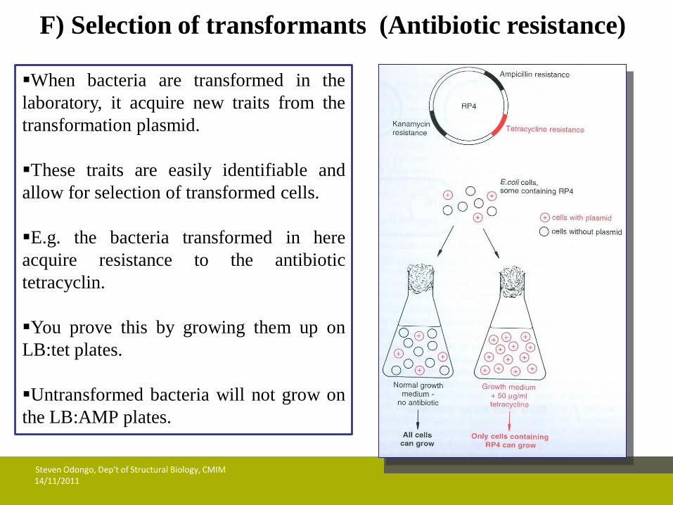
F) Selection of transformants (Antibiotic resistance)
When bacteria are transformed in the
laboratory, it acquire new traits from the
transformation plasmid.
These traits are easily identifiable and
allow for selection of transformed cells.
E.g. the bacteria transformed in here
acquire resistance to the antibiotic
tetracyclin.
You prove this by growing them up on
LB:tet plates.
Untransformed bacteria will not grow on
the LB:AMP plates.

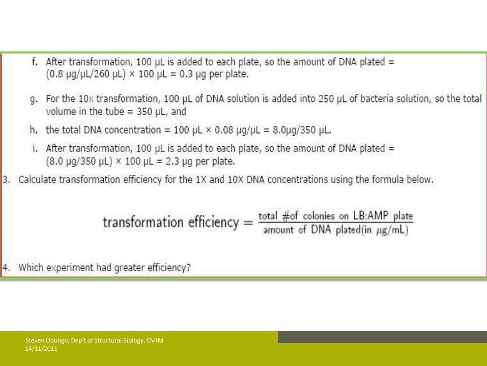
G) Transformation efficiency
Measure of the amount of cells within the bacterial culture able to take up DNA molecules.
Determined experimentally e.g. to detemine transformation
efficiency of an intact plasmid DNA at two different concentrations (1x & 10X):
14/11/2011 Steven Odongo, Dep't of Structural Biology, CMIM
14/11/2011 Steven Odongo, Dep't of Structural Biology, CMIM
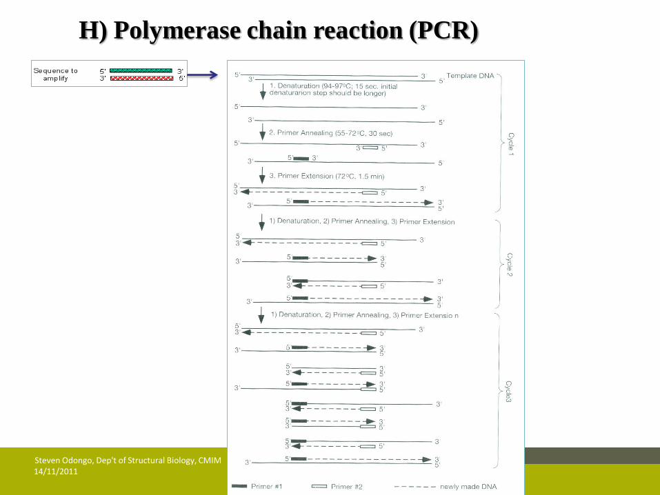
H) Polymerase chain reaction (PCR)
14/11/2011 Steven Odongo, Dep't of Structural Biology, CMIM
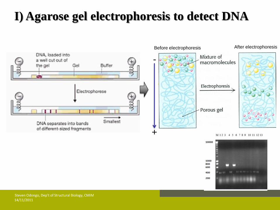
I) Agarose gel electrophoresis to detect DNA
-
+
Before electrophoresis After electrophoresis
14/11/2011 Steven Odongo, Dep't of Structural Biology, CMIM

14/11/2011 Steven Odongo, Dep't of Structural Biology, CMIM
J) Ethidium bromide visualization of DNA bands
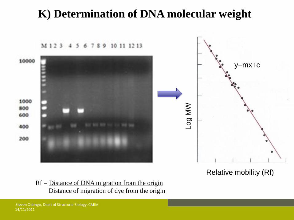
K) Determination of DNA molecular weight
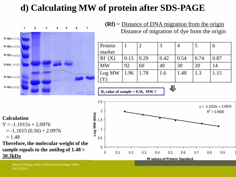
Rf = Distance of DNA migration from the origin
Distance of migration of dye from the origin
14/11/2011 Steven Odongo, Dep't of Structural Biology, CMIM
Lo
g M
W
Relative mobility (Rf)
y=mx+c

PROTEIN TECHNIQUES
a) Expression of recombinant protein
14/11/2011 Steven Odongo, Dep't of Structural Biology, CMIM
14/11/2011 Steven Odongo, Dep't of Structural Biology, CMIM
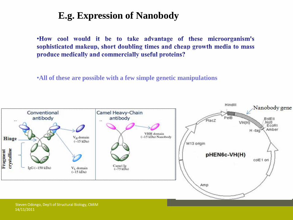
E.g. Expression of Nanobody
•All of these are possible with a few simple genetic manipulations

14/11/2011 Steven Odongo, Dep't of Structural Biology, CMIM
b) Protein expression, extraction and purification

14/11/2011 Steven Odongo, Dep't of Structural Biology, CMIM
c) Protein detection
I) Sodium Dodecyl Sulphate polyacrylamide gel electrophoresis (SDS PAGE)
Principle
14/11/2011 Steven Odongo, Dep't of Structural Biology, CMIM
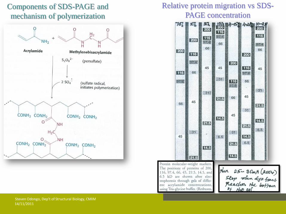
Components of SDS-PAGE and
mechanism of polymerization
Relative protein migration vs SDS-
PAGE concentration
14/11/2011 Steven Odongo, Dep't of Structural Biology, CMIM
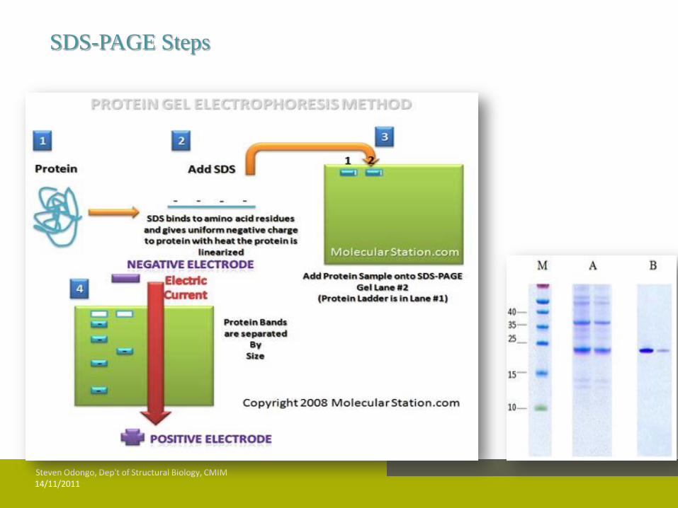
SDS-PAGE Steps
14/11/2011 Steven Odongo, Dep't of Structural Biology, CMIM
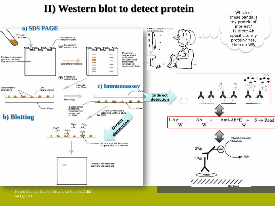
II) Western blot to detect protein
Indirect detection
a) SDS PAGE
c) Immunoassay
b) Blotting
Which of these bands is my protein of
interest? Is there Ab
specific to my protein? Yes, then do WB
Log MW (kDa) against Rf values
y = -1.1015x + 2.0976
R2 = 0.9935
0
0.5
1
1.5
2
2.5
0 0.1 0.2 0.3 0.4 0.5 0.6 0.7 0.8 0.9 1
Rf values of Protein Standard
Lo
g M
W (
kD
a)
Protein
marker 1 2 3 4 5 6
Rf (X) 0.15 0.29 0.42 0.54 0.74 0.87
MW 92 60 40 30 20 14
Log MW
(Y) 1.96 1.78 1.6 1.48 1.3 1.15
(Rf) = Distance of DNA migration from the origin
Distance of migration of dye from the origin
Rf value of sample = 0.56, MW ?
Calculation
Y = -1.1015x + 2.0976
= -1.1015 (0.56) + 2.0976
= 1.48
Therefore, the molecular weight of the
sample equals to the antilog of 1.48 =
30.3kDa
d) Calculating MW of protein after SDS-PAGE
14/11/2011 Steven Odongo, Dep't of Structural Biology, CMIM
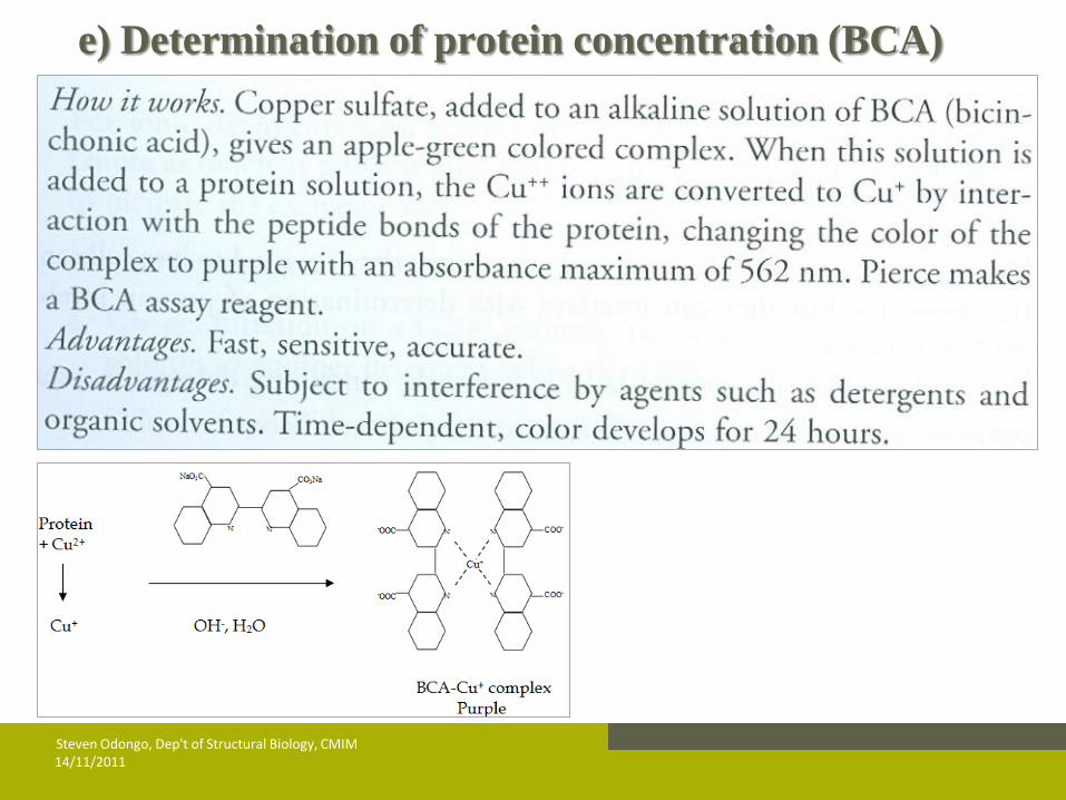
e) Determination of protein concentration (BCA)
14/11/2011 Steven Odongo, Dep't of Structural Biology, CMIM

14/11/2011 Steven Odongo, Dep't of Structural Biology, CMIM
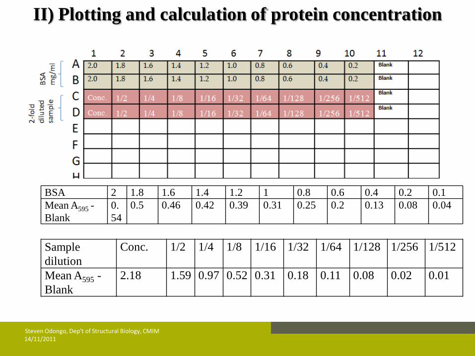
From the stock solution of 2 mg/ml of BSA prepare 1.8 mg/ml, 1.6
mg/ml, 1.4 mg/ml, 1.2 mg/ml, 1.0 mg/ml, 0.8 mg/ml, 0.6 mg/ml, 0.4 mg/ml
and 0.2 mg/ml.
Prepare 2-fold serial dilutions of sample as indicated in the diagram
of microplate
Fill wells of a flat-bottom micro-titer plate by pipetting 10 µl of each
& blank well filled with PBS
Mixing reagents A and B in a 50:1 proportion
Incubate plate at 37°C for 30 mins or at RT for 2h
Read the OD595nm in a micro-titer plate reader
I) BCA Steps
14/11/2011 Steven Odongo, Dep't of Structural Biology, CMIM
BSA 2 1.8 1.6 1.4 1.2 1 0.8 0.6 0.4 0.2 0.1 Mean A595 -
Blank 0.
54 0.5 0.46 0.42 0.39 0.31 0.25 0.2 0.13 0.08 0.04
Sample
dilution Conc. 1/2 1/4 1/8 1/16 1/32 1/64 1/128 1/256 1/512
Mean A595 -
Blank 2.18 1.59 0.97 0.52 0.31 0.18 0.11 0.08 0.02 0.01
II) Plotting and calculation of protein concentration

14/11/2011 Steven Odongo, Dep't of Structural Biology, CMIM
Absorbance (O.D) of standard BSA y = 0.267x + 0.0324
R2 = 0.9898
0
0.1
0.2
0.3
0.4
0.5
0.6
0 0.5 1 1.5 2 2.5
BSA concentration (mg/ml)
An
srb
an
ce (
nm
)
Absorbance at 1/8 dilution (0.52 nm) was used to calculate protein concentration in the sample because it is the
first one within range of standard.
Plotting and calculation of protein concentration
y = 0.267x + 0.0324
0.52 = 0.267x + 0.0324
0.4876 = 0.267x
x = 1.826
Concentration at 1/8 is 1.826 mg/ml, then concentration of undiluted protein
equals to
8 (i.e. dilution factor) x 1.826 = 14.608mg/ml
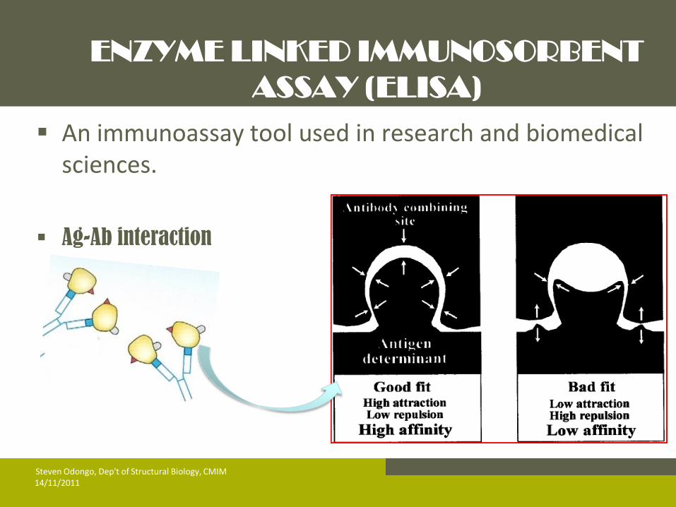
ENZYME LINKED IMMUNOSORBENT
ASSAY (ELISA)
An immunoassay tool used in research and biomedical sciences.
Ag-Ab interaction
14/11/2011 Steven Odongo, Dep't of Structural Biology, CMIM
14/11/2011 Steven Odongo, Dep't of Structural Biology, CMIM
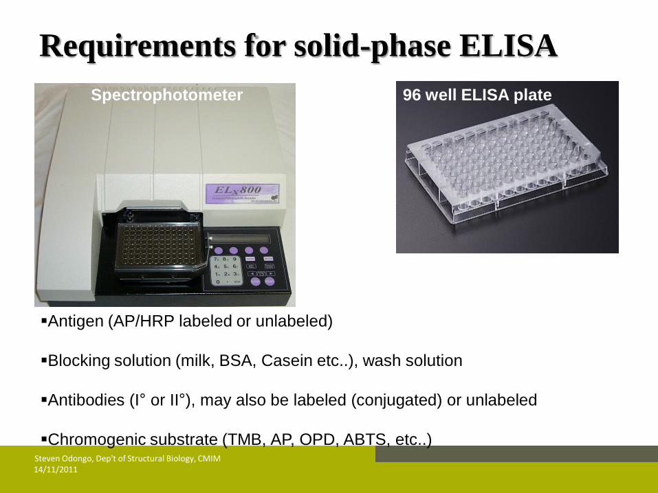
Antigen (AP/HRP labeled or unlabeled)
Blocking solution (milk, BSA, Casein etc..), wash solution
Antibodies (I° or II°), may also be labeled (conjugated) or unlabeled
Chromogenic substrate (TMB, AP, OPD, ABTS, etc..)
Spectrophotometer 96 well ELISA plate
Requirements for solid-phase ELISA
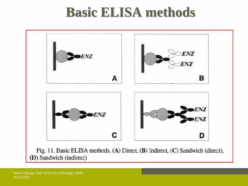
Basic ELISA methods
14/11/2011 Steven Odongo, Dep't of Structural Biology, CMIM

14/11/2011 Steven Odongo, Dep't of Structural Biology, CMIM
Steps of Indirect & sandwich ELISA

14/11/2011 Steven Odongo, Dep't of Structural Biology, CMIM
Steps of a direct solid-phase ELISA
14/11/2011 Steven Odongo, Dep't of Structural Biology, CMIM
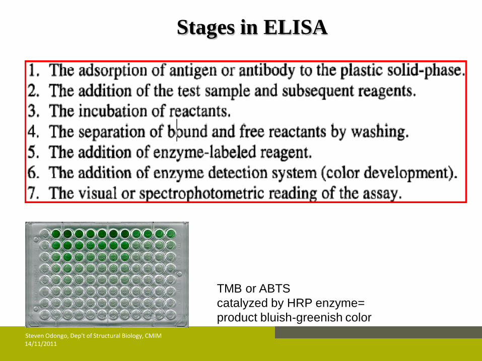
Stages in ELISA
TMB or ABTS
catalyzed by HRP enzyme=
product bluish-greenish color
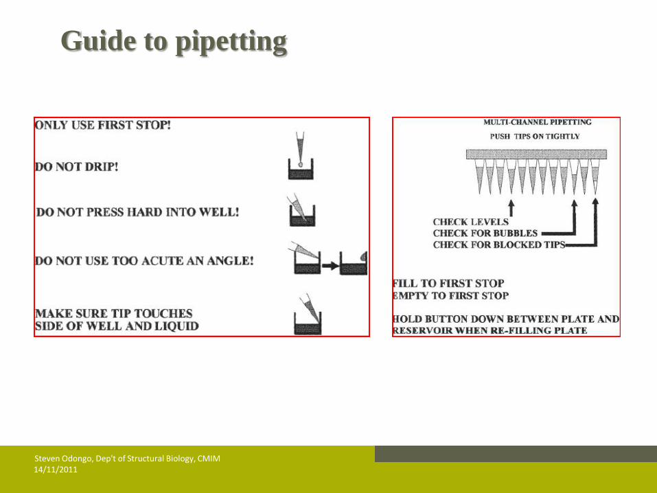
Guide to pipetting
14/11/2011 Steven Odongo, Dep't of Structural Biology, CMIM
14/11/2011 Steven Odongo, Dep't of Structural Biology, CMIM
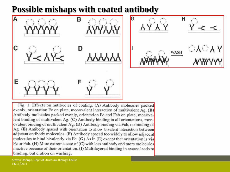
Possible mishaps with coated antibody

WISHING YOU A SUCCESSFUL
PRACTICAL
THANKYOU
&
14/11/2011 Steven Odongo, Dep't of Structural Biology, CMIM