Embed Size (px)
Citation preview
Generation and Characterisation of
Antibodies Specific for the Multidrug
Resistance-Associated Protein, MRP.
A thesis submitted for the degree of Ph D
by
Lisa Connolly (B Sc )
November 1999
The research work described in this thesis was earned out under
the supervision of
Prof Martin Clynes and Dr Elizabeth Moran
National Cell and Tissue Culture Centre,
School of Biological Sciences,
Dublin City University,
Glasnevin,
Dublin 9,
Ireland
i

I hereby certify that this material, which I now submit for
assessment on the programme of study leading to the award of
Ph D is entirely my own work and has not been taken from the
work of others save and to the extent that such work has been
cited and acknowledged within the text of my own work
Date IS IJL HI
Signed
ii
Abstract
Multidrug resistance associated protein 1 (MRP1) is a 190kD integral membrane
glycoprotein which belongs to the ATP-binding cassette (ABC) superfamily o f transport
proteins The MRP1 specific MAbs, MRPrl, MRPm6 and QCRL-1 have facilitated
both clinical and experimental investigations of this protein to date In this thesis we
have used various types of MRP 1-related immunogens in various species and utilising
different immunisation protocols with the objective of producing additional MRP1
specific MAbs which might detect different epitopes Immunogens used included short
synthetic peptides, fusion protems and MRP1 over-expressmg cells Species used for
immunisation included New Zealand White rabbits, Balb\c mice and Wistar rats A
number of immunisation routes, such as mtrapentoneal, subcutaneous and the lymphatic
system via footpad injections were used Variations in immunisation protocols included
in-vivo and in-vitro immunisations
Resulting antibodies included three MRP 1-specific polyclonal rabbit antibodies, a range
of mouse monoclonal antibodies, and a number of apparently MRP 1-specific rat
monoclonal antibodies The mouse MAbs include one which detects a 190kD antigen
present at equal or greater levels in sensitive cells compared to MRP 1-overexpressing
cells It may possibly detect a new form (possibly a conformational isoform which is
less active or has different substrate specificity) of MRP1 One of the rat MAbs
behaves in a manner similar to the well characterised MRPrl MAb and detects both
drug-selected and -transfected MRP1 by western blot analysis and stains drug selected
and transfected MRP1 cells lines homogeneously by immunocytochemistry Another
rat MAb detects a 190kD band in MRP 1-transfected cells but not MRP1 drug-selected
cells by western blot analysis and stains a sub-population of cells in both drug selected
III

and transfected MRP1 over-expressing cell lines None of these antibodies cross-
reacted with the recently described MRP2 and MRP3 lsoforms
IV

Acknowledgements
I would like to sincerely thank Professor Martin Clynes for his tremendous
encouragement and guidance during this thesis, especially for all the help
during the last few months
I would especially like to thank my supervisor Dr Liz Moran for her
unending help, advice and friendship I wouldn’t have made it this far
without your help, Liz
Thanks to all my colleagues m the NCTCC, especially Annemarie for her
friendship and constant support (listening to my worries), Helena, Yizheng,
Rob and Allan for their friendship, advice and humour Thanks to Liz,
Helena, Annemarie and Rob for their help in the final version of this thesis
Thanks to Rob for all his help with the computer problems also I couldn’t
forget Carol, Yvonne and Mairead for their help and kind words of
encouragement
I wish to acknowledge the generosity o f Mr George Schefer for his
donation of antibodies and cell lines, the kind permission of Professor
Marcel Kool to use the MRP3 cell line, the generosity of Professor Sarkadi
for his donation of fusion proteins and Emese Sinko for further analysis of
the polyclonal antibodies
A very special thanks to the boys in Beaumont, Nick, Derek and Martin for
their input to a vital part of this thesis I would especially like to thank
Derek and Martin for their help in the last few immunisations
Thanks to my friend Carmel for her friendship and sense of humour during
the weekends we 'partied* the stress away
Finally, I would like to thank my parents and my sister for their continuous
love and support, without which I would not have reached this far
V

This thesis is dedicated to Eddie, Rosie and Rachel The best
father, mother and sister I could ever have.
VI

TABLE OF CONTENTS
V II
1.0. INTRODUCTION 1
1 1 1 Drug resistance mechanisms 2
1 1 2 Multidrug resistance 2
1 1 3 Other mechanisms 3
1 1 4 The ATP-Binding Cassette (ABC) family 4
1 1 5 P-glycoprotein 5
1 2 The multidrug resistance-associated protein 1 (MRP1) 6
1 2 1 Membrane structure of MRP 1 9
1 2 2 MRP1 drug resistance profile 12
1 2 3 Cellular location of MRP 1 12
12 4 MRP1 expression in cell lines 13
12 5 MRP1 expression in normal human tissues 13
12 6 MRP1 expression in malignant tissues 14
12 7 Physiological function of MRP 1 19
1 2 8 Other ABC transporters involved in MDR 19
13 M R P 1 homologues 21
1 3 1 MRP2 23
1 3 2 MRP3 24
1 3 3 MRP4 26
1 3 4 MRP5 27
1 3 5 MRP6 27
13 6 Summary of MRP 1 homologues 28
1 1 Chemotherapy in cancer 2
V III

1 4 Monoclonal antibodies (MAbs) m research and diagnostics 31
1 4 1 MAbs specific for MRP1 and its homologues 32
14 2 Monoclonal antibody production techniques 34
1 4 3 Immunogens 36
1 4 4 Design of synthetic peptides for immunisation 37
14 5 Species 39
14 6 Route of immunisation 40
1 4 7 Immunisation 40
14 8 Fusion 41
14 9 Screenmg 42
14 10 Maintenance of hybndoma cell lines 42
1 5 Aims of this thesis. 44
1 6 Cell lines used in this thesis 45
IX
2.0. MATERIALS AND METHODS. 47
21 Materials and methods 47
2 1 1 Water 48
2 12 Glassware 48
2 1 3 Sterilisation 48
2 1 4 Media preparation 49
2 2 Cell culture procedures 50
2 2 1 Cell lines 50
2 2 2 Subculturmg cell lines 52
2 2 3 Cell counting 52
2 2 4 Cell freezing 53
2 2 5 Cell thawing 53
2 2 6 Monitoring of sterility of cell culture solutions 53
2 2 7 Mycoplasma analysis of cell lines 54
2 2 7 1 Indirect staining procedure for Mycoplasma analysis 54
2 2 7 2 Direct staining procedure for Mycoplasma analysis 54
2 3 Polyclonal and monoclonal antibody production 55
2 3 1 Immunogens 55
2 3 11 Whole cell lysates 55
2 3 12 Cell membranes 56
2 3 13 Synthetic peptides 57
2 3 14 Fusion protems 58
X

2 3 2 1 Rabbits 59
23 2 2 Mice 59
2 3 2 3 Rats 60
2 3 3 Fusion procedure 60
2 3 3 1 Mouse 60
2 3 3 2 Rat 61
2 3 4 In-vitro immunisation 63
2 3 5 Screening of hybndomas 63
2 3 6 Subculture of hybndomas 64
2 3 7 Single cell cloning by limiting dilution 64
2 3 8 Isotype analysis 65
2 4 Western blot analysis 65
2 4 1 Determination o f protein concentration 65
2 4 2 Sample preparation for gel electrophoresis 65
2 4 3 Gel electrophoresis 66
2 4 4 Western blotting 67
2 4 5 Development of Western blots by enhanced
chemiluminescence (ECL) 68
2 4 6 Immunoprécipitation 69
2 5. Immunocytochemical analysis 70
2 5 1 Coating of slides with poly-L-Lysine 70
2 5 2 Preparation of cytospins 70
2 3 2 In-vivo immunisation procedure 59
X I

2 5 4 Immunofluorescence analysis on fixed cells 71
2 5 5 Immunocytochemical analysis using the Strep AB
complex/HRP method 71
2 5 3 Immunofluorescence analysis on live cells 70
X II

3 0 RESULTS. 73
3 1 Charatensation of M R P lsoform MAbs, M R P 1
over-expressing cell lines, M R P 1 transfected SF9 insect
membranes and M R P isoform transfected cell lines 74
3 2 Characterisation of the rat M R P 1 specific MAb, MRPrl 76
3 2 1 Western blot analysis 77
3 2 2 Immunocytochemical analysis 82
3 2 3 Heat treatment studies by Western blot analysis
on the MRPrl epitope 86
3.3 Characterisation of the M R P 2 specific MAbs, M2DI-6 and M2I-4. 89
3 3 1 Immunocytochemical analysis 89
3 3 2 Western blot analysis 92
3 4 Characterisation of the M R P 3 specific MAbs, M3II-21 and M3II-9 94
3 4 1 Immunocytochemical analysis 94
3 4 2 Western blot analysis 97
3 5 Immunogens 99
3 5 1 Native protein 99
3 5 2 Synthetic peptides and Fusion protems 99
X III

3 6 1 Immunisation and screening o f serum 104
3 6 2 Polyclonal Antibody Production Results 104
3 6 3 Peptide 1 polyclonal serum results (Polyclonal 1) 107
3 6 4 Peptide 2 polyclonal serum results (Polyclonal 2) 111
3 6 5 Peptide 3 polyclonal serum results (Polyclonal 3) 114
3 7 Mouse monoclonal antibody production 117
3 7 1 Immunisations, fusions and screenmg of hybndomas 117
3 7 2 Mouse Monoclonal antibody Production Results 117
3 7 3 Peptide 1 MAb results 120
3 7 4 Peptide 2 MAb results 132
3 7 5 Peptide 3 MAb results 134
3 7 6 Fusion Protein 1 MAb results 147
3 7 7 Fusion Protein 2 MAb results 149
3 7 8 COR-L23R whole cells MAb results 151
3 7 9 COR-L23R cell membrane MAb results 154
3 8 Rat monoclonal antibody production 167
3 8 1 Immunisations, fusions and screening of hybndomas 167
3 8 2 Rat Monoclonal Antibody Results 168
3 8 3 Peptide 2 and 3 combined immunisation MAb results 168
3 8 4 COR-L23R cell membrane MAb results 168
3 8 5 Rat isotyping ELISA results 169
3 8 6 Reactive peptide ELISA results 170
3 6 Polyclonal antibody production 104
X IV

3 8 8 Western blot analysis
3 8 9 Immunocytochemical analysis
3 8 7 Peptide inhibition studies
176
181
170
3 9 Summary of most interesting M R P 1 antibodies 184
3 10 Cell line survey by Western Bot analysis 185
3 10 1 MRPrl 187
3 10 2 P2A8(6) 188
3 10 3 1A2 189
3 10 4 MRP2 190
3 10 5 MRP3 191
3 10 6 MRP5 192
4.0 DISCUSSION 193
41 General 194
4 2 Anti-MRPl rabbit polyclonal antibodies 198
4 3 Anti-MRPl mouse monoclonal antibodies 200
4 4 Anti-MRPl rat monoclonal antibodies 214
4 5 Species difference 217
4 6 Antigen presentation 221
4 7 Production of an external epitope MRP 1-specific
antibody 221
X V

5.0 CONCLUSIONS 223
6 0. FUTURE WORK. 227
7.0. BIBLIOGRAPHY. 230
8.0. APPENDICES. 245
Appendix 1 Abbreviations 246
Appendix 2 List o f Suppliers 249
X V I

1.0. INTRODUCTION.
1
10. INTRODUCTION
1 1 Chemotherapy m cancer
Chemotherapy constitutes the principal therapeutic tool to treat unresectable or
disseminated tumours resulting in a significant impact on survival in several malignancies
such as Hodgkin's disease, testicular cancer and childhood acute leukaemias (De Vita,
1989) However, a major problem m the chemotherapeutic treatment of cancer is the
occurrence of cellular resistance to cytotoxic drugs Chemotherapy against many types
of cancers is often ineffective because tumour cells possess cellular mechanisms that
enable them to resist the effects of these cytotoxic agents (Clynes, 1993, Coop, 1993)
A number of different cellular mechanisms may be responsible for cytotoxic resistance
and have been extensively studied in the laboratory using tumour cells made resistant by
selection and growth in increasing concentrations of various cytotoxic drugs (Clynes,
1993)
1 1 1 Drug resistance mechanisms
1 1 2 Multidrug Resistance (MDR)
One particular mechanism to be revealed from these studies is that of multidrug
resistance (MDR), that is, cells selected for resistance to one type of anti-cancer agent
become simultaneously resistant to a range of structurally different agents These include
anthracyclines, vinca alkaloids, taxanes, actinomycin D and the epipodophyllotoxins
(Borst, 1991, Clynes et a l , 1993) MDR affects patients with a wide variety of
haematological cancers and solid tumours including those of the breast, ovarian, lung and
lower gastrointestinal tract Tumours are often composed of mixed populations of
malignant cells, some drug sensitive and others drug resistant In the progression of
MDR, treatment with chemotherapeutic agents kills drug sensitive cells, leaving a higher
proportion of drug resistant cells As the tumour begins to grow again, chemotherapy
fails because the remaining cells of the tumour are drug resistant These cells can
become resistant by actively lowering the intracellular drug concentration, indicating the
presence of a transporter with the ability to efflux a range of different substrates The
transporters responsible for this phenomenon have been identified as multidrug
transporters To date two major human multidrug transporters have been identified,
MDR1 (P-glycoprotein, P-170, P-gp) (Gottesman and Pastan, 1993) and MRP1
(multidrug resistance-associated protein 1, P-190 or MRP) (Cole et a l , 1992) Both
these membrane proteins are capable of transporting a range of drugs with different
cellular targets and confer resistance by decreasing the intracellular concentration of
drugs However, MDR1 and MRP1 are only distantly related sharing only 15%
homology in their amino acid sequences (Cole e t a l , 1992)
1 1 3 Other mechanisms
Over-expression of other cellular proteins and mechanisms has also been observed, such
as alterations in topoisomerase II levels (reviewed in Hoffman and Mattern, 1993), over-
expression of the T antigen presenting protein (TAP) (Izquierdo et a l , 1996a),
alterations in the glutathiomne system (reviewed in Moscow and Dixon, 1993) and over-
expression of the lung resistance protein (LRP) (Scheper et a l , 1993, Izquierdo et a l ,
1995)
3
1 1 4 The ATP-Binding Cassette (ABC) family
Membrane proteins belonging to the ATP binding cassette (ABC) family of transport
proteins play a central role m the defence of cells against toxic compounds After
exposure of mammalian tumour cells to one single cytotoxic drug, these cells can
become resistant to a whole range of drugs by actively lowering the intracellular drug
concentration Both MDR1 and MRP1 are integral membrane proteins belonging to the
ABC (ATP-binding cassette) super family o f transporter proteins The ABC superfamily
are a structurally diverse group of protein transporters with over 50 members from
eukaryote to prokaryote, but all sharing a common molecular architecture and containing
at least one hydrophobic transmembrane region and a cytoplasmic nucleotide binding
domain (NBD), (Higgins, 1992) These proteins are involved in the energy-dependent
transport of a wide variation of chemical substrates across membranes including vanous
cytotoxic drugs (Bellamy et a1, 1996, Loe et a l , 1996b) In eukaryote cells, ABC genes
often encode four domains composed of two NBDs and two transmembrane regions,
(Leveille-Webster and Arias, 1995) The ATP binding NBDs contain conserved residues
(termed Walker A and B motifs) spaced by 90-120 ammo acids (Walker C motif)
(Walker et a l , 1982) Eukaryotic ABC proteins require two nucleotide-binding regions
and associated transmembrane regions in order to be functional In eukaryotes the two
sites are usually found as two halves o f a single polypeptide, while in prokaryotes ABC
proteins have only one NBD and are therefore functional as dimers MRP1 requires both
NBD’s to actively extrude drug Mutation of lysines 773(NBD1) or 1333(NBD2)
inactivates MRP1 transport possibly by destroying the structural integrity o f these
domains (Zhu et a l , 1997)
4

The first major human multidrug transporter identified was P-glycoprotein (P-170, P-gp)
encoded by the multidrug resistance protein 1 (MDR1) gene (Dano et a l , 1973, Julamo
and Ling, 1976, Gottesman and Pastan, 1993, Reviewed in Gottesman et a l , 1996) P-
170 is a 170 kD glycoprotein composed of 1280 amino acids P-170 is located in the
plasma membrane and acts as an ATP-dependent efflux pump that actively transports
chemotherapeutic agents (Gottesman and Pastan, 1993, Borst et a l , 1991) P-170
substrates include several structurally and functionally unrelated agents, including the
anthracychnes (daunorubicin and doxorubicin), the vinca alkaloids (vinblastine and
vincristine), the epipophyllotoxms (temposide and etoposide), the taxanes (paclitaxel and
docetaxel) and actinomycin D P-170 is predominantly expressed in the apical
membranes of organs with excretory functions (Georges et al f 1990, Van Der Valk et
a l , 1990) It is thought that P-170 may play a role in the elimination of exogenous
toxins or their toxic metabolites from the body (Hono et a l , 1989) P-170 is found in
several normal human tissues (Sugawara et a l , 1988) and is highly expressed in the
adrenal cortex, kidney (proximal tubules), liver (bile camculi), colon and jejunum (apical
brush border cells) P-170 has also been found in untreated human cancers derived from
normal tissues which express P-170, such as carcinomas of the colon, kidney, liver,
adrenal gland and pancreas However, in other cancers P-170 has been more frequently
detected at the time of relapse following initial chemotherapy (Bellamy et a l , 1990)
Studies have shown that P-170 expression in primary tumours has prognostic value in
childhood sarcoma (Chan et a l , 1990), neuroblastoma (Chan et a l , 1991), osteosarcoma
(Baldini et a l , 1995) and acute myeloid leukaemia (Mane et a l , 1995) In other major
cancer types such as in melanoma, and cancers of the lung, breast and ovary, P-170
appears to play a minor role (Lai et a l , 1989, Bellamy et a l , 1990, Cole et a l , 1992)
1 1 5 P-glycoprotein.
5
P-170 chemosensitisers (chemosensitisers are drugs or chemicals that enhance the
therapeutic effects of chemotherapy drugs and therefore improve their effectiveness)
include a variety of chemical classes such as calcium channel blockers (e g verapamil),
calmodulin antagonists (e g trifluoperazine), vinca alkaloids (e g vindohne), steroidal
agents (e g tamoxifen), immunosuppressive drugs (e g cyclosporin, SDZ PSC833) and
antibiotics (eg erythromycin) (Hamada et a l , 1987, Ford et al, 1995) There are
however a number of human tumour cell lines that show decreased cellular accumulation
and increased resistance to drugs associated with the MDR phenotype, but do not
express P-170 (Center et al f 1993, Twentyman and Versantvoort, 1996)
1 2. The Multidrug resistance associated protein 1 (MRP1)*
The second major multidrug transporter termed MRP1 (multidrug resistance-associated
protein 1, P-190 or MRP) (Reviewed in Cole and Deeley, 1998) was cloned and
sequenced from a small-cell-lung cancer line which showed resistance to drugs
associated with the MDR phenotype but did not express MDR1 (Cole et a l , 1992) The
MRP1 gene encodes a 190a membrane-bound glycoprotein of 1531 ammo acids (Cole et
a l, 1992, Knshnamachary and Center, 1993)
MRP1 is an ATP-dependent drug efflux pump (Leier et a l , 1994, Versantvoort et a l ,
1994) Reduced drug accumulation in cell lines expressing MRP1 have been reported
(Barrand et a l , 1993, Davey et a l , 1995) Transfection experiments with different
eukaiyotic expression vectors containing full length complementary DNAs of the MRP1
gene have shown that MRP1 confers resistance to a broad range of natural product
drugs, among which are anthracyclines, vinca alkaloids and epipodophyllotoxins (Cole et
a l , 1994, G ran ts a l , 1994, Kruh et a l , 1994, Zaman et a l f 1994) MRP Is function as
6
a drug extrusion pump has been established (Cole et a l , 1994, Zaman et a l , 1994), but
the nature of the transported drugs and other chemicals is still controversial MRP1
appears to mediate the transport of a broad range of drugs across cellular membranes
Like MDR1, many of the compounds transported by MRP1 are of natural origin
However, MDR1 can directly transport unmodified, relatively large, hydrophobic, either
uncharged or weakly basic molecules (Cole et a l , 1992, Higgins, 1992, Gottesman and
Pastan, 1993), whereas, MRP1 transports drugs in an unmodified form possibly
conjugated to glutathione (Leier et a l , 1994, Loe et a l , 1996a, Zaman et a l , 1996,
Muller et a l , 1994) or conjugated to acidic ligands such as glutathione, glucuromde, or
sulphate On the one hand, it was concluded from the increased cellular drug efflux from
MRP 1-over-expressing or transfected tumour cell lines that MRP1 functions as a
transporter of the typical ‘MDR’ anti-cancer agents (Versantvoort et a l , 1992, Zaman et
a l , 1994) On the other hand, the observation that the efflux of anti-cancer drugs from
MRP 1-over-expressing cells was glutathione-dependent (Lutzky et a l , 1989,
Versantvoort et a l , 1995) suggested that MRP1 is a transporter of anionic compounds
and/or glutathione-conjugated drugs (Jedlitschky et a l , 1994, Broxterman et a l , 1995)
It has been suggested that glutathione plays an essential role in MRP1 mediated drug
resistance Glutathione conjugation of drugs or interaction of glutathione directly with
MRP1 may be critical for drug transport (Loe et a l , 1996b) Experiments using the
glutathione (GSH) synthesis inhibitor, buthiomne sulphoximine (BSO), show that
intracellular GSH levels regulate drug transport in MRP1 tumour cell lines (Versantvoort
et a l , 1995) Important evidence for the identification of MRP 1 as a transporter of
multiple organic anions (MOAT) came from experiments showing ATP-dependent
uptake of glutathione conjugates, such as leukotnene C4, into mside out plasma
membrane vesicles prepared form MRP1 over-expressing cells (Jedlitschky et a l , 1994,
7

Muller et a l , 1994) Transporters with these characteristics of MRP 1 are known as GS-
X pumps (Ishikawa, 1992), multispecific organic anion transporters (MOAT) (Jansen
and Oude Elferink, 1993), or leukotnene C4 (LTC4) transporters (Keppler et a l , 1992)
As yet the mode of action by which MRP1 confers a MDR phenotype on cells is not
known However, the available data suggests that MRP1 acts both as a plasma
membrane outward drug pump and as a pump for drug accumulation in intracytoplasmic
vesicles (Cole et a l , 1994, Zaman et a l , 1994, Breunmger et a l , 1995) By both
mechanisms, cytoplasmic concentrations of free drug may be reduced to sublethal levels,
and in that way MRP1 is able to promote cell survival
The ammo acid sequence of MRP 1 contains various sites known to be relevant for ATP
binding and post-translational modification (Loe et a l , 1996a) MRP1 has been detected
immunologically as a 190 kD N-glycosylated phosphoprotein that binds ATP Various
studies have shown that the unmodified MRP1 polypeptide has an apparent mass of 170
kD and is processed into a mature 190 kD form by addition of N-linked oligosaccharides
It has been established that it takes approximately 90 minutes to produce the mature 190
kD MRP1 molecule from the 170 kD polypeptide, with the mature protein possessing a
half life of 20 hours Human MRP1 contains twelve potential sites for N-linked
glycosylation, but it is believed that only three of these sites are external to the plasma
membrane and so ultimately glycosylated The effect of glycosylation on MRP1 activity
is not fully known It has been demonstrated that tumcamycin-mduced inhibition of
glycosylation has little effect on the cellular drug accumulation characteristics of MRP 1
expressing cells (Almquist et a l , 1995)
8
Due to the difficulty of crystallising large membrane proteins, no detailed 3-dimensional
structure of any members of these transporters is currently available, and empirical
prediction methods are used to obtain molecular models of their structure, especially to
predict the locations and numbers of the membrane spanning helices In most cases these
methods identify six short transmembrane domains (Gottesman and Pastan, 1993, Cole
and Deeley, 1992, Higgins, 1992) The relevance of the prediction for the membrane
topology of CFTR has been confirmed experimentally by insertional mutagenesis (Chang
et a l, 1994), thus reinforcing the 2 x 6 transmembrane helix model The same
arrangement of transmembrane helices has been suggested in the case of P-glycoprotein
(Gros et a l , 1986, Chen et a l , 1986), and a large body of experimental data strongly
favours this model (Kast et a l , 1997) On the other hand, Ling and co-workers (Zhang
and Ling, 1991, Zhang et a l, 1993), by suggesting an alternative 6- and 4- helix
conformation, raised the possibility that P-glycoprotein may exist in two different
topological forms in the cell membrane When the multidrug resistance associated
protein 1 (MRP1) was cloned and sequenced, analysis of its primary amino acid
sequence revealed that MRP1 is more closely related to CFTR than P-gp (Cole et a l ,
1992) Cole et a l , (1992) predicted a unique transmembrane topology for MRP1, with
eight N-terminal and four C-terminal transmembrane segments However, according to
Bakos et a l , (1996), the model predicted by Cole et a l , (1992), is not consistent with
their newly developed membrane topology of MRP1 They found that MRP1 has a
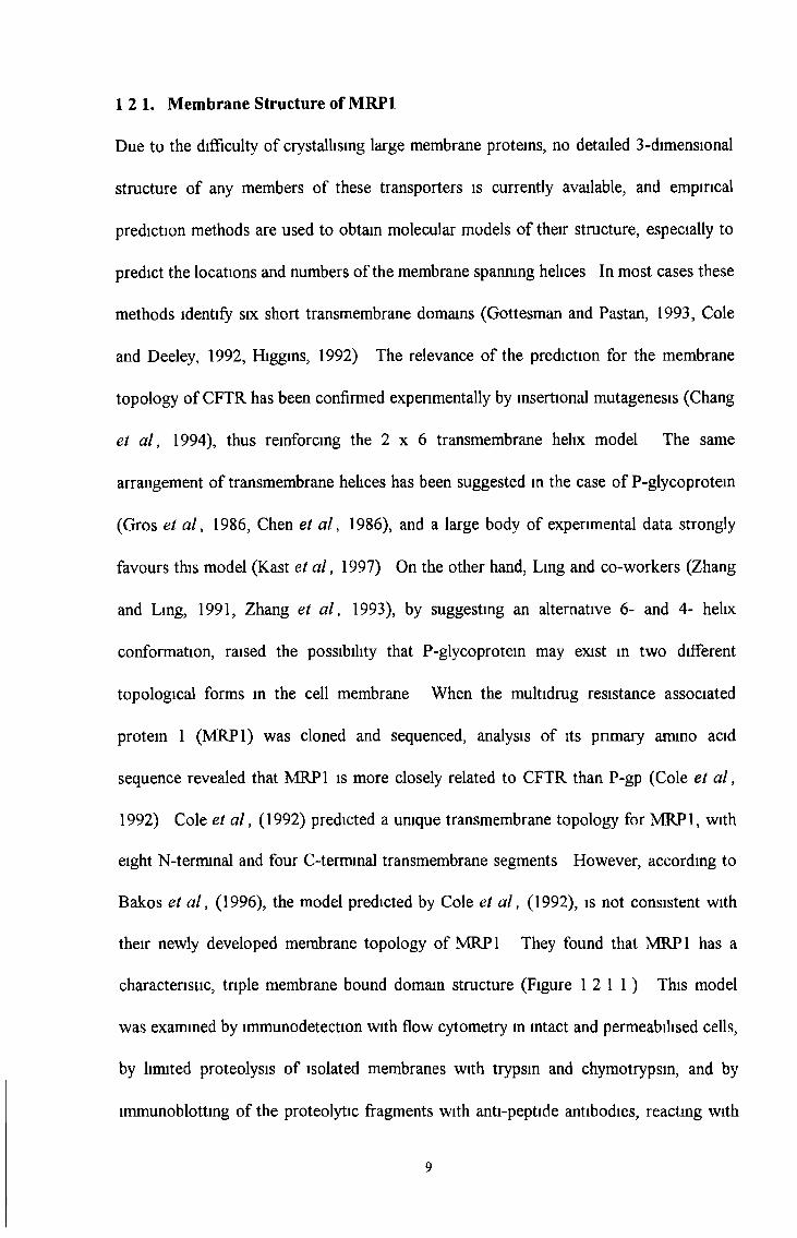
characteristic, triple membrane bound domain structure (Figure 12 1 1 ) This model
was examined by immunodetection with flow cytometry in intact and permeabilised cells,
by limited proteolysis of isolated membranes with trypsin and chymotrypsin, and by
immunoblotting of the proteolytic fragments with anti-peptide antibodies, reacting with
12 1. Membrane Structure of MRP1
9

either the N-terminal or the C-terminal half of the protein By using glycosylated and
unglycosylated forms of MRP1, its major sites of glycosylation could be determined
They compared the experimental findings with a newly developed membrane topology of
MRP1, based on the experimentally confirmed transmembrane topology of CFTR
10

TMO TM1 TM2
F ig u re 1.2.1.1. Membrane topology studies of the MRP1 protein (Bakos et al.,
1996) show three major membrane bound domains (TMO, TM1, and TM2) separated by
two cytoplasmic loops. The first (TMO) and last (TM2) membrane bound domains are
glycosylated when expressed in mammalian cells and a further possible glycosylation
site is situated in (TM1). An ATP binding domain (ABC unit) follows TM1 and TM2.
11
Cell lines over-expressing MRP1 are typically cross-resistant to anthracyclines,
epipodophyllotoxins and vinca alkaloids (Loe et a l , 1996a) Although the resistance
profiles of drug-selected MRP1 or MDR1 over-expressing cell lines are similar,
considerable differences exist particularly with regard to taxol resistance (Loe et a l ,
1996a) The explanation for a cell developing MRP1 rather than MDR1 mediated
resistance during drug exposure remains unclear, but it is believed that overexpression of
MRP1 may confer initial levels of resistance, while MDR1 overexpression develops as
higher levels of resistance are required for survival A study by Brock et a l , (1995),
showed that in the small-cell lung cancer cell line, H69, MRP1 is over-expressed during
selection in low concentrations of VP-16 Following further selection in higher
concentrations of drug, MRP1 expression remained relatively constant, but MDR1
overexpression developed The expression of MRP 1 protein and mRNA is not limited to
drug-selected MRP1 over-expressing cells Berger et a l , (1997a), reported that a
significant number of drug sensitive non-small cell lung cancer cell lines expressed MRP1
mRNA and protein
1 2.3. Cellular location of MRP1
Although MRP1 was initially believed to be predominantly located in the endoplasmic
reticulum of resistant cells (Krishnamachary and Center, 1993), significant levels are now
known to be present in the plasma membrane (Flens et a l , 1994, Muller et a l , 1994,
Zaman et a l , 1994) and also in endocytic vesicles (Almquist et a l , 1995)
Immunohistochemical studies show that MRP1 in normal tissue is predominantly
cytoplasmic whereas in malignant tissue it is predominantly plasma membrane located
with some granular cytoplasmic staining observed (Flens et a l , 1996) However, it has
12 2 MRP1 drug resistance profile
12
been observed that resistant cell lines show predominantly plasma-membrane staining
only (Flens e t a l , 1996, Zaman e t a l , 1994)
12 4 MRP1 expression in cell lines
The MRP1 transporter molecule has been shown to be present in non-MDRl multidrug
resistant cell lines obtained from a variety of tumour types These tumours include
leukaemias, fibrosarcomas, small-cell and non-small cell lung carcinomas, breast, cervix,
prostate and bladder carcinomas (Izquierdo e t a l , 1996b) There have also been reports
of co-expression of MDR1 and MRP1 in a number of drug-selected cell lines (Brock et
a l , 1995) However, the high prevalence of MRP 1 expression observed in a large
number of cell lines which have not been subject to laboratory drug selection suggest that
MDR mechanisms associated with this protein may be widespread in human
malignancies Therefore, while acquired resistance in many human tumours may be
attnbuted to MDR1, MRP1 may play an important role in the intrinsic resistance of
certain human tumours
1 2.5 MRP1 expression in normal human tissue
MRP1 has been detected either at the protein or the mRNA level in normal human
tissues including lung, stomach, colon, peripheral blood macrophages, thyroid, testis,
nerve, bladder, adrenal, ovary, pancreas, gall bladder, duodenum, heart, muscle,
placenta, brain, kidney, liver and spleen (Sugawara e t a l , 1997, Zaman e t a l , 1993, Cole
e t a l , 1992, Kruh e t a l , 1995)
Distinct cell types which express MRP1 include epithelial cells, muscle cells and
macrophages Negative MRP1 expression has been observed in many other cell types
13
including capillary epithelial cells (Flens et a l , 1996)
Studies on a panel of normal human tissues earned out by Flens et a l , 1996, revealed
that the highest level of MRP 1 protein (levels similar to or below that detected in the
parental, drug sensitive lung cancer cell lines GLC4 and SW-1573/S1) are expressed in
adrenal gland, lung, heart and skeletal muscle Lower amounts of MRP 1 were detected
in liver, spleen, kidney and erythrocyte membranes
12 6 MRP1 expression in malignant human tissues,
The widespread expression of MRP 1 in normal human tissues is reflected in a broad
range of adult solid tumours which over-express MRP1 (Kruh et a l , 1995) This has
been observed in breast carcinomas (Nooter et a l , 1995, Filipits et a l , 1996a),
neuroblastomas (Bordow et a l , 1994), anaplastic thyroid carcinomas (Sugawara et a l ,
1994), non-small-cell-lung carcinomas (Ota et a l , 1995), colorectal carcinomas (Nooter
et a l , 1995), ovarian carcinomas (Nooter et a l , 1995, Izquierdo et a l , 1995) and
relapsed acute leukaemias (Schneider et a l , 1995) Generally, MRP1 positive tumours
are derived from tissues that normally express MRP1, suggesting that MRP1 persists
during malignant transformation Importantly, these tumours are also known to be
intrinsically resistant to treatment with chemotherapeutic agents (Goldstein et a l , 1992)
suggesting that MRP1 may therefore contribute to this intrinsic resistance MRP1 may be
an important factor in the clinical drug resistance of several tumour types, most notably
non-small cell lung cancers (Ota et a l , 1995, Nooter et a l , 1996, Chuman et a l , 1996,
Giaccone et a l , 1996), breast carcinomas (Nooter et a l , 1997a, Nooter et a l , 1997b,
Ito et a l , 1998) neuroblastoma (Norris et a l , 1996), certain haematological malignancies
(Kuss et a l , 1996, Ross et a l , 1996), and malignant melanoma (Berger et a l , 1997b)
14

Immunohistochemical studies show that unlike the cytoplasmic staining observed in
normal tissue, tumour tissue shows predominantly strong plasma-membrane staining with
some granular cytoplasmic staining (Flens et a l , 1996) Strong MRP1 staining,
predominantly at the plasma-membrane, was observed in 20% of untreated solid tumours
examined by Flens et a l , (1996) This staining was mainly observed in squamous cell
carcinomas such as the lung and oesophagus
MDR is a serious clinical problem encountered in the chemotherapy of lung cancer
Human lung cancer is characterised by both high incidence and lethality, with non-small
cell lung cancer (NSCLC) accounting for at least 80% of the cases (Boring et a l , 1993)
The poor prognosis of this malignancy may be partially due to the pronounced resistance
to chemotherapeutic drugs that is present intrinsically in most NSCLCs and is also
acquired during treatment in many cases of initially drug sensitive small-cell lung cancer
(SCLC) (Ihde, 1992) The response rate of NSCLC at initial chemotherapy is relatively
low, suggesting the existence of intrinsic drug resistance On the other hand, while the
primary response rate of SCLC is higher than NSCLC, SCLC usually relapses with the
tumours displaying acquired MDR The drug resistance mechanisms in clinical lung
cancer remain poorly understood However, human MDR1 may be linked to acquired
MDR in SCLC and MRP 1 is a potentially important candidate for intrinsic resistance in
NSCLC
Differences in the expression of MRP 1 in various histological types of lung cancer have
also been reported Immunohistochemical studies have shown that in untreated lung
cancer tumours, MRP1 is more prevalent in NSCLC than in SCLC (Wright et a l , 1998)
It has been observed that NSCLC tumours expressing moderate or high levels of MRP 1
15
show significantly poorer prognoses than those with non or low level MRP1 expressing
tumours (Ota et a l , 1995) A clinical survey carried out by Dingemans et a l , 1996,
reported that all SCLC samples surveyed were MRP1 negative while a small number of
NSCLC tumours were MRP1 positive suggesting that MRP1 may be a negative
predictive marker in the response of NSCLC to chemotherapy
Recent work has focused on the role of MRP1 in breast cancer (Beck et a l , 1998,
Filipits et a l , 1996, Ito et a l , 1996, Lacave et a l , 1998, Linn et a l , 1997) A few
studies have suggested that MRP1 may play a role in the prediction of response to
chemotherapy in breast cancer (Mechetner et a l , 1997, Nooter et a l , 1997a, Nooter et
a l , 1997b) In a small immunohistochemical study of breast carcinomas, Larkin et a l ,
(1999a), reported that in cases where intense MRP1 positivity was observed, the level of
P-gp expression was low High MRP1 expression was observed in relapsed patient
groups of primary breast carcinoma, suggesting that MRP1 is of prognostic significance
in primary breast carcinomas and also a marker of a more malignant phenotype in breast
cancer (Nooter et a l , 1997a, Nooter et a l , 1997b, Ito et a l , 1998)
In neuroblastoma high levels of MRP 1 gene expression are associated with reductions in
both survival and event-free survival of patients (Norris et a l , 1996) MRP1 expression
also correlates with N-/wyc amplification, which is a well established prognostic indicator
in this disease (Norris et a l , 1997, Bordow et a l , 1994) In a series of 40
neuroblastomas, Bader et a l , (1999), analysed the relative mRNA levels of the MDR
associated genes encoding MDR1, MRP1, LRP and TOPO II alpha and the N-myc gene
by cDNA PCR The cellular proliferation marker, cyclin A, was included to examine
proliferation activity They observed that tumours with N-myc gene amplification exhibit
16
significantly increased N-rrtyc and MRP1 gene expression levels However, they also
found a correlation of N-myc with TOPO II alpha and MRP1 with LRP Tumours with
an allelic loss of the chromosomal lp region showed lower MDR1 gene expression than
tumours without loss They concluded that MDR in neuroblastomas might be caused by
multiple resistance factors and that a higher proliferation rate of neuroblastoma cells
possibly based on altered N-myc gene expression is associated with enhanced MRP1,
Cyclin A and TOPO II alpha gene expression
Ikeda et a l , (1999), observed an increased expression of MRP1 and LRP in chronic
adult T-cell leukaemia (ATL) and suggested that the MRP1 and LRP genes in ATL are
often activated by human T-cell leukemia-virus-1 (HTLV-1) infection and may confer a
multidrug resistance phenotype on cells in vivo
The over-expression of MRP 1 in de novo acute non-lymphocytic leukaemia (ANLL) is
rare whereas both P-gp and LRP over-expression are common features at the onset of
ANLL (Michieli et a l , 1999) Increased MRP1 gene expression has been reported in
relapsed acute leukaemias (Hart et a l , 1994, Schneider et a l , 1995) Filipits et a l ,
(1997), surveyed MRP1 in de novo AML samples and concluded that unlike P-gp,
MRP1 does not predict for outcome of induction chemotherapy or survival Recently the
over-expression of the MRP1 gene has been implicated as another possible factor in the
multidrug resistance phenotype of acute myeloid leukaemia (AML) Intermediate or
high levels of MRP 1 expression may be associated with shorter overall survival in some
AML patients (Filipits et a l , 1999) Brugger et a l , (1999), carried out functional
analysis studies of P-gp and MRP1 to study the role these two proteins mediate in the
MDR of AML MRP1 and P-gp modulators were applied to blasts from AML patients
17

with relapses, followed by drug treatment studies The results concluded that
chemotherapy responses might not be improved by targeting P-gp or MRP1 exclusively
The clinical relevance of the MRP1 gene in the chemoresistance of prostate carcinomas
has recently been questioned In a study carried out by Schummer et a l , (1999), MRP1
expression levels in human prostate tissue were determined by RT-PCR and capillary
electrophoresis MRP1 expression was found in all 30 organ-confined prostate
carcinoma samples, 9 adjacent normal tissue samples and 4 hormone unresponsive
samples Normal tissue showed the same MRP1 mRNA level as the adriamycin-sensitive
HL60 cells The adnamycin-resistant HL60-ADR cells were used as a positive control
The expression of MRP1 was higher in organ-confined tumours than in hormone-
unresponsive anaplastic tumours A higher tumour stage correlated with an increase of
MRP1 expression, whereas G3 tumours displayed an MRP1 level of expression lower
than that found in G2 tumours This study concluded that the small alterations in MRP1
expression do not seem to be involved in the chemoresistance of prostate carcinomas
Malignant melanoma is considered to be a chemotherapy-refractory tumour The cellular
resistance mechanisms involved in melanoma chemoresistance have not yet been
elucidated However, melanoma derived cell lines are often markedly chemoresistant
suggesting the presence of intrinsic cellular resistance mechanisms MRP1 has been
implicated in this intrinsic resistance in malignant melanoma (Berger et a l , 1997b)
18
1.2.7. Physiological function of MRP1:
The physiological function of MRP1 remains unclear. However, the widespread
expression of MRP 1 in normal human tissues and haematopoietic cells suggests that
MRP1 serves a function common to most cell types. Based on its prominent presence in
many epithelia lining external surfaces, MRP1, like Pgp, may be associated with the
transport of natural xenobiotics (Flens et al, 1996). Studies on the physiological
function of MRP 1 by Wijnholds et al, (1997), showed that MRP1 knockout mice which
are homozygous for the mutant allele MRP'7' were viable and fertile but hypersensitive to
etoposide. Evidence that cancer cells require glutathione for MRP1 drug transport has
been demonstrated by the cellular depletion of glutathione (Versantvoort et al, 1995;
Zaman etal., 1995).
Flens et al, (1996), reported high levels of MRP 1 in cells with endocrine functions such
as those of the adrenal cortex, the islets of Langherans in the pancreas, in the placenta
trophoblast and in testosterone producing cells of the testis and ovary, suggesting that
like Pgp, MRP1 may play a role in hormone transport.
1.2.8. Other ABC transporters involved in MDR:
In addition to MDR1 and MRP1, evidence suggests that there may be other ABC
transporters that are involved in MDR. In the case of natural product drugs, resistant
cell lines that display a multi-drug resistance phenotype associated with a drug
accumulation deficit, but do not over-express MDR1 or MRP1 have been described (Lee
JS. et al, 1997.). ABC transporters have also been linked to cisplatin resistance, and
several lines of evidence suggest the possibility that pumps specific for organic ions may
be involved:
19
(a) Decreased cisplatin accumulation is observed consistently in cisplatin-resistant cell
lines (Gately eta l, 1993)
(b) Cisplatin is conjugated to glutathione in the cell, and this anionic conjugate is toxic in
an in-vitro biochemical assay (Ishikawa et a l , 1993)
(c) Biochemical studies using membrane vesicle preparations have shown that cisplatm-
resistant cell lines have enhanced expression of an ATP-dependent transporter of
cisplatin-glutathione and other glutathione S“Conjugates, such as the cystinyl
leukotnene C4 (Ishikawa et a l , 1994)
Whereas MRP1 is an organic anion transporter, the reported drug resistance profile of
MRP 1-transfected cells does not extend to cisplatin (Breunmger et a l , 1995, Cole et a l ,
1994), and to date only one cisplatin-resistant cell line has been reported to over-express
MRP1 (Ishikawa et a l , 1996) These observations suggest the possibility that organic
anion transporters other than MRP1 may contribute to cisplatin resistance Consistent
with this possibility, MRP2 (canalicular MRP / cMRP / cMOAT), an MRP 1-related
transporter that functions as a major organic amon transporter in liver, has been reported
to be overexpressed in cisplatin-resistant cell lines (Tamguchi et a l , 1996, Kool et a l ,
1997) A more direct link between MRP2 and cytotoxic drug resistance is suggested by
a recent report in which transfection of an MRP2 antisense construct into a liver cancer
cell line resulted in sensitisation to cisplatin, daunorubicin and other cytotoxic agents
(Koike e ta l , 1997)
20

Recently it has been reported that besides MRP1 and MRP2, there are at least four more
MRP homologues expressed in human tissues, called MRP3, MRP4, MRP5 and MRP6
(Kool et a l , 1997, Kool et a l , 1999a, Kool et a l , 1999b) Other groups have also
sequenced and reported these recently discovered MRP homologues (Belinsky et a l ,
1998, Lee et a l , 1998) Therefore, various names for each homologue have arisen The
names of each homologue can be matched by comparison of their cDNA sequence (see
Table 13 1)
1 3 MRP1 Homologues
Table 1.3.1.
MRP Isoform
(as described by Kool et al 1997)
Other names
(as described by Belinsky et al
1998, Lee et al, 1998)
MRP1 MRP
MRP2 Canalicular MOAT
cMOAT
cMRP
MRP3 MOAT-D
MRP4 MOAT-B
MRP5 MOAT-C
MRP6 -
21

The newly identified MRP homologues are all located on chromosomes distinct from
those containing the MRP1 and MRP2 genes confirming that they are new genes and not
splice variants of MRP 1 or MRP2 (see Table 13 2 )
Table 1.3 2
MRP Isoform Gene location
MRPl 16pl3 1
MRP2 10q213
MRP3 17q213
MRP4 13q32
MRP5 3q27
MRP6 16q 13 11
The cDNA sequences of all six human MRP family members, MRP1 (Cole et a l , 1992),
MRP2 (Tamguchi et a l , 1996), MRP3 (Kool et a l , 1999a, Kuichi et a l , 1998, Uchiumi
et a l , 1998, Belinsky et a l , 1998), MRP4 (Lee et a l , 1998), MRP5 (Belinsky et a l ,
1998) and MRP6 (Kool et a l , 1999b) are known Each member is more homologous to
MRP1 than to any other ABC-transporter, justifying the classification of these genes into
one subfamily of ABC-transporter genes called MRP 1-6 by Kool et a l , (1997) Based
on the complete sequences two subgroups can be recognised One group with MRP1,
MRP2, MRP3 and MRP6 sharing 45 - 58% ammo acid identity, is characterised by the
presence of an NLb-terminal membrane-bound extension of about 280 ammo acids,
22
which adds 5 putative transmembrane segments to an MDRl-hke core (Tusnady et a l ,
1997) This extra domain is also present in the yeast MRP1 homologue YCF1 and the
SUR proteins SUR1 and SUR2, but not in the other MRP homologues, MRP4 and
MRP5 MRP4 and MRP5 have less homology to MRP1 (34 - 39% ammo acid identity)
and their structure is more similar to CFTR and MDR1 The function of the NH2-
terminal extension is unknown but recent data suggest that this domain is not required
for catalytic function or routing to the plasma membrane (Bakos e ta l , 1998)
1.3 1 MRP2 (canalicular MRP / cMRP / cMOAT).
The human MRP2 cDNA sequence (Taruguchi et a l , 1996) codes for a protein of 1545
ammo acids MRP2 has been established as another GS-X pump which can confer drug
resistance in cell lines but has yet to be proven to be involved in clinical multidrug
resistance MRP2 has been shown to transport vinblastine and overexpression of MRP2
has been found in many cell lines resistant to cisplatin While MRP1 is expressed
ubiquitously in the basolateral plasma membrane (Zaman et a l , 1994), MRP2 is mainly
expressed in the canalicular membranes of hepatocytes (Paulusma et a l , 1997) and in
other apical domains of polarized cells such as the epithelial cells of the proximal tubules
of the kidney (Schaub et a l , 1997) Like MRP1, MRP2 uses ATP to transport bilirubin
glucuromdes Bilirubin is secreted from the liver into bile mainly as glucuronosyl and
bisglucuronsyl conjugates Studies with mutant rats (TR/GY or EHBR), which lack the
MRP2 protein in the canalicular membrane of hepatocytes, have shown that the substrate
specificity of MRP2 is very similar to that of MRP 1 (reviewed in Oude Elfennk et a l ,
1995, Keppler et a l , 1997) However, Jedlitschky et a l , (1997), demonstrated that
substrate specificity for bilirubin glucuromdes is more strongly favoured by MRP2
MRP2 also contributes to transport of anti-cancer drugs and metals The mutant rats
23
showed a reduced biliary clearance of methotrexate (Masuda et a l , 1997), of the
topoisomerase inhibitor CPT-11 and its metabolites (Sugiyama et a l , 1998) Cells
transduced with an MRP2 cDNA construct transport the cytostatic drug vinblastine
(Evers et a l , 1998) Moreover, overexpression of the MRP2 gene has been found in
several cisplatin resistant cell lines (Tamguchi et a l , 1996, Kool et a l , 1997), and
transfection of an MRP2-antisense construct into liver cells was reported to confer an
increased sensitivity to cytotoxic drugs (Koike et a l , 1997) All these observations
strongly suggest that MRP2 may confer multidrug resistance in these cells, but whether it
does so in cancer patients remains to be established However, defects in MRP2 are
known to cause Dubin-Johnson syndrome (DJS), an autosomal recessive disorder in
humans causing hyperbilirubinemia (an increase in the urinary excretion of
coproporphyrin isomer I), deposition of a melanin-like pigment in hepatocytes, and
prolonged retention of sulfobromophthalein, but otherwise normal liver function
(Kartenbeck et a l , 1996) Therefore, MRP2 mediates heptobilhary excretion of
numerous organic anions and may function as a cellular transporter of cisplatin but as of
yet the involvement of MRP2 in clinical multidrug resistance is unknown
1 3 2 MRP3 (MOAT-D).
Of all the MRP family members, MRP3 has the highest sequence homology to MRP1
with a 58% amino acid identity between these two proteins and 47% ammo acid identity
to MRP2 MRP3 is located on chromosome 17q21 3 and the mature protein yields a
170-190 kD doublet band in MRP3-transfected cells by Western blot analysis (Kool et
a l , 1999a) Like MRP1 and MRP2, MRP3 has also been established as a GS-X pump
and is able to confer resistance to the anti-cancer drugs methotrexate, etoposide and
temposide (Kool et a l , 1999a) Like MRP1, it also exports drug towards the basolateral
24

side of polarised cells (Kool et a l , 1999a) However, MRP3 has a different tissue
distribution from MRP1 In a previous report, Kool et a l , (1997), showed that MRP3
mRNA is mainly expressed in the liver, colon, intestine, and adrenal gland, and to a
lower extent in several other tissues In agreement with Kool et a l , (1997), high levels
of MRP3 mRNA in human liver have also been reported by others (Kiuchi et a l , 1998,
Uchiumi et a l , 1998, Belinsky et a l , 1998, Fromm et a l , 1999) MRP3 mRNA
expression has also been reported in tissues such as the prostate, testis and various
regions of the brain (Fromm et a l , 1999) However, results with new MRP3 antibodies
(Kool et a l , 1999a), show that there is little MRP3 protein in normal human liver These
results may be explained by a massive induction of MRP3 expression in diseased liver
which may have been possibly used in the mRNA studies while normal liver was used in
the protein studies This hypothesis is further supported by Hirohashi et a l , (1998), who
reported that Mrp3 mRNA levels are low or undetectable in normal rat liver but that the
level is increased in rats made cholestatic by bile duct ligation Another study carried out
by Komg et a l , (1999), reported MRP3 mRNA expression in human liver They also
observed a particularly strong expression of the MRP3 protein in the basolateral
hepatocyte membrane of two patients with Dubin-Johnson syndrome who are deficient in
MRP2 They concluded that the basolateral MRP1 isoform, MRP3, may be upregulated
when the canalicular secretion of anionic conjugates is impaired However, more studies
are needed to assess what determines liver MRP3 levels in humans The physiological
role of MRP3 has been linked to biliary excretion Kool et a l , 1999a, have suggested
that MRP3 may be the transporter responsible for secretion of bile salts into the blood
mRNA levels of both MRP1 and MRP3 have been reported to be higher in NSCLC cell
lines than in SCLC cell lines (Young et a l , 1999) The levels of both MRP1 and MRP3
25
mRNA correlated with resistance of 23 unselected lung cancer cell lines to the
chemotherapeutic drugs tested, including a strong correlation of MRP3 protein over-
expression with doxorubicin resistance This difference of MRP3 expression in the two
major subclasses of lung cancer was also reflected in tumour samples Therefore, both
MRP1 and MRP3 may contribute to the multifactonal multidrug resistance phenotype of
lung cancer cells, particularly that of NSCLC
1.3 3 MRP4 (MOAT-B)
The MRP4 cDNA sequence (Lee et a l , 1998) predicts a structure more similar to CFTR
and MDR1 than MRP1 or MRP2 because of the lack of an NH2-terminal membrane-
t
bound extension of about 280 amino acids However, comparison of the MRP4 protein
sequence with other transporters revealed that it is most closely related to MRP1, MRP2
and the yeast organic anion transporter YCF1 In addition, the MRP4 tissue distribution
is distinct from MRP1 and MRP2 In contrast to MRP1, which is widely expressed in
tissues including liver, and MRP2, the expression of which is largely restricted to liver,
the MRP4 transcript is widely expressed, with particularly high levels in prostate, but is
barely detectable in liver (Lee et a l , 1998) A low level of MRP4 mRNA m the pancreas
was also reported (Kool et a l , 1997) These data indicate that MRP4 is a ubiquitously
expressed transporter that is closely related to MRP1 and MRP2 and raises the
possibility that it may be an organic anion pump relevant to cellular detoxification
26

The MRP5 cDNA sequence (Belinsky et a l , 1998) like MRP4, predicts a structure more
similar to CFTR and MDR1 than MRP1 or MRP2 because of the lack of an NH2-
terminal membrane-bound extension of about 280 amino acids However, comparison of
the MRP5 protein sequence of 1437 amino acids with other transporters revealed that it
is most closely related to MRP1, MRP2 and MRP4 sharing about 36% amino acid
identity
MRP5 mRNA is expressed at highest levels in skeletal muscle, kidney, testis, heart and
brain It is also expressed in most other tissues but is barely detectable in the lung and
liver (Belinsky et a l , 1998)
1 3 5 MRP6
MRP6 is located on chromosome 16, band 16ql3 11, immediately next to MRP1
MRP6 is predicted to encode a protein of 1503 amino acids with a predicted molecular
weight of 165 kD and shares a 45% identity to human MRP1 Another recently}
identified MRP-like gene, named Anthracychne Resistance Associated protein (ARA)
(Longhurst et a l , 1996) is almost 100% identical with the 3' end of MRP6 and has been
identified in epirubicin selected leukaemia cells ARA, a 49 5 kD unglycosylated protein
is, therefore, possibly a splice variant of MRP6
Whether the complete MRP6 gene is also transcribed in tissues and cell lines has recently
been investigated (Kool M et a l , 1999b) MRP6 mRNA has been reported to be highly
expressed in liver and kidney and to a lower or very low extent in a few other tissues
No evidence of an independent expression of the ARA gene in tissue was found
1 3 4 MRP5 (MOAT-C)
27
Analysis of a large panel of resistant cell lines for the over-expression of MRP6 did not
reveal a correlation between expression and multidrug resistance Over-expression of
the ARA gene was only found in those cell lines with high over-expression and high
amplification of the MRP1 gene These results suggest that MRP6 does not play a role
in the resistance of the resistant cells analysed and ARA is only co-amplified with MRP1
because of its location immediately next to it on the same chromosome
1 3 6 Summary of MRP1 and its isoforms
MRP1 and its isoforms are expressed in various human tissues (see Table 13 6 1)
MRP1 is widely expressed in tissues including liver MRP2 is expressed mainly in the
liver MRP3 is widely expressed in tissues but not in normal liver at the protein level
MRP4 is widely expressed with particularly high levels in prostate, but barely detectable
in liver MRP5 is expressed in skeletal muscle, kidney, testis, heart and brain Lower
levels are detected in most tissues but MRP5 mRNA is barely detectable in the lung and
liver MRP6 is highly expressed in liver and kidney and to a lower or very low extent in
a few other tissues (similar to MRP1)
MRP1 and MRP2 have been characterised as organic anion transporters, giving rise to
MDR and possibly to cisplatin resistance Not much is known about the substrate
specificity of the putative new transporters However, MRP3 has only recently been
characterised as another organic anion transporter which is able to confer resistance
against a range of drugs similar to that of MRP1 and MRP2 (Kool et a l , 1999a)
MRP3, MRP5 and MRP6 but not MRP4 were found to be over-expressed in some drug
resistant cell lines, but no correlation was found thus far between their expression and the
resistance of these cells (Kool et a l , 1997, Kool et a l , 1999a, Kool et a l , 1999b)
28
Over-expression of the MRP6 gene in MDR cells is invariably associated with the
amplification of the adjacent MRP1 gene and MRP6 probably does not contribute to
resistance Although several members of the MRP family can be up-regulated
simultaneously in MDR or cisplatin resistant cell lines, it is not yet known whether
MRP4-6 can transport drugs
The functions of MRP4 and MRP5 remain to be elucidated Analysis by Kool et a l ,
1997, of a panel of cell lines did not reveal any correlation between MRP4 or MRP5
mRNA expression and MDR or cisplatin resistance The absence of the N-terminal
membrane spanning domain and their weaker amino acid similarities to MRP 1 and MRP2
suggest that their functions and substrate specificities may be different from that of
MRP 1, MRP2 and MRP3
29
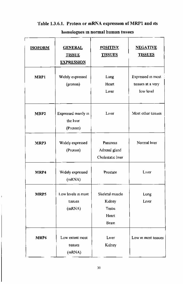
homologues in normal human tissues
Table I.3.6.I. Protein or mRNA expression of MRP1 and its
ISOFORM GENERAL
TISSUE
EXPRESSION
POSITIVE
TISSUES
NEGATIVE
TISSUES
MRP1 Widely expressed
(protein)
Lung
Heart
Liver
Expressed in most
tissues at a very
low level
MRP2 Expressed mainly in
the liver
(Protein)
Liver Most other tissues
MRP3 Widely expressed
(Protein)
Pancreas
Adrenal gland
Cholestatic liver
Normal liver
MRP4 Widely expressed
(mRNA)
Prostate Liver
MRP5 Low levels in most
tissues
(mRNA)
Skeletal muscle
Kidney
Testis
Heart
Brain
Lung
Liver
MRP6 Low extent most
tissues
(mRNA)
Liver
Kidney
Low in most tissues
30
1 4 Monoclonal antibodies in research and diagnostics
The discovery of antibodies at the end of the 19th century provided the means to search
for cancer specific antigens and opened the way for extensive studies of antibodies as
potential diagnostic indicators and immunotherapies for cancer Over the past century,
investigators injected human cancer cells into innumerable horses, sheep, rabbits, mice
and rats, closely analysing the polyclonal antibodies the animals produced in response If
the immune systems of the animals reacted to the foreign tumour cells by producing
antibodies that did not react with normal cells, this finding would signal the presence of
antigens that could subsequently be identified and pressed into service as targets for
antibody based techniques Many workers tried this approach and claimed to identify
cancer-specific antigens Unfortunately, none of these claims held up to careful scrutiny
The search for cancer antigens became easier in 1975, thanks to a discovery made by
Cesar Milstein and Georges J F Kohler of the University of Cambridge This model was
the first mouse model of monoclonal antibody (MAb) secreting hybndomas These
researchers demonstrated that antibody-producing cells could be made to survive
indefinitely if they were fused with cancer cells This method had a profound effect on
cancer immunology for several reasons First, it provided a powerful new method to
search for cancer antigens and second, workers could at last produce defined antibodies
in sufficient amounts to put antibody-based therapies to the test The ability of
antibodies to recognise fine distinctions between molecules is what has made them
extremely useful in the search and study of cancer antigens
31
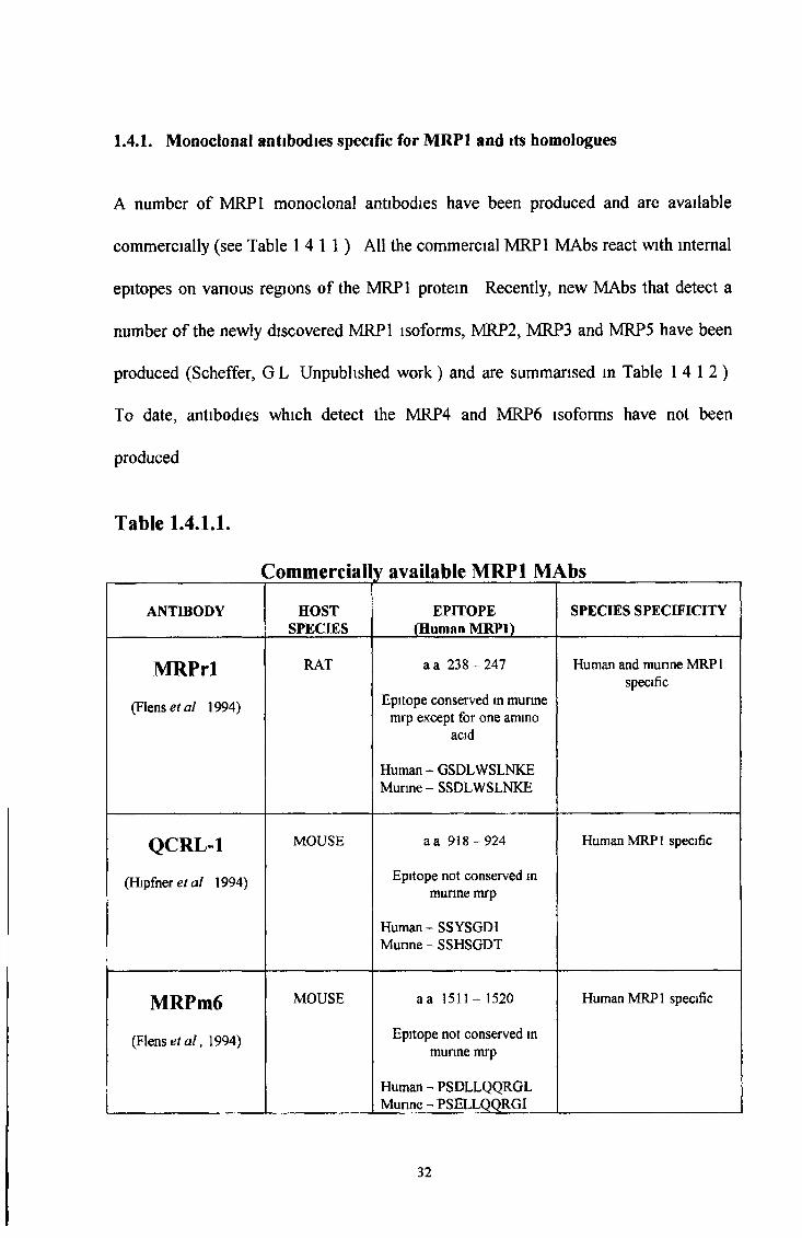
A number of MRP1 monoclonal antibodies have been produced and are available
commercially (see Table 14 1 1 ) All the commercial MRP1 MAbs react with internal
epitopes on various regions of the MRP1 protein Recently, new MAbs that detect a
number of the newly discovered MRP1 isoforms, MRP2, MRP3 and MRP5 have been
produced (Scheffer, G L Unpublished work) and are summarised in Table 14 1 2 )
To date, antibodies which detect the MRP4 and MRP6 isoforms have not been
produced
Table I .4 .U .
1.4.1. Monoclonal antibodies specific for MRP1 and its homologues
Commercially available MRP1 MAbs
ANTIBODY HOSTSPECIES
EPITOPE (Human MRP1)
SPECIES SPECIFICITY
MRPrl
(Flense/ al 1994)
RAT a a 23 8 -2 4 7
Epitope conserved in munne mrp except for one ammo
acid
Human - GSDLWSLNKE Murine - SSDLWSLNKE
Human and munne MRP 1 specific
QCRL-l
(Hipfner et al 1994)
MOUSE a a 9 1 8-924
Epitope not conserved m munne mrp
Human - SSYSGDI Munne-SSHSGDT
Human MRP1 specific
MRPm6
(Flense* a /, 1994)
MOUSE aa 1511-1520
Epitope not conserved in munne mrp
Human - PSDLLQQRGL Munne-PSELLQQRGI
Human MRP 1 specific
32
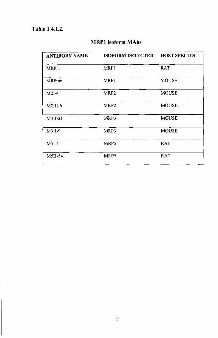
Table 1 4.1.2.
M RPl isoform MAbs
ANTIBODY NAME*
ISOFORM DETECTED HOST SPECIES
MRPrl MRPl RAT
MRPm6 MRPl MOUSE
M2I-4 MRP2 MOUSE
M2III-6 MRP2 MOUSE
M311-21 MRP3 MOUSE
M3II-9 MRP3 MOUSE
M5I-1 MRP5 RAT
M5II-54 MRP5 RAT
33
The techniques conventionally used for the production of MAbs are based on variations
of the original report by Kohler and Milstein (1975) This technology has proven to be
capable of producing monoclonal antibodies of a single pre-determined specificity to a
wide variety of antigens which can potentially be produced in unlimited quantities
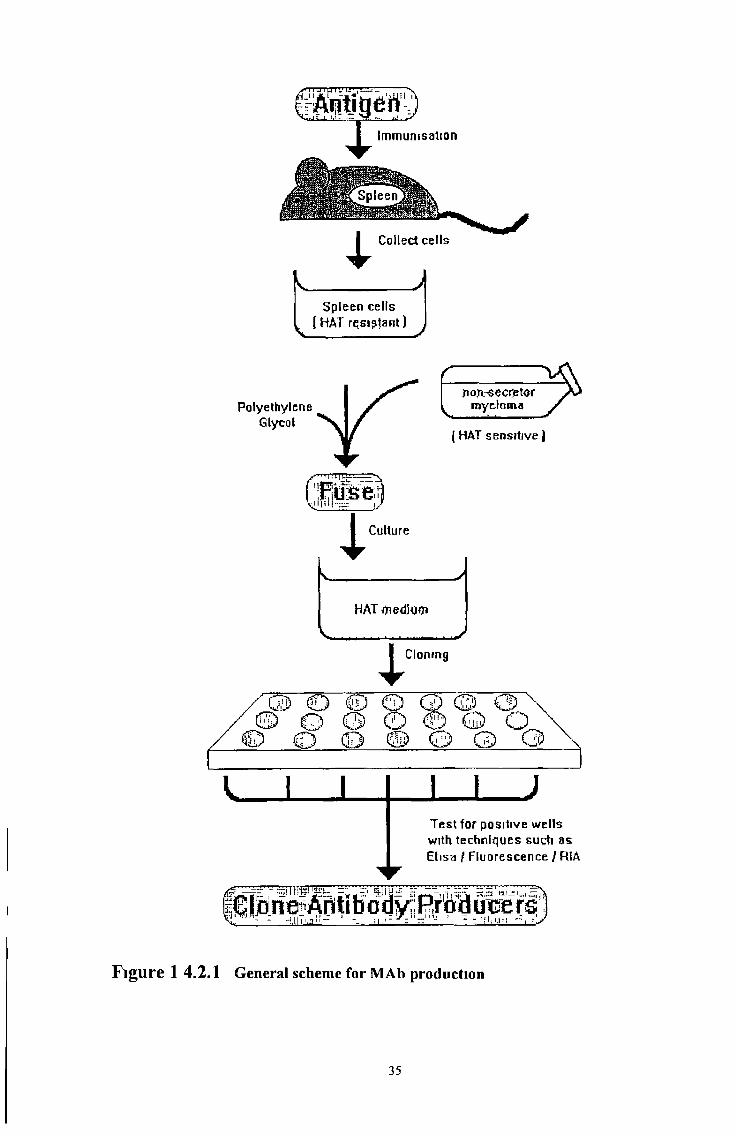
The general scheme for the production of MAbs by hybndoma technology is shown in
Figure 14 2 1 Animals, usually mice are immunised by injection of the antigen to which
a MAb is required When an immune response has been raised, B cells are harvested
from the rodent, usually from the spleen, and these are fused with immortal myeloma
cells by fusion of the cell membranes with polyethylene glycol (PEG) Fusion results in
the production of immortalised hybnd cells or hybndomas The remainder of the process
is then one of isolation and propagation of hybndoma cells which have retained the
ability of the B cell to produce antibody and the good growth characteristics of the
myeloma cell The process of MAb production can therefore be considered in three
parts immunisation, fusion and selection of the required hybndoma Many protocols for
the immunisation and generation of hybndomas are available (Harlow and Lane, 1988)
14 2 Monoclonal antibody production techniques
34
/ r x I u I ü I L1 U 1 , i — i ~
Antigen ;Immunisation
Spleen cells [HAT rçsisJant)
ïiojirsecriBtor my gì am a
I HAT sensitive )
1Cloning
© O © © G) © 03 © © Q 0 © Q Q
O ( D @ © CD 0
V I I l _ l )
Test for positive wells with techniques such as Ehsa / Fluorescence / RIA
joneAntibo dy,, Pro d uce r s; - -U IÏ ..2 M - ‘ 11 ■1 ~ z 1 ' - r l i i . u n ■:
F ig u re 1 4 .2 .1 General scheme for MAb production
35

MAbs can be produced against a wide variety of antigens including proteins, nucleic
acids, carbohydrates and haptens (Harlow and Lane, 1988; Claassen et al, 1993). The
production of MAbs specific for proteins can be performed by immunising with a variety
of sources and forms of antigen. Live cells may be used to generate MAbs specific for
surface antigens. Fusion proteins produced by recombinant DNA technology make
excellent antigens and provide a large quantity of antigen which can be purified. Short
synthetic peptides can be used for site specific MAbs when a proteins coding sequence is
available.
However, all immunisations using peptide antigens run the risk of producing antibodies
that recognise the linear peptide but do not recognise the native protein which may have
a complex conformation, thus blocking the MAb epitope (Claassen et al, 1993).
Therefore, it is important to consider the conformation of the molecule which the MAb is
desired to bind to. For example, will a protein be in its native conformation or
denatured? MAbs of exquisite specificity can be generated which recognise a desired
isomer or molecular form of a particular antigen.
There are two important exceptions to the range of immunogens which can be used
(Harlow and Lane, 1988). Antibodies will not normally be raised to ‘self antigens, and
small antigens (less than approximately 1000 Daltons in molecular weight) will not raise
a response unless conjugated to a high molecular weight carrier protein. For this
purpose carrier proteins such as bovine serum albumin (BSA) or keyhole limpet
hemocyanin (KLH) are used, which are very immunogenic in rodents, easily available
and may be conjugated to small molecules.
1.4.3. Immunogens:
36
Adjuvants produce an emulsion which is injected into the animal and provides a long-
lasting local ‘depot’ of antigen which is slowly released and thus continually boosts the
response (Harlow and Lane, 1988) In addition, other components are sometimes
included in the adjuvant mixture to activate the immune system non-specifically For
example, complete Freund’s adjuvant contains killed mycobactena for activation as well
as oil and an emulsifying agent Complete Freund’s adjuvant is used for the first
immunisation and subsequent booster immunisations are carried out in incomplete
Freund’s adjuvant (without the mycobacteria component) to maximise response to the
antigen and avoid excessive inflammatory responses to mycobacteria
14 4 Design of Synthetic Peptides for Immunisation.
The use of synthetic peptides for MAb production requires careful thought in their
choice and design Searches for the full-length amino acid sequences of a protein can be
earned out in the NCBI SWISS-PROT data bank (www3 nebi nlm mh gov) After
considenng the cntena for choosing peptide sequences for immunisation, chosen peptide
sequences can then be entered into the BLITZ data bank by email (BLITZ@ebi ac uk)
and aligned to compare sequence homology with other protein sequences Usually
sequences shanng five or more amino acids m sequence with any other mammalian
protein are disregarded The main cntena for peptide choice are listed below
CRITERIA FOR CHOOSING THE PEPTIDES
• Choose peptides with a preponderance of polar amino acids and proline residues
(these sequences are more likely to be exposed on the surface of the native protein)
• Choose sequences as near as possible to termini (these sequences are also more likely
to be exposed)
37
• Choose sequences with a higher “mobility” of the amino acid residues (stretches of
amino acids with higher flexibility) as determined by NMR and X-ray structure when
available Higher temperature sensitivity in crystallography and NMR distinguishes
regions of a protein that are more mobile from regions that are more static It has
been suggested that regions of higher mobility present a higher chance of forming a
structure that is similar to the linear peptide (Moore, 1980)
• Avoid hydrophobic regions
• Avoid transmembrane segments (only possible when information on 3D structure is
available)
• Avoid sequences including valines
• Look at sequences used for successful polyclonals
• Avoid sequences sharing five or more amino acids in sequence with any mammalian
protein
To optimise the chance of a MAb being able to gain access to its epitope, it is
advantageous to choose sequences which are more likely to be situated on the exposed
surface of the native protein Peptide sequences with a preponderance of polar amino
acids and proline residues are more likely to be exposed on the surface of the native
protein In contrast, hydrophobic regions are more likely to be encased in the inner core
of a protein, making them less likely to be accessible to MAbs Sequences close to either
terminus of a protein are also more likely to be exposed on the surface of a protein
Obviously transmembrane sequences should be avoided, as the membrane would block
access of the MAb to its epitope (Harlow and Lane, 1988) Crystallography and NMR
distinguishes regions of the protein that are more likely to be exposed on the surface of a
38
protein and present a higher chance of forming a structure that is similar to the linear
peptide (Moore, 1980) Successful polyclonals may be used as a precursor to testing
peptide sequences They may indicate accessible epitopes and pinpoint peptide
sequences which will work optimally for their intended use It is our experience that
peptides including valines are insoluble and therefore unsuitable for use as a soluble
immunogen
14 5 Species*
There are a number of species which are useful for hybndoma production such as rats,
mice, chickens, hamsters and rabbits (Harlow and Lane, 1988) Mice are usually the first
choice for immunisation because they are small, easy to handle and are suitable for fusion
with the most commonly available SP2 murine myeloma cells There are in fact suitable
myeloma cells for each species available now However, cross species fusions with
murine SP2 cells and rat lymphocytes have been earned out successfully by our
laboratory and others (Flens et a l , 1994) Choosing a species which does not express
any proteins shanng a high amino acid homology with the immunogen is advised because
antibodies will not normally be raised to ‘self antigens Various strains of the same
species can react differently to the same antigen, resulting in successful immunisation in
only some of the strains Also, individual species of animals may elicit vanous immune
responses to immunogens For example the rat antibody repertoire is different from that
of the mouse in a number of ways Most importantly some antigens from origins other
than mouse or rat species can induce much stronger responses in rats than in mice and
the reciprocal may be true for other antigens However, obtaining rat-rat hybridomas still
seems to be considered difficult and several experienced investigators have had difficulty
in keeping rat-rat hybrids alive for more than 2 to 3 weeks possibly due to an unusually
39
high number of natural killer cells in the spleens of rats from some colonies Therefore,
alternative immunisation via the lymphatic system and collection of B cells from lymph
nodes may be more suitable in rat models
14 6 Route of immunisation*
There are many routes of injection which can be employed to immunise an animal
However, each route has advantages and disadvantages which must be taken into
consideration when designing an immunisation protocol (Harlow and Lane, 1988) For
example, different routes present varying immunogenicity and the structure of the
antigen can be changed dependent on the environment where antigen is presented
(Claasen et a l , 1993) The most common route of injection for mouse and rat is
intraperitoneally, followed by collection of B cells from the spleen for fusion The most
common route of injection for rabbit is subcutaneously followed by collection of
polyclonal serum from bleeds Other routes include subcutaneous, intravenous,
intramuscular and intradermal The lymphatic system can also be employed by
administering rear footpad injections followed by collection of B cells from the popliteal
node (see Materials and Methods, section 2 2 8 2 )
1 4.7 Immunisation
The immunisation protocol, used to raise an immune response in the animal, is of key
importance (Harlow and Lane, 1988) Immunogens of different molecular types can be
used and they do not need to be pure materials, as a hybndoma secreting a MAb of the
required specificity may be selected out later Different molecules vary greatly in their
immunogenicity and thus require different immunisation protocols for an optimal
response Factors such as the form of the antigen, number and route of immunisations,
40

carrier, adjuvant and the species and the strain of animal used need to be considered To
ensure that an immune response has been elicited against the antigen before harvesting B
cells, blood samples are obtained from the immunised animal and tested for the presence
of specific antibodies If positive, final boosts of soluble antigen are given intravenously
to increase the number of specific immune B cells present in the spleen prior to harvest
The type of antibody produced is dependent on the number of immunisations If IgM is
required, only one immunisation is earned out, as only a pnmary response is required,
whereas IgG antibodies require multiple injections (usually three or four) at intervals of
approximately 3-4 weeks to allow an effective secondary response
14 8 Fusion.
Pnor to fusion, blood samples should be obtained from the immunised animal, tested to
ensure the presence of specific antibodies, followed by a final boost The spleen is the
most common source of immune B cells, although cells can also be harvested from lymph
nodes Fusion of immune B cells with immortal myeloma cells is usually accomplished
with polyethylene glycol (PEG) to induce membrane fusion After fusion, a mixture of
hybndoma cells, B cells and myeloma cells are present in the culture medium B cells
fused to myeloma cells form hybndoma cells which have a selective growth advantage
over B cells and myeloma cells B cells fail to grow in culture because they are not
immortal and die after a few days Myeloma cells used for fusion are deficient in the
hypoxanthinephosphonbosyltranseferase (HPRT) enzyme and consequently are not able
to use the HRPT - based salvage pathway for RNA synthesis Selection is then achieved
by using hypoxanthine, aminopterm and thymidine (HAT) medium Aminoptenn is an
effective inhibitor of RNA and DNA synthesis and thus will block the growth of the
myeloma cells However, hybnd cells which have the HRPT enzyme from the B cells
41
will be able to use the added hypoxanthine and thymidine to produce RNA via the
salvage pathway and survive
14 9 Screening
After selection in HAT medium, hybrid cells are grown and tested to determine which
colonies of cells produce antibodies of interest Not all of the spleen cells which form
hybrids are B cells, and thus there will be some hybrids which do not produce antibody at
all, and also many cells will be present which produce antibodies which are not of
interest An important step is therefore the screening of clones of cells to identify those
which produce the MAbs required A large number of clones may need to be screened
quickly to allow selection of those required for further culture A suitable screening
assay should therefore be rapid, able to screen many samples and capable of giving a
result with the small amount of antibody produced by early clones of cells There are
many types of suitable assays to measure antigen-binding properties including ELISA
(enzyme linked immunoadsorbent assay), dot blots, Western blots,
immunohistochemistry and immunofluorescence If possible, the assay which uses the
antibody in the closest way to that required for the intended end use of the antibody is
the most suitable, as this will ensure that the optimal properties of the antibody are
selected
1 4 10 Maintenance of stable hybndoma cell lines
Once suitable hybndomas have been identified, these must be cloned several times to
ensure a stable, homogeneous colony of cells and to ensure that the antibody is indeed
monoclonal To do this requires the growth of a colony from a single cell This is
achieved by subcultunng the cells by limiting dilution so that each well of the culture
42

plate contains an average of less than one cell Alternatively, cloning can be achieved by
plating out cells in semisolid agar of by single cell manipulation techniques Individual
clones of cells can then be screened for antibody production as before Once cloned, the
hybndoma cell is ready to be used for the production of large amounts of antibody, but it
is also important to ensure that samples of cells are preserved by freezing in liquid
nitrogen at each stage so that valuable hybndomas are not lost
43

In order to elucidate further the role of multidrug transporters in multidrug resistance
specific antibodies can be used to determine the frequency and pattern of expression of a
particular protein in different tumour types MAbs specific for various regions of a
protein can determine variations in topology Each individual protein may contain
various functional epitopes MAbs which bind to a functional epitope of a protein can be
used to determine the substrate specificity of a protein External epitope MAbs are
desirable because of their suitability for use on viable unfixed cells and their potential
clinical applications for treating drug resistance
Several well-performing MAbs have been described that detect both internal and external
epitopes of the MDR1 transporter molecule A limited number of MAbs have also been
described for MRP1 However, all MRP1 MAbs described to date detect internal
epitopes and require permeabihsation of the cells in order for the antibody to bind to
their epitopes It is possible that like P-gp, MRP1 may exist in a number of topological
forms This can only be confirmed by use of a range of MRP 1 antibodies While this
thesis was in progress five homologues of MRP1 were discovered MAbs detecting
internal epitopes of the MRP homologues MRP2 MRP3 and MRP5 have also been
described (SchefFer, G L , Unpublished w ork) MAbs detecting MRP4 and MRP6 have
not been reported
It was the aim of this thesis to produce new MRP1 antibodies which do not cross-react
with other members of the MRP family and can be included in the further study of MRP-
1 These MAbs could then be used as tools to unravel the role of MRP 1 in multidrug
resistance and furthermore may possibly have clinical applications
1.5 Aims of this thesis
44
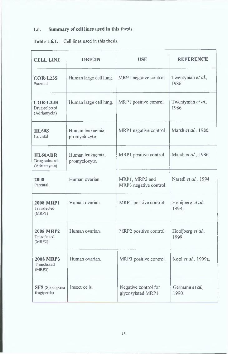
1.6. Summary of cell lines used in this thesis.
Table 1.6.1. Cell lines used in this thesis.
CELL LINE ORIGIN USE REFERENCE
COR-L23SParental
Human large cell lung. MRP1 negative control. Twentyman et al., 1986.
COR-L23RDrug-selected(Adriamycin)
Human large cell lung. MRP1 positive control. Twentyman et al., 1986
HL60SParental
Human leukaemia, promyelocyte.
MRP1 negative control. Marsh e/ al., 1986.
HL60ADRDrug-selected(Adriamycin)
Human leukaemia, promyelocyte.
MRP1 positive control. Marsh ei al., 1986.
2008Parental
Human ovarian. MRP1, MRP2 and MRP3 negative control.
Naredi et al., 1994.
2008 MRP1Transfected(MRP1)
Human ovarian. MRP1 positive control. Hooijberg et al., 1999.
2008 MRP2Transfected(MRP2)
Human ovarian. MRP2 positive control. Hooijberg et al., 1999.
2008 MRP3Transfected(MRP3)
Human ovarian. MRP3 positive control. Kool et al., 1999a.
SF9 (Spodoptera frugiperda)
Insect cells. Negative control for glycosylated MRP 1.
Germann et al., 1990.
45

Table 16 1 (Continued) Cell lines used in this thesis
CELL LINE ORIGIN USE REFERENCE
SF9 MRP1Transfected(MRP1)
Insect cells Positive control for glycosylated MRP1
B akose/a/, 1996
DLKPParental
Human non-small-cell lung carcinoma
Cell line survey L sw eta l, 1992
DLKPADrug-selected(Adnamycin)
Human non -small-cell lung carcinoma
Cell line survey Clynes eta l, 1992
A549Parental
Humanadenocarcinoma, lung
Cell line survey Berger e* a /, 1997a
RPMI 2650Parental
Cultured from pleural effusion of a patient with metastatic anaplastic squamous cell carcinoma of the nasal septum (human)
Cell line survey Moore and Sandberg, 1964
RPMI 2650Drug-selected(Taxol)
Cultured from pleural effusion of a patient with metastatic anaplastic squamous cell carcinoma of the nasal septum (human)
Cell line survey Yizheng Liang, PhD Thesis, 1999
RPM I 2650Drug-selected(Melphalan)
Cultured from pleural effusion of a patient with metastatic anaplastic squamous cell carcinoma of the nasal septum (human)
Cell line survey Yizheng Liang, PhD Thesis, 1999
46

2.0. MATERIALS AND METHODS
47
Addresses of suppliers are provided in Appendix 2
2 11 Water
Ultrapure water was used in the preparation of all media and solutions This water was
purified to a standard of 12-18 MD/cm resistance by a reverse osmosis system
(Millipore Milli-RO 10 Plus, Elgastat UHP) A conductivity meter in the system
continuously monitored the quality of the UHP
2.12 Glassware
Most solutions pertaining to cell culture and maintenance were prepared and stored m
sterile glass bottles All glassware including these bottles, used for cell culture work
were soaked m a 2% (v/v) solution of RBS (Chemical Products) fort at least one hour
after which, they were scrubbed and rinsed several times in tap water The glassware
was washed in an mdustnal dishwasher, using Neodisher detergent (an organic,
phosphate-based acid detergent) nnsed twice with UHP The materials were finally
sterilised by autoclaving as described in section 2 1 3
2 13. Sterilisation
Water, glassware and all thermostable solutions were sterilised by autoclaving at 121°C
for 20 minutes at 15 p s l Thermolabile solutions were filtered through a 0 22|im sterile,
low protem-binding filter (Millipore, Millex-GV SLGV025BS) Large volumes, (up to
10 litres) of thermolabile solutions were filter sterilised through a micro-culture bell
filter (Gelman, 12158)
48
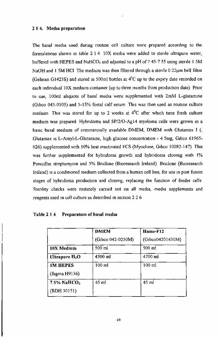
2 14. Media preparation
The basal media used during routine cell culture were prepared according to the
formulations shown in table 2 14 10X media were added to sterile ultrapure water,
buffered with HEPES and NaHC03 and adjusted to a pH of 7 45-7 55 using sterile 1 5M
NaOH and 1 5M HC1 The medium was then filtered through a sterile 0 22jxm bell filter
(Gelman G1423S) and stored in 500ml bottles at 4°C up to the expiry date recorded on
each individual 10X medium container (up to three months from production date) Prior
to use, 100ml aliquots of basal media were supplemented with 2mM L-glutamine
(Gibco 043-0503) and 5-15% foetal calf serum This was then used as routine culture
medium This was stored for up to 2 weeks at 4°C after which time fresh culture
medium was prepared Hybndoma and SP/2/0-Agl4 myeloma cells were grown in a
basic basal medium of commercially available DMEM, DMEM with Glutamax I (,
Glutamax is L-Amyl-L-Glutamine, high glucose concentration - 4 5mg, Gibco 61965-
026) supplemented with 10% heat inactivated FCS (Myoclone, Gibco 10082-147) This
was further supplemented for hybridoma growth and hybridoma clonmg with 1%
Penicillin streptomycin and 5% Bnclone (Bioresearch Ireland) Briclone (Bioresearch
Ireland) is a conditioned medium collected from a human cell line, for use in post fusion
stages of hybridoma production and cloning, replacing the function of feeder cells
Sterility checks were routinely carried out on all media, media supplements and
reagents used in cell culture as described in section 2 2 6
Table 2 14 Preparation of basal media
DMEM
(Gibco 042-0250M)
Hams-F12
(Gibco04201430M)
10X Medium 500 ml 500 ml
Ultrapure H2O 4300 ml 4700 ml
1M HEPES
(Sigma H913 6)
100 ml 100 ml
7 5% NaHCOs
(BDH 30151)
45 ml 45 ml
49
2 2 Cell culture procedures
All cell culture work was carried out in a class II down-flow re-circulating laminar air
flow cabinet (Nuaire Biological Cabinet) All experiments involving toxic compounds
were conducted in a cytoguard laminar air-flow cabinet (Gelman Sciences, CG series)
Strict aseptic technique was adhered to at all times The laminar flow cabinet was
swabbed with 70% industrial methylated spirits (IMS) before and after use, as were all
items brought into the cabinet Each cell line was assigned specific media and waste
bottles At any time, only one cell line was used m the laminar air-flow cabmet, and
upon completion of work with any given cell line the laminar air-flow cabinet was
allowed to clear for at least 15 minutes This was to eliminate any possibilities of cross
contammation between the various cell lines The cabmet itself was cleaned weekly
with industrial disinfectants (Virkon, Antec International or TEGO, TH Goldschmidt) as
were all incubators used in the culture of cell lines and hybridomas These detergents
were alternated every month
2 2 1 Cell lines
Details pertaining to the cell lines used for the experiments detailed in this thesis are
provided in Table 2 2 1 Cell lines were maintained in 25 cm (Costar 3035), 75 cm
(Costar 3075) or 175 cm2 (Nunclon, NUNC) tissue culture flasks at 37°C and fed every
2-3 days Cell lines were cultured through 7-10 passages before they were discarded and
new cultures grown from frozen stocks Confluent hybndoma cell lines were grown for
at least 7 days with no change of growth media when antibody was being harvested
50
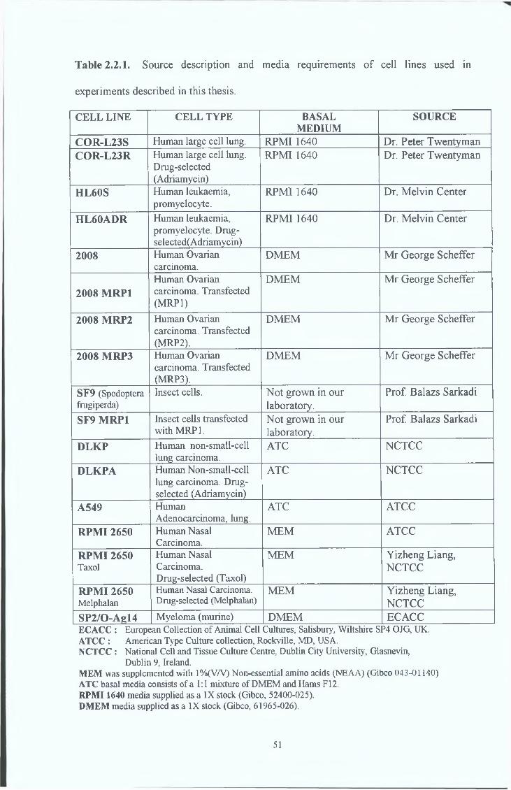
Table 2.2.1. Source description and media requirements of cell lines used in
experiments described in this thesis.
C E L L L IN E C E L L TY P E B A S A LM E D IU M
SOURCE
C O R -L 2 3 S Human large cell lung. RPMI 1640 Dr. Peter TwentymanC O R -L 2 3 R Human large cell lung.
Drug-selected(Adriamycin)
RPMI 1640 Dr. Peter Twentyman
H L 6 0 S Human leukaemia, promyelocyte.
RPMI 1640 Dr. Melvin Center
H L 6 0 A D R Human leukaemia, promyelocyte. Drug- selected(Adriamycin)
RPMI 1640 Dr. Melvin Center
2008 Human Ovarian carcinoma.
DMEM Mr George Scheffer
2008 M R P 1
Human Ovarian carcinoma. Transfected (MRP1)
DMEM Mr George Scheffer
2008 M R P 2 Human Ovarian carcinoma. Transfected (MRP2).
DMEM Mr George Scheffer
2008 M R P 3 Human Ovarian carcinoma. Transfected (MRP3).
DMEM Mr George Scheffer
SF9 (Spodoptera frugiperda)
Insect cells. Not grown in our laboratory.
Prof. Balazs Sarkadi
SF9 M R P 1 Insect cells transfected with MRP 1.
Not grown in our laboratory.
Prof. Balazs Sarkadi
D L K P Human non-small-cell lung carcinoma.
ATC NCTCC
D L K P A Human Non-small-cell lung carcinoma. Drug- selected (Adriamycin)
ATC NCTCC
A 549 HumanAdenocarcinoma, lung.
ATC ATCC
R P M I 2650 Human Nasal Carcinoma.
MEM ATCC
R P M I 2650Taxol
Human Nasal Carcinoma. Drug-selected (Taxol)
MEM Yizheng Liang, NCTCC
R P M I 2650Melphalan
Human Nasal Carcinoma. Drug-selected (Melphalan)
MEM Yizheng Liang, NCTCC
S P 2 /0 -A e l4 Myeloma (murine) DMEM ECACCECACC : European Collection of Animal Cell Cultures, Salisbury, Wiltshire SP4 OJG, UK.ATCC : American Type Culture collection, Rockville, MD, USA.NCTCC : National Cell and Tissue Culture Centre, Dublin City University, Glasnevin,
Dublin 9, Ireland.MEM was supplemented with 1%(V/V) Non-essential amino acids (NEAA) (Gibco 043-01140) ATC basal media consists of a 1:1 mixture of DMEM and Hams F I2.RPMI 1640 media supplied as a IX stock (Gibco, 52400-025).DMEM media supplied as a IX stock (Gibco, 61965-026).
51
2.2.2 Subcultunng cell lines
During routine subcultunng or harvesting of adherent cell lines, cells were removed
from their flasks by enzymatic detachment Waste medium was removed from the cells
which were then rinsed with pre-warmed trypsin-versene/EDTA (TV) solution (0 25%
trypsin (Gibco 043-05090), 0 01% EDTA (Sigma) solution in PBS (Oxoid BR14A)
This ensured that any naturally occurnng tiypsin inhibitor in residual serum was
deactivated Fresh TV was then placed in the flask and incubated until the cells were
seen to have detached (2-10 minutes) The TV solution was deactivated by the addition
of pre-warmed basal medium containing serum The entire solution was then transferred
to a 30ml stenle universal tube (Stenlin 128a) and centrifuged at 1,000 rpm for 5
minutes The resulting pellet was then re-suspended in pre-warmed growth medium,
cells counted and tissue culture flasks re-seeded at the required density
Hybndoma and SP/2 cells are loosely adherent Cells were passaged by tapping the
flask lightly and/or gentle pipetting with a 10ml stenle pipette (Elkay, Ireland) Cell
suspensions were pooled and centrifuged at 1,000 rpm for 5 minutes The cell pellet was
then re-suspended m culture medium and a cell count performed (section 2 6 3 ) and the
cells re-suspended at the desired density Cells were grown in 5% CO2
2 2 3 Cell counting
Cells were trypsimsed, pelleted and re-suspended in media as described in section 2 2 2
Cell counting and viability were carried out by using trypan blue (Gibco, 15250) dye
exclusion technique An aliquot of Trypan blue was added to a single cell suspension at
a ratio of 1 2 After 3 minutes incubation at room temperature, a sample of the mixture
was applied to the chamber of a haemocytometer over which a glass cover slip had been
placed Cells in the 4 outer corner grids of the chamber were counted microscopically,
an average per corner grid was calculated with the dilution factor taken into account and
the final number multiplied by 104 to determine the number of cells per ml Non-viable
cells were those which stained blue while viable cells excluded the trypan blue dye and
remained unstained
52
2 2 4. Cell freezing
Cells of various passage numbers were frozen and cryo-preserved to serve as master
stocks Vials could then be thawed and cultured for study
Cells to be frozen were harvested in the log phase of growth and counted as described in
Section 2 2 3 The pellets were re-suspended in foetal calf serum (pre-cooled to 4°C)
and an equal volume of freezing medium (DMSO (Sigma D-5879)/serum 1 9 (v/v)) was
added drop-wise to the cell suspension to give a final concentration of at least 5x10⁶
cells per ml 1 5ml of the cell suspension was quickly placed in a cryovial (Greiner
122278) which was placed in the vapour phase of liquid nitrogen container for 2 5-3 5
hr After this, the cryovials were stored in liquid nitrogen until required
2 2 5 Cell thawing
The cryopreserved cells were removed from the liquid nitrogen and thawed at 37°C
Immediately prior to removal of a cryovial from the liquid nitrogen, a sterile universal
tube containing growth medium was prepared for the rapid transfer and dilution of
thawed cells (thus reducing the exposure time to DMSO, which is toxic at room
temperature) Following the addition of the thawed cell suspension to the growth
medium, the suspension was centrifuged at 1,000 rpm for 3 minutes, after which the
pellet was re-suspended in fresh growth medium A viability count was earned out
(Section 2 2 3 ) and the thawed cells were placed in tissue culture flasks and allowed to
attach over night, The following day the cultures were re-fed with growth medium to
remove any residual DMSO
2 2 6 Monitoring of sterility of cell culture solutions
Sterility testing was performed for all cell culture media and cell culture related
solutions Samples of prepared basal media were inoculated onto Columbia blood agar
plates (Oxoid CM331), Saburaud dextrose (Oxoid CM217), and Thioglycollate broths
(Oxoid CM173) and incubated for 2 days to 3 weeks at 37°C and 25°C Complete cell
53
media were sterility tested at least four days prior to use, using Columbia blood agar
plates and incubation of 5ml media samples at 37°C
2 2 7 M yc o p la sm a analysis of cell lines
M yco p la sm a exammations were carried out routinely (every 3 months) on all cell lines
used in this study
2 2 7 1 Indirect staining procedure for M yc o p la sm a analysis
M yco p la sm a negative NRK cells (Normal Rat Kidney fibroblasts) were used as
indicator cells for this analysis The cells were cultured with supernatant (harvested
from test cell lines after 2-3 days confluency) from test cell lines for 4 to 5 days, fixed
and stained with Hoescht fluorescent stain which binds specifically to DNA, thus it
would stain the nucleus of the cell and any M yco p la sm a infection, which can be seen as
small fluorescent bodies in the cytoplasm of the NRK cells Positive controls consisted
of NRK cells infected with M yco p la sm a enriched supernatants
2 7 2 2 Direct staining procedure for M yc o p la sm a analysis
The direct procedure for M yco p la sm a involved inoculating test samples onto an
enriched M yco p la sm a culture broth to optimise the growth of any contaminants and
incubated at 37°C for 48h Samples of this broth were then streaked onto plates of
M yco p la sm a agar base and incubated for 3 weeks at 37°C and 5% CO2 Plates were
checked microscopically every 7 days for the growth of small oval shaped colonies
which was indicative of M yco p la sm a infection
54
2 3 Polyclonal and monoclonal antibody production
All experiments on animals were carried out under license and in compliance with the
rules of The Cruelty to Animals Act, 1876, E C Directive 86/609/EC
2 3 1 Immunogens
A range of immunogens were employed in three different animal species (rabbit, mouse
and rat) for this thesis to produce both polyclonal and monoclonal antibodies specific to
the MRP1 protein These included (1) whole cell lysates of the COR-L23R MRP1
over-expressmg cell line, (2) cell membranes isolated by differential centrifugation from
the COR-L23R MRP1 over-expressmg cell line, (3) synthetic peptides and (4) fusion}
proteins
2 3.1 1 (1) Whole cell lysates (Used for immunisation of mice)
The MRP1 over-expressing COR-L23R cells which is an adherent cell line was
trypsinsed as described in section 2 2 2 , counted as descnbed in section 2 2 3 to give a
final concentration of 2 x 10⁶ cells and washed three times in PBS by centrifugation @
lOOOrpm for 10 minutes The cell pellet was re-suspended in 250^1 PBS containing
protease inhibitors (Protease inhibitor cocktail tablets, prepared according to the
manufacturers instructions Boehrmger Mannheim 1697498) Cells were then lysed by
somcation and completion of cell lysis was confirmed by viewing under an inverted
microscope Equal volumes (i e 250^1) of Freund's Complete Adjuvant (Sigma F-5881)
were added immediately prior to primary immunisation and Freund's Incomplete
adjuvant (Sigma F-5506) added for booster injections Mice received one
mtrapentoneal injection of 500|il each
55
The COR-L23R cell membrane isolation, outlined below, was carried out according to a
modification of the method Gerlach et al., 1987.
• At least 2x10⁷ cells were trypsinised (or thawed directly from liquid nitrogen) and
washed 3 times with PBS by centrifugation at 2000rpm.
• NB. Cells are kept on ice from this stage on.
• The cells were then suspended in approximately 5ml of lysis buffer (PBS / Protease
Inhibitors (Boehringer Mannheim 1697498) - added just before use and stored on
ice). The re-suspended cells were then sonicated until cells are lysed - this is
determined by viewing under the microscope after each sonication.
• The lysed cells were centrifuged at 7,800rpm in an ultracentrifuge (Beckman XL80)
for 10 minutes at 4°C.
• The supernatant was collected and centrifuged at 38,000rpm in an ultracentrifuge
(Beckman XL80) for lhour at 4°C.
• The pellet obtained was re-suspended in 500 j l i 1 - 1000(il lysis buffer.
• An aliquot of 50|ul is taken for total protein analysis and the remaining supernatant
was divided into aliquots (equivalent to lOOug protein) which were lyophilised
overnight in a freeze-drier (VirTis Consol 4.5).
• The lyophilised samples are stored at -20°C (closed tightly and sealed with parafilm)
until required.
Mice received one intraperitoneal injection (500^1 per mouse) of lOOug of freeze-dried
membrane protein reconstituted in 250^1 PBS and mixed with equal volumes of
appropriate adjuvant. As in previous section, rats received injections (100|il per
footpad) into both rear footpads of lOOug freeze dried membrane protein reconstituted
to 50|lx1 in PBS and mixed with equal volumes of adjuvant.
2.3.1.2. (2) Cell membranes (Used for immunisation of mice and rats.)
56
2.3.1.3. (3) Synthetic Peptides (Used for immunisation of rabbits, mice and rats.)
The entire amino acid sequence of the encoded MRP1 protein was obtained from
EMBL'S Swiss-Prot (EMBL:http://www.embl-heidelberg.de/) protein sequence
database databank using the NETSERV program. The three short synthetic peptides
(their location within the complete protein sequence for MRP1 is described in Section
3.5.2. of the Results) used in immunisations were chosen after alignment searches of the
above databank using the Mail-Fasta program (Pearson and Lipman 1982). The three
peptides were synthesised by Biosyn Ltd, 10 Malone Road, Belfast, BT9 5BN, Northern
Ireland and Immune Systems Ltd., PO Box 120, Paignton, TQ4 7XD, UK according to
the deduced MRP1 sequence (Cole et al., 1992). Three sequences having the following
sequences were prepared :
Peptide 1 CALFYSMAKDAGLV (amino acids 1520 - 1531 ) conjugated to KLH
Peptide 2 PLEGSDLWSLNK (amino acids 235 - 246) conjugated to BSA
Peptide 3 KEDTSEQVVPVLVKN (amino acids 246 - 260 ) conjugated to KLH
For immunisation of rabbits, lmg of Peptide 1, Peptide 2 or Peptide 3 was reconstituted
in 1ml PBS and mixed thoroughly with equal volumes of Freund's Complete Adjuvant
(Sigma F-5881) for the primary immunisation and Freund's Incomplete Adjuvant
(Sigma F-5506) for all subsequent booster injections. Rabbits received subcutaneous
injections on 10 sites along their back equivalent tolOOug protein/site.
For immunisation of mice, synthetic peptides were reconstituted at lOOug protein in
250(j.l PBS. Equal volumes of Freund's Complete Adjuvant (Sigma F-5881) were added
for primary immunisations and Freund's Incomplete adjuvant (Sigma F-5506) for
booster injections. Mice received one intraperitoneal injection of 500fil each (lOOug
peptide/injection).
For immunisation of rats, lOOug of each of the two synthetic peptides used (Peptide 2
and Peptide 3) were reconstituted together in 100 1 PBS and mixed with equal volumes
of Freund's Complete Adjuvant (Sigma F-5881) for the primary immunisation and
57

Freund's Incomplete adjuvant (Sigma F-5506) for booster injections Rats received
100|il volumes in both rear footpads (50ug mixed peptides/injection)
2 3 1 4 (4) Fusion Proteins (Used for immunisation of mice )
DNA technology provides the ability to isolate, characterise and sequence genes From
these gene sequences (1 e the nucleotide sequence) it is possible to predict the ammo
acid sequence of the encoded gene This information makes it possible to express large
quantities of the protein of interest in cell expression vector systems These protems are
usually expressed fused to a large protein such as Maltose Binding Protein (MBP) for
their purification and are termed ‘’Fusion Proteins’5 Shorter synthetic peptides may be
synthesised using the solid-phase techniques introduced by Merrifield (1963) These
short synthetic peptides are usually coupled to a carrier protein such as bovine serum
albumin (BSA) (Sigma) or keyhole limpet hemacyamn (KLH) (Sigma) The two Fusion
Protems used in immunisations (described in Section 3 5 2 of the Results) were a kind
gift from Dr Balazs Sarkadi, The Institute of Enzymology, Biological Research Center,
Hungarian Academy of sciences, H-1113 Budapest, Hungary Both Fusion protems
(location described in Section 3 5 2 of the Results) used in immunisations were
produced according to the deduced MRP1 sequence ( Cole et a l , 1992) and their amino
acid numbers are listed below
Fusion Protein 1 ( ammo acids 1294 - 1531 ) fused to Maltose Binding Protein (MBP)
Fusion Protein 2 ( ammo acids 647 - 912 ) fused to Maltose Binding Protein (MBP)
Fusion protems were reconstituted at lOOug protein in 250jal PBS Equal volumes of
Freund's Complete Adjuvant (Sigma F-5881) were added for primary immunisations
and Freund's Incomplete adjuvant (Sigma F-5506) for booster injections Mice were the
only species used for immunisation with fusion proteins Each mouse received one
mtrapentoneal injection of 500(4.1 each (lOOug peptide/injection)
58
2 3 2. In-vivo Immunisation procedure
2 3 21 Rabbits (female New Zealand White rabbits)
Prior to immunisations and test bleeds, the rabbits (6 months old) were given 0 5ml
(equivalent to 0 015mg) Tengesic (Buprenorphorme) as an analgesic Before the
primary immunisation a control pre-bleed of l-2ml was taken from a vein visible under
the skin surface of the earlobe after shaving the area Directly after this the rabbits were
injected at least 3 times at 6 week intervals with lOOug peptide in 200^1 injection
volume per site (10 sites on back (lmg of Peptide 1, Peptide 2 or Peptide 3 reconstituted
in 1ml PBS and mixed thoroughly with Freund's Complete Adjuvant (Sigma F-5881)
for the primary immunisation and Freund's Incomplete adjuvant (Sigma F-5506) for all
subsequent immunisation boosts)) After each booster, a blood sample (5-6ml) was
taken as before and tested for specific antibody presence by Western blot analysis A
period of 9-11 days were left after boosters for the test bleeds on immunised rabbits to
allow peaking of the immune response 10 days following the final booster injection, the
animals were administered Pentobarbitone Sodium (Sagatal) (60mg/ml/kilo
bodyweight) until they were completely anaesthetised The animals were then
exsanguinated and finally euthanased using Pentobarbitone sodium (Sagatal)
(120mg/ml/kilo bodyweight) at the end of the procedure
2 3 2.2 Mice (male Balb/c mice)
Mice (6-8 weeks old) received a primary booster of lOOug peptide, lOOug fusion protein
or lOOug COR-L23R cell membranes or 2x10⁶ lysed COR-L23R cells in Freund's
Complete Adjuvant (Sigma F-5881), followed by at least 3-4 booster injections of the
same quantity of immunogen in Freund's Incomplete adjuvant (Sigma F-5506) at three
weekly intervals, injections administered intraperitoneally For the synthetic peptides
and fusion proteins, the same regime applied with each mouse receiving lOOug peptide
reconstituted into 250jil PBS and mixed with equal volumes of appropnate adjuvant In
all cases, three days prior to a fusion being performed, a final booster was given
59
2 3 2 3 Rats (female Wistar rats)
Rats (10-12 weeks old) received at least 4 rear footpad injections (lOOjil per footpad) of
either COR-L23R cell membranes or a combination of both Peptide 2 and Peptide 3 at
three weekly intervals 200ug of freeze-dried cell membrane protein was reconstituted
to lOO il in PBS and mixed with equal volumes of Freund's Complete Adjuvant (Sigma
F-5881) for the primary immunisation and Freund's Incomplete adjuvant (Sigma F-
5506) for booster injections 50ug of each of the two synthetic peptides used (Peptide 2
and Peptide 3) were reconstituted together in 100(il PBS and mixed with equal volumes
of Freund's Complete Adjuvant (Sigma F-5881) for the primary immunisation and
Freund's Incomplete adjuvant (Sigma F-5506) for booster injections In all cases, three
days prior to performing the fusion, a final booster was given
2 3 3 Fusion Procedure
2 3 3 1 Mouse
The fusion procedure used for the production of mouse monoclonal antibodies was a
modification of the protocol outlined by Kohler and Milstein (1975) Prior to the
removal of the spleen from the sacrificed mouse, SP/2/0-Agl4 myeloma cells (Shulman
et al 1978) were prepared for cell fusion by harvesting from 75 cm flasks and
centrifuging at 1,000 rpm for 5 minutes in HEPES-free serum free medium This step
was repeated twice A cell count was then performed (Section 2 2 3 ) and the cells kept
at 37°C A Balb/C mouse was then sacrificed by cervical dislocation The animal was
swabbed with 70% IMS and the spleen removed in a laminar flow cabinet with sterile
dissection instruments Single cells were obtamed by forcing the spleen through a sterile
Falcon cell strainer (Becton Dickinson 2360) using the plunger from a sterile 10ml
syringe into serum free DMEM (the DMEM referred in this section is DMEM with
Glutamax I (high glucose concentration - 4 5mg Gibco 61965-026) containing
pyridoxine and without sodium pyruvate or HEPES This cell suspension was placed in
a 50ml centrifuge tube and the volume adjusted to 20ml Large clumps of cells were
60

allowed to pellet by standing at room temperature for 2-3 minutes The supernatant was
then transferred to a fresh centrifuge tube and centrifuged at 17000 rpm for 5 minutes A
cell count was performed as before (Section 2 2 3 ) Splenocyte and SP/2 myeloma cells
were mixed in a 50ml universal tube at a ratio of 10 1 (a minimum of lxlO7 SP/2 are
required for this procedure), centrifuged at 1,000 rpm for 5 minutes and re-suspended in
serum free medium This step was repeated twice Following the final washing step, 1ml
of PEG (polyethylene glycol, Boehrmger Mannheim, 783641 pre-warmed to 37°C) was
added to the cell pellet with a Pasteur pipette using a gentle swirling and aspirating
action for 30 seconds After 30 seconds the aspiration was discontmued 75 seconds
after the start, 0 5ml of plating medium (DMEM with Glutamax I, 10% heat inactivated
FCS, 5% Briclone (Bioresearch Ireland) and 1% HAT (Hypoxanthine, Aminopterin,
Thymidine) (Boehrmger Mannheim 644579) was added slowly down the side of the
50ml centrifuge tube while continuing to swirl gently 8ml of plating medium was
added over the next 5 minutes (3ml at 1 minute intervals followed by the addition of
5ml) Following this step, the cell suspension was centrifuged at 500 rpm for 5 minutes
The supernatant was removed and the cells were re-suspended in 10ml of plating
medium and incubated at room temperature for 15 minutes Prior to performing the
fusion, 0 5ml of plating medium was dispensed into each well of 8x48 well plates
(Costar, 3548) and the plates were equilibrated in the CO2 incubator @ 37°C
Following the 15 minute incubation at room temperature one drop of fused cells was
added to each well and the plates were incubated for 10-12 days at 37°C, 5% CO2
2 3 3 2 Rat
The fusion procedure for the production of rat monoclonal antibodies was identical to
the procedure used for mouse monoclonal antibody production except that immune
activated B cells were collected from the popliteal node in the lymphatic system as seen
m Diagram 2 3 2 This single node is found in a cavity, the popliteal fossa, amidst the
leg muscles at the back of the knee The rat is placed on its ventral surface and the hind
limbs are stretched out A small longitudinal incision is made in the skin at the back of
the leg behind the knee Superficial connective tissue is cleared, and by careful
observation the area where the semitendinous, the biceps femoris and the triceps sural
muscles cross is located This area is often denoted by a small white fatty area into
61
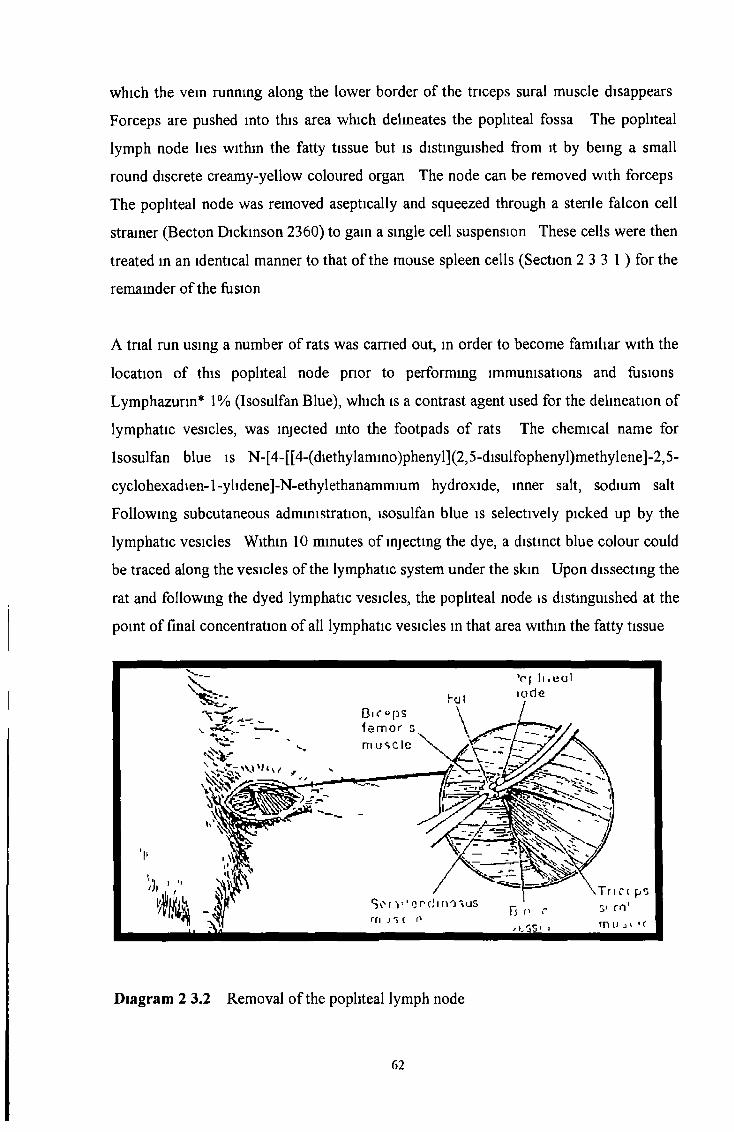
which the vein running along the lower border of the triceps sural muscle disappears
Forceps are pushed into this area which delmeates the popliteal fossa The popliteal
lymph node lies within the fatty tissue but is distinguished from it by being a small
round discrete creamy-yellow coloured organ The node can be removed with forceps
The popliteal node was removed aseptically and squeezed through a sterile falcon cell
strainer (Becton Dickinson 2360) to gam a single cell suspension These cells were then
treated in an identical manner to that of the mouse spleen cells (Section 2 3 3 1 ) for the
remainder of the fusion
A trial run using a number of rats was carried out, in order to become familiar with the
location of this popliteal node prior to performing immunisations and fusions
Lymphazurin* 1% (Isosulfan Blue), which is a contrast agent used for the delineation of
lymphatic vesicles, was injected mto the footpads of rats The chemical name for
Isosulfan blue is N-[4-[[4-(diethylamino)phenyl](2,5-disulfophenyl)methylene]-2,5-
cyclohexadien-l-ylidene]-N-ethylethanammium hydroxide, inner salt, sodium salt
Following subcutaneous administration, isosulfan blue is selectively picked up by the
lymphatic vesicles Within 10 minutes of injecting the dye, a distinct blue colour could
be traced along the vesicles of the lymphatic system under the skin Upon dissecting the
rat and following the dyed lymphatic vesicles, the popliteal node is distinguished at the
point of final concentration of all lymphatic vesicles in that area within the fatty tissue
Diagram 2 3.2 Removal of the popliteal lymph node
62
2.3.4. In-vitro immunisation
In-vitro immunisations for murine monoclonal antibody production were carried out
using a kit (Cell Prime) supplied by Immune Systems, Bristol, UK. The spleen was
removed from a BALB/c mouse, and the spleen cells were immunised in-vitro with
60ug of conjugated peptide following the instructions provided by the in-vitro
immunisation kit manufacturer. Three days after in-vitro immunisation, spleen cells
were fused to SP/2 as described in section 2.3.3.1. Alternatively, a combination of in-
vivo and in-vitro immunisations were carried out by immunising the mouse in-vivo prior
to removal of the spleen for in-vitro immunisation.
2.3.5. Screening of hybridomas
Hybridomas were screened by Peptide ELISA, Western blotting against COR-L23R
whole cell lysates or live cell immunofluoresence. Hybridomas were allowed to form
large colonies and grow for at least 10 days undisturbed before supernatant was
removed to screen for specific antibody production.
For the peptide ELISA, plates were coated with peptide (lug per well in 100|li1
carbonate buffer (BUFFER 1: 1.68g NaHCC>3 dissolved in 100ml distilled H2O;
BUFFER 2: 2.12g Na2CC>3 dissolved in 100ml distilled H20. 74ml of BUFFER 1 was
mixed with 26ml of BUFFER 2. The pH was 9.4-9.7. Stored at 4°C.) and incubated at
4°C overnight. The plates were washed once in PBS and then incubated with blocking
buffer (1% (w/v) BSA, 0.1% (w/v) Sodium Azide, PBS) for 1.5 hours at room
temperature. The plates were washed once in PBS again. lOOfil of hybridoma
supernatant was then added to the peptide coated plates and incubated at 37°C for 1 hr.
This solution was then discarded and the plates washed 3 times with wash buffer (0.1%
(v/v) Tween 20 (Merck) in PBS). 100|il of secondary antibody, alkaline -phosphatase-
linked rabbit anti-mouse immunoglobulins, IgG, IgM (Dako, diluted 1/10,000 in PBS
0.1% (v/v) Tween 20) was added to each well and incubated at 37°C for 1 hr. The
secondary antibody was removed and the plates washed 3 times with wash buffer as
before. Plates were then incubated with the substrate solution (1 mg/ml p-nitrophenyl
63
phosphate (PNPP, Sigma 104-0) in 0 1M glycine, 0 001M MgCk, 0 001 M ZnC^, pH
10 4) at 37°C for 0 5-1 hr or until a yellow colour appeared in the wells The reaction
was stopped by the addition of 1M NaOH which also enhanced colour Absorbencies
were read on a Titerex ELISA plate reader at 405nm Positive reactivity was determined
by comparing supernatant containing wells with those which had been incubated with
PBS instead of supernatant
2.3.6 Subculture of Hybridomas
Positive clones were further sub-cultured to 6 well plates (Costar) and gradually
transferred (two feeds in HAT medium followed by gradual change into HT medium -
half HAT and half HT medium, HT medium) into medium containing HT
(Hypoxanthine, Thymidine) (Boehringer Mannheim, 623091) Eventually (within two
weeks) hybridoma clones were weaned off HT (by decreasing HT gradually in feeds
every three days) and fed with DMEM medium supplemented with 5% Bnclone
(Bioresearch Ireland) and 10% heat inactivated FCS
2 3 7 Single cell cloning by limiting dilution
Using a multi-channel pipette (eppendorf) 100(jl1 of DMEM growth medium was
pipetted into each well of a sterile 96 well tissue culture treated plate IOOjjI of cell
suspension from rapidly growing hybridomas at a concentration of lx l0 4 cells was
added to the top left hand well and mixed by pipetting 1 m 2 doubling dilutions were
performed down the left hand row of the plate (8 wells, 7 dilution steps) and mixed by
pipetting, ensuring to change the pipette tip each time 1 in 2 dilutions were also
performed across the plate using a multi-channel pipette Plates were then incubated for
7 - 1 0 days at 37°C, 5 CO2 Wells with a single colony were chosen Hybridomas were
cultured in 25 cm2 flasks and the procedure repeated The selected clones were screened
by ELISA, Western blotting and immunocytochemistry and frozen stocks made of
positive clones
64
The mouse MAbs were isotyped using an isotypmg kit specific for mouse MAbs, The
Isostnp Mouse Monoclonal Antibody Isotyping Kit (Boehringer Mannheim, 1493027)
The rat MAbs were istoyped using an ELISA based rat MAb isotypmg kit, MAb-based
Rat Ig Isotypmg Kit (PharMingen, 04117K)
'i
2 3 8 Isotype analysis
2 4 Western blot analysis
2 4 1 Determination of Protein concentration
Protein concentration was determined by the BCA method (Smith et a l , 1985) using a
kit obtained from Pierce (Pierce, 23225) Protein samples (cell lysates or cell
membranes) were diluted to a final volume of IOOjlxI in PBS BSA protein standards of
known concentration were also prepared from a 2mg/ml stock solution in PBS in
borosicilliate test tubes Negative controls consisted of PBS 100|il of the BCA reagent
(prepared according to the manufacturers instructions) was added to each protein
sample, mixed and mcubated at 60°C for 30 minutes Following a brief cooling period,
samples were transferred to plastic cuvettes (Elkay) and the absorbencies read on
spectrophotometer (Titertek, Multiscan Plus) at 562 nm A standard curve of absorbance
vs protein concentration of the BSA standards was constructed The protein
concentration of the test samples was then estimated
2 4 2 Sample preparation for gel electrophoresis
At least 2 x 10 cells were collected by trypsimsation as outlined in section 2 2 2
However, instead of re-suspending the cell pellet in medium, the pellet was re
suspended and washed three times in PBS by centrifugation at lOOOrpm for 10 minutes
65

The washed cell pellet was re-suspended in 25 x1 PBS Protein inhibitors were also
added to the cell suspension (protease inhibitor cocktail tablets, Boehringer Mannheim
1697498) Cells were lysed by sonication Complete lysis was confirmed by viewing
under an inverted microscope Protein concentration was determined by the BCA
method (Section 2 4 1) Protein samples were prepared for electrophoresis by adding
equal volumes of 2X loading buffer (5 0ml 1 25M-Tns/HCL pH 6 8, 2 Og SDS, 5 0ml
mercaptoethanol (Sigma M6250), 11 6ml glycerol and lOmg bromophenol blue (Sigma
B8026) reconstituted to a final volume of 50ml in H20 ) Protein samples in 2X loading
buffer were only heat treated (boiled for 3 minutes at 100°C) if necessary for antigen
detection Detection of the antigen by MRPrl, or any other antibody mentioned in this
thesis which worked in Western blotting techniques, did not require heat treatment
Samples were stored at -20°C until required for a maximum of one month
2 4 3 Gel electrophoresis
Proteins for Western blotting were separated by SDS-polyacrylamide gel
electrophoresis (Laemmli et al, 1970) Gels were prepared as outlined m table 2 4 3 and
poured into clean 10cm x 8cm gel casting cassettes (Mighty Small II, Hoefer) which
consisted of one glass plate and one aluminium plate separated by 0 75cm2 plastic
spacers The resolving gel was poured first and allowed to set A layer of saturated
isobuthanol was gently layered over the resolving gel to prevent dry mg out When the
resolving gel was set the layer of isobuthanol was washed off with several changes of
distilled H2O The stacking gel was then poured and a comb was fitted allowing the
formation of wells for sample loadmg Once the gels had set (at room temperature) they
were wrapped in tmfoil and stored at 4°C if not used immediately 10-15 jig of protein
was applied to each well of the polyacrylamide gel Empty wells were loaded with IX
loading buffer to ensure even running of the gel Pre-stamed molecular weight markers
(Sigma, SDS-6H) were also loaded onto the gel for the determination of the molecular
weight of unknown protem samples Gels were run at 250 volts and 45 milliamps (Atto
power pack, Atto Corp, Japan) for 1-1 5 hr (values were halved if only one gel was
being run) When the bromophenol dye front had reached the end of the gel,
66
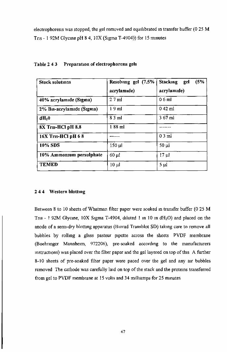
electrophoresis was stopped, the gel removed and equilibrated in transfer buffer (0 25 M
Tris -1 92M Glycine pH 8 4, 10X (Sigma T-4904)) for 15 minutes
Table 2 4 3 Preparation of electrophoresis gels
Stock solutions Resolving gel (7.5%
acrylamide)
Stacking gel (5%
acrylamide)
40% acrylamide (Sigma) 2 7 ml 0 6 ml
2% Bis-acrylamide (Sigma) 1 9 ml 0 42 ml
dH20 8 3 ml 3 67 ml
8X Tns-HCl pH 8.8 1 88 ml
16X Tris-HCl pH 6 8 0 3 ml
10% SDS 150 nl 50 |il
10% Ammonium persulphate 60 (il 17 |il
TEMED 10 ill 5 jal
2 4 4 Western blotting
Between 8 to 10 sheets of Whatman filter paper were soaked in transfer buffer (0 25 M
Tris - 1 92M Glycine, 10X Sigma T-4904, diluted 1 in 10 in dH20) and placed on the
anode of a semi-dry blotting apparatus (Biorad Transblot SD) taking care to remove all
bubbles by rolling a glass pasteur pipette across the sheets PVDF membrane
(Boehrmger Mannheim, 972206), pre-soaked according to the manufacturers
instructions) was placed over the filter paper and the gel layered on top of this A further
8-10 sheets of pre-soaked filter paper were paced over the gel and any air bubbles
removed The cathode was carefully laid on top of the stack and the protems transferred
from gel to PVDF membrane at 15 volts and 34 milliamps for 25 minutes
67
2 4 5 Development of Western blots by enhanced chemiluminescence (ECL)
ECL Western blotting detection is a highly sensitive, rapid, quantifiable, non-isotopic
method for detecting proteins immobilised on membranes The primary antibody is
located with the Horseradish-Peroxidase (HRP)-labelled species specific secondary
antibody The HRP-labelled molecule catalyses the oxidation of luminol, resulting in
the emission of light - chemiluminescence Enhanced chemiluminescence (ECL) is the
sustained emission of light provided by the inclusion of chemical enhancers in the HRP
catalysed oxidation of luminol, and gives approximately 1000 fold more light that the
oxidation of luminol alone
Following Western blotting, the filter paper was removed and the membrane blocked
for non-specific binding by incubating for 2 hours on a rocking platform with non-fat
milk (Marvel, Cadbury) After the blocking step was complete, the blot was nnsed once
in TBS, cut into strips (if required) and incubated with the primary antibody optimally
diluted in TB S/Tween overnight at 4°C on a rocking platform Mouse IgG diluted
1/5,000 in TBS/Tween served as a negative control The following day blots were
washed 3 times within 30 minutes with TBS 0 5% (v/v) Tween 20 Blots were then
incubated with a rabbit anti-mouse HRP-conjugated secondary antibody (Dako, P0447)
diluted 1/4,000 in TBS/Tween for 1 hour at room temperature on a rocking platform
The blots were washed as before, laid out on a glass plate covered in parafilm and
incubated with ECL reagents (Amersham RPN 2109) for 1 minute at room temperature
Or if used to enhance low signals, Super Signal Ultra (Pierce) was mixed according to
the instructions and applied to the blots for 5 minutes at room temperature Following
this the solution was tapped off, the blot laid between two sheets of cling film and
exposed to LUMI film, chemiluminescence film (Boehrmger Mannheim 166657) for
various time periods and processed using standard x-ray developing procedures Dried
film was aligned with pre-stained molecular markers for molecular weight
determination
68

2.4.6. Immunoprécipitation
Immunoprécipitation of specific proteins was carried out using a modification of a
protocol described in a study carried out by Masterson A, PhD Thesis 1998. This
protocol utilised a new cellular labelling and immunoprécipitation kit (Boehringer
Mannheim 1647652) with modifications. The principle of this procedure is based on
non-radioactive labelling of proteins or glycoproteins on intact cells (or in cell lysates)
with D-biotinoyl-e-aminocarporic acid-N-hydroxysuccinimide ester (biotin-7 NSH)
which covalently binds free amino groups (mainly lysine residues). These labelled
proteins can then be immunoprecipitated and detected using a biotin specific antibody.
However, in this study the protein was directly labelled with the primary antibody,
immunoprecipitated and then detected by Western blot analysis using the same antibody
to re-probe the blot.
Unless stated otherwise, all reagents were supplied in the immunoprécipitation kit.
Briefly, the modified procedure was carried out as follows; whole cell lysates were
prepared from cell pellets containing a maximum of 5x10⁷ cells/ml. Cell lysates (kept
on ice for the rest of the procedure) were prepared in lysis buffer and sonicated (as
outlined in Section 2.4.2.), followed by a 20 minute incubation period in lysis buffer (to
solubilise membrane protein. Cell lysates were then diluted 1:1 with dilution buffer and
pre-cleared by incubating with 2 changes of Protein A or G-linked Agarose beads
(depending on Ab isotype and species) for four hours each clearance. Following the
final incubation step, the primary antibody was optimally diluted in the cell lysates (15
fig/ml, mouse immunoglobulins at a concentration of 15 \ig/m\ served a negative
control) and incubated at 4° C over night on a rocking platform. The following day 50|il
of protein A/G-linked agarose beads (Boehringer Mannheim) were added to the cell
lysates and incubated for a minimum of 4 hours at 4°C on a rocking platform to
precipitate the antibody-antigen complex. Pelleted beads were then washed with two
changes of wash buffer 1, two changes of wash buffer 2 or 2 changes of a low salt
buffer (lOOmM NaCl) and one change of wash buffer 3. Following the removal of any
remaining supernatant, pellets were re-suspended in 60(il of loading buffer and heat
treated at 100°C for 3 minutes to disassociate the beads from the complex. After
69

centrifugation at 1 OOOrpm for 5 minutes, the supernatant was collected and analysed by
Western blotting, section 2.4.4. and blot development using ECL section 2.4.5.
2.5. Immunocytochemical analysis
2.5.1. Coating slides with poly-L-lysine
Washed microscope slides were coated with poly-L-Lysine hydrobromide (Sigma
P1274) following the procedure of Huang et al., (1983). Slides were stored at room
temperature for up to 1 month.
2.5.2 Preparation of cytospins
Cells from actively growing cultures were trypsinised (Section 2.2.2.), washed 3 times
in 10 minutes in PBS and diluted to a final concentration of lx l0 6 cells/ml in PBS.
100( 1 of the cell suspension was then applied to modified eppendorf component of a
cytofuge and spun onto Poly-L-lysine coated glass slides at 400 rpm for 4 minutes
Cytospins, consisting of 2 "sections" were allowed to air dry over night. Slides were
allowed to air dry and stored wrapped in tin foil at -20° C until required.
2.5.3. Immunofluorescence analysis of live cells
When immunofluorescence is carried out on live cells only cell surface components are
detected (Schachner et al., 1982). All reagents were kept at 4°C. Cell suspensions at
lx l0 6 cells/ml were prepared in PBS. 100fil of cell suspensions was placed in an
eppendorf tube mixed and incubated at 4°C for 30 minutes. Negative controls consisted
of cell suspension incubated with mouse immunoglobulins (Vector Laboratories, I-
2000), diluted 1/10,000 in PBS. Cells were washed 3 times over 12 minutes in PBS by
70
centrifuging at 2,000rpm in a microfuge. Cell pellets were re-suspended in 100 fil of
anti-mouse IgG FITC-labelled secondary antibody, diluted 1/50 in PBS, (Boehringer
Mannheim 814385) and incubated in the dark for 30 minutes at 4°C. Cell suspensions
were washed as described and following the final washing step the supernatant was
removed. Cells were then aspirated in 1 drop of Vectashield Mounting Medium (Vector
Labs Ltd. UK H1000). 20|nl of the suspension was placed on a microscope slide, covered
with a coverslip and the edges sealed with clear nail polish. Slides were viewed using a
Nikon phase contrast microscope fitted with an FITC filter.
2.5.4. Immunofluorescence analysis of fixed cells
Indirect immunofluorescence on fixed cells was carried out on cytospin preparations
(Section 2.9.2.). Cytospins were fixed in ice cold acetone (BDH) and allowed to air dry
at room temperature. The test antibody was applied to one cytospin section on the
cytospin while mouse IgG diluted 1/10,000 in PBS was applied to the other section. The
cytospins were incubated for 30 minutes at room temperature in a humidified chamber.
Slides were then washed in PBS 0.1% (v/v) Tween 20 3 times over 10 minutes 50|nl of
an anti-mouse FITC-labelled secondary antibody diluted 1/50 in PBS was applied to
each section and incubated at room temperature for 30 minutes Slides were washed as
before. Slides were mounted with 1 drop of Vecta Shield (Vector Laboratories)
mounting medium applied to the centre of the slide, and the coverslip and sealed with
clear nail varnish.
2.5.5. Immunocytochemical analysis using the StrepAB complex/HRP method
All immunocytochemical studies on cytospins or formalin fixed paraffin-embedded
archival material were performed following the method of Hsu et al.y (1981) using an
avidin-biotin (ABC) horseradish peroxidase (HRP) conjugated kit (Dako K337). The
use of diaminobenzidine (DAB) (liquid DAB solution prepared according to
instructions, Dako K3465) as a substrate for the peroxidase enzyme produced an
insoluble brown precipitate which was indicative of primary antibody activity.
71
Frozen cytospm preparations were allowed to equilibrate to room temperature
(approximately 10-15 minutes) Slides were then fixed in ice cold acetone for 4 to 10
minutes, depending on the antibody (all antibodies worked when fixed for 10 minutes),
and allowed to air dry A wax pen (Dako S2002) was used to encircle each "section" to
contain the various solutions mvolved Cytospins were then incubated with 0 3% (v/v)
hydrogen peroxide (BDH) in methanol (BDH) to quench endogenous peroxidase
activity followed by a brief rinse in distilled H2O and incubation in lxTBS for 5
minutes Following this cytospins were incubated in 20% (v/v) normal rabbit serum
(Dako X0902), in lxTBS for 20 minutes at room temperature to block any non-specific
binding of the secondary antibody (a rabbit host) The excess serum was tapped off and
the optimally diluted primary antibody was applied to one of the cytospm sections while
control mouse immunoglobulins (Vector Laboratories) served as a negative control
Slides were placed in a humidified atmosphere and incubated at room temperature for 2
hour or at 4°C overnight Following primary antibody incubation, cytospins were
washed 3 times over 15 minutes in lxTBS, 0 1% (v/v) Tween 20 (TBS/Tween) and then
incubated with an appropriate biotinylated secondary antibody (anti-rat (Dako E0468)
or anti-mouse (Dako E0354)) diluted 1/300 in TBS/Tween, for 30 minutes at room
temperature Following this cytospins were washed as before in TBS/Tween The Strep
AB complex (Dako K337 prepared according to manufacturers instructions) was
applied to the cytospm and incubated at room temperature for 30 minutes Following the
washing steps, liquid DAB (Dako K3565 prepared according to the manufacturers
instructions) was applied to each "section" of the cytospm for 7-10 minutes at room
temperature Cytospins were nnsed m tap water, terminating the reaction,
counterstained in Harris' haematoxylin (Sigma Diagnostics HHS-16) (15 seconds),
differentiated in 1% (v/v) hydrochloric acid in 70% IMS (15 seconds) and blued in
Scotts tap water (Clintech, Clacton-on-Sea, Essex, England )(10 seconds) rinsing in
water between each step Alternatively cells were counterstained with methyl green
(Vector Laboratories) Cytospins were then dehydrated in graded alcohols ( 2 x 3
minutes in 70%, 90% and 100% (v/v) IMS in dH20 and cleared in xylene (BDH, 2
changes of 5 minutes each) Cytospins were mounted in DPX mounting medium (BDH)
and allowed to dry overnight Cytospins were viewed under a light microscope
72

J
3.0 RESULTS.
73
3 1 CHARACTERISATION OF MRP ISOFORM MAbs AND MRP
ISOFORM TRANSFECTED CELL LINES
Various MAbs which detect specific isoforms of the MRP family were a kind gift from
Mr George Scheffer of Professor Rik Scheper’s Laboratory, Department of Pathology
and Oncology, Free University Hospital, 1081 HV Amsterdam (see Table 3 1 1 ) The
MRPrl antibody included is the well characterised MRP1 specific rat MAb (Flens et a l ,
1996) The ovarian 2008 parental cell lines, which were transfected with the MRP1,
MRP2 or MRP3 cDNAs, were also a kind gift from Mr George Scheffer (see Table
3 1 2 ) Permission to use the MRP3 transfected 2008 cell lme was kmdly provided by
Marcel Kool, Academic Medical Center, Laboratory of Neurozintuigen, Room K2-212,
Meibergdreef 9, 1105 AZ Amsterdam Both the MRP isoform MAbs and MRP isoform
transfected cells were characterised by Western blot and immunocytochemical analysis
(see following results) These MAbs and cell lines were further used to characterise the
specificity of the MRP1 antibodies produced in this thesis
74
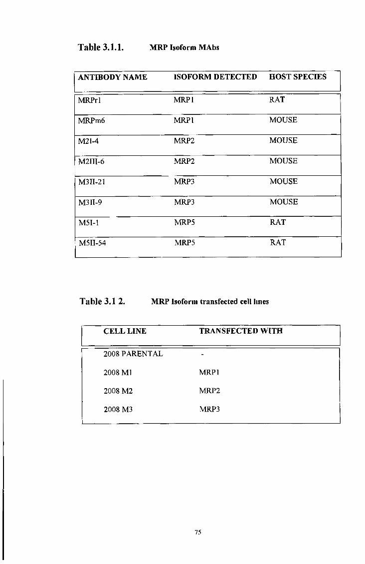
Table 3.1.1. MRP Isoform MAbs
ANTIBODY NAME ISOFORM DETECTED HOST SPECIES
MRPrl MRP1 RAT
MRPm6 MRP1 MOUSE
M2I-4 MRP2 MOUSE
M2III-6 MRP2 MOUSE
M311-21 MRP3 MOUSE
M3II-9 MRP3 MOUSE
M5I-1 MRP5 RAT
M5II-54 MRP5 RAT
Table 3.1 2. MRP Isoform transfected cell lines
CELL LINE TRANSFECTED WITH
2008 PARENTAL -
2008 Ml MRP1
2008 M2 MRP2
2008 M3 MRP3
75
3 2 CHARACTERISATION OF THE MRP1 SPECIFIC MAb, MRPrl
The expression of MRP 1 in various cell lines was examined by Western blotting and
immunocytochemical techniques Known MRP1 over-expressing drug-selected cell
lines and their parental MRP1 negative cell lines (see Table 3 2 1) were compared with
the various MRP isoform transfected cell lines (previously described in section 3 1 )
MRP1 cDNA-transfected SF9 insect cell membranes, which express the unglycosylated
form of MRP1, were also examined (see Table 3 2 2 ) These membranes were a kind
gift from Dr Balazs Sarkadi, The Institute of Enzymology, Biological Research Center,
Hungarian Academy of sciences, H-1113 Budapest, Hungary After characterisation
these cell lines and membranes were then used for controls during antibody screening
and characterisation of the chosen antibodies
Table 3.2.1
Drug-selected MRP1 over-expressing cell lines and their corresponding parental
cell lines
PARENTAL CELL LINE
(VERY LOW MRP1 EXPRESSION)
DRUG SELECTED CELL LINE
(MRP1 OVER-EXPRESSION)
HL60S
(Leukemia cell line)
HL60ADR
COR-L23 S
(lung carcinoma cell line)
COR-L23R
76

Table 3.2.2
MRPl cDNA transfected insect cells
CELL LINE TRANSFECTED WITH
Sf9 insect cells MRPl cDNA
Western Blot analysis of MRPl expression with the rat monoclonal MRPrl
Western Blot analysis of MRPl expression with the rat monoclonal MRPrl showed that
MRPl is over-expressed as a 190kD protein in the drug selected HL60ADR and COR-
L23R cell lmes No MRPl expression was observed in the parental HL60S cell line and
a very faint expression was observed in the parental COR-L23S cell lme (see Figures
3 2 1 and 3 2 2 ) The unglycosylated form of MRPl was observed as a 150kD band in
the MRPl transfected SF9 insect membranes (see Figure 3 2 3 ) Strong MRPl over-
expression at 190kD was apparent in the MRPl transfected ovarian cell lme, 2008
MRPl A very low or faint 190kD band of expression was seen m the parental, MRP2
and MRP3 transfected 2008 cell lines (see Figure 3 2 4 ) The faint expression of
MRPl in some sensitive and un-transfected lines is probably due to an intrinsic basal
level of MRPl present prior to drug selection or transfection
77

&Q< (73o o'sO 'O
190kD
Lane ► l 2
Figure 3.2,1. Detection of MRP1 with the rat monoclonal antibody, MRPrl
MRP1 is detected at 190kD m the drug-resistant HL60ADR cell lme MRP1 is not
detected m the drug-sensitive HL60S cell line
78

P4c o<Nh-1
ip 4Ou
i nm<Nh -l
I
OO
>T
190kD
Lane ► \ 2
Figure 3.2.2 Detection of MRP1 with the rat monoclonal antibody, MRPrl MRP1 is
detected at 190kD in the drug-resistant COR-L23R cell line MRP1 is very weakly detected
in the drug-sensitive COR-L23S cell line
79
190 kD150kD
Figure 3.2.3. Detection of MRP1 with the rat monoclonal antibody, MRPrl
MRP1 is detected at 190kD m the COR-L23R cell line, faintly detected in the COR-
L23S cell Ime and detected at 150kD m the MRP 1-transfected SF9 insect
membranes
80
P4c o<NhJ
I
Oo
GOCO<N
IPinOO
aa>
SPh00oo<N
<3(N cnS & &
2 s so coo<N
00 00 o o o o( N ( N
190kD
Figure 3.2.4. Detection of MRP1 with the rat MAb, M R Prl. MRP1 is detected at
190kD in the drug-resistant COR-L23R cell line and the MRP1-transfected 2008 MRP1 cell
line. MRP1 is very weakly detected at 190kD in the drug-sensitive COR-L23S cell line, the
parental 2008 cell line, the 2008 MRP2 and 2008 MRP3 -transfected cell lines.
81
lmmunocytochenncal analysis of MRP1 expression in various cell lines with the rat
monoclonal MRPrl
Immunocytochemical analysis of MRP1 expression with the rat monoclonal MRPrl
showed that MRP1 is over-expressed in the plasma membrane of the drug-selected
HL60ADR and COR-L23R cell lines Very low basal levels of MRP1 expression were
observed m the parental HL60S and COR-L23S cell lines (see Figures 3 2 5 and 3 2 6 )
Immunocytochemical analysis of the 2008 parental cell line and MRP2 and MRP3
isoform transfectants with the rat monoclonal antibody MRPrl, showed intense staining
of the plasma membrane of the 2008 MRP1 isoform transfected cell line MRPrl
detects a basal level of staining in the 2008 parental and 2008 MRP2 and MRP3 isoform
transfectants (see Figure 3 2 7 )
82
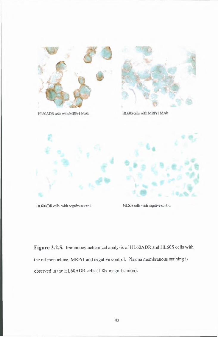
HL60ADR cells with negitive control HL60S cells with negrtive control
F ig u re 3.2.5. Immunocytochemical analysis of HL60ADR and HL60S cells with
the rat monoclonal MRPrl and negative control. Plasma membranous staining is
observed in the HL60ADR cells (lOOx magnification).
83
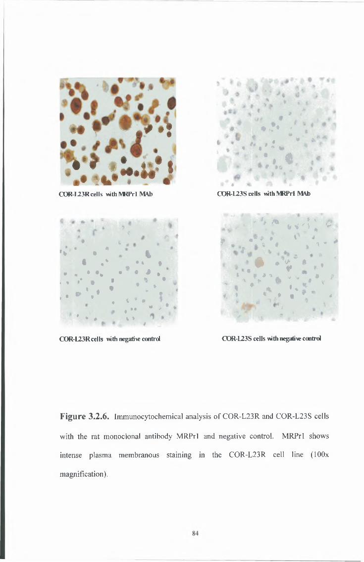
» T * . * • !
i » * • ! » , » ? | V H » . M
COR-L23R cells withlVRPrl MAb
* ** « A
'* • %• •* . ** *
• » * * * * * * •. , •* t * * *
» * *, . » . * *
t
• *♦
« *%
m- *« <*♦% *
up?
> * .* * #
OOR-L23R cells with negatile control
« * : •v t* ■■ «> ♦ ^ i ;i « • * t
* I* *> p i
• i t# ♦ * » « 4%P P #-'M w
« < Ét a ** COR-L23S cells vuth IVRPrl M \b
■y K m * K- K M ; ^ -
M, *W # * *^ - - * « * * * *
• - V * , '* • « -* « V * A * w # • 11
' « ♦ * • * * *4 * • %* * # *
OOR-L23S cells with negative control
Figure 3.2.6. Immunocytochemical analysis of COR-L23R and COR-L23S cells
with the rat monoclonal antibody MRPrl and negative control. MRPrl shows
intense plasma membranous staining in the COR-L23R cell line (lOOx
magnification).
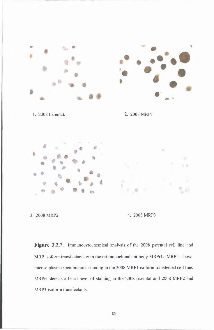
%
*5# W w ^ ^
* * # •
. » ♦ • _ .
1. 2008 Parental. 2. 2008 MRP 1
# * % »# # .
^ f t £# $ “ & 0 \
^ ^ ^ ^ m
3. 2008 MRP2 4. 2008 MRP3
F ig u re 3.2.7. Immunocytochemical analysis of the 2008 parental cell line and
MRP isoform transfectants with the rat monoclonal antibody MRPrl. MRPrl shows
intense plasma-membranous staining in the 2008 MRP1 isoform transfected cell line.
MRPrl detects a basal level of staining in the 2008 parental and 2008 MRP2 and
MRP3 isoform transfectants.
85

Heat treatment studies on the epitope recognised by the MRPrl antibody by
western blot analysis
Heat treatment studies in the MRP1 over-expressing drug selected COR-L23R cell line
and the MRP 1-transfected cell lme 2008 MRP1 prior to electrophoresis (Refer to
section 2 4 2 for usual sample preparation prior to electrophoresis), showed that heat
treatment for 3 minutes @ 100°C destroys the MRP1 epitope recognised by the MRPrl
antibody in the drug selected COR-23R cell line but not the MRP 1-transfected 2008
MRP1 cell lme (see Figure 3 2 8 and 3 2 9 ) This result may be due to a
conformational difference between the drug-selected and the transfected MRP1 protein
86
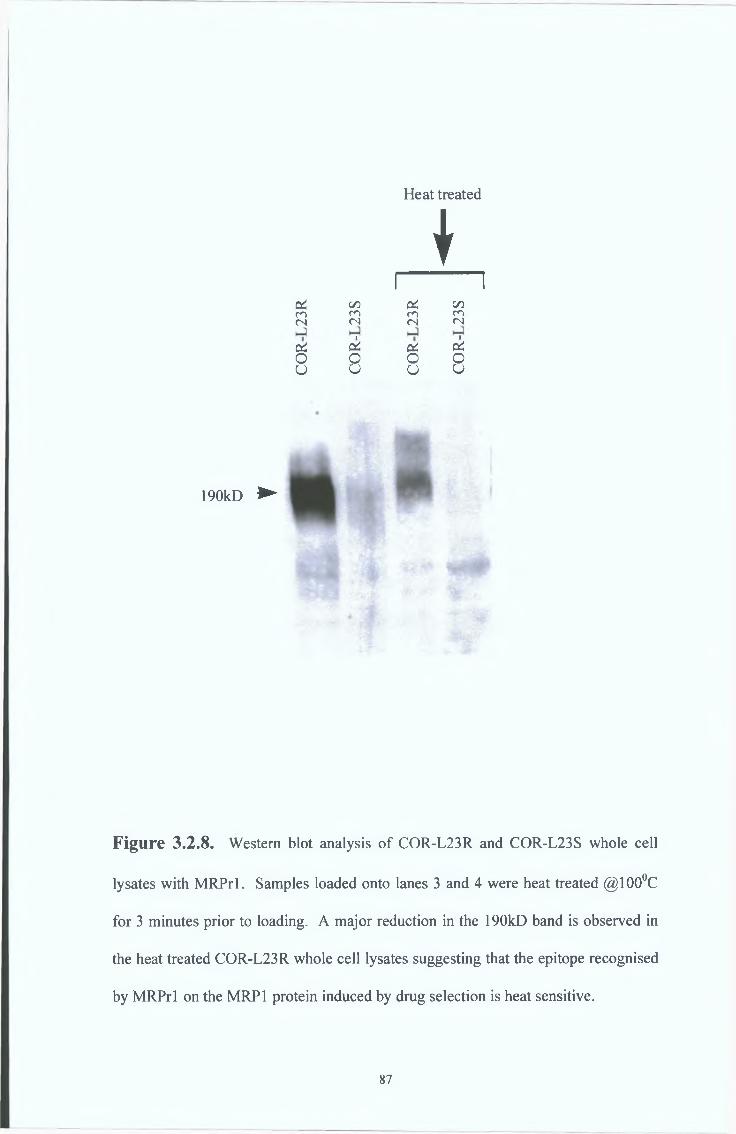
Heat treated
♦I----------------1
p! C/5 PC C/5CO CO CO CO<N <N (N (N
1 1p! pi Pi PiO o o Oo o u u
190kD ►
Figure 3.2.8. Western blot analysis of COR-L23R and COR-L23S whole cell
lysates with MRPrl. Samples loaded onto lanes 3 and 4 were heat treated @100°C
for 3 minutes prior to loading. A major reduction in the 190kD band is observed in
the heat treated COR-L23R whole cell lysates suggesting that the epitope recognised
by MRPrl on the MRP1 protein induced by drug selection is heat sensitive.
87

"T3<DcS<D
"T3<L>
<DVh
190kD
F ig u re 3.2.9. Heat treatment studies in the MRP1 over-expressing drug selected
COR-L23R cell line and the MRP1 transfected cell line 2008 MRP1, by Western
blot analysis with the rat MAb, MRPrl. Heat treatment at 100°C for 3 minutes
destroys the epitope recognised by the MRPrl antibody in the intrinsically expressed
MRP1 protein but not the MRP1 transfected 2008 MRP1 cell line. This result may
be due to a conformational difference of the intrinsic and the transfected protein.
88
3 3 C H A R A C T E R I S A T I O N O F T H E M R P 2 SPECIFIC MAbs, M2I-4 and
M2ITI-6
Immunocytochemical analysis
A number of cell lines, including the 2008 MRP2-transfected cell line were examined
by immunocytochemical analysis with the MRP2 specific antibodies M2I-4 and M2III-
6 The mouse monoclonal antibody M2I-4 weakly stained all the cell lines examined
(see Figure 3 3 1) However, immunocytochemical analysis of the 2008 parental cell
line and MRP isoform transfectants with the mouse monoclonal antibody M2III-6
showed that MAb M2III-6 strongly stained the 2008 MRP2 cell line only (see Figure
3 3 2 ) These results show that MRP2 protein is expressed in the 2008 MRP2
transfected cells While the M2I-4 is unsuitable for immunocytochemical analysis, the
M2III-6 MAb is highly specific for the MRP2 protein expressed in the 2008 MRP2
transfected cells and does not cross-react with the MRP1 or MRP3 2008 transfectants or
the COR-L23RMRP1 over-expressing cell line
89
6 O
V #
#
* •% N i
1 « •
1. COR-L23R
4 % *. *
2. COR-L23S
mi
3. 2008 Parental
m
? * » * « » * •i
• «di ^
4. 2008 MRP1
f
«5. 2008 MRP2
* #- S ;fe *„ w «t -» &
6. 2008 MRP3
Figure 3.3.1. Immunocytochemical analysis of the 2008 parental cell line and
MRP isoform transfectants with the mouse monoclonal antibody M2I-4. MAb M2I-
4 stained all cell lines weakly.
90
1. COR-L23R
•- «#<«* 9
* #• * * *
* m r* * * » 4 •« : ' ♦ . *
• # • ®
2. COR-L23S
% «• *
* v - • «* 4 # • •
I % % *• •
* H ! .3. 2008 Parental
r * * •
• I 0
I 4 *
4. 2008 MRP1
«
• % *
* %
• 4f * #
#
5. 2008 MRP2 6. 2008 MRP3
F ig u re 3.3.2. Immunocytochemical analysis of the 2008 parental cell line and
MRP isoform transfectants with the mouse monoclonal antibody M2III-6. MAb
M2III-6 strongly stained the 2008 MRP2 cell line.
91

Western blot analysis
A number of cell lines, including the 2008 MRP2-transfected cell lme were examined
by Western blot analysis with the MRP2 specific antibody M2III-6 The M2III-6 MAb
was used because of the specific results achieved with this MAb in the MRP2
immunocytochemical study (Figure 3 3 2 ) Analysis of the 2008 parental cell lme and
MRP isoform transfectants with the M2III-6 MAb showed that it strongly reacted with a
190kD band in the 2008 MRP2 cell line (see Figure 3 3 3 ) These results show that
MRP2 protein is expressed m the 2008 MRP2 transfected cells and that MRP2
expression can be detected with the M2III-6 MAb by Western blot analysis The M2III-
6 MAb is highly specific for the MRP2 protein and does not cross-react with the MRP1
or MRP3 2008 transfectants or the COR-L23R MRP1 over-expressing cell lme
92
i§%Ph00oo
Xoo
§ §Xoo
£§00oo
e*r^i(S
ciOU
C/3fO
ciO
P4
5 C/3© OV© sO
JX X
190KD
Figure 3,3.3. Detection of MRP2 by Western blot analysis with the MAb, M2III-
6 MRP2 is detected at 190kD in the MRP2-transfected cell line, 2008 MRP2
MRP2 is not expressed in the 2008 parental, MRP1 -transfectant, MRP3-transfectant
or the COR-L23R, COR-L23S, HL60ADR and HL60S cell lines
93

3 4 CHARACTERISATION OF THE MRP3 SPECIFIC MAbs, M311-21 and
M3H-9
Immunocytochemical analysis
A number of cell lines, including the 2008 MRP3 transfected cell lme were examined
by immunocytochemical analysis with the MRP3 specific M311-21 and M3II-9 The
mouse monoclonal antibody M311-21 showed granular plasma-membranous staining in
a sub-population of the 2008 MRP3 cell line (see Figure 3 4 1) The mouse
monoclonal antibody M311-9 also showed granular plasma-membranous staining in a
sub-population of the 2008 MRP3 cell lme (see Figure 3 4 2 ) These results show that
MRP3 protein is expressed in a subpopulation of the 2008 MRP3 transfected cells The
MRP3 protein was not found to be expressed in any of the other cell lines exammed
Both of these MRP3 MAbs were found to be suitable for immunocytochemical analysis
and did not cross-react with the MRP1 or MRP3 2008 transfectants or the COR-L23R
MRP1 over-expressing cell lme, indicating that they are MRP3 specific
94
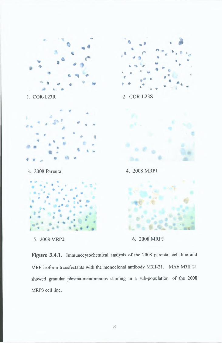
o
1. COR-L23R
a r " * . * 4 * *i £ * <*
• V —
t S >* t ® *
r •* * ♦%
k ♦
« t
2. COR-L23S
•V 9&
<**) 9 <r* <#(P *<r<• ! *
#^ ft
0 • 4« ♦
c • ^ •
% •
♦ 1£- 0
* 4 -
3. 2008 Parental 4. 2008 MRP1
» - <* ^ tf
1 K* %
%% M
, 0 ‘t* • *
0 # • i * *% • ^ %A
5. 2008 MRP2 6. 2008 MRP3
Figure 3.4.1. Immunocytochemical analysis of the 2008 parental cell line and
MRP isoform transfectants with the monoclonal antibody M3II-21. MAb M3II-21
showed granular plasma-membranous staining in a sub-population of the 2008
MRP3 cell line.
95
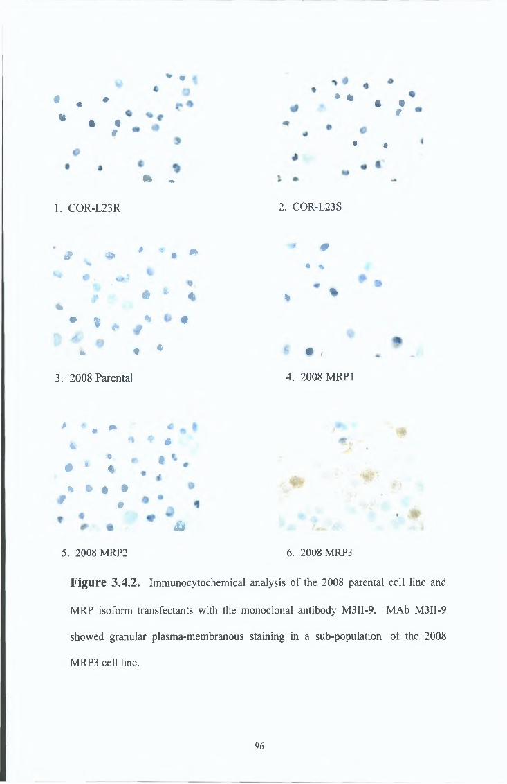
* t
• 4 «r
* <1 •
m -
1. COR-L23R
* % 4 Ir
• i
2. COR-L23S
# * 4* •# * - * w *• %
# # ^ % % ♦%
« I % #’ i J _
• ^ ^ « * # • 0 f>
3. 2008 Parental 4. 2008 MRP 1
# * • *V
9* * «
% « « ** e
5. 2008 MRP2
#
t
&
6. 2008 MRP3
Figure 3.4.2. Immunocytochemical analysis of the 2008 parental cell line and
MRP isoform transfectants with the monoclonal antibody M3II-9. MAb M3II-9
showed granular plasma-membranous staining in a sub-population of the 2008
MRP3 cell line.
96
Western blot analysis
A number of cell lines, including the 2008 MRP3 transfected cell line were examined
by Western blot analysis with the MRP3 specific antibody M311-21 The M311-21 MAb
was used because of its characterisation in publications (Kool et a l , 1999a) Analysis
of the 2008 parental cell line and MRP isoform transfectants with the M311-21 MAb
showed that it strongly reacted with a 190kD band in the 2008 MRP3 cell line (see
Figure 3 4 3 ) These results show that MRP3 protein is expressed m the 2008 MRP3
transfected cells and that MRP3 expression can be detected with the M311-21 MAb by
Western blot analysis The M311-21 MAb is highly specific for the MRP3 protein and
does not cross-react with the MRP1 or MRP2 2008 transfectants or the COR-L23R
MRP1 over-expressmg cell line
97
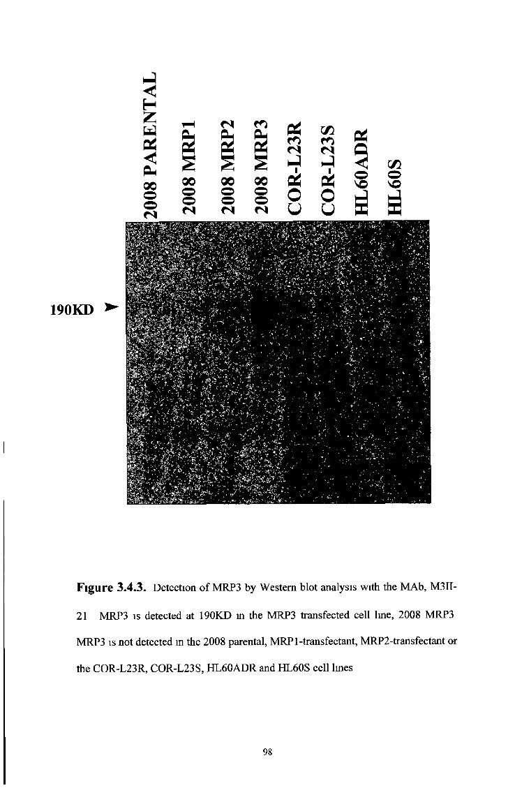
X X Xo o oo o o
P4
I f lo ©
190KD
Figure 3.4.3. Detection of MRP3 by Western blot analysis with the MAb, M3II-
21 MRP3 is detected at 190KD in the MRP3 transfected cell line, 2008 MRP3
MRP3 is not detected in the 2008 parental, MRPl-transfectant, MRP2-transfectant or
the COR-L23R, COR-L23S, HL60ADR and HL60S cell lines
98
3 5 IMMUNOGENS
3 5 1 Native protein
Native protein can be a valuable source for the production of antibodies which are
required to recognise the native protein The most obvious source of native protein is
the protein expressing cell itself The COR-L23R MRP1 over-expressing cells are a
rich source of the native MRP1 protein Whole cell lysate preparations or cell
membranes partially purified by centrifugation techniques may be used as immunogens
The COR-L23R cells were used as sources for whole cell lysates and cell membranes
for immunisation
3 5 2 Synthetic peptides and Fusion proteins
Short synthetic peptides or Fusion proteins expressed in vector systems can be produced
for use as immunogens These proteins are linear and may not possess the original
conformation of the native protein However, a multitude of techniques can help to
predict linear regions on the native protein Therefore, these proteins are another
valuable source of immunogen in certain cases The 3 short peptides used in this thesis
were chosen from the predicted model proposed by Bakos et a l , 1996 (see Figure
12 1 1 ) The locations of each of these 3 peptides on the MRP1 protein are shown in
Diagram 3 5 1 Peptide 1 (amino acids 1520 - 1531) is situated mtracellularly in the C-
terminus Peptide 2 (ammo acids 235-246) and peptide 3 (amino acids 246-260) are
both situated in the cytoplasmic loop separating transmembrane domam 0 (TMO) and
transmembrane domam 1 (TM1) in the N-terminus
99
The locations of the two Fusion Proteins used in this thesis are shown in Diagram 3 5 2
Fusion Protein 1 (amino acids 1294-1531) is situated mtracellularly in the C-terminus of MRP1
Fusion Protein 2 (ammo acids 647-912) is situated mtracellularly in the N-terminus of MRP 1
MRPrl, the MRP 1-specific rat MAb was produced by immunising with a Fusion
Protein (Flens et a l , 1996) Diagram 3 5 3 shows the location of the Fusion Protein
used in the of production MRPrl and the mapped epitope it recognises The fusion
protein incorporates ammo acids 192-360 of human MRP1 This region is mainly
situated in the large cytoplasmic loop separating transmembrane domain 0 (TMO) and
transmembrane domain 1 (TM1) in the N-terminus (see Diagram 3 5 4 , Region 1) The
epitope recognised by MRPrl has been mapped to amino acids 238 to 247 This
epitope is similar to the region encompassed by one of the short synthetic peptides,
Peptide 2, used in this thesis as an immunogen (see Diagram 3 5 1)
100
Peptide 2 Peptide 3* % * »
Peptide 1
Diagram 3.5.1. Presumed location of immunisation Peptides 1, 2 and 3 based on
the model of Bakos et ai, 1996. Peptide 1 (amino acids 1520 - 1531) is situated
intracellularly in the C-terminus. Peptide 2 (amino acids 235-246) and peptide 3
(amino acids 246-260) are both situated in the cytoplasmic loop separating
transmembrane domain 0 (TMO) and transmembrane domain 1 (TM1) in the N-
terminus.

Fusion Protein 2 Fusion Protein 1# + # # « «
Diagram 3.5.2. Presumed location of fusion Proteins 1 and 2 based on the model
of Bakos et al., 1996. Fusion Protein 1 (amino acids 1294-1531) is situated
intracellularly in the C-terminus of MRP 1. Fusion Protein 2 (amino acids 647-912)
is situated intracellularly towards the N-terminus of MRP 1.
Epitope
Region 1 # # # •
Diagram 3.5.3. Presumed location of the Fusion Protein (including amino acids
192-360 of human MRP1) used to produce the rat monoclonal antibody, MRPrl,
based on the model of Bakos et al., 1996. This region is mainly situated in the large
cytoplasmic loop separating transmembrane domain 0 (TMO) and transmembrane
domain 1 (TM1) in the N-terminus (see Region 1). The epitope recognised by
MRPrl has been mapped to amino acids 238 to 247.

3 6 POLYCLONAL ANTIBODY RESULTS
Immunisations
In order to generate polyclonal antibodies for the detection of MRP 1, New Zealand
White Rabbits underwent an immunisation regime with short synthetic peptides (see
Diagram 3 5 1 ) taken from the deduced amino acid sequence of human MRP1 The
resultmg polyclonals were designated Polyclonal 1, Polyclonal 2 and Polyclonal 3
Screening of Polyclonal Serum
Serum collected from test bleeds after each immunisation were tested primarily by
Western blotting on HL60ADR and HL60S whole cell lysates Further characterisation
of each polyclonal was carried out on COR-L23R and COR-L23S cells and SF9 insect
membranes expressing various fragments of MRP 1 and controls (see Table 3 6 1)
Western blotting results
All three polyclonals were found to be suitable for Western blot analysis and reacted
strongly with a 190kD band in the MRP1 over-expressing, drug-selected cell line,
HL60ADR, and weakly or not at all with the parental HL60S cell Ime (see Figures
3 6 1, 3 6 3 and 3 6 5 ) These results strongly suggested that the polyclonals were
reacting with MRP 1
Polyclonal 1, 2 and 3 detected a 150kD band in human MRP 1-expressing SF9 insect
membranes and a shift of the 190kD band to a 150kD band in tumcamycin treated
HL60ADR cells None of the polyclonals reacted with WT-MDR expressing SF9 insect
membranes All three polyclonals also reacted with a 140kD band m SF9 insect
104
membranes expressing DeltaMRPl (Àl), containing the MRPrl rat antibody epitope
(ammo acids 192 - 360), while only polyclonal 1 detected a 120kD band in SF9 insect
membranes expressing DeltaMRP2 (À2), which does not contain the MRPrl epitope
(see Figures 3 6 2 ,3 6 4 and 3 6 6 )
105
the MRP1 Polyclonal antibodies Carried out by Emese Sinkoe from The Institute of
Enzymology, Biological Research Center, Hungarian Academy of sciences, H-l 113
Table 3 6 1 Explanation of various fragments used in the further characterisation of
Budapest, Hungary
106

One New Zealand White rabbit was immunised with Peptide 1 and is summarised in
Table 3.6.2.
POLYCLONAL 1.
• AA 1520 - 1531 (Internal peptide / C-terminus).
• Sequence : CALFYSMAKDAGLV + KLH
IMMUNISATION REGIME : IN VIVO (NEW ZEALAND WHITE RABBITS).
SCREENING SYSTEM : WESTERN BLOTTING ON HL60ADR AND HL60S
CELLS.
RESULTS : IDENTIFIES 190kD ON RESISTANT , MRP POSITIVE HL60ADR
CELLS WITH NO STAINING ON SENSITIVE CELLS, HL60S (see Figure 3.6.1.).
TABLE 3.6.2.
Primary screening.
Primary screening of Polyclonal 1 by Western blot analysis on HL60ADR and HL60S
cell lysates identified a 190kD band in HL60ADR cells but not in HL60S cells (see
Figure 3.6.1.).
Characterisation.
Further screening of Polyclonal 1, raised to the C-terminus of MRP 1, was carried out by
Western Blot analysis using various MRP1 fragments and controls (see Figure 3.6.2.).
As expected, Polyclonal 1 did not react with (TMO), HL60S cell lysates or WT-MDR.
Polyclonal 1 detected MRP1 at 190kD in HL60ADR cell membranes, at 150kD in
107
tunicamycin treated cells and human MRP1 expressed in SF9 insect cells and at 140kD
and 120kD respectively m A1 and A2 MRP 1/2 which both express the C-terminus of
MRP1
108

FIGURE 3.6.1. Western blot analysis of HL60ADR and HL60S cells with
Polyclonal 1 Polyclonal 1 recognises a 190kD band in the MRP1 over-expressing
cell line, HL60ADR but not the parental drug-sensitive HL60S cell line
109
so¡H
O
osH
OïO
<N < < 1
H+ §
H§U
220 kD
97 kD
66kD
46kD
30kD
Lane 1 2 3 4 5 8 9
FIGURE 3.6.2. Western blot analysis of various MRP1 fragments and controls (see
Table 3.6.1. for description of controls) with Polyclonal 1. As expected, Polyclonal 1 does
not react with (TM O ), CONTROL or W T-M D R because these controls do not contain any
MRP1. Polyclonal 1 detects a 190kD band in MRP (fully glycosylated, full length MRP1),
a 150kD band in MRP + TU N IC A M Y C IN and hM RPl (unglycosylated form expressed at
150kD), and at 140kD and 120kD respectively in A1 and A2 (unglycosylated full length
MRP1, resulting in lower molecular weight detection. The M RPrl fusion protein region is
deleted in A2.). A ll glycosylated MRP1 fragments were expressed in human HL60ADR
cells (HL60) and all unglycosylated fragments were expressed in SF9 insect membranes
(SF9) which are unable to undergo glycosylation.
110

One New Zealand White rabbit was immunised with Peptide 2 and is summarised in
Table 3.6.3.
POLYCLONAL 2.
• AA 235 - 246 (Internal peptide (originally thought to be external) / N-terminus).
• Sequence : PLEGSDLWSLNK + BSA
IMMUNIZATION REGIME : IN VIVO (NEW ZEALAND WHITE RABBITS).
SCREENING SYSTEM : WESTERN BLOTTING ON HL60ADR AND HL60S
CELLS.
RESULTS : IDENTIFIES 190kD ON RESISTANT , MRP POSITIVE HL60ADR
CELLS WITH NO STAINING ON SENSITIVE CELLS, HL60S (see Figure 3.6.3.).
Primary screening.
Primary screening of Polyclonal 2 by Western blot analysis on HL60ADR and HL60S
cell lysates identified a 190kD band in HL60ADR cells but not in HL60S cells (see
Figure 3.6.3.).
Characterisation.
Further screening of Polyclonal 2, raised to a region in the N-terminus of MRP1, was
carried out by Western blot analysis using various MRP1 fragments and controls (see
Figure 3.6.4.). Polyclonal 2 did not detect any protein in HL60S cell lysates, WT-MDR
or A2 MRP2 (MRPrl fusion protein region deleted). As expected, Polyclonal 2
detected a 30kD band in (TMO), a 190kD band in HL60ADR cell membranes, a 150kD
band in tunicamycin treated cells and human MRP1 expressed in SF9 insect cells and a
140kD band in A1 MRP1 which does express the MRPrl fusion protein region.
i l l

FIGURE 3.6.3. Western blot analysis of HL60ADR and HL60S cells with
Polyclonal 2 Polyclonal 2 recognises a 190kD band in the MRP1 over-expressing
cell line, HL60ADR but not the parental drug-sensitive HL60S cell line
112
sy
FIGURE 3.6.4. Western blot analysis of various MRP1 fragments and controls (see
Table 3.6.1. for description o f controls) with Polyclonal 2. As expected, Polyclonal 2 does
not detect any protein in CONTROL, W T-M D R or A2 (M RPrl fusion protein region
deleted. This deleted region contains the amino acids to which Polyclonal 2 was raised). As
expected, Polyclonal 2 detected a 30kD band in (TM O ) which contains the region to which
Polyclonal 2 was raised in the N terminus. Polyclonal 2 also detected a 190kD band in MRP
(fully glycosylated, full length MRP1), a 150kD band in MRP + TUNIC A M YC IN and
hMRPl (unglycosylated form expressed at 150kD), and a 140kD band in A1
(unglycosylated full length MRP1, resulting in lower molecular weight detection). All
glycosylated MRP1 fragments were expressed in human HL60ADR cells (HL60) and all
unglycosylated fragments were expressed in SF9 insect membranes (SF9) which are unable
to undergo glycosylation.
113

One New Zealand White rabbit was immunised with Peptide 3 and is summarised in
Table 3.6.4.
POLYCLONAL 3.
• AA 246 - 260 (Internal peptide / N-terminus)
• Sequence : KEDTSEQVVPVLVKN + KLH
IMMUNIZATION REGIME : IN VIVO (NEW ZEALAND WHITE RABBITS).
SCREENING SYSTEM : WESTERN BLOTTING ON HL60ADR AND HL60S
CELLS.
RESULTS : IDENTIFIES 190kD ON RESISTANT , MRP POSITIVE HL60ADR
CELLS WITH REDUCED STAINING ON SENSITIVE CELLS, HL60S (see Figure
3.6.5.).
Table 3.6.4.
Primary screening.
Primary screening of Polyclonal 3 by Western blot analysis on HL60ADR and HL60S
cell lysates identified a 190kD band in HL60ADR cells with reduced staining in HL60S
cells (see Figure 3.6.5.).
Characterisation.
Further screening of Polyclonal 3, raised to the N-terminus of MRP 1, was carried out by
Western blot analysis using various MRP1 fragments and controls (see Figure 3.6.6.).
Polyclonal 3 did not detect any protein in HL60S cell lysates, WT-MDR or A2 MRP2
(MRPrl fusion protein region deleted). As expected, Polyclonal 3 detected a 30kD
band in (TMO), a 190kD band in HL60ADR cell membranes, a 150kD band in
tunicamycin treated cells and human MRP1 expressed in SF9 insect cells and a 140kD
band in A1 MRP1 which does express the MRPrl fusion protein region.
114

ZAOVO
£
&Q<os£
190 kD
v
Lane ► 1 2
FIGURE 3,6.5. Western blot analysis of HL60S and HL60ADR cells with
Polyclonal 3 Polyclonal 3 recognises a 190kD band m the MRP1 over-expressing
cell line, HL60ADR Polyclonal 3 shows reduced staining but a high background in
the parental drug-sensitive HL60S cell line
115
sü
Lane ► 1 2 3 4 5 6 7 8 9
FIGURE 3.6.6. Western blot analysis of various MRP1 fragments and controls (see
Table 3.6.1. for description o f controls) with Polyclonal 3. As expected, Polyclonal 3 does
not detect any protein in CONTROL, W T-M D R or A2 (M RPrl fusion protein region
deleted. This deleted region contains the amino acids to which Polyclonal 3 was raised). As
expected, Polyclonal 3 detected a 30kD band in (TM O ) which contains the region to which
Polyclonal 3 was raised in the N terminus, a 190kD band in MRP (fully glycosylated, full
length MRP1), a 150kD band in MRP + TUNIC A M Y C IN and hMRPl (unglycosylated
form expressed at 150kD), and a 140kD band in A1 (unglycosylated full length MRP1,
resulting in lower molecular weight detection). A ll glycosylated MRP1 fragments were
expressed in human HL60ADR cells (HL60) and all unglycosylated fragments were
expressed in SF9 insect membranes (SF9) which are unable to undergo glycosylation.
116
MOUSE MONOCLONAL ANTIBODY PRODUCTION RESULTS
Immunisations and fusions
In order to generate monoclonal antibodies for the detection of MRP 1, BALB/c mice
underwent a regime of combined in-vivo / in-vitro or purely in-vivo immunisations
The immunogens used included, short synthetic peptides (see Diagram 3 5 1) or fusion
proteins (see Diagram 3 5 2 ) taken from the deduced ammo acid sequence of human
MRP1, and whole cell lysates or cell membrane preparations of the MRP1 over-
expressing COR-L23R cell line Fusion rates for all the immunisations were
approximately 95%
Screening of hybndomas
Screening of confluent supernatants was carried out by ELISA, against the relevant
peptide or by Western blot analysis, against whole cell lysates or cell membrane
preparations of the MRP1 over-expressmg COR-L23R cell line All antibodies chosen
by the ELISA screening method identified MAbs which reacted with the linear peptide
by ELISA but failed to identify the native protein on Western blots Antibodies chosen
by the Western blot screenmg method identified MAbs which detected a 190kD band in
various cell lines, including cell lines which do not over-express MRP1, by Western
blot analysis Results for each fusion and screenmg regime are dealt with individually
in their respective section
\
117
Immunocytochemical analysis of the mouse MAbs using the ABC/HRP method
Supernatants from positive hybridomas were examined by immunocytochemical
analysis on various cell lmes
Glycosylation studies
MRP1 undergoes N-hnked Glycosylation when expressed in mammalian cells To
determine if the epitopes recognised by the MAbs chosen by the ELISA screening
method were masked by glycosylation sugars when expressed in mammalian cells,
COR-L23R MRP1 over-expressing cells were grown in the presence of tumcamycin
(preventing N-hnked glycosylation) and the cell lysates were examined by Western blot
analysis The rat monoclonal MRPrl was included as a control
The MAbs chosen by the Western blotting method identified 190kD bands on various
cell lmes including cell lmes which do not over-express MRP1 The detection of the
190kD protein in the cell lines which do not over-express MRP1 suggested cross
reaction of the antibodies with another protein, possibly another isoform of MRP 1 To
determine if the MAbs chosen by the Western blotting method were recognising MRP1
and another protein or another MRP isoform, MRP1 over-expressmg COR-L23R and
MRP1 non-over-expressing COR-L23S cells were grown the presence of tunicamycin
(preventmg N-lmked glycosylation) and analysed by Western blot analysis The
subsequent detection of a 150kD band or an increase in the 190kD band in the COR-
L23R cells but not the COR-L23S cells would indicate that the MAbs were reacting
with MRP 1
118

Heat treatment of protein samples prior to Western Blot analysis
Heat treatment of MRP 1 protein samples @ 100°C for 3 minutes prior to Western blot
analysis destroys the epitope recognised by the rat monoclonal antibody MRPrl (see
Figure 3 2 8 ) but not the epitope in the MRP1 transfected 2008 cell line (see Figure
3 2 9 ) This indicates that MRPrl recognises an epitope which is heat labile when
expressed by drug inducement but not when it is induced by transfection To determme
if the epitopes recognised by the MAbs identified by the Western blotting screenmg
method were heat labile, heat treatment of COR-L23R and COR-L23S cells was carried
out prior to Western blot analysis
119

Two immunisations/fusions were carried out using Peptide 1 (see Diagram 3 5 2 ) and
are summarised in Table 3 7 1
Peptide 1
• AA 1520- 1531• Internal peptide / C-terminus• Sequence CALF Y SMAKD AGLV + KLH
Fusion 1. Immunisation regime IN VIVO/IN VITRO (BALB/cMICE)Screening system ELISA, AGAINST PEPTIDE & PEPTIDE/KLHSelected MAbs POSITIVE AGAINST PEPTIDE BY ELISA
DO NOT IDENTIFY MRP ON WESTERN BLOTS
Positive clone 58C
Fusion 2. Immunisation regime IN VIVO (B ALB/c MICE)Screening system Selected MAbs
Positive clone
WESTERN BLOTTING ON COR-L23R CELLS IDENTIFY BAND AT 190kD ON COR-L23R AND A LARGER OR DOUBLET BAND ON COR-L23S CELLSPOSITIVE AGAINST PEPTIDE BY ELISA 1A2Table 3.7.1.
Primary screening.
Primary screening of Fusion 1 (Peptide 1) by ELISA identified the monoclonal antibody
58C However, MAb 58C did not detect protein on Western Blots
Primary screening of Fusion 2 (Peptide 1) by Western Blotting identified the
monoclonal antibody 1A2 MAb 1A2 identified a 190kD band on COR-L23R cells, a
larger or doublet 190kD band on COR-L23S cells (see Figure 3 7 1) and a 150kD band
on the un-glycosylated MRP1 expressing SF9 insect cell membranes (see Figure 3 7 2 )
120
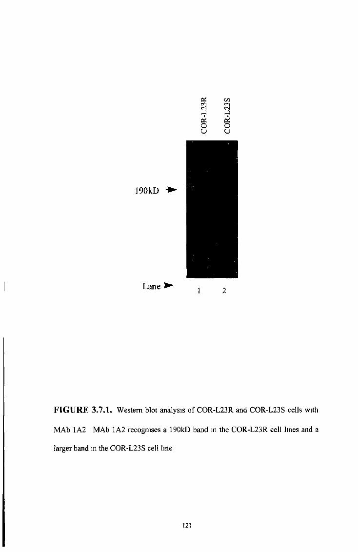
Lane ►1 2
FIGURE 3.7.1. Western blot analysis of COR-L23R and COR-L23S cells with
MAb 1A2 MAb 1A2 recognises a 190kD band in the COR-L23R cell lines and a
larger band m the COR-L23S cell Ime

FIGURE 3.7.2. Western blot analysis of un-glycosylated MRP 1-expressing SF9
membranes and COR-L23R cells with MAb 1A2 MAb 1A2 detects a 190kD band
m the COR-L23R cell line and the un-glycosylated SF9 MRP1 at the lower
molecular weight of 150kD
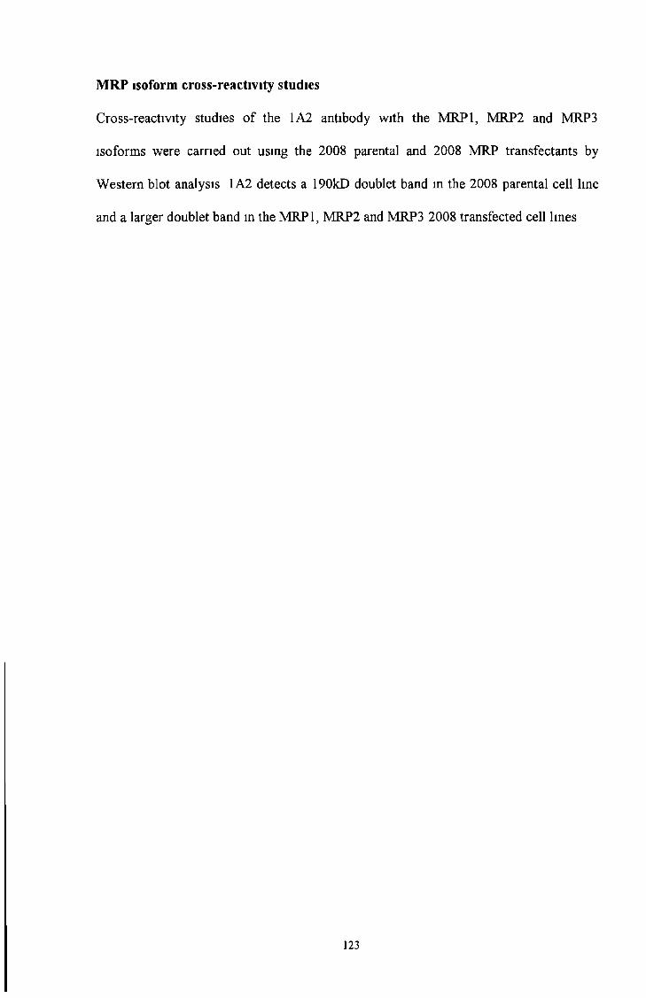
Cross-reactivity studies of the 1A2 antibody with the MRP1, MRP2 and MRP3
isoforms were carried out using the 2008 parental and 2008 MRP transfectants by
Western blot analysis 1A2 detects a 190kD doublet band in the 2008 parental cell line
and a larger doublet band in the MRP1, MRP2 and MRP3 2008 transfected cell lines
MRP isoform cross-reactivity studies
123
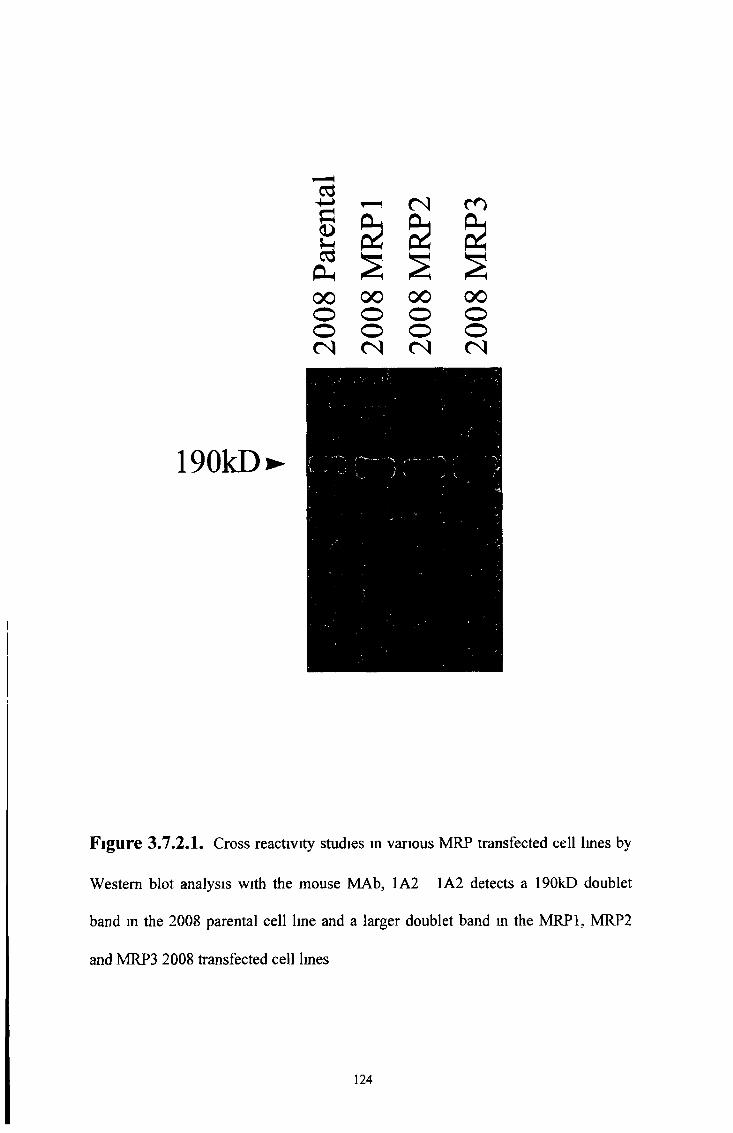
190kD
Figure 3.7.2.I. Cross reactivity studies in various MRP transfected cell lines by
Western blot analysis with the mouse MAb, 1A2 1A2 detects a 190kD doublet
band in the 2008 parental cell line and a larger doublet band in the MRP1, MRP2
and MRP3 2008 transfected cell lmes
124
Heat treatment of cell preparations destroys the epitope recognised by the rat
monoclonal antibody MRPrl in the drug selected COR-L23R cell line but not the 2008
MRP 1-transfected cell line (see section 3 2 Figure 3 2 8 and 3 2 9 ) The monoclonal
antibody 1A2 detects a 190kD band in the COR-L23R cell line and a larger or doublet
190kD band in the parental COR-L23S cell line (see Figure 3 7 1) Heat treatment of
COR-L23R and COR-L23S cell lysates @ 100°C for 3 mmutes does not destroy the
epitope recognised by MAb 1A2 (see Figure 3 7 3 ,3 7 3 1 and 3 73 2 )
Heat treatment of cell lysates prior to Western Blot analysis
125
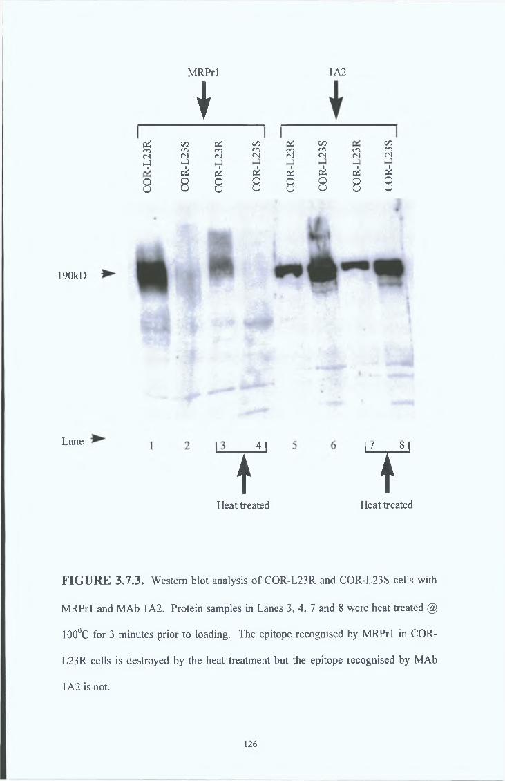
MRPrl
190kD
Lane
&co<NhJi
8
♦c/5co<NH-)pSs8
1A2
c*CO<Ni
Od8
C/5 c* C/5CO CO CO CO<N (N <N (N-)ck ck ck ckO O O OU U U u
c/5CO(N
ck
8
?u
Heat treated
YHeat treated
FIGURE 3.7.3. Western blot analysis of COR-L23R and COR-L23S cells with
MRPrl and MAb 1A2. Protein samples in Lanes 3, 4, 7 and 8 were heat treated @
100°C for 3 minutes prior to loading. The epitope recognised by MRPrl in COR-
L23R cells is destroyed by the heat treatment but the epitope recognised by MAb
1A2 is not.
126
Heat treated
-4-»a<D
03Ph00OO( N
00oo( N
( N m
00 00 o o o o< N < N
a
£<L>
Ph00oo< N
< N c o
00 00 o o o o< N < N
00oo< N
190kD
1 2 3 4 5 6 7 8
Figure 3.7.3.I. Western blot analysis of 2008 parental and MRP transfected cells
with MRPrl Protein samples m Lanes 5 to 8 were heat treated @ 100°C for 3
mmutes prior to loading The epitope recognised by MRPrl m the 2008 MRP1
transfected cells is not destroyed by the heat treatment
127
Heat treated
a d
00 00 00 00 00 00 00 00o o o o o o o oo o o o o o o o(N <N <N <N <N <N (N <N
190kD
1 2 3 4 5 6 7 8
Figure 3.7.3.2. Western blot analysis of 2008 parental and MRP transfected cells
with 1A2 Protein samples in Lanes 5 to 8 were heat treated @ 100°C for 3 minutes
prior to loadmg The epitope recognised by 1A2 in the 2008 parental and
transfected cells is not destroyed by the heat treatment
128
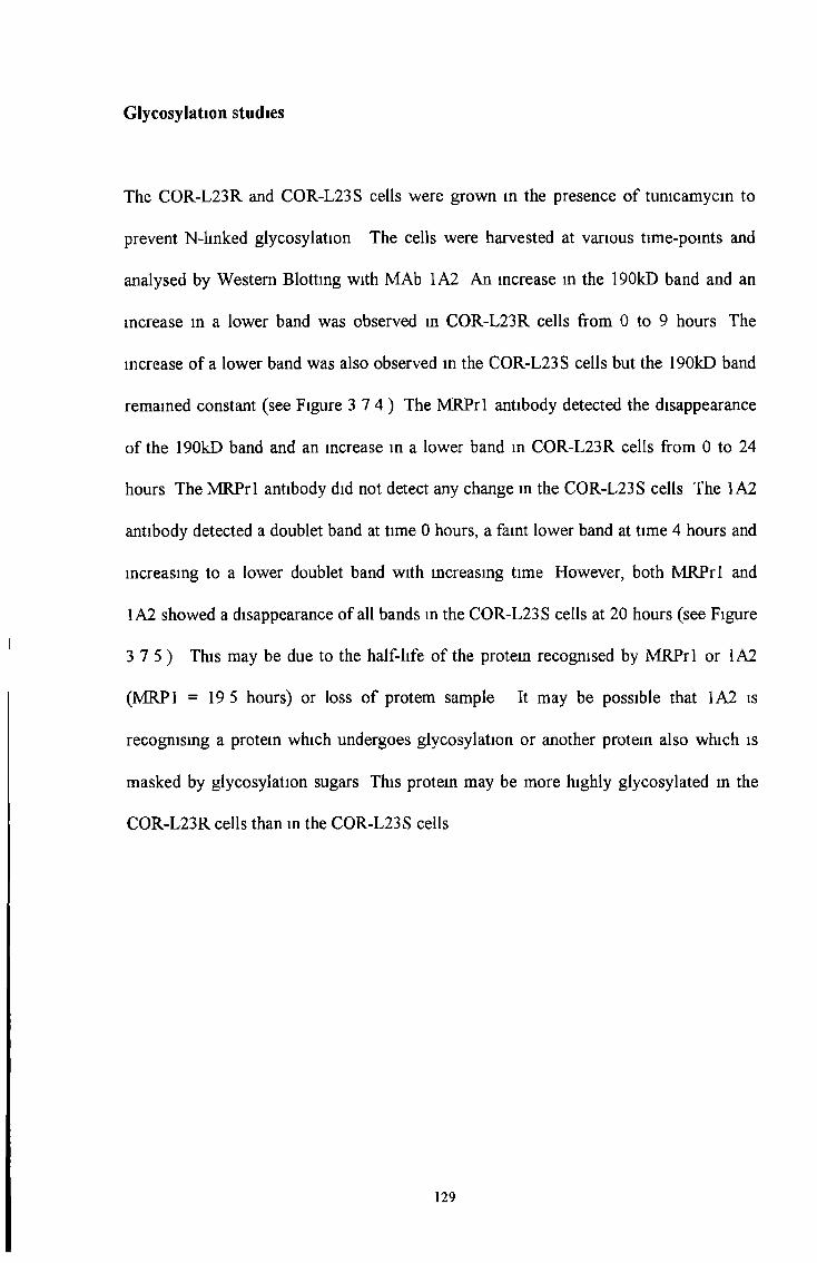
Glycosylation studies
The COR-L23R and COR-L23S cells were grown in the presence of tunicamycin to
prevent N-hnked glycosylation The cells were harvested at various time-points and
analysed by Western Blotting with MAb 1A2 An increase in the 190kD band and an
increase in a lower band was observed in COR-L23R cells from 0 to 9 hours The
increase of a lower band was also observed in the COR-L23S cells but the 190kD band
remained constant (see Figure 3 7 4 ) The MRPrl antibody detected the disappearance
of the 190kD band and an increase in a lower band in COR-L23R cells from 0 to 24
hours The MRPrl antibody did not detect any change in the COR-L23S cells The 1A2
antibody detected a doublet band at time 0 hours, a faint lower band at time 4 hours and
mcreasmg to a lower doublet band with mcreasing time However, both MRPrl and
1A2 showed a disappearance of all bands in the COR-L23S cells at 20 hours (see Figure
3 7 5 ) This may be due to the half-life of the protem recognised by MRPrl or 1A2
(MRP1 = 19 5 hours) or loss of protem sample It may be possible that 1A2 is
recogmsmg a protein which undergoes glycosylation or another protein also which is
masked by glycosylation sugars This protem may be more highly glycosylated m the
COR-L23R cells than in the COR-L23S cells
129

(A) COR-L23R with 1A2
190kD ►
Hours ► 0 0 5 1 2 3 4 6 9
(B) COR-L23S with 1A2
190kD ►
Hours ► 0 05 1 2 3 4 6 9
FIGURE 3.7.4. Western b lot analysis o f (A ) COR-L23R and (B) COR-L23S cells
grown m medium containing 8ug/ml o f Tunicamycin from 0 to 9 hours w ith M A b 1A2
M A b 1A2 detects an increase in a lower band in both (A ) COR-L23R and (B ) COR-L23S
cells w ith increasing exposure in time to Tunicamycin It is possible that 1A2 is recognising
another protein in this second band which is masked by glycosylation
130
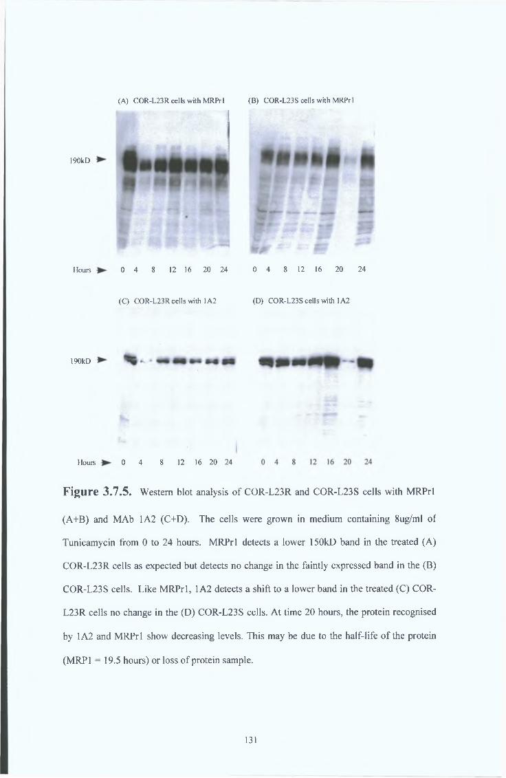
(A) COR-L23R cells with MRPrl (B) COR-L23S cells with MRPrl
190kD
Hours 0 4 8 12 16 20 24 0 4 8 12 16 20 24
( Q COR-L23R cells with 1A2 (D) COR-L23S cells with 1A2
190kD
Hours 0 4 8 12 16 20 24
Figure 3.7.5. Western blot analysis o f COR-L23R and COR-L23S cells w ith M R P rl
(A +B ) and M A b 1A2 (C+D). The cells were grown in medium containing 8ug/ml o f
Tunicamycin from 0 to 24 hours. M R P rl detects a lower 150kD band in the treated (A )
COR-L23R cells as expected but detects no change in the fa intly expressed band in the (B)
COR-L23S cells. L ike M R P rl, 1A2 detects a shift to a lower band in the treated (C) COR-
L23R cells no change in the (D ) COR-L23S cells. A t time 20 hours, the protein recognised
by 1A2 and M R P rl show decreasing levels. This may be due to the ha lf-life o f the protein
(MRP1 = 19.5 hours) or loss o f protein sample.
131
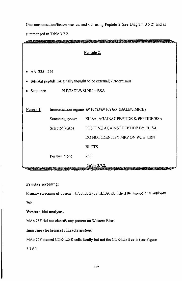
One immunisation/fusion was earned out using Peptide 2 (see Diagram 3 5 2) and is
summarised in Table 3 7 2
Peptide 2.
• AA 235 - 246
• Internal peptide (originally thought to be external) / N-terminus
• Sequence PLEGSDL W SLNK + BSA
Fusion 1. Immunisation regime IN VIVO/IN VITRO (BALB/c MICE)
Screening system ELISA, AGAINST PEPTIDE & PEPTIDE/BSA
Selected MAbs POSITIVE AGAINST PEPTIDE BY ELISA
DO NOT IDENTIFY MRP ON WESTERN
BLOTS
Positive clone 76F
__X.&bl£,.3&7,2j!_______________________,___
Primary screening:
Primary screening of Fusion 1 (Peptide 2) by ELISA identified the monoclonal antibody
76F
Western blot analysis.
MAb 76F did not identify any protein on Western Blots
Immunocytochemical characterisation:
MAb 76F stamed COR-L23R cells faintly but not the COR-L23S cells (see Figure
3 7 6 )
132
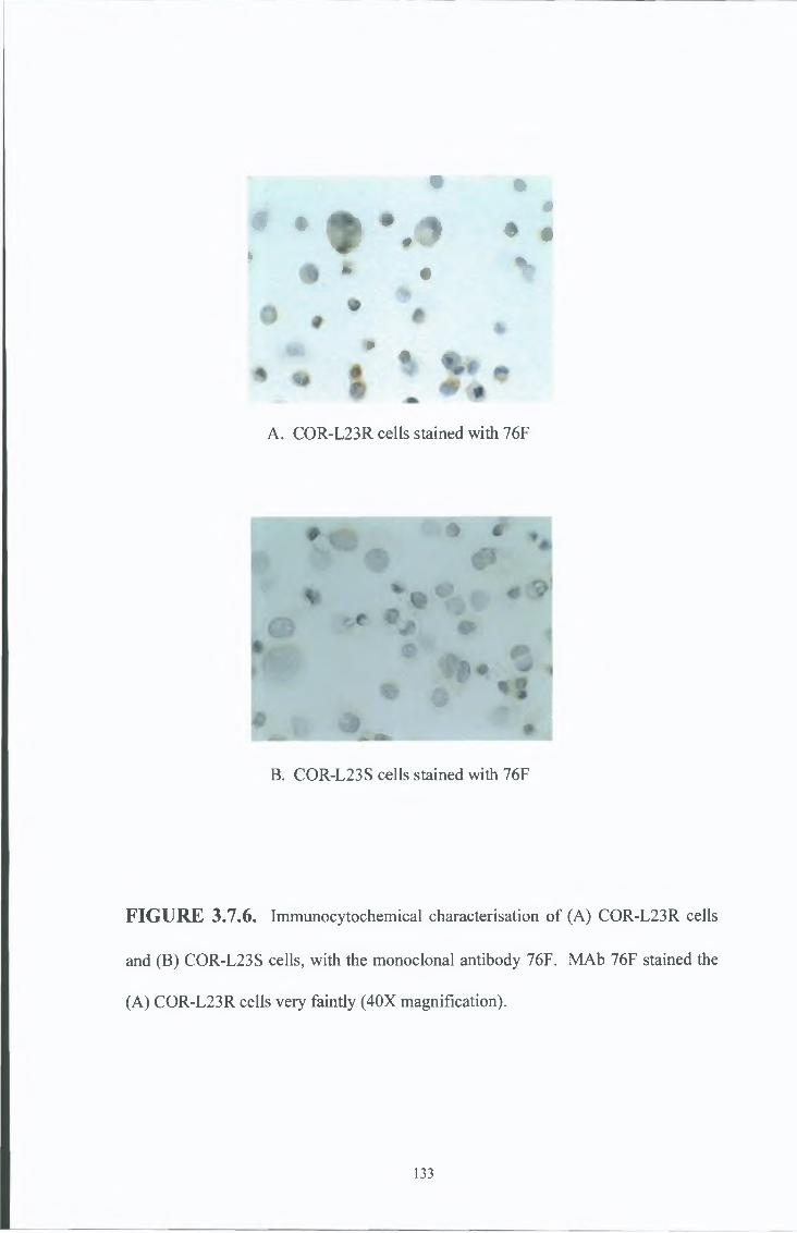
# •I
• •m
J \ 9 • .* 0 %
* . *
A. COR-L23R cells stained with 76F
B. COR-L23S cells stained with 76F
FIGURE 3.7.6. Immunocytochemical characterisation of (A) COR-L23R cells
and (B) COR-L23S cells, with the monoclonal antibody 76F. MAb 76F stained the
(A) COR-L23R cells very faintly (40X magnification).
133
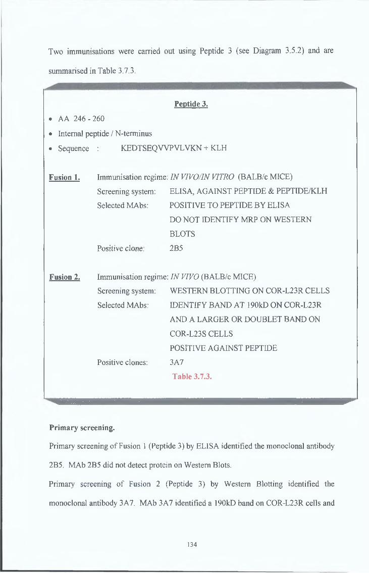
Two immunisations were carried out using Peptide 3 (see Diagram 3.5.2) and are
summarised in Table 3.7.3.
Peptide 3.
• AA 246 - 260
• Internal peptide / N-terminus
• Sequence : KEDTSEQWPVLVKN + KLH
Fusion 1, Immunisation regime: IN VIVO/IN VITRO (BALB/c MICE)
Screening system: ELISA, AGAINST PEPTIDE & PEPTIDE/KLH
Selected MAbs: POSITIVE TO PEPTIDE BY ELISA
DO NOT IDENTIFY MRP ON WESTERN
BLOTS
Positive clone. 2B5
Fusion 2. Immunisation regime: IN VIVO (BALB/c MICE)
Screening system: WESTERN BLOTTING ON COR-L23R CELLS
Selected MAbs: IDENTIFY BAND AT 190kD ON COR-L23R
AND A LARGER OR DOUBLET BAND ON
COR-L23S CELLS
POSITIVE AGAINST PEPTIDE
Positive clones: 3A7
Table 3.7.3.
Primary screening.
Primary screening of Fusion 1 (Peptide 3) by ELISA identified the monoclonal antibody
2B5. MAb 2B5 did not detect protein on Western Blots.
Primary screening of Fusion 2 (Peptide 3) by Western Blotting identified the
monoclonal antibody 3A7. MAb 3A7 identified a 190kD band on COR-L23R cells and
134
a 170-190kD doublet band on COR-L23S cells (see Figure 3 7 8 ), a large or doublet
190kD band on both DLKP and DLKPA cells (see Figure 3 7 9 ), a 190kD doublet band
on both HL60ADR and HL60S cells and a 150kD band on un-glycosylated MRP1-
expressing SF9 membranes (see Figure 3 7 10)
135
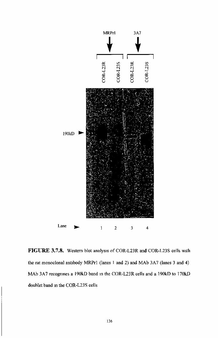
MRPrl 3A7
p4CO<NhJ
Ou
C/3CO
2cioo
PiCOCNhJpi
8
C/3CO
¿5ou
190kD
Lane
FIGURE 3.7.8. Western blot analysis of COR-L23R and COR-L23S cells with
the rat monoclonal antibody MRPrl (lanes 1 and 2) and MAb 3A7 (lanes 3 and 4)
MAb 3A7 recognises a 190kD band in the COR-L23R cells and a 190kD to 170kD
doublet band m the COR-L23S cells
136
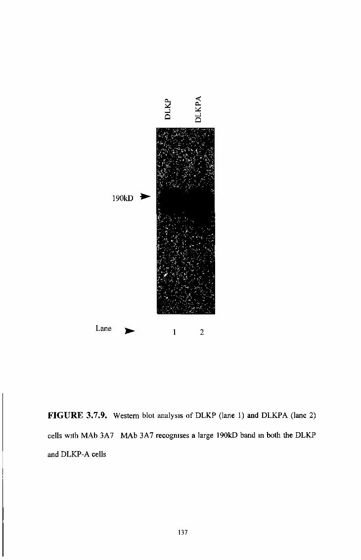
FIGURE 3.7.9. Western blot analysis of DLKP (lane 1) and DLKPA (lane 2)
cells with MAb 3A7 MAb 3A7 recognises a large 190kD band in both the DLKP
and DLKP-A cells
137
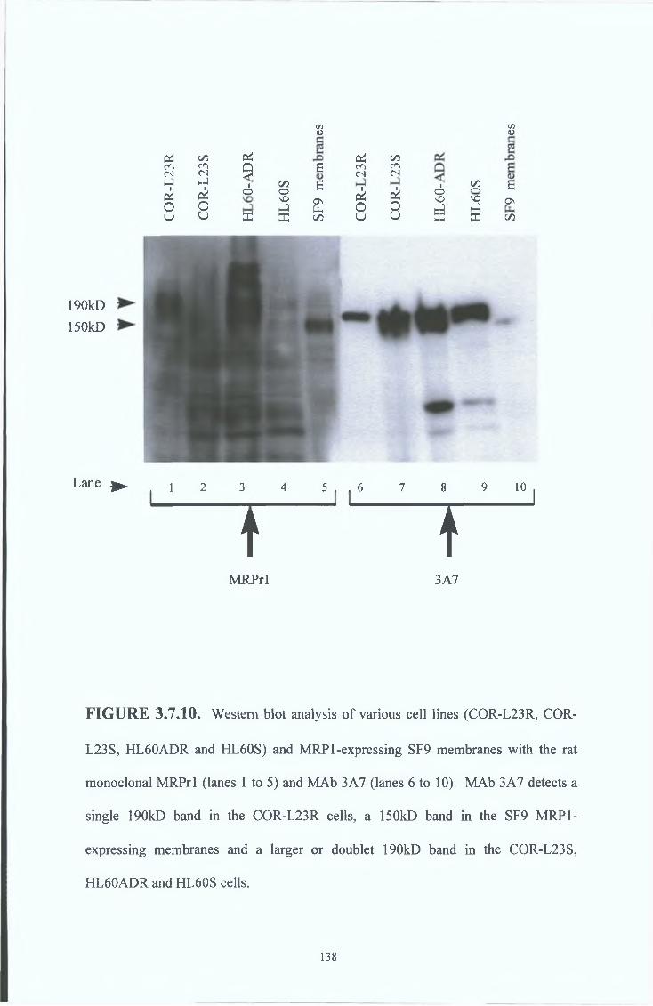
P i C/5 c * P 4 c/5co CO Û g CO CO<N <N y <N <NH-) hJ 1 C/5 8ckO
ckO
0 0s oh J
ONpLi
ci0
ciO
u u a K C/3 0 U
C/5 05<L> O
• £ c
§ S °"5^ K c/5
190kD150kD
Lane ► , i 2 3 4 5 . . 6 7 8 9 10 ^
t îMRPrl 3A7
FIGURE 3.7.10. Western blot analysis of various cell lines (COR-L23R, COR-
L23S, HL60ADR and HL60S) and MRP 1-expressing SF9 membranes with the rat
monoclonal MRPrl (lanes 1 to 5) and MAb 3A7 (lanes 6 to 10). MAb 3A7 detects a
single 190kD band in the COR-L23R cells, a 150kD band in the SF9 MRP1-
expressing membranes and a larger or doublet 190kD band in the COR-L23S,
HL60ADR and HL60S cells.
138

Immunocytochemical analysis of COR-L23R and COR-L23S cells with MAb 3A7
showed staining on both cell lines (see Figure 3 7 11)
Immunocytochemical characterisation
ii
139
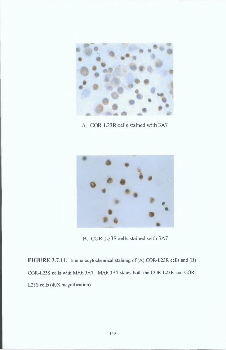
A. COR-L23R cells stained with 3A7
B. COR-L23S cells stained with 3A7
FIGURE 3.7.11. Immunocytochemical staining of (A) COR-L23R cells and (B)
COR-L23S cells with MAb 3A7. MAb 3A7 stains both the COR-L23R and COR-
L23S cells (40X magnification).
140
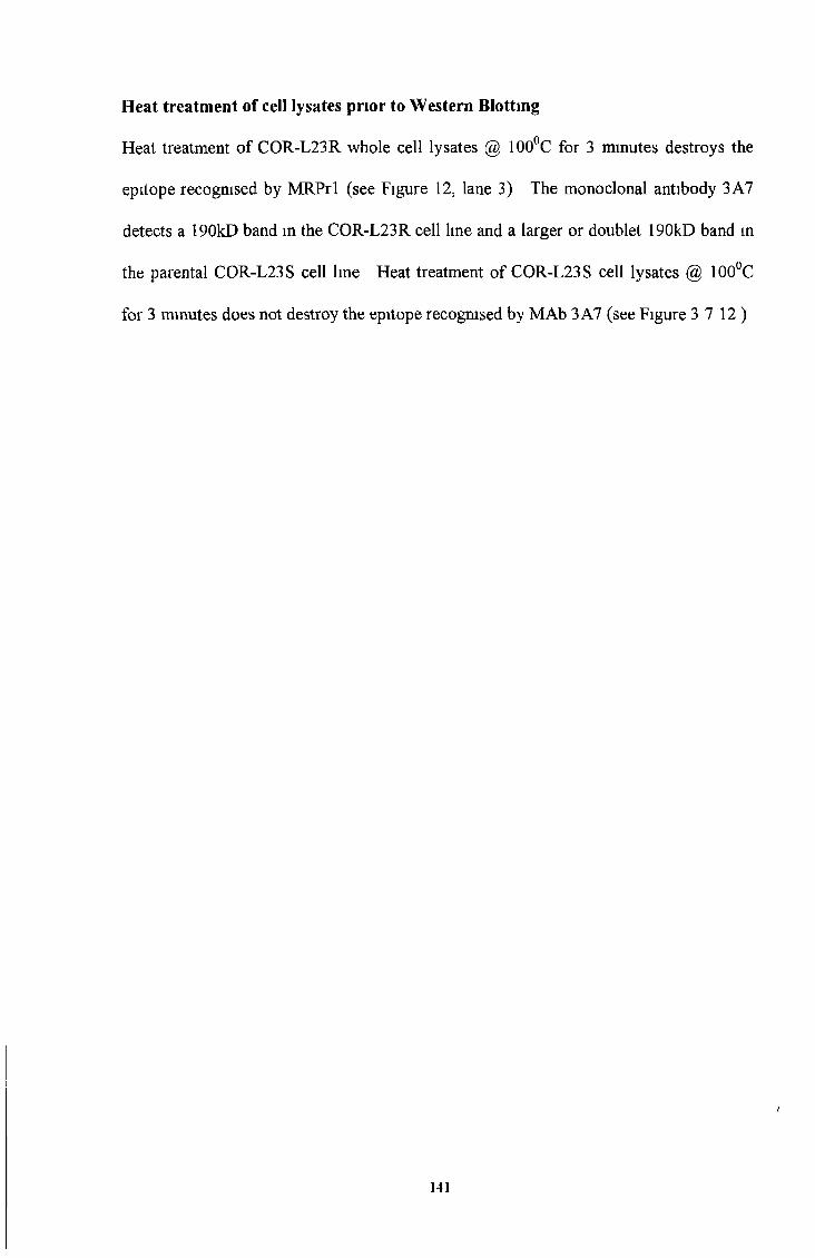
Heat treatment of cell lysates prior to Western Blotting
Heat treatment of COR-L23R whole cell lysates @ 100°C for 3 minutes destroys the
epitope recognised by MRPrl (see Figure 12, lane 3) The monoclonal antibody 3A7
detects a 190kD band in the COR-L23R cell line and a larger or doublet 190kD band in
the parental COR-L23S cell line Heat treatment of COR-L23S cell lysates @ 100°C
for 3 minutes does not destroy the epitope recognised by MAb 3 A7 (see Figure 3 7 12)
141

FIGURE 3.7.12. Western blot analysis of COR-L23R and COR-L23S cells, with
the rat monoclonal MRPrl (lanes 1 to 4) and MAb 3A7 (lanes 5 to 9). Protein
samples in lanes 3, 4, 7 and 8 were heat treated @ 100°C for 3 minutes prior to
loading. The epitope recognised by MAb 3A7 is not destroyed by heat treatment.
142
Immunoprécipitation studies
To determine that the monoclonal antibodies produced recognised MRP 1,
immunoprécipitation studies were earned out (see Figure 3 7 13) COR-L23R and
COR-L23S cells were immunoprecipitated with the MRP1 specific, MRPrl antibody
The precipitates were run out on Western Blots and re-probed with MAb 3A7, MRPrl
and control mouse IgG MAb 3 A7 identified a 190kD band in the COR-L23R cells and
a slightly stronger 190kD band in the COR-L23S cells The control mouse IgG was
negative
143
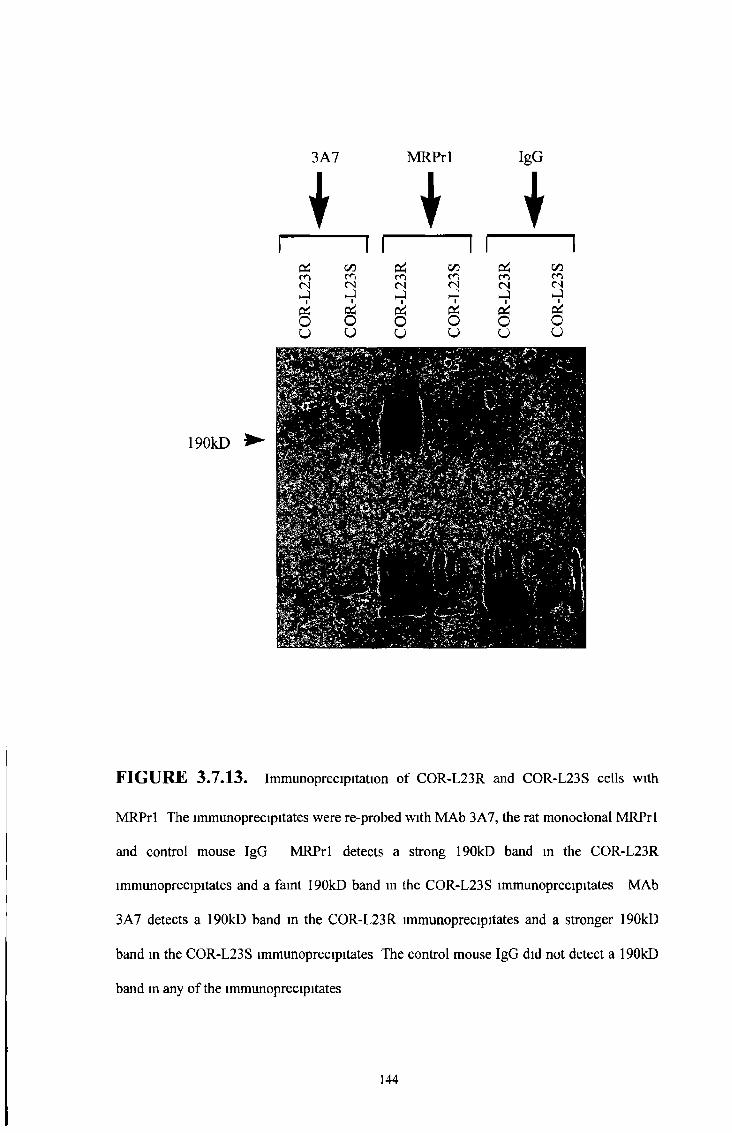
I I \
I 1 I 1 I 1
3A7 MRPrl IgG
od C/3 C/3 C/3CO co co CO co co<N <N <N <N (N <N►J H-)
çéckO o o O O Ou u u u O o
190kD ►
FIGURE 3.7.13. Immunoprécipitation o f COR-L23R and COR-L23S cells w ith
M R P rl The immunoprecipitates were re-probed w ith M A b 3A7, the rat monoclonal M R P rl
and control mouse IgG M R P rl detects a strong 190kD band in the COR-L23R
immunoprecipitates and a faint 190kD band in the COR-L23S immunoprecipitates M A b
3A7 detects a 190kD band in the COR-L23R immunoprecipitates and a stronger 190kD
band in the COR-L23S immunoprecipitates The control mouse IgG did not detect a 190kD
band m any o f the immunoprecipitates
144

Glycosylation studies.
Glycosylation studies with tumcamycin showed that the epitope recognised by MAb
3A7 was destroyed after 40 hours treatment Unexpectedly, MAb 3A7 failed to detect a
band in the MRP1 expressing SF9 membranes (see Figure 3 7 14) Previously MAb
3A7 recognised a faint 150kD band in unglycosylated MRP1 expressing SF9 insect
membranes (Figure 3 7 10) The 3A7 MAb showed a tendency to give varying results
and became unstable, eventually failing to secrete any antibody
145

r ico(N
éu
190kD
Lane
3A7
i
MRPrl
♦
on p ico co
co<L>I
3 I
o g *u o
O P'O ^w C/l
r i <Z3 co co <N <N
ir i Ov
r io
r ico(N
to o
CO
iinCO(N BhJir i Eo ON
u u.
Tunglycosylated
runglycosylated
FIGURE 3.7.14. Western b lot analysis o f COR-L23R cells, COR-L23S cells and
MRP 1-expressing SF9 membranes w ith M A b 3A7 and the rat monoclonal M R P rl Samples
loaded onto lanes 3, 4, 8 and 9 were grown in medium containmg 8ug/ml Tunicamycin fo r
40 hours The epitope recognised by 3A7 is destroyed after 40 hours Unexpectedly, M A b
3A7 failed to detect MRP1 expressed in the SF9 insect membranes (as it had done
previously F igure 3 7 10) possibly because it was becoming an unstable antibody secretor
146
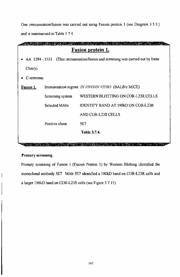
One immunisation/fusion was carried out using Fusion protein 1 (see Diagram 3 5 3 )
and is summarised in Table 3 7 4
Fusion protein 1.
• AA 1294 - 1531 (This immunisation/fusion and screening was carried out by Irene
Cleary)
• C-terminus
Fusion 1. Immunisation regime IN VIVO/IN VITRO (BALB/c MICE)
Screemng system WESTERN BLOTTING ON COR-L23R CELLS
Selected MAbs IDENTIFY BAND AT 190kD ON COR-L23R
AND COR-L23S CELLS
Positive clone 5E7
Table 3.7.4.
Primary screening
Primary screening of Fusion 1 (Fusion Protein 1) by Western Blotting identified the
monoclonal antibody 5E7 MAb 5E7 identified a 190kD band on COR-L23R cells and
a larger 190kD band on COR-L23S cells (see Figure 3 7 15)
147
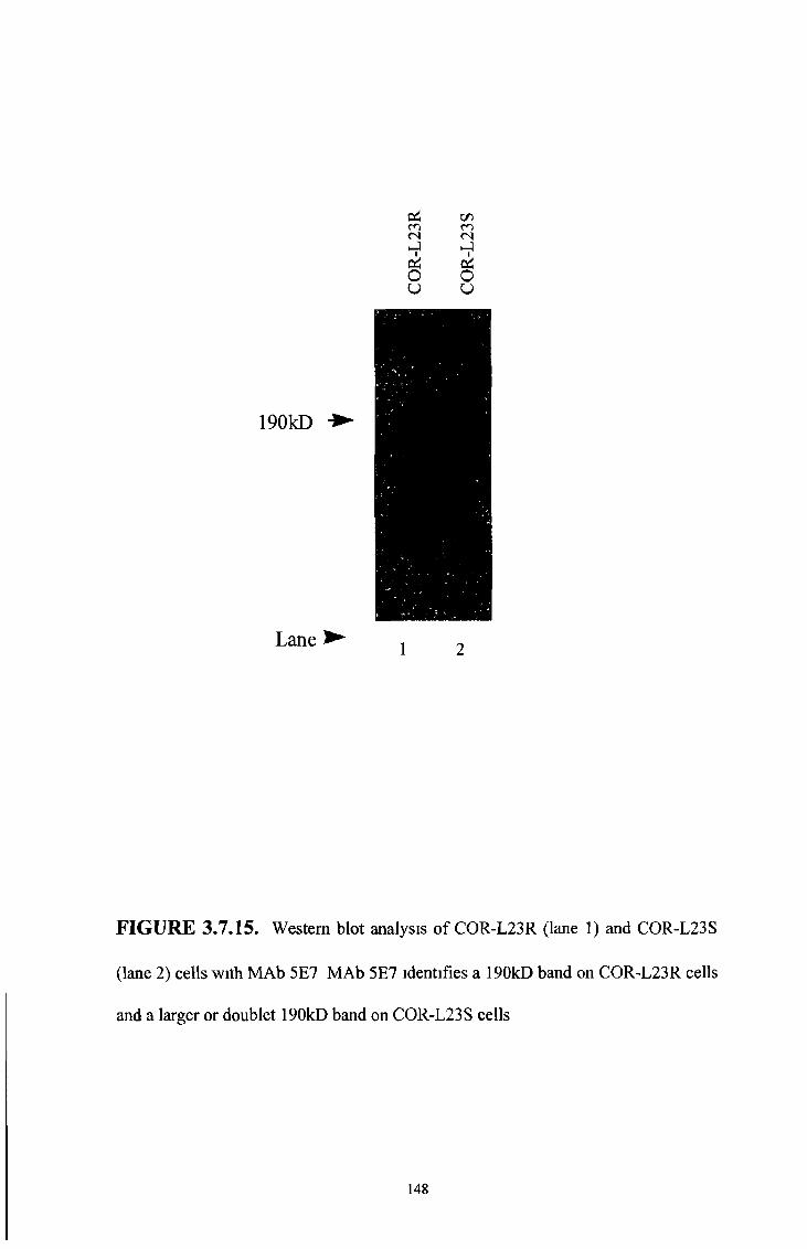
Lane ► 1 2
FIGURE 3.7.15. Western blot analysis of COR-L23R (lane 1) and COR-L23S
(lane 2) cells with MAb 5E7 MAb 5E7 identifies a 190kD band on COR-L23R cells
and a larger or doublet 190kD band on COR-L23S cells

One immunisation/fusion was earned out using Fusion protein 2 (see Diagram 3 5 3 )
and is summarised in Table 3 7 5
m s s s m t
Fusion protein 2.
• AA 647 - 912
• N-terminus
Fusion 1 Immunisation regime IN VIVO/IN VITRO (BALB/c MICE)
Screening system
Selected MAbs
Positive clone
ELISA, AGAINST FUSION PROTEIN & MBP
POSITIVE TO PEPTIDE & MBP
DO NOT IDENTIFY MRP ON WESTERN
BLOTS
2A6
Table 3.7 5
Primary screening.
Primary screening of Fusion 1 (Fusion Protein 2) by ELISA identified the monoclonal
antibody 2A6 MAb 2A6 did not identify any protein on Western Blots However, 2A6
also reacted positively by ELISA with an irrelevant fusion protein, which also contained
the Maltose Binding Protein (MBP) expression tag This indicated that 2A6 was
reacting with MBP and not the peptide immunogen
Immunocytochemical charatensation
MAb 2A6 stained both COR-L23R and COR-L23S cells by immunocytochemistry but
staining was reduced on the COR-L23S cells (see Figure 3 7 16 )
149
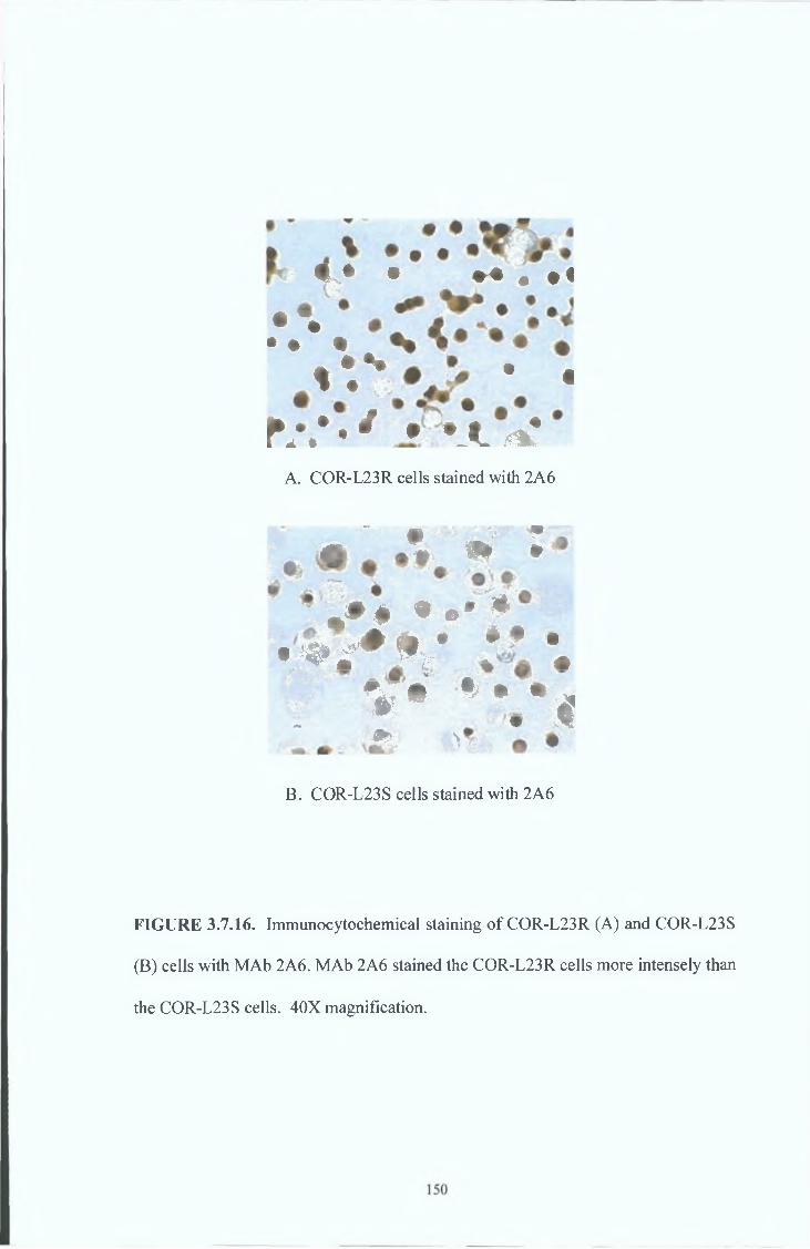
! • • M t • (
.V.1 • . * * • *
K \ * • / • ‘ A .*%*
A. COR-L23R cells stained with 2A6
« . ‘ *. » i
, » * • ® * *A cm • # • ^
• i # ^ P ) , 1 . #
V > * * ‘
W $!
•% • • „ I
\*
B. COR-L23S cells stained with 2A6
FIGURE 3.7.16. Immunocytochemical staining of COR-L23R (A) and COR-L23S
(B) cells with MAb 2A6. MAb 2A6 stained the COR-L23R cells more intensely than
the COR-L23S cells. 40X magnification.

One immunisation/fusion was carried out using COR-L23R whole cell lysates and is
summarised in Table 3.7.6.
COR-L23R WHOLE CELL LYSATE FUSION.
• Immunogen : COR-L23R whole cell lysates
Fusion 1. Immunisation regime: IN VIVO/IN VITRO (BALB/c MICE)
Screening system: WESTERN BLOTTING ON COR-L23R CELLS
Selected MAbs: IDENTIFY BAND AT 190kD ON COR-L23R,
COR-L23S, HL60ADR AND HL60S CELLS.
STAIN COR-L23R CELLS BUT NOT THE
SENSITIVE PARENTAL COR-L23S CELLS BY
IMMUNOCYTOCHEMISTRY.
Positive clone: 4F6.
T a b le 3.7.6.
Primary screening.
Primary screening by Western Blotting identified the monoclonal antibody 4F6. MAb
4F6 identified a 190kD band on COR-L23R, COR-L23S, HL60ADR and HL60S cells
(see Figure 3.7.16.).
Immunocytochemical characterisation.
4F6 also reacted strongly with COR-L23R cells but not with COR-L23S cells by
immunocytochemistry (see Figure 3.7.17.). However, this hybridoma was unstable and
stopped secreting antibody despite cloning.
151
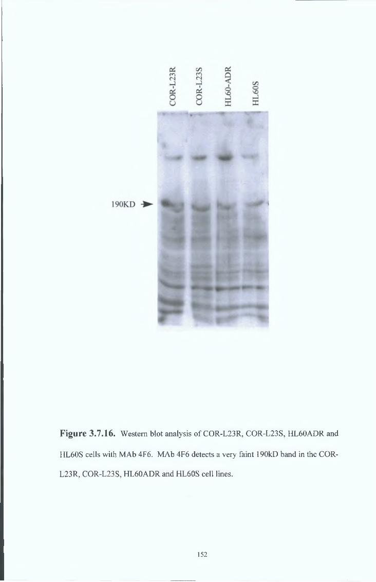
Figure 3.7.16. Western blot analysis of COR-L23R, COR-L23S, HL60ADR and
HL60S cells with MAb 4F6. MAb 4F6 detects a very faint 190kD band in the COR-
L23R, COR-L23S, HL60ADR and HL60S cell lines.
152
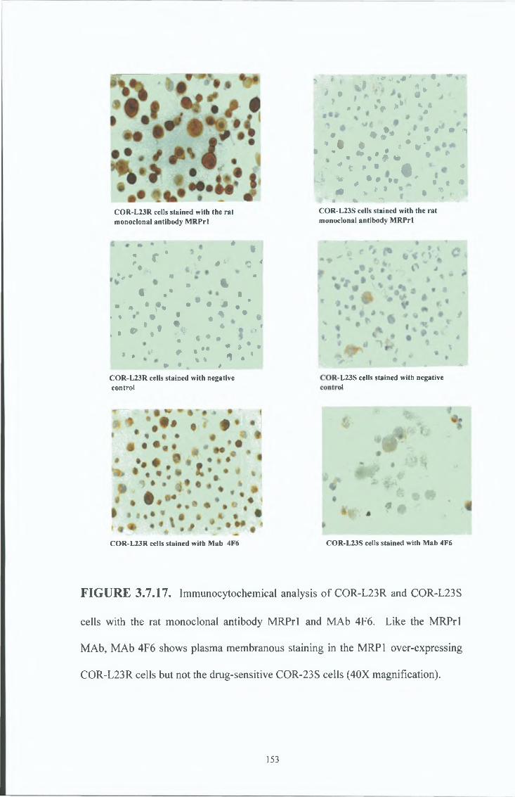
COR-L23R cells stained with the ra t monoclonal antibody M R Prl
; c * f# *
0 & $ 4• a ,
•j, « <• •• %
« .* t• » • f .
. , » •
. . » V * 1 •
, . * * * *
• * « * . •«
1 « .. ••% 1
* . „ * A ^ • • * * • ¥
Ì • 1i
COR-L23R cells stained with negative control
* ì ***> •+ * * * ^».'I a *> » « * f » » *V * * v
* * . * * : V “ *. » t * . V % 'fc * . » . » i 0. • *
* I ♦k * . * . » l * • -•• * t » • * ** * „ * • • • " * • t *. * * * » • * *
** a % ^ * *COR-L23S cells stained with the rat monoclonal antibody M R P rl
-L23S cells stained with negativenl
COR-L23R cells stained with Mab 4F6 COR-L23S cells stained with Mab 4F6
FIGURE 3.7.17. Immunocytochemical analysis of COR-L23R and COR-L23S
cells with the rat monoclonal antibody MRPrl and MAb 4F6. Like the MRPrl
MAb, MAb 4F6 shows plasma membranous staining in the MRP1 over-expressing
COR-L23R cells but not the drug-sensitive COR-23S cells (40X magnification).
153

One immunisation/fusion was earned out using COR-L23R cell membrane preparations
and is summarised m Table 3 7 7
COR-L23R CELL MEMBRANE FUSION,
• Immunogen COR-L23R cell membrane preparations
Fusion 1 Immunisation regime IN VIVO (BALB/c MICE)
Screening system WESTERN BLOTTING ON COR-L23R CELL
MEMBRANES
Selected MAbs IDENTIFY BAND AT 190kD ON COR-L23R
CELLS
Positive clones 6C6 and 2A7
Table 3.7.7.
Primary screening
Primary screening by Western blotting identified the monoclonal antibodies 2A7 and
6C6 Both MAb 2A7 and MAb 6C6 recognised a 190kD band in COR-L23R and COR-
L23S whole cell lysates and cell membrane preparations (see Figure 3 7 18) MAb 2A7
detected two lower bands in the MRP 1-over-expressing HL60ADR cells and a faint
single lower band in the MRP1 negative HL60S cell line at a 1 15 concentration
However, MAb 2A7 detected only a single lower band m the HL60ADR cells and no
bands in the HL60S cells at a 1 20 concentration (see Figure 3 7 19) MAb 6C6 did not
recognise any bands on the HL60ADR or HL60S cells by Western blotting (see Figure
154
3 7 20) MAb 2A7 or MAb 6C6 did not detect un-glycosylated MRP1 in MRP1-
expressing SF9 membranes by Western blotting (see Figures 3 7 21, 3 7 22 and
3 7 23 )
155
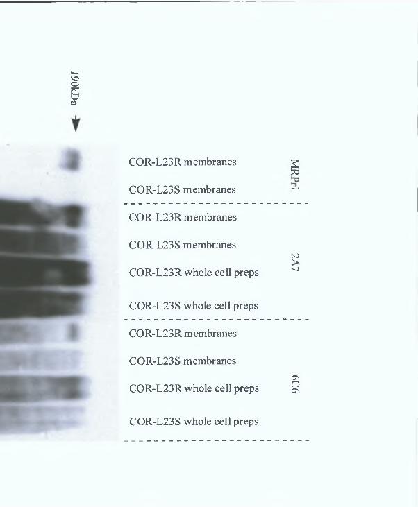
190kDa
COR-L23R membranes ^
COR-L23S membranes ~
COR-L23R membranes
COR-L23S membranests)>
COR-L23R whole cell preps ^
COR-L23S whole cell preps
COR-L23R membranes
COR-L23S membranesas
COR-L23R whole cell preps 9
COR-L23S whole cell preps
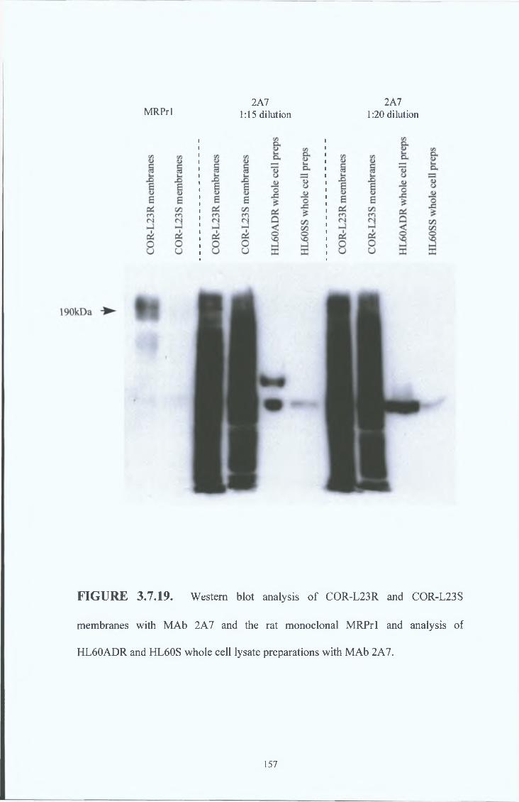
MRPrl2A7
1:15 dilution2A7
1:20 dilution
FIGURE 3.7.19. Western blot analysis of COR-L23R and COR-L23S
membranes with MAb 2A7 and the rat monoclonal MRPrl and analysis of
HL60ADR and HL60S whole cell lysate preparations with MAb 2A7.
157

o0*1rK>U>7*P3CL
O0&1rK>U>C/33CD3a*Icd(/3
O'ONoON
V!inICDCD
P
OnnON
P3a-ErCD
'Cc/3.Eq‘o
2oe
-4N>
CD
£CD3a;o'PP£L
vT03
O•“»>KPONo>a
§a*ECrO nOC/3
NOO
HL60ADR whole cell preps
HL60SS whole cell preps
COR-L23R membranes
COR-L23S membranes
HL60ADR whole cell preps
HL60SS whole cell preps
COR-L23R membranes
COR-L23S membranes
HL60ADR whole cell preps
HL60SS whole cell preps
6C6 6C
6 M
Rl’rl
1:8 dilution
1:5 dilution

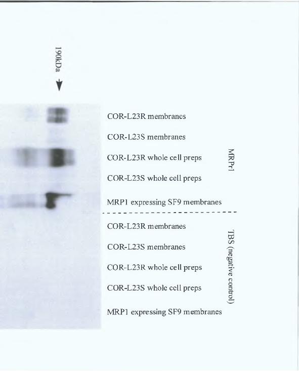
COR-L23R membranes
COR-L23S membranes
COR-L23R whole cell preps
COR-L23S whole cell preps
MRP1 expressing SF9 membranes
COR-L23R membranes
COR-L23S membranes
COR-L23R whole cell preps
COR-L23S whole cell preps
MRP1 expressing SF9 membranes
MRPrl
I TBS
(negative control)
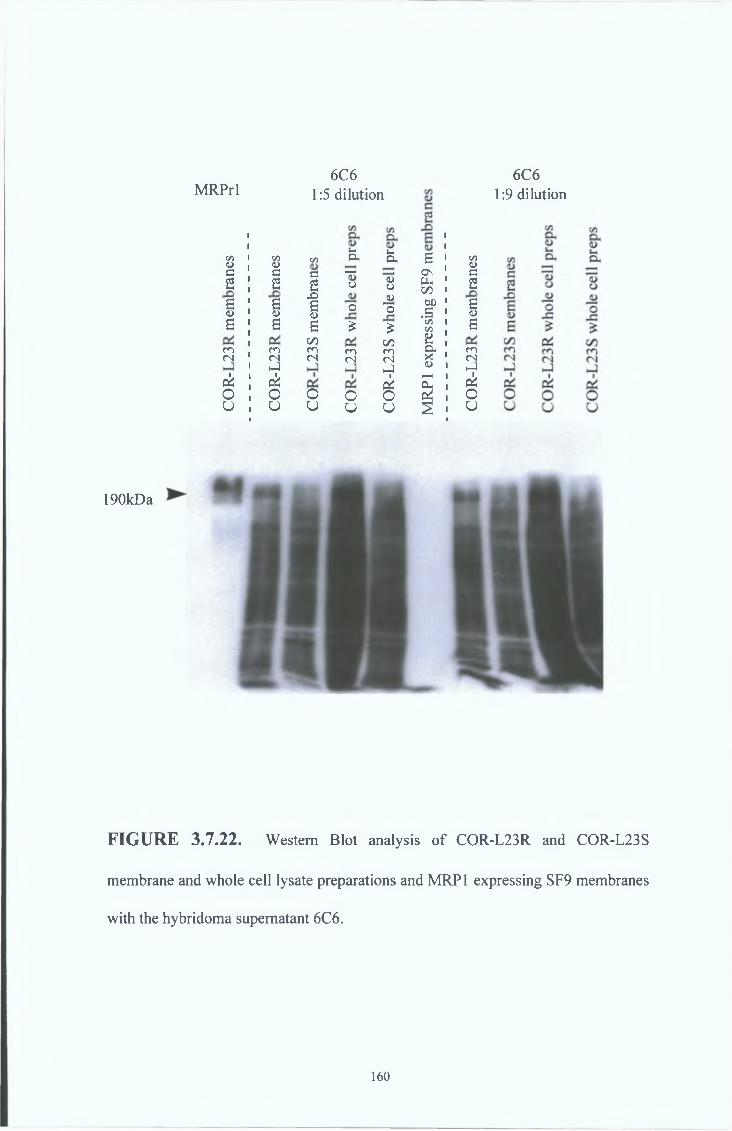
190kDa
FIGURE 3.7.22. Western Blot analysis of COR-L23R and COR-L23S
membrane and whole cell lysate preparations and MRP1 expressing SF9 membranes
with the hybridoma supernatant 6C6.
MRPrl6C6
1:5 dilution
cn <L> C/3<u C/3»-iOh «HDh
1a ■ 1 C/3<L>C c c *—* ; | C \ a
2 2 2 o <UO Uh ' C/5 1 2
X) <ua<D a<D a<D o ’o 00 1 .S '
B<Ds s a £ £ CM I C/3 , s
C/5 C/5 Pfa 1CO co co co CO & 1 co<N <N <N (N (N X 1 (N
iJ hJ 4J I t-1ck p i ck £ ! cko o o O O 3 i Ou o U u u § » U
6C61:9 dilution
160
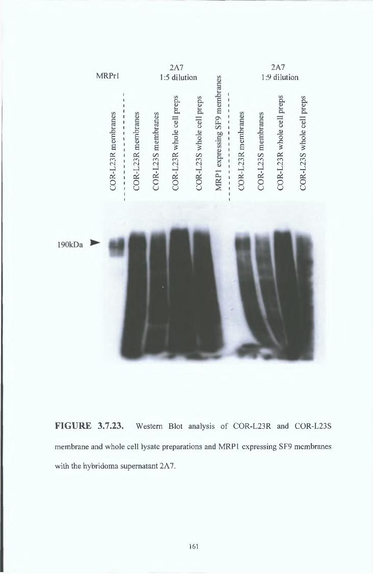
MRPrl2A7
1:5 dilution2A7
1:9 dilution
B<&B
r ico(N
c iOo
J-iI<üS
r ien(Nh-)c iOU
C/3
C/3a<L>«-XCL<D
§S-H
<DOX)B 'o<L>B £
C/5 riCO CO<N <Nh-lck ciO Ou o
C/3P,<L)
<L>OJJo
£C/5co(N
Ou
S-
1<uS
r ico(N
c iOU
a<DC/>co2ckOU
C/3CL<D
<L>OJJOÆ£
r iCO<NhJc i8
C/îa(U>-ia<DOo£
00en(NhJ
éu
FIGURE 3.7.23. Western Blot analysis of COR-L23R and COR-L23S
membrane and whole cell lysate preparations and MRP1 expressing SF9 membranes
with the hybridoma supernatant 2A7.
161
Ivnmunocytochemical characterisation
MRPrl consistently stains the plasma membrane of COR-L23R and HL60ADR cells
more intensely than COR-L23S and HL60S cells (see Figure 3 7 24 ) MAb 2A7
stained COR-L23R and COR-L23S cells m the plasma membrane region but stamed the
HL60ADR and HL60S cells m then* nucleoli (see Figure 3 7 25 ) MAb 6C6 stained
COR-L23R and COR-L23S cells in the plasma membrane region also but stamed
HL60ADR and HL60S cells m the cytoplasmic region (see Figure 3 7 26) The
negative controls can be viewed in Figure 3 7 27
162

(A) COR-L23R cells stained with MRPrl (B) COR-L23S cells stained with MRPrl
(C) HL60ADR cells stained with MRPrl (D) HL60S cells stained with MRPrl
FIGURE 3.7.24. Immunocytochemical analysis of COR-L23R, COR-L23S,
HL60ADR and HL60S cells with the rat monoclonal antibody MRPrl. MRPrl
intensely stains the COR-L23R and HL60ADR cells in the plasma membrane region
but does not stain the drug sensitive COR-L23S or HL60S cells. (100X
magnification).
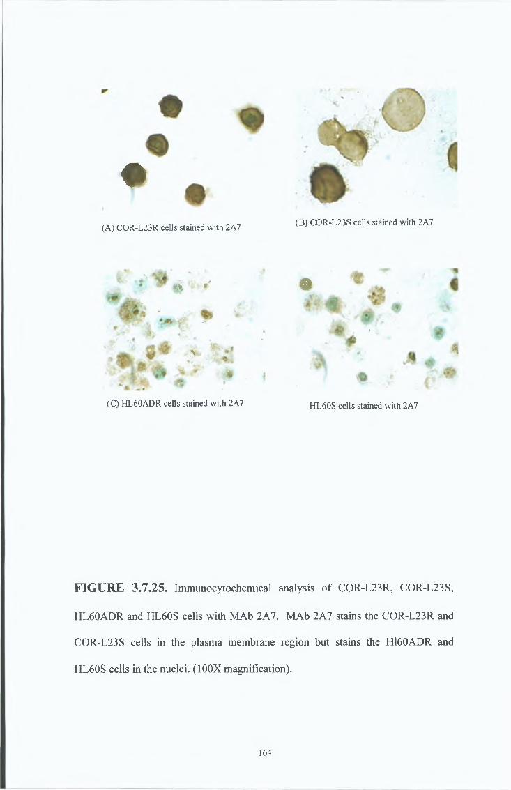
♦
(A) COR-L23R cells stained with 2A7
« r
RtflK “'4y ^
1V
(C) HL60ADR cells stained with 2A7
(B) COR-L23S cells stained with 2A7
«
i
41
HL60S cells stained with 2A7
FIGURE 3.7.25. Immunocytochemical analysis of COR-L23R, COR-L23S,
HL60ADR and HL60S cells with MAb 2A7. MAb 2A7 stains the COR-L23R and
COR-L23S cells in the plasma membrane region but stains the H160ADR and
HL60S cells in the nuclei. (100X magnification).
164
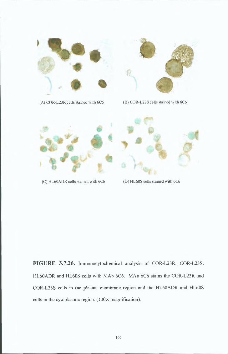
♦
(A) COR-L23R cells stained with 6C6
(C) HL60ADR cells stained with 6C6
'•% gW
(B) COR-L23S cells stained with 6C6
(D) HL60S cells stained with 6C6
FIGURE 3.7.26. Immunocytochemical analysis of COR-L23R, COR-L23S,
HL60ADR and HL60S cells with MAb 6C6. MAb 6C6 stains the COR-L23R and
COR-L23S cells in the plasma membrane region and the HL60ADR and HL60S
cells in the cytoplasmic region. (100X magnification).
165

%
w %
(A) COR-L23R cells stained with TBS (B) COR-L23S cells stained with TBS
r %
* _ :
(C) HL60ADR cells stained with TBS (D) HL60S cells stained with TBS
FIGURE 3.7.27. Immunocytochemical analysis of COR-L23R, COR-L23S,
HL60ADR and HL60S cells with negative control. (100X magnification).
166
3 8 RAT MONOCLONAL ANTIBODY PRODUCTION
Immunisations
In order to generate rat monoclonal antibodies for the detection of MRP 1, female Wistar
rats were immunised with either a double peptide combination or MRP1 over-
expressing COR-L23R membranes Two synthetic peptides corresponding to the
deduced ammo acid sequence of human MRP1 (Peptide 2, amino acids 235-246 and
Peptide 3, ammo acids 246-260) (see Diagram 3 5 2) were used in the double peptide
immunisation The epitope recognised by the MRP1 specific rat antibody, MRPrl, has
been mapped to ammo acids 238 to 247 of the deduced ammo acid sequence of human
MRP1 This ammo acid sequence is similar to that of Peptide 2 and was used as a
positive control The COR-L23R membranes were used as an immunogen in the hope
of obtaining an antibody recognising an external epitope The rats received footpad
injections of 10-25ug of protein initially emulsified in freunds complete, followed two
weeks later by a booster emulsified in freunds incomplete and finally 4 days before
fusion a booster in PBS without adjuvant
Fusions.
Lymphocytes were isolated from the popliteal nodes (see Diagram 2 3 2 ) of the
immunised rats and fused with mouse myeloma Sp2/0 cells using polyethylene glycol
Prior to this, the popliteal lymph nodes were identified and removed in practice runs by
injecting Isosulfan blue into the footpads of non-immumsed rats Isosulfan blue is
selectively picked up by the lymphatic vessels and is dramed into the popliteal nodes
making them discernible from the surrounding tissue Fusion rates for all the
immunisations were approximately 95%
167
Screening of confluent supernatants from the double peptide immunisation was carried
out by ELISA agamst the relevant peptides or by Western blot analysis, against cell
membrane preparations of the MRP 1-over-expressing COR-L23R cell line Screening
of confluent supernatants from the COR-L23R cell membrane immunisation was
carried out by live cell immunofluoresence agamst the COR-L23R and COR-L23S
cells
R esu lts
Antibodies resultmg from the COR-L23R membrane immunisation reacted with both
the COR-L23R and COR-L23S cell Imes by live cell immunofluoresence and therefore
were not characterised any further Approximately 10% of hybndomas analysed from
the double peptide immunisation by ELISA were positive to the linear peptides
Western blot analysis showed that a number of these hybndomas secreted antibodies
that reacted with the COR-L23R but not the COR-L23S cells Further characterisation
showed that these antibodies detected a 190KD band m the COR-L23R, HL60ADR and
MRP 1-transfected cell line, 2008 MRP1, by Western blotting These antibodies were
designated P2A8, P1A6 and P2E4 Of these antibodies P2A8 was cloned and
designated P2A8(6) Immunocytochemical analysis of these antibodies identified one
antibody, P2A8(6), which like the rat monoclonal antibody MRPrl, stained all cell
populations in the COR-L23R and MRP1 2008 transfectants (see Figure 3 8 10 )
However, immunocytochemical analysis also identified an antibody, P1D7, which
stained a subpopulation of cells m the COR-L23R and 2008 MRP1 transfected cell
lines, unlike the MRP1 specific rat monoclonal antibody MRPrl which stains all the
cell population in these cell lines Further characterisation of P1D7 by Western blot
Screening of hybndomas
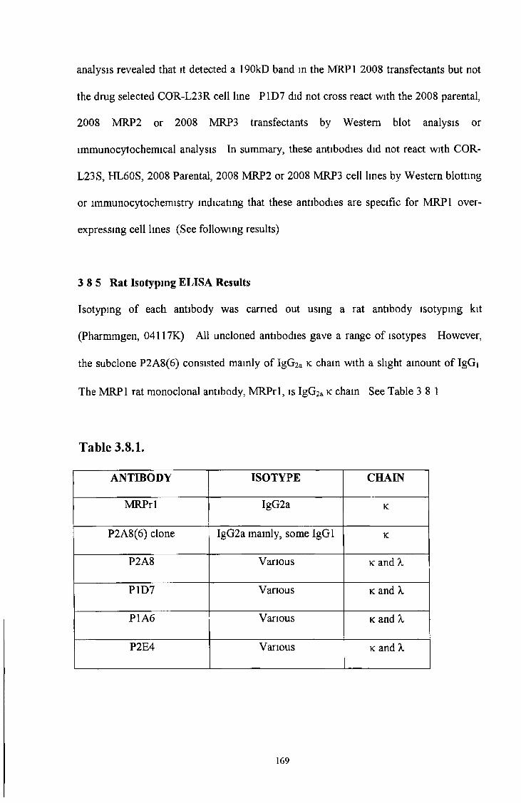
analysis revealed that it detected a 190kD band in the MRP1 2008 transfectants but not
the drug selected COR-L23R cell line P1D7 did not cross react with the 2008 parental,
2008 MRP2 or 2008 MRP3 transfectants by Western blot analysis or
lmmunocytochemical analysis In summary, these antibodies did not react with COR-
L23S, HL60S, 2008 Parental, 2008 MRP2 or 2008 MRP3 cell lines by Western blottmg
or immunocytochemistry indicating that these antibodies are specific for MRP1 over-
expressing cell lines (See following results)
3 8 5 Rat Isotyping ELISA Results
Isotyping of each antibody was carried out using a rat antibody isotypmg kit
(Pharmmgen, 04117K) All uncloned antibodies gave a range of isotypes However,
the subclone P2A8(6) consisted mamly of IgG2a k cham with a slight amount of IgGi
The MRP1 rat monoclonal antibody, MRPrl, is IgG2a k cham See Table 3 8 1
Table 3.8.1.
ANTIBODY ISOTYPE CHAIN
MRPrl IgG2a K
P2A8(6) clone IgG2a mamly, some IgGi K
P2A8 Various k and X
P1D7 Various k and X
P1A6 Various k and X
P2E4 Various k and X
169
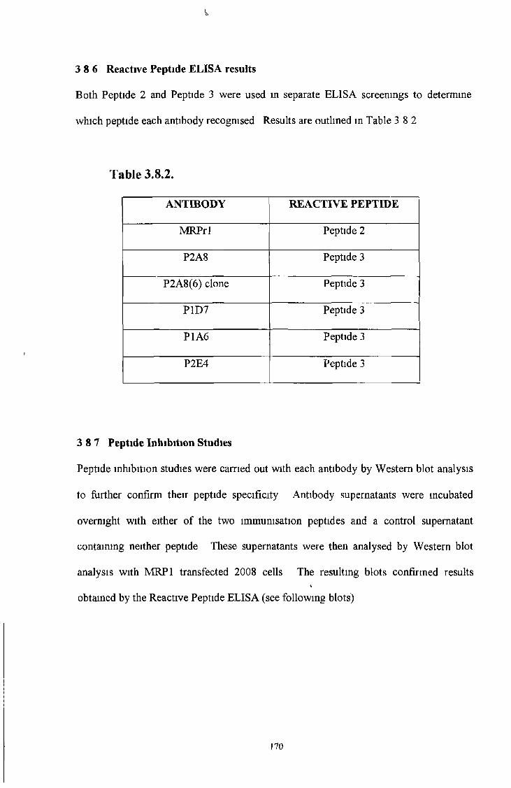
Both Peptide 2 and Peptide 3 were used in separate ELISA screenings to determine
which peptide each antibody recognised Results are outlined m Table 3 8 2
Table 3.8.2.
3 8 6 Reactive Peptide ELISA results
ANTIBODY REACTIVE PEPTIDE
MRPrl Peptide 2
P2A8 Peptide 3
P2A8(6) clone Peptide 3
P1D7 Peptide 3
P1A6 Peptide 3
P2E4 Peptide 3
3 8 7 Peptide Inhibition Studies
Peptide inhibition studies were carried out with each antibody by Western blot analysis
to further confirm their peptide specificity Antibody supernatants were mcubated
overnight with either of the two immunisation peptides and a control supernatant
containing neither peptide These supernatants were then analysed by Western blot
analysis with MRP1 transfected 2008 cells The resulting blots confirmed results*
obtained by the Reactive Peptide ELISA (see following blots)
170
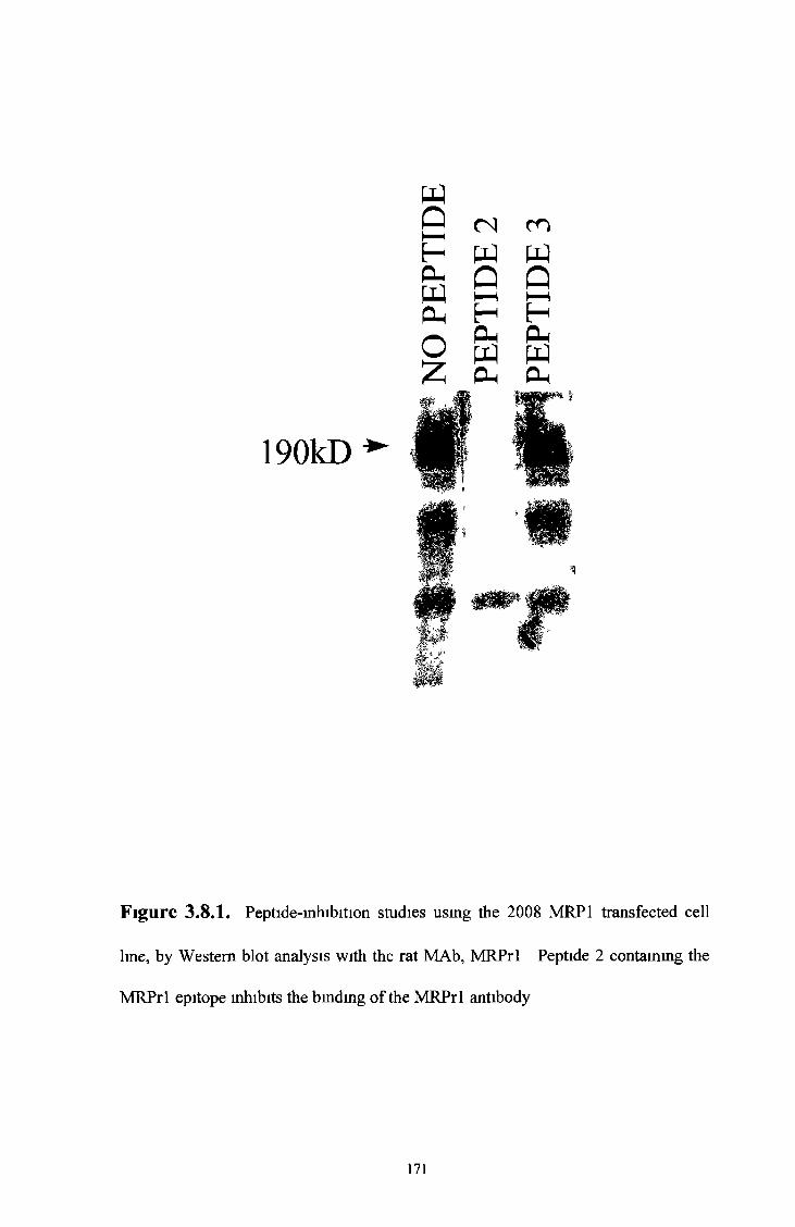
190kD
Figure 3,8.1. Peptide-inhibition studies using the 2008 MRP1 transfected cell
line, by Western blot analysis with the rat MAb, MRPrl Peptide 2 containing the
MRPrl epitope inhibits the binding of the MRPrl antibody
171
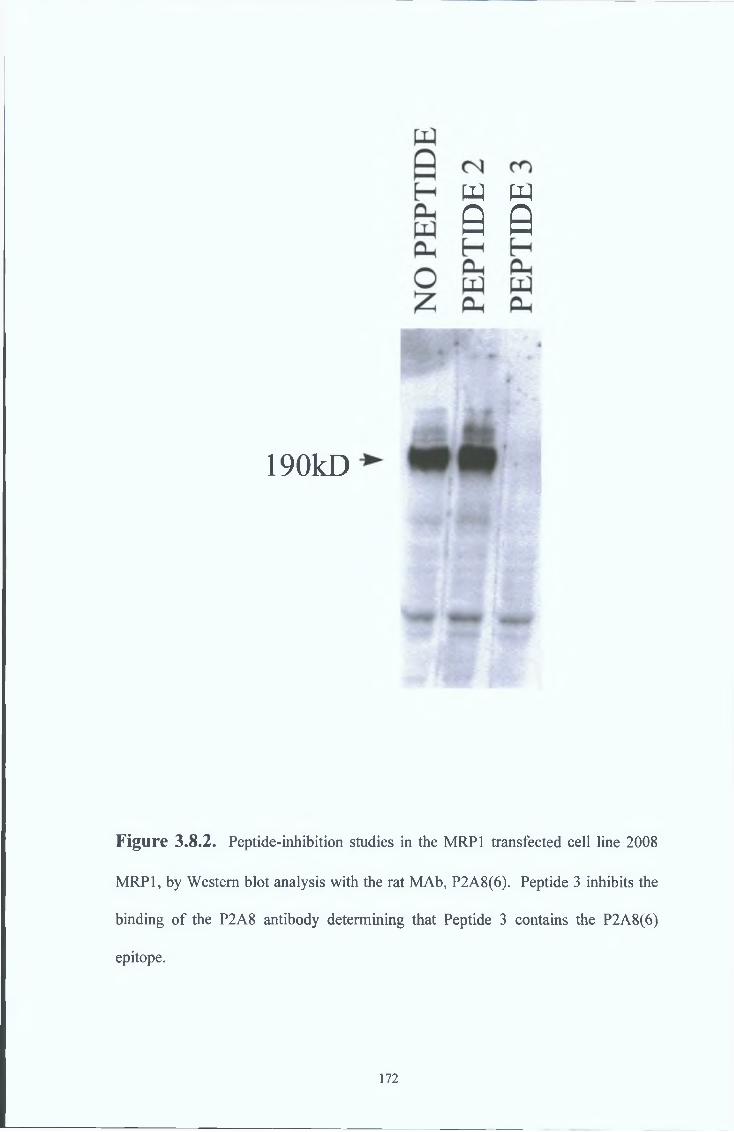
w w
Q 0
190kD
Figure 3.8.2. Peptide-inhibition studies in the MRP1 transfected cell line 2008
MRP1, by Western blot analysis with the rat MAb, P2A8(6). Peptide 3 inhibits the
binding of the P2A8 antibody determining that Peptide 3 contains the P2A8(6)
epitope.
172

190 kD
Figure 3,8.3. Peptide-inhibition studies in the MRP1 transfected cell line 2008
MRP1, by Western blot analysis with the rat MAb, P1D7 Peptide 3 inhibits the
binding of the P1D7 antibody determining that Peptide 3 contains the P1D7 epitope
173
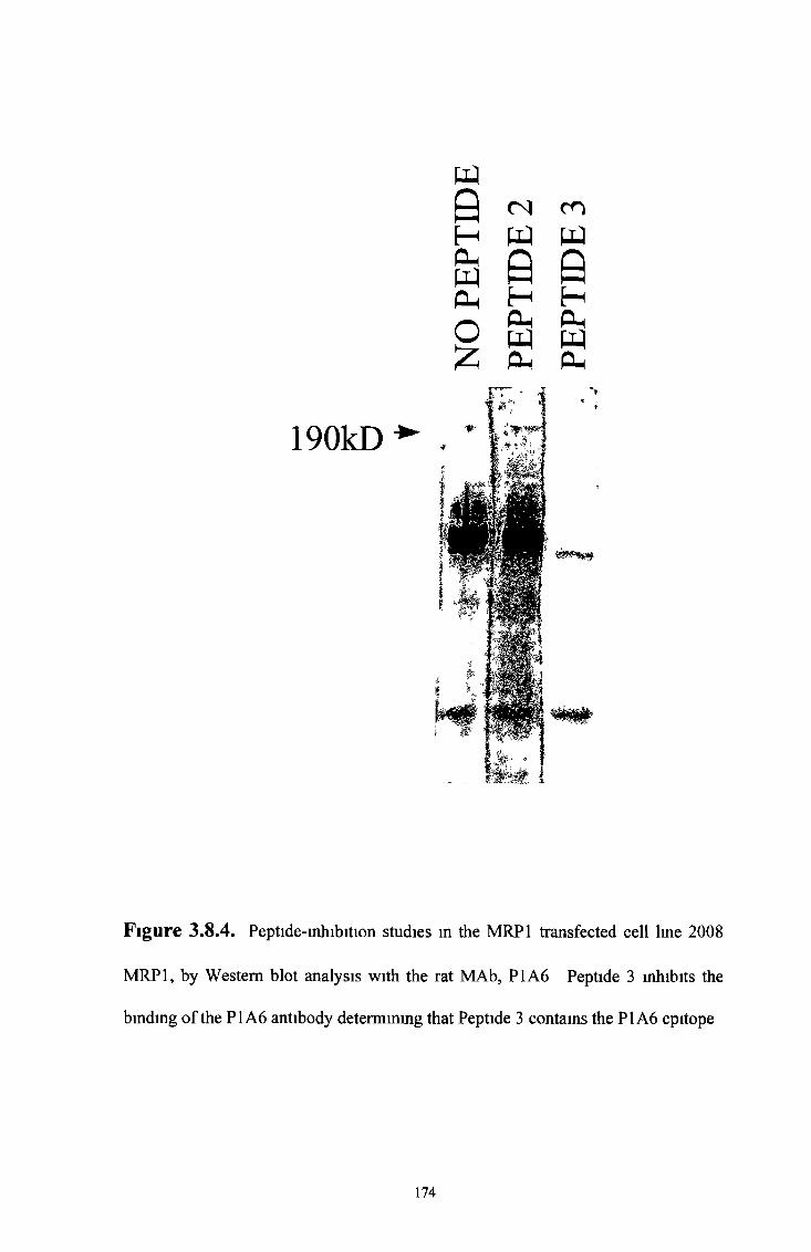
190kD
9HO hWPh
o£
( N c n
WeHPhWO h
w0HP hWPh
Figure 3.8.4. Peptide-inhibition studies in the MRP1 transfected cell line 2008
MRP1, by Western blot analysis with the rat MAb, P1A6 Peptide 3 inhibits the
binding of the PI A6 antibody determining that Peptide 3 contains the PI A6 epitope
174
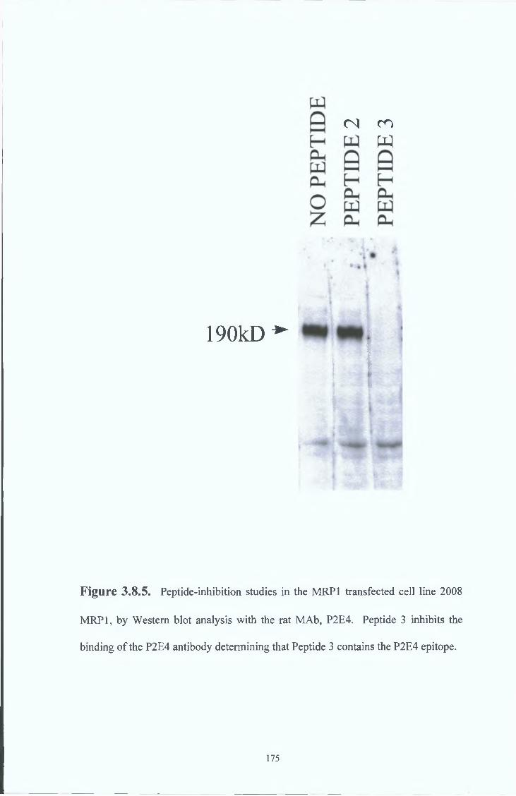
190kD *■
Figure 3.8.5. Peptide-inhibition studies in the MRP1 transfected cell line 2008
MRP1, by Western blot analysis with the rat MAb, P2E4. Peptide 3 inhibits the
binding of the P2E4 antibody determining that Peptide 3 contains the P2E4 epitope.
<N CO
w
175
3 8 8 Western Blot analysis
P2A8(6)
The MAb P2A8(6) was cloned from the MAb P2A8 Western blot analysis of this MAb
revealed that it is specific for a 190kD band in MRP1 over-expressing cells P2A8(6)
detects a 190kD band in drug selected MRP1 over-expressmg COR-L23R cells and
MRP1 transfected 2008 MRP1 cells (see Figure 3 8 6) P2A8(6) also detects a 190kD
band m the drug selected MRP1 over-expressmg HL60ADR cells (see Figure 3 8 7 )
P1D7
The MAb P1D7 detects a 190kD band in the 2008 MRP1 transfectants (see Figure
3 8 8 ) However, P1D7 does not detect a 190kD band in the drug-selected MRP 1-over
expressing COR-L23R cell lme suggesting that this MAb is specific for the transfected
MRP1 and not the drug-induced MRP1 (see Figure 3 8 9 )
176

190KDa
CCfOf S
iP4Ou
(Z )mfN
i
ou
ea>>*A
Oh
00©©(N
▼HOh Oh Ohc c 0- ££ £ s00 00 00o o ©© o ©<N N
'mggg/m
Figure 3.8.6. Western blot analysis of COR-L23R, COR-L23S, 2008 Parental,
2008 MRP1, 2008 MRP2 and 2008 MRP3 cells with MAb P2A8(6). MAb P2A8(6)
detects a 190KD band in the MRP1 over-expressing COR-L23R and MRP1
transfected 2008 MRP1 cell lines.
177
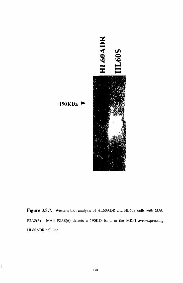
Figure 3.8.7. Western blot analysis of HL60ADR and HL60S cells with MAb
P2A8(6) MAb P2A8(6) detects a 190KD band in the MRP 1-over-expressing
HL60ADR cell line
178
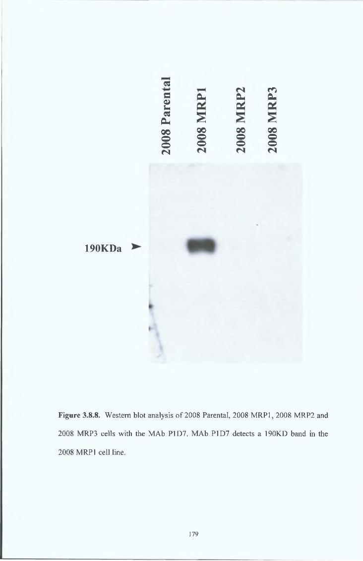
cs
eoucs
PH00oon
mOh a- OhOL X£ s00 00 00o o oo o o
190KDa
F ig u re 3.8.8. Western blot analysis of 2008 Parental, 2008 MRP1, 2008 MRP2 and
2008 MRP3 cells with the MAb P1D7. MAb P1D7 detects a 190KD band in the
2008 MRP1 cell line.
179
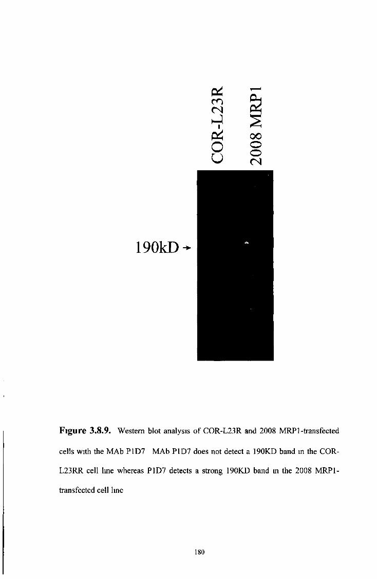
190kD
Figure 3.8,9. Western blot analysis of COR-L23R and 2008 MRP 1-transfected
cells with the MAb P1D7 MAb P1D7 does not detect a 190KD band m the COR-
L23RR cell line whereas P1D7 detects a strong 190KD band in the 2008 MRP1-
transfected cell line
180

3 8 9 Immunocytochemical analysis*
P 2 A 8 (6 )
Immunocytochemical analysis of various cell lines with P2A8(6) shows strong plasma-
membranous staining in every cell population of the COR-L23R and 2008 MRP1 cell
lines A basal level of stammg was detected in the Parental and MRP2 and MRP3 2008
transfectants (see Figure 3 8 10) The stammg by P2A8(6) of these cell lines is similar
to the stammg of these same cell lines by the rat monoclonal antibody, MRPrl, in
Figures 3 2 6 and 3 2 7
P1D 7
Immunocytochemical of various cell lines with P1D7 shows strong plasma-
membranous staining in a sub-population of cells in the COR-L23R and 2008 MRP1
cell lines P1D7 does not stain non or low MRP 1-expressing cells (see Figure 3 8 11)
181

• •• •
1. COR-L23R
%
4 , *
I
i*
2. COR-L23S
* f• 4m*
*
«i
#. •
n i?
3. 2008 Parental. 4. 2008 MRP1
$% *f 0* * • iC" c
9 a*» •
f ** v «»
# ##
Y • . « • *•.
5. 2008 MRP2 6. 2008 MRP3
Figure 3.8.10. Immunocytochemical analysis of various cell lines with the rat
MAb P2A8(6). MAb P2A8(6) shows strong plasma-membranous staining in every
cell population of the COR-L23R and 2008 MRP1 cell lines. A basal level of
staining can be seen on the Parental and MRP2 and MRP3 2008 transfectants. The
staining by P2A8(6) of these cell lines is similar to the staining of these same cell
lines by the rat monoclonal antibody, MRPrl, in Figures 3.2.6. and 3.2.7.
182

1. COR-L23R 2. COR-L23S
t #
mm
* W
3. 2008 Parental
» #* A *
• • •4. 2008 MRP1
c
5. 2008 MRP2 6. 2008 MRP3
Figure 3.8.11. Immunocytochemical analysis of various cell lines with the rat
monoclonal antibody P1D7. MAb P1D7 shows strong plasma-membranous staining
in a sub-population of cells in the COR-L23R and 2008 MRP1 cell lines but does
not stain any of the non or low MRP 1-expressing cell lines.

The antibodies below were chosen because they are specific, consistent, stable
and are applicable in immunocytochemical and Western blotting techniques
3 9 SUMMARY OF MOST INTERESTING ANTIBODIES
T a b le 3 9 1.
ANTIBODY IMMUNOGEN SPECIES PROTEINSPECIFICITY
Polyclonal 1 Peptide 1 Rabbit MRP1
Polyclonal 2 Peptide 2 Rabbit MRP1
Polyclonal 3 Peptide 3 Rabbit MRP1
1A2 Peptide 1 Mouse MRP1 and/or another protein or MRP form
P2A8(6) Peptide 2 and Peptide 3
Rat MRP1
P1D7 Peptide 2 and Peptide 3
Rat Transfected MRP1
184

3 10 CELL LINE SURVEY
A cell line survey was carried out on a small number of cell lines by Western blot
analysis The most interesting MAbs (listed in Table 3 9 1) and MRPrl, were surveyed
j
185
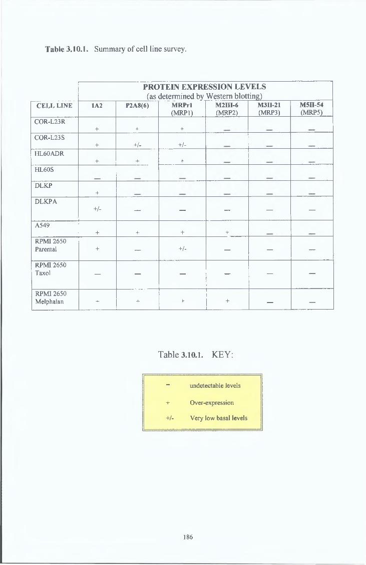
Table 3.10.1. Summary o f ce ll line survey.
PROTEIN EXPRESSION L E \(as determined by Western blotl
/ELS^ g )
CELL LINE 1A2 P2A8(6) MRPrl(MRP1)
M2HI-6(MRP2)
M3II-21(MRP3)
M5II-54(MRP5)
COR-L23R+ + +
COR-L23S+ +/- +/-
HL60ADR+ + +
HL60S
DLKP+
DLKPA+/- — — — — —
A549+ + + +
RPMI 2650 Parental + — +/- — — —
RPMI 2650 Taxol — — — — — —
RPMI 2650 Melphalan + + + + — —
Table 3.10.1. KEY:
- undetectable levels
+ Over-expression
+/- Very low basal levels
186
0 4 &0 q /c n m S<N (N wJ J < wJ , J , O O(X OS v o v oo o j jU U f f i f f i
o«rjno
O n 5 ^ 2 * n Oh
<
P3j2
rH 4 3 O &H
H S
§ 2 £
190kD
Figure 3.10.1. Survey by Western blot analysis, of various cell lines using the
MRP1 specific MRPrl MAb. Cell lines which express high levels of MRP 1 are;
COR-L23R, HL60ADR, A549 and RPMI Melphalan. Cell lines which express very
low levels of MRP 1 are; COR-L23S and RPMI 1650.
187
P h CO &c n c n »( N (N Q
,_ ) < 0 0 ^ ^ O °P h Ph vo voO O - I JV u ffi K
<& sh J h JQ Q
oi n^ o
ON t^H' nT 2 i n O h
< P4
O P h^ *a3
¡ 2 2
i xa c
190kD
Figure 3.10.2. Survey by Western blot analysis, of various cell lines using the
P2A8(6) MAb Cell lines which express high levels of P2A8(6) are, COR-L23R,
HL60ADR, A549 and RPMI Melphalan
188
190kD
O i c r t m r o <N <N h-l h-l
I I /«“s
P=h Pin VOo o o uh-lX
i n©voh-lX
<
£ &h-l h-lQ Q
o
vo
o s H
« n P h
< P4
Gj d1 3X i
1 3
£ £
Figure 3.10.3. Survey by Western blot analysis, of various cell lines using the IA2
MAb Cell lines which express high levels of MAb IA2 are, COR-L23R, COR-
L23S, HL60ADR, DLKP, A549, RPMI 2650 Parental and RPMI Melphalan Cell
lines which express low levels of MRP 1 are, DLKP A
189
P4co<Nh-1
C OP i
< N Q
i - l < in n ji
P i
o
V O
©
V O
O S
t a
O h - l h - 1 H - 1
u f f i < Q
190kD
IO Ph X aS
H
<D
Figure 3.10.4. Survey by Western blot analysis, of various cell lines using the
MRP2 specific MAb, M2III-6 Cell lines which express high levels of MRP2 are,
2008 MRP2 transfectants, A549 and RPMI 2650 Melphalan
190
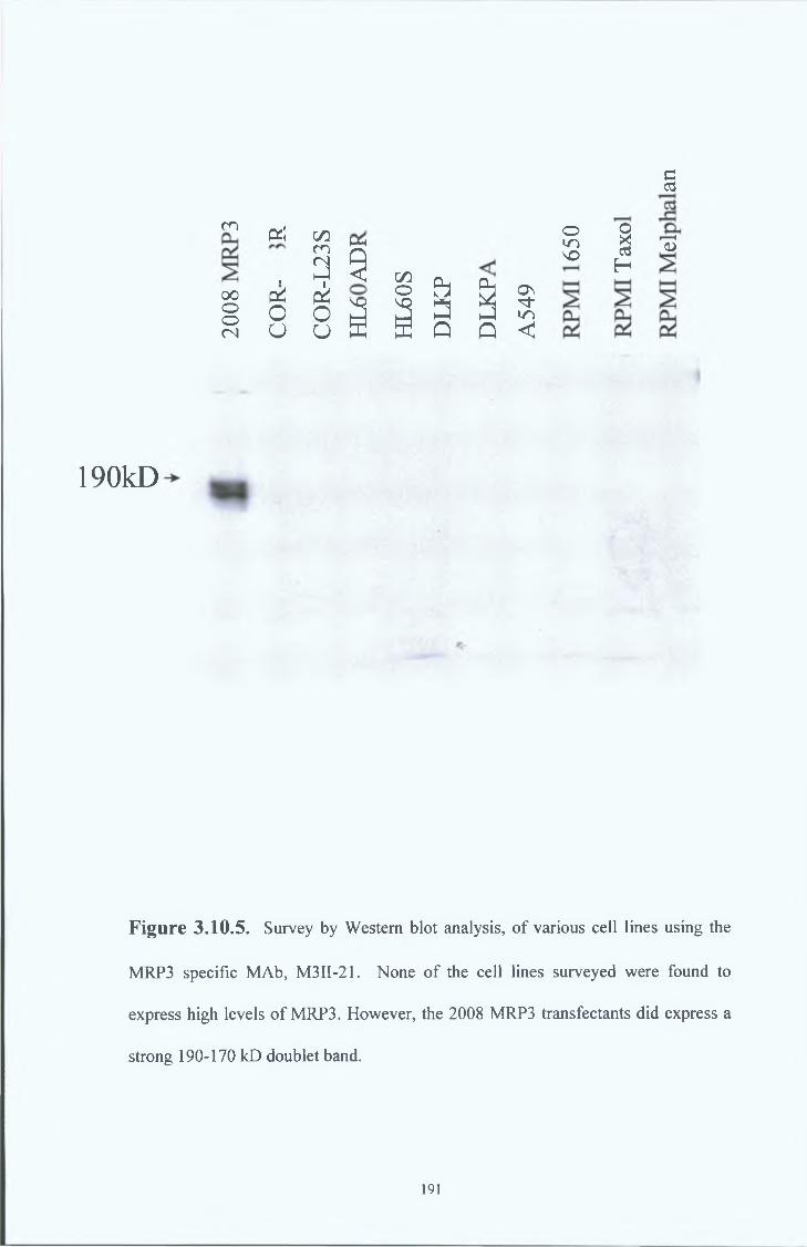
190kD
m
oooo<N
Pi! GOm<N■ hJ
pi eko Ou u
Mh
5 I 6 & a
OHO'O I
H
§
13
Si i 3 3 3
Figure 3.10.5. Survey by Western blot analysis, of various cell lines using the
MRP3 specific MAb, M3II-21. None of the cell lines surveyed were found to
express high levels of MRP3. However, the 2008 MRP3 transfectants did express a
strong 190-170 kD doublet band.
191

m m (N (Nh-li i
u u
P4Q< C/3o OVO vo
X
<
£ & h-l JQ Q
O n
<
©mv o
o
g
IOh
13
£ & £
190kD
¿V:\iV ;w. r*
Figure 3.10.6. MRP5 cell line survey.
Survey by Western blot analysis, of various cell lines using the MRP5 specific MAb,
M5II-54. None of the cell lines surveyed were found to express high levels of
MRP5.
192

4.0. DISCUSSION
193

4 0 DISCUSSION
4 1 General
The choice and synthesis of short synthetic peptides for the production of specific
monoclonal antibodies in our laboratory has proven highly successful Using short
synthetic peptides our laboratory has produced a range of monoclonal antibodies
specific for proteins such as the MDR-1 gene product, P-gp, MDR-3, and Topo II alpha
(Moran et a l , 1997, Larkin et a l , 1999b)
However, many factors may limit the utility of short synthetic peptides Antibodies
recogmse conformational structures (epitopes) consisting of amino acid sequences
which interact with the antigen-binding site of the antibody Epitopes may be either
continuous in the primary amino acid sequence (linear epitopes), or discontinuous,
resulting from juxtaposition of distant linear sequences There is no stnct consensus as
to what might be the minimal size of an antigenic determinant recognisable by
antibodies Ammo acid sequences of 7-10 amino acids may represent such a
determinant As a consequence, synthetic linear peptides can be used to generate
antibodies which cross-react with the native protein This offers the advantage that
antibodies can be raised against antigenic structures to which antibodies would not easily
be generated otherwise (for example, where a source of pure protein is unavailable)
Therefore, when short peptides are utilised for immunisation, anti-peptide responses are
generally obtained However, the recognition of the native protein in these cases is not
always assured (Claassen et a l , 1993) Induction of anti-native proteins by synthetic
peptides is dependent on many parameters The successful application of the synthetic
peptide approach involves a number of not entirely predictable elements, including the
194

selection o f a sequence, immunisation regime and screening system
Therefore, the employment of other forms of immunogens may be required in order to
gam a specific MAb which works under various conditions For example, longer
peptides, such as fusion proteins, may be used to increase the chances of obtaining an
antibody which recognises the native protein by increasing the chances of presenting
unique linear ammo acid sequences which are present in the native protein Immunising
with the native protein itself greatly increases the chances of obtaining a MAb which
recognises the native protein by presenting the antigen in its native conformation In this
case, antibodies should be produced which are specific for the conformation of the
native protein The pitfall in this scenario is that the location of the epitope is unknown
and undirected For example, specific epitopes may be advantageous in functional
assays and studies Short synthetic peptides are therefore useful for producing MAbs
which recognise these specific sites The most abundant source of a cellular protein in
its native form is obviously the cell itself Whole cell lysates or purer sources of the
native protein extracted from the cell, depending on the location of the protein in the
cell, may be used for immunisation In the case of MRP 1, cell membranes are a rich
source of this protein and can be purified from whole cell lysates to give a concentrated
source of the native MRP 1 protein
An important consideration for the successful production of a specific MAb is the choice
of species or strain of an animal for immunisation A number of species are useful for
hybndoma production including rats, and mice However, individual species of animals
may elicit various immune responses to the same immunogen Importantly some
antigens from origins other than mouse or rat species can induce much stronger
195
responses in rats than in mice and evidently the reciprocal could be true for other
antigens. Immunogens must also be compared with individual species for homology to
any proteins expressed by that species. The less homology found between the
immunogen and species, the more likely that an immune response will be elicited against
the immunogen.
The route of immunisation is extremely important for the presentation of the immunogen
(Harlow and Lane 1988). Various routes such as subcutaneous, intraperitoneal and the
lymphatic system present different environments for presentation of the antigen and may
structurally change the conformation of the antigen presented to immune cells (Claassen
et al., 1993). Therefore, antibodies produced from the same antigen in various
environments may detect various conformational forms of the antigen and may not be
suitable for all assays.
Therefore, in our experience the production of specific MAbs using short synthetic
peptides has proven successful for a number of antibodies mentioned above.
Unfortunately, not all proteins will present the perfect scenario and may pose a difficult
challenge in obtaining a specific MAb which recognises the native protein and works in
various assays. The production of an MRP 1-specific MAb has proven to be one such
challenge demanding that a number of the variables discussed above be examined.
Two collaborations were set up to aid the production and characterisation of an MRP1
specific MAb. Our laboratory participated in collaboration with Professor Balazs
Sarkadi of The Institute of Enzymology, Biological Research Center, Hungarian
Academy of sciences, H-1113 Budapest, Hungary. Professor Sarkadi kindly provided
196
our laboratory with various fragments of the MRP1 protein expressed in SF9 insect cells
and two large MRP1 fusion proteins These fusion proteins were used to immunise
mice and screen the resulting hybridomas for an MRP1 specific MAb The SF9 insect
membranes expressing various MRP1 fragments were in a limited quantity and were
used to characterise the rabbit and mouse MRP 1 antibodies produced in our laboratory
In return we made available our three MRP1 polyclonals and our MRP1 MAbs to
Professor Sarkadi’s laboratory Another collaboration was set up between our
laboratory and Mr George Scheffer of Professor Rik Schepers’ laboratory, Department
of Pathology and Oncology, Free University Hospital, 1081 HV Amsterdam Our
laboratory received various MRP isoform MAbs as well as the parental ovarian cancer
2008 cell line, MRP1, MRP2 and MRP3 transfected 2008 cell lines from MR George
Scheffer Permission for the use of the MRP3 transfected cell line (Kool et al 1999a)
came from Professor Marcel Kool, Academic Medical Center, Laboratory of
Neurozintuigen, Room K2-212, Meibergdreef 9, 1105 AZ Amsterdam These MRP
MAbs and MRP transfected cell lines were used to further characterise our MRP1
antibodies and for a small cell line survey
During the course of this thesis many MRP1 antibodies were produced Rabbits, mice
and rats were immunised with peptides, fusion proteins, whole cell lysates or cell
membranes to generate antibodies for the detection of MRP 1 Here efforts to produce
MRP 1-specific antibodies have proven successful in rabbit (polyclonal antibodies, PAbs)
and rat (monoclonal antibodies, MAbs), but has proven more complicated in mouse
(monoclonal antibodies, MAbs) However, these complications may point to the
existence of another form of MRP 1
197

Three polyclonal antibodies (see Table 4 2 1), designated 1, 2 and 3 were obtained by
immunising New Zealand White Rabbits with short synthetic peptide sequences (see
Section 3 6 ) corresponding to three separate amino acid sequences of human MRP1
The suitability of these synthetic peptides for use in monoclonal antibody production
was assessed by polyclonal production
Polyclonal 1 was raised against amino acids 1520-1531, an internal epitope in the C-
terminus Polyclonal 2 was raised against ammo acids 235-246 and Polyclonal 3 was
raised against ammo acids 246-260, both internal epitopes in the N-terminus region (see
Diagram 3 5 2 ) All three polyclonals, found to be suitable for Western blot analysis,
reacted strongly with a 190kD band in the MRP over-expressing, drug-selected cell line,
HL60ADR, and weakly or not at all with the parental HL60S cell line (Refer to Figures
3 6 1 , 3 6 3 and 3 6 5 , Results section) These results strongly suggested that the
polyclonals were reacting with MRP1
To confirm the specificity of these polyclonals, further Western blot analysis was
performed Polyclonal 1, 2 and 3 detected a 150kD band in human MRP-expressing
SF9 insect membranes and a shift of the 190kD band to a 150kD band in tumcamycin-
treated HL60ADR cells None of the polyclonals reacted with WT-MDR expressing
SF9 insect membranes All three polyclonals also reacted with a 140kD band in SF9
insect membranes expressing DeltaMRPl (Al), containing the MRPrl rat antibody
epitope (amino acids 192 - 360), while only polyclonal 1 detected a 120kD band in SF9
insect membranes expressing DeltaMRP2 (A2), which does not contain the MRPrl
epitope (see Figures 3 6 2 , 3 6 4 , 3 6 6 and 3 6 7)
4.2. Anti-MRPl rabbit polyclonal antibodies.
198
In summary, we have generated three polyclonal antibodies which are MRP 1-specific
and are suitable for Western blot analysis (see Table 4.1.1.). As expected Polyclonal 3,
raised against the C-terminus, behaved exactly as the mouse monoclonal MRPm6
(Bakos E. et al. 1996), also raised to the C-terminus, does in trypsinised and
chymotrypsinised experiments (Results not shown, carried out by Emese Sinkoe of
Professor Sarkadis’ laboratory, The Institute of Enzymology, Biological Research
Center, Hungarian Academy of sciences, H-1113 Budapest Hungary). As expected
Polyclonals 1 and 2 reacted with SF9 insect membranes, expressing the N-terminus of
human MRP1 which contains the MRPrl fusion peptide sequence intact, but did not
when this region was deleted (see Figures 3.6.2. and 3.6.4.).
Heat treatment of the MRP expressing whole cells lysates, @ 100°C for 3 minutes, did
not affect the ability of the three polyclonals to detect their epitopes (results not shown).
Taken together, these results provide strong confirmation of the MRP1 specificity of
Polyclonals 1, 2 and 3 and the suitability of peptides 1, 2 and 3 for the production of
monoclonal antibodies against MRP 1.
Table 4.2.1. Summary of Polyclonal antibodies.
PO LY C LO N A L IM M UNO G EN SPECIFICITY SUITABLE A PPLICATIO NS
Polyclonal 1 Peptide 1 MRP1 Western blot analysis and immuno-cytochemical analysis.
Polyclonal 2 Peptide 2 MRP1 Western blot analysis and immuno-cytochemical analysis.
Polyclonal 3 Peptide 3 MRP1 Western blot analysis and immuno-cytochemical analysis.
199
A number of MAbs were obtained by immunising B ALB/c mice with short synthetic
peptides or fusion proteins taken from the amino acid sequence of human MRP1 or
whole cell lysates or cells membranes of MRP 1 over-expressing COR-L23R cells (see
Section 3 7 ) The production of these MRP 1-specific mouse MAbs using various
immunogens resulted in a number of antibodies (see Table 4 3 1) However, the quest
for an anti MRP1 MAb involved the changing of many variables in the immunisation and
screening protocols
Immunisations using the synthetic peptides 1, 2 and 3 were carried out following
confirmation of their suitability by the MRP1 polyclonal results The subsequent fusions
were screened by the ELISA screening method using the peptide immunogen All
MAbs obtained by this regime identified MAbs which reacted with the linear peptide but
did not identify any protein by Western Blotting The resulting anti-peptide MAbs, 58C,
76F and 2B5, indicated that an immune response was elicited against each of the short
peptides The fact that they did not recognise the native protein by Western blot
analysis suggests that the MAbs were reacting with a linear epitope which was
conformationally different in the native protein Other possibilities may be that the
epitope is destroyed by Western blotting procedures or blocked by glycosylation sugars
Various Western blotting conditions, such as immunoprecipitation, non-denatunng and
non-reducing conditions were examined Glycosylation studies in which sugar
formation was prevented in MRP1 over-expressing cells were also examined However,
the various western blotting conditions and glycosylation studies did not reveal any
destroyed or hidden epitopes The most likely case for these MAbs is that immunising
with the short synthetic and the ELISA screening system employed produced MAbs to
4 3 Anti-MRPl mouse monoclonal antibodies
200
linear epitopes w hich do not recognise the native conformational protein
Following these results variations in both screening methods and immunogens were
developed A Western blotting screening system was set up to optimise the chances of
obtaining MAbs which recognise the native conformational protein and work under
Western blotting assay procedures Immunisations with Peptide 1 and Peptide 3 were
earned out and screened under this screening system Variations in immunogens
included fusion proteins, whole cell lysates and cell membranes containing the native
MRP 1 protein In an attempt to increase the chances of finding common linear epitopes
between the linear peptide and the mature native MRP1 protein, fusion proteins
containing 200 to 300 amino acids were used in immunisations Further efforts to
obtain a MAb which recognised the native MRP1 protein were made by immunising
with the native protein itself and screening against the native protein by Western
Blotting COR-L23R whole cell lysates and cell membranes over-expressing the native
MRP1 protein were used as immunogens All MAbs obtained by the Western blotting
method identified MAbs which reacted with a 190kD band in the MRP1 over-expressing
COR-L23R cell line in Western blots However, these MAbs also reacted with a
190kD, sometimes doublet band, in the sensitive parental COR-L23S cell line and
various other cell lines Fusion Protein 1 also yielded an antibody, 5E7, which
recognised a 190kD band in the MRP1 over-expressing COR-L23R cell line and also
reacted with a 190kD, sometimes doublet band, in the sensitive parental COR-L23 S cell
line
Another immunisation, using a purer source of native MRP1 protein than whole cell
lysates, was carried out using COR-L23R plasma membranes Initial screening
201
identified two hybndomas designated MAb 2A7 and MAb 6C6 which identified a
190kD band on COR-L23R but not COR-L23S cells (see Figure 3 7 18) Both these
MAbs appeared to be extremely “sticky” on Western blots and required high dilution in
buffers to produce clearer results This high background is possibly due to the fact that
neither of these MAbs were sub-cloned at this stage and other membrane protein MAbs
were possibly present in the hybndoma supernatants However, despite recognising a
190kD band in the COR-L23R cell line, neither MAb 2A7 nor MAb 6C6 recognised a
190kD band in the HL60ADR MRP1 over-expressing cell line suggesting that they were
not reacting with MRP1 (see Figures 3 7 19 and 3 7 20 ) MAb 2A7 detected two
lower bands in the drug resistant HL60ADR cells which decreased to one lower band
with higher dilution of MAb 2A7 MAb 2A7 did not detect any protein in the drug
sensitive HL60S cell line (see Figure 3 7 19) Preliminary immunocytochemical analysis
showed that MAb 2A7 stained both COR-L23R and COR-L23S cells in the plasma
membrane region similar to the staining observed in COR-L23R cells by the rat
monoclonal MRPrl (see Figure 3 7 25 ) However, MAb 2A7 stained the nucleoli of
both HL60ADR and HL60S cells (see Figure 3 7 25 ) This suggests that MAb 2A7 is
recognising a plasma membrane protein in COR-L23R and COR-L23S cells which is
located in the nucleoli of HL60ADR and HL60S cells Other investigators
(Krishnamachary et a l , 1993) have reported MRP1 expression in the membranes of
intracellular vesicles such as the endoplasmic reticulum It may be possible that the
epitope recognised by 2A7 may only be exposed on these intracellular membranes
An immunisation using COR-L23R whole cell lysates resulted in the MAb 4F6 MAb
4F6 recogmsed a 190kD band in the drug resistant COR-L23R and HL60ADR cell lines
but also their drug sensitive parental cell lines COR-L23S and HL60S (see Figure
202

3 7 16 ) However, lmmunocytochemical analysis of COR-L23R and COR-L23S cells
with 4F6 demonstrated strong staining of COR-L23R cells but not the COR-L23S cells
(see Figure 3 7 17) Unfortunately the hybndoma secreting MAb 4F6 was unstable and
stopped secreting antibody before further characterisation was carried out
The use of the Western blotting screening method to screen the immunisations earned
out with Peptide 1 and Peptide 3 resulted in two antibodies, 3A7 (Peptide 3) and 1A2
(Peptide 1) Both of these antibodies recognised a 190kD band in the COR-L23R and
COR-L23S cells However, after further characterisation, the hybndoma producing
MAb 3 A7 ceased antibody secretion MAb 3A7 proved to be unstable despite efforts to
re-clone it However, the 1A2 antibody has remained stable and consistently recognises
a 190kD band in the COR-L23R, COR-L23S and HL60ADR cells but not the HL60S
cells Due to the fact that 1A2 was the most stable mouse MAb produced which
worked under many procedures such as Western blotting and immunocytochemistry,
further characterisation concentrated on the 1A2 MAb
In summary, none of the mouse MAbs obtained behaved in a manner similar to that of
the rat monoclonal MRPrl antibody (see Table 4 2 1 ) Anti-peptides MAbs were
achieved which detected the linear peptide but not the native MRP1 Changes in
immunogens and screening procedures resulted in antibodies which detected a 190kD
band by Western blotting and stained cells by lmmunocytochemical procedures The
only mouse MAb which behaved in a similar manner to the MRPrl antibody by
lmmunocytochemical techniques was the 4F6 antibody (whole cell lysate fusion) which
unfortunately was unstable The most stable mouse MAb which worked in western
blotting and lmmunocytochemical techniques was the 1A2 MAb (Peptide 1 Fusion)
203
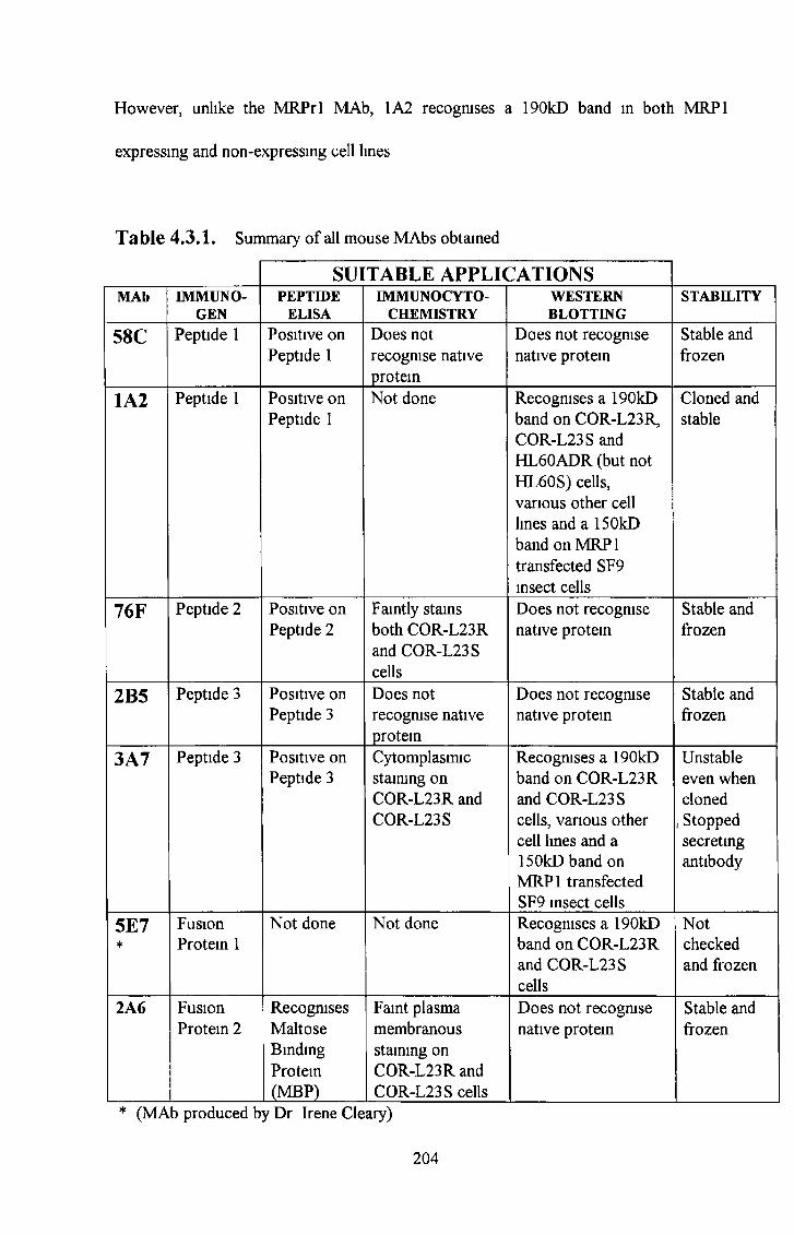
However, unlike the MRPrl MAb, 1A2 recognises a 190kD band in both MRP1
expressing and non-expressing cell lines
Table 4.3.1. Summary of all mouse MAbs obtained
SUITABLE APPLICATIONSMAb IMMUNO
GENPEPTIDE
ELISAIMMUNOCYTO-
CHEMISTRYWESTERNBLOTTING
STABILITY
58C Peptide 1 Positive on Peptide 1
Does not recognise native protein
Does not recognise native protein
Stable and frozen
1A2 Peptide 1 Positive on Peptide 1
Not done Recognises a 190kD band on COR-L23R, COR-L23 S and HL60ADR (but not HL60S) cells, various other cell lines and a 150kD band on MRP1 transfected SF9 insect cells
Cloned and stable
76F Peptide 2 Positive on Peptide 2
Faintly stains both COR-L23R and COR-L23 S cells
Does not recognise native protein
Stable and frozen
2B5 Peptide 3 Positive on Peptide 3
Does not recognise native protein
Does not recognise native protein
Stable and frozen
3A7 Peptide 3 Positive on Peptide 3
Cytomplasmic staining on COR-L23R and COR-L23S
Recognises a 190kD band on COR-L23R and COR-L23 S cells, various other cell lines and a 150kD band on MRP1 transfected SF9 insect cells
Unstable even when cloned
, Stopped secreting antibody
5E7*
Fusion Protein 1
Not done Not done Recognises a 190kD band on COR-L23R and COR-L23S cells
Notchecked and frozen
2A6 Fusion Protein 2
RecognisesMaltoseBindingProtein(MBP)
Faint plasma membranous staining on COR-L23R and COR-L23 S cells
Does not recognise native protein
Stable and frozen
* (MA 3 produced by Dr Irene Cleary)
204
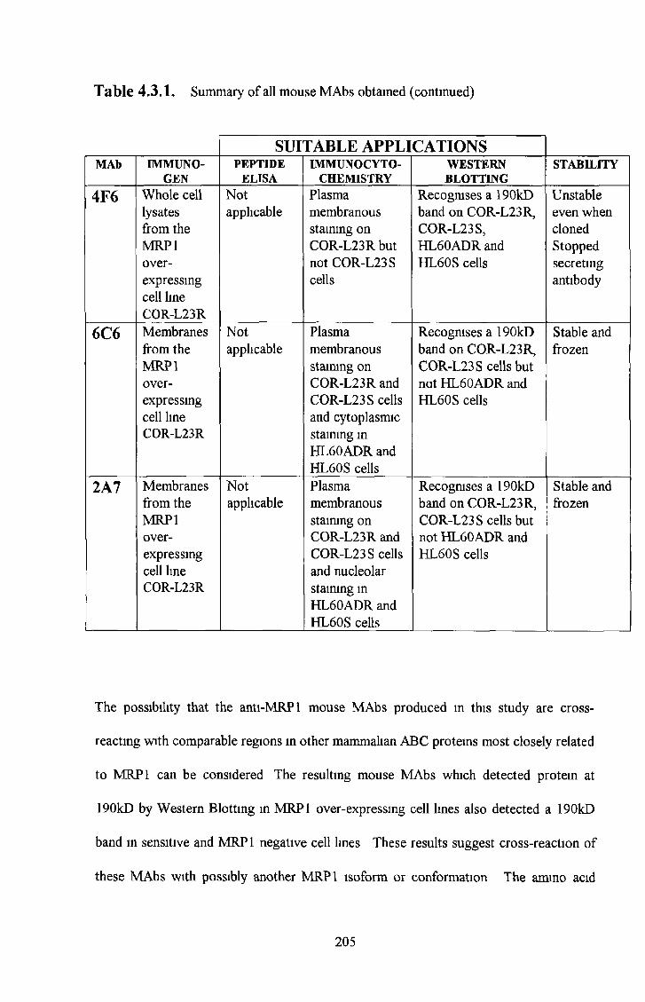
Table 4.3.1. Summary o f all mouse M A bs obtained (continued)
SUITABLE APPLICATIONSMAb IMMUNO
GENPEPTIDE
ELISAIMMUNOC YTO-
CHEMISTRYWESTERNBLOTTING
STABILITY
4F6 Whole cell lysates from the MRP1 overexpressing cell line COR-L23R
Notapplicable
Plasma membranous staining on COR-L23R but not COR-L23 S cells
Recognises a 190kD band on COR-L23R, COR-L23S, HL60ADR and HL60S cells
Unstableeven whenclonedStoppedsecretingantibody
6C6 Membranesfrom theMRP1over-expressingcell lineCOR-L23R
Notapplicable
Plasma membranous staining on COR-L23R and COR-L23S cells and cytoplasmic staining in HL60ADR and HL60S cells
Recognises a 190kD band on COR-L23R, COR-L23 S cells but not HL60ADR and HL60S cells
Stable and frozen
2A7 Membranes from the MRP1 overexpressing cell line COR-L23R
Notapplicable
Plasma membranous staining on COR-L23R and COR-L23S cells and nucleolar staining in HL60ADR and HL60S cells
Recognises a 190kD band on COR-L23R, COR-L23 S cells but not HL60ADR and HL60S cells
Stable and frozen
The possibility that the anti-MRP 1 mouse MAbs produced m this study are cross
reacting with comparable regions in other mammalian ABC proteins most closely related
to MRP1 can be considered The resulting mouse MAbs which detected protein at
190kD by Western Blotting in MRP1 over-expressing cell lines also detected a 190kD
band in sensitive and MRP1 negative cell lines These results suggest cross-reaction of
these MAbs with possibly another MRP1 isoform or conformation The amino acid
205
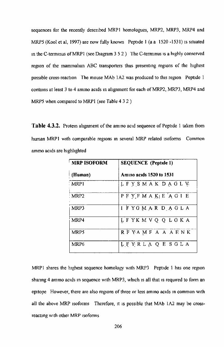
sequences for the recently described MRP1 homologues, MRP2, MRP3, MRP4 and
MRP5 (Kool et al, 1997) are now fully known Peptide 1 (a a 1520 -1531) is situated
in the C-terminus of MRP 1 (see Diagram 3 5 2 ) The C-terminus is a highly conserved
region of the mammalian ABC transporters thus presenting regions of the highest
possible cross-reaction The mouse MAb 1A2 was produced to this region Peptide 1
contains at least 3 to 4 ammo acids in alignment for each of MRP2, MRP3, MRP4 and
MRP5 when compared to MRP1 (see Table 4 3 2 )
Table 4.3.2. Protein alignment of the amino acid sequence of Peptide 1 taken from
human MRP1 with comparable regions in several MRP related isoforms Common
ammo acids are highlighted
MRP ISOFORM
(Human)
SEQUENCE (Peptide 1)
Amino acids 1520 to 1531
MRP1 L F Y S M A K D A G L V.
MRP2 P F Y^F M A Kj E A G I E
MRP3 I F Y G M A R D A G L A
MRP4 L F Y K M V Q Q L G K A
MRP5 R F Y A M F A A A E N K
MRP6 L F Y R L A Q E S G L A
MRP1 shares the highest sequence homology with MRP3 Peptide 1 has one region
sharing 4 ammo acids in sequence with MRP3, which is all that is required to form an
epitope However, there are also regions of three or less ammo acids in common with
all the above MRP isoforms Therefore, it is possible that MAb 1A2 may be cross-
reacting with other MRP isoforms
206
Cross-reactivity studies by Western blot analysis with MAb 1A2 and the 2008 Parental,
2008 MRP1, 2008 MRP2 and 2008 MRP3 transfectants showed that 1A2 detected a
190kD band in all these cell lines (see Figure 3 7 2 1 ) However, characterisation of the
MRP isoform antibodies (section 3 1 ) revealed that neither MRP2 nor MRP3 is
expressed in the COR-L23R, COR-L23S or HL60ADR cell lines As MAb 1A2
recognises a 190kD band in all these cell lines it appears to be more likely that 1A2 is
cross-reacting with another protein, or possibly with a different conformation of MRP 1
The possibility that MAb 1A2 is recognising another protein or conformationally
different form of MRP 1 is also evident A cell line survey (section 3 10 ) was earned
out on a small number of cell lines (see section 1 6 for information on cell lines used)
with the MRPrl, P2A8(6), 1A2, M2III-6, M311-21 and M5II-54 antibodies As
expected, the results showed that the MRPl-specific MRPrl MAb and the P2A8(6)
MAb detected a 190kD band in the MRP1 over-expressing cell lines, COR-L23R,
HL60ADR, A549 and RPM I2650 Melphalan selected The MRPrl and P2A8(6) MAbs
did not detect a 190 kD band in the COR-L23S, HL60S, DLKP, DLKPA, RPMI 2650
parental or RPMI 2650 Taxol-selected MRP2 was detected in the A549 and RPMI
2650 Melphalan selected cell lines with the M2III-6 MAb These results also correlate
with an RT-PCR study earned out in our lab (Yizheng Liang, PhD Thesis submitted,
1999) The 1A2 MAb detected a 190kD band in all these cell lines except HL60S,
DLKPA and RPMI 2650 Taxol selection Therefore, the antigen recognised by the 1A2
MAb appears to be down-regulated in Pg-P over-expressing cell lines The down-
regulation of MRP 1 in P-gp over-expressing cell lines is commonly observed (Brock et
a l , 1995) However, a survey of the 1A2 MAb in a wider selection of cell lines is
required to confirm this pattern MRP3 was detected with the M311-21 MAb in the
207
2008 MRP3 transfected cell line only MRP5 was not detected in any cell line with the
M5II-54 MAb See Table 4 2 3 for summary of protein expression in these cell lines
Hipfner et a l , 1998, reported that the MRPrl MAb recognised a linear epitope It was
found in this study that the MRPrl epitope was destroyed in heat treatment studies (see
Figure 3 2 9 ) which suggested that MRPrl may recognise a conformational epitope
However, heat treatment studies with the 2008 Ml cell lines did not destroy the MRPrl
epitope (see Figure 3 2 9 ) This may be due to the fact that the 2008 cells are MRP1
transfectants and the MRP1 may be incorporated into the cell membrane in a slightly
different conformation to that of MRP1 expressed intrinsically in the cell Heat
treatment studies showed that the epitope recognised by MAb 1A2 was not destroyed in
MRP1 drug-selected or -transfected cells suggesting that 1A2 recognises a linear
epitope
The cell line survey carried out in section 3 10 of the 1A2, P2A8(6), MRPrl (MRP1),
M2II-6 (MRP2), M311-21 (MRP3) and M5II-54 (MRP5) antibodies on various cells
lines was compared to P-gp expression in Table 4 3 3 This cell line survey showed that
the P2A8(6) followed an expression pattern similar to that of the well characterised
MRPrl antibody The 1A2 protein was found to be expressed in all of the examined
parental cell lines except the HL60S cell line It was expressed in the MRP1 induced
HL60ADR cell line However, it was also noted that the 1A2 protein appeared to be
downregulated (compared to the parental cell lines) in P-gp induced cell lines such as
the DLKPA and RPMI 2650 cell lines Therefore, these results suggest a link between
MRP1 expression, P-gp expression and expression of the protein detected by the 1A2
antibody It may be possible that the P-gp pump downregulates expression of the
208

protein detected by the 1A2 antibody and this protein is dependent on the MRP1
pathway
209
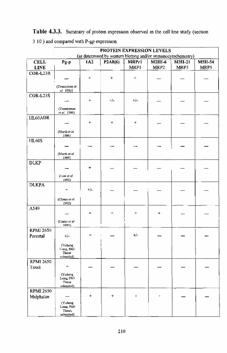
Table 4.3.3, Summary o f protein expression observed in the cell line study (section
3 10) and compared with P-gp expression
PROTEIN EXPRESSION LEVELS(as determined by western blotting and/or immunocytochemistry)
CELLLINE
Pg-P 1A2 P2A8(6) MRPrlMRP1
M2III-6MRP2
M3II-21MRP3
M5II-54MRP5
COR-L23R
(Twentyman et al 1986)
+ + + — — —
COR-L23S
(Twentyman et al 1986)
+ +/- +/- — — —
HL60ADR
(Marsh et al 1986)
+ + + — — —
HL60S
(Marsh et al 1986)
— — — — — —
DLKP
(Law et al 1992)
+ — — — — —
DLKPA+
(Clynes et al 1992)
+ /- — — — — —
A549
(Center et al 1993)
+ + + + — —
RPMI 2650 Parental + /-
(Yizheng Liang, PhD
Thesis submitted)
+ — + /- — — —
RPMI 2650 Taxol +
(Yizheng Liang, PhD
Thesis submitted)
— — — — — —
RPMI 2650 Melphalan
(Yizheng Liang, PhD
Thesis, submitted)
+ + + + — —
210
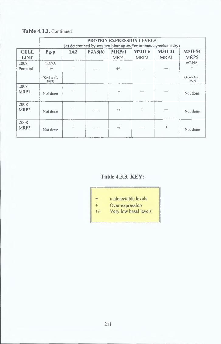
Table 4.3.3. Continued.
PROTEIN EXPRESSION LEVELS(as determined by western blotting and/or immunocytochemistry)
CELLLINE
Pg-P 1A2 P2A8(6) M R PrlMRP1
M2III-6MRP2
M3n-21MRP3
M5II-54MRP5
2008Parental
mRNA+/-
(Kool et al., 1997)
+ — +/- — —
mRNA+
(Kool et al, 1997)
2008MRP1 Not done
+ + + — — Not done
2008MRP2 Not done
+ — +/- + — Not done
2008MRP3 Not done
+ — +/- — +Not done
Table 4.3.3. KEY:
undetectable levels + Over-expression+/- Very low basal levels
211
In summary the antigen recognised by 1A2 is expressed in MRP1 over-expressing drug-
selected and MRP 1 transfected cell lines The 1A2 antigen is also expressed in sensitive
cell lines but appears to be down-regulated in Pg-p over-expressing cell lines It may be
possible that MRP1 is present in sensitive cells in a different conformation or active state
from that of drug selected MRP1 over-expressing cells A number of results point to
this conclusion The Western blotting screening system identified the 1A2 MAb Heat
treatment did not destroy the 1A2 MAb epitope The MRPrl antibody detects MRP1 in
COR-L23R and 2008 MRP1 transfected cells Heat treatment destroys the MRPrl
epitope in drug selected MRP1 over-expressing cells COR-L23R cells but not the
MRP1 transfected 2008 MRP1 cell line This suggests a difference in conformation
between drug selected and transfected MRP1 The conformational difference may be
due to another pattern of insertion of the protein in the cell membrane caused by
different lipid composition in the separate cell lines It may be possible that the drug-
selected MRP1 is heat labile while the transfected MRP1 is more stable because the
conformation difference creates a stronger anchorage for the transfected MRP1 into the
cell membrane Taken together these results suggest that the conformation of active
drug selected MRP 1 is unique to resistant cells and another MRP 1 conformation which
is probably inactive may be present in sensitive cells It is, therefore, possible that the
epitope recognised by 1A2 is present in both these conformations
Alternatively, the 1A2 MAb may recognise MRP1 and cross react with all or some of
the recently discovered MRP1 homologues Cross reactivity studies showed that the
1A2 MAb detected a 190kD band in the MRP1, MRP2 and MRP3 transfected 2008 cell
lines Unfortunately the 1A2 MAb also reacted with a 190kD band in the parental un
transfected 2008 cell line However, there are basal levels of MRP 1 expressed in the
212
parental un-transfected 2008 cell line also Therefore the 1A2 MAb may be cross
reacting with MRP1, MRP2 and MRP3, detecting the basal level of MRP 1 in all the cell
lines or cross-reacting with another MRP 1-related protein expressed in all these cell
lines The fact that the parental un-transfected 2008 cell line expresses a basal level of
MRP1 is an obstacle in the interpretation of these results The 1A2 MAb recognises a
heat stable conformation of the antigen This could be due to a different conformation
of the MRP1 protein and may also be reflected in a poorer efflux activity of this same
conformation 1A2 detects a 190kD band in the sensitive COR-L23S cell Preliminary
pharmacological experiments in this cell line which does not express MRP1, suggests
the presence of another MRP related protein (S Tuohey Personal communication)
Toxicity assays were earned out and the results compared between the COR-L23R and
2008 MRP1 cell lines to investigate whether there is any difference in the levels of drug
resistance (Tuohey, S , PhD Thesis, to be submitted) Lower levels of resistance were
found in the transfected cells Recently, Kool et a l , (1999a), described a limited range
of resistance in the transfected 2008 MRP3 cell line
Therefore the antigen recognised by the 1A2 MAb is probably MRP 1-related For
example, the 1A2 MAb also recognises the MRP1 transfected SF9 insect membranes but
not the control SF9 membranes and is inhibited by peptide 1 in peptide inhibition
studies Punfication by immunoprecipitation, followed by N-terminal sequence analysis
may determine the specificity of this MAb for MRP 1
213
A number of rat MAbs were obtained by immunising female Wistar rats with Peptide 2
and Peptide 3 in combination (see Table 4 4 1) The subsequent fusions were screened
and a number of monoclonal antibodies resulted These antibodies were designated
P2A8(6), P1D7, P1A6 and P2E4 All MAbs, except P1D7, obtained m this regime
recognised a 190KD band in MRP1 over-expressing cell lines but not the sensitive
counterparts by Western blotting P1D7 did not react with the COR-L23R cell line by
Western blotting but did react with a 190kD band in the MRP1 transfected 2008 cell
line Therefore, P1D7 appears to recognise the transfected MRP1 but not the drug-
selected MRP1 This difference may be due to a slight difference in the conformation of
the transfected and drug-selected MRP1 The transfected MRP1 may be incorporated
into the transfected cells membrane in a slightly different conformation to that of the
drug selected MRP1 over-expressing cells, possibly due to different lipid conformations
in the membranes of these cell lines The P2A8(6) MAb also reacted with MRP1 over-
expressing cell lines by immunocytochemical techniques in a similar manner to that of
the well characterised rat monoclonal antibody MRPrl That is P2A8(6) stained all cells
in MRP1 over-expressing cell lines However, unlike the rat MAb, MRPrl, the P1D7
antibody stained a subpopulation of cells in MRP 1 over-expressing cells These results
suggest that this P1D7 MAb may recognise a conformationally or structurally different
form of MRP 1 All of these MAbs were MRP1 specific and did not cross-react with
MRP2 or MRP3 as determined by Western blot analysis and immunocytochemical
techniques Peptide inhibition studies, confirmed by Western blot analysis, indicated that
the characterised selected rat MAbs reacted with an epitope present on Peptide 3
However, further stocks of selected hybndomas remain frozen Further characterisation
of these frozen hybndomas may detect MAbs which recognise an epitope on Peptide 2
4 4 Anti-MRPl rat monoclonal antibodies
214

An attempt to raise rat MAbs specific for external MRP1 epitopes was unsuccessful
Resulting MAbs reacted with both the COR-L23R and COR-L23S cell lines by live cell
immunofluoresence Other investigators have reported similar results in similar attempts
(Scheffer, G L Unpublished work )
215
Table 4.4,1. Summary o f rat M R P1 monoclonal antibodies
MAb PEPTIDESPECIFICITY
SUITABLEAPPLICATIONS
MRP1 SPECIFICITY
P2A8(6) Peptide 3Western blot analysis and lmmuno- cytochemistry
Specific for MRP1 in a similar manner to the rat monoclonal antibody, MRPrl, by Western blot and immunocytochemical analysis
PID7 Peptide 3Western blot analysis and lmmuno- cytochemistry
Specific for MRP1 in MRP1 transfected cells but not drug selected MRP 1 expressing cells byWestern blot analysis Recognises a sub-population of cells in MRP1 transfected and drug selected cells by immunocytochemical analysis
P1A6 Peptide 3Western blot analysis Very sticky in immuno- cytochemistry
Specific for MRP1 in MRP1 tansfected and drug selected cell lines in a similar manner to the rat monoclonal antibody, MRPrl, by Western blot analysis
P2E4 Peptide 3Western blot analysis and lmmuno- cytochemistry
Specific for MRP 1 in MRP 1 tansfected and drug selected cell lines in a similar manner to the rat monoclonal antibody, MRPrl, by Western blot analysis Recognises a sub- population of cells in MRP1 transfected and drug selected cells by immunocytochemical analysis in a similar manner to P1D7
216

The failure to produce a drug-induced MRP1 specific mouse monoclonal, which
behaves in a manner similar to that of the well characterised MRPrl rat MAb, may also
be due to a species difference. Considering the success of the peptides 1, 2 and 3 for the
production of rabbit polyclonals and the success of co-immunising rats with peptides 2
and 3, another possibility may be that the BALB/c mice may not be a suitable host
species for immunisation of the MRP1 protein. The epitopes recognised by the MRP1
specific MAbs MRPrl and MRPm6 (Flens et al, 1994) have recently been published
(Hipfner et al, 1998). MAb MRPrl was mapped to the decapeptide a.a. 238 to 247 of
human MRP1. This region is similar to Peptide 2 (a.a. 235 to 246 of human MRP1)
which was used in this study to successfully produce a rabbit polyclonal antibody but
failed to produce a mouse MAb specific for the mature MRP1 protein. The mouse
monoclonal MRPm6 was mapped to the decapeptide a.a. 1511 to 1520. This epitope is
adjacent to Peptide 3 (a.a. 1520 to 1531) used in this study to successfully produce a
rabbit polyclonal antibody but which failed to produce a mouse MAb specific for the
mature MRP1 protein. The murine mrp mRNA encodes a protein of 1528 amino acids
that is 88% identical to human MRP (Stride et al, 1996). Rat and rabbit MRP are less
homologous to human MRP and may be a more suitable choice of species for
immunisation. That is, murine MRP is so closely related to human MRP1 that it may be
seen as self-protein resulting in a lack of immunological response. Opposing this theory
is the fact that there are two mouse monoclonals to MRP1, MRPm6 (Flens et a l, 1994)
and QCRL-1 (Hipfner et al, 1994). However, both MRPm6 and QCRL-1 reinforce the
species theory. Their epitopes are specific for rare un-conserved regions in murine
MRP, which are not as closely homologous to human MRP as most of the regions.
Hipfner et al., 1998, stated that alignment of the MRPrl and MRPm6 epitope sequences
4.5. Species d ifference:
217
with the comparable regions in mammalian ABC proteins most closely related to MRP1
indicates, with the exception of murine mrp, the sequences are poorly conserved This
suggests that antibodies produced to Peptide 2, which contains the MRPrl epitope
sequence, will not cross-react with any other members of the mammalian ABC
transporter family
Murine mrp is 88% identical to human MRP1 Table 4 5 1 shows that the epitopes
mapped for the commercial MAbs, MRPm6 and QCRL-1, are rare regions which are
not highly conserved in mouse mrp That is, they both have at least two ammo acids
which are in contrast to corresponding ammo acids in mouse MRP These sequences
may be more immunogenic in mice than a more highly conserved sequence The
sequence used for the production of the commercial MRPrl antibody is more highly
conserved However, the MRPrl antibody was produced in rat The rat model of
immunisation may have a higher chance of eliciting a strong immune response against
most regions of human MRP1 because of a lower conservation of amino acid homology
between human MRP1 and rat MRP isoforms The same case is true for EBCR, the
rabbit homologue of MRP 1
218
Table 4.5.1. AMINO ACID SEQUENCE COMPARISONS OF COMMERCIAL
MRP1 ANTIBODY EPITOPES IN VARIOUS SPECIES. Differences in amino
acids present in the antibody epitope and corresponding amino acids in various species
highlighted in red.
M R P r l M R P m 6 Q C R L -1
5r V ]
(RatMAb) (Mouse MAb) (Mouse MAb)H§ epitope human MRP1 epitope human MRP1 epitope human MRP1wmÛ- a.a.238 - 247 a.al511-1520 a.a.918-924
Human and murine Human specific. Human specific.specific.
§eCQa GSDLWSLNKE PSDLLQQRGL SSYSGDIm3B
»«■1 A
1 &SSDLWSLNKE PSELLQQRGI SSHSGDT
I s
H<0§
LEDVWDIDEG PEEOOQIPGP ENSLRRT+ -)
«um fk
w LEDVWDIDEE PEELLESAGP SRSSRSS15..O
£
219
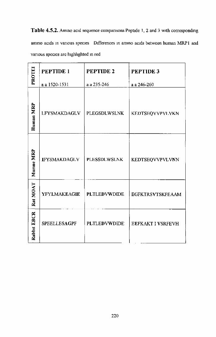
Table 4.5.2. Amino acid sequence comparisons Peptide 1, 2 and 3 with corresponding
amino acids in various species Differences in amino acids between human MRP1 and
various species are highlighted in red
PRO
TE
I
PEPTIDE 1
a a 1520-1531
PEPTIDE 2
a a 235-246
PEPTIDE 3
a a 246-260
£§ LF Y SMAKD AGL V PLEGSDLWSLNK KEDTSEQWPVLVKNM«ssW
§2§li IF Y SMAKD AGLV PLESSDLWSLNK KEDTSEQWPVLVNNsu§
Hus YFYLMAKEAGIE PLTLEDVWDIDE EGFKTRSVTSKFEAAM
Pi
P*UWw SPEELLESAGPF PLTLEDVWDIDE EEFKAKTIVSRFEVH-EpQPÌ
220

The route of immunisation may also play a role in the production of antibodies which
recognise the native protein (Harlow and Lane, 1988) All three species of animals were
immunised by various routes The rabbits were immunised subcutaneously, the rats in
the footpad and the mice mtrapentoneally Various routes create various environments
for presentation of the antigen and may structurally change the conformation of the
antigen presented to immune cells Therefore, the antibodies produced from similar
antigens in various species may detect various forms of the antigen and may not be
suitable for all assays This may explain the production of mouse MAbs specific for the
linear MRP1 peptide but unable to recognise the native MRP1 protein
4 7 Production of an external epitope MRPl-specific antibody*
The production of external epitope MRPl-specific antibodies has proven unsuccessful in
both rat and mouse MRP1 specific external epitope PAbs (rabbit) were not attempted
because immunisation with whole cells lysates or membranes would probably result in
polyclonal serum containing many antibodies specific for various components of the cell
or cell membrane thus masking the polyclonals specificity for the MRP 1 protein alone
Future work using external epitope peptides could be valuable, but it is possible that the
external epitopes may be masked by glycosylation sugars An obvious solution would
be to remove the glycosylation sugars from whole cells prior to immunisation
However, the resulting MAbs would probably be unable to recognise the mature, fully
glycosylated protein found in the native protein, thus destroying the end objective
Hipfner et a l , 1997 and Kast et a l , 1997 have determined that MRP1 does have
external protein fragments protruding from the membrane However, they do not tell us
how well these fragments are available to the immune response It is possible that these
4 6 Antigen presentation
221
external epitopes do not elicit an immune response Scheffer et a l , 1997, have tried
directing the immune response to the antigen of interest by masking unwanted cell
surface epitopes as described by Shen et a l , 1994, without success They also tried
fusion proteins corresponding to external fragments proposed in the membrane model by
Hipfner et a l , 1997 and Kast et a l , 1997 MAbs reacting with these peptides were
easily selected but these MAbs did not react with the membranes of MRP 1 expressing
cells They suggested that the immune response was directed mainly towards aberrant
epitopes due to e g peptide aggregation and not towards the native epitopes
222

5.0. CONCLUSIONS.
223

The use of various immunogens, species, immunisation protocols and screening
procedures can greatly enhance the chances of obtaining a desired antibody Antibodies
were produced in rabbits, mice and rats for the detection of MRP1 The rabbit
immunisations produced three MRP1 specific polyclonals The mouse monoclonals
produced include MAbs which recognise the peptide but not the native protein and the
1A2 MAb which detected MRP1 expressed in un-glycosylated form in SF9 membranes
and an MRP 1 -like antigen in cells, possibly a different conformational form of MRP 1 A
collaboration between our laboratory and Professor Balazs Sarkadi’s laboratory in
Budapest, Hungary made available MRP1 fusion proteins and a limited amount of
various MRP1 segments expressed in SF9 insect cells The rat immunisations which also
included two of the peptides used in the mouse immunisations produced a number of
MAbs One rat MAb (P2A8(6), behaved in a similar manner to that of the well
characterised MRPrl MAb and detected MRP1 homogeneously in drug selected or
transfected cells by Western blotting and immunocytochemical analysis Another rat
MAb (P1D7), detected transfected but not drug selected MRP1 by Western blot analysis
and detected a subpopulation of cells in both transfected and drug selected MRP1 cell
lines Prehmmaiy characterisation of these novel antibodies suggests that there are
various conformations or isoforms of MRP1 opening up the possibility of a more
complex mode of multidrug resistance executed by MRP1 It is possible that there are
different conformations of MRP 1 (perhaps produced by as yet unknown cofactors, e g
insertion into membrane lipids) which differ in drug efflux capability and/or substrate
specificity Whether there are various degrees of conformational change or a number of
active and less active isoforms and whether each conformation or active/less active
isoform elicits varying degrees of resistance is still not clear
5 0 CONCLUSIONS
224

Immunisation with the same peptides in mice, rats and rabbits produced different results
MRP 1-specific antibodies were obtained with the rats and rabbits while the mice
generally produced MAbs which recognised the linear peptide but not the native protein
and the 1A2 MAb which may recognise a conformationally different (possible less
active) MRP1 isoform However, immunisations in mice with MRP1 over-expressing
membranes produced the 4F6 MAb which detected plasma-membranous staining in
MRP1 over-expressing COR-L23R cells but not the COR-L23S parental cells by
immunocytochemistry 4F6 also recognised a 190kD band in COR-L23R, COR-L23S,
HL60ADR and HL60S cells by western blotting These bands may be due to the fact
that 4F6 was un-cloned at this stage Unfortunately 4F6 was unstable and ceased MAb
secretion despite sub-cloning Although it seems to be extremely difficult to obtain an
MRP 1-specific MAb in mice, the 4F6 results do suggest that it is possible Further
proof of this is the two commercial mouse MRP1 MAbs which exist, MRPm6 and
QCRL-1 The difficulty may be that mouse MRP is 88% identical to human MRP1 and
an immune response may therefore be difficult to elicit as many regions of the amino
acid sequence may be seen as self-proteins Both of the commercially available mouse
MAbs mentioned above are specific for epitope regions which are unusually un
conserved m mouse MRP Both of these MAbs are also human specific and do not
cross react with mouse MRP suggesting that these epitopes are different in mouse and
human MRP Therefore they may be regions of higher immunogenicity in mice The
4F6 MAb may have been detecting one of these rare un-conserved regions also The
success of the rat and rabbit antibodies using the same peptides to produce MRP1
specific antibodies may be attributed to the fact that rat and rabbit MRP 1/MRP 1
homologues are highly un-conserved when compared to human MRP1, therefore posing
225

no problems with the majority of the human MRP1 ammo acid sequence in eliciting a
strong immunological response in either of these species
The recent sequencing of the genes for several new MRP homologues has led to new
MRP isoform transfected cell lines and MRP isoform antibodies MRP 1, 2 and 3
transfected 2008 ovarian cell lines and antibodies specific for MRP1, 2, 3, and 5 were
made available just before the end of the thesis to our lab in a collaboration set up with
Marcel Kool and George Scheffer from Professor Rik Scheper’s lab in Amsterdam
These cell lines and antibodies were invaluable in determining the specificity of the
MAbs produced in the course of this thesis for MRP 1, MRP2 or MRP3
226

6.0 FUTURE WORK.
227
6 0 FUTURE WORK
Purification by immunoprecipitation of the antigen recognised by the mouse MAb 1A2
followed by N-terminal sequence analysis of the protein would aid confirmation of the
specificity of this MAb A full cell line survey with MAb 1A2 should be carried out to
further confirm or disprove the link between P-gp over-expression and down-regulation
of the antigen recognised by the 1A2 MAb which was observed in the small cell line
survey carried out in this thesis already
The success of raising MRP1 specific rat MAbs suggests that the rat may be the most
suitable species for eliciting a strong immunological response against the MRP 1 protein
Therefore, further efforts to raise an external MRP1 epitope MAb using various
immunogens (external peptides, cell membranes, fusion proteins) and screening methods
(live cell immunofluorescence) should be tried
The epitope recognised by the rat MAb P2A8(6) is unique compared to other
commercially available MRP1 antibodies Therefore further characterisation of this
novel MRP1 specific MAb would facilitate both clinical and experimental investigations
in unravelling the role of MRP 1 in multidrug resistance
Interestingly the P1D7 rat MAb distinguishes between transfected and drug-selected
MRP1 by Western blot analysis and detects a subpopulation in transfected and drug
selected MRP1 cell lines by immunocytochemical analysis suggesting that there may be
at least two conformations of the MRP1 molecule A full cell line survey with the P 1D7
MAb should be carried out to determine the expression in various cell line types
228

A number of interesting antibodies have been produced during this thesis and have
raised the possibility that there are different MRP1 conformations (possibly less active
or have a different substrate specificity) present in cells and therefore possibly tissues
An investigation of the expression of these proteins should be carried out in a tissue
survey, including human normal and tumour samples
229

7.0 BIBLIOGRAPHY.
230
Almquist, K C , Loe, D W , Hipfner, D R , Mackie, J E , Cole, S P and Deeley, R G(1995) Characterisation of theMr 190,000 Multidrug Resistance Protein (MRP) in Drug- selected and Transfected Human Tumour Cells Can Res (55) 102-110
Bader, P , Schilling, F , Schlaud, M , Girgert, R , Handgretinger, R , Klingebiel, T , Treuner, J , Liu, C , Niethammer, D and Beck, JF (1999) Expression analysis of multidrug resistance associated genes in neuroblastomas Oncol Rep 6(5) 1143-6
Bakos, E , Hegedus, T , Hollo, Z , Welker, E , Tusnady, G E , Zaman, G J R , Flens, M J , Varadi, A and Sarkadi, B (1996) Membrane Topology and Glycosylation of the Human multidrug Resistance-associated Protein J Biol Chem 271(21) 12322-12326
Bakos, E , Evers, R , Szakacs, Tusnady, G E , Welker, E , Szabo, K , de Haas, M , van Deemter, L , Borst, P , Varadi, A and Sarkadi, B (1998) Functional multidrug resistance protein (MRP 1) lacking the N-terminal transmembrane domain J Biol Chem 273(48) 32169-75
Baldirn, N , Scotlandi, K , Barbanti-Brodano, G , Manara, M C Maunci D , Bacci, G , Bertom, F , Picci, P , Sottili, S , Campanacci, M , et al (1995) Expression of P- glycoprotein in high-grade osteosarcomas in relation to clinical outcome N Engl J M ed 333(21) 1380-1385
Barrand, M A , Rhodes, T , Center, M S and Twentyman, P R (1993)Chemosensitisation and drug accumulation effects of cyclosporin A, PSC-833 and verapamil in human MDR large cell lung cancer cells expressing a 190kD membrane protein distinct from P-glycoprotein Eur J Cancer 29A(3) 408-415
Beck, J , Bohnet, B , Brugger, D , Bader, P , Died, J , Scheper, R J , Kandolf, R , Liu, C , Niethammer, D and Gekeler, V (1998) Multiple gene expression analysis reveals distinct differences between G2 and G3 stage breast cancers, and correlations of PKCN with MDR1, MRP and LRP gene expression Br J Cancer 77(1) 87-91
Belinsky, M G , Bam, L J , Balsara, B B , Testa, J R and Kruh, D (1998)Characterisation of MOAT-C and MOAT-D, New Members of the MRP/cMOAT Subfamily of Transporter Proteins J Natl Can Inst 90(22) 1735-1741
Bellamy, W T (1990) Multidrug resistance in human tumours Curr Opin Oncol 2 (6) 1126-1132
Bellamy, W T (1996) P-glycoprotein and multidrug resistance Ann Rev Pharmacol Toxocol (26) 161-183
Berger, W , Elbling, L , Hauptmann, E and Micksche, M (1997a) Expression of the multidrug resistance-associated protein (MRP) and chemoresistance of human non-small- cell lung cancer cells Int J Cancer (73) 84-93
Berger, W , Hauptmann, E , Elbling, L , Vetterlein, M , Kokoschka, E and Micksche, M (1997b) Possible role of the multidrug resistance-associated protein (MRP) in chemoresistance of human melanoma cells Int J Cancer (71) 108-115
231
Bordow, S B , Haber, M , Madafiglio, J , Cheung, B , Marshall, G M and Norris, M D (1994) Expression of the Multidrug Resistance-associated Protein (MRP) Gene Correlates with Amplification and Overexpression of the N-myc Oncogene in Childhood Neuroblastoma Can Res (54) 5036-5040
Boring, C C , Squires, T S , and Tong, T (1993) Cancer statistics, 1993 CA Cancer J Clin 43(1) 7-26
Borst, P Genetic mechanisms of drug resistance (Review) (1991) Acta Oncol (30) 87
Breumnger, L M , Paul, S , Gaughan, K , Miki, T , Chan, A , Aaronson, S A and Kruh, G D (1995) Expression of multidrug resistance associated with increased drug efflux and altered intracellular drug distribution Cancer Res 55(22) 5342-5347
Brock, I , Hipfher, D R , Nielsen, B S , Jensen, P B , Deeley, R G , Cole, S P and Shested, M (1995) Sequential coexpression of the multidrug resistance genes MRP and mdrl and their products in VP-16 (etoposide)-selected H69 small cell lung cancer cells Cancer Res 55(3) 459-62
Broxterman, H J , Giaccone, G and Lankelma, J (1995) Multidrug resistance proteins and other drug transport-related resistance to natural product agents Curr Opin Oncol 7(6) 532-540
Brugger, D , Herbart, H , Gekeler, V , Seitz, G , Liu, C , Klingebiel, T , Orlikowsky, T , Einsele, H , Dennlinger, C , Bader, P , Niethammer, D and Beck, J F (1999) Functional analysis of P-glycoprotein and multidrug resistance associated protein related multidrug resistance in AML-blasts Leuk Res (5) 467-75
Center, M S (1993) Non-P-glycoprotein multidrug resistance in cell lines which are defective in the cellular accumulation of drug Cytotechnology (12) 109-125
Chan, H S L , Haddad, G and Thorner, PS , de Boer, G , Lin, Y , Ondrusek, N , Yeger, H and Ling, V (1991) P-glycoprotein expression as a predictor of the outcome of therapy for neuroblastoma N Engl J Med (325) 1608-1614
Chan, H S L , Thorner, P S , Haddad, G and Ling, V (1990) Immunohistochemical detection of P-glycoprotein prognostic correlation in soft tissue sarcoma of childood J Clin Oncol (8) 689-704
Chang, X B , Hou, Y X , Jensen, T J and Riordan, J R (1994) Mapping of cystic fibrosis transmembrane conductance regulator membrane topology by glycosylation site insertion J Biol Chem 269(28) 18572-18575
Chen, C J , Chin, J , Ueda, K , Clark, D , Pastan, I , Gottesman, M and Romnson, I (1986) Internal duplication and homology with bacterial transport proteins in the mdr-1 (p-glycoprotein) gene from multidrug resistant cells Cell (47) 381-389
232

Chuman, Y , Sumizawa, T , Takebayashi, Y , Njwa, K , Yamada, K , Haraguchi, M , Furukawa, T , Akiyama, S and Aikou, T (1996) Expression of the multidrug-resistance- associated protein (MRP) gene in human colorectal, gastric and non-small-cell lung carcinomas Int J Can (66) 274-279
Claassen, E , Zegers, N D , Laman, J D and Boersma, W J A (1993) Use of Synthetic Peptides for the Production of Site (Ammo Acid) Specific Polyclonal and Monoclonal Antibodies Year Immunol (7) 150-161
Clynes, M , Redmond, A , Moran, E and Gilvarry, U (1992) Multiple drug resistance in variants of a human non-small-cell lung carcinoma cell line, DLKPA Cytotechnology (10) 75-89
Clynes, M , Heenan, M and Hall, K (1993) Human cell lines as models for multidrug resistance in solid tumours Cytotechnology (12) 231-256
Cole, S P , Bhardwaj, G , Gerlach, J H , Mackie, J E , Grant, C E , Almquist, K C , Stewart, A J , Kurz, E U , Duncan, A M V and Deeley, R G (1992) Overexpression of a Transporter Gene in a Multidrug-Resistant Human Lung Cancer Cell Line Science (258) 1605-1654
Cole, S P , Sparks, K E , Fraser, K , Loe, D W , Grant, C E , Wilson, G M and Deeley, R G (1994) Pharmacological characterisation of multidrug resistant MRP-transfected human tumour cells Cancer Res 54(22) 5902-5910
Cole, S P and Deeley, R G (1998) Multidrug resistance mediated by the ATP-binding cassette transporter protein MRP Bioessays (2) 931-940
Coop, J M (1993) P-glycoprotein structure and evolutionary homologues In Multidrug Resistance in cancer cellular molecular and clinical approaches Clynes, M (Ed) Kluwer Academic Press London 1 -32
Dano, K (1973) Active outward transport of daunomycin in resistant Ehrlich ascites tumour cells Biochem Biophys Acta (323) 466-483
Davey, R A , Longhurst, T J , Davey, M W , Belov, L , Harvie, R M , Hancox, D and Wheeler, H (1995) Drug resistance mechanisms and MRP expression in response to epirubicin treatment in a human leukaemia cell line Leuk Res 19(4) 275-282
De Vita, V T (1989) principles of chemotherapy In Principles and practice of Oncology De Vita, Helman and Rosenberg (Eds) JP lippincott Company, Philadelphia 276-300
Dingemans, A M C , van Ark-otte, J , van der Valk, P , Apolinario, R M , Scheper, R J , Postmus, P E and Giaccone, G (1996) Expression of the human major vault protein LRP in human lung cancer samples and normal lung tissue Ann Oncol (7) 625-630
233
Evers, R , Kool, M , van Deemter, L , Janssen, H , Calafat, J , Oomen, L C , Paulusma, C C , Oude Elfennk, R P , Baas, F , Schinkel, A H and Borst, P (1998) Drug export activity of the human canalicular multispecific organic anion transporter in polarized kidney MDCK cells expressing cMOAT (MRP2) cDNA J Clin Invest 101(7) 1310- 1319
Filipits, M , Suchomel, R W , Dekan, G , Haider, K , Valdimarsson, G , Despich, D and Pirker, R (1996a) MRP and MDR1 Gene Expression in Primary Breast Carcinomas Clin Can Res (2) 1231-1237
Filipits, M , Suchomel, R W , Zochbauer, S , Malayen, R and Pirker, R (1996b) Clinical relevance of drug resistance genes in malignant diseases Leukemia 10(3) S10-S17
Filipits, M , Suchomel, R W , Zochbauer, S , Brunner, R , Lechner, K and Pirker, R(1997) Multidrug Resistance-associated Protein in Acute Myeloid Leukemia No impact on Treatment Outcome Clin Can Res (3) 1419-1425
Filipits, M , Stranzl, T , Pohl, G , Suchomel, R W , Zochbauer, S , Brunner, R , Lechner, K and Pirker, R (1999) MRP expression in acute myeloid leukemia An update Adv Exp Med Biol (457) 141-50
Flens, M J , Izquierdo, M A , SchefFer, G L , Fritz, J M , Meijer, C J , Scheper, R J and Zaman, G J (1994) Immunocytochemical detection of the multidrug resistance- associated protein MRP in human multidrug-resistant tumour cells by monoclonal antibodies Cancer Res 54(17) 4557-45563
Flens, M J , Zaman, G J R , van der Valk, P , Izquierdo, M A , Schroeijers, A B , SchefFer, G L , van der Groep, P , de Haas, M , Meijer, C J L M and Scheper, R J (1996) Tissue Distribution of the Multidrug Resistance Protein Am J Path 148(4) 1237-1247
Ford, JM (1995) Modulators of multidrug resistance Preclmical studies Hematol Oncol Clin North Am 9(2) 337-361
Fromm, M F , Leake, B , Roden, L D M , Wilkinson, G R and Kim, R B (1999) Human MRP3 transporter identification of the 55-flanking region, genomic organization and alternative splice variants Biochimica et Biophysica A eta (1415) 369-3 74
Gately, D P and Howell, S B (1993) Cellular accumulation of the anticancer agent cisplatin a review Br J Cancer (67) 1171 - 1176
Georges, E , Bradley, G , Ganepy, J and Ling, V (1990) Detection of P-glycoproteinisoforms by gene-specific monoclonal antibodies Proc Natl Acad Sei USA (87) 152- 156
Gerlach JH et al, (1987) P-glycoprotein in human sarcoma evidence for multidrugresistance J Clin Oncol 5 1452 - 1460
234

Germann, U A , Willingham, M C , Pastan, I and Gottesman, M M (1990) Biochemistry (29) 2295-2303
Giaccone, G , van Ark-otte, J , Rubio, G J , Gazdar, A F , Broxterman, H J , Dingemans, A M , Flens, M J , Scheper, R J and Pinedo, H M (1996) MRP is frequently expressed in human lung-cancer cell lines, in non-small-cell lung cancer and in normal lungs Int J Cancer 6 6 (6) 760-767
Goldstein, L J , Pastan, I and Gottesman, M M (1992) Multidrug resistance in human cancer Crit Rev Oncol Hematol 12(3) 243-253
Gottesman, M M , and Pastan, I (1993) Biochemistry of multidrug resistance mediated by the multidrug transporter Ann Rev Biochem (62) 385-427
Gottesman, M M , Pastan, I , and Ambudkar, S V (1996) P-glycoprotein and multidrug resistance Current Opinion in Genetics & Development (6) 610-617
Grant, C E , Valdimarsson, G , Hipfher, D R , Almquist, K C , Cole, S P and Deeley, R G (1994) Overexpression of multidrug resistance-associated protein (MRP) increases resistance to natural product drugs Cancer Res 54(2) 357-361
Gros, P , Croop, J and Housman, D (1986) Mammalian Multidrug Resistance Gene Complete cDNA Sequence Indicates Strong Homology to Bacterial Transport Proteins Cell (47) 371-380
Hamada, H , Hagiwara, K , Nakajima, T and Tsuruo, T (1987) Phosphorylation of the Mr 170,000 to 180,000 glycoprotein specific to multidrug-resistant tumour cells effects of verapamil, trifluoperazine, and phorbol esters Cancer Res 47(11) 2860-2865
Harlow, E and Lane, D (1988) Antibodies A Laboratory Manual Cold Spring Harbour Laboratory (Pub)
Hart, S M , Ganeshaguru, K , Hoffbrand, A V , Prentice, H G and Mehta, A B (1994) Expression of the multidrug resistance-associated protein (MRP) in acute leukaemia Leukemia 8(12) 2163-2168
Higgins, C F (1992) ABC transporters, from microorganisms to man Ann Rev Cell Biol (8) 67-113
Hipfher, D R , Gauldie, S D , Deeley, R G and Cole, S P (1994) Detection of the Mr190,000 Multidrug Resistance Protein, MRP, with Monoclonal Antibodies Can Res (54) 5788-5792
Hipfner, D R , Almquist, K C , Stride, B D , Deeley, R G and Cole, SP (1996) Location of a Protease-hypersensitive Region in the Multidrug Resistance Protein (MRP) by Mapping of the Epitope of MRP-specific Monoclonal Antibody QCRL-1 Can Res (56) 3307-3314
235

Hipfner, D R., Almquist, K.C., Leslie, E.M., Gerlach, J.H., Grant, C.E., Deeley, R.G. and Cole, S.P. (1997) Membrane topology of the multidrug resistance protein (MRP). A study of glycosylation-site mutants reveals an extracytosolic NH2 terminus. J. Biol. Chem. 272(38): 23623-236230.
Hipfner, D R., Gao, M., Scheffer, G., Scheper, R.J., Deeley, R.G. and Cole, S.P. (1998) Epitope mapping of monoclonal antibodies specific for the 190-kDa multidrug resistance protein (MRP). Br. J. Can. 78(9): 1134-1140.
Hirohashi, T., Suzuki, H., Ito, K., Ogawa, k., Kume, K., Shimizu, T. and Sugiyama, Y.(1998) Hepatic expression of multidrug resistance-associated protein-like proteins maintained in eisai hyperbilirubinemic rats. Mol Pharmacol 53(6): 1068-1075.
Hoffman, G. and Mattern, M. (1993) Topoisomerase II in multidrug resistance. Cytotechnology. 12(1-3): 137-154.
Hooijberg, J.H., Broxterman, H.J., Kool, M., Assaraf, Y.G., Peters, G.J., Noordhuis, P., Scheper, R.J., Borst, P., Pinedo, H.M. and Jansen, G. (1999) Antifolate resistance mediated by the multidrug resistance proteins MRP1 and MRP2. Cancer Res. 59(11): 2532-5.
Horio, M., Chin, K.V., Currier, S.J., Goldenberg, S., Williams, C., Pastan, I., Gottesman, M.M. and Handler, J. (1989) Transepithelial transport of drugs by the multidrug transporter in cultured Madin-Darby canine kidney derived cell line epithelia. J. Biol Chem. (264): 14880-14884.
Ihde, D.C. (1992) Chemotherapy of lung cancer. N. Engl J. Med. 327(20): 1434-1441.
Ikeda, K., Oka, M., Yamada, Y., Soda, H., Fukuda, A., Tsukamoto, K., Nogucji, Y., Isomoto, H., Takeshima, F., Murase, K., Kambira, S., Tomonaga, M. and Kohno, S.(1999) Adult T-cell leukemia cells over-express the multidrug-resistance-protein (MRP) and lung-resistance-protein (LRP) genes. Int. J. Cancer. 82(4): 599-604.
Ishikawa, T. (1992) The ATP-dependent glutathione S-conjugate export pump. Trends Biochem. Sci. (17): 463-468.
Ishikawa, T., and Ali-Osman, F. (1993) Glutathione-associated cis- diamminedichloroplatinum (II) metabolism, and ATP-dependent efflux from leukemia cells. Molecular characterisation of glutathione-platinum complex and its biological significance. J. Biol. Chem. (268): 20116-20125.
Ishikawa, T., Wright, C.D. and Ishizuka, H. (1994) GS-X pump is functionally overexpressed in cis-diamminedichloroplatinum (Il)-resistant human leukemia HL-60 cells and down-regulated by cell differentiation. J. Biol. Chem. 269(46): 29085-29093.
Ishikawa, T .4 Bao, J.J., Yamane, Y., Akimaru, K., Frindrich, K., Wright, C D and Kuo? M.T. (1996) Coordinated induction of MRP/GS-X pump and gamma-glutamycysteine synthase by heavy metals in human leukemia cells. J. Biol Chem. 271(25). 14981-14988.
236

Ito, K , Fujimori, M , Nakata, S , Hama, Y , Shingu, K , Kobayashi, S , Tsuchiya, S , Kohno, K , Kuwano, Ml and Amano, J (1998) Clinical Significance of the Increased Multidrug Resistance-Associated Protein (MRP) Gene Expression in Patients With Primary Breast Cancer Oncol Res (10) 99-109
Izquierdo, M A , van der Zee, A G J , Vermorken, JB , et al (1995) Drug resistance associated marker LRP for prediction of response to chemotherapy and prognosis in advanced ovarian carcinoma J Natl Can Inst 87(16) 1230-1237
Izquierdo, M A , Neefjes, J J , Mathan, A E , Flens, M J , Scheffer, G L and Scheper, R J (1996a) Overexpression of the ABC transporter TAP in multidrug-resistant human cancer cell lines Br J Cancer 74(12) 1961-1967
Izquierdo, M A , Shoemaker, R H , Flens, M J , Scheffer, G L , Wu, L , Prather, T R and Scheper, R J (1996b) Overlapping phenotyes of multidrug resistance among panels of human cancer-cell lines Int J Cancer (65), 1-8
Jansen, P L and Oude Elfennk, R P (1993) Hereditary conjugated hyperbilirubinemia in Wistar rats a model for the study of ATP-dependent hepatocanalicular organic anion transport Adv Vet Sci Comp M ed (37) 175-195
Jedlitschky, G , Leir, I , Buchloz, U , Barouin, K , Center, M and Keppler, D (1994) ATP-dependent transport of glutathione S-conjugates by multidrug resistant small-cell lung cancer cells Can Res (54) 4833-4836
Jedlitschky, G , Leier, I , Bucholz, U , Hummel-Eisenbeiss, J , Burchell, B and Keppler, D (1997) ATP-dependent transport of bilirubin glucuromdes by the multidrug resistance protein MRP1 and its hepatocyte canalicular isoform MRP2 Biochem J 327(1) 305- 310
Juliano, R L and Ling, V (1976) A surface glycoprotein modulating drug permeability in Chinese hamster ovary cell mutants Biochem Biophys Acta (455) 152-162
Kast, C and Gros, P (1997) Topology mapping of the amino-terminal half of multidrug resistance-associated protein by eiptope insertion and immunofluorescence J Biol Chem 272(42) 26479-87
Keppler, D , Muller, M , klunemann, C , Guhlmann, A , Krauss, K , Muller, J , Berger, U , Leier, I and Mayatepek, E (1992) Transport and in vivo elimination of cysteinyl leukotnenes Adv Enzyme Regul (32) 107-116
Keppler, D , Komg, J and Buchler, M (1997) The canalicular multidrug resistance protein, cMRP/MRP2, a novel conjugate export pump expressed in the apical membrane of hepatocytes Advan Enzyme Regul (37) 321-333
Kiuchi, Y s Suzuki, H , Hirohashi, T , Tyson, C A and Sugiyama, Y (1998) cDNA cloning and inducible expression of human multidrug resistance associated protein 3 (MRP3) FEBS Letters (433) 149-152
237
Kohler, G and Milstein, C (1975) Continuos cultures of fused cells secreting antibody of predefined specificity Nature 256(5517) 495-497
Koike, K , Kawabe, T , Tanaka, T , Toh, S , Uchiumi, T , Wada, M , Akiyama, S , Ono, M and Kuwano, M (1997) A canalicular multispecific organic anion transporter (cMOAT) antisense cDNA enhances drug sensitivity in human hepatic cancer cells Cancer Res 57(24) 5475-5479
König, J , Rost, D , Cui, Y and Keppler, D (1999) Characterisation of the human multidrug resistance protein isoform MRP3 localised to the basolateral hepatocyte membrane Hepatology (4) 1156-63
Kool, M , de Haas, M , Scheffer, G L , Scheper, R J , van Eijk, M J T , Juijn, J A , Baas, F and Borst, P (1997) Analysis of Expression of cMOAT (MRP2), MRP3, MRP4, and MRP5, Homologues of the Multidrug Resistance-associated Protein Gene (MRP1), in Human Cancer Cell Lines Can Res (57) 3537-3547
Kool, M , van der Linden, M , de Haas, M , Scheffer, G L , De Vree, M L , Smith, A J , Jansen, G , Peters, G J , Ponne, N , Scheper, R J , Oude Elferink, R P J , Baas, F and Borst, P (1999a) MRP3, an organic anion transporter able to transport anti-cancer drugs Proc Natl Acad Sei USA (96) 6914-6919
Kool, M , van der Linden, M , de Haas, Baas, F and Borst, P (1999b) Expression of Human MRP6, a Homologue of the Multidrug Resistance Protein Gene MRP1, in Tissues and Cancer Cells Can Res (59) 175-182
Krishnamachary, N and Center, M S (1993) the MRP Gene Associated with a Non-P- glycoprotein Multidrug Resistance Encodes a 190-kDa Membrane Bound Glycoprotein Can Res (53) 3658-3661
Kruh, G D , Chan, A,Myers, K , Gaughan, K , Miki, T and Aaranson, S A (1994) Expression Complementary DNA Library Transfer Establishes mrp as a Multidrug Resistance Gene Can Res (54) 1649-1652
Kruh, G D , Gaughan, K T , Godwin, A and Chan, A (1995) Expression Pattern of MRP in Human Tissues and Adult Solid Tumour Cell Lines Journal o f the National Cancer Institute 87(16) 1256-1258
Kuichi, Y , Suzuki, H , Hirohashi, T , Tyson, C A and Sugiyama, Y (1998) FEBS Lett (4 3 3 ) 149-152
Kuss, B J , Deeley, R G , Cole, S P , William, C L , Kopecky, K J , Wolman, S R , Eyre, H J and Callen, D F (1996) The biological significance of the multidrug resistance gene MRP in inversion 16 leukemias Leuk Lymphoma 20(5-6) 357-364
Lacave, R , Coulet, F , Ricci, S , Touboul, E , Flahault, A , Rateau, J G , Cesari, D , Lefranc, JP and Bernaudin, JF (1998) Comparative evaluation by semiquantitative reverse transcnptive polymerase chain reaction of MDR-1, MRP and GSTrc gene expression in breast carcinomas Br J Cancer (77) 694-702
238

Lai, S.L., Goldstein, L.J., Pastan, M.M., Tsai, C.M., Johnson, B.E., Mulshine, J.L., Ihde, DC., Kayser, K. and Gazdar. (1989) MDR-1 gene expression in lung cancer. J. Natl. Cancer Inst. (81): 1144-1150.
Larkin, A.M., Moran, E., Kennedy, S. and Clynes, M. (1999a) Investigations of multidrug resistance (MDR) protein levels in pre- and post-treatment samples from breast cancer patients. Br. J. Cancer. (78) SI (Abstract).
Larkin, A.M., Moran, E., Alexander, D., Doherty, J., Connolly, L., Kennedy, S. and Clynes, M. (1999b) A new monoclonal antibody that specifically recognises the mdr-3 gene product. Int. J. Can. 80(2): 265-271.
Law, E., Gilvarry, U., Lynch, V., Gregory, B., Grant, G. and Clynes, M. (1992) Cytogenetic comparison of two poorly differentiated human lung squamous cell carcinoma lines. Cancer Genet. Cytogenet. (59): 111-118.
Lee, J.S., Scala S., Matsumoto Y., Dickstein B., Robey R., Zhan Z., Altenberg G., and Bates SE. (1997) Reduced drug accumulation and multidrug resistance in human breast cancer cells without associated P-glycoprotein or MRP expression. J. Cell. Biochem. (65): 513 - 526.
Lee, K., Belinsky, M.G., Bell, D.W., Testa, J R. and Kruh, G. (1998) Isolation of MOAT-B, a Widely Expressed Multidrug Resistance-associated Protein/Canalicular Multispecific Organic Anion Transporter-related Transporter. Can. Res. (58): 2741- 2747.
Legrand, O., Perrot, J.Y., Tang, R., Simonin, G., Gurbuxani, S., Zittoun, R., Marie, J.P. (1996) Expression of the multidrug resistance-associated protein (MRP) mRNA and protein in normal peripheral blood and bone marrow haemopoietic cells. Br. J. Haematol. 94(1): 23-33.
Leier, I., Jedlitschky, G., Bucholz, U., Cole, S.P., Deeley, R.G. and Keppler, D. (1994) the MRP gene encodes an ATP-dependent export pump for leukotriene C4 and structurally related conjugates. J. Biol. Chem. 269(45): 27807-10.
Leveille-Webster, C. and Arias, I. (1995) The biology of the P-glycoproteins. J. Membrane Biol. (143): 89-102.
Linn, S.C., Pinedo, H.M., van Arke-Otte, J., van der Valk, P., Hoekman, K., Honkool, A.H., Vermrken, J.B. and Giaccone, G. (1997) Expression of drug resistance proteins in breast cancer, in relation to chemotherapy. Int. J. Cancer. (71): 787-795.
Loe D.W., Deeley R.G., and Cole S.P.C. (1996a) Biology of the multidrug resistance associated protein, MRP. Europ. J. Cancer. (32A): 945-957.
Loe D.W., Almquist, K.C., Deeley R.G., and Cole S.P.C. (1996b) Multidrug resistance protein (MRP)-mediated transport of leukotriene C4 and chemotherapeutic agents in membrane vesicles. Demonstration of glutathione-dependent vincristine transport. J. Biol. Chem. 271(16): 9683-9.
239

Longhurst, T J , O’Neill, G M , Harvie, R M and Davey, R A (1996) The anthracycline resistance-associated (ara) gene, a novel gene associated with multidrug resistance in a human leukaemia cell line Br J Can (74) 1331-1335
Lutzky, J , Astor, M B , Taub, R N , Baker, M A , Bhalla, K , Gervasom, J E , Rosado, M , Stewart, V , Krishna, S and Hindenburg, A A (1989) Role of glutathione and dependent enzymes in anthracycline-resistant HL60/AR cells Cancer Res 49(15) 4120- 4125
Marie, JP (1995) P-glycoprotein in adult hematologic malignancies Hematol Oncol Clin North Am (2 ) 239-249
Masuda, S , Saito, H and Inui, K I (1997) Interactions of nonsteroidal antiinflammatory drugs with rat renal organic anion transporter, OAT-K1 J Pharmacol Exp Ther 283(3) 1039-1042
Marsh, W , Sichen, D and Center, M S (1986) Isolation and characterisation of Adnamycin-resistant HL-60 cells which are not defective in the initial intracellular accumulation of drug Cancer Res (46) 4053-4057
McGrath, T , Latoud, C , Arnold, S T , Safa, A R , Felsted, L and Center, M S (1993) Mechanisms of multidrug resistance in HL60 cells Analysis of resistance associated membrane proteins and levels of mdr gene expression Biochem Pharmacol 38(20) 3611-3619
Mechetner, E , Cummiskey, R , Kyshtoobayeva, A , Garcia, R , Mera, R , Salwen, H , Cohn, S , Fruehauf, J and Levine, E (1997) MRP expression as a predictive marker in multidrug resistant breast cancer (Abstract) Proc Am Assoc Ca Res (38) 389
Michieli, M , Damiam, D , Ermacora, A , Masolim, P , Raspadon, D , Visam, G , Scheper, R J and Baccaram, M (1999) P-glycoprotein, lung resistance-related protein and multidrug resistance associated protein in de novo acute non-lymphocytic leukaemias biological and clinical implications Br J Haematol 104(2) 328-35
Moore, G E and Sandberg, A A (1964) Studies of human tumour cell line with a diploid karyotype Cancer (17) 170-175
Moore, S (1980) William H Stem J Biol Chem 255(20) 9517-9518
Moran, E , Larkin, A , Doherty, G , Kelehan, P , Kennedy, S and Clynes, M (1997) A new mdr-1 encoded P-170 specific monoclonal antibody (6/1C) on paraffin wax embedded tissue without pretreatment of sections J Clin Pathol 50(6) 465-71
Moscow, J and Dixon, H (1993) Glutathione related enzymes, glutathione and multidrug resistance Cytotechnology (12) 155-170
240
Muller, M , Meijer, C , Zaman, G J , Borst, P , Scheper, R J , Mulder, N H , de Vries, E G and Jansen,PL (1994) Overexpression of the gene encoding the multidrug resistance-associated protein results in increased ATP-dependent glutathione S-conjugate transport Proc Natl Acad Sei USA 91(26) 13033-7
Naredi, P , Heath, D D , Enns, R E and Howell, S B (1994) Cross resistance between cisplatin and antimony in a human ovarian carcinoma cell line Cancer Res (54) 6464- 6468
Nooter, K , Westerman, A M , Flens, M J , Zaman, G J R , Scheper, R J , van Wingerden, K E , Burger, H , Oostrum, R , Boersma, T , Sonneveld, P , Gratama, J W , Kok, T , Eggermont, A M M , Bosman, F T and Stoter, G (1995) Expression of the Multidrug Resistance-associated Protein (MRP) Gene in Human Cancers Clin Can Res (11) 1301-1310
Nooter, K , Bosman, F T , Burger, H , van Wingerden , K E , Flens, M J , Scheper, R J , Oostrum, R G , Boersma, A W M , van der Gast, A and Stoter, G (1996) Expression of the multidrug resistance-associated protein (MRP) gene in primary non-small-cell lung cancer Annals o f Oncology (7) 75-81
Nooter, K , de la Riviere, G B , Look, M P , van Wingerden, K E , Henzen-Logmans, S C , Scheper, R J , Flens, M J , Klijn, J G M, Stoter, G and Foekens, J A (1997a) The prognostic significance of expression of the multidrug resistance-associated protein (MRP) in pnmaiy breast cancer Br J Can 76(4) 486-493
Nooter, K , de la Riviere, G B , Klijn, J , Stoter, G and Foekens, J (1997b) Multidrug resistance protein in recurrent breast cancer Lancet 349(9069) 1885-6
Norns, M D , Bordow, S B , Marshall, G M , Haber, P S , Cohn, S and Haber, M (1996) Expression of the gene for multidrug-resistance-associated protein and outcome in patients with neuroblastoma New Engl J Med 334(4) 231-238
Norris, M D , Bordow, S , Haber, P S , Marshall, G M , Kavallaris, M , Madafiglio, J , Cohn, S L , Salwen, H , Schmidt, M L , Hipfner, D R , Cole, S P C , Deeley, R G and Haber, M, (1997) Evidence that the MYCN Oncogene Regulates MRP Gene Expression in Neuroblastoma Eur J Can 33(12) 1911-1916
Ota, E , Abe, Y , Oshika, Y , Ozeki, Y , Iwasaki, M , Inoue, H , Yamazaki, H , Ueyama, Y , Takagi, K , Ogata, T , Tamaoki, N and Nakamua, M (1995) Expression of the multidrug resistance-associated protein (MRP) gene in non-small-cell lung cancer Br J Can (72) 550-554
Oude Elferink, R P , Meijer, D K , Kuipers, F , Jansen, P L , Groen, A K and Groothuis, G M (1995) Hepatobiliary secretion of organic compounds, molecular mechanisms of membrane transport Biochim Biophys Acta 1241(2) 215-268
241
Paulusma, C C and Oude Elferink, R P (1997) The canalicular multispecific organic anion transporter and conjugated hyperbilirubinemia in rat and man J Mol M ed 75(6) 420-428
Ross, D D , Doyle, L A , Schiffer, C A , Lee, E J , Grant, C E , Cole, S P , Deeley, R G , Yang, W and Tong, Y (1996) Expression of multidrug resistance-associated protein (MRP) mRNA in blast cells from acute myeloid leukemia (AML) patients Leukemia 10(1) 48-55
Schachner, M et a l , (1977) Brain surface antigens detected by anti-corpus callosum antiserum Brain Research 127 87-97
Schaub, T P , Kartenbeck, J , Komg, J , Vogel, O , Witzgall, R , Kriz, W and Keppler, D (1997) Expression of the conjugate export pump encoded by the mrp2 gene in the apical membrane of kidney proximal tubules J Am Soc Nephrol (8) 1213-1221
Scheper, R J , Broxterman, H J , Scheffer, G L , et al (1993) Overexpression of a M(r)110,000 vesicular protein in non-P-glycoprotein mediated multidrug resistance Can Res (53) 1475-1479
Schneider, E , Cowan, K H , Bader,H, Toomey, S , Schwartz, G N , Karp, J E , Burke, P J and Kaufmann, SH (1995) Increased Expression of the Multidrug Resistance- Associated Protein Gene in Relapsed Acute Leukemia Blood 85(1) 186-193
Schummer, B , Siegsmund, M , Steidler, A , Toktomambetova, L , Kohrmann, K U and Aiken, P (1999) Expression of the gene for the multidrug resistance-associated protein in human prostate tissue Urol Res 27(3) 164-8
Shen, R , Su, Z Z , Olsson, C A , Goldstein, N I and Fisher, P B (1994) Surface-epitope masking a strategy for the development of monoclonal antibodies specific for molecules expressed on the cell surface J Natl Cancer Inst 86(2) 91-98
Stride, B D , Valdmarsson, G , Gerlach, J H , Wilson, G M , Cole, S P and Deeley, R G(1996) Structure and expression of the messenger RNA encoding the murine multidrug resistance protein, an ATP-binding cassette transporter Mol Pharmacol 49(6) 962- 971
Sugawara, I , Aral, T , Yamashita, T , Yoshida, A , Masunaga, A and Itoyama, S(1994) Expression of multidrug resistance-associated protein (MRP) in anaplastic carcinoma of the thyroid Cancer Lett 82(2) 185-185
Sugawara, I , Akiyama, S , Scheper, R J and Itoyama, S (1997) Lung resistance protein (LRP) expression in human normal tissues in comparison with that of MDR1 and MRP Cancer lett (112) 23-31
Sugawara, I , Kataoka, I , Monshita, Y , Hamada, H , Tsuruo, T , Itoyama, S and Mori, S (1988) Tissue distribution of P-glycoprotein encoded by a multidrug-resistant gene as revealed by a monoclonal antibody, MRK 16 Cancer Res 48(7) 1926-9
242
Sugiyama, Y., Kato, Y. and Chu, X. (1998) Cancer Chemother. Pharmacol Suppl.42: 44-49.
Taniguchi, K., Wada, M., Kohno, K., Nakamura, T., Kawabe, T., Kawakami, M., Kagotani, K., Okumura, K., Akiyama, S. and Kuwano, M. (1996) A human canalicular multispecific organic anion transporter (cMOAT) gene is overexpressed in cisplatin- resistant human cancer cell lines with decreased drug accumulation. Cancer Res. 56(18): 4124-9.
Tusnady, G.E., Bakos, E., Varadi, A. and Sarkadi, B. (1997) Membrane topology distinguishes a subfamily of the ATP-binding cassette (ABC) transporters. FEBS Letters. 402(1): 1-3.
Twentyman, P R., Fox, N.E., Wright, K.A. and Bleehen, N.M. (1986) Derivation and preliminary characterisation of adriamycin resistant lines of human cancer cells. Br. J.Cancer. (53): 529-537.
Twentyman, P.R. and Versantvoort, C.H. (1996) Experimental modulation of MRP (multidrug resistance-associated protein)-mediated resistance. Eur. J. Cancer. 32A(6): 1002-9.
Uchiumi, T., Hinoshita, E., Haga, S., Nakmura, T., Tanaka, T., Toh, S., Furukawa, M., Kawabe, T., Wada, M., Kagotani, K., Okumura, K., Kohno, K., Akiyam, S. and Kuwano, M. (1998) Isolation of a novel human canalicular multispecific organic anion transporter, cMOAT2/MRP3, and its expression in cisplatin-resistant cancer cells with decreased ATP-dependent drug transport. Biochem. Biophys. Res. Commun. 252(1): 103-10.
van Der Valk, P., van Kalken, C.K., Ketelaars, H., Broxterman, H.J., Scheffer, G., Kuiper, C.M., Tsuruo, T., Lankelma, J., Meijer, C.J., Pinedo, H.M., et al. (1990) Distribution of multidrug resistance associated P-glycoprotein in normal and neoplastic human tissues. Analysis with 3 monoclonal antibodies recognising different epitopes of the P-glycoprotein molecule. Ann. Oncol ( 1): 54-64.
Versantvoort, C.H.M., Broxterman, H.J., Pinedo, H.M., de Vries, E.G., Feller, N, Kuiper. C M. and Lankelma, J. (1992) Energy-dependent processes involved in reduced drug accumulation in multidrug-resistant human lung cancer cell lines without P- glycoprotein expression. Cancer Res. 52(1): 17-23.
Versantvoort, CH., Broxterman, H.J., Lankelma, J., Feller, N., Pinedo, H.M. (1994) Competitive inhibition by genistein and ATP dependence of daunorubicin transport in intact MRP overexpressing human small cell lung cancer cells. Biochem. Pharmacol. 48(6): 1129-36.
Versantvoort, C.H.M., Broxterman, H.J., Bagrij, T., Scheper, R.J. and Twentyman, P R.(1995) Regulation by glutathione of transport in multidrug resistant human lung tumour cell lines overexpressing multidrug resistance associated protein. Br. J. Can. (72): 82-89.
243

Walker, J E , Saraste, M , Runswick, M J and Gray, N J (1982) EMBO J (1) 945- 951
Wijnholds, J , Evers, R , van Leusden, M R , Mol, C A , Zaman, G J , Mayer, U , Beijnen, J H , van der Valk, M , Krimpenfort, P and Borst, P (1997) Increased sensitivity to anticancer drugs and decreased inflammatory response in mice lacking the multidrug resistance-associated protein Nature Medicine (11) 1275-1279
Wright, S R , Boag, A H , Valdmarsson, G , Hipfner, D R , Campling, B G , Cole, S P and Deeley, R G (1998) Immunohistochemical detection of multidrug resistance protein in human lung cancer and normal lung Clin Can Res 4(9) 2279-89
Young, L C , Campling, B G , Voskoglou-Nomiskos, T , Cole, S P , Deeley, R G and Gerlach, JH (1999) Expression of Multidrug Resistance Protein-related Genes in Lung Cancer Correlation with Drug Response Clin Can Res (5) 673-680
Zaman, G J R , Versantvoort, C H M , Smit, J J M , Eijdems, E W H M , de Haas, M , Smith, A J , Broxterman, H J , Mulder, N H , de Vries, E G E , Baas, F and Borst, P (1993) Can Res (53) 1747-1750
Zaman, G J R , Flens, M J , van Leusden, M R , de Haas, M , Mulder, H S , Lankelma, J , Pinedo, H M , Scheper, R J , Baas, F , Broxterman, H J and Borst, P (1994) The human multidrug resistance-associated protein MRP is a plasma membrane drug-effiux pump Proc Natl Acad Sei USA (91) 8822-8826
Zaman, G J R , Lankelma, J , van Telhngen, O , Beijnen, J , Dekker, H , Paulusma, C , Oude Elfermk, R P , Baas, F and Borst, P (1995) Role of glutathione in the export of compounds from cells by the multidrug-resistance-associated protein Proc Natl Acad Sei USA (92) 7690-7694
Zaman, G J R , Cnubben, N H , van Bladeren, P J , Evers, R and Borst, P (1996)Transport of the glutathione conjugate of ethaciymc acid by the human multidrugresistance protein MRP FEBS Lett 391(1-2) 126-30
Zhang, J T and Ling, V (1991) Study of membrane orientation and glycosylatedextracellular loops of mouse P-glycoprotein by in vitro translation J Biol Chem 266(27) 18224-32
Zhang, J T , Duthie, M and Ling, V (1993) Membrane topology of the N-terminal half of the hamster P-glycoprotein molecule J Biol Chem 268(20) 15101-10
Zhu, Q Sun, H and Center, M S (1997) Functional Analysis of the Nucleotide Binding Domains of the Multidrug Resistance Protein (MRP) Oncol Res (9) 229-236
244

8.0. APPENDICES.
245
Appendix 1
ABBREVIATIONS
MRP1 Multidrug resistance-associated protein 1 (protein or gene)
P-gp P-glycoprotein or P-170 (MDR1 gene protein product)
MDR1 Multidrug resistance protein 1 gene (P-gp encoding gene)
ABC/HRP Streptavidin/biotin-horseradish peroxidase conjugate
Adr Adnamycin
ATCC American Tissue Culture Collection
BCA Bicinchommc acid
BSA Bovine serum albumin
DAB Diaminobenzidine
d l^ O Deionised water/glass distilled water
DMEM Dulbeccos Modified Eagles Mediumv
DMSO Dimethyl sulfoximide
DNA Deoxyribonucleic acid
DOX Doxorubicin (Adnamycin)
ECL Enhanced chemiluminescence
ECACC European Collection of Animal Cell Culture
EDTA Ethylene diamino tetra-acetic acid
ELISA Enzyme-linked immunoabsorbent assay
FCS Foetal calf serum
FITC Fluoroscein-isocyanate
246
HAT Hypoxanthine, aminopterin, thymidine
HCL Hydrochloric acid
HEPES 4-(2-hydroxethyl-)-piperazine ethane sulphomc acid
HT Hypoxanthine, thymidine
IMS Industrial methylated spirits
kD KiloDalton
LRP Lung resistance related protein
MAb/s Monoclonal antibody/monoclonal antibodies
MDR Multidrug resistance
MEM Minimum Essential Medium
MgCh Magnesium chloride
mRNA Messenger RNA
Mw Molecular weight
NaCl Sodium chloride
N aH C 03 Sodium bicarbonate
NaOH Sodium hydroxide
NCTCC The National Cell and Tissue Culture Centre
ND Not done
NEAA Non-essential amino acids
NSCLC Non-small cell lung carcinoma
OD Optical density
PAGE Polyacrylamide gel electrophoresis
PBS Phosphate buffered saline
PEG Polyethylene glycol
247
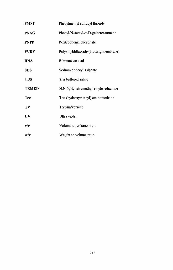
PMSF Phenylmethyl sulfonyl fluoride
PNAG Phenyl-N-acetyl-a-D-galactosammide
PNPP P-mtrophenyl phosphate
PVDF Polyvmyldifluonde (blotting membrane)
RNA Ribonucleic acid
SDS Sodium dodecyl sulphate
TBS Tns buffered saline
TEMED N,N,N,N,-tetramethyl-ethylenediamine
Tris Tns (hydroxymethyl) aminomethane
TV Trypsin/versene
UV Ultra violet
v/v Volume to volume ratio
w/v Weight to volume ratio
248
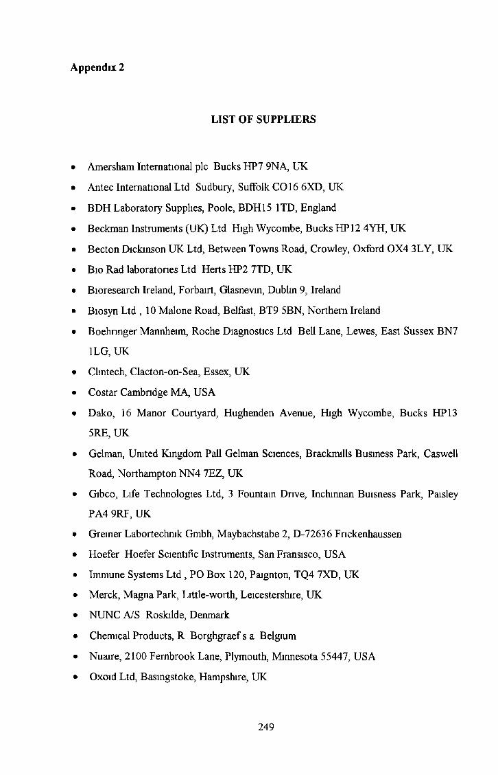
Appendix 2
LIST OF SUPPLIERS
• Amersham International pic Bucks HP7 9NA, UK
• Antec International Ltd Sudbury, Suffolk CO 16 6XD, UK
• BDH Laboratory Supplies, Poole, BDH15 1TD, England
• Beckman Instruments (UK) Ltd High Wycombe, Bucks HP 12 4YH, UK
• Becton Dickinson UK Ltd, Between Towns Road, Crowley, Oxford 0X4 3LY, UK
• Bio Rad laboratones Ltd Herts HP2 7TD, UK
• Bioresearch Ireland, Forbairt, Glasnevin, Dublin 9, Ireland
• Biosyn Ltd , 10 Malone Road, Belfast, BT9 5BN, Northern Ireland
• Boehnnger Mannheim, Roche Diagnostics Ltd Bell Lane, Lewes, East Sussex BN7
1LG, UK
• Clintech, Clacton-on-Sea, Essex, UK
• Co star Cambridge MA, USA
• Dako, 16 Manor Courtyard, Hughenden Avenue, High Wycombe, Bucks HP 13
5RE, UK
• Gelman, United Kingdom Pall Gelman Sciences, Brackmills Business Park, Caswell
Road, Northampton NN4 7EZ, UK
• Gibco, Life Technologies Ltd, 3 Fountain Drive, Inchinnan Buisness Park, Paisley
PA4 9RF, UK
• Greiner Labortechmk Gmbh, Maybachstabe 2, D-72636 Fnckenhaussen
• Hoefer Hoefer Scientific Instruments, San Fransisco, USA
• Immune Systems Ltd , PO Box 120, Paignton, TQ4 7XD, UK
• Merck, Magna Park, Little-worth, Leicestershire, UK
• NUNC A/S Roskilde, Denmark
• Chemical Products, R Borghgraef s a Belgium
• Nuaire, 2100 Fernbrook Lane, Plymouth, Minnesota 55447, USA
• Oxoid Ltd, Basingstoke, Hampshire, UK
249

• Pharmingen, BD Diagnostic Systems, Pottery Road, Kill O'The Grange, Dun
Laoghaire, Co Dublin, Ireland
• Pierce, Post Office Box 117, Rockford, Illinois 61105 USA
• Sigma Diagnostics, St Louis, MO 63178 USA
• Sigma, Sigma-Aldrich Company LTD Fancy Road, Poole, Dorset, BH12 4QH, UK
• Sterlin Ltd, Middlesex TW148QS, UK
• Vector Laboratories Ltd , 16 Wulfnc Square, Bretton, Peterborough PE3 8RF, UK
• Vector Laboratories, Inc Burlingame, CA 94010 USA
250