Embed Size (px)
Citation preview

Generation of recombinant antibodies for Ebola diagnostics XXXXXXXX
Mentors: Barbara D. Lipes, PhD & Michael D. Gunn, MD Duke University Department of Immunology
Duke University, Durham, North Carolina April, 2016
Abstract:
We used phage display technology to generate recombinant single chain antibodies to use
as diagnostic reagents for a variety of diseases. This project focused on generating antibodies for the Ebola virus. By using phage display technology, the screening of antibodies can be accomplished in a few days, whereas the previous method of using monoclonal antibodies would take months. The usage of bacteriophages, in this case the M13 phage, is a novel method of expressing antibody fragments to detect bacterial pathogens. The antibody’s structure includes the variable fragment (Fv), comprised of variable regions from heavy and light chains, which gives the antibody its specificity to an antigen. These fragments are joined together by a linker and are called a single chain antibody (scFv). In our process of selection and screening of different scFv, we used the EBOV sGP protein as our target. To avoid cross-reactivity with the Sudan virus sGP, we aimed to identify clones that were specific for EBOV sGP by incorporating negative selection with Sudan virus sGP. By using phage display technology, we were able generate a pool of phage clones enriched for binding to EBOV sGP. From this pool, we used ELISA to screen 192 clones, 82 of which were positive for EBOV sGP. We chose the 24 clones with high binding activity and specificity for EBOV sGP for characterization by restriction digests. Through further analysis, we have identified two clones that show great promise for generating antibodies that could be used as an EBOV diagnostic reagent in the future. Introduction:
Ebola virus (EBOV) is a severe, highly pathogenic disease that affects both humans and primates (Leroy et al., 2004). Ebola was first discovered in 1976 in Zaire, a country in Central Africa. Ebola is a negative-stranded RNA virus that belongs to a family of viruses known as Filoviridae (Leroy et al., 2004). There are four species of the genus Ebolavirus that can infect humans, Ebola virus (Zaire ebolavirus), Sudan virus (Sudan ebolavirus), Taï Forest virus (Taï Forest ebolavirus), and Bundibugyo virus (Bundibugyo ebolavirus) (Centers for Disease Control and Prevention, 2016). EBOV can lead to the most severe form of hemorrhagic fever (diseases that interfere with the body’s ability to clot) and can be fatal if left untreated (Leroy et al., 2004). In 90% of human cases, death is the outcome (LaVega, 2015). It has caused several outbreaks occurring in locations all over the world. The outbreak in West Africa from 2014-2015 is the most severe outbreak recorded in history, with more than 11,000 deaths reported from the outbreak (“Outbreaks”, 2016).
EBOV can spread rapidly throughout a population if not controlled or contained. During

2
the 2014 outbreak in West Africa, the reproduction numbers for Sierra Leone, Guinea, and Liberia were 2.53, 1.59, and 1.51, respectively (WHOER Team, 2014). Humans can be infected by EBOV through contact with bodily fluids of infected animals and non-human primates such as chimpanzees (World Health Organization, 2016). EBOV can spread between humans through direct contact or through contact with a material that has been contaminated with the bodily fuels of an infected individual (World Health Organization, 2016). It is important to note that EBOV can transmit through both the sick and the dead (World Health Organization, 2016). This makes burials ceremonies extremely dangerous if any individual comes into contact with the infected body. There is also evidence that EBOV can be transmitted sexually (Christie et al, 2015).
Currently, there are no vaccines or medications for EBOV. Because the symptoms of
EBOV are treated as they appear, and the earlier they get treated the better it is for the patient. Diagnosis of EBOV is difficult during the first few days of infection because the symptoms that are exhibited by the infected individual are not specific to EBOV (World Health Organization, 2016). The symptoms are often mistaken for other diseases, such as malaria, typhoid, and meningitis, because they share the same common symptoms (World Health Organization, 2016). An effective method for detecting EBOV in relatively early stages is using the reverse transcriptase-polymerase chain reaction assay (RT-PCR), which was found in 2000 (Leroy et al., 2000). However, this method requires specialized equipment and highly trained personnel, making it difficult to administer this method in the developing countries that are typically struck by EBOV in a timely method (Leroy et al., 2000). There is also a concern of false positives being generated from this diagnostic method (Leroy et al., 2000). Therefore, there is a need for development of novel assays that can reliably diagnose EBOV prior to the onset of symptoms.
We are aiming to overcome these limitations that exist in current diagnostics for EBOV
by targeting the EBOV soluble envelope glycoprotein (sGP) to develop a new point-of-care test for Ebola. SGP is a variant of GP, which is a membrane-bound protein that is found on the surface of the virus and is needed for viral infection to occur (Sanchez et al, 1996). The GP gene that encodes sGP is found in all five species of Ebolavirus (LaVega, 2015). Pre-SGP is encoded by gene four of the EBOV genome (Falzarano et al., 2006). EBOV SGP is a homodimer of parallel orientation with intermolecular disulfide bonds that are critical for the anti-inflammatory function of sGP (Falzarano et al., 2006).
Figure 1: Structure of EBOV sGP. The site of C-mannosylation (red), N-glycosylation (green), and disulfide bonding
(blue) are all illustrated in the schematic (Falzarano et al., 2007). We are choosing EBOV sGP as a diagnostic target based on what is known about the

3
biology of the EBOV infection. EBOV leads to the secretion of high levels of sGP six hours after infection, which is significantly shorter that the times it takes for significant viremia to occur which is 48 hours (Volchkova et al., 1999). Because sGP concentration levels are much higher than the concentrations of other proteins and are found to be high in patients with EBOV, we believe that an sGP diagnostic antibody will be able to detect EBOV infection prior to the onset of symptoms with high sensitivity. Wilson et al. found that monoclonal antibodies that immunoprecipitated EBOV sGP bound to Ebola Zaire, Ebola Ivory Coast, and Ebola Sudan viruses. While this suggests promise in the development of an antibody that can protect against all species of Ebola (Wilson et al. 2002), it also highlights the need for diagnostic antibodies that can discriminate between the highly conserved sGP proteins of the Ebolavirus family members.
We aim to develop a diagnostic assay that can evade the harmful consequences of
generating false negatives by generating recombinant single chain antibodies. The method we will use to target the EBOV sGP is to create specific antibodies that detect sGP epitopes using scFV (single chain F variable) phage display technology (Barbas et al., 2001). The usage of bacteriophages is a novel method of expressing antibody fragments to detect bacterial pathogens that allows rapid screening of a high number of antibodies (Barbas et al., 2001). In the M13 phage display method, an antibody fragment is expressed as a fusion with pIII phage protein resulting in the display of a functional antibody on the phage tip (Figure 2). By incorporating antibody fragments called the single chain F variable portions (scFv) that are fused to become phagemids, libraries of phage clones can be screened to determine the antibodies that identify EBOV sGP (Barbas et al., 2001).
Figure 2: Generation of M13 scFv library for phage display. VH and VL cDNAs are joined as scFv inserts and cloned
into phagemids to allow expression of scFv-pIII fusion proteins in E. coli host cells. Infection with M13K07 helper phage results in production of M13 phage particles displaying scFv antibodies
While a whole antibody consists of variable and constant regions, the scFv is composed of variable chains only, and consists of variable heavy chains (VH) and variable light chains (VL). The variable regions together create an antigen-binding site that recognizes a specific epitope. In a single chain variable fragment, there are two regions of VH and VL chains connected together by a peptide linker (Figure 3).

4
Figure 3. The native antibody contains 4 polypeptide chains, 2 heavy chains and 2 light chains. The scFv is composed of a heavy chain and a single chain. The molecular weights are indicated. Image is from Olafsen, Tove, Vania E. Kenanova, and Anna M. Wu. "Tunable pharmacokinetics: modifying the in vivo half-life of antibodies by directed mutagenesis of the Fc fragment." Nature protocols 1.4 (2006): 2048-2060.
The steps of phage display technology allow multiple rounds of selection can be
performed efficiently, which is one major advantage phage display technology has over previous methods of generating antibodies (Barbas et al., 2001). The procedure for generating the anti-EBOV sGP antibodies consists of generating an scFV phage display library and then identifying the scFV clones that are reactive to the target we are aiming for (Barbas et al., 2001). The whole process of a single round can be performed in 3 weeks, which allows flexibility in adjusting and repeating steps in the procedure in pre-clearing, selection, and screening. Within our phage display protocol, we are utilizing anti-HA magnetic beads, which are beads coated with anti-HA antibodies that are able to immobilize the sGP protein through the N-terminal HA tag that exists in sGP. We are using these beads to bind to the target sGP in effort to isolate phage clones that are bound to sGP from the pool of phage in solution. Overall, we are using phage display technology to create single chain F variable anti-EBOV sGP antibodies in effort to develop a novel diagnostic reagent for Ebola. Methods: Binding sGP target protein to magnetic beads
250 µl of 10 mg/ml Pierce anti-HA magnetic beads was pipetted into a sterile 1.5ml tube, where the beads in the magnet were left to stand for at least 1 minute. The beads gathered onto the side of the tube against the magnet due to the force of attraction. After the beads have gathered, the liquid was removed by slowly pipetting and avoiding contact with the beads. Then, the 1.5ml tube was removed from the magnet and 1 ml of sterile PBS was added to re-suspend and mix the beads. The tube was then put on the magnet again and left to stand until the beads gathered after 1 minute. The liquid was then pipetted out. The steps of adding PBS and removing the liquid were repeated. 1 ml of PBS containing 25 g of purified recombinant EBOV sGP was added to the tube. The tube was inverted 5-6 times to mix and placed on a rotator overnight at 4ºC.
Selection:
The unbound sGP was removed by placing the 1.5ml tube that was rotating overnight on the magnet for 1 minute. The liquid was pipetted out and 1 ml of sterile PBS + 2% BSA was
5
added to mix. This wash step was repeated two more times. The tube was then put on a rotator for one hour at room temperature then washed as above 3 times with 1 ml of PBS + 2% BSA. Then, 1011 phage, either from the library or the previous round of selection, were added to the tube. The sGP-coated beads were rotated with the phage for 1.5-2 hours at room temperature. Then, the tube was placed back on the magnet where the liquid was pipetted out and washed 3 times with 1 ml of sterile PBS + 0.1% Tween20 (PBST). After the final wash, the beads were suspended in 1 ml of sterile PBST and transferred to a 50 ml conical tube where an additional 25ml of PBST was added. The conical tube was then placed on a large magnet and left for 1-2 minutes for the magnetic beads to gather. The liquid was pipetted out with a 50ml pipette. Four additional 25 ml PBST wash steps were performed.
After removing the final wash, 1 ml PBST was used to transfer the beads to a new 1.5ml
tube for elution of the bound phage. The 1.5ml tube was then placed on the smaller magnet, where the liquid was removed and 1ml of 0.1 HCL was added. The tube was then rotated at room temperature for 10 minutes to elute the phage. After elution 1M Tris buffer [pH 8.0] was added to neutralize the phage solution. Again, the tube was placed on the magnet and the liquid containing the eluted phage, was transferred to a 50ml conical tube. 10ml of log phase E. coli strain TG1 host cells were added to the phage in the conical tube and then placed in a 37oC water bath for 30 minutes to allow the cells to be infected. The TG1 host cells in the conical tube were then centrifuged at 3800 rpm for 12 minutes. The supernatant was discarded and the pellet was re-suspended in 750µl of LB. The cells were plated of 4 large LB+ carbenicillin plates and left to grow overnight at 30oC.
A second round of this iterative selection process was carried out using the phage
resulting from the first round. Two versions of the second round were done, with one including a negative selection step in which the input phage were negatively selected with Sudan Virus sGP prior to the positive selection with EBOV sGP.
Rescue:
Approximately 35µl of bacterial suspension from the library or previous round of selection was used to inoculate 25 ml LB + 25 µl carb. The mixture was then shaken at 37oC until the OD600 was approximately 0.7. 250µl of 1011 pfu/ml stock of M13K07 helper phage was then added and incubated at 37oC for 30 minutes, and then shaken at 37oC for another 30 mins. The bacteria and helper phage mixture was then centrifuged at 4500g at 4oC for 10 minutes. The pelleted cells were then re-suspended in 50 ml LB + Ampicillin + Kanamycin and placed in a 150-250ml culture flask. The flask was then shaken for 30 mins at 37 oC and then overnight at 30oC.
Phage Prep:
The bacteria were centrifuged at 4500g at 4oC for 10 minutes. The supernatant, the rescued phage, was collected and 10ml of ice-cold 20%PEG/2.5M NaCl was added. The mixture was placed on ice for around 30 minutes. Then the mixture was centrifuged at 10,000g at 4oC for 15 mins, and the pellet containing the phage particles was re-suspended in 10 ml sterile PBS and 2ml of 20%PEG/2.5M NaCl. It was then placed on ice for another 30 mins. The mixture was

6
then centrifuged again at 10,000g at 4oC for 15 mins, and then removed of the supernatant. The pellet was re-suspended in 1.5 ml PBS and centrifuged at 10,000g at 4oC for 10 minutes. The pellet containing debris was discarded and the supernatant was taken and filtered through a 45µl syringe filter to remove residual bacteria or other insolubles. Through a titer process involving serial dilutions the number of phage particles per ml was determined. 100 µl of the 10-8, 10-10, and 10-11 dilutions were each plated on LB+carbenicillin plates to grow overnight at 30oC . Screening Individual Phagemid Clones: In a 96-well culture plate, 400µl of LB+ carbenicillin was added to each well. Using sterile toothpicks to transfer from the plates to the wells, individual clones were picked from plates made during the selection process and then placed into each well. This created the master plate that was left to shake overnight at 30ºC. 400µl of the helper phage solution was added to each well of a 96 well culture plate. A solution of 40ml LB + carbenicillin containing 250µl of helper phage M13K07 was created. 400µl was added to each well, and this created the rescue plate. Then, 80µl from each well of the master plate was transferred to the corresponding well in the rescue plate. After a 2 hour incubation of the rescue plate at 37 ºC, the plate was centrifuged at 3800 rpm at 4 ºC for 20 minutes to pellet the cells. The supernatant was pipetted out and discarded, leaving the pellet behind. Then, 400µl of Lb + Amp + Kan was added to each well in the plate. The plate was grown overnight at 30 ºC, while shaking. The next day, the rescue plate was centrifuged at 3800 rpm at 4 ºC for 20 minutes. The supernatant containing the rescued phage was removed and placed into a fresh plate marked for ELISA screenings. Screening:
An enzyme-linked immunosorbent assay (ELISA) screening was used to detect the affinity of the antibodies. The wells were coated with 250-500ng of target antigen per well in a 96-well plate. Then, the target protein solution was removed, and 150µl of 2% milk in PBS was added and blocked for a minimum of 1 hour on a shaker. The purpose of blocking with excess protein is to prevent phage antibodies from becoming stuck to the plastic in the wells. The wells were washed 3 times with 300µl of PBST. Phage diluted in PBS were then added to the well to a total volume of 100µl per well. The plate was left to shake for 1 hour. The wells were washed 3 times with 300µl of PBST. After that, 100µl of secondary antibody made of anti-M13 antibody conjugated to horseradish peroxidase (HRP) at 1:5000 was added and the plate was left to shake for an hour. The wells were washed 3 more times with 300µl of PBST, then 100µl of OPD substrate was added. In around 30 minutes, the plate was analyzed to see if any wells had positive reactions, which would be indicated by a change to a blue color. Finally, the reaction was stopped with 100µl of 2 M H2SO4 and the absorbances of each well were measured. The absorbance values were read at λ= 492nm.
Selecting the Top 24 Clones: The process described above was completed again using Sudan sGP instead of EBOV sGP. After obtaining the results from the ELISA with EBOV sGP and Sudan sGP, we analyzed the clones’ binding activity to select the clones that exhibited high activity with EBOV and low
7
activity with Sudan. DNA Fingerprinting: For each reaction for each clone that was being analyzed, a tube containing 1µl of dNTP, 1µl of VLK3 forward, 1µl of Hinge VH Tap reverse, 10µl of GC enhancer, 10µl of 5x Q5N, 26.5µl of dH2O, 0.5µl Q5 Hot Start, and a swirled toothpick culture containing DNA. The reactions underwent PCR amplification. Afterwards, a BstN1 digest was performed to the reactions and a gel was made. Gel Electrophoresis:
A 1% agarose gel was heated in a microwave, cooled until room temperature, and poured until it solidified. The PCR reactions were mixed with loading dye. A 1kb ladder was used in the gel. The gel was left to run at 100 volts for around 40 minutes and the soaked in cyber green dye for around 20 minutes. The DNA bands for unique clones were cut out for DNA gel extraction.
DNA gel extraction: Using the Monarch DNA Gel Extraction Kit, each gel fragment was put into separate 1.5ml centrifuge tubes. Four volumes of Monarch Gel Dissolving Buffer was added to each fragment and incubated at 55°C for around 5 minutes with periodic vortexing. The resulting solution was then added to a column tube, which was inserted in a collection tube. The tubes were spun at 13,000x g for 1 minute and the flow through was discarded. Then, 200µl of DNA Wash Buffer was added to the column. The tubes were spun again for 1 minute and the flow through was discarded. This Wash Buffer was added again and the tubes were centrifuged once more. The column tube was then transferred to a new 1.5µl tube and 15µl of DNA Elution Buffer was added. After it was spun for 1 minute, the DNA was eluted and collected in a new tube. Cloning into vector and Transformation:
For each clone, 3µl of purified scFv DNA insert, 3µl of pFUSE Fc expression vector, 10µl Gibson assembly mastermix (New England Biolabs), and 4µl dH2O were incubated at 50°C for 1 hour.
The transformation procedure was then performed. 2µl of assembled scFv-Fc expression
plasmid DNA was mixed with NEB competent cells and placed on ice for 30 minutes. They were then heat shocked at 42°C for 30 seconds and placed on ice for 5 minutes. 950µl of SOC medium was then added to the cell. The tube was shaken at 37°C for 1 hour and plated on TB+zeocin plates. The plates were incubated at 37°C.
Minipreparation:
From the plates, colonies were picked and mixed in 4mL of Terrific broth with zeocin for overnight growth shaking at 37°C. 1.5 ml of the resulting culture was centrifuged for 30 seconds
8
at 13,000 RPM. After the supernatant was discarded, the pellet was resuspended in 200µl of Plasmid Resuspension Buffer. Afterwards, the tube was inverted 5-6 times and incubated at room temperature for 1 minute. 400µl of Plasmid Neutralization Buffer was added, the tube was inverted, and incubated at room temperature for 2 minutes. The lysate was centrifuged for 3.5 minutes and then the supernatant was transferred to the spin column tube. This was centrifuged for 1 minute and the flow-through was discarded. The 200µl of Plasmid Wash Buffer 1 was added and the tube was centrifuged for 1 minute. This was repeated with 400µl of Plasmid Wash Buffer 2. The column was transferred to a new tube and 30µl of DNA Elution Buffer was added. The tube was left for 1 minute and then centrifuged for 1 minute to elute the DNA.
Absorbance Spectrometer:
Using a spectrometer, absorbance values were measured to calculate the concentration of DNA. The spectrometer was blanked with 80µl of dH2O before mixtures of 5µl of DNA and 75µl of dH2O were measured. The absorbance was measured at wavelength values of 260nm and 280nm. The 260 nm value was used to calculate the DNA concentration.
Transfection:
The DNA of each clone was mixed with Expifectamine transfection reagent in Opti-MEM for 20 minutes then added to 5 ml cultures of Expi293 cells. After shaking at 180 rpm at 37°C in 8% CO2 Transfection Enhancers 1 and 2 were added. After incubating for 3 days, culture supernatants from the cells containing secreted antibody were collected and used for ELISA screening. Results: Incorporation of anti-HA magnetic beads
Our objective was to create single chain F variable anti-EBOV sGP antibodies. The long-term goal of our research is to develop a novel diagnostic reagent for Ebola that is not vulnerable to false negatives. We believe that the anti-EBOV sGP antibody will be able to diagnose EBOV before the onset of symptoms because of the biological characteristics of sGP. To accomplish this, we used phage display technology to because of the advantages of being able to manipulate and repeat steps in the procedure in an efficient, timely manner. Within our procedure for phage display, we employed the use of anti-HA magnetic beads for selecting the phages that was bound to sGP. Incorporating anti-HA magnetic beads into the phage display procedure had not been done prior to this project. The reason why we wanted to utilize the magnetic beads is because of its ability to separate sGP protein-bound phage clones from unbound phage clones remaining in the solution. This is so we can obtain scFv antibodies that bind to epitopes that correspond to the native structure of sGP.
Selection Process
EBOV or SUDV sGP proteins were bound to Pierce anti-HA magnetic beads. Phage from the library or a previous round of selection was added to the magnetic beads. Bound phage were

9
eluted and used to infect E. coli strain TG1 host cells. We went through rounds of positive and negative selection to select clones that have our desired affinity activity.
We were able to successfully perform 2 rounds of iterative selection process, where the
phage resulting from the first round was used in the second round. Two versions of the second round was performed, one including a negative selection step where the input phage was negatively selected with Sudan Virus sGP prior to the positive selection with EBOV sGP. Figure 4 illustrates how we were able to employ the anti-HA magnetic beads in negative selection and positive selection to isolate the clones with the binding activity that we desired.
Figure 4: Schematic of the selection process using anti-HA magnetic beads with negative and positive selection. We
employed anti-HA magnetic beads for selecting the phage bound to sGP. Incorporating anti-HA magnetic beads into the phage display procedure had not been done prior to this project. Magnetic beads allow separation of sGP protein-bound phage clones from unbound phage clones, which remain in the solution. Beads also allowed us to obtain scFv antibodies that bind to epitopes that correspond to the native structure of sGP.
Capture Antibody for EBOV
After the second round of phage selection, we were able to screen 192 phage clones with an ELISA. We found 82 clones to have readings higher than 1.80, which we considered as our cutoff for being considered highly positive (Figure 5). We compared the binding activities of the EBOV Phage Clones we found to EBOV sGP and Sudan sGP to find the Top 24 clones that exhibited high activity for EBOV sGP and low activity for Sudan sGP in the ELISA results

10
(Figure 6). CC11 and B6 exhibited significantly low binding activity for Sudan sGP while also exhibiting high binding activity for EBOV sGP. H1, A6, H3, A3, F12, GG3, and F3 were clones that had Sudan sGP binding activity less than 1. For the rest of the 24, we picked the clones that had highest EBOV sGP activity.
Figure 5: ELISA screening results for individual phage clones. This graph shows the binding activity to the target EBOV sGP for 192 clones. These clones are the result from the second round of phage selection. The absorbance values were read at λ= 492nm. Out of the 192 clones, 82 exhibited readings of higher than 1.80 and were considered highly positive clones.
Figure 6. The ELISA results from the Top 24 Clones were selected from the second round of phage selection. The blue bars represent the EBOV binding activity and the red bars represent the Sudan Virus binding activity. Ideal clones are the ones that have high EBOV activity and low Sudan activity, which were CC11 and B6.
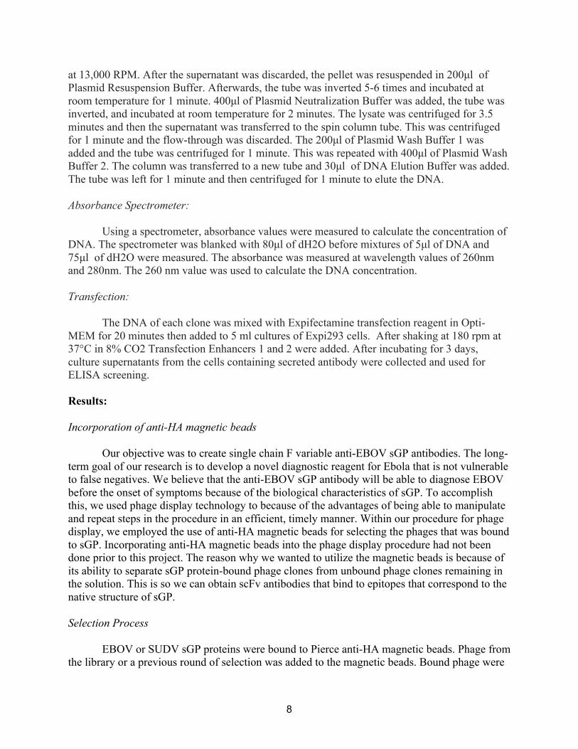
After selecting the Top 24 Clones, we used DNA fingerprinting and gel electrophoresis to
identify the unique clones from our pool by looking at the results from the gel. By using the Alu1 enzyme and BstN1 enzyme, we were able to cut the DNA into different fragments to determine which clones were the same. The clones with the same gel pattern would most likely be the same clones (Figure 7). For groups of clones that yielded the same results in the gel electrophoresis, we only selected one clone from each group for further analysis. After this process, we had narrowed down our pool to two clones, AA5 and B6.
11
Figure 7: DNA Fingerprinting gel. From our top 24 clones, we used DNA fingerprinting to determine
which clones were unique. This figure illustrates the results of a representative gel.
After identifying clones AA5 and B6, we sequenced the clones before moving to converting the phage clone to scFv-Fc antibodies. We were able to isolate the scFv insert DNA using DNA gel extraction before cloning the DNA into an expression vector containing the hinge and Fc portion of the antibody and transforming it using plasmids. A minipreparation of the plasmid was then successfully performed to separate the plasmid DNA from the bacteria. We used sequencing to ensure that the DNA was ready for transfection. We were able to create scFv-Fc antibodies through the transfection process. To test the scFv-Fc antibodies, we used two ELISA screenings for each clone, one with the target as EBOV sGP and the other with the target as SUDV sGP. This was to confirm the binding activity of the scFv-Fc antibodies. We used successive ¼ dilutions to test the specificity and concentration at which the IgG could recognize the targets. We expected to see a decrease in activity as the dilutions factor increased, which we were able to see (Figure 8). We saw that AA5 exhibited high binding activity and detection for both EBOV sGP and SUDV sGP. B6 only had high binding activity and specificity for EBOV sGP. This was expected since the binding activities depicted in Figure 6 showed similar patterns for both clones.

12
Figure 8: ELISA screening results from transfection of Clones AA5 and B6. . The absorbance values were read at λ= 450nm. Results demonstrate that we were able to successfully express clones AA5 and B6 as scFv-Fc antibodies. AA5 shows high binding activity to EBOV sGP and SUDV sGP. B6 shows high binding activity for EBOV sGP only, with very small absorbance values for SUDV sGP. Although only B6 showed the desired specificity for EBOV only, AA5 will also be useful in developing Ebola diagnostics. Discussion:
Our experiment aimed to use phage display technology to produce scFv-Fc antibodies that have high binding activity for the EBOV sGP target protein. We were able to successfully incorporate the use of anti-HA magnetic beads into our phage display protocol.
We also incorporated using negative selection with Sudan sGP to yield clones that would be specific in their binding activity to EBOV sGP. We were able to successfully identify clones that exhibited high activity toward EBOV sGP and low activity toward Sudan sGP. With our results thus far, the anti-HA magnetic beads and the negative selection with Sudan sGP are a promising direction in terms of adjusting the phage-display protocol to produce the clones with the highest detection for our target.
Through our experiment, we found that phage display technology is certainly able to isolate phage clones with the specificities that we desired. Through the screening portion of the methods, we were able to use positive selection to identify the clones that bound with the greatest strength to the EBOV sGP. We were able to accomplish 2 rounds of selection in a timely, efficient manner. We were successfully able to transfect the 2 phage clones selected, AA5 and B6, to produce scFv-Fc antibodies that were able to detect EBOV sGP. AA5 exhibited high detection for both EBOV sGP while B6 only exhibited high detection for SUDV sGP. These results confirm that using phage display technology and anti-HA magnetic beads for negative and positive selection is effective. We were able to isolate the antibodies capable of

13
discriminating between highly homologous EBOV and SUDV sGP proteins. These two clones show great promise for generating antibodies to be used in developing a novel diagnostic assay for the Ebola virus.
Future directions for our experiments include further exploring how negative selection with other sGP proteins (Taï Forest and Bundibugyo virus) to yield the best clones and antibodies. Because the negative selection was performed in our second round of phage phage selection, future directions will include incorporating negative selection in the first round. Through continuously cycling through the steps of phage display and performing multiple rounds of selection, highly specific anti-EBOV sGP antibodies will eventually be selected.
Citations: "About Ebola Virus Disease." Centers for Disease Control and Prevention. Centers for Disease Control and Prevention, 18 Feb. 2016. Web. 07 Dec. 2016. Barbas, C. F., III; Burton, D. R.; Scott, J.K., Silverman, G.J. Eds. (2001) ”Phage Display: A Laboratory Manual.” Cold Spring Harbor Laboratory Press: Cold Spring Harbor, New York. "Ebola Virus Disease." World Health Organization. World Health Organization, Jan. 2016. Web. 07 Dec. 2016. Christie, Athalia, et al. "Possible sexual transmission of Ebola virus-Liberia, 2015." MMWR. Morbidity and mortality weekly report 64.17 (2015): 479-481. Leroy, E. M., et al. "Diagnosis of Ebola haemorrhagic fever by RT- PCR in an epidemic setting." Journal of medical virology 60.4 (2000): 463-467. Leroy, Eric M., et al. "Multiple Ebola virus transmission events and rapid decline of central African wildlife." Science 303.5656 (2004): 387-390. Olafsen, Tove, Vania E. Kenanova, and Anna M. Wu. "Tunable pharmacokinetics: modifying the in vivo half-life of antibodies by directed mutagenesis of the Fc fragment." Nature protocols 1.4 (2006): 2048-2060. "Outbreaks Chronology: Ebola Virus Disease." Centers for Disease Control and Prevention. Centers for Disease Control and Prevention, 14 Apr. 2016. Web. 07 Dec. 2016. Sanchez, Anthony, et al. "The virion glycoproteins of Ebola viruses are encoded in two reading frames and are expressed through transcriptional editing." Proceedings of the National Academy of Sciences 93.8 (1996): 3602-3607. Team, WHO Ebola Response. "Ebola virus disease in West Africa—the first 9 months of the epidemic and forward projections." N Engl J Med 2014.371 (2014): 1481-1495.
14
Volchkova VA, Klenk HD, Volchkov VE. Delta-peptide is the carboxy-terminal cleavage fragment of the nonstructural small glycoprotein sGP of Ebola virus. Virology. 1999;265(1):164-71. Wilson, Julie A., et al. "Epitopes involved in antibody-mediated protection from Ebola virus." Science 287.5458 (2000): 1664-1666. de La Vega, Marc-Antoine, et al. "The Multiple Roles of sGP in Ebola Pathogenesis." Viral Immunology 28.1 (2015): 3. Yang, Zhi-yong, et al. "Identification of the Ebola virus glycoprotein as the main viral determinant of vascular cell cytotoxicity and injury." Nature medicine 6.8 (2000): 886-889. Falzarano, Darryl, et al. "Structure- Function Analysis of the Soluble Glycoprotein, sGP, of Ebola Virus." Chembiochem 7.10 (2006): 1605-1611. Falzarano, Darryl, et al. "Ebola sGP—the first viral glycoprotein shown to be C-mannosylated." Virology 368.1 (2007): 83-90.