Embed Size (px)
Citation preview
Tumorigenesis and Neoplastic Progression
Genetic Ablation of �v Integrins in Epithelial Cells ofthe Eyelid Skin and Conjunctiva Leads to SquamousCell Carcinoma
Joseph H. McCarty,* Marc Barry,*Denise Crowley,*† Roderick T. Bronson,‡
Adam Lacy-Hulbert,* and Richard O. Hynes*†
From the Center for Cancer Research,* and the Howard Hughes
Medical Institute,† Massachusetts Institute of Technology,
Cambridge; and the Rodent Histopathology Laboratory,‡ Harvard
Medical School, Boston, Massachusetts
Integrin-mediated cell adhesion and signaling eventsare essential for the proper development and ho-meostasis of most epithelial tissues. Dysregulation ofintegrin expression and function can cause abnormalepithelial cell proliferation and/or differentiation,contributing to the pathogenesis of malignant epithe-lial cancers. Here we report on the use of a condi-tional knockout strategy exploiting the Cre/Lox tech-nology to study the in vivo functions of �v integrinsduring epithelial cell proliferation and differentia-tion. We show that genetic ablation of �v integrinexpression in basal epithelial cells of the eyelid skinand conjunctiva causes the formation of tumors thatare strikingly similar to the malignant epithelial can-cer, squamous cell carcinoma. These data suggest amechanism whereby �v integrins normally suppressepithelial cell proliferation, likely via adhesion toECM ligands, as well as by the modulation of intracel-lular signaling cascades. We propose that �v genedeletion eliminates normal integrin-mediated growthsuppression, ultimately leading to cellular transfor-mation and tumorigenesis. Hence, these studies re-veal a novel tumor suppressor-like function of �vintegrins and provide a genetically tractable mousemodel for studying the pathogenesis of squamous cellcarcinoma and related cancers of epithelial origin, aswell as to test and develop novel therapeutic com-pounds to treat or prevent squamous cell carcinomaof the skin. (Am J Pathol 2008, 172:1740–1747; DOI:10.2353/ajpath.2008.070700)
The skin is a dynamic organ composed of two multicel-lular layers, the dermis and epidermis, which are sepa-
rated by an intervening basement membrane.1 An assort-ment of extracellular matrix (ECM) proteins within thebasement membrane supply instructive cues that regu-late basal cell proliferation, differentiation, and migra-tion.2 Abnormal regulation of these events can lead to thepathogenesis of a variety of epithelial abnormalities, includ-ing the malignant cancer squamous cell carcinoma (SCC).3
SCC is the most common form of skin cancer and arises viadefective growth regulatory pathways in stem cells or transitamplifying cells located in the epidermal basal layer.
Members of the integrin family of ECM receptors playcritical roles during the development and homeostasis ofmost stratified epithelial tissues.4 Several integrins andtheir associated intracellular signaling effectors are ex-pressed in basal epithelial cells, and gene ablation stud-ies in mice reveal essential functional roles for thesemolecules in the formation and maintenance of the epi-thelial tissues, particularly the skin.5 For example, micegenetically null for the �6 or �4 integrin genes developskin pathologies due to defective epidermal-dermal ad-hesion.6,7 Loss of �3 integrin expression leads to skinblistering phenotypes related to defective assembly ofepidermal basement membranes.8 Selective ablation ofthe murine �1 integrin gene in the skin leads to severedefects in epidermal development and homeostasis,9
and activating mutations in the human �1 integrin geneare found in rare cases of SCC.10
The �v integrin subfamily consists of �v�1, �v�3,�v�5, �v�6, and �v�8, and various genetic ablation stud-
Supported by grants from the National Cancer Institute (RO1CA17007)and the National Heart Lung and Blood Institute (PO1HL066105) and bythe Howard Hughes Medical Institute of which R.O.H. is an Investigator.A.L.H. was a United Kingdom Research Council Fellow.
J.H.M. and M.B. contributed equally to this work.
Accepted for publication March 11, 2008.
Current address of J.H.M.: Department of Cancer Biology, MD AndersonCancer Center, Houston, TX. Current address of M.B.: Department of Pathol-ogy, University of New Mexico, Albuquerque, NM. Current address of A.L.-H.: Massachusetts General Hospital, Department of Pediatrics, Boston, MA.
Address reprint requests to Richard O. Hynes, Ph.D., Center for CancerResearch, and the Howard Hughes Medical Institute, MassachusettsInstitute of Technology, Cambridge, MA 02139. E-mail: [email protected].
The American Journal of Pathology, Vol. 172, No. 6, June 2008
Copyright © American Society for Investigative Pathology
DOI: 10.2353/ajpath.2008.070700
1740
ies in mice have shown that these integrins play importantroles in multiple physiological and pathological con-texts.11–16 However, in vivo genetic models to study �vintegrin-mediated regulation of epithelial cell growth havenot been reported. In this study we use Cre/lox technol-ogy to analyze the in vivo growth regulatory functions forthe �v integrin subunit. We show that genetic ablation of�v expression selectively in basal cells of the eyelid skinand conjunctiva leads to development of epithelial tu-mors with pathological similarities to SCC. These data aredirect molecular genetic evidence that �v integrins pro-vide critical growth regulatory functions during epithelialproliferation and homeostasis. To our knowledge, thesefindings represent the first experimental data showingthat genetic ablation of �v integrin expression and func-tion in epithelial cells leads to dysregulation of normal cellgrowth and homeostasis, and suggest a physiological tu-mor suppressor-like function for �v integrins. Furthermore,this study provides a novel mouse genetic model to studythe pathogenesis of SCC in the eyelid skin and conjunctiva.
Materials and Methods
Mouse Strains and Genotyping
This murine glial fibrillary acidic protein (mGFAP)-Cretransgene consists of a genomic fragment encompass-ing the minimal murine GFAP promoter, as well as regu-latory intronic sequences flanking the Cre cDNA.17 Gen-eration and characterization of mGFAP-Cre transgenicmice have been described elsewhere.18 The �v-floxmouse strain has been previously described.14 The Ro-sa26-LoxSTOPLox-LacZ reporter strain was purchasedfrom The Jackson Laboratories.19 All mouse genotypeswere confirmed by standard PCR-based genotyping ofgenomic DNA isolated from tail snips. The followingprimer sequences were used for PCR genotyping: Cre,5�-ACCAGCCAGCTATCAACTC-3�, and 5�-TATACGCG-TGCTAGCGAAGATCTCCATCTTCCAGCAG-3�. The Creprimers yield a single PCR product of �200 bp. �v-floxprimers, F1: 5�-GTTGAGTATGCTCCATGCAGGTCA-3�,F2: 5�-TTCAGGACGGCACAAAGACCGTTG-3�, and R:5�-CACAAATCAAGGATGACCAAACTGAG-3�. The F1-Rprimer pair generates a PCR product of approximately350 bp. The F2-R primer pair generates an 850 bp bandrepresenting the non-recombined �v-flox allele, or a250bp band representing the recombined �v-flox allele.The �v�/� or �v�/� alleles yield a PCR product of 250 bpusing either the F1-R primer pair, or a PCR product of 550bp using the F2-R primer pair.
Antibodies, Immunohistochemistry, and Histology
The anti-�v antiserum was used at a 1:300 dilution.20 Tominimize nonspecific immunoreactivity, the diluted anti-body was first pre-absorbed using �v-null acetone-ex-tracted protein, prepared as previously described.14 Theanti-�-catenin antibody was purchased from Chemicon,Inc. For immunofluorescence and immunohistochemicalanalyses, samples of eye tumors from GFAP-Cre�;
�vflox/� mutant animals, or eyelid tissue from control an-imals were fresh-frozen in Tissue Tek OCT (Miles). Sec-tions (7 �m) were immunostained with rabbit IgG (10�g/ml), or anti-�v antibody. A secondary antibody con-jugated to horseradish peroxidase (Vector Laboratories)in combination with diaminobenzidine chromagenic sub-strate was used for immunohistochemistry. Alternatively,an Alexa488-conjugated goat anti-rabbit secondary anti-body (Molecular Probes) was used for immunofluores-cence. For histopathology studies, eyelids or eye tumorswere excised and fixed overnight at 4°C with 4% para-formaldehyde in phosphate buffered saline. Tissue wassubsequently dehydrated and processed for standardparaffin embedding and H&E staining. Alternatively, tovisualize mucin-expressing goblet-like cells, paraffin sec-tions from eye tumors were counterstained with periodicacid-Schiff and diastase, or Alcian Blue. Oil Red O stain-ing was performed on sections prepared from unfixed,fresh-frozen ocular tumors.
Results
A Murine GFAP-Cre Transgene Is Expressed inEpithelial Cells of the Developing Eyelid andConjunctiva
Previously, we used Cre/lox technology to selectivelyablate the murine �v integrin gene in central nervoussystem (CNS) neural cells.14 These efforts involved ana-lyzing the temporal and spatial expression patterns ofvarious Cre transgenes reportedly expressed in specificcell types in the CNS. One such transgene, consisting ofa fragment of the mGFAP promoter inserted 5� to thecDNA encoding Cre recombinase (mGFAP-Cre), was re-ported to be expressed specifically in postnatal CNS gliaand neurons.21 Indeed, using the ROSA26-loxSTOPlox-lacZ reporter mouse,19 we confirmed Cre activity in someneuronal and glial cells of the developing neural tube, aswell as some cells in the subventricular zone of the brain(data not shown). However, we also detected transgeneexpression outside of the CNS. As shown in Figure 1, Band D, embryos (E13.5) harboring the mGFAP-Cre andROSA26-loxSTOPlox-lacZ transgenes expressed robustlevels of Cre in epithelial cells of the developing lens andconjunctiva. Additionally, analysis of neonatal (P7) trans-genic mice revealed Cre expression in epithelial cells ofthe conjunctiva and cornea (Figure 1F), eyelid (Figure1H), as well as occasional hair follicles in the eyelidepidermis (Figure 1J). We did not detect lacZ activity inembryos or neonates lacking the GFAP-Cre transgene(Figure 1, A, C, E, G, and I). The pattern of Cre expressionwas primarily localized to epithelial cells of the eyelidskin; we did not detect Cre activity in epithelia of otherneonatal organs, including the intestine and lung (datanot shown). It is likely that the epithelial expression pat-tern of the GFAP-Cre transgene is an aberrant conse-quence of the transgene insertion site. For example, thetransgene may have inserted into a genomic region thatis regulated by enhancer elements that activate geneexpression in specific epithelial cells of the developing
�v Integrins in Epithelial Cells and SCC 1741AJP June 2008, Vol. 172, No. 6
eye. A second transgene that we have previously char-acterized,14 consisting of the human GFAP promoter reg-ulating Cre expression, is active primarily in CNS glia,and is not expressed in skin epithelial cells (unpublisheddata). However, it also remains possible that some cellsin the eyelid skin and conjunctiva originate from GFAP-expressing progenitors, or that some basal epithelialcells express GFAP as others have recently shown.22
Targeted Deletion of the �v Integrin Gene Usingthe mGFAP-Cre Transgene
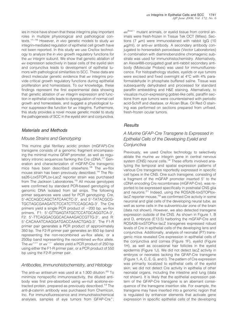
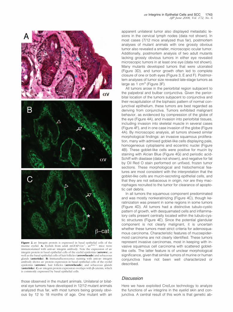
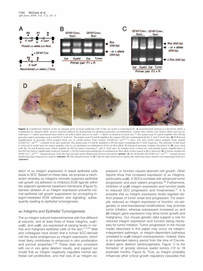
We used an anti-�v integrin antibody14,20 to analyze thespatial expression pattern of �v integrin protein in thepostnatal murine eyelid. �v integrin protein expressionwas detected in the basal epithelium of the normal eyelid,as well as by basal epithelial cells in sebaceous glandsand hair follicles (Figure 2, A and B). The expressionpattern of �v integrin protein was very similar to the patterndetected for �-catenin (Figure 2C), a protein commonlyexpressed in basal cells of stratified epithelial tissues.23 Weselectively ablated the �v integrin gene by generating miceharboring a conditional �v allele (�vflox/flox) in combinationwith the mGFAP-Cre transgene (Figure 3A). First, mG-FAP-Cre hemizygotes were bred with �v�/� mice to gen-erate GFAP-Cre�; �v�/� progeny, which were subse-quently bred with �vflox/flox mice.13,14 The resultingmutant progeny are hemizygous for the mGFAP-Cretransgene, and carry one �v-flox allele and one �v-nullallele. Littermate controls were hemizygous for the mG-FAP-Cre transgene, and carry one �v-flox allele and one�v wild-type allele.
We used genomic PCR to test for mGFAP-Cre-medi-ated deletion of the �v integrin gene. We isolatedgenomic DNA from eyelid tissue, and analyzed �v-floxdeletion using PCR to monitor a 350 bp PCR band rep-resenting the �v-flox allele (Figure 3B). Analysis of ear,eyelid, or eye tumor genomic DNA samples from controland mutant mice revealed recombination of the �v-floxallele selectively in the eye or eye tumor samples. Wemonitored deletion of the conditional �v allele using asecond primer pair designed to amplify an 850 bp bandrepresenting the non-recombined �v-flox allele (Figure3C). The same primer pair amplified a 250 bp bandrepresenting the recombined �vflox/flox gene. Amplifica-tion of the complementary allele, which lacked loxP sitesand is either wild-type or null for �v, yielded a 550 bpPCR product. Analysis of ear, eyelid, or eye tumorgenomic DNA samples from control and mutant micerevealed recombination of the �v-flox allele selectively inthe eye or eye tumor samples. This correlated with re-duced intensity of the intact 850 bp �v-flox cassette, aswell as an increase in the recombined 250 bp PCR prod-uct (Figure 3C).
Genetic Ablation of �v Integrin Causes theFormation of Eyelid Skin Tumors DisplayingPathological Characteristics of Squamous CellCarcinoma
mGFAP-Cre�, �vflox/� and mGFAP-Cre�, �vflox/� mu-tants were born in expected ratios, and displayed nogrossly obvious developmental or behavioral abnormali-ties (data not shown). However, beginning as early asnine postnatal months, mGFAP-Cre�; �vflox/� mutant an-imals developed tumors surrounding one or both eyes(Figure 3, D–F). mGFAP-Cre�; �vflox/� control littermates(17/17 analyzed thus far) did not develop eye tumors like
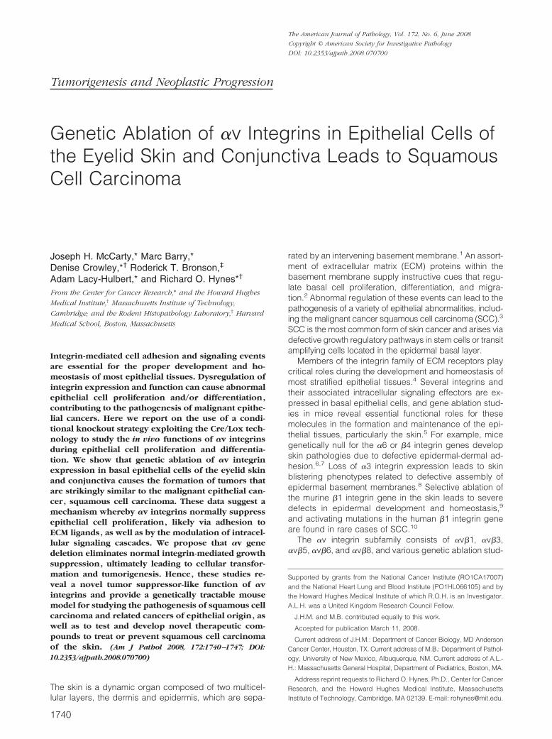
Figure 1. A murine GFAP-Cre transgene is expressed in epithelial cells of theembryonic and postnatal eye. Embryos (E13.5) expressing the ROSA26-loxSTOPLox-LacZ reporter transgene in the absence (A) or presence (B) ofthe mGFAP-Cre transgene were dissected and whole-mounts were stained todetermine the spatial pattern of �-galactosidase activity. �-galactosidase isminimally expressed in embryos lacking the mGFAP-Cre transgene (A);however, embryos harboring the mGFAP-Cre transgene display Cre-medi-ated expression of �-galactosidase (B). C, D: Embryonic heads were sec-tioned horizontally and the pattern of �-galactosidase activity was analyzedmicroscopically. Note the �-galactosidase activity in epithelial cells of thelens (arrow in D) and conjunctiva (arrowheads in D). Sagittal histologicalsections through the center of the neonatal eye (P7) from ROSA26-lox-STOPlox-LacZ transgenics in the absence (E, G, I) or presence (F, H, J) of themGFAP-Cre transgene. Cre-mediated �-galactosidase activity is present inepithelial cells in the developing conjunctiva (arrows in F), cornea (arrow-heads in F), eyelid (arrows in H), and hair follicles (arrows in J).
1742 McCarty et alAJP June 2008, Vol. 172, No. 6
those observed in the mutant animals. Unilateral or bilat-eral eye tumors have developed in 12/12 mutant animalsanalyzed thus far, with most tumors being grossly obvi-ous by 12 to 18 months of age. One mutant with an
apparent unilateral tumor also displayed metastatic le-sions in the cervical lymph nodes (data not shown). Inmost cases (7/12 mice analyzed thus far), postmortemanalyses of mutant animals with one grossly obvioustumor also revealed a smaller, microscopic ocular tumor.Additionally, postmortem analysis of two adult mutantslacking grossly obvious tumors in either eye revealedmicroscopic tumors in at least one eye (data not shown).Many mutants developed tumors that were ulcerated(Figure 3D), and tumor growth often led to completeclosure of one or both eyes (Figure 3, E and F). Postmor-tem analyses of tumor size revealed late-stage tumors aslarge as 1 cm3 (Figure 3F).
All tumors arose in the periorbital region subjacent tothe palpebral and bulbar conjunctiva. Given the perior-bital location of the tumors subjacent to conjunctiva andtheir recapitulation of the biphasic pattern of normal con-junctival epithelium, these tumors are best regarded asderiving from conjunctiva. Tumors exhibited malignantbehavior, as evidenced by compression of the globe ofthe eye (Figure 4A), and invasion into periorbital tissues,including invasion into skeletal muscle in several cases(Figure 4F), and in one case invasion of the globe (Figure4A). By microscopic analysis, all tumors showed similarmorphological findings: an invasive squamous prolifera-tion, many with admixed goblet-like cells displaying pale,homogeneous cytoplasms and eccentric nuclei (Figure4B). These goblet-like cells were positive for mucin bystaining with Alcian Blue (Figure 4G) and periodic acid-Schiff with diastase (data not shown), and negative for fatby Oil Red O stain performed on unfixed, frozen tumorsections. These morphological and histochemical fea-tures are most consistent with the interpretation that thegoblet-like cells are mucin-secreting epithelial cells, andthat they are not sebaceous in origin, nor are they mac-rophages recruited to the tumor for clearance of apopto-tic cell debris.
In all tumors the squamous component predominatedand was mostly nonkeratinizing (Figure 4C), though ke-ratinization was present in some regions in some tumors(Figure 4D). All tumors had a distinctive tubulo-cysticpattern of growth, with desquamated cells and inflamma-tory cells present centrally located within the tubulo-cys-tic structures (Figure 4E). Since the potential glandularcomponent is not clearly malignant, it is uncertainwhether these tumors meet strict criteria for adenosqua-mous carcinoma. Characteristic features of mucoepider-moid carcinoma are not clearly identified. These tumorsrepresent invasive carcinomas, most in keeping with in-vasive squamous cell carcinoma with scattered goblet-like cells. The latter feature is of unclear morphologicalsignificance, given that similar tumors of murine or humanconjunctiva have not been well characterized ordescribed.
Discussion
Here we have exploited Cre/Lox technology to analyzethe functions of �v integrins in the eyelid skin and con-junctiva. A central result of this work is that genetic ab-
Figure 2. �v Integrin protein is expressed in basal epithelial cells of themurine eyelid. A: Eyelids from adult mGFAP-Cre�; �vflox/� mice wereimmunostained with anti-�v integrin antibody. Note the expression of �vintegrin protein in basal epithelial cells of the eyelid epidermis (arrows), aswell as the basal epithelial cells of hair follicles (arrowheads) and sebaceousglands (asterisks). B: Immunufluorescence staining with anti-�v integrinantibody shows �v protein expression in basal epithelial cells of the eyelidepidermis (arrows), hair follicles (arrowheads), and sebaceous glands(asterisks). C: �v integrin protein expression overlaps with �-catenin, whichis commonly expressed by basal epithelial cells.
�v Integrins in Epithelial Cells and SCC 1743AJP June 2008, Vol. 172, No. 6
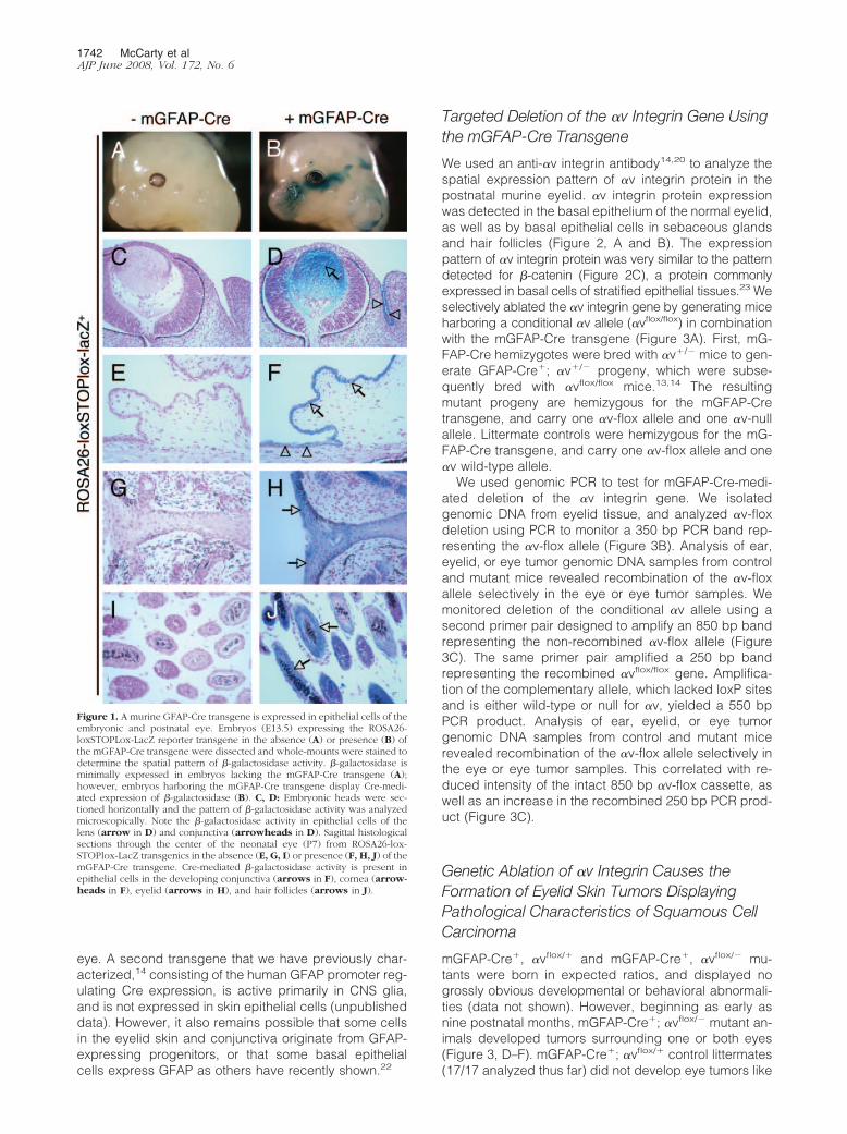
lation of �v integrin expression in basal epithelial cellsleads to SCC. Based on these data, we propose a mech-anism whereby �v integrins normally suppress epithelialcell growth via adhesion to inhibitory ECM ligands withinthe adjacent epidermal basement membrane (Figure 5).Genetic ablation of �v integrin expression prevents nor-mal epithelial cell growth suppression by uncoupling in-tegrin-mediated ECM adhesion and signaling, subse-quently leading to epithelial tumorigenesis.
�v Integrins and Epithelial Tumorigenesis
The �v integrin subunit heterodimerizes with five different� subunits, and at least three of these integrins, �v�1,�v�5, and �v�6, are expressed at varying levels in nor-mal and malignant epithelial cells of the skin.2,3,24 Wattand colleagues have shown that a human SCC-derivedcell line lacks endogenous �v integrin expression, whichmost likely contributes to enhanced in vitro proliferationand survival properties.3,25 These data are consistentwith our in vivo gene deletion results, and support ourmodel that �v integrin negatively regulates normal epi-thelial cell proliferation, and that loss of �v integrin ex-
pression or function causes aberrant cell growth. Otherreports show that increased expression of �v integrins,particularly �v�6, in SCCs correlate with advanced tumorprogression and poor patient prognosis.26 Furthermore,inhibition of �v�6 integrin expression and function leadsto reduced SCC progression and invasiveness.27 It ispossible that �v integrin expression levels regulate dis-tinct phases of tumor onset and progression. For exam-ple, reduced �v integrin expression or function, via epi-genetic or post-translational modifications, may promotetumor initiation, whereas subsequent increased �v and�6 integrin gene expression may drive tumor growth andmalignancy. Our mouse genetic data support a role forreduced integrin expression and function being neces-sary for tumor initiation. Tumor progression in the mousemodel described in this paper may occur via integrin-independent pathways, or integrin-dependent pathwaysunrelated to �v�6 integrin overexpression. Indeed, thereis an extended latency period from the time of Cre-me-diated gene deletion (embryogenesis, Figure 1) to theformation of grossly obvious eyelid tumors (12 to 18postnatal months, Figure 3). Thus, �v integrin probablyinfluences other critical growth regulatory cascades that
Figure 3. Conditional ablation of the �v integrin gene in basal epithelial cells of the eye leads to tumorigenesis. A: Experimental strategy to selectively ablate aconditional �v integrin allele. Arrows indicate primers for monitoring Cre-mediated genomic recombination. Control mice harbor one �vflox allele and one �vwild-type (�) allele and mutant mice harbor an �vflox allele and an �v null (�) allele via deletion of exon one.14 The primer pair F1 and R amplifies the 350-bpgenomic region spanning exon 4 and the 3� loxP site. The primer pair F2 and R amplifies the region (850 bp) containing both the 5� and 3� loxP sites. B: PCR-basedamplification of genomic DNA isolated from ear or eyelid tissues from control (GFAP-Cre�;�vflox/�) mice. Ear and eyelid tumor samples from mutant(GFAP-Cre�;�vflox/�) animals were also analyzed. The primer pair, F1 and R, amplifies a 350 bp band, containing the 3� loxP sequence. The intensity of this bandis reduced in eyelid and eye tumor samples, due to cre-mediated recombination of the �v-flox allele. C: Identical genomic samples described in (B) were usedwith the F2 and R primer pair, which amplify an 850 bp band containing 5� and 3� loxP sites. In samples from control eye and mutant tumor, amplification ofthis 850 bp band is significantly reduced. Instead, a 250 bp band representing the recombined �v-flox allele (lower panel in A) is detected. D: A twelve month-oldGFAP-Cre�; �vflox/� mutant mouse. Note the macroscopic tumor displaying obvious ulceration (arrow). E: An 18 month-old GFAP-Cre�; �vflox/� mutant mousedisplaying large bilateral eye tumors (arrows). F: The mutant mouse in (E) with the skin removed to expose the skull and tumor masses encompassing both eyes(arrows).
1744 McCarty et alAJP June 2008, Vol. 172, No. 6

are progressively altered following gene deletion; to-gether, these events contribute to the onset and progres-
sion of eyelid SCC. We are currently investigating themolecular alterations that occur as a result of �v genedeletion, for example, whether tumor suppressor or on-cogene signaling pathways are dysregulated, and howthese events collectively lead to SCC. All five murine �subunit genes that pair with �v have been ablated indi-vidually or in various combinations, yet none of the pub-lished studies reveals a phenotype that relates to SCC.5
Thus, it is likely that the combined loss of two or more�v-containing integrins, eg, �v�6, and �v�8, may con-tribute to SCC. We are currently generating mice that lackmultiple � integrin genes to address this possibility.
�v Integrins and Functional Links withTransforming Growth Factor � Signaling in SCC
�v integrins bind to RGD tripeptide motifs within the latentassociated peptides of transforming growth factor (TGF)�1 and TGF�3.28 Latent associated peptides non-co-valently associate with TGF�1/3 in the ECM and maintain
Figure 5. A model for �v integrin-mediated regulation of epithelial prolifer-ation and homeostasis. �v integrins expressed in basal epithelial cells of theconjunctiva and eyelid skin bind to the latent forms of TGF�1 and 3 (latentassociated peptide-TGF�1/3) in the epidermal basement membrane. Integrinbinding leads to activation of TGF� signaling and suppression of epithelialcell proliferation, likely via an autocrine loop. Genetic ablation of �v integrinexpression in basal epithelia causes dysregulation of TGF� growth inhibition,leading to epithelial cell hyperplasia and tumor progression.
Figure 4. Genetic ablation of �v integrin in the murine eye epithelium leads to tumors with histological characteristics of squamous cell carcinoma. H&E stainingof eye tumor sections from GFAP-Cre�; �vflox/� mutant animals. A: Tumor compressing and invading globe of the eye (arrows). B: Tumor showing admixedsquamous cells (arrowheads) and goblet-like cells (arrows). C: Tumor with predominantly squamous differentiation (arrows) and intralumenal apoptosis(asterisks). D: Tumor showing keratinization (arrow). E: Tumor tubulo-cystic structures with marked lumenal accumulation of desquamated tumor cells(arrows). F: Tumor invading skeletal muscle of the eye (arrows). G: Tumor showing biphasic squamous and goblet-like cell differentiation. The Alcian Bluemucin stain highlights goblet-like cells (arrows). H, I: H&E stained sections from the ocular region of an �vflox/� mouse that did not harbor the GFAP-Cretransgene. Note the normal cytoarchitecture of the conjunctiva (arrows in H) and eyelid skin epidermis (arrows in I).
�v Integrins in Epithelial Cells and SCC 1745AJP June 2008, Vol. 172, No. 6
TGF� in an inactive form. Both �v�6 and �v�8 integrinsmediate the physical dissociation of latent associatedpeptides, leading to release of bioactive TGF�1 andTGF�3 from the ECM.29–32 TGF�’s and their receptorshave been shown to negatively regulate epithelial cellgrowth.33 A recent report reveals that selective ablationof TGF�-receptor I signaling leads to development ofadmixed squamous cell carcinomas and mucoepider-moid carcinomas in the periorbital and perianal re-gions.34 Fuchs and colleagues more recently published astudy showing that genetic ablation of the TGF� receptorII gene in basal cells of the mouse skin epidermis (via theKeratin5-Cre transgene) results in SCC development inperianal and perivaginal areas, and these results corre-late with reduced expression of TGF� receptor II in hu-man SCC samples.35 The TGF� receptor I/II knockoutresults are consistent with a previous report showing thatmice lacking Smad4, an intracellular signaling proteinregulated by TGF� receptors, develop SCC of the skin.36
Interestingly, genetic deletion of �v integrins in mousedendritic cells leads to colitis and colon tumor formation,and these events are mostly due to defective �v�8 inte-grin-mediated TGF� activation.13,16 These various datastrongly support our model that �v integrins, via activa-tion of TGF� signaling events, normally serve to suppressepithelial cell growth (Figure 5). Genetic ablation of �vintegrins or TGF� signaling components dysregulate thisbalance, leading to epithelial cell hyperplasia and tumorprogression.
In conclusion, our molecular genetic strategies revealimportant functions for �v integrins in regulating epithelialcell proliferation and homeostasis in the eyelid skin andconjunctiva. To our knowledge, these are the first directgenetic data supporting a tumor suppressor-like functionfor �v integrins in epithelial cells. Since the expression ofthe GFAP-Cre transgene has not been detected in epi-thelial cells outside of the eyelid skin and conjunctiva, orother stratified epithelial organs, we cannot yet concludethat �v integrins play more general roles in suppressingbasal epithelial cell growth. We are currently deleting �vintegrin expression using other Cre transgenes that areexpressed in a broader range of epithelial organs to testthis possibility. These various integrin knockout modelswill likely be useful translational tools to study SCC onsetand progression, as well as to test and develop noveltherapeutic compounds to treat or prevent SCC of theskin.
Acknowledgments
The GFAP-Cre transgenic mice were kindly provided byDr. Anton Berns (Netherlands Cancer Institute). The �vintegrin anti-serum was provided by Dr. Louis Reichardt(UCSF). We thank Aaron Cook for technical assistancewith mouse genotyping. We are also grateful to Dr. BitaEsmaeli and Dr. Alexander Lazar (both at MD AndersonCancer Center) for helpful discussions regarding the oc-ular tumor pathologies.
References
1. Fuchs E, Raghavan S: Getting under the skin of epidermal morpho-genesis. Nat Rev Genet 2002, 3:199–209
2. Watt FM: Role of integrins in regulating epidermal adhesion, growthand differentiation. EMBO J 2002, 21:3919–3926
3. Janes SM, Watt FM: New roles for integrins in squamous-cell carci-noma. Nat Rev Cancer 2006, 6:175–183
4. Hynes RO: Integrins: bidirectional, allosteric signaling machines. Cell2002, 110:673–687
5. Bouvard D, Brakebusch C, Gustafsson E, Aszodi A, Bengtsson T,Berna A, Fassler R: Functional consequences of integrin gene muta-tions in mice. Circ Res 2001, 89:211–223
6. Dowling J, Yu QC, Fuchs E: Beta4 integrin is required for hemides-mosome formation, cell adhesion and cell survival. J Cell Biol 1996,134:559–572
7. Georges-Labouesse E, Messaddeq N, Yehia G, Cadalbert L, DierichA, Le Meur M: Absence of integrin alpha 6 leads to epidermolysisbullosa and neonatal death in mice. Nat Genet 1996, 13:370–373
8. DiPersio CM, Hodivala-Dilke KM, Jaenisch R, Kreidberg JA, HynesRO: alpha3beta1 Integrin is required for normal development of theepidermal basement membrane. J Cell Biol 1997, 137:729–742
9. Brakebusch C, Grose R, Quondamatteo F, Ramirez A, Jorcano JL,Pirro A, Svensson M, Herken R, Sasaki T, Timpl R, Werner S, FasslerR: Skin and hair follicle integrity is crucially dependent on beta 1integrin expression on keratinocytes. EMBO J 2000, 19:3990–4003
10. Evans RD, Jones J, Taylor C, Watt FM: Sequence variation in the I-likedomain of the beta1 integrin subunit in human oral squamous cellcarcinomas. Cancer Lett 2004, 213:189–194
11. Bader BL, Rayburn H, Crowley D, Hynes RO: Extensive vasculogen-esis, angiogenesis, and organogenesis precede lethality in micelacking all alpha v integrins. Cell 1998, 95:507–519
12. Hodivala-Dilke KM, McHugh KP, Tsakiris DA, Rayburn H, Crowley D,Ullman-Cullere M, Ross FP, Coller BS, Teitelbaum S, Hynes RO:Beta3-integrin-deficient mice are a model for Glanzmann thrombas-thenia showing placental defects and reduced survival. J Clin Invest1999, 103:229–238
13. Lacy-Hulbert A, Smith AM, Tissire H, Barry M, Crowley D, Bronson RT,Roes JT, Savill JS, Hynes RO: Ulcerative colitis and autoimmunityinduced by loss of myeloid alphav integrins. Proc Natl Acad Sci USA2007, 104:15823–15828
14. McCarty JH, Lacy-Hulbert A, Charest A, Bronson RT, Crowley D,Housman D, Savill J, Roes J, Hynes RO: Selective ablation of alpha vintegrins in the central nervous system leads to cerebral hemorrhage,seizures, axonal degeneration, and premature death, Development2005, 132:165–176
15. Reynolds LE, Wyder L, Lively JC, Taverna D, Robinson SD, Huang X,Sheppard D, Hynes RO, Hodivala-Dilke KM: Enhanced pathologicalangiogenesis in mice lacking beta3 integrin or beta3 and beta5integrins. Nat Med 2002, 8:27–34
16. Travis MA, Reizis B, Melton AC, Masteller E, Tang Q, Proctor JM,Wang Y, Bernstein X, Huang X, Reichardt LF, Bluestone JA, SheppardD: Loss of integrin alpha(v)beta8 on dendritic cells causes autoim-munity and colitis in mice. Nature 2007, 449:361–365
17. Miura M, Tamura T, Mikoshiba K: Cell-specific expression of themouse glial fibrillary acidic protein gene: identification of the cis- andtrans-acting promoter elements for astrocyte-specific expression.J Neurochem 1990, 55:1180–1188
18. Tronche F, Kellendonk C, Kretz O, Gass P, Anlag K, Orban PC, BockR, Klein R, Schutz G: Disruption of the glucocorticoid receptor genein the nervous system results in reduced anxiety. Nat Genet 1999,23:99–103
19. Soriano P: Generalized lacZ expression with the ROSA26 Cre re-porter strain. Nat Genet 1999, 21:70–71
20. Bossy B, Reichardt LF: Chick integrin alpha V subunit molecularanalysis reveals high conservation of structural domains and associ-ation with multiple beta subunits in embryo fibroblasts. Biochemistry1990, 29:10191–10198
21. Marino S, Vooijs M, van Der Gulden H, Jonkers J, Berns A: Inductionof medulloblastomas in p53-null mutant mice by somatic inactivationof Rb in the external granular layer cells of the cerebellum. Genes Dev2000, 14:994–1004
22. Danielyan L, Tolstonog G, Traub P, Salvetter J, Gleiter CH, Reisig D,Gebhardt R, Buniatian GH: Colocalization of glial fibrillary acidic
1746 McCarty et alAJP June 2008, Vol. 172, No. 6
protein, metallothionein, and MHC II in human, rat. NOD/SCID, andnude mouse skin keratinocytes and fibroblasts. J Invest Dermatol2007, 127:555–563
23. Perez-Moreno M, Fuchs E: Catenins: keeping cells from getting theirsignals crossed. Dev Cell 2006, 11:601–612
24. Stepp MA: Alpha9 and beta8 integrin expression correlates with themerger of the developing mouse eyelids. Dev Dyn 1999, 214:216–228
25. Janes SM, Watt FM: Switch from alphavbeta5 to alphavbeta6 integrinexpression protects squamous cell carcinomas from anoikis. J CellBiol 2004, 166:419–431
26. Hazelbag S, Kenter GG, Gorter A, Dreef EJ, Koopman LA, VioletteSM, Weinreb PH, Fleuren GJ: Overexpression of the alpha v beta 6integrin in cervical squamous cell carcinoma is a prognostic factor fordecreased survival. J Pathol 2007, 212:316–324
27. Nystrom ML, McCulloch D, Weinreb PH, Violette SM, Speight PM,Marshall JF, Hart IR, Thomas GJ: Cyclooxygenase-2 inhibition sup-presses alphavbeta6 integrin-dependent oral squamous carcinomainvasion. Cancer Res 2006, 66:10833–10842
28. Annes JP, Munger JS, Rifkin DB: Making sense of latent TGFbetaactivation. J Cell Sci 2003, 116:217–224
29. Cambier S, Gline S, Mu D, Collins R, Araya J, Dolganov G, EinheberS, Boudreau N, Nishimura SL: Integrin alpha(v) beta8-mediated ac-tivation of transforming growth factor-beta by perivascular astrocytes:an angiogenic control switch. Am J Pathol 2005, 166:1883–1894
30. Cambier S, Mu DZ, O’Connell D, Boylen K, Travis W, Liu WH, Broad-
dus VC, Nishimura SL: A role for the integrin alphavbeta8 in thenegative regulation of epithelial cell growth. Cancer Res 2000,60:7084–7093
31. Morris DG, Huang X, Kaminski N, Wang Y, Shapiro SD, Dolganov G,Glick A, Sheppard D: Loss of integrin alpha(v) beta6-mediated TGF-beta activation causes Mmp12-dependent emphysema. Nature2003, 422:169–173
32. Mu D, Cambier S, Fjellbirkeland L, Baron JL, Munger JS, KawakatsuH, Sheppard D, Broaddus VC, Nishimura SL: The integrin alpha(v)beta8mediates epithelial homeostasis through MT1-MMP-dependent acti-vation of TGF-beta1. J Cell Biol 2002, 157:493–507
33. Massague J, Gomis RR: The logic of TGFbeta signaling. FEBS Lett2006, 580:2811–2820
34. Honjo Y, Bian Y, Kawakami K, Molinolo A, Longenecker G, BoppanaR, Larsson J, Karlsson S, Gutkind JS, Puri RK, Kulkarni AB: TGF-betareceptor I conditional knockout mice develop spontaneous squa-mous cell carcinoma. Cell Cycle 2007, 6:1360–1366
35. Guasch G, Schober M, Pasolli HA, Conn EB, Polak L, Fuchs E: Lossof TGFbeta signaling destabilizes homeostasis and promotes squa-mous cell carcinomas in stratified epithelia. Cancer Cell 2007,12:313–327
36. Yang L, Mao C, Teng Y, Li W, Zhang J, Cheng X, Li X, Han X, Xia Z,Deng H, Yang X: Targeted disruption of Smad4 in mouse epidermisresults in failure of hair follicle cycling and formation of skin tumors.Cancer Res 2005, 65:8671–8678
�v Integrins in Epithelial Cells and SCC 1747AJP June 2008, Vol. 172, No. 6