Embed Size (px)
Citation preview
ORIGINAL RESEARCHpublished: 15 May 2015
doi: 10.3389/fmicb.2015.00464
Frontiers in Microbiology | www.frontiersin.org 1 May 2015 | Volume 6 | Article 464
Edited by:
Christina Maria Joseph Elisabeth
Vandenbroucke-Grauls,
VU University Medical Center,
Netherlands
Reviewed by:
Magdalena Chirila,
Iuliu Hatieganu University of Medicine
and Pharmacy, Romania
Malgorzata Anna
Mikaszewska-Sokolewicz,
The Medical University of Warsaw,
Poland
*Correspondence:
Patricio Retamal,
Departamento de Medicina
Preventiva, Facultad de Ciencias
Veterinarias y Pecuarias, Univerisdad
de Chile, Av. Sta. Rosa 11735, La
Pintana, Santiago, Chile
Specialty section:
This article was submitted to
Infectious Diseases,
a section of the journal
Frontiers in Microbiology
Received: 11 March 2015
Accepted: 28 April 2015
Published: 15 May 2015
Citation:
Retamal P, Fresno M, Dougnac C,
Gutierrez S, Gornall V, Vidal R, Vernal
R, Pujol M, Barreto M,
González-Acuña D and Abalos P
(2015) Genetic and phenotypic
evidence of the Salmonella enterica
serotype Enteritidis human-animal
interface in Chile.
Front. Microbiol. 6:464.
doi: 10.3389/fmicb.2015.00464
Genetic and phenotypic evidence ofthe Salmonella enterica serotypeEnteritidis human-animal interface inChilePatricio Retamal 1, 2*, Marcela Fresno 3, Catherine Dougnac 3, Sindy Gutierrez 1,
Vanessa Gornall 1, Roberto Vidal 2, 4, Rolando Vernal 5, Myriam Pujol 3, Marlen Barreto 6,
Daniel González-Acuña 2, 7 and Pedro Abalos 1, 2
1Departamento de Medicina Preventiva, Facultad de Ciencias Veterinarias y Pecuarias, Universidad de Chile, Santiago,
Chile, 2 Emerging and Remerging Zoonosis Research Network, Santiago, Chile, 3 Programa de Doctorado en Ciencias
Silvoagropecuarias y Veterinarias, Universidad de Chile, Santiago, Chile, 4 Programa de Microbiología, Facultad de Medicina,
Universidad de Chile, Santiago, Chile, 5Departamento de Odontología Conservadora, Facultad de Odontología, Universidad
de Chile, Santiago, Chile, 6 Facultad de Ciencias de la Salud, Universidad Autónoma, Santiago, Chile, 7 Facultad de Ciencias
Veterinarias, Universidad de Concepción, Chillán, Chile
Salmonella enterica serotype Enteritidis is a worldwide zoonotic agent that has been
recognized as a very important food-borne bacterial pathogen, mainly associated
with consumption of poultry products. The aim of this work was to determine
genotypic and phenotypic evidence of S. Enteritidis transmission among seabirds,
poultry and humans in Chile. Genotyping was performed using PCR-based virulotyping,
pulse-field gel electrophoresis (PFGE) and multi-locus sequence typing (MLST).
Pathogenicity-associated phenotypes were determined with survival to free radicals,
acidic pH, starvation, antimicrobial resistance, and survival within human dendritic cells.
As result of PCR and PFGE assays, some isolates from the three hosts showed identical
genotypic patterns, and through MLST it was determined that all of them belong to
sequence type 11. Phenotypic assays show diversity of bacterial responses among
isolates. When results were analyzed according to bacterial host, statistical differences
were identified in starvation and dendritic cells survival assays. In addition, isolates
from seabirds showed the highest rates of resistance to gentamycin, tetracycline, and
ampicillin. Overall, the very close genetic and phenotypic traits shown by isolates from
humans, poultry, and seabirds suggest the inter-species transmission of S. Enteritidis
bacteria between hosts, likely through anthropogenic environmental contamination that
determines infection of seabirds with bacteria that are potentially pathogenic for other
susceptible organism, including humans.
Keywords: Salmonella enterica, Enteritidis, humans, poultry, seabirds, Chile
Introduction
Worldwide, reported human Salmonella infections are caused by many serotypes, although atpresent, the highest incidence is represented by S. enterica serotype Enteritidis (Hendriksen et al.,2011; Jackson et al., 2013). The changing epidemiology of S. Enteritidis infection over the last 20

Retamal et al. S. Enteritidis in the human-animal interface
years has allowed this serotype to become the most prevalentamong S. enterica serotypes, and is therefore considered anemergent pathogen. Sources of S. Enteritidis are generallyassociated with commercial poultry products, mainlyundercooked eggs and meat (Jackson et al., 2013). In Chile,S. Enteritidis emerged in 1994, causing food-borne disease inhumans and is now included in surveillance systems for bothanimal and public health services (Fernandez et al., 2003).However, its current endemic condition has shown increasingincidence during the last years, in spite of sanitary regulationsand a previous successful campaign against S. Typhi (Ficaet al., 2012). The epidemiological link between poultry andhuman S. Enteritidis infection has been confirmed throughgenotypic analysis that has shown the presence of two majorS. Enteritidis subtypes distributed between both hosts in theChilean territory (Fernandez et al., 2003). However, becausesome clinical strains show unique genetic patterns, it seems thatinfection of humans is also coming from different unknownsources.
Regarding wildlife hosts, seabirds have been associatedelsewhere with zoonotic serotypes of S. enterica, with evidencethat suggests a direct bacterial transmission either amongthemselves, with other animals or humans (Reche et al., 2003;Pennycott et al., 2006; Dhama et al., 2008; Skov et al., 2008;Horton et al., 2013; Gruszynski et al., 2014). In addition, alongthe Chilean coast, zoonotic and multi-drug resistant (MDR)Salmonella strains have been detected in seashore animals,specifically seabirds and pinnipeds (Lopez-Martin et al., 2011;Sturm et al., 2011; Rodriguez et al., 2012a; Fresno et al.,2013).
The extended geographical distribution, host range andgenome plasticity of S. Enteritidis have determined genotypicand phenotypic diversity among strains, which contain astriking number of variably detected chromosomal and plasmidgenes that may be related to diverse clinical outcomes andadaptive changes favoring survival in different hosts (Panet al., 2009; Huehn et al., 2010). However, direct correlationsbetween genotypes and phenotypes would not be obvious,since indistinguishable genetic patterns have shown majordifferences in pathogenicity-associated phenotypes (Yim et al.,2010).
Because of the increasing impact of S. Enteritidis on publichealth in Chile and the unknown role of wildlife in itsepidemiology, the aim of this work was to determine genotypicand phenotypic evidence of S. Enteritidis transmission amongseabirds, poultry and humans in Chile.
Materials and Methods
IsolatesNinety S. Enteritidis isolates previously isolated from humans,poultry, and seabirds (n = 30 each) were analyzed (Table 1).Bacteria were grown routinely in liquid culture with Luria-Bertani (LB) medium (Bacto Tryptone, 10 g/L; Bacto YeastExtract, 5 g/L; NaCl, 5 g/L) adjusted to pH 7 (NaHPO4/NaH2PO425mM), at 37◦C for 24 h, with shaking. When necessary, mediawere solidified by the addition of agar (15 g/L).
Genotypic AssaysVirulotypingThis PCR based test was performed for the identification of genesinvA, pefA, spvC, sirA, gipA, SEN1417, trhH, and prot6e, all ofthem associated with virulence that have been variably detectedin S. Enteritidis (Pan et al., 2009; Huehn et al., 2010). Afterbacterial growth, PCR reactions were performed under standardconditions, as previously described (Fresno et al., 2013).
Pulse-Field Gel Electrophoresis (PFGE)This assay followed the PulseNet protocol (Ribot et al.,2006). The electrophoresis was performed using CHEF DRIIICHILER (Bio-Rad) equipment. DNA was digested with XbaI(50U/sample) endonuclease. PFGE patterns were analyzedwith the GEL COMPAR II software (Applied Maths), usingthe Dice similarity coefficient with a 1% tolerance in bandposition.
ClusteringResults from virulotyping and PFGE were analyzed throughthe construction of a binary matrix using “1” for presence and“0” for absence of genes or bands from each isolate. Clusterswere determined using via the unweighted pair group method(UPGMA) using TREECON software, and the discriminatorypower (DP) was calculated with the Simpson’s index of diversity,as reported in a previous study (Hunter and Gaston, 1988).
MultiLocus Sequence Typing (MLST)This procedure was based on sequencing seven housekeepinggenes of S. enterica (Achtman et al., 2012), using the primersdescribed in the MLST public database (http://mlst.warwick.ac.uk/mlst/). The sequence type was determined according to thescheme provided on this site.
Phenotypic AssaysHydrogen Peroxide and Sodium Nitrite Survival
AssaysThese assays were performed as described by Lu et al. (2002).Briefly, after an overnight growth of bacteria in LB broth thecultures were diluted 1/100 in LB pH 7 or LB pH 5 and challengedwith 15mMH2O2 or 10mMNaNO2, respectively. Then, cultureswere incubated at 37◦C with shaking for 30min with H2O2, orfor 3 h with NaNO2. For survival rate analysis, aliquots of culturewere plated in triplicate before (time 0) and after challenge(30min or 3 h). Survival was expressed as the percentage ofcolony forming units (CFU) after the assay; the count before thechallenge was considered as 100%.
Acidic pH SurvivalAfter overnight growth in LB broth, bacteria were washed threetimes with LB pH 3 (citric acid 0.1M), diluted 1/100 in the samemedium and incubated with shaking at 37◦C for 3 h. Survivalanalysis was performed as with H2O2 and NaNO2 assays, withCFU counts at 0 and 3 h.
Starvation Survival AssayThis procedure was based on published reports (Spector andCubitt, 1992; O’neal et al., 1994), with some modifications.
Frontiers in Microbiology | www.frontiersin.org 2 May 2015 | Volume 6 | Article 464
Retamal et al. S. Enteritidis in the human-animal interface
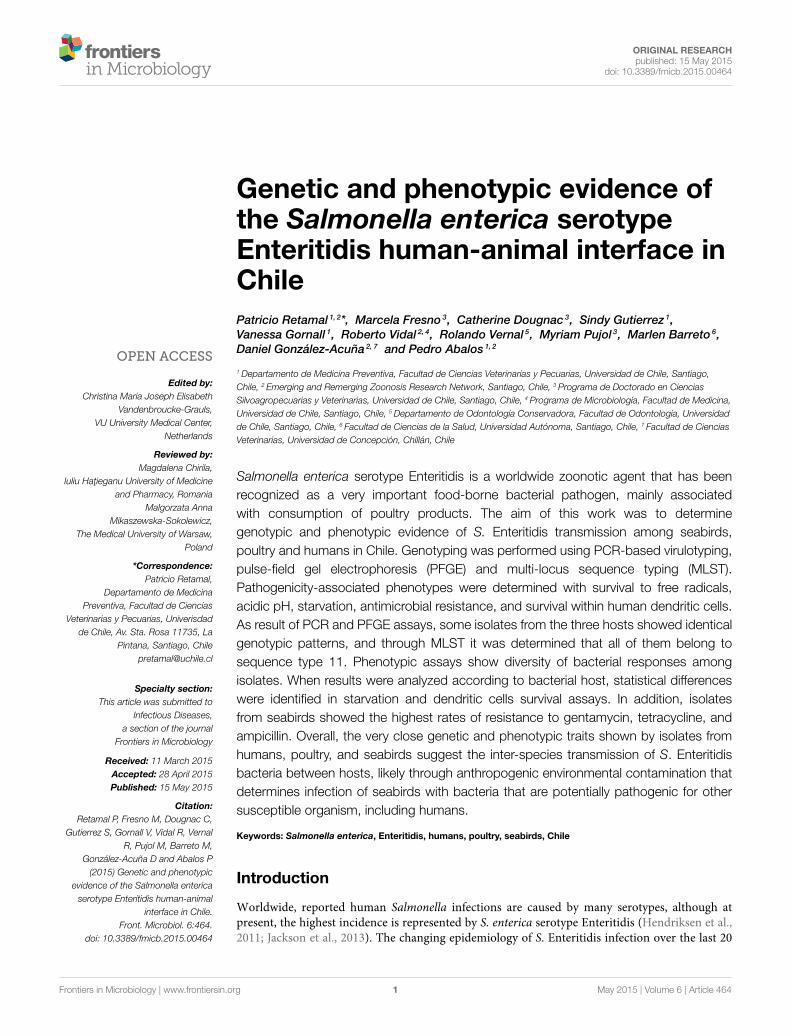
TABLE 1 | Salmonella enterica ser. Enteritidis strains utilized in this study.
Strain ID Source Hosta Virulence genes Antimicrobial resistanceb
invA pefA spvC sirA gipA SEN1417 trhH prot6e ENR AMC CTX CN TE STX EFT CE AMP CFR
SEN1 Valparaíso Poultry
SEN2 Valparaíso Poultry
SEN3 Valparaíso Poultry
SEN4 Valparaíso Poultry
SEN5 Valparaíso Poultry
SEN6 Valparaíso Poultry
SEN7 Valparaíso Poultry
SEN8 Valparaíso Poultry
SEN9 Valparaíso Poultry
SEN10 Valparaíso Poultry
SEN11 Valparaíso Poultry
SEN12 Valparaíso Poultry
SEN13 Valparaíso Poultry
SEN14 Valparaíso Poultry
SEN15 Biobío Kelp gull
SEN16 Biobío Kelp gull
SEN17 Biobío Kelp gull
SEN18 Biobío Kelp gull
SEN19 Biobío Kelp gull
SEN20 Biobío Kelp gull
SEN21 Antofagasta Human
SEN22 Metropolitana Human
SEN23 Metropolitana Human
SEN24 Metropolitana Human
SEN25 Arica Human
SEN26 Arica Human
SEN27 Antofagasta Human
SEN28 Coquimbo Human
SEN29 Valparaíso Human
SEN30 Biobío Human
SEN31 Arica Poultry
SEN32 O’Higgins Poultry
SEN46 Arica Poultry
SEN47 Arica Poultry
SEN48 Valparaíso Human
SEN49 Atacama Human
SEN50 Metropolitana Human
SEN51 Metropolitana Human
SEN52 Metropolitana Human
SEN53 Valparaíso Human
SEN54 Valparaíso Human
SEN55 O’Higgins Human
SEN56 Biobío Human
SEN57 Metropolitana Human
SEN58 Metropolitana Human
SEN59 Los Lagos Human
SEN72 Coquimbo Human
SEN73 Magallanes Human
SEN74 Los Lagos Human
(Continued)
Frontiers in Microbiology | www.frontiersin.org 3 May 2015 | Volume 6 | Article 464

Retamal et al. S. Enteritidis in the human-animal interface
TABLE 1 | Continued
Strain ID Source Hosta Virulence genes Antimicrobial resistanceb
invA pefA spvC sirA gipA SEN1417 trhH prot6e ENR AMC CTX CN TE STX EFT CE AMP CFR
SEN75 Coquimbo Human
SEN76 Valparaíso Human
SEN77 Valparaíso Human
SEN78 Metropolitana Human
SEN79 Metropolitana Human
SEN80 Metropolitana Poultry
SEN81 Los Ríos Poultry
SEN82 Los Ríos Poultry
SEN83 Arica Poultry
SEN85 Metropolitana Poultry
SEN86 Metropolitana Poultry
SEN87 Metropolitana Poultry
SEN88 Metropolitana Poultry
SEN89 Metropolitana Poultry
SEN90 Metropolitana Poultry
SEN91 Metropolitana Poultry
SEN92 Metropolitana Poultry
SEN95 Arica Franklin gull
SEN96 Magallanes Penguin
SEN97 Coquimbo Kelp gull
SEN98 Coquimbo Kelp gull
SEN99 Coquimbo Kelp gull
SEN100 Coquimbo Kelp gull
SEN101 Coquimbo Kelp gull
SEN102 Coquimbo Kelp gull
SEN103 Coquimbo Kelp gull
SEN104 Coquimbo Kelp gull
SEN105 Coquimbo Kelp gull
SEN106 Coquimbo Kelp gull
SEN107 Coquimbo Kelp gull
SEN108 Valparaíso Kelp gull
SEN109 Valparaíso Kelp gull
SEN110 Valparaíso Kelp gull
SEN111 Valparaíso Kelp gull
SEN112 Valparaíso Kelp gull
SEN113 Valparaíso Kelp gull
SEN114 Valparaíso Kelp gull
SEN115 Valparaíso Kelp gull
SEN116 Valparaíso Kelp gull
SEN117 Valparaíso Kelp gull
SEN118 Valparaíso Kelp gull
Virulence genes and antimicrobial resistance phenotypes appear with shaded squares.aPoultry, Gallus gallus; Human, Homo sapiens; Kelp gull, Larus dominicanus; Franklin’ gull, Leucophaeus pipixcan; Penguin, Spheniscus magellanicus.bAMC, Amoxicillin–clavulanic acid; AMP, Ampicillin; CTX, Cefotaxime; CFR, Cefadroxil; CE, Cefradine; EFT, Ceftiofur; ENR, Enrofloxacin; CN, Gentamicin; STX, Trimethoprim–
sulfamethoxazol; TE, Tetracycline.
Briefly, bacterial isolates were inoculated into MOPS-bufferedsalts (MS) hiPCN (MS, 25mM KH2PO4/K2HPO4 pH 7.4, 0.4%glucose, 15mM NH4Cl) media, and then incubated for 16–18 h at 37◦C. Cultures were washed with distilled water, diluted1/10 in MS loPCN (MS, 1mM KH2PO4/K2HPO4 pH 7.4, 0.2%
glucose, 10mM NH4Cl) media and incubated at 37◦C up toOD600 0.3–0.4. Then, 1mL of this suspension was washed withdistilled water, inoculated into 5mL MS media and incubatedat 37◦C for 40 d. Aliquots of culture were plated in triplicate atdifferent times. Survival was expressed as a percentage of CFU in
Frontiers in Microbiology | www.frontiersin.org 4 May 2015 | Volume 6 | Article 464

Retamal et al. S. Enteritidis in the human-animal interface
relation with the maximal CFU count reached between day 0 and5, which was considered 100%.
Survival within Dendritic Cells (DCs)For this assay we used nine isolates, three from each host, whichshowed the highest resistance in the four previous survival assays.For selection, a survival ranking was performed in each assay,assigning 1 to the most susceptible and 90 to the most resistantstrain. Then, an average ranking value was calculated for everyisolate.
Human peripheral blood mononuclear-derived DCs wereobtained from the buffy coats of six healthy donors and preparedas previously described (Vernal et al., 2008). For the infection,day 6 DCs were maintained in culture medium (RPMI-1640 with10% fetal calf serum) and seeded into tissue culture plates at aconcentration of 4×10⁵ cells per well. Exponential-phase (OD600,0.6) grown bacteria were pelleted and suspended in the samemedium. Aliquots of bacteria were added to DCs at a multiplicityof infection (MOI) of 50:1. After 1 h of infection, cells werewashed three times with PBS, and incubated with cellular culturemedium containing gentamicin (200µg/mL). After additionalincubation for 2 and 24 h, DCs were washed with PBS andpermeabilized for 30min with 0.1% Triton X-100, and the titersof intracellular bacteria were determined by serial dilution ofcell lysates on LB agar plates. The percentage of survival wascalculated at 2 h considering the initial inoculant as 100%, andat 24 h considering the CFU counted at 2 h as 100%.
Ethics StatementThe human DCs protocol included a written consent of alldonors, which was approved by the University of Chile ClinicalHospital Scientific Ethics Committee (OAIC Reference #508/11,Exempt Resolution #570).
Antimicrobial SusceptibilityAntimicrobial susceptibility was evaluated by the disk diffusionmethod following CLSI criteria (CLSI, 2010). Antimicrobialstested were (µg/disk) ampicillin (10), amoxicillin–clavulanicacid (20/10), cefotaxime (30), gentamicin (10), trimethoprim–sulfamethoxazol (1.25/23.75), tetracycline (30), ciprofloxacin (5),cefradine (30), ceftiofur (30), and enrofloxacin (10) (Oxoid R©).Escherichia coli ATCC 25922 was utilized as a control strain.The multidrug resistance condition was determined by thesimultaneous resistance to three or more antimicrobial classes.
Statistical AnalysisStatistical analyses were performed using the ANOVA andKruskal–Wallis test for independent samples. Categorical dataand principal components analyses were performed using datafrom survival assays and hosts. These tests were calculated usingINFOSTAT (2010v) software.
Results
Genotypic AssaysPCR detection of virulence genes from 90 S. Enteritidis isolatesshowed 16 distinct virulotypes, resulting in a low discriminatorypower (DP) methodology (0.773) that clustered 80% of isolates
within three of these gene combinations (Table 2, Figure S1).Higher diversity was observed in isolates from poultry andseabirds (9 and 8 virulotypes, respectively) than in isolatesdetected in humans (5 virulotypes). In addition, virulotypestended to associate with a specific host (P < 0.05), as themost frequent gene combination detected in poultry and humanisolates is different from that identified in seabird isolates(virulotypes H and C, respectively. Table 2, Figure S1). Thisvariation was also observed at genetic level, because spvC and sirAwere differentially detected among hosts (P < 0.05), being thefirst most frequent in bacteria found in humans and poultry andthe second in those isolated from seabirds (Table 3).
Through the PFGE procedure we obtained fingerprints with8–12 bands, resulting in 10 major clusters (two or more isolateswith identical PFGE profiles) that represent 89% of isolates(Figure S2). The DP of this method was 0.891, identifying fiveidentical patterns between isolates from seabirds and humans.However, combined PFGE and PCR results showed the highestDP (0.949), with 16 major clusters that contain 70% of isolates.Six of these are mixed clusters, and two of them contain isolatesfrom humans and seabirds (Figure 1). The MLST analysesdetermined that all S. Enteritidis isolates in this study belong tothe sequence type (ST) 11.
Phenotypic AssaysIn pathogenicity-associated phenotypes there was a significantdiversity (P < 0.05) of bacterial survival responses amongisolates, which was observed in all these assays (data not shown).When results from individual strains were grouped and analyzedaccording to bacterial host, statistical differences (P < 0.05)
TABLE 2 | Virulence gene combinations (Virulotypes) of Salmonella
enterica ser. Enteritidis strains and their frequency according to host.
Virulotypes Host (N◦ of strains)
ID* invA-pefA-spvC-
sirA-gipA-SEN1417-
trhH-prot6e
Human (30) Poultry (30) Seabirds (30) Total
O 10000100 0 0 3 3
K 10010001 0 1 0 1
P 10011000 0 1 0 1
G 11000101 0 0 1 1
L 11010001 0 0 2 2
B 11010101 0 0 2 2
F 11100001 0 2 0 2
H 11100101 18 14 2 34
N 11101100 1 0 0 1
D 11101101 0 1 0 1
M 11101110 1 0 0 1
C 11110001 6 4 10 20
A 11110101 4 5 9 18
J 11111000 0 1 0 1
I 11111001 0 1 0 1
E 11111101 0 0 1 1
*Letters were assigned correlatively according to the order in which isolates appear in the
dendogram (Figure S1).
Frontiers in Microbiology | www.frontiersin.org 5 May 2015 | Volume 6 | Article 464

Retamal et al. S. Enteritidis in the human-animal interface
TABLE 3 | Frequency of virulence associated genes in Salmonella enterica
ser. Enteritidis strains grouped by host.
Gene Host1 Total N (%)
Human N (%) Poultry N (%) Seabirds N (%)
invA 30 (100) 30 (100) 30 (100) 90 (100)
pefA 30 (100) 28 (93) 27 (90) 85 (94)
spvC 30 (100)a 28 (93)a 22 (73)b 80 (89)
sirA 10 (33)a 13 (43)a 24 (80)b 47 (52)
gipA 2 (7) 4 (13) 1 (3) 7 (8)
SEN1417 24 (80) 20 (67) 18 (60) 62 (69)
trhH 1 (3) 0 (0) 0 (0) 1 (1)
prot6e 28 (93) 28 (93) 27 (90) 83 (92)
1Different letters represents statistical differences between groups (p < 0.05).
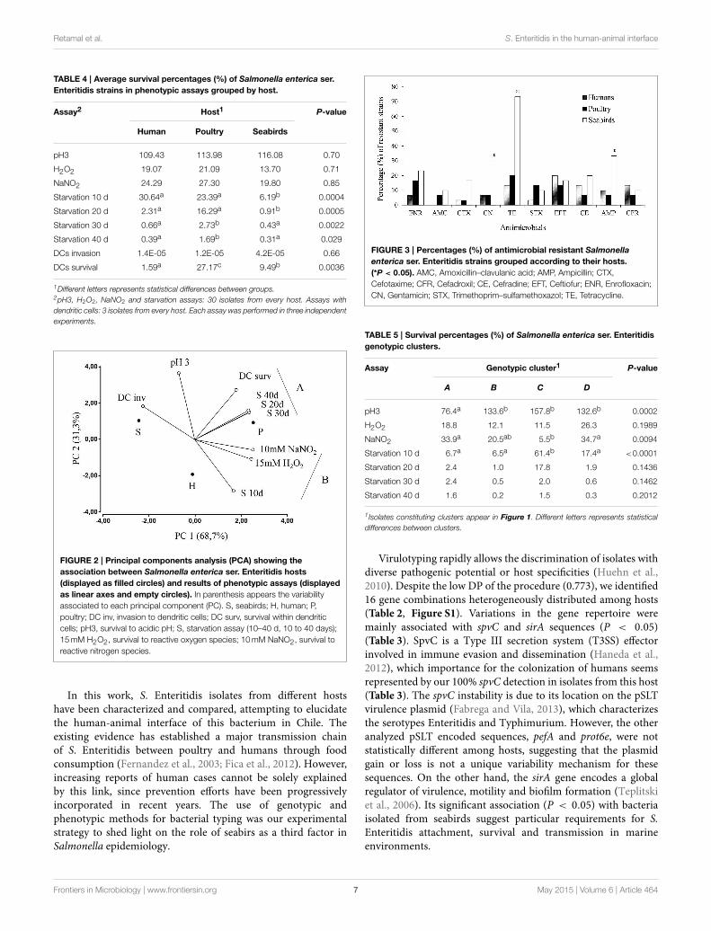
were identified in starvation and dendritic cells survival assays(Table 4), in which isolates from poultry were the most resistant.Isolates from humans expressed higher resistance to short-termstarvation (10 d), but later showed the steepest survival decreaseand at 30 and 40 d showed similar CFU counts as bacteriabelonging to seabirds. Among the top 10 most resistant isolatesin each phenotypic assay, those recovered from poultry wereconsistently the most frequent (Figure S3). Within dendriticcells, isolates from humans were the most susceptible (Table 4).The relative survival performance of bacteria analyzed accordingto their hosts can be graphically seen in Figure 2. The poultryisolates have the closest position to most of assays, representingtheir highest survival capabilities in these challenges.Besides, some phenotypic variables are located forming twogroups (A and B, Figure 2), depicting a high correlationbetween them.
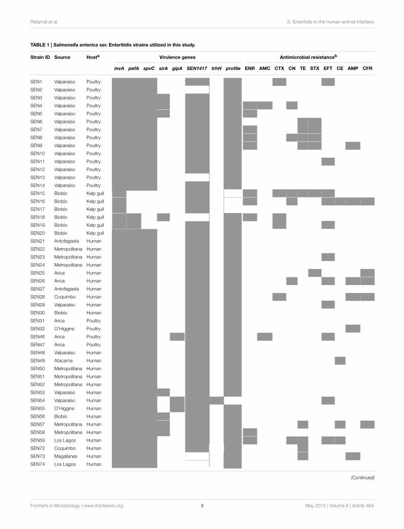
Resistance to gentamycin, tetracycline and ampicillin wasstatistically associated with S. Enteritidis isolates from seabirds(P < 0.05, Figure 3). Additionally, we detected multidrugresistance (simultaneous resistance to three or more differentCLSI antimicrobial clasess) in 13 (43%) isolates recoveredfrom seabirds; in contrast, only 3 (10%) and 4 (13%) humanand poultry isolates showed multidrug resistance, respectively(Table 1).
Significant associations (P < 0.05) between genotypic andphenotypic results were detected when isolates from majorclusters (A–D in Figure 1) were compared (Table 5). The clusterA, which only contains isolates from humans and poultry, showsthe lowest survival to acidic pH. The cluster C, which is composedby isolates from the three hosts, shows the lowest survival tonitrite-derived free radicals and the highest survival to short-termstarvation (Table 5).
Discussion
Wild birds have received attention from sanitary authoritiesbecause of both their association with several highly transmissiblezoonotic pathogens and their ability to disseminate agents with awide host range, over wide geographical areas (Hubalek, 2004).
FIGURE 1 | Dendogram showing genetic similarities (%) between
Salmonella enterica serovar Enteritidis strains resulting from
combined PFGE and PCR data. The ID and host are shown for each isolate.
Clusters with at least five isolates sharing more than 95% similarity are
indicated with letters A, B, C, and D. Mixed clusters are indicated with letters
according to hosts (P, poultry; H, human; S, seabird).The tree was constructed
using the UPGMA method with the software TREECON.
Frontiers in Microbiology | www.frontiersin.org 6 May 2015 | Volume 6 | Article 464
Retamal et al. S. Enteritidis in the human-animal interface
TABLE 4 | Average survival percentages (%) of Salmonella enterica ser.
Enteritidis strains in phenotypic assays grouped by host.
Assay2 Host1 P-value
Human Poultry Seabirds
pH3 109.43 113.98 116.08 0.70
H2O2 19.07 21.09 13.70 0.71
NaNO2 24.29 27.30 19.80 0.85
Starvation 10 d 30.64a 23.39a 6.19b 0.0004
Starvation 20 d 2.31a 16.29a 0.91b 0.0005
Starvation 30 d 0.66a 2.73b 0.43a 0.0022
Starvation 40 d 0.39a 1.69b 0.31a 0.029
DCs invasion 1.4E-05 1.2E-05 4.2E-05 0.66
DCs survival 1.59a 27.17c 9.49b 0.0036
1Different letters represents statistical differences between groups.2pH3, H2O2, NaNO2 and starvation assays: 30 isolates from every host. Assays with
dendritic cells: 3 isolates from every host. Each assay was performed in three independent
experiments.
FIGURE 2 | Principal components analysis (PCA) showing the
association between Salmonella enterica ser. Enteritidis hosts
(displayed as filled circles) and results of phenotypic assays (displayed
as linear axes and empty circles). In parenthesis appears the variability
associated to each principal component (PC). S, seabirds; H, human; P,
poultry; DC inv, invasion to dendritic cells; DC surv, survival within dendritic
cells; pH3, survival to acidic pH; S, starvation assay (10–40 d, 10 to 40 days);
15mM H2O2, survival to reactive oxygen species; 10mM NaNO2, survival to
reactive nitrogen species.
In this work, S. Enteritidis isolates from different hostshave been characterized and compared, attempting to elucidatethe human-animal interface of this bacterium in Chile. Theexisting evidence has established a major transmission chainof S. Enteritidis between poultry and humans through foodconsumption (Fernandez et al., 2003; Fica et al., 2012). However,increasing reports of human cases cannot be solely explainedby this link, since prevention efforts have been progressivelyincorporated in recent years. The use of genotypic andphenotypic methods for bacterial typing was our experimentalstrategy to shed light on the role of seabirs as a third factor inSalmonella epidemiology.
FIGURE 3 | Percentages (%) of antimicrobial resistant Salmonella
enterica ser. Enteritidis strains grouped according to their hosts.
(*P < 0.05). AMC, Amoxicillin–clavulanic acid; AMP, Ampicillin; CTX,
Cefotaxime; CFR, Cefadroxil; CE, Cefradine; EFT, Ceftiofur; ENR, Enrofloxacin;
CN, Gentamicin; STX, Trimethoprim–sulfamethoxazol; TE, Tetracycline.
TABLE 5 | Survival percentages (%) of Salmonella enterica ser. Enteritidis
genotypic clusters.
Assay Genotypic cluster1 P-value
A B C D
pH3 76.4a 133.6b 157.8b 132.6b 0.0002
H2O2 18.8 12.1 11.5 26.3 0.1989
NaNO2 33.9a 20.5ab 5.5b 34.7a 0.0094
Starvation 10 d 6.7a 6.5a 61.4b 17.4a <0.0001
Starvation 20 d 2.4 1.0 17.8 1.9 0.1436
Starvation 30 d 2.4 0.5 2.0 0.6 0.1462
Starvation 40 d 1.6 0.2 1.5 0.3 0.2012
1 Isolates constituting clusters appear in Figure 1. Different letters represents statistical
differences between clusters.
Virulotyping rapidly allows the discrimination of isolates withdiverse pathogenic potential or host specificities (Huehn et al.,2010). Despite the low DP of the procedure (0.773), we identified16 gene combinations heterogeneously distributed among hosts(Table 2, Figure S1). Variations in the gene repertoire weremainly associated with spvC and sirA sequences (P < 0.05)(Table 3). SpvC is a Type III secretion system (T3SS) effectorinvolved in immune evasion and dissemination (Haneda et al.,2012), which importance for the colonization of humans seemsrepresented by our 100% spvC detection in isolates from this host(Table 3). The spvC instability is due to its location on the pSLTvirulence plasmid (Fabrega and Vila, 2013), which characterizesthe serotypes Enteritidis and Typhimurium. However, the otheranalyzed pSLT encoded sequences, pefA and prot6e, were notstatistically different among hosts, suggesting that the plasmidgain or loss is not a unique variability mechanism for thesesequences. On the other hand, the sirA gene encodes a globalregulator of virulence, motility and biofilm formation (Teplitskiet al., 2006). Its significant association (P < 0.05) with bacteriaisolated from seabirds suggest particular requirements for S.Enteritidis attachment, survival and transmission in marineenvironments.
Frontiers in Microbiology | www.frontiersin.org 7 May 2015 | Volume 6 | Article 464
Retamal et al. S. Enteritidis in the human-animal interface
Because of its high DP and reproducibility for bacterial typing,the PFGE technique has been utilized to compare Salmonellastrains isolated from a diversity of hosts and substrates (Ribotet al., 2006; Zheng et al., 2011; Sandt et al., 2013). In this work,the combination of XbaI PFGE with virulotyping has allowedthe best DP (0.949), comparable to PFGE using a combinationof XbaI and BlnI restriction enzymes (Zou et al., 2012). Thisprocedure is showing several indistinguishable patterns amonghuman, poultry and seabirds isolates (Figure 1). Moreover, theMLST analysis has classified all analyzed isolates as belonging toST11, which is distributed worldwide (http://mlst.warwick.ac.uk/mlst/) and has also been associated with prevalent phage types(Pan et al., 2009),
Overall, genotypic data suggests that wild birds are sharingbacteria, whether directly or indirectly, with poultry and humans,participating in the transmission cycle of S. Enteritidis in Chile.
Phenotypic assays were performed in order to determine thepathogenic potential of Salmonella isolates from different hosts.The analyzed conditions are mainly found when bacteria facethe gastrointestinal lumen upon entering a host via ingestionand within the phagolysosomal environment in phagocytic cells(Behnsen et al., 2015), although survival as free-living bacteria inwater, soil or within protozoa in extra-host settings represents asimilar challenge (Spector and Kenyon, 2012).
In this work we were able to determine differences among S.Enteritidis strains (P < 0.05) in all phenotypic assays. Whenresults were grouped according to host source, starvation survivalconstitutes the unique in vitro assay that demonstrates differences(P < 0.05) among hosts (Table 4), showing that seabird isolatesare the most susceptible to nutrient deprivation at 37◦C. Thehuman isolates demonstrated a critical downshift between 10 and30 days (Table 4), suggesting a better adaptation to shorter ratherthan longer periods of starvation. Moreover, poultry strains notonly had the highest survival rate to starvation during the entireexperiment, but also showed the highest resistance when allin vitro assays were considered. Because of that, they are the bestrepresented among the top 10 most resistant strains within everysurvival assay (Figure S3).
Unexpectedly, genotypic clustering at 95% similarity(Figure 1) was associated with three survival phenotypes(Table 5) in contrast to previous reports with this serotypewhich, although not using the same methods, did not findsuch association (Betancor et al., 2009; Yim et al., 2010). Thecluster A appear the most defective in pH 3 assay, probablydue to absence of seabirds’ isolates, which showed the best(non-statistical, P > 0.05) fitness in this challenge (Figure 2,Table 4). Interestingly, the cluster C, which is the unique clustercomposed by isolates from the three hosts, is associated withthe lowest performance in NaNO2-derived free radicals and thehighest survival to a short-term starvation condition (Table 5).Whether the transmission between aquatic and terrestrial hosts,suggested by the high genetic similitude of bacteria withincluster C, could be facilitated by such combination of phenotypicresponses, is a question that remains to be elucidated.
During the infective process it has been determined thata critical survival challenge facing S. Enteritidis inside itshost is DCs, because it cannot replicate within these cells
as well as in macrophages after invasion (Swart and Hensel,2012). In order to determine a correlation between in vitrophenotypes and survival within human dendritic cells, weperformed a gentamicin protection assay using differentiatedhuman peripheral blood monocytes infected with the mostresistant Salmonella strains isolated from the three hosts.Interestingly, the highest correlation was found with starvationassays at 20 (r = 0.93), 30 (r = 0.92), and 40 days (r =
0.94), suggesting this in vitro assay is a predictor of bacterialbehavior in the intracellular environment and the pathogenicpotential in the human host. In this assay, poultry isolateshave again shown the highest survival rate, and unexpectedly,human isolates were the most susceptible (Table 4). Consistently,poultry strains have shown the highest survival rates in mostassays, suggesting an increased virulence. It is probable that thehigh contact rates within poultry flocks promote selection ofthe most rapidly replicating and most virulent clones, becausethe transmission to other animals will occur no matter theclinical outcome in infected hosts (Berngruber et al., 2013).Whenrepresented graphically (Figure 2), the relationship betweensurvival to pathogenicity-associated stresses and the source ofSalmonella isolates highlights thementioned differential bacterialperformances. Furthermore, two groups of assays are formedaccording to their correlations, one including the short-termstarvation survival (S 10 d) with resistance against free radicals(B, Figure 2), and the other including long-term starvationsurvival (S 20, 30, 40 d) with survival within DCs (A, Figure 2).This reflects that within each group, the bacterial mechanismsto resist these challenges are sharing stimuli, regulatory factorsor effectors, in agreement with previous studies in other bacteria(Watson et al., 1998; Cuny et al., 2005).
The high amount of prescribed antimicrobials in human andveterinary medicine represent a global concern because of thespread of antimicrobial resistant infectious pathogens (WHO,2014). In general, S. Enteritidis isolates have shown low resistancelevels against antimicrobials (Huehn et al., 2010; Sandt et al.,2013), contrasting with our results that show host-associatedvariability in this matter and suggest an overall high frequencyof drug resistance and MDR phenotypes, especially in wildbirds. From antimicrobials tested, the highest frequencies ofresistances were detected against ceftiofur in humans (20%) andagainst tetracycline in poultry (20%) and wild birds (73%). Thisconstitutes a concerning situation since ceftiofur is prescribedfor animal use only, suggesting transmission of resistant strainsfrom animals to humans. Isolates belonging to seabirds expressedsignificantly higher percentages of resistance (P < 0.05) thanhuman and poultry isolates with antimicrobials gentamicin(30%), ampicillin (33%), and tetracycline (73%) (Figure 3).The long established environmental persistence of tetracyclines(Hamscher et al., 2002), could explain the high resistance tothis antimicrobial. The environment can persistently spreadresistant bacteria and sublethal antimicrobial concentrationsthat, derived from anthropogenic activities (mainly animalfarms and wastewater), can select for resistance (Tello et al.,2012; Andersson and Hughes, 2014). Besides, the appearingof virulence-resistance plasmids that encode antimicrobialresistance and virulence factors, determines co-selection of these
Frontiers in Microbiology | www.frontiersin.org 8 May 2015 | Volume 6 | Article 464

Retamal et al. S. Enteritidis in the human-animal interface
functions even in the absence of antimicrobials (Rodriguez et al.,2012b; Gullberg et al., 2014), which could also explain ourfindings. In any case, these situations represents a potential riskto both public and animal health (Wellington et al., 2013), andjustifies the study of these hosts as bio-indicators of resistancetraits dispersion into the environment (da Costa et al., 2013).
Altogether, genotypic and phenotypic evidence gathered inthis study suggest that S. Enteritidis is circulating amongwildlife, domestic animals and humans, with human beingsparticipating as incidental (spill-over) hosts. Seabirds can bereservoirs of Salmonella strains with potential risk to publicand animal health, and could partially explain the progressiverise in the incidence of these serotype-associated outbreaks.Whether such transmission among hosts is direct or indirectis a question that should be addressed in future analyses. Ourresults support the establishment of biosecurity measures foranimal farms and systematic Salmonella surveillance campaignsin seabirds, determining not only the genetic similarities ofbacterial strains but also their pathogenic potential in susceptiblehosts.
Acknowledgments
We thank Natalia Paredes, Pilar Lillo, Paula Vicencio and PilarRodriguez for their collaboration in experimental procedures.
We also thank María Esther Saldías (Servicio Agrícola yGanadero) and Alda Fernández (Instituto de Salud Pública) forproviding Salmonella strains used in this study. This researchreceived financial support from Fondecyt projects # 11110398,1070464 and from the International Society for InfectiousDiseases through its Small Grants program.
Supplementary Material
The Supplementary Material for this article can be foundonline at: http://journal.frontiersin.org/article/10.3389/fmicb.2015.00464/abstract
Figure S1 | Dendogram showing genetic similarities (%) between
Salmonella enterica ser. Enteritidis strains resulting from PCR data. For
each strain, detected genes (black boxes) and host are also shown. The tree was
constructed using the UPGMA method with the software TREECON (1000
replicates).
Figure S2 | Dendogram showing genetic similarities (%) between
Salmonella enterica ser. Enteritidis strains resulting from PFGE XbaI data.
For each strain, the genotypic pattern and host are also shown. The tree was
constructed using the UPGMA method with the GEL COMPAR II software (1000
replicates) with a 1% of tolerance in band position.
Figure S3 | Number of Salmonella enterica ser. Enteritidis strains within
the top 10 most resistant isolates in every stressful challenge, according
to their host source.
References
Achtman, M., Wain, J., Weill, F. X., Nair, S., Zhou, Z., Sangal, V., et al. (2012).
Multilocus sequence typing as a replacement for serotyping in Salmonella
enterica. PLoS Pathog. 8:e1002776. doi: 10.1371/journal.ppat.1002776
Andersson, D. I., and Hughes, D. (2014). Microbiological effects of sublethal levels
of antibiotics. Nat. Rev. Microbiol. 12, 465–478. doi: 10.1038/nrmicro3270
Behnsen, J., Perez-Lopez, A., Nuccio, S. P., and Raffatellu, M. (2015). Exploiting
host immunity: the Salmonella paradigm. Trends Immunol. 36, 112–120. doi:
10.1016/j.it.2014.12.003
Berngruber, T. W., Froissart, R., Choisy, M., and Gandon, S. (2013).
Evolution of virulence in emerging epidemics. PLoS Pathog. 9:e1003209. doi:
10.1371/journal.ppat.1003209
Betancor, L., Yim, L., Fookes, M., Martinez, A., Thomson, N. R., Ivens, A., et al.
(2009). Genomic and phenotypic variation in epidemic-spanning Salmonella
enterica serovar Enteritidis isolates. BMC Microbiol. 9:237. doi: 10.1186/1471-
2180-9-237
CLSI. (2010). Performance Standards for Antimicrobial Susceptibility Testing;
Twentieth Informational Supplement. Wayne, PA: Clinical and Laboratory
Standards Institute.
Cuny, C., Dukan, L., Fraysse, L., Ballesteros, M., and Dukan, S. (2005).
Investigation of the first events leading to loss of culturability during Escherichia
coli starvation: future nonculturable bacteria form a subpopulation. J. Bacteriol.
187, 2244–2248. doi: 10.1128/JB.187.7.2244-2248.2005
da Costa, P. M., Loureiro, L., and Matos, A. J. (2013). Transfer of multidrug-
resistant bacteria between intermingled ecological niches: the interface between
humans, animals and the environment. Int. J. Environ. Res. Public Health 10,
278–294. doi: 10.3390/ijerph10010278
Dhama, K., Mahendran, M., and Tomar, S. (2008). Pathogens transmitted
by migratory birds: threat perceptions to poultry health and
production. Int. J. Poultry Sci. 7, 516–525. doi: 10.3923/ijps.2008.
516.525
Fabrega, A., and Vila, J. (2013). Salmonella enterica serovar Typhimurium skills to
succeed in the host: virulence and regulation. Clin. Microbiol. Rev. 26, 308–341.
doi: 10.1128/CMR.00066-12
Fernandez, J., Fica, A., Ebensperger, G., Calfullan, H., Prat, S., Fernandez, A.,
et al. (2003). Analysis of molecular epidemiology of chilean Salmonella
enterica serotype enteritidis isolates by pulsed-field gel electrophoresis
and bacteriophage typing. J. Clin. Microbiol. 41, 1617–1622. doi:
10.1128/JCM.41.4.1617-1622.2003
Fica, A., Acosta, G., Dabanch, J., Perret, C., Torres, M., Lopez, J., et al. (2012).
[Salmonellosis outbreaks and the size and role of the Chilean State]. Rev.
Chilena Infectol. 29, 207–214. doi: 10.4067/S0716-10182012000200014
Fresno, M., Barrera, V., Gornall, V., Lillo, P., Paredes, N., Abalos, P., et al. (2013).
Identification of diverse Salmonella Serotypes, Virulotypes, and antimicrobial
resistance phenotypes in waterfowl from chile. Vector Borne Zoonotic Dis. 13,
884–887. doi: 10.1089/vbz.2013.1408
Gruszynski, K., Pao, S., Kim, C., Toney, D. M., Wright, K., Colon, A., et al.
(2014). Evaluating gulls as potential vehicles of Salmonella enterica serotype
Newport (JJPX01.0061) contamination of tomatoes grown on the eastern shore
of Virginia. Appl. Environ. Microbiol. 80, 235–238. doi: 10.1128/AEM.02809-13
Gullberg, E., Albrecht, L. M., Karlsson, C., Sandegren, L., and Andersson,
D. I. (2014). Selection of a multidrug resistance plasmid by sublethal
levels of antibiotics and heavy metals. MBio 5, e01918–e01914. doi:
10.1128/mBio.01918-14
Hamscher, G., Sczesny, S., Hoper, H., and Nau, H. (2002). Determination of
persistent tetracycline residues in soil fertilized with liquid manure by high-
performance liquid chromatography with electrospray ionization tandemmass
spectrometry. Anal. Chem. 74, 1509–1518. doi: 10.1021/ac015588m
Haneda, T., Ishii, Y., Shimizu, H., Ohshima, K., Iida, N., Danbara, H., et al. (2012).
Salmonella type III effector SpvC, a phosphothreonine lyase, contributes to
reduction in inflammatory response during intestinal phase of infection. Cell.
Microbiol. 14, 485–499. doi: 10.1111/j.1462-5822.2011.01733.x
Hendriksen, R. S., Vieira, A. R., Karlsmose, S., Lo Fo Wong, D. M., Jensen, A.
B., Wegener, H. C., et al. (2011). Global monitoring of Salmonella serovar
distribution from theWorld Health Organization Global Foodborne Infections
Network Country Data Bank: results of quality assured laboratories from 2001
to 2007. Foodborne Pathog. Dis. 8, 887–900. doi: 10.1089/fpd.2010.0787
Horton, R. A., Wu, G., Speed, K., Kidd, S., Davies, R., Coldham, N. G., et al. (2013).
Wild birds carry similar Salmonella enterica serovar Typhimurium strains to
Frontiers in Microbiology | www.frontiersin.org 9 May 2015 | Volume 6 | Article 464

Retamal et al. S. Enteritidis in the human-animal interface
those found in domestic animals and livestock. Res. Vet. Sci. 95, 45–48. doi:
10.1016/j.rvsc.2013.02.008
Hubalek, Z. (2004). An annotated checklist of pathogenic microorganisms
associated with migratory birds. J. Wildl Dis. 40, 639–659. doi: 10.7589/0090-
3558-40.4.639
Huehn, S., La Ragione, R. M., Anjum, M., Saunders, M., Woodward, M. J., Bunge,
C., et al. (2010). Virulotyping and antimicrobial resistance typing of Salmonella
enterica serovars relevant to human health in Europe. Foodborne Pathog. Dis. 7,
523–535. doi: 10.1089/fpd.2009.0447
Hunter, P. R., and Gaston, M. A. (1988). Numerical index of the discriminatory
ability of typing systems: an application of Simpson’s index of diversity. J. Clin.
Microbiol. 26, 2465–2466.
Jackson, B. R., Griffin, P. M., Cole, D., Walsh, K. A., and Chai, S.
J. (2013). Outbreak-associated Salmonella enterica serotypes and food
Commodities, United States, 1998-2008. Emerg. Infect. Dis. 19, 1239–1244. doi:
10.3201/eid1908.121511
Lopez-Martin, J., Junod, T., Riquelme, F., Contreras, C., and Gonzalez-Acuna,
D. (2011). [Detection of Salmonella and Mycobacterium species in seagulls
captured in Talcahuano, Chile]. Rev. Med. Chil. 139, 1496–1502. doi:
10.4067/S0034-98872011001100017
Lu, S., Killoran, P. B., Fang, F. C., and Riley, L. W. (2002). The global regulator
ArcA controls resistance to reactive nitrogen and oxygen intermediates in
Salmonella enterica serovar Enteritidis. Infect. Immun. 70, 451–461. doi:
10.1128/IAI.70.2.451-461.2002
O’neal, C. R., Gabriel, W. M., Turk, A. K., Libby, S. J., Fang, F. C., and Spector,
M. P. (1994). RpoS is necessary for both the positive and negative regulation of
starvation survival genes during phosphate, carbon, and nitrogen starvation in
Salmonella typhimurium. J. Bacteriol. 176, 4610–4616.
Pan, Z., Carter, B., Nuñez-García, J., Abuoun, M., Fookes, M., Ivens, A., et al.
(2009). Identification of genetic and phenotypic differences associated with
prevalent and non-prevalent Salmonella Enteritidis phage types: analysis
of variation in amino acid transport. Microbiology 155, 3200–3213. doi:
10.1099/mic.0.029405-0
Pennycott, T. W., Park, A., andMather, H. A. (2006). Isolation of different serovars
of Salmonella enterica from wild birds in Great Britain between 1995 and 2003.
Vet. Rec. 158, 817–820. doi: 10.1136/vr.158.24.817
Reche, M. P., Jimenez, P. A., Alvarez, F., Garcia De Los Rios, J. E., Rojas, A. M., and
De Pedro, P. (2003). Incidence of salmonellae in captive and wild free-living
raptorial birds in central Spain. J. Vet. Med. B Infect. Dis. Vet. Public Health 50,
42–44. doi: 10.1046/j.1439-0450.2003.00623.x
Ribot, E. M., Fair, M. A., Gautom, R., Cameron, D. N., Hunter, S. B., Swaminathan,
B., et al. (2006). Standardization of pulsed-field gel electrophoresis protocols
for the subtyping of Escherichia coli O157:H7, Salmonella, and Shigella for
PulseNet. Foodborne Pathog Dis 3, 59–67. doi: 10.1089/fpd.2006.3.59
Rodriguez, F., Moreno, J., Ortega, R., Mathieu, C., Garcia, A., Cerda-Leal, F.,
et al. (2012a). Evidence for kelp gulls (Larus dominicanus) and Franklin’s Gulls
(Leucophaeus pipixcan) as carriers of salmonella by real-time polymerase chain
reaction. J. Wildl. Dis. 48, 1105–1108. doi: 10.7589/2012-04-104
Rodriguez, I., Rodicio, M. R., Guerra, B., and Hopkins, K. L. (2012b). Potential
international spread ofmultidrug-resistant invasive Salmonella enterica serovar
enteritidis. Emerg. Infect. Dis. 18, 1173–1176. doi: 10.3201/eid1807.120063
Sandt, C. H., Fedorka-Cray, P. J., Tewari, D., Ostroff, S., Joyce, K., and M’ikanatha,
N., M. (2013). A comparison of non-typhoidal Salmonella from humans
and food animals using pulsed-field gel electrophoresis and antimicrobial
susceptibility patterns. PLoS ONE 8:e77836. doi: 10.1371/journal.pone.0077836
Skov, M. N., Madsen, J. J., Rahbek, C., Lodal, J., Jespersen, J. B., Jorgensen, J. C.,
et al. (2008). Transmission of Salmonella between wildlife andmeat-production
animals in Denmark. J. Appl. Microbiol. 105, 1558–1568. doi: 10.1111/j.1365-
2672.2008.03914.x
Spector, M. P., and Cubitt, C. L. (1992). Starvation-inducible loci
of Salmonella typhimurium: regulation and roles in starvation-
survival. Mol. Microbiol. 6, 1467–1476. doi: 10.1111/j.1365-2958.1992.
tb00867.x
Spector, M. P., and Kenyon, W. J. (2012). Resistance and survival strategies of
Salmonella enterica to environmental stresses. Food Res. Int. 45, 455–481. doi:
10.1016/j.foodres.2011.06.056
Sturm, N., Abalos, P., Fernandez, A., Rodriguez, G., Oviedo, P., Arroyo, V.,
et al. (2011). Salmonella enterica in pinnipeds, Chile. Emerg. Infect. Dis. 17,
2377–2378. doi: 10.3201/eid1712.111103
Swart, A. L., and Hensel, M. (2012). Interactions of Salmonella enterica with
dendritic cells. Virulence 3, 660–667. doi: 10.4161/viru.22761
Tello, A., Austin, B., and Telfer, T. C. (2012). Selective pressure of antibiotic
pollution on bacteria of importance to public health. Environ. Health Perspect.
120, 1100–1106. doi: 10.1289/ehp.1104650
Teplitski, M., Al-Agely, A., and Ahmer, B. M. (2006). Contribution of the SirA
regulon to biofilm formation in Salmonella enterica serovar Typhimurium.
Microbiology 152, 3411–3424. doi: 10.1099/mic.0.29118-0
Vernal, R., Leon, R., Herrera, D., Garcia-Sanz, J. A., Silva, and Sanz, M.
(2008). Variability in the response of human dendritic cells stimulated with
Porphyromonas gingivalis or Aggregatibacter actinomycetemcomitans.
J. Periodontal. Res. 43, 689–697. doi: 10.1111/j.1600-0765.2007.
01073.x
Watson, S. P., Clements, M. O., and Foster, S. J. (1998). Characterization of
the starvation-survival response of Staphylococcus aureus. J. Bacteriol. 180,
1750–1758.
Wellington, E. M., Boxall, A. B., Cross, P., Feil, E. J., Gaze, W. H., Hawkey, P.
M., et al. (2013). The role of the natural environment in the emergence of
antibiotic resistance in gram-negative bacteria. Lancet Infect. Dis. 13, 155–165.
doi: 10.1016/S1473-3099(12)70317-1
WHO. (2014). Antimicrobial Resistance: Global Report on Surveillance. [Online].
World Health Organization. Available online at: http://apps.who.int/iris/
bitstream/10665/112642/1/9789241564748_eng.pdf (Accessed May 2014).
Yim, L., Betancor, L., Martinez, A., Giossa, G., Bryant, C., Maskell,
D., et al. (2010). Differential phenotypic diversity among epidemic-
spanning Salmonella enterica serovar enteritidis isolates from humans
or animals. Appl. Environ. Microbiol. 76, 6812–6820. doi: 10.1128/AEM.
00497-10
Zheng, J., Keys, C. E., Zhao, S., Ahmed, R., Meng, J., and Brown, E. W. (2011).
Simultaneous analysis of multiple enzymes increases accuracy of pulsed-field
gel electrophoresis in assigning genetic relationships among homogeneous
Salmonella strains. J. Clin. Microbiol. 49, 85–94. doi: 10.1128/JCM.00
120-10
Zou, M., Keelara, S., and Thakur, S. (2012). Molecular characterization of
Salmonella enterica serotype Enteritidis isolates from humans by antimicrobial
resistance, virulence genes, and pulsed-field gel electrophoresis. Foodborne
Pathog. Dis. 9, 232–238. doi: 10.1089/fpd.2011.1012
Conflict of Interest Statement: The authors declare that the research was
conducted in the absence of any commercial or financial relationships that could
be construed as a potential conflict of interest.
Copyright © 2015 Retamal, Fresno, Dougnac, Gutierrez, Gornall, Vidal, Vernal,
Pujol, Barreto, González-Acuña and Abalos. This is an open-access article
distributed under the terms of the Creative Commons Attribution License (CC BY).
The use, distribution or reproduction in other forums is permitted, provided the
original author(s) or licensor are credited and that the original publication in this
journal is cited, in accordance with accepted academic practice. No use, distribution
or reproduction is permitted which does not comply with these terms.
Frontiers in Microbiology | www.frontiersin.org 10 May 2015 | Volume 6 | Article 464


![SALMONELLA ENTERICA SUBSP. ENTERICA 1,4,[5],12:i:-](https://img.pdfslide.net/doc/110x75/6297d8bb7423086b1b094e2e/salmonella-enterica-subsp-enterica-14512i.jpg)