Embed Size (px)
Citation preview
Articledoi:10.1006/geno.2001.6595, available online at http://www.idealibrary.com on IDEAL
homologous to human chromosomes 1q or 7p [3]. The
Genetic and Physical Delineation of the Region Overlapping the Progressive Motor Neuropathy (pmn)
Locus on Mouse Chromosome 13 Natalia Martin,1 Jean Jaubert,1 Philippe Glaser,2 Marek Szatanik,1
and Jean-Louis Guénet1,*
1Unité de Génétique des Mammifères and 2Laboratoire de Génomique des Micro-organismes Pathogènes, Institut Pasteur, 25 Rue du Docteur Roux, F-75724 Paris Cedex 15, France
*To whom correspondence and reprint requests should be addressed. Fax: 33 1 45 68 86 34. E-mail: [email protected].
The mouse autosomal recessive mutation progressive motor neuropathy (pmn) results inearly onset motor neuron disease with rapidly progressing hindlimb paralysis, severe mus-cular wasting, and death at 4–6 weeks of age. pmn is thus considered a good animal modelfor motor neuron diseases and the characterization of the causative gene should help inunderstanding the biological causes of human spinal muscular atrophies. Here we reportthe generation of a physical map based on a high-resolution and high-density genetic mapencompassing the pmn locus on mouse chromosome 13. We have positioned the pmn locusand a cluster of markers cosegregating with it within a genetic interval of 0.30 cM, delineated by two clusters of markers. We have constructed an ~850-kb contig of BACsspanning the pmn critical region. This BAC contig contains the breakpoint of syntenybetween mouse chromosome 13 and human 1q and 7p regions and lays the foundation for identifying at the molecular level such a breakpoint region. The physical and geneticmaps provided a support for the identification of five transcription units positioned in thenonrecombinant interval, and constitute invaluable tools for the identification of othercandidate genes for the pmn mutation.
Key Words: positional cloning, progressive motor neuropathy (pmn), motor neuron disease, breakpoint of synteny
INTRODUCTION
The pmn mutation is autosomal recessive and fully pene-trant. It is defined by a single mutant allele segregating in arandom-bred stock of NMRI/Pan mice [1]. Mice homozy-gous for pmn can be first identified between two and threeweeks of age, when they begin to develop paralysis of thehindlimbs. They suffer caudio-cranial degeneration ofmotor axons and die four to six weeks after birth, probablydue to respiratory muscle denervation [1,2]. Neither senso-ry neurons nor the central nervous system are affected.Muscle histology and the specific alteration of the motorneurons has led the pmn mutation to be considered as amodel for the Werdnig-Hoffmann type of human spinalmuscular atrophy (SMA) [1]. Despite these phenotypic sim-ilarities, however, pmn and the Werdnig-Hoffmann type ofSMA are of different molecular origin. The pmn mutationmaps to mouse chromosome 13, very close to the Gli3 locus,between markers D13Mit80 and D13Mit3 in a region
GENOMICS Vol. 75, Numbers 1–3, July 2001Copyright © 2001 by Academic Press. All rights of reproduction in any form res
causative gene for the Werdnig-Hoffmann type of SMA(SMN1) maps to human 5q [4–6] and its mouse orthologue,Smn, has been assigned to the distal portion of chromosome13 [7]. However, pmn can still be viewed as a genetic modelof degenerative motor neuron disease (MND), involving adying-back process with distal axon degeneration and rela-tive preservation of proximal axons and cell bodies [1].
The pmn/pmn mice have been used to determine thepathogenic mechanisms underlying motor neuron degener-ation [8] and to evaluate potential therapeutic strategies.Using neurotrophic factors (through injection or infectionwith genetically engineered viruses), it has been demon-strated that it is possible to alleviate the phenotype and toincrease the lifespan of pmn/pmn mice [2,9–14]. These resultsindicate that characterization of the pmn gene might lead toa better understanding of the pathogenesis of neurodegen-erative disorders, which, in turn, might open the way forbetter therapies.
erved.9

Article
FIG. 1. Genetic maps of the pmn
doi:10.1006/geno.2001.6595, available online at http://www.idealibrary.com on IDEAL
region of mouse chromosome 13.These maps are based on 902 F2progeny of four intercrosses. Thenumber of F2 animals used for eachintercross is indicated at the bottom.*Markers genotyped in at least twocrosses. **Markers genotyped in allfour crosses. The 95% confidenceinterval of the genetic distances (cM)are indicated in brackets. Humanorthologues and their chromosomalassignment are shown on the rightand are respectively LGALS8 (mouseEST AI595603), mRNA encodingRBP1-like protein (mouse ESTAW047204). NID, RYR2, LYST,GNG4, TBCE, GLI3, CHRM3, andSFRP4 human genes have the sameabbreviations in the mouse.
As a step toward the molecular identification of the pmn chromosomes. We genotyped these 86 mice with markers
gene, we constructed a genetic and a physical map encom-passing the pmn locus on mouse chromosome 13. Here wepresent the localization of pmn in a genetic interval of 0.30cM and the generation of a dense BAC contig coveringapproximately 850 kb around the pmn locus and encom-passing the synteny breakpoint between mouse chromo-some 13 and human chromosomes 1q and 7p. In addition,five transcription units have been mapped to the nonre-combinant interval, which provide candidates for the muta-tion. These maps also constitute invaluable tools both forthe mouse genome sequencing and for the identification ofadditional candidate genes for the pmn mutation.RESULTS AND DISCUSSION
High-Resolution and High-Density Genetic MapWe carried out high-resolution and high-density geneticmapping between two markers flanking the mutation(D13Mit152 and D13Mit3 or D13Mit14) by genotyping 902pmn/pmn F2 progeny of four intercrosses representing 1804meioses. Of these 902 animals, we obtained 227, 164, 330,and 181 from IC1, IC2, IC3, and IC4, respectively. Animalsfrom intraspecific crosses IC2 and IC4 were useful toincrease the resolution of the genetic map, but, as most ofthe markers were not polymorphic between C57BL/6 orDBA/2 and the genetic background of origin of the pmnmutation, we concentrated our efforts on the two interspe-cific intercrosses. Of the 902 mice genotyped with the flank-ing markers, 85 inherited a single recombinant chromo-some, whereas one (from IC2) inherited two recombinant
Copyrig10
(Table 1) to refine the position of each recombination event.Based on this analysis, we determined the order of markersin the pmn region (Fig. 1). We observed one recombinationevent out of 660 meioses in IC3, between pmn and a clusterof proximal markers (332O23-F/322E16-F/Gng4/AW611066).Another recombination event out of 660 meioses in IC3 wasfound between pmn and a cluster of distal markers(D13Uth3/7O14-R/378J7-F/266O18-R/144K13-F). Theseresults placed pmn in a 0.30-cM region between these twoclusters of loci and unresolved from the clusterD13Uth2/Tbce/AW047204 (Fig. 2).
Physical Map of the pmn RegionTo initiate a physical map across the pmn interval, weordered one YAC clone (21B2) containing markersD13Uth2/D13Uth3 [15,16] from the Research GeneticsWI/MIT 820 Mouse YAC library [17]. This clone was prop-agated as eight independent subclones. DNA was isolatedand digested with rare-cutter restriction enzymes and ana-lyzed by pulsed field gel electrophoresis (PFGE). Weobserved at least three different forms of this YAC. Thelongest form (430 kb) was chosen, but further analysisshowed that even this form was deleted (Fig. 3).
As an initial step for the generation of a BAC contig, the129/Sv CITB BAC library was screened with two PCRmarkers: D13Uth2 (no recombination with pmn out of 1804meioses) and D13Uth3 (one recombination out of 1804meioses, distal to pmn). One clone (CITB-332O23) and fourclones (CITB-7O14, -203A16, -266O18 and -319M16) wereidentified with each marker, respectively. We analyzed each
GENOMICS Vol. 75, Numbers 1–3, July 2001ht © 2001 by Academic Press. All rights of reproduction in any form reserved.

Articledoi:10.1006/geno.2001.6595, available online at http://www.idealibrary.com on IDEAL
TABLE 1: Molecular markers used to construct the genetic map
New STS markers from BACs endsa,b
Marker Accession No. Polymorphism Primers Sequence
332O23-F* AZ579068 SSCP (IC3) 332023-F-1F CTCCTTCATTCTAAAGCCTCC332023-F-230R CCTTTCTTCATTCCATACTAGC
322E16-F* AZ579069 SSCP (IC3) 322F6-F-368F TACCCTTGTCCAACAGCCAT322F6-F-478R GGACATCCCTTTCAACCTCA
7O14-F*e AZ579070 SSCP (IC1,3) 7014-F-145F CTAAGAAGTCCATCACCCGC7014-F-335R CACAGAGACACACAGCTGGC
7014-R* AZ579071 SSCP (IC1,3) 7014-F-107F GGCTGTATGAATGGCATCCTAA198410c 7014-F-305R TCATTGAGGTGGTTAGGGGA
266O18-R* AZ579072 SSCP (IC3) 266O18-R-148F CAGCTCCCCCTCAATAATCA266O18-R-394R TGAAGGTAGGGACCACAAGG
144K13-F* AZ579079 SSCP (IC3) 144K13-F-379F CATCGGTGAGGTCTTTTGGT144K13-F-493-R GTCAGAGCTGCCAACAATCA
378J7-F* (TDB) AZ011196 SSCP (IC1,3) 378J7-F-7F CCTTTGCAGCACAGCTTAAA378J7-F-208R TCTTCCCTAATTCCCAGCCT
GENES/ESTs (MGI-Chr. Committee Report: Chr 13)
Marker Accession No. Polymorphism Primers Sequence
D13Abb1ed Z74629 SSCP (IC1,3) D13Abb1e-pA, D13Abb1e-pB
Ryr2*d X83933 SSCP (IC1) Ryr2-pA, Ryr2-pB
D13Ertd463e C86771 SSCP (IC3) D13Ertd463-83F TGACTGATATGGGACAGCCAD13Ertd463-299R GCGCAACAGTTATGTTGTTT
D13Ertd524e AU022205 SSLP (IC3) D13Ertd524-244F TTGAGCCCCCAATATCTCAGD13Ertd524-346R TAATTTGTACGGAGGCCAGG
Gng4* AF038593 SSCP (IC3) Gng4-95F GCTTTGCAGAGTCACCATGAGng4-281R TGGGTATGGAAAGATGCTCC
Nid1* X14480 SSCP (IC1,3) D13Mit44d
Lyst* U70015 SSCP (IC1) Lyst-22F GAAGGACCAGCAGCGTGTLyst-270R GCCTTCCACGCCATTATG
RFLP (EcoRV)(IC3) Lyst-264F AAGCCAGGGAAGAAGAAGAALyst-1257R GCCCTAAGCAATTCAGTCAA
Gpx5 U13705 SSLP (IC1,2,3) D13Mit17d
Sfrp4 U88569 SSLP (IC1,2) Sfrp4-113F AGAGAAACAGCTCCCTGAAGGSfrp4-429R TCGATCAGATTAACCAACATGC
Gli3* X95255 SSCP (IC1) Gli3-4881F CCCTTGCAGAAGAAAGCAAGGli3-5032R ACATCCCAATCAGGTGAGATG
Genes from homology with human 1q
Marker Accession No. Polymorphism Primers Sequence
Chrm3 S74908 SSCP (IC1) Chrm3-138-F GCTCAGAGACCAGAGCCATCBLAST CHRM3 Chrm3-334-R ACAGTTGTCACGGTCATCCA
AI595603 AI595603 SSCP (IC3) AI595603-91F GCCACACCCCAACCCCAGCCBLAST LGALS8 AI595603-220R CCGTTTCCGCGAGAACGACT
AW047204*e AW047204 SSCP (IC3) AW047204-131F GTTCTGGCTGATTGCTACTGBLAST mRNA for RBP1-like protein AW047204-350R GTCACTATAACAGAAGCTAG
ESTs from TJL RH database
Marker/GenBank Accession No. Polymorphism Primers Sequence
AA517551* SSCP (IC3) AA517551-79F GCTGAGACTGGTGAGATAAGAGAA517551-211R GCCAGCCATTAGCAAGAC
AI256733* SSCP (IC3) AI256733-22F TTGAGACCCTCCTTCCTCCTAI256733-228R TTTGGGGTGTGAAGATGACA
Genes/ESTs from shotgun sequencing of BAC CITB-332O23b
Marker Accession No. Polymorphism Primers Sequence
Tbce* AF330048 SSLP (IC3) AF330048-5502-F GACAGAAGACAGTGACGGCAU61232 (TBCE)c AF330048-5677-R GTGCTATGGAGAGAAGAATGGG
AF330047* AF330047 SSLP (IC3) AF330047-7944-F CCTGTTAACTACGTGAGACACAW611066C AF330047-8080-R TGCCACTGAGTAGCGGTTTAG
Primers are named according to the following scheme: name of the marker, position number (in nucleotides) from the beginning of the sequence, orientation of primer (F, forward; R,reverse. BAC ends sequenced previously and available at TIGR database (http://www.tigr.org) are indicated (TDB).aMarkers derived from BAC ends are named with the address of the BAC from which the STS was designed, followed by a "F" for the T7 end and a "R" for the SP6 end.bThe sequence of the new markers derived from the pmn locus contig has been deposited in dbGSS with the GenBank accession number shown. cWhen a homology with a mouse or human EST/gene was identified after a BLAST search, its accession number is also shown. dMarkers and primers data can be found at http://www.informatics.jax.org.eThese markers were used as PCR markers and as probes for RPCI-23 BAC library screening.*Markers used also for the physical mapping.
GENOMICS Vol. 75, Numbers 1–3, July 2001Copyright © 2001 by Academic Press. All rights of reproduction in any form reserved.
11

doi:10.1006/geno.2001.6595, available online at http://www.idealibrary.com on IDEAL
BAC by PFGE to establish its size and restriction map. We
Article
developed sequence-tagged sites (STSs) from the end ofeach BAC (Tables 1 and 2) and used them to establish thepositional relationships of the five BAC clones and the YACclone by cross-screening and by placing them, when poly-morphic, into the genetic map. Analysis of the CITB-332O23BAC showed that it was not contiguous with the other fourBAC clones (Fig. 3). The 332O23-F end was placed in therecombinant interval (IC3; Figs. 1 and 2) and was not pres-ent in the YAC, whereas 332O23-R was present in the YAC,close to D13Uth2. These results gave directionality to thewalk. We performed a similar analysis with the D13Uth3-positive set of BACs. Re-screening the CITB BAC librarywith 332O23-R identified three BACs (CITB-194O7, -299A21,and -322E16), which we analyzed as described above. TheseBACs were not contiguous with the four BACs previouslyidentified with D13Uth3 (Fig. 3)
Completion of the central portion of the contig wasaccomplished using the C57BL/6 RPCI-23 BAC library. Thislibrary has three advantages: a longer insert size; the BACends are characterized for several clones; and it is the BAClibrary that fuels the mouse genome sequencing effort.Seven BACs (44K14, 60B24, 138B22, 158L23, 257K8, 332P8,335J16) were obtained from a screening with a probe corre-sponding to the EST AW047204 (previously placed near299A21-R BAC end) and five BACs (5A19, 144K13, 174L15,269J1, 378J7) with a probe corresponding to the BAC end7O14-F. We analyzed DNA from these BACs as describedpreviously (Tables 1 and 2). BACs 60B24 (130 kb), 257K8(180 kb), 332P8 (210 kb), and 335J16 (200 kb) were discardedfrom further analysis because both ends of each BAC wererepetitive sequences. BAC 138B22 (200 kb) was not analyzedfurther because inconsistencies between markers wereobserved. We achieved completion of the BAC contig (Fig.3) with BACs 108A22, 293H2, and 399G8 by screening withprobes AV273951 and 174L15-R (Table 2).
Assessment of Candidate GenesWe placed all the transcription units localized either in theregion of interest of mouse chromosome 13 or in human1q42.3 and 7p13 regions in our genetic and physical maps. Ina first step, we excluded seven genes (Chrm3, Nid, Ryr2, Lyst,Sfrp4, Gli3, and Gng4) and five ESTs (D13Abb1e, D13Ertd463e,D13Ertd524e, AI595603 and AA517551) as candidates becauseat least one recombination event with the pmn locus wasfound (Table 1 and Fig. 1). Only one EST (GenBank acc. no.AW047204) that cosegregates with the mutation and is pres-ent in the physical map was found. This EST was retrieved byhomology in a BLAST search with the human mRNA encod-ing RBP1-like protein (AB030181), placed on the human1q42–q43 region (Figs. 1, 2, and 3). Another BLAST analysisshowed that at least seven other ESTs corresponded to thesame transcript (AI849387, AA510967, AI614062, AW050083,AA177691, AA645992, and AA178059).
Once the BAC contig was established, we obtained newcandidate transcription units with a BLAST analysis of the
Copyrig12
sequences derived from the ends of the BACs and the shot-gun sequencing of BAC CITB-332O23. The BAC end 194O7-F corresponds to the EST AI049377. We were not able tolocalize it on the genetic map, but results derived from thephysical map place this EST in the nonrecombinant interval(Fig. 3). Analysis of the BAC end 174L15-R by BLAST in thehtgs (high-throughput genome sequence) databaseretrieved a significant homology to a human BAC localizedin the human 7p13 region (clone GS1-587J1, AC006365).This BAC contains the NEDL1 mRNA (AB048365), which inturn identified the mouse EST AV273951 in a BLAST analy-sis with the mouse EST database (later, three additionalmouse ESTs were detected: R74895, R74894, and BE862678).We found this EST to be present in two BACs of our map(RPCI-23-44K14 and -158L23) by PCR and Southern blot. Ascreening of the RPCI-23 BAC library carried out withprobes derived from this EST and the BAC end 174L15-Rallowed us to complete the physical map with the BACclone 108A22. These data confirm the localization ofAV273951 in the nonrecombinant region of the physicalmap (Fig. 3).
The shotgun sequencing performed with the BAC cloneCITB-332O23 confirmed the presence of Gng4 in the regionproximal to pmn. We also identified two more transcriptionunits: the EST AW611066 (proximal to pmn) and the partialgenomic sequence of the mouse orthologue of human TBCE(no recombination was found between Tbce and the pmnlocus; Figs. 1, 2, and 3).
We identified another candidate transcription unitusing data from the human 1q43 genome sequence: thehuman geranylgeranyl pyrophosphate synthase mRNA
FIG. 2. Haplotypes of the 29 recombinant animals found in IC3. The MAI andthe “pmn” allele are depicted with filled and open boxes, respectively. Thenumber of affected animals for each haplotype is shown at the bottom of eachhaplotype. Notation * and ** are the same as in Fig. 1.
GENOMICS Vol. 75, Numbers 1–3, July 2001ht © 2001 by Academic Press. All rights of reproduction in any form reserved.
Articledoi:10.1006/geno.2001.6595, available online at http://www.idealibrary.com on IDEAL
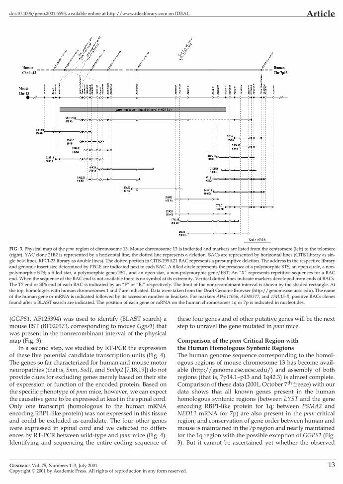
(GGPS1, AF125394) was used to identify (BLAST search) a these four genes and of other putative genes will be the next
FIG. 3. Physical map of the pmn region of chromosome 13. Mouse chromosome 13 is indicated and markers are listed from the centromere (left) to the telomere(right). YAC clone 21B2 is represented by a horizontal line; the dotted line represents a deletion. BACs are represented by horizontal lines (CITB library as sin-gle bold lines, RPCI-23 library as double lines). The dotted portion in CITB-299A21 BAC represents a presumptive deletion. The address in the respective libraryand genomic insert size determined by PFGE are indicated next to each BAC. A filled circle represents the presence of a polymorphic STS; an open circle, a non-polymorphic STS; a filled star, a polymorphic gene/EST; and an open star, a non-polymorphic gene/EST. An “X” represents repetitive sequences for a BACend. When the sequence of the BAC end is not available there is no symbol at its extremity. Vertical dotted lines indicate markers developed from ends of BACs.The T7 end or SP6 end of each BAC is indicated by an “F” or “R,” respectively. The limit of the nonrecombinant interval is shown by the shaded rectangle. Atthe top, homologies with human chromosomes 1 and 7 are indicated. Data were taken from the Draft Genome Browser (http://genome.cse.ucsc.edu). The nameof the human gene or mRNA is indicated followed by its accession number in brackets. For markers AW611066, AI049377, and 174L15-R, positive BACs clonesfound after a BLAST search are indicated. The position of each gene or mRNA on the human chromosomes 1q or 7p is indicated in nucleotides.
mouse EST (BF020173, corresponding to mouse Ggps1) thatwas present in the nonrecombinant interval of the physicalmap (Fig. 3).
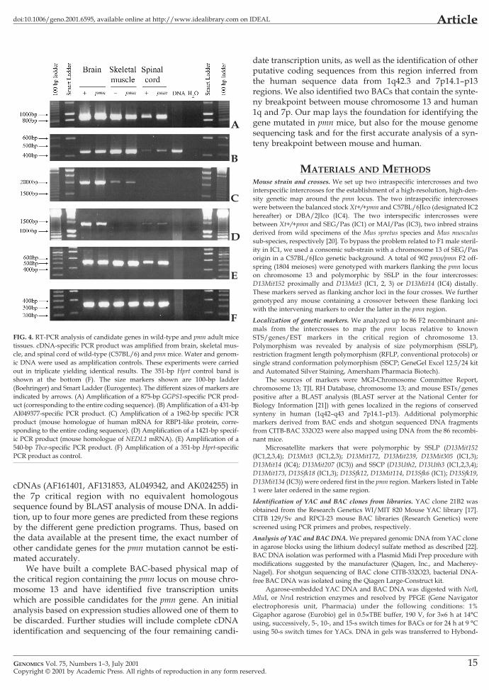
In a second step, we studied by RT-PCR the expressionof these five potential candidate transcription units (Fig. 4).The genes so far characterized for human and mouse motorneuropathies (that is, Smn, Sod1, and Smbp2 [7,18,19]) do notprovide clues for excluding genes merely based on their siteof expression or function of the encoded protein. Based onthe specific phenotype of pmn mice, however, we can expectthe causative gene to be expressed at least in the spinal cord.Only one transcript (homologous to the human mRNAencoding RBP1-like protein) was not expressed in this tissueand could be excluded as candidate. The four other geneswere expressed in spinal cord and we detected no differ-ences by RT-PCR between wild-type and pmn mice (Fig. 4).Identifying and sequencing the entire coding sequence of
GENOMICS Vol. 75, Numbers 1–3, July 2001Copyright © 2001 by Academic Press. All rights of reproduction in any form res
step to unravel the gene mutated in pmn mice.
Comparison of the pmn Critical Region with the Human Homologous Syntenic RegionsThe human genome sequence corresponding to the homol-ogous regions of mouse chromosome 13 has become avail-able (http://genome.cse.ucsc.edu/) and assembly of bothregions (that is, 7p14.1–p13 and 1q42.3) is almost complete.Comparison of these data (2001, October 7th freeze) with ourdata shows that all known genes present in the humanhomologous syntenic regions (between LYST and the geneencoding RBP1-like protein for 1q; between PSMA2 andNEDL1 mRNA for 7p) are also present in the pmn criticalregion; and conservation of gene order between human andmouse is maintained in the 7p region and nearly maintainedfor the 1q region with the possible exception of GGPS1 (Fig.3). But it cannot be ascertained yet whether the observed
erved.13

Article doi:10.1006/geno.2001.6595, available online at http://www.idealibrary.com on IDEAL
change is a true inversion or an error because the assembly present in this human region. Two of our BACs (44K14 and
TABLE 2: Molecular markers used to construct the physical mapNon-polymorphic markers present on the contig
Markera,b GenBank Primers Sequence
Acc. No.
332O23-R not available 332023-R-1F GTTGTTGTTGAGTGAGTGAGTA332023-R-1R AGTCATCTCTTCAGCCAAGCA
194O7-F AZ579062 AI049377-15F GGTTTATGCTGTCCATGGCTAI049377c AI049377-187R TGGAGAGAATGGATTGGAGC
299A21-F AZ579063 299A21-F-6F CAGTTCTCTCCAGAGGTATC299A21-F-206R GAACTAGGCCTACCCACTAC
299A21-R AZ579064 299A21-F-42F ACGTGGGAAGAGCTAGAAAC299A21-F-228R ACTGTTCTCAACCACACCCC
322E16-R AZ579065 322E16-R-180F TTCACTGGTCAAGAGGTCCA322E16-R-363R AGCCTGAGGGAGGTTTGAAT
203A16-F AZ579066 203A16-F-177F TCCTGCCATAAATTTCCCTG203A16-F-401R CAGGCCATGTTTTCAGTCCT
203A16-R AZ579067 203A16-R-140F GGTGACTAAGGGTGGAGTGG203A16-R-353R TTAGGCTGTTGTGGGAGCTT
44K14-R AZ579073 44K14-R-8F TACACTTACCTTCCATGATG44K14-R-110R GAAGACTGTCTTCATGTTCTC
158L23-F AZ579074 158L23-F-61F CCCACTCTCGTATATATACAC158L23-F-522R CATATAGAATGAATGAATGAATGAAAAG
5A19-F AZ579075 5A19-F-346F GGCAGTCATTGACCCAGTTT5A19-F-503R CAAAGGTCCTGAACGGAAAA
5A19-R AZ579076 5A19-R-47F ATTCTCTTTCCTCATACCTC5A19-R-128R GGTGGGGTGTAAACATATAA
174L15-Re AZ579077 174L15-R-16F GCTGTTTCCCACCATACTATTC174L15-R-244R AAGCGATTGAGAAACACCCATC
269J1-F AZ579078 269J1-F-16F CTATCTGACCACTACAAATG269J1-F-101R TTGAGAGTGGATAACCTCAA
269J1-R (TDB) AZ022261 269J1-R-25F CATCTAAAACTTGAGGCATG269J1-R-432R CATTGTTTCCAAGATGAGAG
378J7-R (TDB) AZ011192 378J7-R-1F AGGTATAGACTAAGAGACTAG378J7-R-153R AAAATACCCTCAAGTGACTC
293H2-R (TDB) AQ922186 293H2-R-345F TCTAGGAGCTCATTCTTCCCA293H2-R-493R TGATTCACCTATAGTGATGAACAAGA
399G8-F (TDB) AZ052416 399G8-F-315F CACATGGCAGACACTTGCTT399G8-F-492R ACAGGGTTCTGGCAAATCAG
399G8-R (TDB) AZ052413 399G8-R-164F TGTGGTTCAAATTGACAAAAGAA399G8-R-443R TGCATTTTTAACCAGCCAAA
AV273951e,f AV273951 AV273951-55F TCCTTTGTTTCCCATGAACTGAV273951-190R CCATTTCGTCAAGTGTAGCA
BF0201731g BF020173 BF020173-82F CCACAGGCCTCAATTTGTTTBF020173-274R CCCCACTATACCATGCCATT
Accession No. and primers names and sequences for polymorphic markers used in the establishment of the physical map are not included, as they are shown in Table 1.Primers and markers derived from BAC ends are named according to the same scheme as in Table 1.a, b, c, d, eNotations are the same as in Table 1.fMouse EST similar to human NEDL1 mRNA, placed in human chromosome 7p13.gMouse EST corresponding to Ggps1 mouse gene, homologous to human geranylgeranyl pyrophosphate synthase mRNA, placed in human chr. 1q43.
Polymorphic markers present on the contig
332O23-F 266O18-R Gng4
322E16-F 144K13-F AW611066
7O14-F 378J7-F Tbce
7014-R D13Uth2 D13Uth3
Polymorphic markers not found on the contig
ESTs from TJL RH database
Ryr2 AA517551
Nid1 AI256733
Lyst
Gli3
Non-polymorphic ESTs (acc. no.)not found on the contig
From TJL RH database
C80633 AW209491 AI447560
AI663969 AU023367 AI746446
C80161 AV379049 AI132359
AA408594 AI841875 AA516957
AI448615
of human sequence is still a draft. Also, at least one EST(AI049377) is present in our map with, so far, no equivalentgene or full-length mRNA present in the human 1q assem-bly (BLAST search). Nevertheless, according to two differ-ent gene prediction programs, another gene might be
Copyri14
158L23) contain the breakpoint of synteny between mouseand human.
There may be other genes present in our maps. There arethree cDNAs present in the 1q critical region of humanassembly (AF119882, AK022437, and AK022422) and four
GENOMICS Vol. 75, Numbers 1–3, July 2001ght © 2001 by Academic Press. All rights of reproduction in any form reserved.
Articledoi:10.1006/geno.2001.6595, available online at http://www.idealibrary.com on IDEAL
date transcription units, as well as the identification of other
cDNAs (AF161401, AF131853, AL049342, and AK024255) inthe 7p critical region with no equivalent homologoussequence found by BLAST analysis of mouse DNA. In addi-tion, up to four more genes are predicted from these regionsby the different gene prediction programs. Thus, based onthe data available at the present time, the exact number ofother candidate genes for the pmn mutation cannot be esti-mated accurately.
We have built a complete BAC-based physical map ofthe critical region containing the pmn locus on mouse chro-mosome 13 and have identified five transcription unitswhich are possible candidates for the pmn gene. An initialanalysis based on expression studies allowed one of them tobe discarded. Further studies will include complete cDNAidentification and sequencing of the four remaining candi-
FIG. 4. RT-PCR analysis of candidate genes in wild-type and pmn adult micetissues. cDNA-specific PCR product was amplified from brain, skeletal mus-cle, and spinal cord of wild-type (C57BL/6) and pmn mice. Water and genom-ic DNA were used as amplification controls. These experiments were carriedout in triplicate yielding identical results. The 351-bp Hprt control band isshown at the bottom (F). The size markers shown are 100-bp ladder(Boehringer) and Smart Ladder (Eurogentec). The different sizes of markers areindicated by arrows. (A) Amplification of a 875-bp GGPS1-specific PCR prod-uct (corresponding to the entire coding sequence). (B) Amplification of a 431-bpAI049377-specific PCR product. (C) Amplification of a 1962-bp specific PCRproduct (mouse homologue of human mRNA for RBP1-like protein, corre-sponding to the entire coding sequence). (D) Amplification of a 1421-bp specif-ic PCR product (mouse homologue of NEDL1 mRNA). (E) Amplification of a540-bp Tbce-specific PCR product. (F) Amplification of a 351-bp Hprt-specificPCR product as control.
A
B
C
D
E
F
GENOMICS Vol. 75, Numbers 1–3, July 2001Copyright © 2001 by Academic Press. All rights of reproduction in any form res
putative coding sequences from this region inferred fromthe human sequence data from 1q42.3 and 7p14.1–p13regions. We also identified two BACs that contain the synte-ny breakpoint between mouse chromosome 13 and human1q and 7p. Our map lays the foundation for identifying thegene mutated in pmn mice, but also for the mouse genomesequencing task and for the first accurate analysis of a syn-teny breakpoint between mouse and human.
MATERIALS AND METHODSMouse strain and crosses. We set up two intraspecific intercrosses and twointerspecific intercrosses for the establishment of a high-resolution, high-den-sity genetic map around the pmn locus. The two intraspecific intercrosseswere between the balanced stock Xt+/+pmn and C57BL/6JIco (designated IC2hereafter) or DBA/2JIco (IC4). The two interspecific intercrosses werebetween Xt+/+pmn and SEG/Pas (IC1) or MAI/Pas (IC3), two inbred strainsderived from wild specimens of the Mus spretus species and Mus musculussub-species, respectively [20]. To bypass the problem related to F1 male steril-ity in IC1, we used a consomic sub-strain with a chromosome 13 of SEG/Pasorigin in a C57BL/6JIco genetic background. A total of 902 pmn/pmn F2 off-spring (1804 meioses) were genotyped with markers flanking the pmn locuson chromosome 13 and polymorphic by SSLP in the four intercrosses:D13Mit152 proximally and D13Mit3 (IC1, 2, 3) or D13Mit14 (IC4) distally.These markers served as flanking anchor loci in the four crosses. We furthergenotyped any mouse containing a crossover between these flanking lociwith the intervening markers to order the latter in the pmn region.
Localization of genetic markers. We analyzed up to 86 F2 recombinant ani-mals from the intercrosses to map the pmn locus relative to knownSTS/genes/EST markers in the critical region of chromosome 13.Polymorphism was revealed by analysis of size polymorphism (SSLP),restriction fragment length polymorphism (RFLP, conventional protocols) orsingle strand conformation polymorphism (SSCP; GeneGel Excel 12.5/24 kitand Automated Silver Staining, Amersham Pharmacia Biotech).
The sources of markers were MGI-Chromosome Committee Report,chromosome 13; TJL RH Database, chromosome 13; and mouse ESTs/genespositive after a BLAST analysis (BLAST server at the National Center forBiology Information [21]) with genes localized in the regions of conservedsynteny in human (1q42–q43 and 7p14.1–p13). Additional polymorphicmarkers derived from BAC ends and shotgun sequenced DNA fragmentsfrom CITB-BAC 332O23 were also mapped using DNA from the 86 recombi-nant mice.
Microsatellite markers that were polymorphic by SSLP (D13Mit152(IC1,2,3,4); D13Mit3 (IC1,2,3); D13Mit172, D13Mit239, D13Mit305 (IC1,3);D13Mit14 (IC4); D13Mit207 (IC3)) and SSCP (D13Uth2, D13Uth3 (IC1,2,3,4);D13Mit173, D13Sfk18 (IC1,3); D13Sfk12, D13Mit114, D13Sfk6 (IC1); D13Sfk19,D13Mit134 (IC3)) were ordered first in the pmn region. Markers listed in Table1 were later ordered in the same region.
Identification of YAC and BAC clones from libraries. YAC clone 21B2 wasobtained from the Research Genetics WI/MIT 820 Mouse YAC library [17].CITB 129/Sv and RPCI-23 mouse BAC libraries (Research Genetics) werescreened using PCR primers and probes, respectively.
Analysis of YAC and BAC DNA. We prepared genomic DNA from YAC clonein agarose blocks using the lithium dodecyl sulfate method as described [22].BAC DNA isolation was performed with a Plasmid Midi Prep procedure withmodifications suggested by the manufacturer (Qiagen, Inc., and Macherey-Nagel). For shotgun sequencing of BAC clone CITB-332O23, bacterial DNA-free BAC DNA was isolated using the Qiagen Large-Construct kit.
Agarose-embedded YAC DNA and BAC DNA was digested with NotI,MluI, or NruI restriction enzymes and resolved by PFGE (Gene Navigatorelectrophoresis unit, Pharmacia) under the following conditions: 1%Gigaphor agarose (Eurobio) gel in 0.5×TBE buffer, 190 V, for 3×6 h at 14°Cusing, successively, 5-, 10-, and 15-s switch times for BACs or for 24 h at 9 °Cusing 50-s switch times for YACs. DNA in gels was transferred to Hybond-
erved.15
Article doi:10.1006/geno.2001.6595, available online at http://www.idealibrary.com on IDEAL
N+ membranes (Amersham) by conventional Southern blot analysis [23] and 3. Bueno Brunialti, A.L., Poirier, C., Schmalbruch, H., and Guénet, J.-L. (1995). The
hybridized with radiolabeled probes.Generation of novel STSs. We derived sequences for STS- content mappingfrom BAC ends by cloning of extremities of BACs from CITB 129/Sv library,using the vectorette PCR technique [24,25]; direct sequencing of RPCI-23library BAC ends for the forward direction (T7 primer), and for the reversedirection (SP6 or KBR/ TJ primers), according to TIGR Reaction Protocol forBAC End Sequencing (http://www.tigr.org/tdb/bac_ends/mouse/bac_end_intro.html); or analyzing BAC-end sequences that are available on-line at the TIGR site, for some RPCI-23 library BAC clones.
We carried out shotgun sequencing of the first BAC isolated with D13Uth2(CITB-332O23), which cosegregates with pmn locus. Briefly, BAC DNA (30 µg)isolated as described above was sheared by nebulization, end-repaired usingT4 DNA polymerase, and ligated to BstXI adaptors (Invitrogen). The ligationmixture was fractionated by agarose-gel electrophoresis and fragments from 1kb to 3 kb were ligated into BstXI-digested pcDNA2.1 (Invitrogen). We used theplasmids as templates for cycle sequencing reactions consisting of 35 cycles(96°C for 30 s; 50°C for 15 s; 60°C for 4 min) in a thermocycler. Samples wereprecipitated and loaded onto an ABI 3700 automatic capillary DNA sequencer(Applied Biosystem). Shotgun clone sequences from both ends were assembledinto 51 contigs and edited using the PHRED [26,27], PHRAP (P. Green, unpub-lished data), and CONSED [28] softwares.
Analysis of candidate transcription units by RT-PCR. Reverse transcriptionreactions using Superscript reverse transcriptase (Gibco-BRL) were per-formed from total RNA prepared (Qiagen, RNAeasy) from brain, spinal cord,and skeletal muscle of adult wild-type (C57BL/6 background) and pmn miceaccording to the manufacturer’s instructions. For cDNA amplification of can-didate genes we used the following primers: mouse homologue of humanmRNA for RBP1-like protein, 5′-GTCACTATAACAGAAGCTAG-3′ and 5′-AATTAAATACAAGTGCCAAACAG-3′; mouse EST AI049377, 5′-GGTT-TATGCTGTCCATGGCT-3′ and 5′-CAGGTTGTGGTATATATG-GATATTTTG-3′; mouse homologue of human NEDL1 mRNA, 5′-CCATTTCGTCAAGTGTAGCA-3′ and 5′-CAGGGTCCGGGCAAAAT-TAAG-3′; mouse homologue of human TBCE, 5′-GAACAGGTGCCA-GATTCTTC-3′ and 5′-GGCAAAGGTTTTGTTTCTGG-3′; mouse Ggps1, 5′-GAGATCATCGTGGAACCGTC-3′ and 5′-CCACAGGCCTCAATTTGTTT-3′.Mouse Hprt was amplified as control: 5′-CCTGCTGGATTACATTAAAG-CACTG-3′ and 5’-GTCAAGGGCATATCCAACAACAAAC-3′.
ACKNOWLEDGMENTSWe thank our colleagues and friends of the Unité de Biologie du Développement, inparticular Sandrine Vandormael-Pournin, for support and advice; and PatriciaBaldacci, Michel Cohen-Tannoudji, and Xavier Montagutelli for comments on themanuscript. This work was supported by the Institut Pasteur and the AssociationFrançaise contre les Myopathies (A.F.M.).
RECEIVED FOR PUBLICATION DECEMBER 26, 2000; ACCEPTED APRIL 7, 2001.
REFERENCES1. Schmalbruch, H. M. D., Jensen, H., Bjearg, M., Kamienniecka, Z., and Kurland, L. B. S.
(1991). A new mouse mutant with progressive motor neuronopathy. J. Neuropathol.Exp. Neurol. 50: 192–204.
2. Sendtner, M., et al. (1992). Ciliary neurotrophic factor prevents degeneration of motorneurons in mouse mutant progressive motor neuronopathy. Nature 358: 502–504.
Copyrig16
Sequence data from this article have been deposited with the EMBL/GenBank Data LibrCITB-332O23), and AZ579062–AZ579079 (markers derived from BAC ends).
mouse mutation progressive motor neuronopathy (pmn) maps to chromosome 13.Genomics 29: 131–135.
4. Brzustowicz, L.M., et al. (1990). Genetic mapping of chronic childhood-onset spinalmuscular atrophy to chromosome 5q11.2–13.3. Nature 344: 540–541.
5. Melki, J., et al. (1990). Gene for chronic proximal spinal muscular atrophies maps tochromosome 5q. Nature 344: 767–768.
6. Burglen, L., et al. (1996). Structure and organization of the human survival motorneuron (SMN) gene. Genomics 32: 479–482.
7. Viollet, L., et al. (1997). cDNA isolation, expression, and chromosomal localizationof the mouse survival motor neuron gene (Smn). Genomics 40: 185–188.
8. Sagot, Y., et al. (1995). Bcl-2 overexpression prevents motoneuron cell body loss butnot axonal degeneration in a mouse model of a neurodegenerative disease. J.Neurosci. 15: 7727–7733.
9. Duong, F., et al. (1998). The effect of the nonpeptide neurotrophic compound SR57746A on the progression of the disease state of the pmn mouse. Br. J. Pharmacol.124: 811–817.
10. Haase, G., et al. (1998). Adenovirus-mediated transfer of the neurotrophin-3 geneinto skeletal muscle of pmn mice: therapeutic effects and mechanisms of action. J.Neurol. Sci. 160 (suppl. 1): S97–S105.
11. Haase, G., et al. (1999). Therapeutic benefit of ciliary neurotrophic factor in progres-sive motor neuronopathy depends on the route of delivery. Ann. Neurol. 45:296–304.
12. Sagot, Y., et al. (1995). Polymer encapsulated cell lines genetically engineered torelease ciliary neurotrophic factor can slow down progressive motor neuronopathyin the mouse. Eur. J. Neurosci. 7: 1313–1322.
13. Sagot, Y., Tan, S. A., Hammang, J. P., Aebischer, P., and Kato, A. C. (1996). GDNFslows loss of motoneurons but not axonal degeneration or premature death ofpmn/pmn mice. J. Neurosci. 16: 2335–2341.
14. Sagot, Y., Rosse, T., Vejsada, R., Perrelet, D., and Kato, A. C. (1998). Differentialeffects of neurotrophic factors on motoneuron retrograde labeling in a murinemodel of motoneuron disease. J. Neurosci. 18: 1132–1141.
15. Barbosa, M. D., et al. (1996). Identification of the homologous beige and Chediak-Higashi syndrome genes. Nature 382: 262–265.
16. Perou, C. M., et al. (1997). Comparative mapping in the beige-satin region of mousechromosome 13. Genomics 39: 136–146.
17. Haldi, M. L., et al. (1996). A comprehensive large-insert yeast artificial chromosomelibrary for physical mapping of the mouse genome. Mamm. Genome 7: 767–769.
18. Brown, R. H. J. (1995). Amyotrophic lateral sclerosis: recent insights from geneticsand transgenic mice. Cell 80: 687–692.
19. Cox, G. A., Mahaffey, C. L., and Frankel, W. N. (1998). Identification of the mouseneuromuscular degeneration gene and mapping of a second site suppressor allele.Neuron 21: 1327–1337.
20. Bonhomme, F., and Guénet, J.-L (1996). The laboratory mouse and its wild relatives. InGenetic Variants and Strains of the Laboratory Mouse (M. F. Lyon, S. Rastan, and S. D. M.Brown, Eds.), pp. 1577–1596. Oxford University Press, Oxford.
21. Altschul, S. F., et al. (1997). Gapped BLAST and PSI-BLAST: a new generation of pro-tein database search programs. Nucleic Acids Res. 25: 3389–3402.
22. Huxley, C., Hagrino, Y., Schlessinger, D., and Olson, M. V. (1991). The human HPRTgene on a yeast artificial chromosome is functional when transferred to mouse cellsby fusion. Genomics 9: 742–750.
23. Southern, E.M. (1975). Detection of specific sequences among DNA fragments sepa-rated by gel electropohoresis. J. Mol. Biol. 98: 503–517.
24. Riley, J., et al. (1990). A novel rapid method for the isolation of terminal sequencesfrom yeast artificial chromosome (YAC) clones. Nucleic Acids Res. 18: 2887–2890.
25. Cohen-Tannoudji, M., et al. (2000). A 2-Mb YAC/BAC-based physicial map of theOvum mutant (Om) locus region on mouse chromosome 11. Genomics 68: 273–282.
26. Ewing, B., and Green,. P. (1998). Base-calling of automated sequencer traces usingphred. II. Error probabilities. Genome Res. 8: 186–194.
27. Ewing, B., Hillier, L., Wendl, M. C., and Green, P. (1998). Base-calling of automatedsequencer traces using phred. I. Accuracy assessment. Genome Res. 8: 175–185.
28. Gordon, D., Abajian, C., and Green, P. (1998). Consed: a graphical tool for sequencefinishing. Genome Res. 8: 195–202.
GENOMICS Vol. 75, Numbers 1–3, July 2001ht © 2001 by Academic Press. All rights of reproduction in any form reserved.
aries under accession numbers AF330047 and AF330048 (shotgun sequencing of BAC