Embed Size (px)
Citation preview
3/26/2010fatchiyah, lab of Molecular Biology Brawijaya
University1
Genetic Control of
Immune Responses
3/26/2010fatchiyah, lab of Molecular Biology Brawijaya
University2
B
T
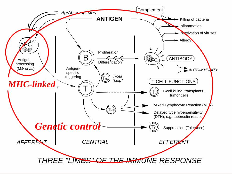
Antigen- specifictriggering
Proliferation
Differentiation
T H2T-cell"help"
T-cell killing: transplants, tumor cells
Delayed type hypersensitivity,(DTH); e.g. tuberculin reaction
Mixed Lymphocyte Reaction (MLR)
Suppression (Tolerance)
ANTIBODY
T-CELL FUNCTIONS
ANTIGENAg/Ab complexes
Inactivation of viruses
Allergy
Complement
AUTOIMMUNITY
THREE "LIMBS" OF THE IMMUNE RESPONSE
AFFERENT CENTRAL EFFERENT
APC
Antigenprocessing
(M et al.)
CT
T S
AFC
Antigen"presentation"
Inflammation
Killing of bacteria
T H1
Genetic control
MHC-linked
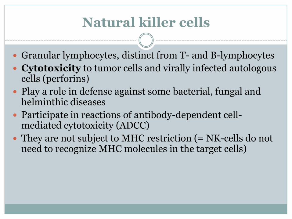
Natural killer cells
Granular lymphocytes, distinct from T- and B-lymphocytes
Cytotoxicity to tumor cells and virally infected autologous cells (perforins)
Play a role in defense against some bacterial, fungal and helminthic diseases
Participate in reactions of antibody-dependent cell-mediated cytotoxicity (ADCC)
They are not subject to MHC restriction (= NK-cells do not need to recognize MHC molecules in the target cells)
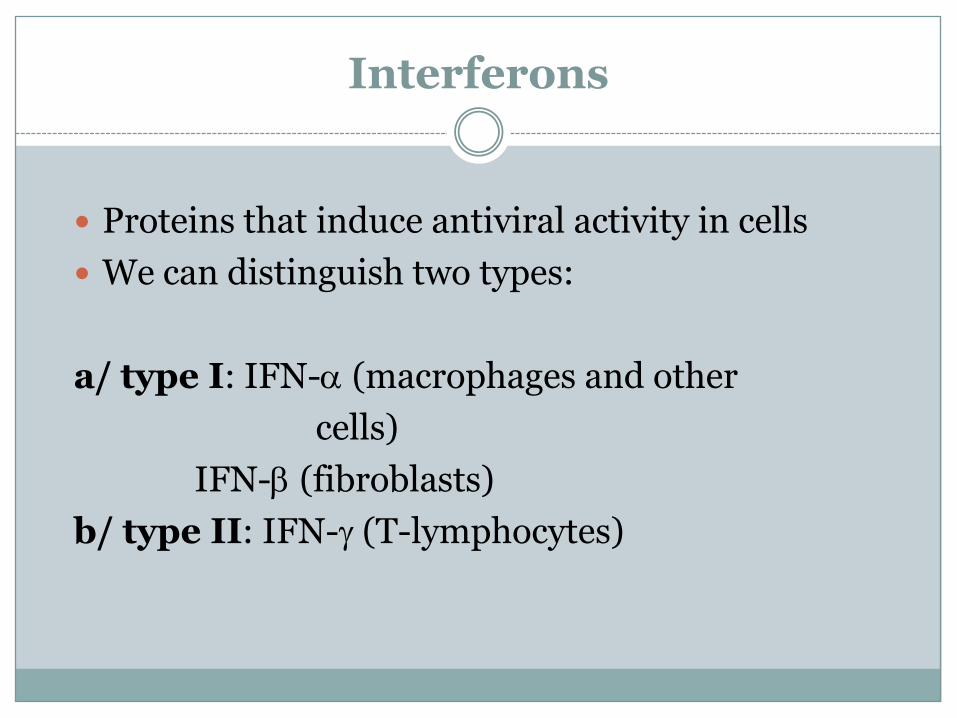
Interferons
Proteins that induce antiviral activity in cells
We can distinguish two types:
a/ type I: IFN-a (macrophages and other
cells)
IFN-b (fibroblasts)
b/ type II: IFN-g (T-lymphocytes)
Function of interferons
Induce cells to produce antiviral proteins (protein kinase, oligonucleotide polymerase – interferencewith the translation of viral mRNA)
Enhance T-cell activity
Activate macrophages
Increase the cytotoxic action of NK-cells
Basophils and mast cells
Very similar type of cells, however, basophils circulate in blood circulation, whereas mast cells reside in tissues (connective tissue, mucosa)
IgE antibodies are bound on the surface of basophils and mast cells by FceRI
Abundant granules containing biogenic amines (histamine), proteases (tryptase) and proteoglycans (heparin) in cytoplasm
Basophils and mast cells
If IgE molecules bound on the surface of the cells are cross-linked by an antigen, then occurs:
a/ degranulation – release of content of granules to the cell’s surroundings
b/ activation of arachidonic acid’s metabolism – production of prostaglandins a leukotriens which are released from cells
The release of these substances leads to vasodilation, increased vascular permeability, bronchoconstriction, increased mucus secretion etc.
Basophils and mast cells - function
Defense against helminthic parasites
Allergic reactions (I.type)
Mast cells contribute to the normal function of mucosa and connective tissue
Major Histocompatibility Complex System of glycoproteins bound on cell membrane
which can be recognized by immune system
Genes coding MHC are localized on chromosome 6, some of these genes are extremely polymorphic (signs of Mendelian heredity, codominancy, en bloc transfer)
MHC haplotype = unique combination of alleles encoding MHC molecules which are localized on one chromosome
Major histocompatibility complex
Class I – HLA A,B,C (E,F,G)
– expressed on the surface of all
nucleated human cells
– antigen presentation to Tc-lymphocytes
Class II – HLA DR, DP, DQ
– expressed on the surface of APC
(macrophages, B lymphocytes)
– antigen presentation to Th-lymphocytes
Major histocompatibility complex
Class III – HLA C2, C4, FB etc.– numerous genes located in MHC
chromosomal region (e.g.gen of two C4-isotypes, C2, factor B, TNF-alfaand beta)
– function – processing and transport of
T-lymphocyte epitopes – heat-shock proteins– inflammation mediators
HLA
3/26/2010fatchiyah, lab of Molecular Biology Brawijaya
University
12
Human Leukocyte Antigen
human MHC
cell-surface proteins
important in self vs. nonself
distinction
present peptide antigens to T cells
CLASS I: A,B,C CLASS II: DR,DQ,DP
J. Noble
3/26/2010fatchiyah, lab of Molecular Biology Brawijaya
University13
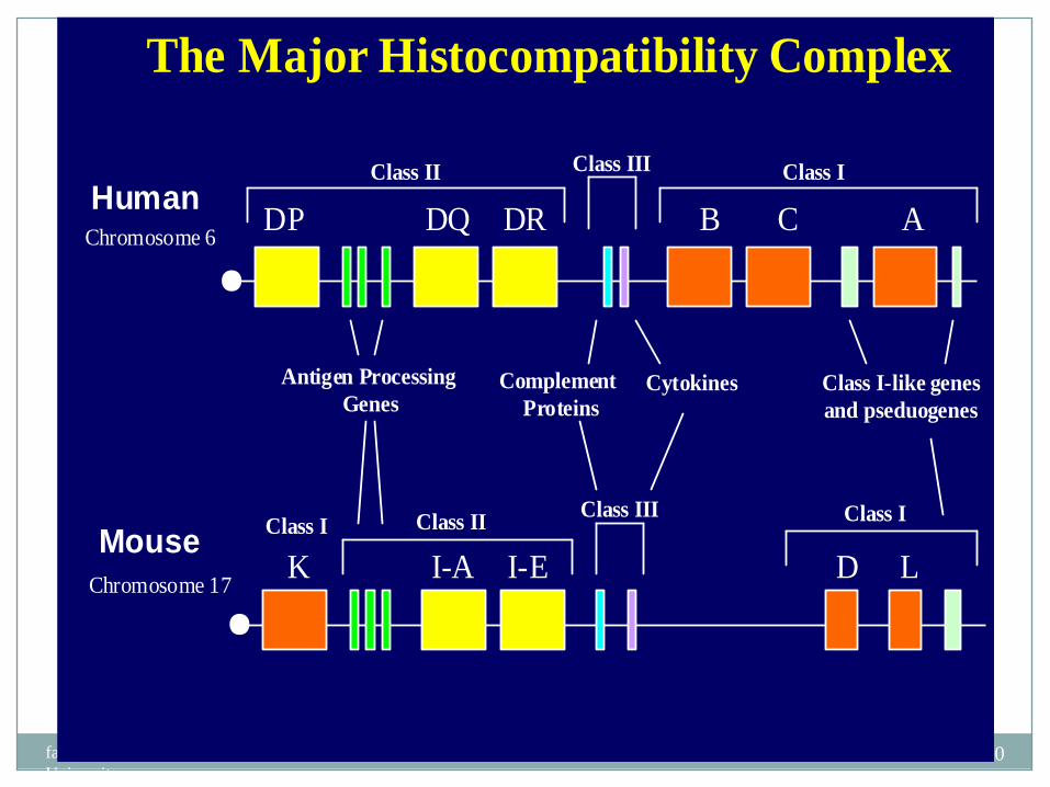
The Major Histocompatibility Complex
Human
Mouse
DP DQ DR B C A
K I-A I-E D L
Chromosome 6
Chromosome 17
Class II Class III Class I
Class IIClass III Class I
Class I
Complement
ProteinsCytokines Class I-like genes
and pseduogenes
Antigen Processing
Genes
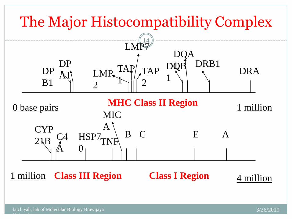
The Major Histocompatibility Complex
3/26/2010fatchiyah, lab of Molecular Biology Brawijaya
University
14
0 base pairs 1 million
1 million 4 million
DP
B1
DP
A1 LMP
2
TAP
1
LMP7
TAP
2
DQB
1
DQA
1 DRB1DRA
CYP
21B C4
A
HSP7
0TNF
B C E A
MIC
A
Class I Region
MHC Class II Region
Class III Region
3/26/2010fatchiyah, lab of Molecular Biology Brawijaya
University15
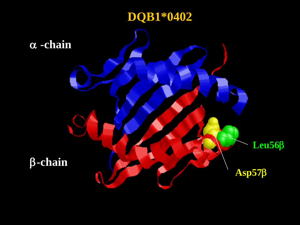
DQB1*0402
Asp57b
Leu56b
a -chain
b-chain
BDC
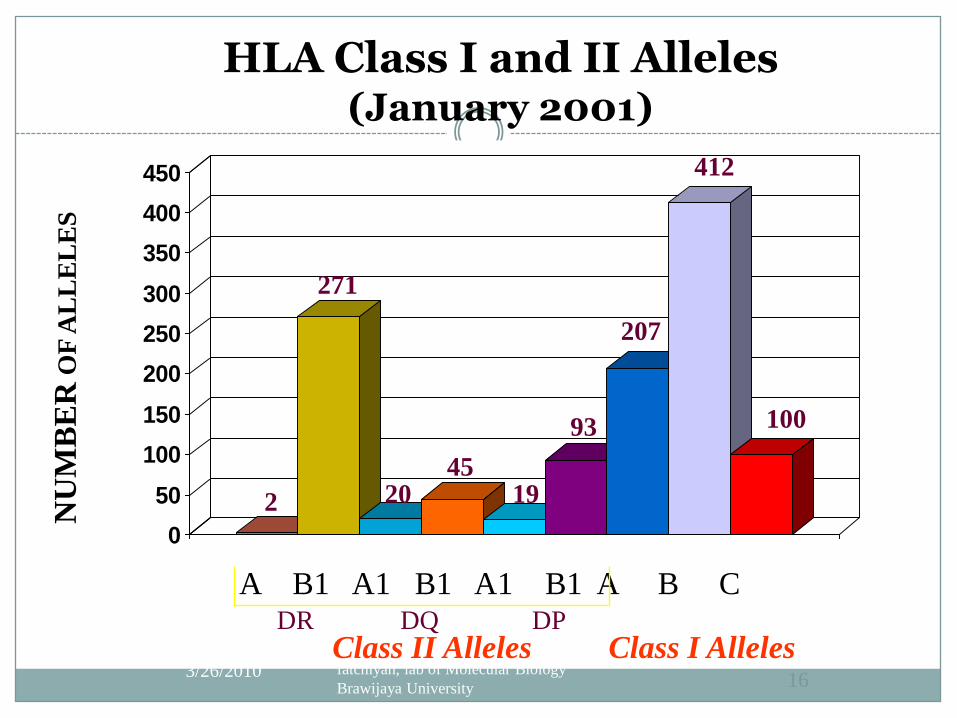
HLA Class I and II Alleles(January 2001)
0
50
100
150
200
250
300
350
400
450
3/26/2010 fatchiyah, lab of Molecular Biology
Brawijaya University 16
Class II Alleles
NU
MB
ER
OF
AL
LE
LE
S
A B1 A1 B1 A1 B1 A B C
Class I AllelesDR DQ DP
207
412
100
2
271
2045
19
93
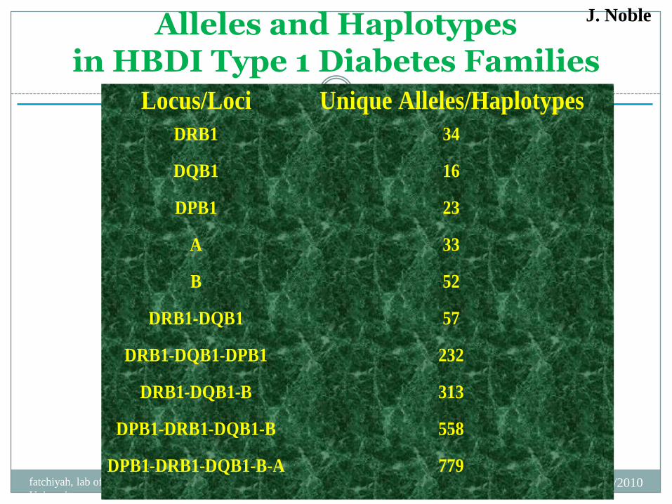
Alleles and Haplotypes in HBDI Type 1 Diabetes Families
3/26/2010fatchiyah, lab of Molecular Biology Brawijaya
University
17Locus/Loci Unique Alleles/Haplotypes
DRB1 34
DQB1 16
DPB1 23
A 33
B 52
DRB1-DQB1 57
DRB1-DQB1-DPB1 232
DRB1-DQB1-B 313
DPB1-DRB1-DQB1-B 558
DPB1-DRB1-DQB1-B-A 779
J. Noble
MHC testing
1/ Sera typing – identification of specific class I and class II MHC molecules using sera typing
Less time-consuming method, however, also less accurate
2/ DNA typing – human DNA testing by PCR
low resolution (groups of alleles), high resolution (single alleles)
More time-consuming method, however, also highly accurate
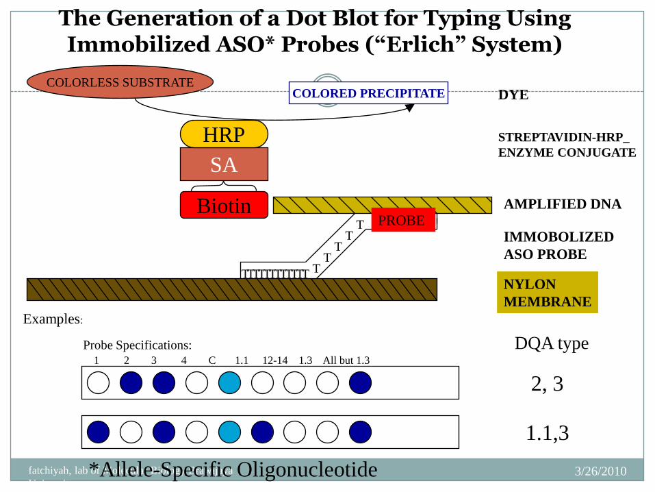
The Generation of a Dot Blot for Typing Using Immobilized ASO* Probes (“Erlich” System)
3/26/2010fatchiyah, lab of Molecular Biology Brawijaya
University
19 DYE
STREPTAVIDIN-HRP_
ENZYME CONJUGATE
AMPLIFIED DNA
IMMOBOLIZED
ASO PROBE
NYLON
MEMBRANE
TT
TT
T
T
Examples:
Probe Specifications:
1 2 3 4 C 1.1 12-14 1.3 All but 1.3
Biotin
SA
HRP
*Allele-Specific Oligonucleotide
COLORLESS SUBSTRATECOLORED PRECIPITATE
DQA type
PROBE
TTTT TTT TTT T
2, 3
1.1,3
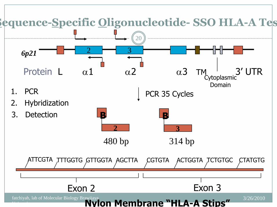
Sequence-Specific Oligonucleotide- SSO HLA-A Test
3/26/2010fatchiyah, lab of Molecular Biology Brawijaya
University
20
2 3
Protein L a1 a2 a3 TM 3’ UTRCytoplasmic
Domain
6p21
PCR 35 Cycles
480 bp 314 bp
B
2
B
3
1. PCR
ATTCGTA TTTGGTG GTTGGTA AGCTTA CGTGTA ACTGGTA TCTGTGC CTATGTG
Nylon Membrane “HLA-A Stips”
2. Hybridization
3. Detection
Exon 2 Exon 3
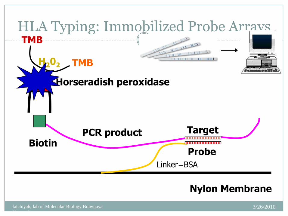
HLA Typing: Immobilized Probe Arrays
3/26/2010fatchiyah, lab of Molecular Biology Brawijaya
University
21
Nylon Membrane
BiotinPCR product Target
Probe
Linker=BSA
Horseradish peroxidaseSA
H202
TMB
TMB
Antigen presentation
An antigen is a substance recognized by immune system that reacts to its presence.
For induction of specific immune response to antigen, first of all antigen processing and its presentation to APC is necessary.
The professional antigen presenting cells (APC) are cells expriming MHC class II molecules (macrophages, dendritic cells, B-lymphocytes).
Processing and presentation of protein antigens
1/ Exogenous antigens
Bacterial, helminthic or viral antigens (either if they form immune complexes swallowed by APC, or if they are processed together with infected cells)
They are presented in a complex with MHC class II to T helper (CD4+) cells
Processing and presentation of protein antigens
2/ Endogenous antigens
Intracellular auto-antigens, antigens of viruses or other intracellular parasites (infecting APC) or tumorous antigens
Present in complex with MHC class I molecules to cytotoxic (CD8+) T cells
1 . T H E S T R U C T U R E O F I M M U N O G L O B U L I N S
Immunoglobulins
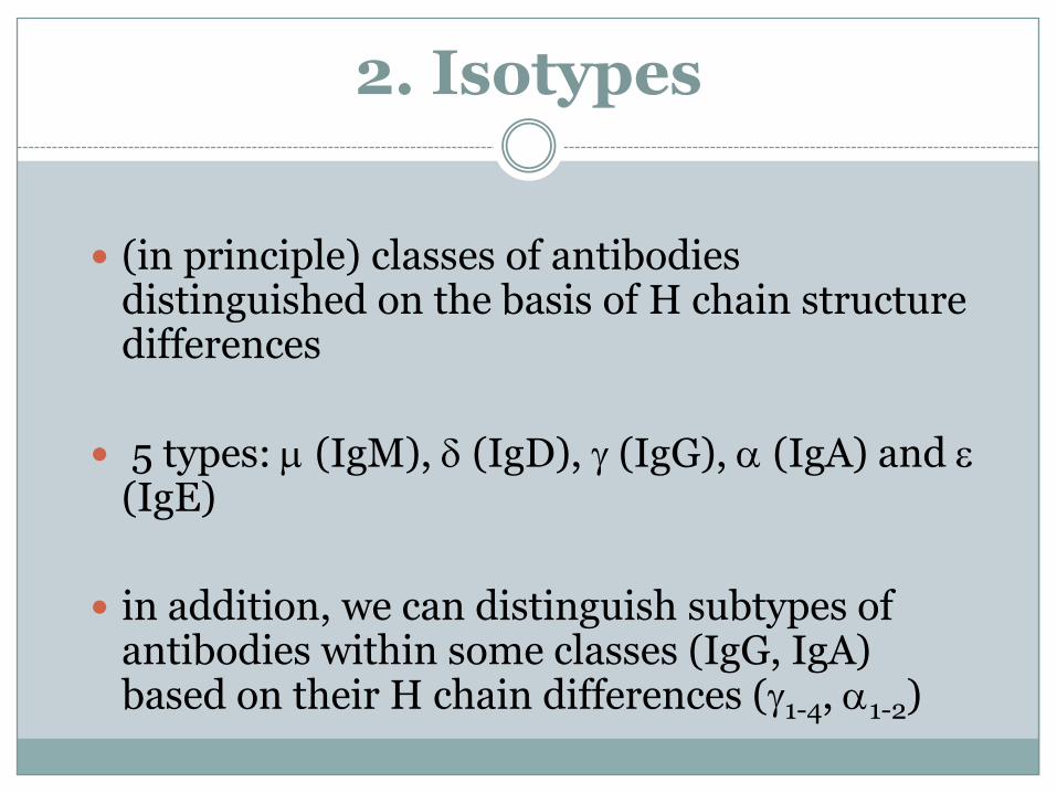
2. Isotypes
(in principle) classes of antibodies distinguished on the basis of H chain structure differences
5 types: m (IgM), d (IgD), g (IgG), a (IgA) and e (IgE)
in addition, we can distinguish subtypes of antibodies within some classes (IgG, IgA) based on their H chain differences (g1-4, a1-2)
3. Domains and their biological function
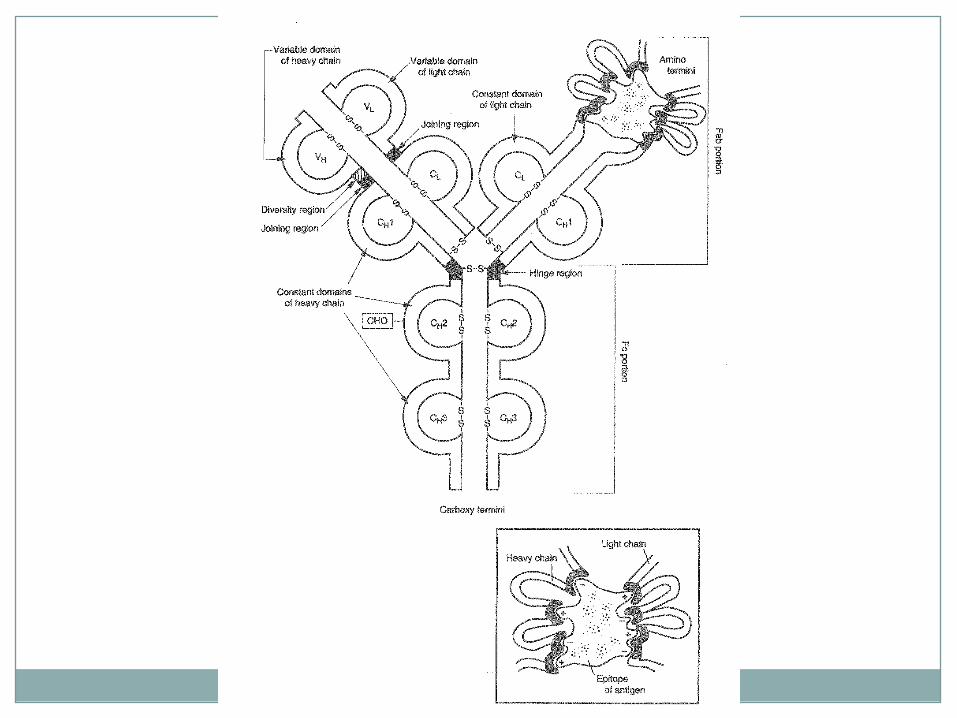
In principle: domains of V regions form a recognizing unit and domains of C regionsdetermine secondary biological functions of antibody (i.e. biological half life, distribution in the body, binding complement, binding to cells through Fc-receptor)
4. Variable region of Ig molecule
Hypervariable loops are concentrated at the spikes of variable regions where antigen binding sites are localized
The binding site specificity is determined by aminoacid sequentions and both by morphology and shape of the loop
5. The biological features of distinct Ig classes
IgG
the most abundant serum Ig
the most important Ig of secondary immune response
the only Ig which passes through the placenta
the main opsonizing Ig
activates complement via classical pathway
biological half life 21 day
IgA
present both in serum and seromucinous secretions
defence of mucosa
opsonization
does not activate complement
IgM
in pentamer form is present in serum; in monomer form is bounded on membrane of B cells
prevailing antibody of primary immune response
high-effective agglutinant and cytolytic agent
usually isohaemagglutinins and natural antibodies
the best classical way complement activator
does not bind phagocytes Fc receptor, but substantially enhances phagocytosis through complement activation
IgD
free form in serum, bound on B cells membrane
antigen receptor on B cells
IgE
in normal conditions low amounts in serum
mainly bound on mast cells (binds through FceR)
anti-helminth defense
immediate type allergic reactions
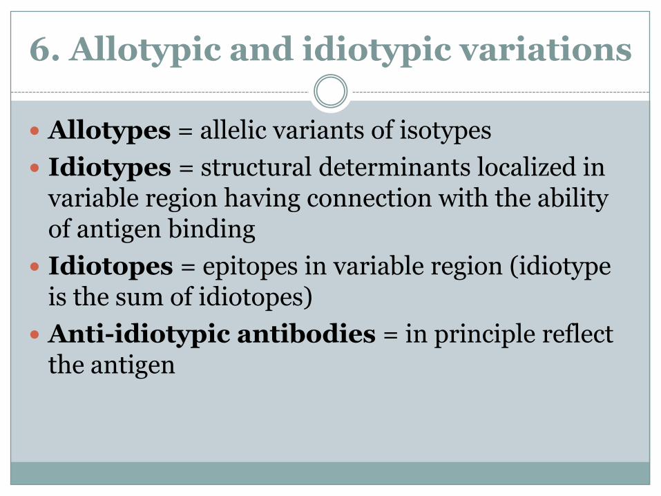
6. Allotypic and idiotypic variations
Allotypes = allelic variants of isotypes
Idiotypes = structural determinants localized in variable region having connection with the ability of antigen binding
Idiotopes = epitopes in variable region (idiotype is the sum of idiotopes)
Anti-idiotypic antibodies = in principle reflect the antigen
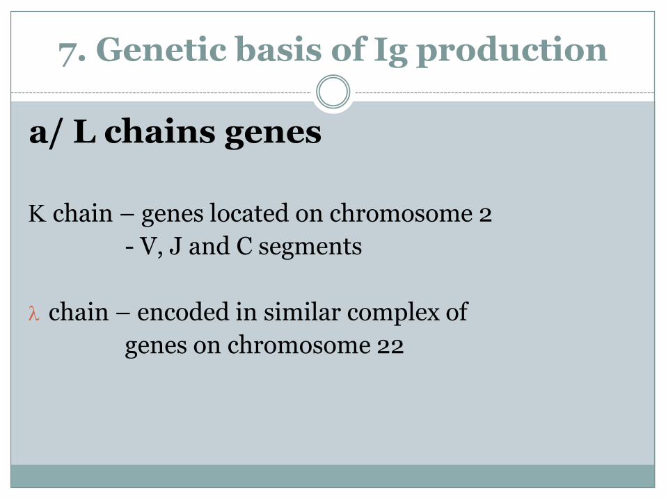
7. Genetic basis of Ig production
a/ L chains genes
K chain – genes located on chromosome 2
- V, J and C segments
chain – encoded in similar complex of
genes on chromosome 22
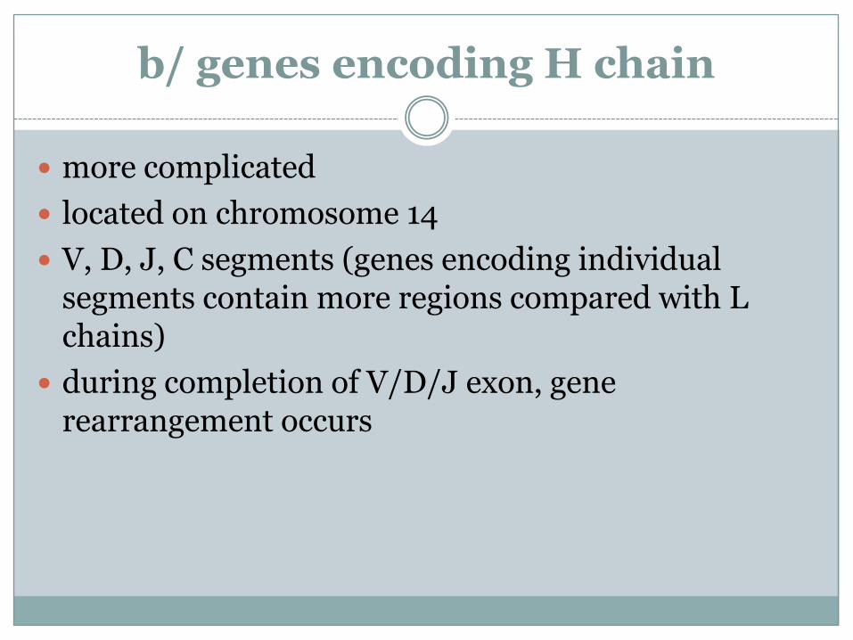
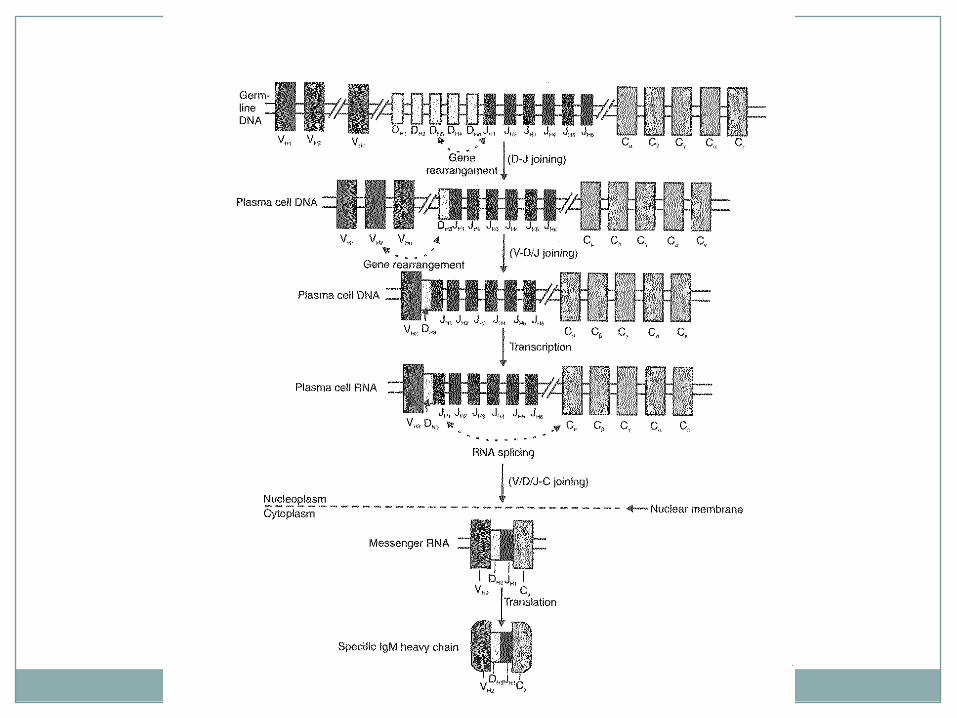
b/ genes encoding H chain
more complicated
located on chromosome 14
V, D, J, C segments (genes encoding individual segments contain more regions compared with L chains)
during completion of V/D/J exon, gene rearrangement occurs
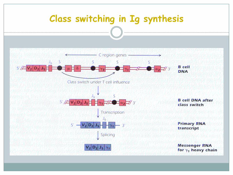
Class switching in Ig synthesis
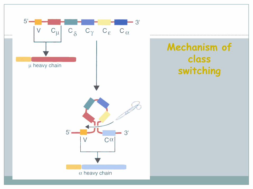
Mechanism of class
switching
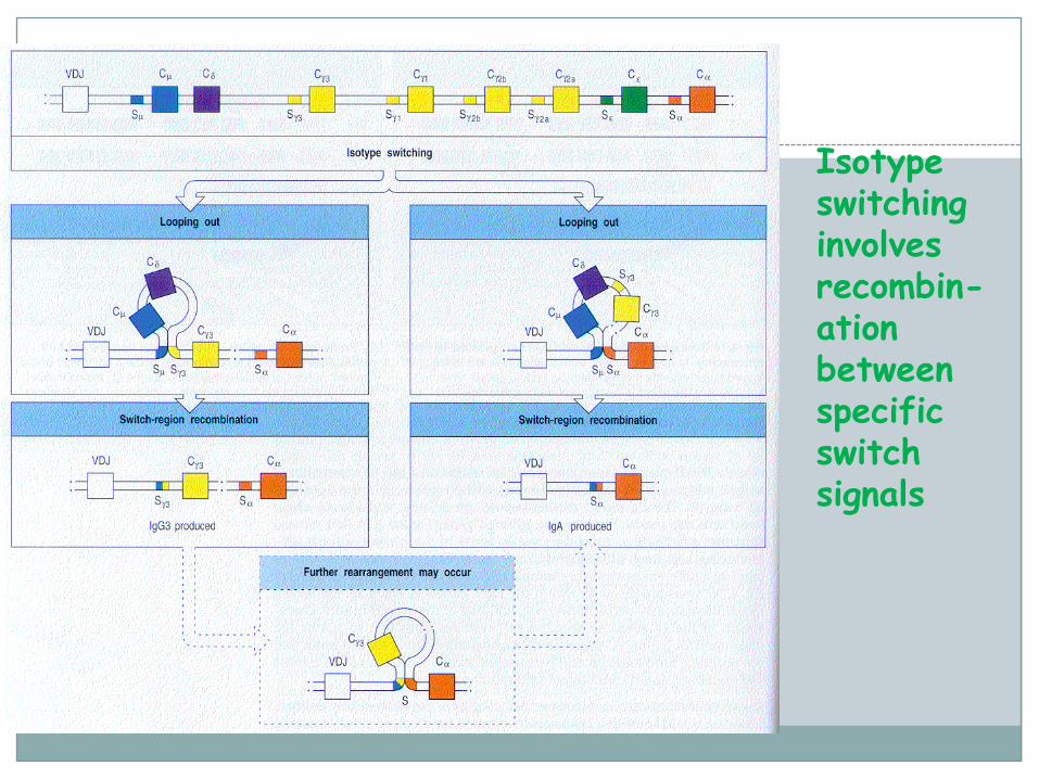
Isotype switching involves recombin-ation between specific switch signals
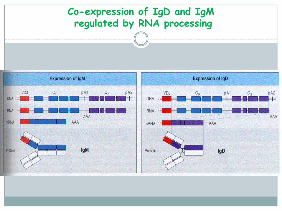
Co-expression of IgD and IgMregulated by RNA processing
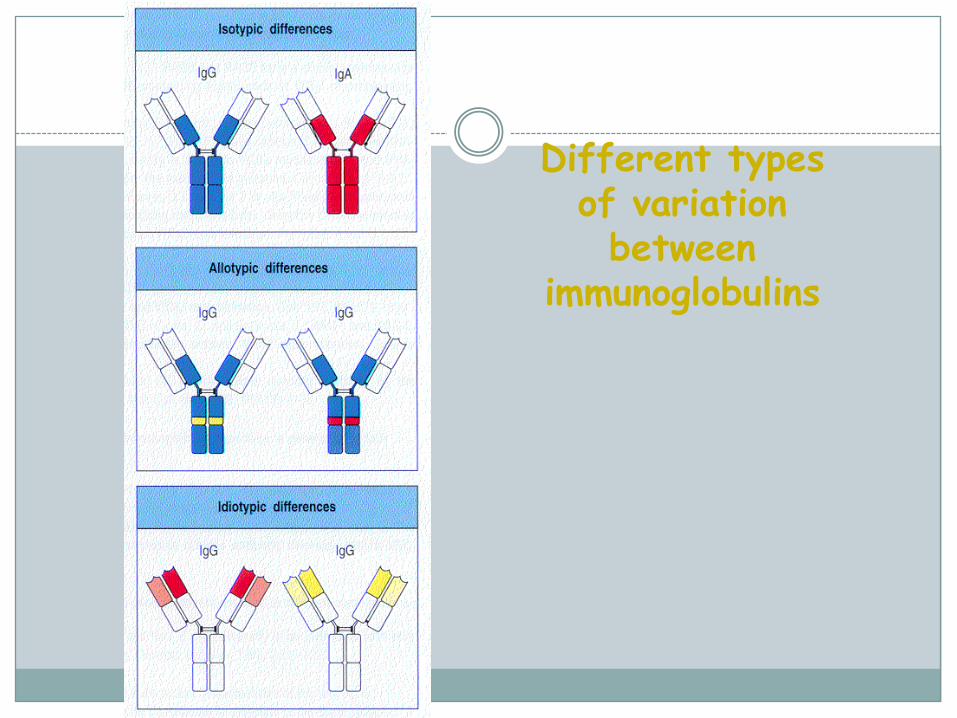
Different types of variation between
immunoglobulins
Mechanisms contributing to antibody diversity:
chance recombinations
imprecise joining of V, D, J genes
N-region additions
extensive mutations involving variable-region genes after antigen exposure
Isotype switching
during the immune response, plasma cells switch from producing IgM to IgG or to another Ig class (IgA, IgE)
the switch involves a change in the H-chain constant domains (CH)
no change in antigen-binding specificity !
(no alteration in the L chain or in the variable portion of H chain)
Allelic exclusion
once the process of rearrangement on one of chromosomes is successful, then all attempts on second chromosome are stopped
the same rule governs both for H- and L-chains
every single B cell produces only one type of H-and one type of L-chain
Clonal restriction
each B cell expresses identical copies of an antibody that is specific for single epitope
when a B cell divides, the chromosomes in its progeny cells bear the selected allelic genes, and these genes do not undergo any further V/J or V/D/J rearrangements
immunoglobulins produced by given B cell and its progeny are identical in epitope specificity and in k- or -chain isotype

The development of B-lymphocytes
B-lymphocytes originates from stem-cell Bone marrow: pre-B-lymphocytes (synthesis of
H chains, Ig genes rearrangement antigen specificity, IgM expression on the surface of the cell)
Blood, peripheral lymphoid organs: mature B-lymphocytes (IgD expression), ready to react with an antigen contact with an antigen division of cells and differentiation to plasma cells (secretion of huge amounts of Ig) + generation of memory B-lymphocytes
B-lymphocytes – surface markers
CD19, CD35 – complement receptors
IgM, IgD = BCR
B7 protein – adhesin, contact with T-lymphocyte
MHC class II – antigen-presenting molecules
B-lymphocytes - function
B-cells activation:
1/ thymus independent – polysacharide antigens, a cooperation with T cells is not necessary for B cells activation
2/ thymus dependent - first of all, the development of antigen-specific Th cells is necessary, then, thanks to cooperation between B cells and Th cells the antibody production could be sufficient and appropriate
B-lymphocytes - function
Antibody production
Antigen presentation
Ontogenesis of the antibody production
Although the production of specific antibodies already begins about week 20-24 of gestation, IgA+M concentrations are very low until the birth
IgG production begins only after the birth, but IgG level is at this time sufficient thanks to maternal IgG
About 4 to 6 months of age maternal IgG is eliminated from the child’s organism (possible onset of humoral deficiency symptoms)
Phases of humoral response
Primary response – typical delay of the antibody production (antigen presentation to Th cells is necessary)
Secondary response – thanks to memory antibodies and memory lymphocytes, the response is stronger and faster