Embed Size (px)
Citation preview
Genetic Diversity of the "Mediterranean" Glucose-6-Phosphate Dehydrogenase Deficiency Phenotype
GEORGESTAMATOYANNOPOULOS,VOLKERVOIGTLANDER, PANAYOTIS KOTSAKIS, andANGELOSAKRIVAKIS
From the Division of Medical Genetics, Department of Medicine, Universityof Washington, Seattle, Washington 98105
ABSTRACT Genetic diversity of the "Mediterranean"phenotype of G-6-PD (glucose-6-phosphate dehydroge-nase) deficiency was revealed when detailed studies wereperformed on blood specimens from 79 Greek males withG-6-PD levels 0-10% of normal. Four different mutantswere found to be responsible for the severely deficientphenotypes: two mutants, G-6-PD U-M (Union-Mark-ham) and G-6-PD Orchomenos, were distinguishable byelectrophoresis, while the other two, G-6-PD Athens-likeand G-6-PD Mediterranean, were distinguishable on thebasis of their kinetic characteristics. Of the kinetic testsapplied, the most useful for differentiating the variantswere those measuring utilization rates of the analoguesubstrates deamino-NADP, 2-deoxyglucose-6-phosphate,and galactose-6-phosphate. Among unrelated males withsevere G-6-PD deficiency, the relative frequencies ofthe four variants were: G-6-PD U-M, 5%; G-6-PDOrchomenos, 7%; G-6-PD Athens-like, 16%; G-6-PDMediterranean, 72%. Genetic, biochemical, and clinicalimplications of the findings are discussed.
INTRODUCTION
Glucose-6-phosphate dehydrogenase (G-6-PD)' has amultimeric structure, each of its identical polypeptidechains having a molecular weight of 40,000-50,000 (1, 2).Over 70 variants, representing mutations at the sex-linked structural locus of this enzyme, have been reported
This work was presented at the 62nd Annual Meeting ofthe American Society for Clinical Investigation in AtlanticCity, N. J., 3 May 1970.
Received for publication 16 November 1970.'Abbreviations used in this paper: ACD, acid-citrate-dex-
trose; BCB, brilliant cresyl blue; CNSHA, chronic non-spherocytic hemolytic anemia; deamino-NADP, deaminc-nicotinamide adenine dinucleotide phosphate; 2-deoxy-G-6-P,2-deoxyglucose-6-phosphate; G-6-P glucose-6-phosphate;G-6-PD, glucose-6-phosphate dehydrogenase; G-6-PD U-M,glucose-6-phosphate dehydrogenase Union-Markham; km,Michaelis constant; NADP, nicotinamide adenine dinucleo-tide phosphate.
(3). Many of these variants have no clinical effect; theyare detected by electrophoresis because of a difference inelectrical charge. Others are associated with mild, mod-erate, or severe reduction of enzymatic activity, whichis reflected in altered cellular metabolic behavior, mostobvious in the red cell. In these G-6-PD variants, theelectrophoretic mobility may be normal, fast, or slow,depending upon the particular amino acid substitution (s)involved. The currently known variants, characterizedby electrophoretic and kinetic measurements, have beenclassified (4, 5) into four categories: variants with (a)normal G-6-PD activity in red blood cells; (b) mildG-6-PD deficiency; (c) severe G-6-PD deficiency; and(d) G-6-PD deficiency associated with chronic non-spherocytic hemolytic anemia (CNSHA). Of the vari-ants with reduced enzymatic activity, the most commonare G-6-PD A-, virtually confined to Negroes, and theMediterranean variant, found in Caucasians and Asiatics(4, 5).
The Mediterranean variant is characterized by verylow G-6-PD activity (0-10% of the normal level), elec-trophoretic mobility indistinguishable from the normaltype, G-6-PD B (4, 5), and a particular kinetic behaviorpattern, described by Kirkman, Schettini, and Pickard(6). G-6-PD-deficient persons of Mediterranean originhave drastically decreased red cell G-6-PD activity, andare usually considered to have this Mediterranean vari-ant. However, such a designation may not be justified,since structurally different forms of an enzyme can bemanifested as identical phenotypes when only assays ofenzyme activity are applied. Performance of more com-plete studies on the enzyme kinetic behavior could un-cover differences of potential importance because of theirassociation with both the metabolism of the cell and theclinical phenotype of the carrier.
This study was designed in order to determine whethersevere G-6-PD deficiency in Mediterraneans is not asingle entity but includes several structurally differentmutants. For this purpose, detailed characterization of
The Journal of Clinical Investigation Volume 50 1971 1253
G-6-PD was performed on the blood of Greek males.To avoid ambiguities in the interpretation of the kineticdata, a relatively large number of deficient persons wasexamined. Since a given G-6-PD variant is expected tohave the same kinetic and electrophoretic expression inthe hemizygous offspring of heterozygous females, sev-eral pairs of G-6-PD deficient brothers were included inthe study. The correlation of the findings in these sib-pairs was to provide the evidence for or against geneticorigin of any observed diversity.
METHODSDetection of males with G-6-PD deficiency. The G-6-
PD-deficient males were detected during population studiesin two areas of Greece (Karditsa and Orchomenos). Initialscreening was performed with brilliant cresyl blue (BCB)by the method of Motulsky and Campbell-Kraut (7). Inthis test, the red cells of males who have less than 10% ofnormal G-6-PD activity fail to decolorize the BCB within3 hr. Of the 79 G-6-PD-deficient males chosen for this study,27 were unrelated, while the other 52 consisted of 26 sib-pairs.
Screening program for G-6-PD diversity. Blood samples(30-60 ml) were collected in acid-citrate-dextrose (ACD,National Institutes of Health formula B) and shipped re-frigerated to Seattle, Wash., where they arrived within 72hr of sampling. Every shipment included six to eight G-6-PD-deficient samples and one normal control. Specimenswere rechecked for G-6-PD activity on arrival; subsequentlythey were partially purified. Further tests on the partiallypurified preparations consisted of starch gel electrophoresisin phosphate buffer at pH 7.0, measurement of deamino-nicotinamide adenine dinucleotide phosphate (deamino-NADP) utilization, and measurements of 2-deoxyglucose-6-phosphate (2-deoxy-G-6-P) utilization. The electrophoreticplates were reviewed by two observers, one of whom wasnot aware of the kinetic fiindings. All kinetic characteriza-tions were performed by one worker, who did not knowwhich samples were obtained from sibs. Subsequently, galac-tose-6-phosphate utilization, Km's for nicotinamide adeninedinucleotide phosphate (NADP) and glucose-6-phosphate(G-6-P) and pH-dependent G-6-PD activities were studied.In addition to the above procedures, hemolysates from 17samples were examined by starch gel electrophoresis forG-6-PD pattern.
Experimental procedures. Techniques recommended bythe World Health Organization Committee on G-6-PD teststandardization were applied (4). Preparation of hemolysates,assay of G-6-PD activity in hemolysates, and partial puri-fication of G-6-PD were carried out as described by Motulskyand Yoshida (5). For electrophoresis, partially purifiedpreparations from normal and enzyme-deficient blood weredialyzed and adjusted to similar G-6-PD activities; separa-tion was performed in starch gels using a phosphate buffersystem at pH 7.0, a Tris (tris [hydroxymethyl] amino-methane)-HCl buffer system at pH 8.8, and a Tris-EDTA-borate buffer system at pH 8.6; (for details, see Reference5). Hemolysates, adjusted to a hemoglobin concentration of2 g/100 ml for normal samples and 10 g/100 ml for deficientsamples were electrophoresed in a phosphate buffer systemat pH 7.0. The development of the G-6-PD staining wasobserved at frequent intervals and the staining solution re-moved as soon as a faint band appeared in the G-6-PD-de-ficient samples.
Michaelis constants (Km's) for G-6-P and NADP weredetermined in a pH 8.0 buffer containing 10' M Tris-HCl,and 7 X 10-' M MgC12, the G-6-P concentration rangingfrom 1.5 X 10' M to 2 X 10' M and the NADPconcentra-tion ranging from 1.35 X 10⁻⁶ M to 6 X 10⁻⁵ M (five to sevendifferent concentrations for each substrate). The relativeutilization rate of analogue substrates (2-deoxy G-6-P,galactose-6-P, deamino-NADP) was expressed as a per-centage of the rate at which the same amount of the enzymecould utilize G-6-P or NADP; it was determined using thefollowing concentrations of substrates: 10-' M deamino-NADPwith 8 X 10- M to 10-' M NADP; 10' M galactose-6-phosphate, with 10-' M G-6-P; 10' M 2 deoxy G-6-P with10' M G-6-P. For measurement of pH-dependent G-6-PDactivity (7) dialyzed, partially purified G-6-PD-deficient,and normal control preparations were tested in a 5 X 10⁻² MTris-HCl, 5 X 10' M glycine, 5 X 10⁻² M KHPO4buffer, thepH being adjusted at intervals from 5.5 to 10.5 with HCl orNaOH. The substrate concentrations were those used inG-6-PD assay of hemolysates (5) and the enzyme activityat each pH was expressed as percentage of the maximumactivity measured.
RESULTS
The findings in the 27 unrelated G-6-PD-deficient malesappear in Table I, while those in sib-pairs appear inTable II and III. A summary of the findings is pro-vided in Table IV.
Diversity revealed by electrophoresisElectrophoretic screening in phosphate buffer, pH 7.0,
revealed two G-6-PD variants: in three persons the en-zyme moved more rapidly, and in eight, it moved moreslowly than normal.
With phosphate buffer at pH 7.0, the migration rateof the slow G-6-PD was 92-94% of G-6-PD B (Fig. 1).In TEB buffer, pH 8.6, and in Tris-HCl buffer, pH 8.8,it migrated 94-96% as far as G-6-PD B. Although theeight examples of this slow variant were detected inde-pendently, they were subsequently found to belong to fourpairs of brothers. The electrophoretic findings, kineticdata (Table IV), and the genetic evidence indicated thatthis enzyme was different from the common Mediter-ranean variant. It was preliminarily called G-6-PDOrchomenos.
The mobility of the fast G-6-PD was 102-104% thatof G-6-PD B. Its electrophoretic pattern and the kineticproperties (Table IV) were similar in some respects tothose of G-6-PD Markham (8) as well as G-6-PD Union(9). These variants are characterized by electrophoreticmigration rates faster than G-6-PD B, by severe re-duction of red cell enzyme activity, and by distinctlyhigher than normal rates of 2-deoxy-G-6-P and galac-tose-6-P utilization. Direct comparisons of these twovariants with that detected in our study could not bedone. Therefore, this Greek G-6-PD was tentativelycalled G-6-PD Union-Markham (G-6-PD U-M). LikeG-6-PD Markham, G-6-PD UMwas found to be un-
1254 G. Stamatoyannopoulos, V. Voigtlander, P. Kotsakis, and A. Akrivakis
TABLE IG-6-PD Characterization in 27 Unrelated Greek Males with Severe G-6-PD Deficiency
Utilization ofKm (X10-6) for Assigned
Case G--PD 2-deoxy- Deamino- G-6-PDno. activity* G-6-P NADP G-6-PI NADPII Galactose-6-Pi variant
1 4 7.8 2.0 225.0 375 114.5 U-M2 0 1.6 200.0 421 it
3 1 18.5 3.7 11.9 153 20.5 "Athens-like"4 5 18.4 3.7 15.7 164 23.3 "5 7 13.1 3.5 10.0 151 18.26 3 17.6 2.9 11.0 173 20.07 2 14.8 139 -8 0 12.6 2.3 58.0 343 49.0 Mediterranean9 0 10.7 1.9 49.0 303 -
10 0 9.5 1.6 60.0 312 40.7 "11 0 11.5 2.1 60.8 331 48.2 "12 0 11.5 2.4 53.1 303 43.3 "13 3 11.6 2.0 49.3 271 44.4 "14 2 12.6 2.4 51.3 326 47.1 "15 2 10.1 1.8 53.2 300 39.3 "16 1 10.9 1.7 44.4 311 "17 3 10.4 2.1 51.8 333 -18 3 11.5 2.5 54.8 341 38.7 "19 0 11.8 2.1 52.3 339 40.520 0 12.4 1.9 42.4 339 41.621 2 10.2 2.1 48.7 295 33.722 6 12.3 2.2 51.5 314 34.723 3 - 57.5 280 -24 6 42.5 315 -25 6 44.0 306 -26 0 9.6 2.0 50.5 312 36.927 3 10.4 1.8 52.2 304 41.3
*I 11, § as in footnote of Table IV.
stable. Partially purified preparations were almost devoidof activity after storage at 4VC for 2-3 wk. Furthermore,although the dialyzed, partially purified preparations ofG-6-PD U-M were adjusted before electrophoresis toactivities similar to the activity of G-6-PD B controls,the variant was always faintly stained, probably becauseof loss in catalytic activity during electrophoresis.
Diversity revealed by kinetic techniquesUtilization of deamino-NADP. Utilization of de-
amino NADPfor the electrophoretically distinguishableG-6-PD U-M and Orchomenos as well as for the remain-ing 68 cases are plotted in Fig. 2. Differences in de-amino-NADP utilization rates separated the enzyme inthe 68 cases with similar electrophoretic mobility intotwo nonoverlapping groups. In one group of 56 cases, thedeamino-NADP utilization ranged from 240 to 385%(mean = 312.86 ±26.7) and in the other group of 12cases the range was 115-175% (mean = 152.67 ±11.8);the difference between the 2 groups was statistically sig-nificant (P < 0.0001).
To test whether this difference in kinetic behaviorwas genetically determined, the correlation of deamino-NADPutilization values in sibships was examined. Ofthe total of 26 pairs of brothers included in this study,22 pairs (Table II) had mothers who were heterozygousfor G-6-PD deficiency, and thus, the brothers in eachpair had the same G-6-PD deficiency gene. Four ofthese sib-pairs had G-6-PD Orchomenos. The correla-tion of utilization values in the remaining 18 pairs isshown in Fig. 3. In 15 pairs, the deamino-NADP utili-zation value was high; in 3 pairs it was low. On thebasis of the bimodality in the distribution of measure-ments and the correlation of the findings in sibs, the 12males with deamino-NADP utilization values between115 and 175% (Fig. 2) were considered to possess aG-6-PD mutant different from the more common Medi-terranean type. Since similar values of deamino-NADPutilization have been described in G-6-PD Athens (ref-erence 10 and unpublished data), the enzyme in these12 individuals was preliminarily called "Athens-like"G-6-PD.
Genetic Diversity of the "Mediterranean" G-6-PD Deficiency Phenotype 1255
Utilization of 2-deoxyglucose-6-phosphate. The find- detected with the methods used, the males of this groupings for persons with G-6-PD U-M, G-6-PD Orcho- were considered to possess the Mediterranean variant ofmenos, Athens-like G-6-PD, and for the remaining 56 G-6-PD deficiency. Mean values of 2-deoxy-G-6-P uti-G-6-PD-deficient individuals are given in Fig. 4. Since lization appear in Table IV; the differences between thein this latter group of cases no further diversity was Athens-like, Medit rranean, and Orchomenos variants
TABLE I IG-6-PD Characterization in 22 Pairs of G-6-PD-Deficient Brothers Having G +/Gd- Heterozygous Mothers
Utilization ofSibship KM (X1O-6) for Assigned
and G-6-PD 2-deoxy- Deamino- G-6-PDcase activity* G-6-P NADP G-6-P§ NADPII Galactose-6-P§ variant
0 10.60 9.24 11.17 10.70 10.44 12.90 11.50 10.57 17.54 16.95 16.16
10 16.510 18.310 12.9
2 10.20 10.32 11.92 12.40 11.86 12.62 11.48 12.60 11.82 10.46 12.1
11.25 12.46 12.1
12.52020 -0 -00 12.580 -045
102
2.4 111.02.0 103.82.2 91.22.3 103.91.9 97.02.2 98.32.1 117.01.9 116.53.2 16.43.1 21.73.1 21.6
16.13.4 17.43.6 11.02.4 58.61.9 47.82.2 51.02.1 45.02.2 54.82.3 59.22.1 58.92.0 63.72.7 48.02.3 58.12.1 48.42.1 53.92.0 55.62.1 42.02.1 39.02.1 51.51.5 51.7- 54.0
53.041.942.355.3
1.6 49.238.2
2.1 51.8- 48.3
47.845.749.2
- 53.1
*, 11, § as in footnote of Table IV.
1256 G. Stamatoyannopoulos, V. Voigtlander, P. Kotsakis, and A. Akrivakis
Orchomencs,,0
II
it
.,
"Athens-like", I
, I
MediterraneanPI
,,
, ,
,,
II
I,
,,
,,
,,
,,
,II
,,
,,
I,,
,,
I,
,I,
,,
, ,
,,9
I.
I..
I.I
,.
,.9
,,9
I I
II
I,,
lab
2ab
3ab
4ab
Sab
6ab
7ab
8ab
9ab
10ab
1lab
12ab
13ab
14ab
15ab
16ab
17ab
18ab
19ab
20ab
21ab
22ab
59.462.858.350.064.254.862.055.022.520.7
22.024.238.943.845.645.039.443.650.847.353.048.838.543.342.233.336.431.0
386350306334303364393366150167152156144153347340338280277327380331370298319286361293253276333319282351312336300312319297335286314280

TABLE IIIG-6-PD Characterization in Four Pairs of G-(-PD-Deficient Brothers Having
Severely G-6-PD-Deficient Mothers
Utilization ofSibship Km (X10-6) for Assigned
and G-6-PD 2-deoxy- Deamino- G-6-PDcase activity* G-6-P NADP G-6-P§ NADPJI Galactose-6-P§ variant
23a 7 13.1 2.3 50.0 291 48.2 Mediterraneanb 5 11.5 2.1 44.0 310 37.1 I
24a 3 11.0 2.4 37.0 295 43.2 "b 4 9.5 1.7 52.8 262 37.8 "
25a 0 9.3 1.7 52.8 305 50.0 "b 3 6.2 1.4 176.0 396 111.7 U-M
26a 0 11.4 1.7 51.0 327 42.8 Mediterraneanb 4 17.9 3.1 17.4 130 26.6 "Athens-like"
*1, § as in footnote of Table IV.
are statistically significant (P <0.0001). That the ob-served differences are genetically determined is indicatedby the correlation of findings in pairs of brothers whosemothers are heterozygous for G-6-PD deficiency (Fig.5).
Utilization of galactose-6-phosphate. This measure-ment was performed in 55 individuals; the findings aregiven in Table IV. There is a clear-cut discriminationof values between Athens-like G-6-PD and Mediter-raneaft G-6-PD (P <0.0001). The values in G-6-PDOrchomenos are significantly higher than in Mediter-ranean G-6-PD (P < 0.0001), although their distribu-tions overlap.
Km for G-6-P and NADP. Km for G-6-P was mea-sured in 60 individuals and for NADPin 63 individuals.Distribution of measurements in G-6-PD Orchomenosand G-6-PD U-M were within the range characteristicof Mediterranean G-6-PD. In G-6-PD Athens-like Km'sfor NADPand G-6-P were significantly higher (P <0.0001) than in Mediterranean G-6-PD and within therange reported for G-6-PD Athens (10).
pH-dependent G-6-PD activity. G-6-PD activity asa function of pH was measured in 11 persons with theMediterranean variant, 2 with G-6-PD Orchomenos and2 with G-6-PD U-M. The mean G-6-PD activities perpH unit are plotted in Fig. 6. The pH-dependent ac-
TABLE IVCharacterization of Four G-6-PD Variants Found among 79 Greek Males with Severe G-6-PD Deficiency
G-6PD G-6-PD G-6-PD G-6-PDG--PD B Mediterranean "Athens-like" Orchomenos U-M
G-6-PD activity* 100 0-10 1-10 0-7 0-4Electrophoretic migrations 100 98-99¶ 98-99¶ 92-94 102-104
Km (X 10-6) forG-6-P 47.3 414.0 11.41 ±1.05 17.08 41.62 10.86 ±1.06 7.0 ±1.13NADP 3.8 40.7 2.06 40.26 3.33 ±0.29 2.11 40.19 1.67 ±0.30
Utilization of2-deoxy-G-6-P§ 4.5 ±1.0 50.34 ±6.07 15.42 i3.91 104.8 ±9.36 200.3 ±24.5deamirno-NADPIj 58.4 43.1 312.86 +26.7 152.67 411.8 350.25 ±-33.8 397.33 ±23.1galactose-6-P§ 7.1 +1.6 42.2 ±5.3 22.0 ±t2.5 58.3 44.8 113.0 ±t1.98
pH dependent G-6-PD activitypH curve Truncate Biphasic Biphasic BiphasicpH peaks 9.0 6.5, 9.5 6.0, 9.5 6.0, 9.5
* Per cent of mean G-6-PD activity in RBCof normals.Per cent of the rate of G-6-PD B migration.
§ Expressed as per cent of the rate of glucose-6-phosphate utilization.Expressed as per cent of the rate of NADPutilization.
¶ Slightly slower than G-6-PD B.
Genetic Diversity of the "Mediterranean" G-6-PD Deficiency Phenotype 1257
(
:..
*. ; . ......*Y * A;
.: :.c.;
| 2 3 4 5 6 7
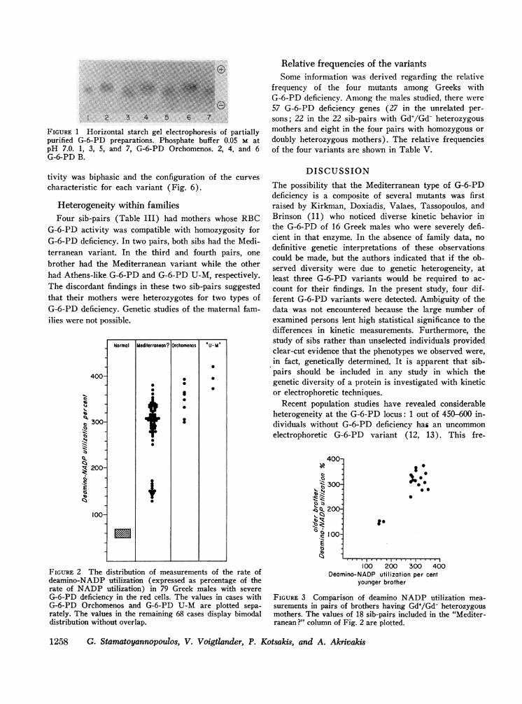
FIGURE 1 Horizontal starch gel electrophoresis of partiallypurified G-6-PD preparations. Phosphate buffer 0.05 M atpH 7.0. 1, 3, 5, and 7, G-6-PD Orchomenos. 2, 4, and 6G-6-PD B.
Relative frequencies of the variantsSome information was derived regarding the relative
frequency of the four mutants among Greeks withG-6-PD deficiency. Among the males studied, there were57 G-6-PD deficiency genes (27 in the unrelated per-sons; 22 in the 22 sib-pairs with Gd+/Gd- heterozygousmothers and eight in the four pairs with homozygous ordoubly heterozygous mothers). The relative frequenciesof the four variants are shown in Table V.
tivity was biphasic and the configuration of the curvescharacteristic for each variant (Fig. 6).
Heterogeneity within familiesFour sib-pairs (Table III) had mothers whose RBC
G-6-PD activity was compatible with homozygosity forG-6-PD deficiency. In two pairs, both sibs had the Medi-terranean variant. In the third and fourth pairs, onebrother had the Mediterranean variant while the otherhad Athens-like G-6-PD and G-6-PD U-M, respectively.The discordant findings in these two sib-pairs suggestedthat their mothers were heterozygotes for two types ofG-6-PD deficiency. Genetic studies of the maternal fam-ilies were not possible.
400-
E 300-
-.
Q 200-
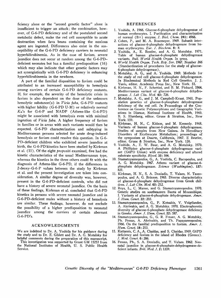
1E
100-
FIGURE 2 The distribution of measurements of the rate ofdeamino-NADP utilization (expressed as percentage of therate of NADP utilization) in 79 Greek males with severeG-6-PD deficiency in the red cells. The values in cases withG-6-PD Orchomenos and G-6-PD U-M are plotted sepa-rately. The values in the remaining 68 cases display bimodaldistribution without overlap.
DISCUSSION
The possibility that the Mediterranean type of G-6-PDdeficiency is a composite of several mutants was firstraised by Kirkman, Doxiadis, Valaes, Tassopoulos, andBrinson (11) who noticed diverse kinetic behavior inthe G-6-PD of 16 Greek males who were severely defi-cient in that enzyme. In the absence of family data, nodefinitive genetic interpretations of these observationscould be made, but the authors indicated that if the ob-served diversity were due to genetic heterogeneity, atleast three G-6-PD variants would be required to ac-count for their findings. In the present study, four dif-ferent G-6-PD variants were detected. Ambiguity of thedata was not encountered because the large number ofexamined persons lent high statistical significance to thedifferences in kinetic measurements. Furthermore, thestudy of sibs rather than unselected individuals providedclear-cut evidence that the phenotypes we observed were,in fact, genetically determined. It is apparent that sib-pairs should be included in any study in which thegenetic diversity of a protein is investigated with kineticor electrophoretic techniques.
Recent population studies have revealed considerableheterogeneity at the G-6-PD locus: 1 out of 450-600 in-dividuals without G-6-PD deficiency has an uncommonelectrophoretic G-6-PD variant (12, 13). This fre-
400-
,*
< 300IZ'll.< X 200-
'C°'c I00-z
P.
@000
to
100 200 300 400Deomino-NADP utilization per cent
younger brother
FIGURE 3 Comparison of deamino NADPutilization mea-
surements in pairs of brothers having Gd+/Gd- heterozygousmothers. The values of 18 sib-pairs included in the "Mediter-ranean ?" column of Fig. 2 are plotted.
1258 G. Stamatoyannopoulos, V. Voigtlander, P. Kotsakis, and A. Akrivakis
Normal Mediterranean? Orchomenos 0U-M
0~~~~~
I 0

quency of the "nonpolymorphic" G-6-PD variants hasbeen considered as an indication that the structure of theG-6-PD molecule is such that amino acid substitutionsare permitted which would not be tolerated in severalother proteins. Of the variants observed in this study,none can be placed in the category of the rare nonpoly-morphic mutants, since their frequencies ranged from 1%in the case of G-6-PD U-M to as much as 13% in thecase of Mediterranean variant (Table V). These findingssuggest that a number of different Gd locus mutationsassociated with deficiency of the enzyme may be favoredby selection and may coexist in the same population. Itremains to be seen whether our findings are character-istic only of the Greek population or whether a similarmolecular diversity exists among the other Mediter-ranean ethnic groups with high frequencies of G-6-PDdeficiency.
The kinetic and electrophoretic studies of G-6-PDhave so far been useful in detecting and discriminatingbetween variants, when comparisons of kinetic and elec-trophoretic behavior are made under standard conditions(4, 5). Even in the case of variants with small differencesin electrophoretic migration or kinetic constants, thesetechniques have been adequate when the comparisons
Normal "Athens" Mediterranean Orchomenos U - M0
200-
15."
00
.00
~ 50-
FIGURE 4 The distribution of measurements of the rate of2-deoxy-glucose-6-phosphate utilization (expressed as per-centage of the rate of glucose-6-phosphate utilization) in 79Greek males with severe G-6-PD deficiency in the red cells.The differences in mean values between G-6-PD Athens-like,G-6-PD Mediterranean, and G-6-PD Orchomenos (TableIV) are highly significant (P < 0.0001) .
150-
.Isz
.N
,, q) I 00-
q, -
o 50-C.:"5l110r
0r(j
.
.
1, 4
AA A
.
50 100 1502-deoxyglucose-6-phosphate utilization per cent
younger brother
FIGURE 5 Comparison of 2-deoxy-glucose-6-phosphate utili-zation measurements in 22 pairs of brothers having Gd+/Gd-heterozygous mothers. (*): G-6-PD Orchomenos; (*):G-6-PD Mediterranean; (A): G-6-PD Athens-like.
were made in the same, rather than in separate, labora-tories. Conclusions about differences or similarities be-tween variants based on reported properties are possibleonly when they are significantly larger than the errorsinherent in the experimental procedures. However, withover 70 different G-6-PD's already reported (3), mean-ingful comparisons are now probably beyond the powerof the differentiation techniques. It is thus not possiblefor us to conclude that the three "new" variants de-scribed in this study (G-6-PD U-M, Athens-like, Orcho-menos) are really new, since one or more of them couldrepresent previously recognized mutants. For example,G-6-PD U-M could be identical with either G-6-PDUnion (8) or G-6-PD Markham (9). Its electropho-
100 6
0
50-~~~
9, 10
pH
FIGURE 6. pH-dependent G-6-PD activity. (v) G-6-PDB; (O) G-6-PD Mediterranean; (O) G,6-PD Orchomenos-(FIG) G-6-PD UeM. G-6-PD activity at individual pH point
is expressed as percentage of the higher measured activity.
Genetic Diversity of the "Mediterranean" G-6-PD Deficiency Phenotype 1259

TABLE VRelative and Population Frequencies of the Four G-6-P Deficiency Mutants
No. of No. of Gd Gd Gd GdClass of individuals cases genes Mediterranean Athens-like Orchomenos U-M
Unrelated 27 27 20 5 2Sib-pairs (heterozygous 44 22 15 3 4
mother)Sib-pairs (homozygous 8 8 6 1 1
or double heterozygousmother)
Totals 79 57 41 9 4 3
Relative frequency of variants, % (Gd- 100%) 71.9 15.8 7.0 5.3
Population frequency of variants, % (Gd- = 18%) 12.94 2.85 1.26 0.95
retic migration and substrate utilization properties arevery similar to those reported for these two enzymes,but proof of identity is impossible in the absence of directcomparisons.
The kinetic profile of G-6-PD Athens-like is identicalwith that of G-6-PD Athens (10), but the two enzymesdiffer in the degree of G-6-PD deficiency. In individualswith G-6-PD Athens, G-6-PD activity is moderatelyreduced, while in G-6-PD Athens-like, the deficiency issevere. It is possible that variations in erythrocyteG-6-PD activity are sometimes secondary to other causesand thus not directly related to a structural change in theenzyme molecule. On the other hand, different mutationsmay cause the same alteration in kinetic characteristicsbut different degrees of G-6-PD instability or rate ofsynthesis. Without comparative structural analyses, onecannot speculate usefully about whether or not theAthens and Athens-like G-6-PD deficiency mutants areidentical.
The third variant, G-6-PD Orchomenos, appears elec-trophoretically and kinetically different from the othermutants which are associated with severe G-6-PD de-ficiency but conclusive proof requires comparison ofpeptide maps and amino acid analyses of isolated peptides.
When the aberrant G-6-PD could not be classified asAthens-like, U-M, or Orchomenos, the individual wasconsidered to have the Mediterranean variant. Thisdesignation seemed reasonable, because there was noapparent bimodality in the values obtained from kineticmeasurements and also because in analysis of varianceof these measurements in sib-pairs, the intrafamilial simi-larities did not differ statistically from the interfamilialsimilarities. It is true that the rates of utilization of2-deoxy-G-6-P and galactose-6-P were higher among thesubjects considered to have the Mediterranean variantthan those assigned to the Mediterranean variant byKirkman et al. (6). However, this disparity probably
reflects slight differences in substrate concentrations andalso differences in the batches of the reagents used, sincehigher values were also obtained with our normal con-trols. Although the Mediterranean variant has been de-scribed as electrophoretically indistinguishable fromG-6-PD B (4-6), we found its migration to be slightlyslower than the normal enzyme, particularly when par-tially purified G-6-PD preparations were electrophoresed.With hemolysates, the slight retardation was observedonly when the normal control hemolysate was dilutedand the development of enzyme staining was interruptedwhen the Mediterranean G-6-PD zone first becamevisible.
The males included in this study were chosen on thebasis of a single criterion, i.e., the results of a screeningtest for G-6-PD deficiency. Hematologic and clinicalobservations were not done, and thus the clinical im-plications of these variants remain unknown. However,there is no a priori reason to assume that all the variantscomprising severe G-6-PD deficiency in inhabitants ofthe Mediterranean area have the innocuous hematologicalphenotype of the Mediterranean mutant or that theyhave a similar degree of predisposition to favism. Thisacute hemolytic syndrome does not occur at randomamong G-6-PD-deficient individuals, but rather has afamilial predisposition even in areas where the frequencyof G-6-PD deficiency is high (14, 15). Family studieshave indicated that a second genetic factor may act syn-ergistically with G-6-PD deficiency in predisposing theindividual to acute hemolysis after Vicia faba ingestion(14). The postulated second genetic factor might repre-sent a polymorphism of the enzyme(s) metabolizing thehemolytic agent in Vicia faba; or a polymorphism of in-traerythrocytic enzymes regenerating NADH, NADPH,or GSH; or even a polymorphism involving hemoglobinstability. In this view, favism appears to be a multi-factorial disease in which the occurrence of G-6-PD de-
1260 G. Stamatoyannopoulos, V. Voigtlander, P. Kotsakis, and A. Akrivakis
ficiency alone or the "second genetic factor" alone isinsufficient to trigger an attack; the combination, how-ever, of G-6-PD deficiency and of the postulated secondmetabolic defect, make the red cell susceptible to acutedestruction when fava beans containing the noxiousagent are ingested. Differences also exist in the sus-ceptibility of the G-6-PD deficiency carriers to neonatalhyperbilirubinemia. As in the case of favism, severejaundice does not occur at random among the G-6-PD-deficient neonates but has a familial predisposition (16)which may also indicate that other genetic factors mayact synergistically with G-6-PD deficiency in enhancinghyperbilirubinemia in the newborn.
A part of the familial disposition to favism could beattributed to an increased susceptibility to hemolysisamong carriers of certain G-6-PD deficiency mutants.If, for example, the severity of the hemolytic crisis infavism is also dependent on the dose of the unknownhemolytic substance(s) in Vicia faba, G-6-PD mutantswith higher lability (G-6-PD U-M) or relatively normalKm's for G-6-P and NADP (Athens-like G-6-PD)might be associated with hemolysis even with minimalingestion of Vicia faba. A higher frequency of favismin families or in areas with such variants would then beexpected. G-6-PD characterization and subtyping inMediterranean persons selected for acute drug-inducedhemolysis or favism could resolve this question. In G-6-PD-deficient children who exhibited severe jaundice atbirth, the G-6-PD kinetics have been studied by Kirkmanet al. (11). Of the eight propositi examined, five had thekinetic characteristics of the usual Mediterranean type,whereas the kinetics in the three others could fit with thediagnosis of Athens-like G-6-PD, if the differences in2-deoxy-G-6-P values between the study by Kirkmanet al. and the present investigation are taken into con-sideration. A similar degree of diversity was, however,present in the G-6-PD-deficient controls, who did nothave a history of severe neonatal jaundice. On the basisof these findings, Kirkman et al. concluded that G-6-PDkinetics in persons with severe neonatal jaundice and inG-6-PD-deficient males without a history of hemolysisare similar. These findings, however, do not excludethe possibility of a higher predisposition to neonataljaundice among the carriers of certain aberrantG-6-PD's.
ACKNOWLEDGMENTSWeare indebted to Dr. A. Yoshida for his guidance duringthe study and to Dr. E. Giblett and Dr. A. G. Motulsky forhelpful comments during the preparation of this manuscript.
This investigation was supported by Grant GM15253 fromthe National Institutes of Health, U. S. Public HealthService.
REFERENCES1. Yoshida, A. 1966. Glucose-6-phosphate dehydrogenase of
human erythrocytes. I. Purification and characterizationof normal (B+) enzyme. J. Biol. Chem. 241: 4966.
2. Cohen, P., and M. A. Rosemeyer. 1969. Subunit inter-actions of glucose-6-phosphate dehydrogenase from hu-man erythrocytes. Eur. J. Biochem. 8: 8.
3. Yoshida, A., E. Beutler, and A. G. Motulsky. 1971.Table of human glucose-6-phosphate dehydrogenasevariants. Bull. World Health Organ. In press.
4. World Health Organ. Tech. Rep. Ser. 1967. Number 366(Standardization of procedures for the study of glucose-6-phosphate dehydrogenase).
5. Motulsky, A. G., and A. Yoshida. 1969. Methods forthe study of red cell glucose-6-phosphate dehydrogenase.In Biochemical Methods in Red Cell Genetics. J. J.Yunis, editor. Academic Press Inc., New York. 51.
6. Kirkman, H. N., F. Schettini, and B. M. Pickard. 1964.Mediterranean variant of glucose-6-phosphate dehydro-genase. J. Lab Clin. Med. 63: 726.
7. Motulsky, A. G., and J. M. Campbell-Kraut. 1961. Pop-ulation genetics of glucose-6-phosphate dehydrogenasedeficiency of the red cell. In Proceedings of the Con-ference on Genetic Polymorphisms and Geographic Vari-ations in Disease, National Institute of Health, 1960.B. S. Blumberg, editor. Grune & Stratton, Inc., NewYork. 159.
8. Kirkman, H. N., C. Kidson, and M. Kennedy. 1968.Variants of human glucose-6-phosphate dehydrogenase.Studies of samples from New Guinea. In HereditaryDisorders of Erythrocyte Metabolism; proceedings ofthe symposium at Duarte, California, 1967. E Beutler,editor. Grune & Stratton, Inc., New York. 126.
9. Yoshida, A., E. W. Baur, and A. G. Motulsky. 1970.A Phillipino glucose-6-phosphate dehydrogenase vari-ant (G6PD Union) with enzyme deficiency and alteredsubstrate specificity. Blood. 35: 506.
10. Stamatoyannopoulos, G., A. Yoshida, C. Bacopoulos, andA. G. Motulsky. 1967. Athens variant of glucose-6-phasphate dehydrogenase. Science (Washington). 157:831.
11. Kirkman, H. N., S. A. Doxiadis, T. Valaes, N. Tasso-poulos, and A. G. Brinson. 1965. Diverse characteristicsof glucose-&phosphate dehydrogenase from Greek chil-dren. J. Lab Clin. Med. 65: 212.
12. Reys, L., C. Manso, and G. Stamatoyannopoulos. 1970.Genetic studies on southeastern Bantu of Mozambique.I. Variants of glucose-6-phosphate dehydrogenase. Amer.J. Hum. Genet. 22: 203.
13. Stamatoyannopoulos, G., P. Kotsakis, V. Voigtlander,A. Akrivakis, and A. G. Motulsky. 1970. Electrophoreticdiversity of glucose-6-phosphate dehydrogenase deficiencyin Greeks. Amer. J. Hum. Genet. 22: 587.
14. Stamatoyannopoulos, G., G. R. Fraser, A. G. Motulsky,Ph. Fessas, A. Akrivakis, and Th. Papayannopoulou.1966. On the familial predisposition to favism. Amer. J.Hum. Genet. 18: 253.
15. Kattamis, C. A., A. Chaidas, and S. Chaidas. 1969. G6PDdeficiency and favism in the island of Rhodes (Greece).J. Med. Genet. 6: 286.
16. Fessas, Ph., S. A. Doxiadis, and T. Valaes. 1962. Neo-natal jaundice in glucose-6-phosphate-dehydrogenase-de-ficient infants. Brit. Med. J. 2: 1359.
Genetic Diversity of the "Mediterranean" G-6-PD Deficiency Phenotype 1261


![lezione sensibilit insulinica 091115 [modalit compatibilit ]) resistenza - definizione e... · HK-II glucose + insulin + glucose - 6 - phosphate glycogen GS glucose - 1 - phosphate](https://img.pdfslide.net/doc/110x75/5f0573df7e708231d4130924/lezione-sensibilit-insulinica-091115-modalit-compatibilit-resistenza-definizione.jpg)