Embed Size (px)
Citation preview
Genetic Engineering, Volume 28, Edited by J. K. Setlow©Springer Science+Business Media, LLC, 2007 159
INTRODUCTION
Interactions between eukaryotic transcription factors and their cognateDNA binding sites form fundamental networks within cells that control criticalsteps during development and tissues-specific gene expression. These interactionsalso are important in regulating cellular responses to stresses, and their dysfunc-tion contributes to numerous diseases. Therefore, determining the in vivo genome-wide binding distribution of transcription factors is an important step towardsdeveloping an understanding of the regulatory networks in a living cell as well astheir changes in response to specific stimuli. Methods based on chromatinimmunoprecipitation (ChIP) are beginning to provide an increasing detailed viewof these dynamic events. This assay was originally developed to monitor histonemodifications and then modified to detect binding of specific transcription fac-tors to native chromatin (1-3). In this method, transcription factors are reversiblycross-linked to their binding sites using formaldehyde to freeze intracellular pro-tein-DNA complexes, the DNA is sonicated to generate fragments with lengths
PAIRED-END GENOMIC SIGNATURE TAGS: A METHOD FOR THE FUNCTIONAL ANALYSIS OF GENOMES ANDEPIGENOMES
John J. Dunn, Sean R. McCorkle, Logan Everett and Carl W. Anderson
Biology DepartmentBrookhaven National LaboratoryUpton, NY 11973-5000
of ~500 to ~2,000 bp, and individual transcription factor-DNA complexes areimmunoprecipitated using specific antibodies to the native protein or to a suitableepitope fused in-frame to the target protein’s coding sequence. The DNA frag-ments enriched by ChIP can be identified by a variety of means such as cloningor amplification with gene specific primers, hybridization to microarrays con-taining subsets of the genomic sequences or by Serial Analysis of GeneExpression (SAGE)-type approaches (4) that extract short sequence identifiertags or Genomic Signature Tags (GSTs) from the ChIP DNA and then use thisinformation to map the DNA back to the genome. In this article we will reviewthe basic steps for generating GSTs, their application to analysis of ChIP dataand will introduce several modifications of our original SACO (for SerialAnalysis of Chromatin Occupancy) method (5) that can simultaneously generatetags from both ends of the ChIP fragments and preserve their spatial relationshipto each other. The sequences of each tag in combination uniquely identify theregion of the genome from which the original SACO fragment was derived andencompass the sequence of the site to which the transcription factor was bound.
This same approach can also be used to obtain paired sequence tags fromthe ends of any DNA fragment. At the whole genome level any changes in theresulting paired-end profile can provide a sensitive method for distinguishingbetween closely-related genomes or genomes that have undergone deletions,insertions or other rearrangements that cause the appearance of new diTAGpairs. Detection and characterization of discrepancies between observed diTAGpairs from reference and test genomes can, in principle, detect structural varia-tions with the same precision as afforded by paired-end sequencing of fosmid orbacterial-artificial chromosome libraries (6). Such changes are characteristic ofmany cancers as are changes in CpG methylation in CpG islands, which are clus-ters of CpG dinucleotides that are found in front of about half of human genes(7). Methylation of cytosine within these islands caused inhibition of downstreamgene expression, and aberrant methylation is an important mechanism for geneactivation or inactivation in cancer. In this article, we briefly review how paired-end diTAGs can be obtained from DNA fragments associated with methylatedCpG islands.
WHAT ARE GENOMIC SIGNATURE TAGS?
Genomic Signature Tags (GSTs) are the products of a method we devel-oped for identifying and qualitatively analyzing genomic DNAs (8). Two majorprinciples underlie this method: first, short DNA sequences (18-21 bp) are suffi-cient to identify unique sites within a genome; second, concatenation of theseshort DNA sequences, as in SAGE (9), greatly increases sequence throughput.The original GST method begins with cutting the DNA sample with a type IIrestriction enzyme, also termed the fragmenting enzyme, to produce fragmentswith cohesive ends. After digestion with the first enzyme, the cohesive ends arebiotinylated, and the sample is digested with Nla III, also called the anchoringenzyme, which cleaves leaving 4 base cohesive ends. Since Nla III has a 4 bprecognition sequence (CATG), it theoretically cleaves on average every 256 bp,and nearly every fragment in the original digest will be cleaved at least once to
160 J. DUNN ET AL.
produce two biotinylated end fragments which are recovered by binding tostreptavidin-coated magnetic beads. The bound DNA fragments are then ligatedwith a linker cassette that creates partially overlapping Mme I (TCCRAC) andNla III (CATG) recognition sites; i.e., TCCRACATG with the C in bold beingshared by both recognition sequences. Mme I is a type IIS restriction enzyme,with cut sites 20-21/18-19 bp past its recognition site. Cutting the linkered DNAwith Mme I releases the linker and 17-18/15-16 bp immediately 3′ to the Nla IIIsite. These CATG+17 or 18 bp sequences become the identifier tags which arePCR amplified and ligated together to form ≥500 bp long concatemers prior tocloning and DNA sequencing. Because each clone contains multiple tags,sequencing throughput increases accordingly.
SERIAL ANALYSIS OF CHROMATIN OCCUPANCY (SACO)
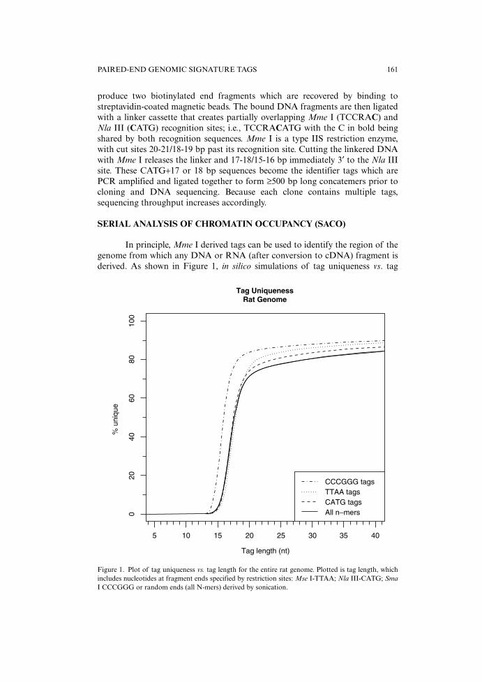
In principle, Mme I derived tags can be used to identify the region of thegenome from which any DNA or RNA (after conversion to cDNA) fragment isderived. As shown in Figure 1, in silico simulations of tag uniqueness vs. tag
PAIRED-END GENOMIC SIGNATURE TAGS 161
Tag UniquenessRat Genome
020
4060
8010
0
% u
niqu
e
5 10 15 20 25 30 35 40
Tag length (nt)
CCCGGG tagsTTAA tagsCATG tagsAll n−mers
Figure 1. Plot of tag uniqueness vs. tag length for the entire rat genome. Plotted is tag length, whichincludes nucleotides at fragment ends specified by restriction sites: Mse I-TTAA; Nla III-CATG; SmaI CCCGGG or random ends (all N-mers) derived by sonication.
length for the rat genome show that uniqueness rapidly increases for lengthslonger than ~12 bp and is limited only by the presence of highly repetitive regionsin genomes. Similar profiles are obtained for the mouse and human genomes.With these as background, we reasoned that a ChIP-to-tag sequencing approachcould be used to identify the genomic locations of ChIP-derived DNA fragments.To establish the effectiveness of the method, we set out to map globally the c-AMP response element binding protein (CREB) binding sites in the genome ofrat PC12 cells (5). CREB was known to bind the cAMP-response element (CRE)(TGACGTCA) present in the promoters of many inducible genes (reviewed in10). To increase the chances that CREB would be associated with CREsequences, we first incubated the cells with forskolin to activate the enzymeadenylcyclase and increase the intracellular levels of cyclic AMP. The cells werethen treated with formaldehyde, and, after randomly fragmenting the entiregenome by sonication, the samples were subjected to ChIP using an anti-CREBantibody or, as a control, non-specific IgG. Real-time quantitative PCR showedthat the CREB antibody provided an ~100-fold enrichment for c-fos (and otherCREB targets) in the immunoprecipitates as compared to the IgG control. Theends of the CREB ChIP DNA were polished (protruding 3′ and 5′ ends weremade flush by incubation with E. coli (Klenow fragment) and T4 DNA poly-merases plus all four deoxynucleotide triphosphates and ligated to adapters forlimited PCR amplification using biotinylated adapter-specific primers). Theresulting DNA was digested with Nla III, and a modified Long-SAGE procedurewas used to create concatemerized chains of randomly-associated 21 bp GSTswhich were then cloned and sequenced. We termed this approach SACO; todemonstrate its utility, the sequences of ~75,000 tags from the PC12-derivedlibrary were determined. More than 40,000 CREB-SACO tags that mapped tounique loci in the rat genome were identified; 6,302 of these were identified twoor more times. When these data were integrated with sequence annotation mapsof the rat genome, forty percent of these loci were within 2 kb of the transcrip-tional start site of an annotated gene, and 72% were within 1 kb of a putativecAMP response element. In addition, CREB binding was confirmed for all locisupported by multiple tag hits (53 of 53 that were tested), and many of theseloci were located upstream from genes not previously known to be regulated byCREB. These included genes for transcriptional regulators, chromatin modifyingenzymes, coactivators, and co-repressors. A surprising result of the CREB SACOstudy was that CREB binding sites were commonly located in bi-directional pro-moters. Thus, the CRE that controls c-fos expression, for example, also regulatesexpression of a noncoding RNA transcribed in the opposite direction (5).
Since publication of the SACO method, several papers have appeared thatutilized similar approaches, attesting to the overall utility of tag-to-genome map-ping of ChIP DNA fragments (11-14). In all of these procedures the tags,whether they are generated from an internal restriction site or directly from the 5′and 3′ ends of the sonicated ChIP DNA fragments, are analyzed separately asindependent bits of sequence data. When mapped correctly to the genomesequence, these tags locate within about 1 to 2 kb the site that was cross-linkedin vivo to the immunoprecipitated protein. In practice finding these sites involvesscanning the genome sequence in both directions from a tag’s location for a
162 J. DUNN ET AL.
nearby binding motif. The distance scanned is usually set at around twice theupper limit of the size of the ChIP DNA since when tags are analyzed separately,it is not known where they originated in the fragment, i.e., were they close to anend or more towards the middle of the ChIP fragment. To overcome this limita-tion, a new cloning strategy was developed by Ng and co-workers (15) that cova-lently links the tag sequences from each end of a DNA fragment into a paireddiTAG structure. This approach, which was originally developed for identifyingsimultaneously both ends of full-length cDNAs, can also be used to map ChIPfragments with high precision.
PAIRED-END GENOMIC SIGNATURE TAGS (PE-GST)
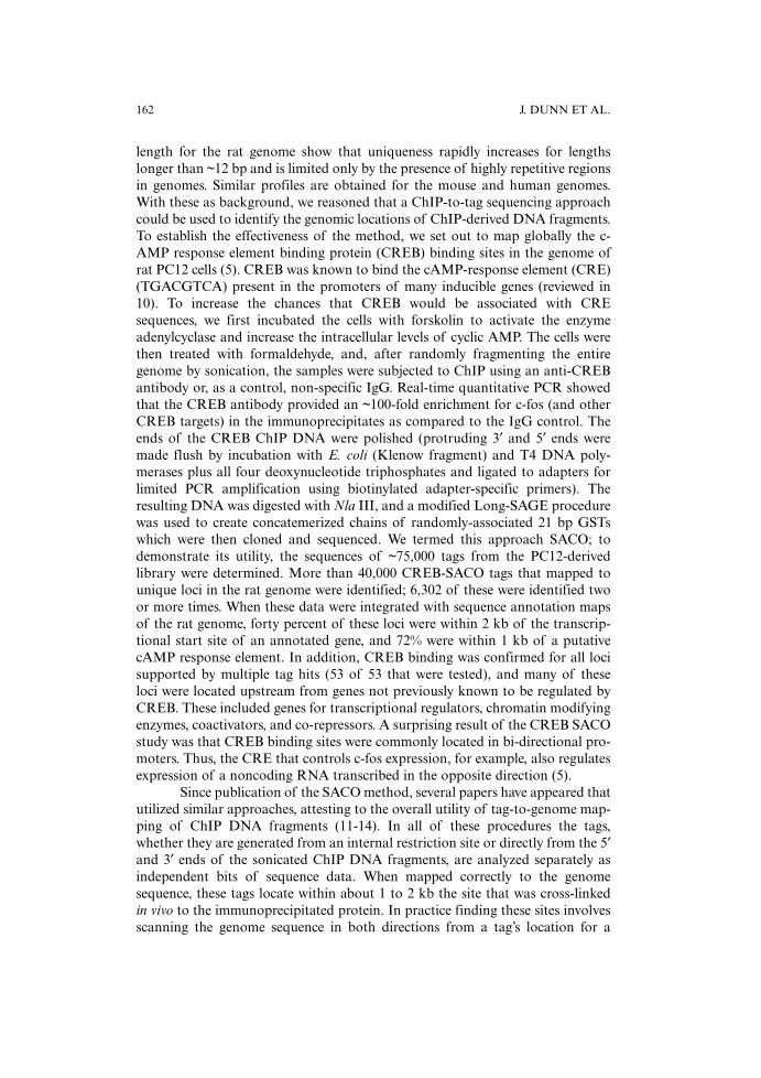
The first step in the procedure is cloning of the DNA fragments into a spe-cial vector, pBEST (Both End Signature Tags), which is based on the pSCANSvector developed at BNL (http://genome.bnl.gov/Vectors/pscans.php). This low-copy number vector, with an isopropyl-beta-D-thiogalactopyranoside (IPTG)inducible origin of replication, was modified for efficient cloning of single DNAfragments in a manner that places them immediately adjacent to oppositely ori-ented Mme I recognition sequences (Figure 2). These are the only Mme I sites inthe vector. Two Bbs I sites were placed between the Mme I sites in opposite orien-tations such that when the vector is cut with Bbs I, the linearized vector DNA willhave non-self-ligatable ends with 4 nt overhangs (5′-GTCG-3′). A synthetic
PAIRED-END GENOMIC SIGNATURE TAGS 163
Figure 2a. Schematic diagram of pBEST paired-end vector. oriS, repE and inc C are from the E. coliF factor, lacOP is the wild-type lac promoter, repL is the lytic origin of replication from bacteriophageP1, and bla encodes β-lactamase activity (ampR). Several of the plasmid’s unique restriction sites areindicated. MCS represents the cloning region, which is shown in greater detail in Figure 2b.
double-stranded DNA cassette is used to append simultaneously BtgZ I and BbsI recognition sites to the ends of blunt-ended ChIP DNAs. The bottom strand ofthe cassette is 5′ phosphorylated (p) and its 3′ end is amino modified to preventself-ligation higher than dimers.
Cassette #1
BtgZ I Bbs I5′ TCCGGTCTAC TGAATTCCGA ACGCGATGCT GAAGACCACG AC3′ Amino-AGGCCAGATG ACTTAAGGCT TGCGCTACGA CTTCTGGTGC TGp
Similar cassettes with appropriate overhangs are used if dealing with frag-ments with cohesive ends. Cutting these cassettes with either BtgZ I or Bbs I gen-erates 4 bp overhangs (5′-CGAC-3′) on the ends of the linkered DNA that arecomplementary to the overhangs of the Bbs I cut vector. After overnight ligationwith excess linker, the ligation products are purified on a Qiagen Qiaquick PCRpurification column, and the eluant is PCR amplified using 5′-biotin-TCCG-GTCTACTGAATTCCGAAC-3′ as primer. Ideally one should set up several dif-ferent PCR reactions varying the amount of input template and PCR cycles.Amplified material should then be analyzed by agarose gel electrophoresis. Theproducts should produce a smear that is similar in its size range to the DNA frag-ments in the sonicated ChIP starting material. The appropriate samples are phe-nol-chloroform extracted; then a portion is digested with BtgZ I and a similarportion with Bbs I to minimize loss of fragments with internal BtgZ I or Bbs Isites. After digestion the samples are combined, the cleaved linker cassettes areremoved by gel electrophoresis or by binding to streptavidin beads, and the ChIPfragments are ligated into Bbs I cut pBEST to generate recombinant plasmids
164 J. DUNN ET AL.
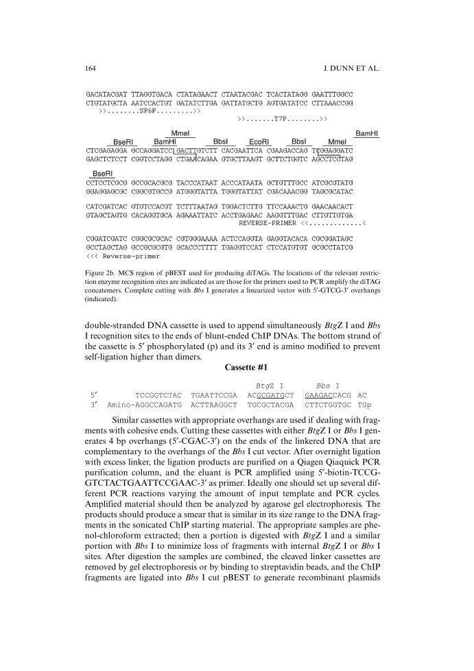
Figure 2b. MCS region of pBEST used for producing diTAGs. The locations of the relevant restric-tion enzyme recognition sites are indicated as are those for the primers used to PCR amplify the diTAGconcatemers. Complete cutting with Bbs I generates a linearized vector with 5′-GTCG-3′ overhangs(indicated).
with only single inserts. These are then electroporated into E. coli D1210, and thelibrary is plated on ZYM5052 plus ampicillin (50 µg/ml) plates (16). Growth onZYM5052 agar provides for solid-phase plasmid amplification without having toadd IPTG to the plates, which should maintain library representation better thangrowth in liquid culture. The number of colonies required at this stage is deter-mined by the estimated number of targets in the genome being investigated; weroutinely target 1-10 X 105 cfu as a convenient benchmark. Cells can be plated ata density just below what is needed to provide for a confluent lawn. Afterovernight 37˚C incubation, the resultant lawn of bacterial colonies is harvestedby scraping into several ml of liquid medium and pelleted by centrifugation.Plasmid DNA preparation is performed e.g., by using a Qiagen Tip500 kit.
These clones now contain an Mme I site (TCCGAC) on each side of theDNA insert oriented so that digestion with Mme I cleaves 20-21 bp into theinserts from both their 5′ and 3′ ends. Consequently, despite the variable sizes ofthe original inserts, the vector-plus the two 20-21 bp tags on each end will be ofa constant size (approx. 4,500 bp) that can be easily recognized upon agarose gelelectrophoresis and can be purified from the unwanted internal ChIP-derivedfragments that are produced during the Mme I digestion step. Approximately 5-10 µg of plasmid DNA is digested using Mme I as per the manufacturer’s condi-tions (NEB), and the entire digestion reaction is then electrophoresed on a 0.7%low melt agarose gel. After staining, the vector plus tags band is excised, and theDNA is recovered. These molecules will eventually be ligated under dilute condi-tions to form circles that bring the tags at each end physically adjacent to eachother as paired-end diTAGs. However, at this stage only 1 in 16 of the overhangsleft following Mme I digestion are expected to be complementary to each otherand able to form monomeric circles. Therefore, they either have to be removed bythe 3′ exonuclease activity of T4 DNA polymerase prior to blunt end ligation or,alternatively, ligated with a special DNA adapter cassette with a 16-fold degener-ate two-base 3′ overhang, which makes it compatible with all possible 3′ over-hangs generated by Mme I digestion. Plasmid maps and detailed protocols areavailable on our web site (http://genome.bnl.gov/pBEST).
We initially used a blunt-ending approach to analyze DNA sequencesassociated with the product of the human p53 tumor suppressor gene (TP53).This 393 amino acid long polypeptide is known to function as a homotetrameric,sequence-specific transcription factor controlling cell cycle progression, DNArepair, and the induction of apoptosis and senescence in response to a variety ofgenotoxic and non-genotoxic stress signals (17-20). Genomic studies have shownthat p53 induces or inhibits the expression of more than 1,500 human genes, butonly a handful of p53 response elements (p53REs) have been characterized. Thep53 tetramer binds a consensus DNA sequence, 5′-RRRCWWGYYY(N = 0-14)RRRCWWGYYY -3′, which consists of pairs of inverted repeats separated by 0to 14 bp to create a 20 bp binding site (21-22). p53 also promotes the expressionof some genes through elements that are of limited similarity to the consensusbinding motif (e.g., PIG3, PAC1) (23-25); therefore, sequence pattern discoveryalgorithms alone cannot reliably predict where p53 will interact with its chromo-somal targets nor does the presence of a consensus sequence itself determinewhether the site will be occupied in vivo by p53. An added complication is that
PAIRED-END GENOMIC SIGNATURE TAGS 165
the nuclear concentration of p53 increases one to two orders of magnitude, froma few hundred molecules per cell to perhaps a few thousand of tetramers per cell,in response to certain genotoxic and non-genotoxic stresses. Furthermore, post-translational protein modifications and the presence of other binding partnersand their concentration all are thought to modulate p53’s ability to transcrip-tionally activate or conversely repress target genes. Considerable effort will there-fore be needed to map the global binding distribution of p53 in mammalian cells.
For our studies we are treating human lung tumor A549 cells with adri-amycin for 15 hr and then carrying out standard ChIP enrichment of the cross-linked DNA using D01 as the anti-p53 antibody. After the cross-links werereversed and the repaired DNA ends were ligated with the adapter shown above,limited PCR was used to amplify the fragments with cassette-specific primers,then the DNA was digested with Bbs I and cloned into pBEST. Purified plasmidDNA from this clone pool was digested with Mme I, and the protruding 3′ endswere removed by incubation with T4 DNA polymerase and deoxynucleotidetriphosphates. After blunt-end ligation to form circles, the sample was elec-trophoresed on a low melt agarose gel and the monomeric circle band was recov-ered and electroporated into electrocompetent D1210 cells. The cells were platedon ZYM5052 agar plates, and plasmid DNA was prepared as above. These mol-ecules now have the following paired-end diTAG structure:
TAG (18-19bp) TAG (18-19bp)Vector—CGACNNNNNNNNNNNNNNNNNN (N) (N) NNNNNNNNNNNNNNNNNNGTCG—Vector
DNA—-GCTGNNNNNNNNNNNNNNNNNN (N) (N) NNNNNNNNNNNNNNNNNNCAGC-DNA
Each tag is 18 (or 19) bp long which, in most cases, is sufficient to allowthe site from which the fragments were derived to be uniquely positioned on thegenomic map. In practice, since it can be hard to tell just from inspection whereone tag ends and the next begins, tags of only 18 nucleotides are extracted.
INCREASING TAG LENGTH AT THE 3′ END
With the strategy described above, the two unique bp at the 3′ end of eachtag are lost, which results in an inability to uniquely identify a tag’s location inlarge genomes (Figure 1). One strategy for capturing these nucleotides in the tagis based on the approach used in the process called TALEST (tandem arrayed lig-ation of expressed sequence tags) developed by Spinella et al. (26) and modifiedby our laboratory for our original GST protocol (8). It employs ligation with a16-fold degenerate oligonucleotide to capture all the sequence information in theMme I′ site’s 3′ extensions. To further simplify downstream processing of thedata, we designed the linker to contain tandem copies of a Bcl I recognition site(5′-pTGATCACGTGATCANN-3′). After it is ligated to the Mme I 3′ overhangs,digestion with Bcl I leaves a single cohesive GATC overhang on the end of eachtag, and these linear DNAs can now easily be ligated to form circles. Because BclI cutting is blocked by dam methylation, the enzyme will not cut in the tags or inthe vector as the plasmid DNA was prepared from E. coli D1210, a dam+ strain.After ligation and purification of the monomeric circles by agarose gelelectrophoresis, the DNA is treated as before by being electroporated into
166 J. DUNN ET AL.
electrocompetent D1210 cells, and the library is plated on ZYM5052 agar plates.The resulting plasmid DNAs now have the following structure:
TAG (20-21 bp) TAG (20-21 bp)Vector-CGACNNNNNNNNNNNNNNNNNNNN (N) TGATCA (N)
NNNNNNNNNNNNNNNNNNNNGTCG-VectorDNA-GCTGNNNNNNNNNNNNNNNNNNNN (N) ACTAGT (N)
NNNNNNNNNNNNNNNNNNNNCAGC-DNA
Each tag is 20 (or 21) bp long with the Bcl I recognition sequence servingas a clear punctuation mark to divide diTAGs into their respective left and rightends. It is also easy to tell if a tag is 20 or 21 bp long. In principle, several addi-tional linkers based on the above Bcl I paradigm could be used provided the cog-nate methylase is available, e.g., BamH I or EcoR I.
BSER I DIGESTION TO RELEASE PAIRED-END DITAGS
Approximately 5-10 µg of plasmid DNA is prepared from plate scrapingsand then digested using BseR I as the manufacturer’s conditions (NEB). Sinceeach diTAG is flanked by a suitably positioned BseR I recognition site, digestionreleases one diTAG pair from every DNA circle. These are the only BseR I sitesin the vector, and the paired-end diTAGs can be easily purified from the lin-earized vector on a 1.5% low melt agarose gel and then concatemerized asdescribed previously for Long-SAGE tags. After size fractionation, the concate-mers are cloned back into pBEST cut with BseR I and dephosphorylated to formthe paired-end diTAG library. We routinely plate out this library on non-induc-ing agar plates, e.g., 2xYT, and then pick colonies into 96-well cultures usingZYM-5052 liquid autoinduction medium. Dilutions (1 to 10) of the overnightcultures are boiled for 10 min. to release DNA, which is then used as template inPCR reactions to amplify the concatemer inserts. After incubation with alkalinephosphatase and exonuclease I, the samples are sequenced using the sameprimers as were used for the PCR reactions. The concatemers have the followingarchitecture if the degenerate Bcl I linker was used:
GTCGAC-Tag1-TGATCA-Tag1′-GTCGAC-Tag2-TGATCA-Tag2′-GTCGAC-Tag3-TGATCA-Tag3′-GTCGAC-Tag4-TGATCA-Tag4′. . . .etc.
Each diTAG pair begins and ends with the sequence GTCGAC (a Sal Isite), and in-between each set of paired-end tags is a single copy of the Bcl Irecognition sequence (TGATCA), which makes parsing of the 20 or 21 bp tagsstraightforward.
PAIRED-END PROFILING OF THE METHYLOME
Because the degenerate linker strategy maximizes the information contentat the 3′ ends of the tags, it has become the core strategy for our ongoing analy-sis of p53 binding sites, and it also is being adapted for global analysis of alter-ations in the human genome involving 5′ methylation of cytosine in CpGdinucleotides. These alterations are regarded as epigenetic as they control gene
PAIRED-END GENOMIC SIGNATURE TAGS 167
expression in cells and during development but do not change the DNAsequence. Seventy percent of all cytosines in CpG dinucleotides in the humangenome are methylated and prone to deamination, resulting in a cytosine tothymine transition, CpG to TpG or CpA on the complementary DNA strand(27-28). This process is believed to have led to an overall reduction in the fre-quency of guanine and cytosine in the human genome to about 40% of allnucleotides and a further reduction in the frequency of CpG dinucleotides toabout a quarter of their expected frequency (29). The exception to CpG underrepresentation in the genome is within CpG islands, which were originally calledHTFs, for Hpa II tiny fragments that remained uncut after digestion with the 5mC sensitive restriction enzyme Hpa II (CCGG) (29). CpG islands were later for-mally defined as sequences >200 bp in length with a GC content >0.5, and aCpGobs/CpGexp (observed to expected ratio based on GC content) >0.6 (29-30).However, more recent studies have shown that CpG islands located near tran-scription start sites are usually longer than 500 bp while those less than 500 bptend to be associated with repetitive elements (31-32).
Determining the global pattern of DNA methylation, or the methylome(33), and its variation in cells is an area of considerable interest because of itspotential use as an early diagnostic biomarker for cancer (34-35). Tumor cellsexhibit hypomethylation of their genomes, but the promoters of certain tumorsuppressor genes (e.g., p16ARF) frequently are silenced in tumor cells throughhypermethylation (reviewed in 36). Accordingly, numerous approaches are beingdeveloped to identify methylation-silenced or demethylation activated genes. Inone approach we are taking, total genomic DNA is digested to completion withMse I (T/TAA), whose recognition site is found rarely within CpG islands butoccurs about once every 140 bp in bulk DNA. DNA fragments with methylatedcytosines in the digest are then separated from the remainder of the genomic frag-ments by affinity chromatography (37-39). The methyl CpG fragments can beligated with
Cassette #2
BtgZ I Bbs I5′ TCCGGTCTAC TGAATTCCGA ACGCGATGCT GAAGACCACG AC3′ Amino-AGGCCAGATG ACTTAAGGCT TGCGCTACGA CTTCTGGTGC TGATp
and then digested with BtgZ I and Bbs I as in the ChIP protocol. After cloningand Mme I digestion, the resulting paired-end diTAGs have the followingstructure
TAG (20-21bp) TAG (20-21bp)Vector-CGACTAANNNNNNNNNNNNNNNNN (N) TGATCA (N)
NNNNNNNNNNNNNNNNNTTAGTCG-VectorDNA-GCTGATTNNNNNNNNNNNNNNNNN (N) ACTAGT (N)
NNNNNNNNNNNNNNNNNAATCAGC-DNA
with the nucleotides in bold coming from the Mse I recognition sequence. In thiscase, as shown in Figure 1, tag length is critical since the first 3 bases are alreadyfixed by the remainder of the Mse I recognition sequence. Decreasing tag lengthby trimming off the 3′ extensions after Mme I cutting would inflict a sizable
168 J. DUNN ET AL.
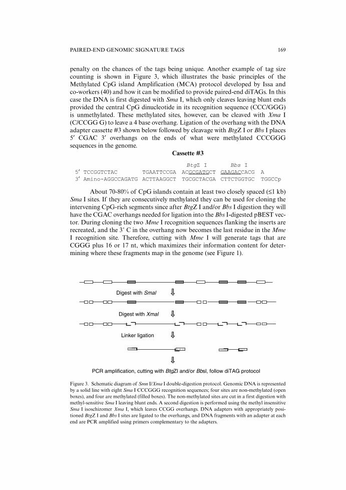
penalty on the chances of the tags being unique. Another example of tag sizecounting is shown in Figure 3, which illustrates the basic principles of theMethylated CpG island Amplification (MCA) protocol developed by Issa andco-workers (40) and how it can be modified to provide paired-end diTAGs. In thiscase the DNA is first digested with Sma I, which only cleaves leaving blunt endsprovided the central CpG dinucleotide in its recognition sequence (CCC/GGG)is unmethylated. These methylated sites, however, can be cleaved with Xma I(C/CCGG G) to leave a 4 base overhang. Ligation of the overhang with the DNAadapter cassette #3 shown below followed by cleavage with BtgZ I or Bbs I places5′ CGAC 3′ overhangs on the ends of what were methylated CCCGGGsequences in the genome.
Cassette #3
BtgZ I Bbs I5′ TCCGGTCTAC TGAATTCCGA ACGCGATGCT GAAGACCACG A3′ Amino-AGGCCAGATG ACTTAAGGCT TGCGCTACGA CTTCTGGTGC TGGCCp
About 70-80% of CpG islands contain at least two closely spaced (≤1 kb)Sma I sites. If they are consecutively methylated they can be used for cloning theintervening CpG-rich segments since after BtgZ I and/or Bbs I digestion they willhave the CGAC overhangs needed for ligation into the Bbs I-digested pBEST vec-tor. During cloning the two Mme I recognition sequences flanking the inserts arerecreated, and the 3˚ C in the overhang now becomes the last residue in the MmeI recognition site. Therefore, cutting with Mme I will generate tags that areCGGG plus 16 or 17 nt, which maximizes their information content for deter-mining where these fragments map in the genome (see Figure 1).
PAIRED-END GENOMIC SIGNATURE TAGS 169
Digest with Smal
Digest with Xmal
Linker ligation
PCR amplification, cutting with BtgZI and/or Bbsl, follow diTAG protocol
Figure 3. Schematic diagram of Smn I/Xma I double-digestion protocol. Genomic DNA is representedby a solid line with eight Sma I CCCGGG recognition sequences; four sites are non-methylated (openboxes), and four are methylated (filled boxes). The non-methylated sites are cut in a first digestion withmethyl-sensitive Sma I leaving blunt ends. A second digestion is performed using the methyl insensitiveSma I isoschizomer Xma I, which leaves CCGG overhangs. DNA adapters with appropriately posi-tioned BtgZ I and Bbs I sites are ligated to the overhangs, and DNA fragments with an adapter at eachend are PCR amplified using primers complementary to the adapters.
SUMMARY
Because paired-end genomic signature tags are sequenced-based, theyhave the potential to become an alternate tool to tiled microarray hybridizationas a method for genome–wide localization of transcription factors and othersequence-specific DNA binding proteins. As outlined here the method also canbe used for global analysis of DNA methylation. One advantage of this approachis the ability to easily switch between different genome types without having tofabricate a new microarray for each and every DNA type. However, the methoddoes have some disadvantages. Among the most rate-limiting steps of our PE-GST protocol are the need to concatemerize the diTAGs, size fractionate themand then clone them prior to sequencing. This is usually followed by additionalsteps to amplify and size select for long (≥500) concatemer inserts prior tosequencing. These time-consuming steps are important for standard DNAsequencing as they increase efficiency ~20-30-fold since each amplified concate-mer can now provide information on multiple tags; the limitation on data acqui-sition is read length during sequencing. However, the development of newsequencing methods such as Life Sciences’ 454 new nanotechnology-basedsequencing instrument (41) could increase tag sequencing efficiency by severalorders of magnitude (≥100,000 diTAG reads/run), which is sufficient to providein-depth global analysis of all ChIP PE-GSTs in a single run. This is because thelengths of our paired-end diTAGs (~60 bp) fall well within the region of highaccuracy for read lengths on this instrument. In principle, sequence analysis of
170 J. DUNN ET AL.
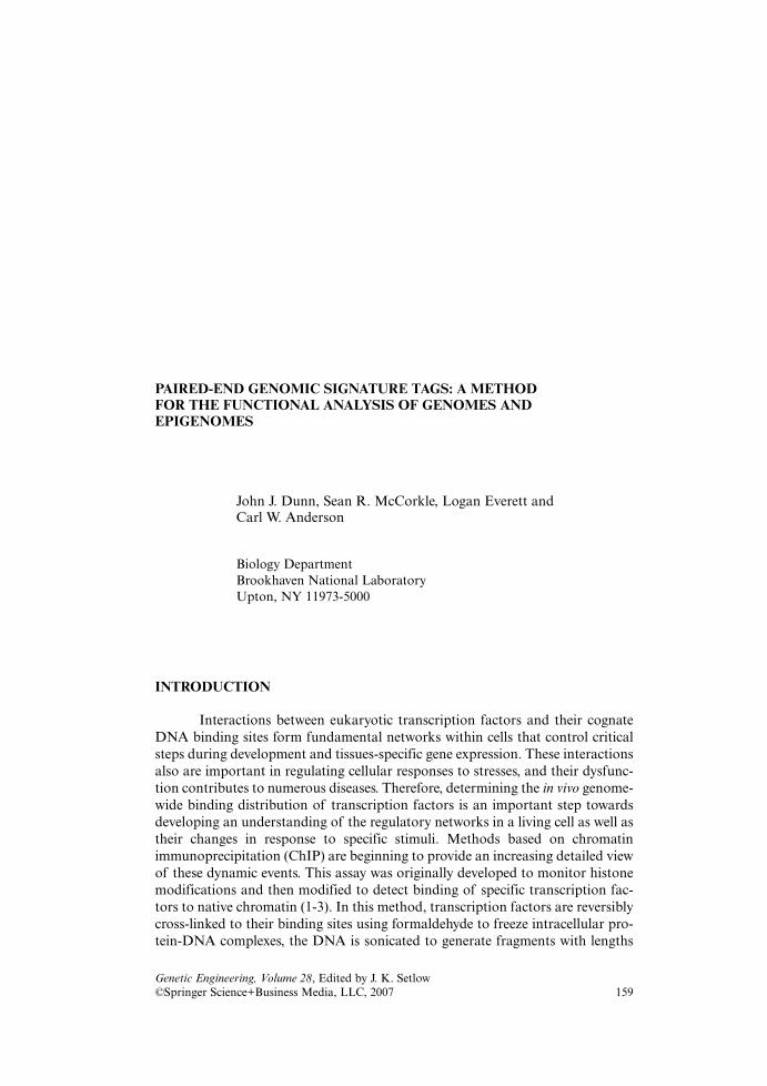
ChiP or CpG island DNA
Ligate with appropriate linker
Digest with BtgZ I and/or Bbs I
Ligate
Bbs I cut pBEST vector
Cut with Mmo I, gel purify, ligate with appropriatedegenerate linker and cut to generate cohesive ends;self-ligate to form circles,
Purify monomeric circles, electroporate
Cut with BseR I, purifydiTAGS, concatmerize,clone and sequence
or Directly sequence diTAGS
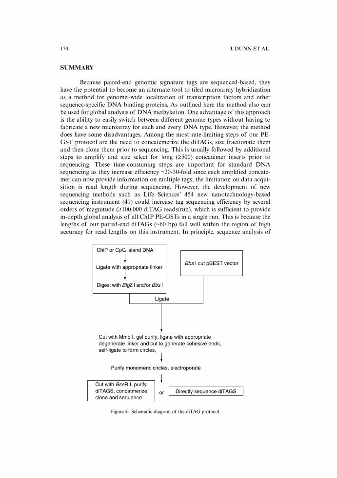
Figure 4. Schematic diagram of the diTAG protocol.
diTAGs could begin as soon as they are generated, thereby completely bypassingthe need for the concatemerization, sizing, downstream cloning steps andsequencing template purification. In addition, our protocol places any one of sev-eral unique four-base long nucleotide sequences, such as GATC, between eachand every diTAG pair, which could be used to help the instrument’s software keepbase register and also provide a well-located peak height indicator in the middleof every sequence run. This additional feature could permit multiplexing of thedata by simultaneous sequencing of several pooled libraries if each used a differ-ent linker sequence during diTAG formation (Figure 4).
ACKNOWLEDGMENTS
Support from National Institutes of Health Grant AI056480, the LowDose Radiation Research Program of the Office of Biological and EnvironmentalResearch (BER), U.S. Department of Energy and the BNL Laboratory DirectedResearch and Development Program is gratefully acknowledged.
REFERENCES
1 Orlando, V. (2000) Trends Biochem. Sci. 25, 99-104.2 Ren, B., Robert, F., Wyrick, J.J., Aparicio, O., Jennings, E.G., Simon, I.,
Zeitlinger, J., Schreiber, J., Hannett, N., Kanin, E., Volkert, T.L., Wilson,C.J., Bell, S.P. and Young, R.A. (2000) Science 290, 2306-2309.
3 Wells, J., Graveel, C.R., Bartley, S.M., Madore, S.J. and Farnham, P.J.(2002) Proc. Nat. Acad. Sci. U.S.A. 99, 3890-3895.
4 Saha, S., Sparks, A.B., Rago, C., Akmaev, V., Wang, C.J., Vogelstein, B.,Kinzler, K.W. and Velculescu, V.E. (2002) Nat. Biotechnol. 20, 508-512.
5 Impey, S., McCorkle, S.R., Cha-Molstad, H., Dwyer, J.M., Yochum,G.S., Boss, J.M., McWeeney, S., Dunn, J.J., Mandel, G. and Goodman,R.H. (2004) Cell 119, 1041-1054.
6 Tuzun, E., Sharp, A.J., Bailey, J.A., Kaul, R., Morrison, V.A., Pertz,L.M., Haugen, E., Hayden, H., Albertson, D., Pinkel, D., Olson, M.V.and Eichler, E.E. (2005) Nat. Genet. 37, 727-732.
7 Bird, A.P. (1986) Nature 321, 209-213.8 Dunn, J.J., McCorkle, S.R., Praissman, L.A., Hind, G., Van Der Lelie,
D., Bahou, W.F., Gnatenko, D.V. and Krause, M.K. (2002) Genome Res.12, 1756-1765.
9 Velculescu, V.E., Zhang, L., Vogelstein, B. and Kinzler, K.W. (1995)Science 270, 484-487.
10 Mayr, B. and Montminy, M. (2001) Nat. Rev. Mol. Cell Biol. 2, 599-609.11 Chen, J. and Sadowski, I. (2005) Proc. Nat. Acad. Sci. U.S.A. 102, 4813-
4818.12 Kim, J., Bhinge, A.A., Morgan, X.C. and Iyer, V.R. (2005) Nat.
Methods 2, 47-53.13 Labhart, P., Karmakar, S., Salicru, E.M., Egan, B.S., Alexiadis, V.,
O’Malley, B.W. and Smith, C.L. (2005) Proc. Nat. Acad. Sci. U.S.A. 102,1339-1344.
PAIRED-END GENOMIC SIGNATURE TAGS 171
14 Roh, T.Y., Cuddapah, S. and Zhao, K. (2005) Genes Dev. 19, 542-552.15 Ng, P., Wei, C.L., Sung, W.K., Chiu, K.P., Lipovich, L., Ang, C.C.,
Gupta, S., Shahab, A., Ridwan, A., Wong, C.H., Liu, E.T. and Ruan, Y.(2005) Nat. Methods 2, 105-111.
16 Studier, F.W. (2005) Protein Expr. Purif. 41, 207-234.17 Wahl, G.M. and Carr, A.M. (2001) Nat. Cell Biol. 3, E277-286.18 Vousden, K.H. and Lu, X. (2002) Nat. Rev. Cancer 2, 594-604.19 Yang, A., Kaghad, M., Caput, D. and McKeon, F. (2002) Trends Genet.
18, 90-95.20 Oren, M. (2003) Cell Death Differ. 10, 431-442.21 el-Deiry, W.S., Kern, S.E., Pietenpol, J.A., Kinzler, K.W. and Vogelstein,
B. (1992) Nat. Genet. 1, 45-49.22 Hoh, J., Jin, S., Parrado, T., Edington, J., Levine, A.J. and Ott, J. (2002)
Proc. Nat. Acad. Sci. U.S.A. 99, 8467-8472.23 Contente, A., Dittmer, A., Koch, M.C., Roth, J. and Dobbelstein, M.
(2002) Nat. Genet. 30, 315-320.24 Kim, E. and Deppert, W. (2003) Biochem. Cell Biol. 81, 141-150.25 Yin, Y., Liu, Y.X., Jin, Y.J., Hall, E.J. and Barrett, J.C. (2003) Nature
422, 527-531.26 Spinella, D.G., Bernardino, A.K., Redding, A.C., Koutz, P., Wei, Y.,
Pratt, E.K., Myers, K.K., Chappell, G., Gerken, S. and McConnell, S.J.(1999) Nucl. Acids Res. 27, e22.
27 Coulondre, C., Miller, J.H., Farabaugh, P.J. and Gilbert, W. (1978)Nature 274, 775-780.
28 Bird, A.P. and Taggart, M.H. (1980) Nucl. Acids Res. 8, 1485-1497.29 Bird, A. (2002) Genes Dev. 16, 6-21.30 Gardiner-Garden, M. and Frommer, M. (1987) J. Mol. Biol. 196, 261-282.31 Ponger, L., Duret, L. and Mouchiroud, D. (2001) Genome Res. 11, 1854-
1860.32 Ponger, L. and Mouchiroud, D. (2002) Bioinformatics 18, 631-633.33 Feinberg, A.P. (2001) Nat. Genet. 27, 9-10.34 Baylin, S.B., Herman, J.G., Graff, J.R., Vertino, P.M. and Issa, J.P.
(1998) Adv. Cancer Res. 72, 141-196.35 Marsit, C.J., Kim, D.H., Liu, M., Hinds, P.W., Wiencke, J.K., Nelson,
H.H. and Kelsey, K.T. (2005) Int. J. Cancer 114, 219-223.36 Feinberg, A.P., Ohlsson, R. and Henikoff, S. (2006) Nat. Rev. Genet. 7,
21-33.37 Cross, S.H., Charlton, J.A., Nan, X. and Bird, A.P. (1994) Nat. Genet.
6, 236-244.38 Shiraishi, M., Chuu, Y.H. and Sekiya, T. (1999) Proc. Nat. Acad. Sci.
U.S.A. 96, 2913-2918.39 Rauch, T. and Pfeifer, G.P. (2005) Lab. Invest. 85, 1172-1180.40 Toyota, M. and Issa, J.P. (2002) Methods Mol. Biol. 200, 101-110.41 Margulies, M., Egholm, M., Altman, W.E., Attiya, S., Bader, J.S.,
Bemben, L.A., Berka, J., Braverman, M.S., Chen, Y.J., Chen, Z., Dewell,S.B., Du, L., Fierro, J.M., Gomes, X.V., Godwin, B.C., He, W., Helgesen,S., Ho, C.H., Irzyk, G.P., Jando, S.C., Alenquer, M.L., Jarvie, T.P.,
172 J. DUNN ET AL.

Jirage, K.B., Kim, J.B., Knight, J.R., Lanza, J.R., Leamon, J.H.,Lefkowitz, S.M., Lei, M., Li, J., Lohman, K.L., Lu, H., Makhijani, V.B.,McDade, K.E., McKenna, M.P., Myers, E.W., Nickerson, E., Nobile,J.R., Plant, R., Puc, B.P., Ronan, M.T., Roth, G.T., Sarkis, G.J., Simons,J.F., Simpson, J.W., Srinivasan, M., Tartaro, K.R., Tomasz, A., Vogt,K.A., Volkmer, G.A., Wang, S.H., Wang, Y., Weiner, M.P., Yu, P.,Begley, R.F. and Rothberg, J.M. (2005) Nature 437, 376-380.
PAIRED-END GENOMIC SIGNATURE TAGS 173