Embed Size (px)
Citation preview
Am. J. Hum. Genet. 52:99-109, 1993
Genetic Recombination Events Which Position the FriedreichAtaxia Locus Proximal to the D9S I 5/D9S5 Linkage Groupon Chromosome 9qSusan Chamberlain, * Martin Farrall,t Jacqui Shaw, * David Wilkes, * Jaime Carvajal,*Renate Hillerman, * Kit Doudney, * A. E. Harding,$ Robert Williamson, * Giorgio Sirugo,§ RicardoFujita,§ Michel Koenig,§ Jean-Louis Mandel,§ Francisco Palau, Eugenia Monros, Juan Vilchez,Felix Prieto, Andrea Richter, # Michel Vanasse, * * Serge Melancon, # Sergio Cocozza, ttElena Redolfi,§§ Francesca Cavalcanti,tT Luigi Pianese,tt Allesandro Filla,$I Stefano DiDonato,§§and Massimo Pandolfo§§
'Department of Biochemistry and Molecular Genetics, Saint Mary's Hospital Medical School; tMedical Research Council Molecular Medicine Group,Royal Postgraduate Medical School; and *Institute of Neurology, London; §Laboratoire de G6n6tique, Institut de Chimie Biologique, Strasbourg;IlUnidad de Genetica, Hospital "La Fe," Valencia; Services de #G6netique Medicale, and **Neurologie, H6pital Sainte Justine, Montreal;Departments of ttCellular and Molecular Pathology and Biology, and ttNeurology, Universita di Napoli, "Federico II," Naples; and §§Divisionof Biochemistry and Genetics of the Nervous System, Instituto Nazionale Neurologico "Carlo Besta," Milan
Summary
The absence of recombination between the mutation causing Friedreich ataxia and the two loci which originallyassigned the disease locus to chromosome 9 has slowed attempts to isolate and characterize the geneticdefect underlying this neurodegenerative disorder. A proximity of less than 1 cM to the linkage group hasbeen proved by the generation of high maximal lod score (Z) to each of the two tightly linked markersD9S15 (Z = 96.69; recombination fraction [0] = .01) and D9S5 (Z = 98.22; 0 = .01). We report hererecombination events which indicate that the FRDA locus is located centromeric to the D9S15/D9S5 linkagegroup, with the most probable order being cen-FRDA-D9S5-D9S15-qter. However, orientation of themarkers with respect to the centromere, critical to the positional cloning strategy, remains to be resolveddefinitively.
Introduction
Friedreich ataxia is a progressive neurodegenerativedisorder affecting both the central and peripheral ner-vous systems. The primary lesion is thought to occurin the dorsal root ganglia, where selective loss of thelarge myelinated neurons is observed. The incidenceof this recessively inherited disorder is estimated tobe 1/50,000 in the United Kingdom, with a carrierfrequency of 1/110.
Received May 27, 1992; final revision received September 9,1992.
Address for correspondence and reprints: Susan Chamberlain,Ph.D., Department of Biochemistry and Molecular Genetics, St.Mary's Hospital Medical School, Norfolk Place, London W2 1PG,England.o 1993 by The American Society of Human Genetics. All rights reserved.0002-9297/93/5201-0013$02.00
The disease characteristicially presents with ataxiaof gait, between the ages of 8 and 12 years. Hypo- orareflexia and small-sensory-nerve action potentials areusually present before symptoms develop. Progressiveataxia, dysarthria, and loss of vibration-and-positionsense follow, with patients losing the ability to walk,on average, 15 years after onset. Hypertrophic cardio-myopathy is thought to occur in more than 90% ofpatients and is the major cause of death.We have previously mapped the Friedreich ataxia
disease locus (FRDA) by linkage analysis to chromo-some 9 (Chamberlain et al. 1988; Fujita et al. 1989a).A more precise location, 9q13-21.1, immediately dis-tal to the variable heterochromatin region 9qh, wasdefined by in situ hybridization studies using the twomost closelylinkedmarkers, MCT1 12 (D9S15) (Shawet al. 1990) and DR47 (D9S5) (Fujita et al. 1989b).
99

Chamberlain et al.
We subsequently constructed a long-range physicalmap of the region and identified 10 CpG-rich regionswithin the 1.7-Mb interval thought to contain theFRDA locus. The two marker loci were shown to liein close proximity, by the identification of a common460-kb NotI fragment (Fujita et al. 1991; Wilkes etal. 1991).The D9S15 and D9S5 loci have remained the most
closely linked markers to date; no recombinationevents between FRDA and either marker (or betweenthe markers themselves) had been detected during theanalysis of more than 200 multiply affected pedigreescollected worldwide. Attempts were therefore madeto increase informativity at the marker loci, in order todetect rare useful recombination events which wouldorientate the linkage group and position the diseasemutation precisely.
Increased informativity at the D9S15 locus wasachieved by the identification of both an additionalpolymorphism detected by AccI (Wallis and Naka-mura 1989a) and a hypervariable microsatellite se-quence (MS) (Fujita et al. 1990; Wallis et al. 1990).More recently, two additional microsatellite sequences,GS4 and GS2, have been identified which flank theD9S15 locus at 90 kb and 60 kb, respectively (Fujita etal. 1992). The D9S5 locus had proved to be extremelyuninformative (PIC = .18). Physical expansion of thislocus resulted in the isolation of the clone 26P, whichdetects three RFLPs defined by the enzymes BstXI,XbaI, and DraI (Fujita et al. 1990), substantially in-creasing informativity.
Physical mapping studies in both genomic and yeastartificial chromosome clones (Fujita et al. 1991;Wilkes et al. 1991) have shown the order of these lociwithin the linkage group to be GS4-MCT1 12/MS-MCT112/MspI-GS2-26P-DR47. The strength ofthe linkage data would suggest a proximity for thedisease locus of no more than 1 cM from the linkagegroup, by the lod -1 confidence interval approach.At this proximity, linkage disequilibrium might be ob-served, hence providing information on the locationof the disease locus. Disequilibrium, predominantlywith D9S15, has been reported in populations witha potential founder effect, but the association hasproved much weaker in several more heterogeneouspopulations studied.We now report evidence of genetic recombination
events which position the disease locus relative to thetwo closest markers. These data are discussed in rela-tion to the clinical phenotypes and the potential prob-lems with definitive diagnosis when one is working at
this level of resolution. The relevance of disequilib-rium as an indicator for proximity is reviewed.
Subjects and Methods
SubjectsFamilies accepted for inclusion in this analysis con-
formed to the basic diagnostic criteria for Friedreichataxia as defined by Geoffroy et al. (1976) and Har-ding (1981), with two exceptions-(1) an absoluterequirement that the age at onset be before 20 yearsof age and (2) the presence of typical electrocardio-graphic changes (Harding and Langton-Hewer 1983)in at least one affected member ofeach family. Geneticlinkage analysis was carried out in five centers andincluded 274 families with two or more affected indi-viduals: St. Mary's Hospital Medical School, London(SM) (104 families); Laboratoire de Genetique Molec-ulaire, Strasbourg (LGM) (46 families); Hopital Ste.Justine, Montreal (SJ) (35 families); Instituto Nazio-nale Neurologico "Carlo Besta," Milan and Universitadi Napoli (CB-UN) (58 families); and Hospital "LaFe," Valencia (LF) (31 families). This heterogeneousgroup of families is predominantly European or NorthAmerican, although some patients were of Asian ornorthern African origin.
Linkage Analysis
Linkage analysis between the FRDA locus and thepolymorphic loci, D9S15 and D9M5, was carried outin all families. After the detection of recombination,positioning of the crossover events with respect to theD9S15/D9S5 linkage group was investigated by in-cluding in the analysis the highly polymorphic micro-satellite loci, GS4 and GS2 (table 1), known to flankD9S15 at 90 kb and 60 kb, respectively.Lod score (Z) values were computed for combined
sexes (recombination fraction [0] for males [Om] = 0for females [0f]), as tests for a sex difference in re-combination were insignificant in both pairwise andmultipoint analysis. To establish a multipoint link-age map ofproximal 9q and in an attempt to orientatethe linkage group, the SM pedigrees were also ana-lyzed with polymorphic loci previously shown toflank the Friedreich ataxia region- i.e., D9S48 (9ql 1)and ALDH1 (9q21.1) (Chamberlain et al. 1989b)(table 1).
Pairwise Z values were calculated, using the LINK-AGE computer program (Lathrop et al. 1985), be-tween FRDA and (a) the individual polymorphic sys-
100

Positioning of the FRDA Locus
Table I
Polymorphic Characteristics of Chromosome 9q Marker Loci
RegionalLocus Probe Assignment Enzyme PIC Source of Data
D9S48 ...... MCT7 9ql 1 PstI ... Y. Nakamura, personalcommunication
D9S15 ...... MCT112 9q13-21.1 MspI .33 Carlson 1987AccI .34 Wallis and Nakamura
1989D9S5 ...... MS .75 Wallis et al. 1990
DR47 9q13-21.1 TaqI .18 Orzechowski et al. 1987ALDH1 .... 26P BstXI .55 Fujita et al. 1990
9q21.2 MspI .33 Yoshida and Chen 1989
Length ofAmplified DNA
(bp) PCR Primers
GS2 ...... 124 Direct: 5'AATGAAATAGAATTTCACAGG 3'Reverse: 5'AACCCTTCTGTCAGACAAGGA 3'
GS4 ...... 131 Direct: 5'GGGAAGAGCAAATTCCCTGAACCCCG 3'Reverse: 5'CCTGGGCGACAGAGTGAGACTCG 3'
tems; (b) the compound haplotype at the D9S15 locus(MS/ / MCT1 12/MspI), where the polymorphic sitesare known to lie within an interval of no more than5 kb; and (c) the D9S5 locus (DR47/TaqI//26P/BstXI), where the sites lie within 30 kb. Multipointanalysis was carried out in the SM pedigrees, over thegenetic interval containing the linkage group D9S48-[D9S15-D9S5]-ALDH1.
Linkage DisequilibriumLinkage disequilibrium studies between FRDA and
the marker loci focused on D9S15 and D9S5. Associa-tion studies for the individual loci and for the extendedhaplotypes in the French (Fujita et al. 1990), Italian(Pandolfo et al. 1990), French-Canadian (Richter etal. 1990), and Spanish (Monr6s et al., submitted) pa-tient populations have been published or submittedfor publication. In the St. Mary's pedigree resource,allelic association was investigated for individual poly-morphisms and for the haplotype at the D9S15 (MS/ /MCT1 12/MspI) locus. Although these loci lie in closephysical proximity, association with the extendedhaplotype D9S15/D9S5 was not investigated in thisinstance, because of the recombination observed inthis region.
Clinical Evaluation of Recombinant FamiliesThe clinical features observed in the affected mem-
bers of the recombinant families are described in table2. Consanguinity was present in family LF2 only.
In all cases, onset occurred before the age of 20years, the disease commonly presenting as general un-steadiness. Affected members have progressive ataxiaaccompanied by dysarthria, posterior column signs,and diminished or absent tendon reflexes. Cardiomy-opathy, considered to be a primary feature of the dis-order, was present in at least one affected member ofeach family.Of the six recombinant families, two (SM1 and
SM2) are from England, two (LF1 and LF2) are fromSpain, one (SJ1) is from Canada, and one (CB-UN1)is from Italy. Clinical diagnosis in these families hasbeen reviewed and confirmed by a neurologist fromeach group.
Family SM 1. - This is a nuclear pedigree with two ofthe three offspring affected. Age at onset and rate ofprogression vary considerably between the sibs, thefemale sib becoming severely disabled and confined toa wheelchair at about the age of puberty. The male sibremained ambulant until the age of 22 years.
Family SM2.- This is a nuclear pedigree with two ofthe three offspring affected. Despite a very similar ageat onset in both sibs, the rate ofprogression has varied.While one sib became confined to a wheelchair at theage of 25 years, the sister could still walk unaidedfor considerable distances, until approximately age 35years. It is surprising that evidence of heart diseasecould not be detected in the more severely affectedsib when last examined, while individual IL.1 had anabnormal electrocardiogram.
101

Chamberlain et al.
Table 2
Clinical Features of the Six Recombinant Pedigrees, Indicating the Primary Diagnostic Criteria
FAMILY FAMILY FAMILY FAMILY FAMILY FAMILYSMi SM2 LF1 LF2 SJ1 CB-UN1
11.1 11.2 11.1 11.2 11.1 11.2 11.2 I1.6 11.7 I1.2 I1.3 11.1 1.2
Age (years) ..................... 23 18 50 38 20 14 28 20 18 35 31 25 24Sex ........ ............. M F F F F F F M M F F M FAge at onset (years) ................. 13 7 16 13 5 7 12 10 12 4 4 14 13Ataxia .......... ........... + + + + + + + + + + + + +
Dysarthria ..................... + + + + + + + + + + + + ±Tendon reflexes ..................... - - - - - - - - - - - - -Position and vibration sense ....-Electrocardiogram ................. A A A N A NT A A A A A A ADiabetes .............. ....... N N NT NT N N N N N + + N N
NOTE.- + = Present; - = diminished or absent; A = abnormal; N = normal; and NT = not tested.
Family LF 1. - This is a nuclear pedigree with two off-spring, both affected.
Family LF2. - This is a nuclear pedigree with three ofseven offspring affected. Enlargement of the fourthventricle and vermis atrophy in individual II.3 and an
anomalous morphology of the fourth ventricle in 11.6were detected on computed-tomography scan. Theparents are third cousins.
Family SJ I. -This is a family with two of four off-spring affected. Age at onset was comparatively early(about 4 years of age), but otherwise the features wereunremarkable. Abnormal glucose tolerance has beendetected in the affected sibs. The unaffected sister hasbeen investigated extensively within the past 6 mo,
and normal nerve conduction velocities (median, 56m/s; peroneal, 53 m/s) have been recorded.
Family CB-UNI.-This is a nuclear pedigree withboth offspring affected. Enlargement ofthe supra- andinfratentorial subarachnoid space and fourth ventriclewas detected on computed-tomography scan after a
disease duration of 11 years. Nerve biopsy showedloss of large myelinated fibers.
Results
Linkage Analysis
Combined pairwise Z values between the FRDAlocus and chromosome 9q markers are summarized intable 3. Data generated from the analysis of the SJ,LGM, CB-UN, and LF pedigrees with the markersD9S15 and D9S5 have been published (in part) else-
where (Richter et al. 1989; Fujita et al. 1990; Pandolfoet al. 1990).Maximal Z values were generated between the
FRDA locus and D9S15 (Z = 96.69; 0 = .01) andD9S5 (Z = 98.22; 0 = .01), reflecting the detectionof recombination events for each marker locus. These0's should be regarded as good estimates, since the lodtable is somewhat sparse around the maximum. Inaddition, analysis of the SM and LF pedigree sets re-vealed recombination between the D9S15 and D9S5loci (0 = .02).The D9S48 locus remains closely linked to FRDA
(Z = 11.2;0 = .06),thedisparitybetweenthegeneticand the estimated physical distance (greater than 10Mb) in this region being maintained. In contrast, anal-ysis of this extended pedigree set resulted in a weaken-ing of the linkage between FRDA and the ALDH1locus (Z = 3.75; 0 = .12).
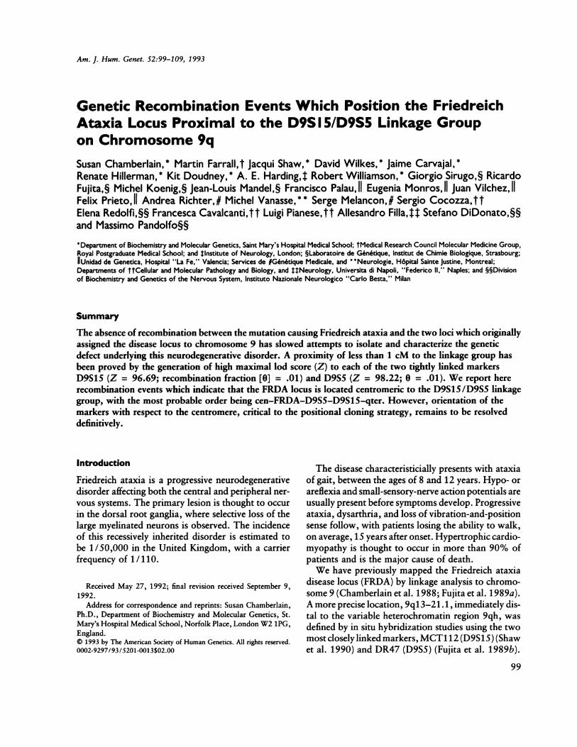
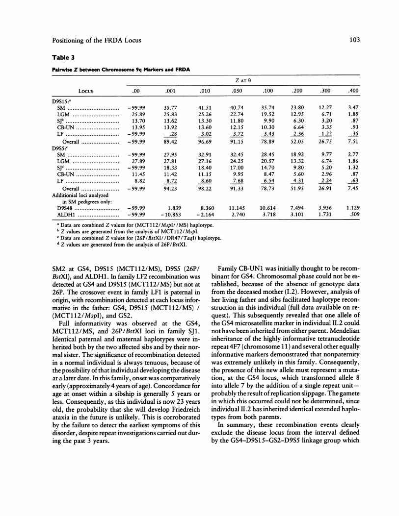
Recombination EventsRecombination events were detected between the
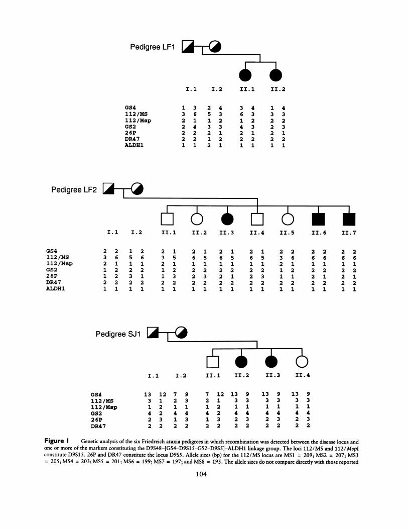
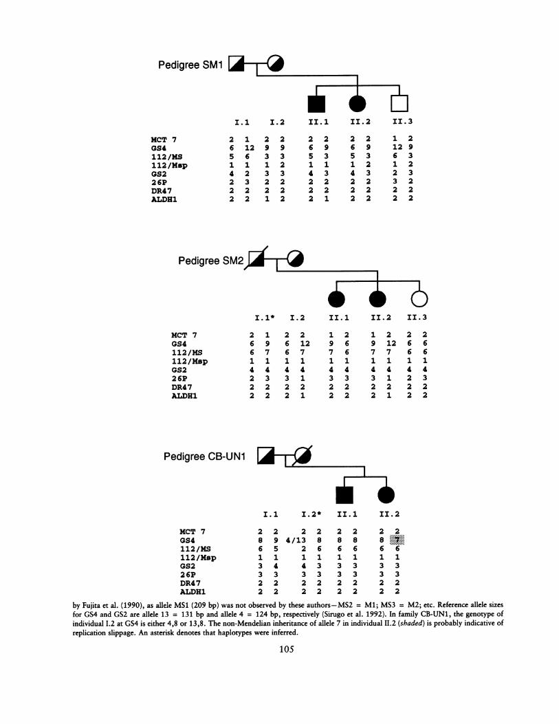
disease locus and one or more of the marker loci insix of the families analyzed. Figure 1 summarizes theanalysis in these families for the markers D9S48(MCT7/PstI), GS4, D9S15 (MCT112/MS), D9S15(MCT112/MspI), GS2, D9S5 (26P/BstXI), D9S5(DR47/ TaqI), and (if informative) ALDH1.
In each of the families SM1, SM2, and LF2, the cross-over event appears to be maternal in origin. In familiesSM1 and SM2, recombination was detected at each lo-cus informative in the respective mothers: family SM1at D9S15 (MCT112/MspI) and ALDH1; and family
102
Positioning of the FRDA Locus
Table 3
Pairwise Z between Chromosome 9q Markers and FRDA
Z AT 0
Locus .00 .001 .010 .050 .100 .200 .300 .400
D9S15:aSM ............ ........ - 99.99 35.77 41.51 40.74 35.74 23.80 12.27 3.47LGM .................... 25.89 25.83 25.26 22.74 19.52 12.95 6.71 1.89Sjb ........... ......... 13.70 13.62 13.30 11.80 9.90 6.30 3.20 .87CB-UN.................... 13.95 13.92 13.60 12.15 10.30 6.64 3.35 .93LF ........... ......... - 99.99 .28 3.02 3.72 3.43 2.36 1.22 .35
Overall ................... - 99.99 89.42 96.69 91.15 78.89 52.05 26.75 7.51D9Ss:cSM ............ ........ - 99.99 27.95 32.91 32.45 28.45 18.92 9.77 2.77LGM .................... 27.89 27.81 27.16 24.25 20.57 13.32 6.74 1.86SJd ........... ......... - 99.99 18.33 18.40 17.00 14.70 9.80 5.20 1.32CB-UN.................... 11.45 11.42 11.15 9.95 8.47 5.60 2.96 .87LF .................... 8.82 8.72 8.60 7.68 6.54 4.31 2.24 .63
Overall ................... - 99.99 94.23 98.22 91.33 78.73 51.95 26.91 7.45Additionial loci analyzed
in SM pedigrees only:D9S48.................... -99.99 1.839 8.360 11.145 10.614 7.494 3.956 1.129ALDH1 ................... - 99.99 - 10.853 -2.164 2.740 3.718 3.101 1.731 .509a Data are combined Z values for (MCT1 12/MspI/ /MS) haplotype.b Z values are generated from the analysis of MCT112/MspI.c Data are combined Z values for (26P/BstXI/ /DR47/ TaqI) haplotype.d Z values are generated from the analysis of 26PIBstXI.
SM2 at GS4, D9S15 (MCT112/MS), D9S5 (26P/BstXI), and ALDH1. In family LF2 recombination wasdetected at GS4 and D9S15 (MCT112/MS) but not at26P. The crossover event in family LF1 is paternal inorigin, with recombination detected at each locus infor-mative in the father: GS4, D9S15 (MCT112/MS) /
(MCT112/MspI), and GS2.Full informativity was observed at the GS4,
MCT112/MS, and 26P/BstXI loci in family SJ1.Identical paternal and maternal haplotypes were in-herited both by the two affected sibs and by their nor-mal sister. The significance of recombination detectedin a normal individual is always tenuous, because ofthe possibility of that individual developing the diseaseat a later date. In this family, onset was comparativelyearly (approximately 4 years of age). Concordance forage at onset within a sibship is generally 5 years or
less. Consequently, as this individual is now 23 years
old, the probability that she will develop Friedreichataxia in the future is unlikely. This is corroboratedby the failure to detect the earliest symptoms of thisdisorder, despite repeat investigations carried out dur-ing the past 3 years.
Family CB-UN1 was initially thought to be recom-binant for GS4. Chromosomal phase could not be es-tablished, because of the absence of genotype datafrom the deceased mother (I.2). However, analysis ofher living father and sibs facilitated haplotype recon-struction in this individual (full data available on re-quest). This subsequently revealed that one allele ofthe GS4 microsatellite marker in individual I.2 couldnot have been inherited from either parent. Mendelianinheritance of the highly informative tetranucleotiderepeat 4F7 (chromosome 11) and several other equallyinformative markers demonstrated that nonpaternitywas extremely unlikely in this family. Consequently,the presence of this new allele must represent a muta-tion, at the GS4 locus, which transformed allele 8into allele 7 by the addition of a single repeat unit-probably the result ofreplication slippage. The gametein which this occurred could not be determined, sinceindividual I.2 has inherited identical extended haplo-types from both parents.
In summary, these recombination events clearlyexclude the disease locus from the interval definedby the GS4-D9S15-GS2-D9SS linkage group which
103
Pedigree LF1
GS4112/MS112 /MspGS226PDR4 7ALDR1
I.1 i.2
2 2 1 2 2 13 6 5 6 3 52 1 1 1 2 11 2 2 2 1 21 2 3 1 1 32 2 2 2 2 21 1 1 1 1 1
Pedigree SJ1
1322221
3 2 4 3 4 16 5 3 6 3 31 1 2 1 2 24 3 3 4 3 22 2 1 2 1 22 1 2 2 2 21 2 1 1 1 1
4323121
II.1 II.2 II.3 II.4 II.5 II.6 II.7
261222
1
1
5
1
2321
2612221
1
5
1
2
1
2
1
21651122232211
23
21
1
2
1
26
1
21
2
1
2612221
26
1
21
2
1
2612221
2612121
I.1 i.2
GS4112/MS112 /MspGS226PDR47
13 12 7 93 1 2 31 2 1 14 2 4 42 3 1 32 2 2 2
7 122 11 2421 32 2
13 93311442322
13 93 31 14 42 32 2
13 93 31 14 42 32 2
Figure I Genetic analysis of the six Friedreich ataxia pedigrees in which recombination was detected between the disease locus andone or more of the markers constituting the D9S48-[GS4-D9S15-GS2-D9SS]-ALDH1 linkage group. The loci 112/MS and 112/MspIconstitute D9S15. 26P and DR47 constitute the locus D9S5. Allele sizes (bp) for the 112/MS locus are MS1 = 209; MS2 = 207; MS3= 205; MS4 = 203; MS5 = 201; MS6 = 199; MS7 = 197; and MS8 = 195. The allele sizes do not compare directly with those reported
104
Pedigree LF2
9 1
LbGS4112/MS112 /MspGS226PDR4 7ALDH1
I
II.1 II.2 II.3 IIA
I.1 i.2 II. 1 II. 2
I-- - -- I
Pedigree SM1
MCT 7GS4112 INS112 /MapGS226PDR47ALDH1
I.1 I.2 11.1 II.2 II.3
2 1 2 2 2 2 2 2 1 26 12 9 9 6 9 6 9 12 95 6 3 3 5 3 5 3 6 31 1 1 2 1 1 1 2 1 24 2 3 3 4 3 4 3 2 32 3 2 2 2 2 2 2 3 22 2 2 2 2 2 2 2 2 22 2 1 2 2 1 2 2 2 2
Pedigree SM2
MCT 7GS4112/MS112/MapGS226PDR47ALDH1
Pedigree CB-UN1
I.1* i.2
2 1 2 26 9 6 126 7 6 71 1 1 14 4 4 42 3 3 12 2 2 22 2 2 1
II..1 II .2 II..3
1 2 1 2 2 29 6 9 12 6 67 6 7 7 6 61 1 1 1 1 14 4 4 4 4 43 3 3 1 2 32 2 2 2 2 22 2 2 1 2 2
MCT 7GS4112 /MS112 /MapGS226PDR47ALDR1
I.1 I.2* II.1
2 2 2 2 2 28 9 4/13 8 8 86 5 2 6 6 61 1 1 1 1 13 4 4 3 3 33 3 3 3 3 32 2 2 2 2 22 2 2 2 2 2
II. 2
2 28 76 61 13 33 32 22 2
by Fujita et al. (1990), as allele MS1 (209 bp) was not observed by these authors-MS2 = Ml; MS3 = M2; etc. Reference allele sizesfor GS4 and GS2 are allele 13 = 131 bp and allele 4 = 124 bp, respectively (Sirugo et al. 1992). In family CB-UN1, the genotype ofindividual 1.2 at GS4 is either 4,8 or 13,8. The non-Mendelian inheritance of allele 7 in individual 11.2 (shaded) is probably indicative ofreplication slippage. An asterisk denotes that haplotypes were inferred.
105

Chamberlain et al.
spans approximately 550 kb of proximal 9q. Thesedata, in association with recombination events ob-served between the disease locus and additional distal9q markers including ALDH1, positionFRDA centro-meric to the linkage group. Interpretation ofthe analy-sis in family LF2, where recombination is detectedbetween FRDA and the marker loci GS4 and D9S15(MCT112/MS) but, despite full informativity, not atD9S5 (26P/BstXI), implies that the most probable or-der is cen-FRDA-D9S5-D9S15-qter.
Multipoint Analysis
The multipoint genetic map of the region, as basedon marker-marker recombination events, was con-structed using the data set generated from the SMpedigrees, both including and excluding the recombi-nant families. Zero recombination was assumed be-tween the two polymorphic loci (MCT112/MS andMCT112/MspI) at D9S15 and also between the twoloci (26P/BstXI and DR47/TaqI) at D9S5. The ge-netic distances were subsequently computed by iterat-ing the three 0's between the markers. The resultinggenetic map of the linkage group was entirely consis-tent with the physical mapping order: cen-D9S48-(.16)-[D9S15-( .02)-D9S5]-(.14)-ALDH1. For bothdata sets, the order D9S48-D9S15-D9S5-ALDH1was favored by odds of only 9.6:1 over the alternativeorder D9S48-D9S5-D9S15-ALDH1.
Linkage DisequilibriumAssociation studies in the French-Canadian (SJ)
(Richter et al. 1989), northern-Spanish (SM) (Cham-berlain et al. 1989a), French (LGM) (Fujita et al.1990), and Italian (CB-UN) (Pandolfo et al. 1990)patient populations have been reported elsewhere. Insummary, no statistically significant evidence of asso-ciation between FRDA and the D9S5 locus has beenpublished for any patient population except theFrench (Fujita et al. 1990), while significant linkagedisequilibrium has been reported between the diseaseand D9S15-in particular, the 205-bp allele of theMCT112/MS polymorphism (Hanauer et al. 1990a).These data implied a closer proximity of the FRDAlocus to D9S15 than to D9S5.
Disequilibrium studies in the SM pedigree set werecarried out between the FRDA locus and the individ-ual polymorphisms and the haplotypes generated atthe D9S15 locus. The analysis of allelic associationbetween FRDA and the DR47/ TaqI polymorphism atD9S5 was excluded because of low informativity inthese families.
As the SM pedigrees constitute families collected incollaboration throughout the world, the possibility ofdisequilibrium was investigated separately in (a) theU.K. pedigrees (n = 41) and (b) the complete pedigreeresource. As both data sets are comparatively small,the families were not subdivided further according togeographical origin. No evidence of allelic associationbetween the FRDA locus and the microsatellite poly-morphism (i.e., MS) at D9S15, the MspI polymor-phism at D9S15, the BstXI polymorphism at D9S5, orthe haplotype (MCT112/MS/ /MCT1 12/MspI) atD9S15 was found (data available on request).
Discussion
The isolation and subsequent characterization of adisease locus depends on the construction of physicaland genetic maps of the linked region and on the posi-tioning of genetic recombination events in relation tothe physical location of potential "candidate" codingsequences. Progress toward the isolation of the muta-tion causing Friedreich ataxia has, ironically, beenslowed by the tight linkage between FRDA and twoanonymous markers MCT112 (D9S15) and DR47(D9S5), preventing ordering within the linkage group.However, the tight linkage has facilitated the provi-sion of reliable genetic counseling in affected families(Wallis et al. 1989; Hanauer et al. 1990b).We now report recombination events between
FRDA and the closest markers, detected after the iden-tification of more highly informative polymorphisms.Combined maximal Z values between FRDA andD9S15 and FRDA and D9S5 are Z = 96.69 (0 = .01)and Z = 98.22 (0 = .01), respectively. Using theconfidence interval approach, linkage analysis pro-vides firm evidence that the FRDA locus lies within1 cM of the linkage group; the recombination dataindicate a position for the disease locus centromeric tothe D9S15/D9S5 linkage group on proximal 9q. Thecritical issue remains the orientation of the linkagegroup with respect to the centromere, which in turndetermines the directional cloning strategy.
This could not be resolved by multipoint linkageanalysis; the probability of the order cen-D9S15-D955-ALDH1 is only approximately 10 times morelikely than the order cen-D9S5-D9S15-ALDH1.Only the analysis of family LF2 provides evidence fororder indicating that D9S5 and, hence, FRDA lie cen-tromeric to D9S15- consistent with the evidence fororder generated by extended haplotype analysis of theCajun population (Sirugo et al. 1992). However, as
106
Positioning of the FRDA Locus
the parents are third-degree cousins, homozygositywould be predicted at each of the informative markerloci in the affected individuals. As homozygosity is lostat the 26P locus, a recombinant event in a formermeiosis can therefore be deduced.
Corroborating evidence from disequilibrium pro-
files across the region has been sought. The analysisof the SM pedigrees provides no evidence for linkagedisequilibrium. In addition, while previous associa-tion studies have demonstrated significant disequilib-rium only between FRDA and D9S15 (Fujita et al.1990; Pandolfo et al. 1990), recent extension of theFrench, Italian, and French-Canadian pedigree sets
has resulted in the detection of statistically significantdisequilibrium at D9S5, with a concomitant diminish-ing degree of association with D9S15 (M. Koenig, M.Pandolfo, and A. Richter, unpublished data). Thisshift in the disequilibrium profile could be the resultof the inclusion of affected pedigrees from more het-erogeneous populations, as exemplified in the Spanishpopulation. Strong disequilibrium with D9S5 was ini-tially observed from the study of families originatingin the immediate vicinity ofValencia. Extension of thestudy to include families collected throughout Spainhas subsequently resulted in the loss of significant dis-equilibrium at either locus.However, this is still a comparatively small data set.
In addition, we can only speculate on the number ofdifferent mutations which can cause Friedreich ataxia.In view of the variation in age at onset, rate ofprogres-sion, and severity, more than one mutation would bepredicted. These factors, singly or in combination,could explain the absence of linkage disequilibrium inthis particular patient group. In summary, support forlocus order based on linkage disequilibrium is ambigu-ous, although it would seem (from unpublished data)that association between FRDA and D9S5 (26P/BstXI) is at least as significant as that seen betweenFRDA and D9S15, in certain populations.The inclusion of GS4, a microsatellite polymor-
phism, for genetic counseling and in disequilibriumstudies is questionable, after the detection of probablereplication slippage in family CB-UN1. This type ofpolymorphism, with a large number of alleles, may beevolving rapidly by mechanisms other than meioticcrossover (Levinson and Gutman 1987; Wolff et al.1988). It would seem likely that certain microsatellitepolymorphisms, including GS4, may not share the dis-equilibrium relationships found for conventionalRFLPs. Consequently, data including such loci mustbe interpreted with caution.
The possibility ofheterogeneity must always be con-sidered when any genetic disorder is analyzed, particu-larly if there is marked clinical variation. In the caseof Friedreich ataxia, locus homogeneity has been con-clusively demonstrated in all patient populations con-forming to the basic diagnostic criteria used here, andwe regard accurate diagnosis as central to this study.Furthermore, analysis of these data by using the HO-MOG program revealed no evidence of heterogeneity.
However, all groups have encountered cases with clin-ical phenotypes initially indistinguishable from Fried-reich ataxia. Following the detection of recombinationacross the entire D9S48-D9S15-D9S5-ALDH1 regionin one family, further investigation showed a vitamin Edeficiency (M. Pandolfo, unpublished data). In addi-tion, several "recombinant" families in which the onlydeviation from the classical criteria is the absence of car-diomyopathy, despite long disease duration, also showexclusion from the chromosome 9 locus (S. Chamber-lain and A. Harding, unpublished data).
It therefore appears that the presence of heart dis-ease is critical for diagnosis of Friedreich ataxia with alocus on 9q. However, the age at which this pathologybecomes evident varies even within families (Hardingand Langton-Hewer 1983). In the Cajun patient popu-lation reported by Keats et al. (1989), where the dis-ease phenotype and course are more benign, cardio-myopathy is a less prominent feature of the disorder,with increased longevity of affected members.
For the six families reported here, clinical notes havebeen reviewed by clinicians from each research group,and the diagnosis has been confirmed. Resampling hasalso been undertaken to eliminate laboratory error.As inversion of the 9qh region is common, with anincidence of 1%, cytogenetic analysis of family LF2has been undertaken. No evidence of pericentric in-version has been detected, supporting the proposedorder for the marker loci. Taking all data into consid-eration, we propose that the most likely gene/probeorder in this region is cen-FRDA-D9S5-GS2-D9S15-GS4-qter.
AcknowledgmentsThis study was supported as follows: center SM-the
Friedreich's Ataxia Group (U.K.), the Muscular DystrophyAssociation (USA), the Medical Research Council (U.K.),the Joseph Levy Trust, and the Buttle Trust; center LGM-Association Francaise contre les Myopathies, CNAMTS, andMinistere de la Recherche; center LF-Fondo de Investiga-tion Sanitaria grants 89/1932 and 91/0445, Fundacion de
107
108 Chamberlain et al.
Ataxias Hereditarias 'Adriana de Luz Caballer,' and Gener-alitat Valenciana; center SJ- L'Association Canadienne deL'Ataxie de Friedreich and the Juvenile Diabetes FoundationInternational; center CB-UN-Italian Telethon and the Ital-ian Association against Ataxic Syndromes.
ReferencesCarlson M, Nakamura Y, Krapcho K, Fujimoto E, O'Con-
nell P, Leppert M, Lathrop M, et al (1987) Isolation andmapping of a polymorphic DNA sequence pMCT1 12 onchromosome 9q (D9S15). Nucleic Acids Res 15:10614
Chamberlain S, Shaw J, Rowland A, Wallis J, South S, Na-kamura Y, von Gabain A, et al (1988) Mapping of muta-tion causing Friedreich's ataxia to human chromosome 9.Nature 334:248-249
Chamberlain S, Shaw J, Wallis J, Rowland A, Chow L,Farrall M, Keats B, et al (1989a) Genetic homogeneity atthe Friedreich ataxia locus on chromosome 9. Am J HumGenet 44:518-521
Chamberlain S, Shaw J, Wallis J, Wilkes D, Farrall M, Wil-liamson R (1989b) Linkage analysis of DNA markersaround the Friedreich's ataxia locus on chromosome 9.Cytogenet Cell Genet 51:975
Fujita R, Agid Y, Trouillas P, Seck A, Tomassi-Davenas C,Driesel AJ, Olek K, et al (1989a) Confirmation of linkageof Friedreich's ataxia to chromosome 9 and identificationof a new closely linked marker. Genomics 4:110-111
Fujita R, Hanauer A, Chery M, Gilgenkrantz S, Noel B,Mandel JL (1989b) Localization of the Friedreich's ataxiagene to 9q13-21 by physical and genetic mapping ofclosely linked markers. Cytogenet Cell Genet 51:1001
Fujita R, Hanauer A, Sirugo G, Heilig R, Mandel JL (1990)Additional polymorphisms at marker loci D9S5 andD9S15 generate extended haplotypes in linkage disequi-librium with Friedreich ataxia. Proc Natl Acad Sci USA87:1796-1800
Fujita R, Hanauer A, Vincent A, Mandel JL, Koenig M(1991) Physical mapping of two loci (D9S5 and D9S15)tightly linked to the Friedreich's ataxia locus (FRDA) andidentification ofnearby CpG islands by pulse-field gel elec-trophoresis. Genomics 10:915-920
Fujita R, Sirugo G, Duclos F, Abderrahim H, Le Paslier D,Cohen D, Brownstein BH, et al (1992) A 530kb YACcontig tightly linked to the Friedreich ataxia locus contains5 CpG clusters and a new highly informative polymorphicmicrosatellite. Hum Genet 89:531-538
Geoffroy G, Barbeau A, Breton G, Lemieux B, Aube M,Leger C, Bouchard JP (1976) Clinical description androentgenolgic evaluation of patients with Friedreich'sataxia. Can J Neurol Sci 3:279-287
Hanauer A, Chery M, Fujita R, Driesel AJ, Gilgenkrantz S,Mandel JL (1990a) The Friedreich ataxia gene is assignedto chromosome 9ql3-q21 by mapping of tightly linked
markers and shows linkage disequilibrium with D9S15.Am J Hum Genet 46:133-137
Hanauer A, Fujita R, Trouillas P, Tommasi-Davenas C,Agid Y, Seck A, Mandel JL (1990b) Prenatal diagnosis ofFriedreich ataxia. Lancet 1:1102
HardingAE (1981) Friedreich's ataxia: a clinical and geneticstudy of 90 families with an analysis of early diagnosticcriteria and intrafamilial clustering of clinical features.Brain 104:589-620
Harding AE, Langton-Hewer RL (1983) The heart diseaseof Friedreich's ataxia: a clinical and electrocardiographicstudy of 115 patients, with an analysis of serial electrocar-diographic changes in 30 cases. Q J Med 208:489-502
Keats B, Ward LJ, ShawJ, Wickremasinghe A, ChamberlainS (1989) Acadian and classical forms ofFriedreich's ataxiaare most probably caused by mutation at the same locus.Am J Med Genet 33:266-268
Lathrop GM, Lalouel JM, Julier C, Ott J (1985) Multilocuslinkage analysis in humans: detection of linkage and esti-mation of recombination. Am J Hum Genet 37:482-498
Levinson G, Gutman GA (1987) Slipped-strand mispairing:a major mechanism for DNA sequence evolution. MolBiol Evol 4:203-221
Monr6s E, Carvajal J, Lopez-Arlandis J, Vilchez JJ, PrietoF, Palau F. Friedreich ataxia shows genetic homogeneityand linkage disequilibrium with the D9S15 and D9S5 lociin the Spanish population (submitted)
Orzechowski HD, HennigJ, Winter P, Grzeschik K-H, OlekK, Driesel AJ (1987) A human single-copy DNA probe(DR47) detects a TaqI RFLP on chromosome 9. NucleicAcids Res 15:6310
Pandolfo M, Sirugo G, Antonelli A, Weitnauer L, FerrettiL, Leone M, Dones I, et al (1990) Friedreich's ataxia inItalian families: genetic homogeneity and linkage disequi-librium with the marker loci D9S5 and D9S15. AmJHumGenet 47:228-235
Richter A, Melancon S, Farrall M, Chamberlain S (1989)Friedreich's ataxia: confirmation of gene localisation tochromosome 9 in the Quebec French-Canadian popula-tion. Cytogenet Cell Genet 51:1066
Richter A, Morgan K, Poirier J, Mercier J, ChamberlainS, Mandel J-L, Melancon SB (1990) Friedreich's ataxia:linkage disequilibrium in the Quebec French Canadianpopulation. Am J Hum Genet Suppl 47:A144
Sirugo G, Keats B, Fujita R, Duclos F, Purohit K, KoenigM, Mandel JL (1992) Friedreich ataxia in Louisiana Aca-dians: demonstration of a founder effect by analysis ofmicrosatellite-generated extended haplotypes. Am J HumGenet 50:559-566
Shaw J, Lichter P, Driesel AJ, Williamson R, ChamberlainS (1990) Regional localisation of the Friedreich's ataxialocus to human chromosome 9q13-21.1. Cytogenet CellGenet 53:221-224
Wallis J, Nakamura Y (1989) A new AccI polymorphismfor pMCT112 (D9S15). Nucleic Acids Res 17:4904

Positioning of the FRDA Locus 109
WallisJ, ShawJ, Wilkes D, Farrall M, Williamson R, Cham-berlain S, Skare J, et al (1989) Prenatal diagnosis forFriedreich's ataxia. Am J Med Genet 34:458-461
WallisJ, Williamson R, Chamberlain S (1990) Identificationof a hypervariable microsatellite polymorphism withinD9S15 tightly linked to the Friedreich's ataxia locus. HumGenet 85:98-100
Wilkes D, Shaw J, Anand R, Riley J, Winter P, Wallis J,Driesel A, et al (1991) Identification of CpG islands in a
physical map encompassing the Friedreich's ataxia locus.Genomics 9:90-95
Wolff RK, Nakamura Y, White R (1988) Molecular charac-terization of a spontaneously generated new allele at aVNTR locus: no exchange of flanking DNA sequence.Genomics 3:347-351
Yoshida A, Chen SH (1989) Restriction fragment lengthpolymorphism of human aldehyde dehydrogenase 1 andaldehyde dehydrogenase 2 loci. Hum Genet 83:204