Embed Size (px)
Citation preview
Perspective
Genetic testing for retinal dystrophies and dysfunctions:benefits, dilemmas and solutionsRobert K Koenekoop MD PhD,1 Irma Lopez PhD,1 Anneke I den Hollander PhD,2 Rando Allikmets PhD3 andFrans PM Cremers PhD2
1McGill Ocular Genetics Center, McGill University Health Center, Montreal, Quebec, Canada, 2Department of Human Genetics andNijmegen Centre for Molecular Life Sciences, Radboud University Nijmegen Medical Centre, Nijmegen, the Netherlands, and3Department of Ophthalmology and Pathology, Columbia University, New York, NY, USA
ABSTRACT
Human retinal dystrophies have unparalleled genetic and clini-cal diversity and are currently linked to more than 185 geneticloci. Genotyping is a crucial exercise, as human gene-specificclinical trials to study photoreceptor rescue are on their way.Testing confirms the diagnosis at the molecular level andallows for a more precise prognosis of the possible futureclinical evolution. As treatments are gene-specific and the‘window of opportunity’ is time-sensitive; accurate, rapid andcost-effective genetic testing will play an ever-increasing crucialrole. The gold standard is sequencing but is fraught withexcessive costs, time, manpower issues and finding non-pathogenic variants. Therefore, no centre offers testing of allcurrently 132 known genes. Several new micro-array tech-nologies have emerged recently, that offer rapid, cost-effectiveand accurate genotyping. The new disease chips from AsperOphthalmics (for Stargardt dystrophy, Leber congenital amau-rosis [LCA], Usher syndromes and retinitis pigmentosa) offeran excellent first pass opportunity. All known mutations areplaced on the chip and in 4 h a patient’s DNA is screened.Identification rates (identifying at least one disease-associatedmutation) are currently ~70% (Stargardt), ~60–70% (LCA)and ~45% (Usher syndrome subtype 1). This may be com-bined with genotype–phenotype correlations that suggest thecausal gene from the clinical appearance (e.g. preserved para-arteriolar retinal pigment epithelium suggests the involvementof the CRB1 gene in LCA). As ~50% of the retinal dystrophygenes still await discovery, these technologies will improvedramatically as additional novel mutations are added. Genetictesting will then become standard practice to complement theophthalmic evaluation.
Key words: CEP290, gene therapy, Leber congenital amau-rosis, retinal dystrophies, Retinitis pigmentosa, RPE65.
INTRODUCTION
Human retinal dystrophies and dysfunctions are a commongroup of inherited retinal diseases that are geneticallycomplex, and exhibit significant clinical overlap between thedifferent types. Retinal dystrophies lead to photoreceptordeath or dysfunction and blindness with a profound impacton the individual and society, as the blindness is lifelong andcurrently irreversible. Over the past decade, our understand-ing of the causes and consequently the potential therapies forretinal dystrophies has dramatically improved. The purposeof this review is to highlight these new insights, by illustrat-ing the importance of diagnosing retinal dystrophies throughgenetic testing. We will also discuss the daunting task of thisendeavour, provide several possible solutions to this problemwith the aid of new diagnostic technology and discussseveral spectacular treatment successes in animal modelswith retinal dystrophies.
With this profound new knowledge, humankind is on theverge of gaining immense new power to heal. Genomescience will have a real impact on all our lives, and evenmore on the lives of our children. It will revolutionize thediagnosis, prevention, and treatment of most, if not all,human diseases (June 26, 2000, President Bill Clinton,announcing the completion of the first draft of the humangenome).
Despite the unparalleled advances made by and as a resultof the human genome project, sadly, the practical impact ofnew retinal gene discoveries have had little impact on thegeneral ophthalmic community, thus far. Is it possible that
� Correspondence: Dr Robert K Koenekoop, Pediatric Ophthalmology division, Montreal Children�s Hospital, McGill University Health Centre, 2300 Tupper,
Montreal, PQ, Canada H3H 1P3, Email: [email protected]
Received 10 October 2006; accepted 9 May 2007.
Clinical and Experimental Ophthalmology 2007; 35: 473–485doi: 10.1111/j.1442-9071.2007.01534.x
© 2007 The AuthorsJournal compilation © 2007 Royal Australian and New Zealand College of Ophthalmologists
expectations have been raised unreasonably? Are we toexpect a real practical impact of molecular diagnostics in ourpractices in the near future? Or are there unforeseen orunderestimated problems in bringing genetic diagnostic datafrom the bench to the bedside?1 We will identify some of theproblems in bringing genotype information from the benchto the bedside, and suggest several solutions that may facili-tate this important process. First we will show with fourconcrete examples, the utility of genetic testing in the man-agement of blind patients, as it improves the diagnostic andvisual prognostic capabilities for the practising ophthalmolo-gist, while at the same time identifies disease pathways forthe vision scientist. Second, we will discuss the state of theart micro-array technology including the latest disease chipsby Asper Ophthalmics for Stargardt macular dystrophy,Leber congenital amaurosis (LCA), autosomal recessiveretinitis pigmentosa (ARRP), autosomal dominant retinitispigmentosa (ADRP) and Usher syndrome. We will thendiscuss the gene specific nature of the successful genereplacement.
Genetic testing and determining a molecular diagnosis ofthe complex group of diseases such as the retinal dystrophiesallows for a more specific characterization of the disease than theclinical phenotyping can provide. At the same time it pro-vides the ophthalmologist and involved families an estimateof the probable clinical course of the disease. We will now discussseven purposes for doing genetic testing as it aids ophthal-mologists in the clinical management of retinal dystrophypatients and facilitates vision scientists in their quest to iden-tify novel disease genes.
1. Improve diagnostic accuracy2. Provide prognostic information3. Establish a genotype–phenotype correlation system,
in order to suggest the causal gene from the retinalphenotype
4. Identify new retinal pathways5. Provide prenatal screening6. Identify new genes7. Guide therapy
Background information
Inherited retinal degenerations, such as adult onset retinitispigmentosa (RP), congenital onset Leber congenital amau-rosis (LCA), complete achromatopsia and congenital station-ary night blindness (CSNB), are a highly heterogeneous,currently untreatable group of human diseases of the photo-receptors that lead to blindness. The genetic and clinicaldiversity is unparalleled in Mendelian human diseases asmore than 185 chromosomal loci have now been identified,harbouring retinal degeneration genes and over 132 of thesegenes have now been cloned.2 It is speculated that these 185loci harbour approximately 50% of the genetic defects in
patients, so that the remaining 50% of genes still remain tobe identified. The retinal gene discoveries have led to newinsights into disease mechanisms, which in turn has led tocautious optimism regarding retinal cell rescue. Therapieswith gene replacements, neurotrophic factor administrationand several pharmacological agents have been shown torescue photoreceptors in animal models of retinal dystro-phies, predicting human clinical trials in the near future.3–5
This enthusiasm has been tempered somewhat by the real-ization of the magnitude of the genotyping endeavour and itstribulations. The current genotyping dilemma can be dividedinto genetic and clinical problems. Genetically, the problemsare the unparalleled genetic diversity (>132 retinal genes andcounting), the multiple inheritance patterns (autosomaldominant, autosomal recessive, X-linked, digenic and mito-chondrial), the number of mutations per gene (e.g. >100rhodopsin mutations), the possible existence of modifieralleles, and the finding of non-disease causing genetic vari-ants (polymorphisms). Clinically, the difficulties are theoverlapping symptoms and signs of the genetically distinctentities and the intra- and inter-familial variability, even inpatients with the same mutation and gene. To illustrate thecomplexity of genetic testing: a simplex male patient with RPcould carry one autosomal dominant, one X-linked mutation,or two autosomal recessive mutations, in any of the currentlyknown 37 RP genes, which would require screening(sequencing) of almost 500 exons (DNA regions that arecoded into the protein), representing ~120.000 base pairs, analmost impossible task.
In a study by Weiss and Biersdorf,6 the authors found thatpatients with retinal dystrophies and congenital blindnessvisit on average seven ophthalmologists before the final diag-nosis is made. This final diagnosis is usually made in theteenage years. However, with carefully chosen genetictesting (as we will illustrate), retinal dystrophies can be diag-nosed before the age of 1 year.
Clinical and genetic overview of LCA, achromatopsia,CSNB and RP
Leber congenital amaurosis, complete achromatopsia andCSNB are three types of congenital retinal dystrophies (dys-functions) that overlap clinically, as all patients present inearly childhood with visual impairment and nystagmus. LCAis predominantly an autosomal recessive entity and can becaused by mutations in nine genes, which encode proteinsthat participate in a wide variety of retinal functionalpathways. LCA-associated proteins participate in pho-totransduction (GUCY2D), vitamin A metabolism (RPE65,RDH12), photoreceptor development (CRX), photoreceptormorphology and retinal architecture (CRB1), biosynthesisand farnesylation of cGMP phosphodiesterase (PDE)(AIPL1), GTP biosynthesis (IMPDH1), and protein traffick-ing (RPGRIP1, CEP290). LCA genes at chromosomal loci6q11, 14q24 and 1p36 remain to be identified. The ninecurrently known LCA genes account for ~60% of the cases,while genes underlying the remaining 40% of cases await
474 Koenekoop et al.
© 2007 The AuthorsJournal compilation © 2007 Royal Australian and New Zealand College of Ophthalmologists
discovery. LCA represents the most severe entity in theretinal dystrophy spectrum and the visual prognosis is poor.Patients present early in life (at around 6 weeks of age) withnystagmus, poor fixation and vision, amaurotic pupils, ocu-lodigital behaviour, a non-detectable ERG and a spectrum ofretinal appearances, ranging from essentially normal to adiversity of pigmentary changes.7,8
In contrast, complete achromatopsia is a rare autosomalrecessive disease of the cone system, and three causal genes(CNGB3, CNGA3 and GNAT2) have been identified. All threegenes encode proteins that have important functions in thephototransduction cascade. Symptoms and signs of thisdisease overlap substantially with LCA and CSNB. Symp-tomatically, patients with complete achromatopsia typicallypresent with striking photoaversion and blepharospasm inthe light; while in the dark their eyes open and vision seemsto improve. ERG cone responses are non-detectable orseverely impaired in complete and incomplete achromatop-sia, and the rod-mediated ERG is often normal. In somepatients with complete achromatopsia, however, residualcone function can be demonstrated psychophysically.Retinal appearance is essentially normal and acuities areusually 6/60. The most characteristic aspect of the diseaseand the reason that genetic testing and confirmation of thediagnosis is so essential is that the disease is stationary.
Congenital stationary night blindness is a group of con-genital retinal dystrophies with two X-linked genes (NYX,CACNA1F) and two autosomal recessive genes (GRM6 andCABP4) identified thus far. Mutations in GRM6 cause auto-somal recessive CSNB with a distinctive scotopic 15-Hzflicker electroretinogram,55 while mutations in CABP4, thegene encoding the Ca2+-binding protein 4,56 also cause auto-somal recessive night blindness. CSNB overlaps significantlywith LCA and achromatopsia. The X-linked genes encodeproteins that are involved in Ca2+ ion exchange (CACNA1F)and in cell adhesion, differentiation and migration(Nyctalopin). The GRM6-encoded protein, metabotropicglutamate receptor 6, is involved in the signal transmissionfrom the photoreceptors to ON-bipolar cells. CABP4, amember of the calcium-binding protein (CABP) family, islocated in photoreceptor synaptic terminals and is directlyassociated with the C-terminal domain of the Cav1.4 alphaprotein. Patients suffer from complete night blindness andmay have visual acuity loss (up to 6/60). Retinal appearanceis not normal as many patients have significant refractiveerrors including myopic astigmatism. Again, this group isstationary. The ERG in the X-linked form is also character-istic and consists of a Schubert-Bornschein waveform, whichis recognized as an electronegative ERG. This appearancemay not be obvious in the first year of life.
In addition to the above challenges with congenitalretinal dystrophies, there are clinical and genetic challengesfor the group of adult onset retinal dystrophies, namely RP.RP phenotypes are very similar, as overlaps exist for diseaseonset, severity, progression and retinal appearances. It istherefore still nearly impossible to distinguish the differenttypes of RP from the clinical appearance alone. This may
change as more detailed and sophisticated technology isappearing to probe the phenotype (autofluorescence, opticalcoherence tomography and others). Also this may be easierfor LCA as several genotype–phenotype associations havebeen published and confirmed that allow for the predictionof the gene from phenotypic characteristics. The fact thatLCA appears to have genotype–phenotype correlations maybe due to the fact that LCA represents a developmentaldisorder. Coupled with the fact that RP has significant clini-cal overlap with other retinal diseases (cone and cone-roddystrophy [CRD], trauma, posterior uveitis and others), andwith a variety of syndromes (Bardet–Biedl syndrome, Joubertsyndrome, Alström syndrome, Usher syndromes and others),one can see that a new genotyping methodology isnecessary. This editorial will start with a discussion of thereasons to perform genetic testing, their benefits and illus-trations with actual cases.
THE BENEFITS OF GENETIC TESTING
To secure the diagnosis at the molecular level
Case 1
A baby with a remote family history of LCA presented withpoor vision and nystagmus at 4 months. There was a history oftachypnea, acidosis and mild chronic pulmonary disease,psychomotor developmental delay and increased CSF spaces.Eye examinations showed vertical and horizontal nystagmus,inability to fix and follow on faces and small objects and-6.00 D myopia at 4 months. An ERG showed markedlydecreased rod and cone responses, compatible with LCA. Theretinal appearance was significant for mild optic pallor, vesselnarrowing and RPE mottling. LCA mutation screening wasnegative. At age 2 years the child was protesting in the dark.Repeat ERG at age 2 years was unchanged. Because of thenyctalopia, nystagmus high myopia and the negative LCAgene screening, the diagnosis of CSNB was entertained andthe CACNA1F gene was screened, which revealed ac.2488C > T which is predicted to lead to a stop mutationp.R830X. At age 3 years the scotopic ERG was electronega-tive (with the a-wave larger than the b-wave) and the coneERG was non-detectable. The electronegative ERG was notpresent on the first and second ERG.
This case illustrates the ability of genetic testing to distin-guish between two overlapping retinal dystrophies. Insummary, this child was initially diagnosed with LCA, basedon the absent visual responses, nystagmus, family history andmarkedly reduced ERG recordings. Genetic testing stronglysuggested CSNB and this was confirmed a year later by thethird ERG and the clinical phenotype. This case illustrates thedifficulties in making the exact diagnosis of the retinal dystro-phy, especially early, at the first time the parents bring thevisually impaired child, usually at around age 6 weeks. Impli-cation for recurrence risk calculation is as follows; autosomalrecessive LCA has a 25% recurrence rate while X-linkedCSNB has a recurrence of 50% in men and 0% in women. LCAis usually progressive, while CSNB is a stationary disease.
Genetic testing for retinal dystrophies 475
© 2007 The AuthorsJournal compilation © 2007 Royal Australian and New Zealand College of Ophthalmologists
Case 2
A 3-month-old boy presented with nystagmus by a paediat-ric ophthalmologist and was found to fix intermittently, butdid not follow, cycloplegic refraction was +4.50 both eyes,the retinal appearance was found to be within normal limits,and the presumptive diagnosis was congenital motor nystag-mus, with differential diagnosis LCA, achromatopsia andCSNB. ERG testing revealed a non-detectable rod and coneERG and LCA was discussed with the parents. A secondpaediatric ophthalmologist added that the child seemed tolike the lights and objected and cried in the dark, and notednarrowing of the retinal arterioles. LCA gene screening wasnegative. At 6 months improved fixation and following inboth the light and dark was reported, and no photoaversionwas documented. ERG testing at 9 months was very difficult,but a large mixed rod-cone b-wave (153 mV/121 mV, while>200 V is normal) but essentially a non-detectable coneb-wave could be recorded as the child vehemently protestedduring the procedure. Examination showed oculodigital signwith mild enophthalmos, and marked arteriolar narrowingwith no pigment degeneration was found. In view of thestriking mixed rod and cone ERG findings the diagnosis ofLCA was not maintained, but in view of the unreliable coneERG results, a new diagnosis was not yet given. At age 1 year,the child was found to have head bobbing, and strikingphotoaversion (with shutting of the eyes to slits in the lightand opening of the eyes and fixing in the dark). ERG was lessdifficult, and showed a robust mixed rod-cone signal and anon-detectable cone signals, and the diagnosis of completeachromatopsia was given. Molecular testing identifiedcompound heterozygous CNGB3 mutations c.1148delC(p.T383fsX) and c.888C > A (p.R283Q) in the b-subunit ofthe cGMP gated cone channel, confirming the diagnosis ofcomplete achromatopsia.
In this case the ERG was technically difficult andappeared to be non-detectable at age 3 months. At age1 year, it was found that some development of the ERG hadtaken place as an essentially normal mixed rod-cone ERGwas recorded. This illustrates that the ERG, althoughextremely helpful at age 1 year, was not diagnostic at age3 months. The clinical sign of liking the lights at 6 months,and developing photoaversion at 12 months illustrates thefact that clinical symptoms can be confusing and non-contributory. The clinical diagnosis of complete achro-matopsia became obvious at age 1 year when the childbecame extremely photophobic and preferred the dark, andthe ERG revealed a non-detectable cone response. Genetictesting before the age of 1 year was extremely helpful inmaking the correct diagnosis.
Case 3
A 23-month-old patient presented with light perceptionvision, nystagmus and a normal retinal appearance. Themother had been diagnosed with stage V cervical cancerduring pregnancy and required emergency cis-platinum che-
motherapy at the 28th week of pregnancy and was inducedat 31. The mother passed away from disseminated cancer.The specific question on consultation was whether the che-motherapeutic agents might have caused the blindness. VEPand ERG were non-detectable while the brain CT wasnormal. LCA gene screening revealed a homozygous c.2074-81del8bp in GUCY2D (p.E692FsX), which predicts a trunca-tion of the GUCY2D protein. Our conclusion was that thispatient had LCA with a GUCY2D defect, and that the cis-platinum was not the cause of the visual loss.
These cases illustrate the difficulties in diagnosing retinaldystrophies, because of overlapping symptoms and signs ofCSNB, complete achromatopsia and LCA, technical difficul-ties with ERGs in this age group and the usefulness of genetictesting in distinguishing between the three entities.
In a study by Lambert et al.9 a diagnostic reappraisal of 75patients diagnosed with LCA was performed and in 30 (40%)of the cases, the diagnosis was revised to CSNB (in five cases),to complete achromatopsia (in four cases), juvenile RP (in fourcases) and to a systemic disorder (in 17 cases). Weleber et al.10
documented two children with congenital blindness, nystag-mus, poor fixation, slightly abnormal retinal appearances, andmarkedly abnormal photopic and scotopic ERG responses,which were diagnosed with LCA. On follow up, these twochildren started to fix and follow and developed a measurableERG, which at age 1 year became electronegative, suggestingthe diagnosis CSNB. Fulton et al.11 documented that normalinfants have significant immaturities of retinal processes, andshowed that the cone and rod responses of children aresignificantly smaller than those of adults. ERG amplitudesincrease with age and are similar to adults only at approxi-mately age 1 year. In the largest cohort of normal term infants,Fulton et al. showed that ERG analysis revealed that theyoungest group of subjects, from 1 to 5 weeks old, had nodetectable ISCEV rod response. A normal developmentalincrease in ERG responses was subsequently documented inthese normals. Also, a developmental increase in the ERGparameters is noted in patients with retinal dystrophy, as inWeleber et al.10 and in our examples above.
To provide a visual prognosis
Four small longitudinal studies of LCA cohorts have identifiedthree categories of visual prognosis.7 Irrespective of howvisual function was measured and followed, Snellen visualacuity, grating acuities, dark adapted visual thresholds or flashvisual evoked potentials, most LCA patients were found tohave stable visual function (75%), followed by deteriorationin 15%, and improvement in 10% of the cases.9,12–14 How thenatural history of visual function corresponds to the specificLCA gene defect is now being studied and reported, but muchwork remains to be done in this important area. Lorenz et al.found that retinal dystrophy patients with RPE65 defects havemeasurable vision, transient improvements in function, fol-lowed by eventual deterioration,15 while Koenekoop et al.found improvements in visual acuity, visual field and coneERG b-wave amplitudes in an LCA patient with a CRX gene
476 Koenekoop et al.
© 2007 The AuthorsJournal compilation © 2007 Royal Australian and New Zealand College of Ophthalmologists
defect that was followed by the authors for 12 years.16 Patientswith mutations in the AIPL1 gene have deteriorating visualfunction.17 Patients with GUCY2D mutations have essentiallypoor but stable visual function (small series).7
Case 4
A male baby presented at 2 months with nystagmus andvisual impairement. At 14 months we found vertical nystag-mus, the oculo-digital sign, paradoxical and amauroticpupils, a cycloplegic refraction of -1.00 + 1.00 ¥ 90° botheyes, and we noted that the child could follow large brightobjects, he protested in the dark. ERG testing revealed that10% remained of the cone b-waves and the mixed rod-coneERG was non-detectable. Retinal exam revealed narrowingof vessels, optic disc pallor, absence of a pigmentary retin-opathy and a very striking translucency of the RPE layer, sothat the choroid and choriocapillaris were easily visible. Wediagnosed the child with LCA. LCA gene screening revealeda heterozygous c.700C > T (p.R234X) nonsense mutationand a heterozygous c.272G > A (p.R91Q) missense mutationin RPE65. At age 3 years we found acuities of 6/30 (Allen),and 6/15 at near.
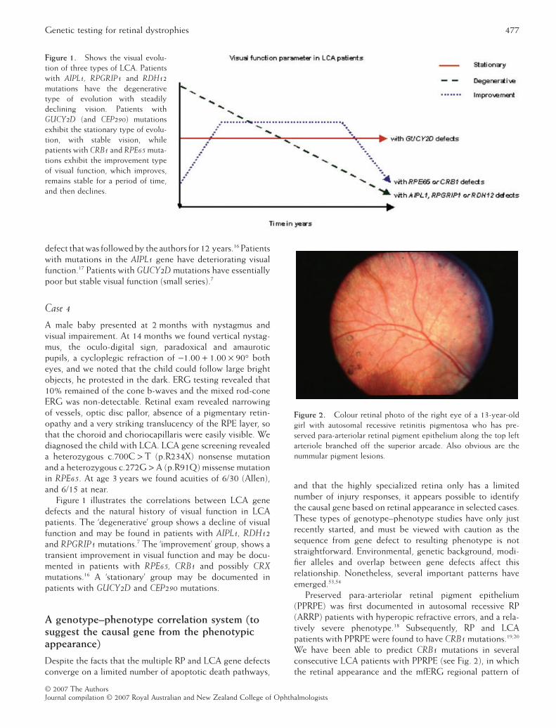
Figure 1 illustrates the correlations between LCA genedefects and the natural history of visual function in LCApatients. The ‘degenerative’ group shows a decline of visualfunction and may be found in patients with AIPL1, RDH12and RPGRIP1 mutations.7 The ‘improvement’ group, shows atransient improvement in visual function and may be docu-mented in patients with RPE65, CRB1 and possibly CRXmutations.16 A ‘stationary’ group may be documented inpatients with GUCY2D and CEP290 mutations.
A genotype–phenotype correlation system (tosuggest the causal gene from the phenotypicappearance)
Despite the facts that the multiple RP and LCA gene defectsconverge on a limited number of apoptotic death pathways,
and that the highly specialized retina only has a limitednumber of injury responses, it appears possible to identifythe causal gene based on retinal appearance in selected cases.These types of genotype–phenotype studies have only justrecently started, and must be viewed with caution as thesequence from gene defect to resulting phenotype is notstraightforward. Environmental, genetic background, modi-fier alleles and overlap between gene defects affect thisrelationship. Nonetheless, several important patterns haveemerged.53,54
Preserved para-arteriolar retinal pigment epithelium(PPRPE) was first documented in autosomal recessive RP(ARRP) patients with hyperopic refractive errors, and a rela-tively severe phenotype.18 Subsequently, RP and LCApatients with PPRPE were found to have CRB1 mutations.19,20
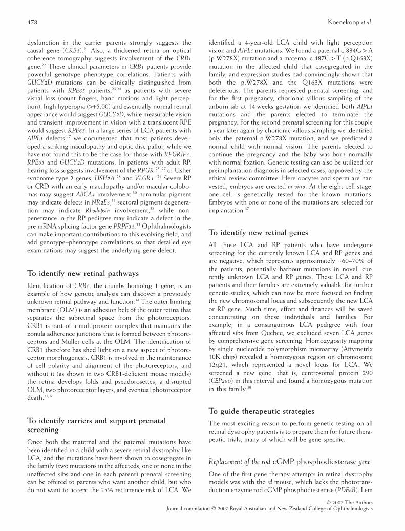
We have been able to predict CRB1 mutations in severalconsecutive LCA patients with PPRPE (see Fig. 2), in whichthe retinal appearance and the mfERG regional pattern of
Figure 1. Shows the visual evolu-tion of three types of LCA. Patientswith AIPL1, RPGRIP1 and RDH12mutations have the degenerativetype of evolution with steadilydeclining vision. Patients withGUCY2D (and CEP290) mutationsexhibit the stationary type of evolu-tion, with stable vision, whilepatients with CRB1 and RPE65 muta-tions exhibit the improvement typeof visual function, which improves,remains stable for a period of time,and then declines.
Figure 2. Colour retinal photo of the right eye of a 13-year-oldgirl with autosomal recessive retinitis pigmentosa who has pre-served para-arteriolar retinal pigment epithelium along the top leftarteriole branched off the superior arcade. Also obvious are thenummular pigment lesions.
Genetic testing for retinal dystrophies 477
© 2007 The AuthorsJournal compilation © 2007 Royal Australian and New Zealand College of Ophthalmologists
dysfunction in the carrier parents strongly suggests thecausal gene (CRB1).21 Also, a thickened retina on opticalcoherence tomography suggests involvement of the CRB1gene.22 These clinical parameters in CRB1 patients providepowerful genotype–phenotype correlations. Patients withGUCY2D mutations can be clinically distinguished frompatients with RPE65 patients,23,24 as patients with severevisual loss (count fingers, hand motions and light percep-tion), high hyperopia (>+5.00) and essentially normal retinalappearance would suggest GUCY2D, while measurable visionand transient improvement in vision with a translucent RPEwould suggest RPE65. In a large series of LCA patients withAIPL1 defects,17 we documented that most patients devel-oped a striking maculopathy and optic disc pallor, while wehave not found this to be the case for those with RPGRIP1,RPE65 and GUCY2D mutations. In patients with adult RP,hearing loss suggests involvement of the RPGR 25–27 or Ushersyndrome type 2 genes, USH2A 28 and VLGR1. 29 Severe RPor CRD with an early maculopathy and/or macular colobo-mas may suggest ABCA4 involvement,30 nummular pigmentmay indicate defects in NR2E3,31 sectoral pigment degenera-tion may indicate Rhodopsin involvement,32 while non-penetrance in the RP pedigree may indicate a defect in thepre mRNA splicing factor gene PRPF31.33 Ophthalmologistscan make important contributions to this evolving field, andadd genotype–phenotype correlations so that detailed eyeexaminations may suggest the underlying gene defect.
To identify new retinal pathways
Identification of CRB1, the crumbs homolog 1 gene, is anexample of how genetic analysis can discover a previouslyunknown retinal pathway and function.34 The outer limitingmembrane (OLM) is an adhesion belt of the outer retina thatseparates the subretinal space from the photoreceptors.CRB1 is part of a multiprotein complex that maintains thezonula adherence junctions that is formed between photore-ceptors and Müller cells at the OLM. The identification ofCRB1 therefore has shed light on a new aspect of photore-ceptor morphogenesis. CRB1 is involved in the maintenanceof cell polarity and alignment of the photoreceptors, andwithout it (as shown in two CRB1-deficient mouse models)the retina develops folds and pseudorosettes, a disruptedOLM, two photoreceptor layers, and eventual photoreceptordeath.35,36
To identify carriers and support prenatalscreening
Once both the maternal and the paternal mutations havebeen identified in a child with a severe retinal dystrophy likeLCA, and the mutations have been shown to cosegregate inthe family (two mutations in the affecteds, one or none in theunaffected sibs and one in each parent) prenatal screeningcan be offered to parents who want another child, but whodo not want to accept the 25% recurrence risk of LCA. We
identified a 4-year-old LCA child with light perceptionvision and AIPL1 mutations. We found a paternal c.834G > A(p.W278X) mutation and a maternal c.487C > T (p.Q163X)mutation in the affected child that cosegregated in thefamily, and expression studies had convincingly shown thatboth the p.W278X and the Q163X mutations weredeleterious. The parents requested prenatal screening, andfor the first pregnancy, chorionic villous sampling of theunborn sib at 14 weeks gestation we identified both AIPL1mutations and the parents elected to terminate thepregnancy. For the second prenatal screening for this couplea year later again by chorionic villous sampling we identifiedonly the paternal p.W278X mutation, and we predicted anormal child with normal vision. The parents elected tocontinue the pregnancy and the baby was born normallywith normal fixation. Genetic testing can also be utilized forpreimplantation diagnosis in selected cases, approved by theethical review committee. Here oocytes and sperm are har-vested, embryos are created in vitro. At the eight cell stage,one cell is genetically tested for the known mutations.Embryos with one or none of the mutations are selected forimplantation.37
To identify new retinal genes
All those LCA and RP patients who have undergonescreening for the currently known LCA and RP genes andare negative, which represents approximately ~60–70% ofthe patients, potentially harbour mutations in novel, cur-rently unknown LCA and RP genes. These LCA and RPpatients and their families are extremely valuable for furthergenetic studies, which can now be more focused on findingthe new chromosomal locus and subsequently the new LCAor RP gene. Much time, effort and finances will be savedconcentrating on these individuals and families. Forexample, in a consanguinous LCA pedigree with fouraffected sibs from Quebec, we excluded seven LCA genesby comprehensive gene screening. Homozygosity mappingby single nucleotide polymorphism microarray (Affymetrix10K chip) revealed a homozygous region on chromosome12q21, which represented a novel locus for LCA. Wescreened a new gene, that is, centrosomal protein 290(CEP290) in this interval and found a homozygous mutationin this family.38
To guide therapeutic strategies
The most exciting reason to perform genetic testing on allretinal dystrophy patients is to prepare them for future thera-peutic trials, many of which will be gene-specific.
Replacement of the rod cGMP phosphodiesterase gene
One of the first gene therapy attempts in retinal dystrophymodels was with the rd mouse, which lacks the phototrans-duction enzyme rod cGMP phosphodiesterase (PDE6B). Lem
478 Koenekoop et al.
© 2007 The AuthorsJournal compilation © 2007 Royal Australian and New Zealand College of Ophthalmologists
et al.39 showed that it was possible to rescue the photorecep-tors by a transgenic introduction of the normal gene bysubretinal injection in 1-day-old mouse embryos. It is impor-tant to know that at the day 1 stage, the photoreceptors arestill dividing and have not yet undergone completedegeneration.
Replacement of the Peripherin/RDS gene before theretinal degeneration is established
Travis et al.40 then showed similar rescue results in the rdsmouse, which lacks the structural photoreceptor proteinperipherin/RDS. Both genes (bPDE and RDS) werere-introduced into the mouse models at a time when thedegenerative disease process had not yet fully taken place,and when cell division was still active. The question emergedwhether it would be possible to replace genes later in thedisease process, when outer segments have been lost owingto retinal degeneration.
Replacement of the Peripherin/RDS gene after the retinaldegeneration is well established
Ali et al.41 performed subretinal injections of recombinantadeno-associated virus (AAV) containing the normalperipherin/RDS gene in the rds mice that had an establishedretinal degeneration. Post-injection immunostaining revealednormal peripherin protein and rhodopsin localization to theouter segments of the photoreceptors, indicating successfulexpression of the introduced gene in the correct layer in theretina. Photoreceptor outer segments and the characteristicstacked discs were very similar to the wild type mice and veryunlike the diseased rds mice. Functional studies showed sig-nificantly improved b-waves in the treated animals.
Replacement of a RPE gene that encodes a retinoid cycleenzyme (RPE65)
To determine whether gene replacement would work in alarger animal model, Acland et al.3 studied the effects ofRPE65 replacement in the Briard dog. This dog model har-bours a naturally occurring, homozygous 4-bp deletion inthe RPE65 gene (which predicts a lack of functional RPE65protein in the RPE) and is essentially blind at birth. ERGfunction is non-detectable, despite the normal appearance ofthe retinas, including the essentially normal histologicalappearance of the photoreceptor layer in this dog model.Subretinal injections in one eye of three dogs containing theAAV virus with cDNA of dog RPE65, with a CMV promotor;b-actin enhancer and internal ribosome entry sequence wereperformed at age 4 months. Rod and cone mediated ERGs,visual evoked potentials, pupillometry and behaviouraltesting all showed dramatic improvements in visual functionat about 8 months of age. Wild type RPE65 was found in theretina and in RPE cells. These experiments were repeatedin other laboratories42 and found to be accurate, while
follow-up visual function testing revealed stable function forat least 3 years, after one subretinal injection of the gene.
Photoreceptor gene RPGRIP1 replacement rescues blindmice with established retinal degeneration
Three important aspects of the successful RPE65 genereplacement may have biased a favourable outcome; (i)RPE65 is a gene expressed in the RPE layer and in conephotoreceptors, (ii) RPE65 encodes an enzyme (not a struc-tural protein), and (iii) the retinal architecture of the RPE65dog model was not severely degenerated. The question nowis; does rescue work for photoreceptor genes that encode struc-tural proteins, when the degenerative process is well established?Pawlyk et al.43 using the well-known RPGRIP1 knockoutmouse answered this question. RPGRIP1 is expressed in theciliary axoneme that connects the inner to the outer photo-receptor segment and RPGRIP1 mutations cause abnormali-ties in protein trafficking from the inner segment to the outersegment (especially rhodopsin), aberrant disc morphogen-esis of the outer segment discs and a rapid early degenerationof the entire retina. Five months post injection of RPGRIP1cDNA by an AAV vector, they showed that RPGRIP1 (andits partner RPGR) correctly colocalized to the connectingcilia and documented preservation of photoreceptor nucleiin the outer nuclear layer. ERGs showed a marked improve-ment of the rod photoreceptor b-wave amplitudes and therates of ERG decline were also significantly smaller in thetreated eyes.
Replacement of a retinal gene that encodes a protein thatfunctions as glue between the inner and outer retina
Can retinal gene therapy rescue other cell types in the retina,when the gene product is an extra-cellular molecule involvedin keeping the retina together as natural glue? Retinoschisin(RS) gene replacement rescues the RS null mice as shown byMin et al.44 and Zeng et al.45 Viral constructs containing thenormal human RS cDNA, injected subretinally in the RSdeficient mouse led to RS expression in retinal extracts and ledto marked improvements in both the rod-mediated and cone-mediated signals (including the reversal of the electro-negative ERG, which is the hallmark of the human and mousedisease phenotype) all of which indicates a rescue effect onthe retinal architecture.44 The effects on the cone ERG, espe-cially the flicker ERG were the most dramatic. Also apparentwere the retinal changes seen by scanning laser ophthalmos-copy (SLO), as the untreated eyes revealed the characteristiccystic lesions, while the transfected retinas were devoid ofthese pathological structures and resembled wild type retinas.Photoreceptor inner and outer nuclear layers, outer plexiformlayer and bipolar cells of the inner nuclear layer in addition tothe inner plexiform layers all revealed RS staining, whichillustrates the important concept that retinal gene replace-ment injected subretinally may lead to protein localizationlongitudinally and laterally into the retina.
Genetic testing for retinal dystrophies 479
© 2007 The AuthorsJournal compilation © 2007 Royal Australian and New Zealand College of Ophthalmologists
DILEMMAS OF GENETIC TESTING
When a patient with a retinal degeneration presents to anOphthalmologist and requests molecular testing for his/hercondition, theoretically a maximum of 132 retinal degenera-tion genes would need to be screened.2 If a patient is foundto have RP, 37 RP genes would need to be screened. Com-prehensive genetic testing of RP patients is a problematic andchallenging endeavour because it is extremely time-consuming, expensive and labour intensive, as RP patientswith unknown inheritance would need to be screened foralmost 500 exons, which represents almost 120 000 basepairs. There are also other problems: the current technologyis not 100% sensitive, and the screening results will be full offalse positive results (polymorphic variants). Once asequence variant is identified, it must be determined whetherit is disease causing or may be a polymorphism. Short ofperforming functional assays on mutations in in vitro cellculture, mutations can be compared with polymorphisms forthe following five attributes which improves the probabilitythat the mutation causes a defect in the resulting protein: (i)the predicted effect of the base pair change on the RNAand/or protein product, (ii) the relative frequency of thevariation in LCA patients versus normal ethnically matchedcontrols (>1% in the controls will be assigned as a polymor-phism), (iii) cosegregation of the mutant allele(s) in theaffected members of the families, (iv) homozygosity orcompound heterozygosity in recessive disease, and (v) con-servation of the mutant codon across other species. Severalweb-based programs exist that are helpful in distinguishingbenign from deleterious effects (polyphen, SIFT, Blossum).Therefore, comprehensive RP screening is currently notavailable in the world in a single laboratory.
The endeavour is slightly better for RP patients withknown inheritance patterns. RP patients with a clear X-linkedinheritance pattern (XLRP) would only need to be screenedfor two genes (RPGR and RP2), which are divided into 24exons and include almost 7000 base pairs. RPGR mutationsmay account for 70–90% of all XLRP patients, and may bethe most common RP gene, overall. For ARRP patients,which represents the most common RP inheritance subtype,20 RP genes are required for screening, divided over 337exons, which include almost 75 000 base pairs. Finally, if theRP patient has clear dominant inheritance, 15 genes need tobe screened, divided over 135 exons, including almost38 000 base pairs.
In addition to this insurmountable task, conventionalmutation screening methods, such as SSCP and dHPLC,can miss mutations, and are not 100% sensitive. Automatedsequencing of almost 500 exons for one retinal dystrophypatient would cost about $10 000.00 (CDN) and evaluationof all sequence data and repeat analysis may add another$2–3000. Another problem would be the enormous numberof DNA variations that would be found which havenothing to do with the disease. Each variation would haveto be tested in the family to verify whether it cosegregateswith the disease phenotype. Deciding which of the varia-
tions in which gene is the actual mutation responsible forthe disease would be extremely difficult, especially in themost common type of RP which is sporadic (also calledisolated RP, in which there is only one affected patient).Fortunately, there are several possible solutions to thisproblem, some of these solutions are being actively testedand evaluated.
POSSIBLE SOLUTIONS FOR GENETIC TESTING
We will discuss three possible solutions: (i) utilize the retinalphenotype (including retinal aspect and natural history ofvision) to suggest the genetic defect; (ii) start with the mostfrequently mutated gene or with the most frequent muta-tion(s); and (iii) utilize new high throughput microarraytechnology.
Solution 1: Utilize genotype–phenotypecorrelations to suggest the causal gene
Ideally, an ophthalmologist could predict the causal gene ina RP patient by documenting the clinical phenotype,including age of onset, rate of progression, optical status,retinal appearance and associated features, to name a few.This is not possible in all cases, because of many reasons,including the fact that there is no straightforward anddirect relation between the gene and the phenotype, theaction of interfering factors from the environment andgenetic background, the influence of modifier alleles andthe fact that RP genes may have similar functions in theretina or participate in similar retinal cycles. Also, this areaof investigation is in its infancy, and there are more genesknown than robust genotype–phenotype correlations.Despite these shortcomings, there are several goodexamples of retinal dystrophy genotype–phenotype corre-lations (see Possible solutions for genetic testing and Table 1). Thisexciting area of investigation is also in its infancy and needsto be expanded and tested.
Solution 2: Focus on the most frequentmutation(s) and/or mutated gene(s) first
Another solution to screening may be to start with the mostcommonly mutated RP genes, that is, RPGR screening mayprovide a molecular diagnosis in ~10% of all RP patients,70–90% of patients with XLRP, and up to 25% of males withsporadic RP. Another common gene mutated in 15–20% ofall ADRP is the rhodopsin gene, especially the Pro23Hismutation, which is common in North America but notelsewhere. USH2A screening is recommended for patientswith Usher (USH) syndrome as 40–50% have mutations inUSH2A, but also for patients with ARRP, as up to 14% mayharbour mutations in USH2A. For LCA, it appears that themost frequently mutated gene is CEP290. In more than 20%of new LCA patients, a single mutation p.C998X can befound. With an inexpensive and simple blood test this muta-tion can be found, establishing the genotype.38
480 Koenekoop et al.
© 2007 The AuthorsJournal compilation © 2007 Royal Australian and New Zealand College of Ophthalmologists
Solution 3: High throughput mutationmicroarrays
Disease chips (asper ophthalmics)
The first microarray (‘disease chip’) was designed for Star-gardt macular dystrophy (STGD1) and contains all currentlyknown disease-associated alleles of the ABCA4 gene. Otherretinal diseases may be caused by or are associated withABCA4 mutations, namely fundus flavimaculatus, CRD, RPand age-related macular degeneration, and can be screenedon this chip as well. Other disease chips are now available forLCA, Usher syndrome ARRP, Bardet Biedl Syndrome, ADRP,XLRP and age-related macular degeneration (see http://www.asperbio.com). The screening by the disease chips is rapid(~4 h per patient), reliable and affordable (~150–200 USD),and can be updated, as new genes and new mutationsbecome available.
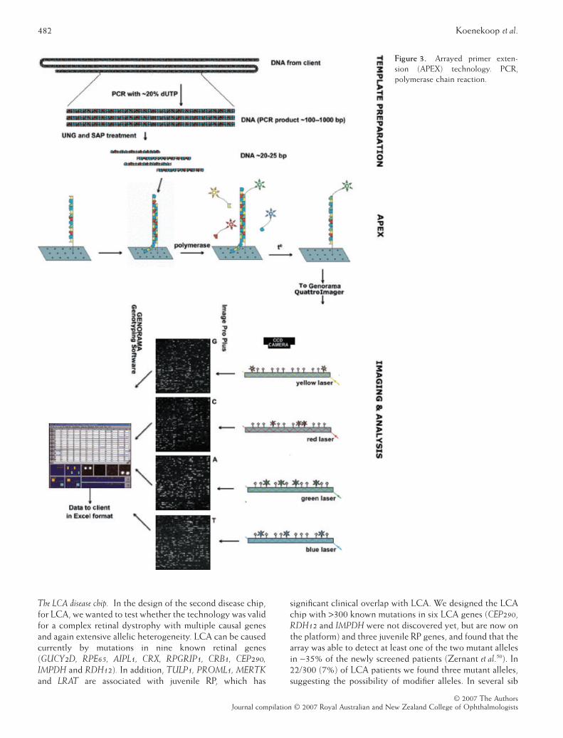
Microarrays are designed and manufactured with thearrayed primer extension (APEX) method, also known assolid-phase minisequencing and details can be found in Kurget al.46 and Tonisson et al.47 There are basically six steps (seeFig. 3): in Step 1 Oligonucleotides (oligos) are designed foreach known mutation, with the 5′ end immobilized on theglass slide and the 3′ end immediately adjacent to the vari-able (queried) site. Step 2 is polymerase chain reaction(PCR) amplification of each DNA segment (which harboursthe known mutations) of the new (to be tested) patient�sgenome. In Step 3, all the patient�s PCR fragments are spreadover the slide containing the oligos and annealing is allowedto take place, between the patient�s DNA fragments and theimmobilized oligos. In Step 4, DNA polymerase is added,
with dye labelled dideoxynucleotides (C, T, G and A) so thata sequence specific extension of one nucleotide only takesplace at the 3′ end of the oligo, using the patient DNA as atemplate. In Step 5, the patient�s DNA not annealed to ahomologous oligo is washed off, to reduce noise, and in Step6 signal detection takes place by laser and computer. Advan-tages are the low cost (~150–200 USD), rapid results (4 hper sample), ability to update the chip and the ability toenter only known pathogenic mutations to the platform. Adisadvantage is that APEX technology only detects theknown mutations, although a new mutation in a nucleotidealready included on the chip will be detected. In patientswith autosomal recessive diseases, sequence analysis may berequired to identify a second allele once APEX technologyhad detected the first.
The ABCA4 disease chip. The first disease chip designed was theABCR400 chip specifically for the ABCA4 gene, which has 50exons. Unlike, for example, cystic fibrosis, which is alsocaused by a member of the ABC super family and in whichthe p.F508del is a common mutation found on ~70% ofcystic fibrosis alleles, the commonest STGD1 mutation isonly found in ~10% of patients. The array now contains~500 ABCA4 mutations, and was >98% effective in detectinga patient�s variations. Jaakson et al.48 showed that the diseasechip was 54–78% effective in detecting at least one of thetwo STGD1 patients mutations, depending on the geo-graphical cohorts. In a second study, Klevering et al.49
showed that the ABCA4 chip is also effective in 33% ofARCRD patients, and 6% effective in severe ARRP patientsin locating at least one of the two mutant alleles.
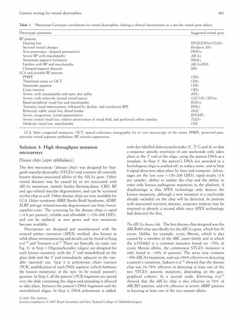
Table 1. Phenotype-Genotype correlations for retinal dystrophies, linking a clinical characteristic to a specific retinal gene defect.
Phenotypic parameter Suggested retinal gene
RP patientsHearing loss RPGR/USH2A/VLGR1Sectoral retinal changes Rhodopsin, RP1Non-penetrance (skipped generations) PRPF31Severe RP with maculopathy ABCA4Nummular pigment formation NR2E3Families with RP and maculopathy ABCA4/RDSClumped pigment deposits NRL
LCA and juvenile RP patientsPPRPE CRB1Thickened retina on OCT CRB1Nummular pigment CRB1Coats reaction CRB1Severe, with maculopathy and optic disc pallor AIPL1Severe, with relatively normal retinal aspect GUCY2D, CEP290Rapid peripheral visual loss and maculopathy RDH12Transient visual improvement, followed by decline, and translucent RPE RPE65Relatively stable visual loss, blond fundus LRATSevere, progressive, retinal pigmentation RPGRIP1Severe central visual loss, relative preservation of visual field, and perifoveal yellow annulus TULP1Moderate visual loss, maculopathy CRX
LCA, leber congenital amaurosis; OCT, optical coherence tomography for in vivo microscopy of the retina; PPRPE, preserved para-arteriolar retinal pigment epithelium; RP, retinitis pigmentosa.
Genetic testing for retinal dystrophies 481
© 2007 The AuthorsJournal compilation © 2007 Royal Australian and New Zealand College of Ophthalmologists
The LCA disease chip. In the design of the second disease chip,for LCA, we wanted to test whether the technology was validfor a complex retinal dystrophy with multiple causal genesand again extensive allelic heterogeneity. LCA can be causedcurrently by mutations in nine known retinal genes(GUCY2D, RPE65, AIPL1, CRX, RPGRIP1, CRB1, CEP290,IMPDH and RDH12). In addition, TULP1, PROML1, MERTKand LRAT are associated with juvenile RP, which has
significant clinical overlap with LCA. We designed the LCAchip with >300 known mutations in six LCA genes (CEP290,RDH12 and IMPDH were not discovered yet, but are now onthe platform) and three juvenile RP genes, and found that thearray was able to detect at least one of the two mutant allelesin ~35% of the newly screened patients (Zernant et al.50). In22/300 (7%) of LCA patients we found three mutant alleles,suggesting the possibility of modifier alleles. In several sib
Figure 3. Arrayed primer exten-sion (APEX) technology. PCR,polymerase chain reaction.
482 Koenekoop et al.
© 2007 The AuthorsJournal compilation © 2007 Royal Australian and New Zealand College of Ophthalmologists
pairs we were able to demonstrate altered phenotypes in thesib with three alleles versus those with two alleles. Severalrecent studies have confirmed the utility and efficiency of thenew LCA chip. Pathogenic mutations were identified in 19out of 58 LCA patients for an efficiency rate of 33% by Yzeret al.51 These results are encouraging for at least two reasons.In comprehensive screening studies (by sequencing all theexons of all the LCA genes) an 47.5% efficiency is reached(Hanein et al.24). While the LCA disease chip will identifymostly the known mutations entered onto the platform, a33–35% detection rate is surprising and will increase propor-tionately to the addition of new mutations added to the chip.Recently, with the addition of several new LCA genes andmutations, the success rate of the LCA chip has increased to~60–70%. As the chip is fast, relatively cheap and accurate,we propose to use the LCA chip as a first pass screening toolfor all patients with LCA and juvenile RP.
The usher disease chip. Three clinical Usher syndrome subtypeshave been described (USH1-3) and eight USH genes havecurrently been reported (USH1 genes: CDH23, MYO7A,PCDH15, Harmonin and SANS; USH2 genes: USH2A andVLGR1; and USH3 gene: USH3A). Identification of thecausal Usher mutations is important for all the above reasons,but also to identify the patient with congenital hearing loss,in order to implant cochlear devices as early as possible. As isthe case for RP and allied retinal degenerations, testing forUsher disease is also hampered by genetic heterogeneity(eight genes), many exons (179 coding units) and the greatnumber of known mutations. In a recent study, Cremerset al.52 designed a new Usher disease chip containing thecurrently known 298 Usher mutations (from the eight cur-rently known Usher genes), and the efficiency of the arraywas tested using DNA from 370 untested Usher patients.Mutations were found in 45% of USH1, 23% of USH2patients and 28% of the USH3 patients, which represents avery encouraging detection rate, considering that not allUsher genes nor all mutations have been identified.
Conclusions. Now more than ever, the genotyping of retinaldystrophy patients is a crucial exercise, as human gene-specific clinical trials to study rescue of photoreceptors are ontheir way. Genetic testing confirms the diagnosis at themolecular level and allows for a more precise prognosis of thepossible future clinical evolution of the disease based on thegene defect. Also, as human treatments are gene specific andthe treatment ‘window of opportunity’ is likely time sensitive;accurate, rapid and cost-effective genetic testing will play anever increasing crucial role. Currently there is no single‘perfect’ technology to quickly and affordably determine thegenotype in a particular retinal dystrophy patient. The goldstandard technique of automated sequencing is fraught withexcessive costs, time and manpower issues and finding non-pathogenic variants (which are not a problem if strict criteriaare used with regard to their pathogenicity, they do however,add much time and work to the genotyping effort). Therefore,no current RP laboratory or centre offers testing of all known
RP genes (37 genes and counting). In the past few yearsseveral new and exciting (micro array) technologies haveemerged that offer the possibility to genotype retinal dystro-phy patients rapidly, cost-effectively and accurately. Each ofthese new technologies has advantages and disadvantages andeach one needs to be tested in large genotyping efforts. Wesuggest that the new disease chips from Asper Ophthalmicsoffer an excellent first pass opportunity. This may be com-bined with genotype–phenotype correlations that suggest thecausal gene from the clinical appearance (e.g. PPRPE suggeststhe involvement of the CRB1 gene in LCA and juvenile RP)and regular sequencing of the suggestive gene. Also startingwith the most frequently mutated genes (RPGR, Rhodopsin,USH2A) may be prudent.
ACKNOWLEDGEMENTS
The authors would like to sincerely thank the FoundationFighting Blindness (Canada), Retina International and theFRSQ (Canada) for their support. We would also like tothank Ms Renee Pigeon and Ms Claudine Robert from TheMontreal Children�s Hospital for their help in the prepara-tion of the manuscript.
REFERENCES
1. Black GCM, Donnai D. Genetic testing-swings and round-abouts: a view from the United Kingdom. Br J Ophthalmol 2001;85: 1402–4.
2. Daiger S. RetNet. http://www.sph.uth.tmc.edu/retnet/disease.html.
3. Acland GM, Aguirre GD, Ray J et al. Gene therapy restoresvision in a canine model of childhood blindness. Nat Genet 2001;28: 92–5.
4. Van Hooser JP, Aleman TS, He YG et al. Rapid restoration ofvisual pigment and function with oral retinoid in a mousemodel of childhood blindness. Proc Natl Acad Sci USA 2000; 97:8623–8.
5. Sieving PA, Caruso RC, Tao W et al. Ciliary neurotrophic factor(CNTF) for human retinal degeneration: phase I trial of CNTFdelivered by encapsulated cell intraocular implants. Proc NatlAcad Sci USA 2006; 103: 3896–901.
6. Weiss A, Biersdorf W. Visual sensory disorders in congenitalnystagmus. Ophthalmology 1989; 96: 517–23.
7. Koenekoop RK. An overview of recent developments in Lebercongenital amaurosis: a model to understand human retinaldevelopment. Surv Ophthalmol 2004; 49: 379–98. Review.
8. Cremers FPM, van den Hurk JA, den Hollander AI. Moleculargenetics of Leber congenital amaurosis. Hum Mol Genet 2002; 11:1169–76. Review.
9. Lambert SR, Kriss A, Taylor D et al. Follow-up and diagnosticreappraisal of 75 patients with Leber�s congenital amaurosis.Am J Ophthalmol 1989; 107: 624–31.
10. Weleber RG, Tongue AC. Congenital stationary night blind-ness presenting as Leber�s congenital amaurosis. Arch Ophthalmol1987; 105: 360–5.
11. Fulton AB, Hansen RM, Westall CA. Development of ERGresponses: the ISCEV rod, maximal, and cone responses innormal subjects. Doc Ophthalmol 2003; 107: 235–41.
Genetic testing for retinal dystrophies 483
© 2007 The AuthorsJournal compilation © 2007 Royal Australian and New Zealand College of Ophthalmologists
12. Heher KL, Traboulsi EI, Maumenee IH. The natural history ofLeber�s Congenital Amaurosis. Ophthalmology 1992; 99: 241–5.
13. Fulton AB, Hansen RM, Mayer DL. Vision in Leber CongenitalAmaurosis. Arch Ophthalmol 1996; 114: 698–703.
14. Brecelj J, Stirn-Kranjc B. ERG and VEP follow up study inchildren with Leber Congenital Amaurosis. Eye 1999; 13: 47–54.
15. Lorenz B, Gyurus P, Preising M et al. Early-onset severe rodcone dystrophy in young children with RPE 65 mutations. InvestOphthalmol Vis Sci 2000; 41: 2735–42.
16. Koenekoop RK, Loyer M, Dembinska O, Beneish R. Improve-ment in visual function in Leber congenital amaurosis and theCRX genotype. Ophthalm Genetics 2002; 23: 49–59.
17. Dharmaraj S, Leroy B, Sohocki M et al. The phenotype of Lebercongenital amaurosis in patients with AIPL1 mutations. ArchOphthalmol 2004; 122: 1029–37.
18. Heckenlively J. Preserved para-arteriole retinal pigment epithe-lium (PPRPE) in retinitis pigmentosa. Br J Ophthalmol 1982; 66:26–30.
19. den Hollander AI, Heckenlively JR, van den Born IL et al. Lebercongenital amaurosis and retinitis pigmentosa with Coats-likeexudative vasculopathy are associated with mutations in thecrumbs homologue 1 (CRB1) gene. Am J Hum Genet 2001; 69:198–203. Erratum in: Am J Hum Genet 2001; 69 : 1160.
20. Lotery A, Jacobson SG, Fishman GA et al. Mutations in theCRB1 gene cause Leber congenital amaurosis. Arch Ophthalmol2001; 119: 415–20.
21. Yzer S, Fishman GA, Racine J et al. CRB1 heterozygotes withregional retinal dysfunction: implications for genetic testing ofLeber congenital amaurosis. Invest Ophthalmol Vis Sci 2006; 47:3736–44.
22. Jacobson S, Cideciyan A, Aleman PS et al. Crumbs homolog 1(CRB1) mutations result in a thick human retina with abnormallamination. Hum Mol Genet 2003; 12: 1073–8.
23. Perrault I, Rozet JM, Ghazi I et al. Different functional outcomeof RetGC1 and RPE65 gene mutations in Leber congenitalamaurosis. Am J Hum Genet 1999; 64: 1225–8.
24. Hanein S, Perrault I, Gerber S et al. Leber congenital amaurosis:comprehensive survey of the genetic heterogeneity, refinementof the clinical definition, and genotype-phenotype correlationsas a strategy for molecular diagnosis. Hum Mutat 2004; 23:306–17.
25. Zito I, Downs SM, Patel RJ et al. RPGR mutation associatedwith retinitis pigmentosa, impaired hearing, and sinorespira-tory infections. J Med Genet 2003; 40: 609–15.
26. Koenekoop RK, Loyer M, Hand C et al. Novel RPGR mutationswith distinct retinitis pigmentosa phenotypes in French-Canadian families.Am J Ophthalmol 2003; 136: 678–87.
27. Iannaconne A, Breuer DK, Wang XF et al. Clinical and immuno-histochemical evidence for an X linked retinitis pigmentosasyndrome with recurrent infections and hearing loss in associa-tion with an RPGR mutation. J Med Genet 2003; 40: 118.
28. Rivolta C, Sweklo EA, Berson EL, Dryja TP. Missense mutationin the USH2A gene: association with recessive retinitis pig-mentosa without hearing loss. Am J Hum Genet 2000; 66:1975–8.
29. Weston MD, Luijendijk MW, Humphrey KD, Moller C,Kimberling WJ. Mutations in the VLGR1 gene implicateG-protein signaling in the pathogenesis of Usher syndrometype II. Am J Hum Genet 2004; 74: 357–66.
30. Cremers FPM, van de Pol DJ, van Driel M et al. Autosomalrecessive retinitis pigmentosa and cone-rod dystrophy causedby splice site mutations in the Stargardt�s disease gene ABCR.Hum Mol Genet 1998; 7: 355–62.
31. Sharon D, Sandberg MA, Caruso RC, Berson EL, Dryja TP.Shared mutations in NR2E3 in enhanced S–cone syndrome,Goldmann–Favre syndrome, and many cases of clumped pig-mentary retinal degeneration. Arch Ophthalmol 2003; 121:1316–23.
32. Heckenlively JP, Rodriguez JA, Daiger SP. Autosomal domi-nant sectoral retinitis pigmentosa. Two families with transver-sion mutation in codon 23 of rhodopsin. Arch Ophthalmol 1991;109: 84–91.
33. Vithana EN, Abu-Safieh L, Allen MJ et al. A human homolog ofyeast pre-mRNA splicing gene, PRPF31, underlies autosomaldominant retinitis pigmentosa on chromosome 19q13.4(RP11). Mol Cell 2001; 8: 375–81.
34. den Hollander AI, ten Brink JB, de Kok YJM, van Soest S.Mutations in a human homologue of Drosophila crumbs causeretinitis pigmentosa (RP12). Nat Genet 1999; 23: 217–21.
35. Mehalow AK, Kameya S, Smith RS et al. CRB1 is essential forexternal limiting membrane integrity and photoreceptor mor-phogenesis in the mammalian retina. Hum Mol Genet 2003; 12:2179–89.
36. van de Pavert SA, Kantardzhieva A, Malysheva A et al. Crumbshomologue 1 is required for maintenance of photoreceptor cellpolarization and adhesion during light exposure. J Cell Sci 2004;117: 4169–77.
37. Van Voorhis BJ. In vitro fertilization. N Engl J Med 2007; 356:379–86.
38. den Hollander AI, Koenekoop RK, Yzer S et al. Mutations inthe CEP290 (NPHP6) gene are a frequent cause of Leber con-genital amaurosis.Am J Hum Genet 2006; 79: 556–61.
39. Lem J, Flannery JG, Li T et al. Retinal degeneration is rescued intransgenic rd mice by expression of the cGMP phosphodi-esterase beta subunit. Proc Natl Acad Sci USA 1992; 89: 4422–6.
40. Travis G, Groshan KR, Lloyd M, Bok D. Complete rescue ofphotoreceptor dysplasia and degeneration in transgenic retinaldegeneration slow (rds) mice. Neuron. 1992; 9: 113–9.
41. Ali RR, Sarra GM, Stevens C et al. Restoration of photoreceptorultrastructure and function in retinal degeneration slow mice bygene therapy.Nat Genet 2000; 25: 306–10.
42. Narfstrom K, Katz ML, Ford M et al. In vivo gene therapy inyoung and adult RPE65-/- dogs produces long-term visualimprovement. J Hered 2003; 94: 31–7.
43. Pawlyk BS, Smith AJ, Buch PK et al. Gene replacement therapyrescues photoreceptor degeneration in a murine model of Lebercongenital amaurosis lacking RPGRIP. Invest Ophthalmol Vis Sci2005; 46: 3039–45.
44. Min SH, Molday LL, Seeliger MW et al. Prolonged recovery ofretinal structure/function after gene therapy in an Rs1h-deficient mouse model of x-linked juvenile retinoschisis. MolTher 2005; 12: 644–51.
45. Zeng Y, Takada Y, Kjellstrom S et al. RS-1 Gene delivery to anadult Rs1h knockout mouse model restores ERG b-wave withreversal of the electronegative waveform of X-linkedretinoschisis. Invest Ophthalmol Vis Sci 2004; 45: 3279–85.
46. Kurg A, Tonisson N, Georgiou I et al. Arrayed primer extension:solid-phase four-color DNA resequencing and mutation detec-tion technology. Genet Test 2000; 4: 1–7.
484 Koenekoop et al.
© 2007 The AuthorsJournal compilation © 2007 Royal Australian and New Zealand College of Ophthalmologists

47. Tonisson N, Kurg A, Lohmussaar E, Metspalu A. Arrayedprimer extension on the DNA chip—method and applications.In: Schena M, ed. Microarray Biochip Technology 5th edn. Natick,MA: Eaton Publishing, 2000; 247–63.
48. Jaakson K, Zernant J, Kulm M et al. Genotyping microarray(gene chip) for the ABCR (ABCA4) gene. Hum Mutat 2003; 22:395–403.
49. Klevering BJ, Yzer S, Rohrschneider K et al. Microarray-basedmutation analysis of the ABCA4 (ABCR) gene in autosomalrecessive cone-rod dystrophy and retinitis pigmentosa. Eur J HumGenet 2004; 12: 1024–32.
50. Zernant JM, Külm S, Dharmaraj A et al. Genotyping microarray(disease chip) for Leber congenital amaurosis: detection ofmodifier alleles. Invest Ophthalmol Vis Sci 2005; 46: 3052–9.
51. Yzer S, Leroy BP, De Baere E et al. Microarray-based mutationdetection and phenotypic characterization of patients withLeber congenital amaurosis. Invest Ophthalmol Vis Sci 2006; 47:1167–76.
52. Cremers FPM, Kimberling WJ, Kulm M et al. Development of agenotyping microarray for usher syndrome. J Med Genet 2006;44: 153–60.
53. Koenekoop RK, Fishman GA, Iannaccone A et al. Electroretino-graphic (ERG) abnormalities in parents of Leber CongenitalAmaurosis children with known GUCY2D mutations. Arch Oph-thalmol 2002; 120: 1325–30.
54. Galvin J, Fishman G, Stone EM, Koenekoop RK. Clinical phe-notypes associated with the heterozygous carriers of variousgenotypes in leber congenital amaurosis. Ophthalmology 2005;112: 349–56.
55. Zeitz C, van Genderen M, Neidhardt J et al. Mutationsin GRM6 cause autosomal recessive congenital stationarynight blindness with a distinctive scotopic 15-Hz flickerelectroretinogram. Invest Ophthalmol Vis Sci 2005; 46: 4328–35.
56. Zeitz C, Kloeckener-Gruissem B, Forster U et al. Mutations inCABP4, the gene encoding the Ca2+-binding protein 4, causeautosomal recessive night blindness. Am J Hum Genet 2006; 79:657–7.
Genetic testing for retinal dystrophies 485
© 2007 The AuthorsJournal compilation © 2007 Royal Australian and New Zealand College of Ophthalmologists