Embed Size (px)
Citation preview
BioMed CentralGenetic Vaccines and Therapy
ss
Open AcceResearchDNA vaccine constructs against enterovirus 71 elicit immune response in miceWong Siew Tung1, Sazaly Abu Bakar2, Zamberi Sekawi3 and Rozita Rosli*1Address: 1Dept. of Human Growth and Development, Faculty of Medicine and Health Sciences, Universiti Putra Malaysia, 43400 Serdang, Selangor, Malaysia, 2Department of Medical Microbiology, Faculty of Medicine, Universiti Malaya Medical Center, 50603 Kuala Lumpur, Malaysia and 3Dept of Clinical Laboratory Sciences, Faculty of Medicine and Health Sciences, Universiti Putra Malaysia, 43400 Serdang, Selangor, Malaysia
Email: Wong Siew Tung - [email protected]; Sazaly Abu Bakar - [email protected]; Zamberi Sekawi - [email protected]; Rozita Rosli* - [email protected]
* Corresponding author
AbstractBackground: Enterovirus 71 (EV71) is a major causative viral agent responsible for largeoutbreaks of hand, foot and mouth disease (HFMD), a common rash illness in children and infants.There is no effective antiviral treatment for severe EV71 infections and no vaccine is available. Theobjectives of this study were to design and construct a DNA vaccine against Enterovirus 71 usingthe viral capsid protein (VP1) gene of EV71 and to verify the functionality of the DNA vaccine invitro and in vivo.
Methods: The VP1 gene of EV71 from two local outbreak isolates were amplified using PCR andthen inserted into a eukaryotic expression vector, pVAX1. The 3.9 kb recombinant constructswere transformed into competent E. coli cells and the positive clones were screened and selectedusing PCR analysis, restriction digestion analysis and DNA sequencing. The constructs were thentested for protein expression in Vero cells. Subsequently, in the in vivo studies, female Balb/c micewere immunized with the DNA vaccine constructs. Enzyme Linked Immunosorbent Assay (ELISA)and virus neutralizing assay were performed to detect the presence of anti-VP1 IgG in mice and itsneutralizing effect against the EV71.
Results: The pVAX1 vector was successfully cloned with the VP1 gene from each of the isolate(S2/86/1 and 410/4) in the correct orientation and in-frame. The DNA vaccine constructs with theVP1 gene were shown to be expressed in a cell-free in vitro expression system. The VP1 proteinwas successfully expressed in the mammalian cell line and was detected using RT-PCR, IndirectImmunofluorescence Assay (IFA) and western blotting. The anti-VP1 IgG levels in mice immunizedwith the DNA vaccine constructs increased after the first booster but declined following thesecond booster. The anti-VP1 IgG in the mice immunized with the DNA vaccine constructsexhibited neutralising activity against EV71.
Conclusion: The promising results obtained in the present study have prompted further testingto improve the expression and immunogenicity of this potential EV71 DNA vaccine.
Published: 19 April 2007
Genetic Vaccines and Therapy 2007, 5:6 doi:10.1186/1479-0556-5-6
Received: 5 September 2006Accepted: 19 April 2007
This article is available from: http://www.gvt-journal.com/content/5/1/6
© 2007 Tung et al; licensee BioMed Central Ltd. This is an Open Access article distributed under the terms of the Creative Commons Attribution License (http://creativecommons.org/licenses/by/2.0), which permits unrestricted use, distribution, and reproduction in any medium, provided the original work is properly cited.
Page 1 of 13(page number not for citation purposes)
Genetic Vaccines and Therapy 2007, 5:6 http://www.gvt-journal.com/content/5/1/6
1. BackgroundEnterovirus 71 (EV71) belongs to the genus of enterovi-ruses from the family Picornaviridae. It possesses a singlestranded RNA genome of approximately 7500 nucleotidesof positive polarity, which is encapsulated in a capsid con-taining 60 copies of each of the four structural proteins,VP1 through VP4 [1]. The antigenic diversity among theenteroviruses is caused by variations within capsid pro-teins VP1 to VP3, but neutralization epitopes are mostdensely clustered on VP1 [2].
Enterovirus 71, along with coxsackievirus A16 (CA16), isa major causative viral agent responsible for large out-breaks of hand, foot and mouth disease (HFMD), a com-mon rash illness in children and infants. EV71 is thoughtto spread by contact with fecal contaminated materials.Infection by the virus is often asymptomatic or may man-ifest as mild self-limiting illness which is often character-ized by the presence of characteristic lesions on the palms,soles and oral mucosa. EV71 and CA16 are geneticallyclosely related. However, unlike CA16 that is more lim-ited in its pathogenicity to HFMD, EV71 is also associatedwith severe complications involving the central nervoussystem (CNS) such as aseptic meningitis, encephalitis andpoliomyelitis-like paralysis [3,4].
Since the first report of EV71 infection which occurred inCalifornia in 1969 [5], world-wide reports of outbreakshave followed. The neurovirulence of EV71 first came toattention in 1975 in Bulgaria when 44 people died of apolio-like disease [6]. Epidemics of EV71 causing CNS dis-eases subsequently occurred in New York, Australia,Europe and Asia [7-10]. An unusual epidemic of HFMDcomplicated by fatal myocarditis and pulmonary edemaoccurred in Malaysia in 1997, and EV71 had been impli-cated as the etiology of the outbreak [11]. Thirty-one chil-dren in Sarawak, Malaysia and four children in PeninsularMalaysia succumbed to the infection within hours ofadmission to the hospitals [12]. The largest EV71 epi-demic reported to date occurred from April to Decemberof 1998 in Taiwan in which a variety of clinical manifesta-tions were observed. These included HFMD, encephalitis,meningitis, herpangina and poliomyelitits-like paralysis.In this outbreak, more than 90,000 children infected withHFMD were reported. Among these patients, more than320 children were hospitalized with suspected meningitis,encephalitis, or acute flaccid paralysis, and at least 55died, suggesting neurovirulence of the pathogen [13].
There is no effective antiviral treatment for severe EV71infections and no vaccine is available. Thus, the only cur-rent means to prevent EV71 infection is through avoid-ance of contact between infected and susceptibleindividuals [14]. Since no effective antiviral agents areavailable, the need for an effective EV71 vaccine is urgent
to immunize the public before an outbreak occurs. A for-malin-inactivated EV71 vaccine was developed inresponse to the Bulgarian epidemic in 1975 [6,15] butwas not used to control the epidemic and has not beenused since. Thus, no data on the efficacy of the Bulgarianvaccine is available. Recently, several candidates of EV71vaccines using different approaches are being investi-gated. These include formalin-inactivated whole virus vac-cine, DNA vaccine and recombinant protein vaccine.These vaccine constructs remain promising vaccine strate-gies that require further refinement, thus further study anddevelopment are required [16-18].
In this study, a DNA vaccine encoding the VP1 gene ofEV71 was designed and constructed. This vaccine candi-date was tested in vitro for expression of VP1 protein inmammalian cell culture and followed by in vivo testing ofthe ability for the protein to be expressed and to elicit animmune response in mice.
2. Materials and methods2.1 Construction and validation of DNA vaccineThe VP1 gene of EV71 isolate 410/4 (genotype B4) andEV71 isolate S2/86/1 (genotype B4) which have beencloned into pCR® Blunt Vector (Zero Blunt™ PCR CloningKit, Invitrogen) were provided by Prof. Mary Jane Car-dosa, Universiti Malaysia Sarawak (UNIMAS). The VP1gene of both isolates was amplified by polymerase chainreaction (PCR). A forward primer 5'-GATCTGCAG-GCCACCATGGGGGATAGAGTGGCAGATG-3' and areverse primer 5'-GAAGCGGCCGCCTAAAGGGTAG-TAATGGCAGTACG-3' were used to amplify the VP1 geneof EV71 isolate 410/4, whereas a forward primer 5'-GATCTGCAGGCCACCATGGGAGATAGAGCGGCA-GATG-3' and a reverse primer 5'-GAAGCG-GCCGCCTAAAGGGTAGTAATGGCAGTACG-3' wereused to amplify the VP1 gene of EV71 isolate S2/86/1. ThePst I site and Not I site (underlined) were included in theforward and reverse primers, respectively for the purposeof cloning. The PCR temperature profile included dena-turation at 95°C for 5 min, followed by 30 cycles of dena-turation at 95°C for1 min, annealing at 62°C andextension at 72°C for 1 min 20s, followed by 10 min ofheating at 72°C. The PCR products were then purified andsubjected to automated sequencing for confirmation.Subsequently, the amplified VP1 gene was digested withPst I and Not I and ligated with the pVAX1 vector. ThepVAX1/VP1 plasmid was transformed into One Shot® TOP10 Chemically Competent Cells (Invitrogen).
2.2 Detection of cell-free in vitro expression of VP1 proteinThe Transcend™ Non-Radioactive Translation DetectionSystems (Promega, U.S.A.) was used to detect the VP1 pro-teins synthesized in vitro. Biotinylated lysine residues areincorporated into nascent proteins during translation in a
Page 2 of 13(page number not for citation purposes)
Genetic Vaccines and Therapy 2007, 5:6 http://www.gvt-journal.com/content/5/1/6
cell-free single-tube, coupled transcription/translationreaction for eukaryotic in vitro protein expression. Afterrunning SDS-PAGE and electroblotting, the biotinylatedVP1 proteins can be visualised by binding to Streptavidin-Horseradish Peroxidase, followed by chemiluminescentdetection.
2.3 In-vitro protein expression in eukaryotic cellsVero cells (ATCC CCL-81) were cultured in RPMI 1640with 10% fetal calf serum added until growth reach 80%confluence. Lipofectamine™ 2000 (Invitrogen) was usedto transfect the DNA vaccine constructs following themanufacturer's instruction. The negative control experi-ment was performed using the backbone vector, pVAX1.The transfected cells were incubated at 37°C in a 5% CO2incubator for 48 hours before detection of VP1 geneexpression was carried out.
2.4 Detection of VP1 mRNAThe expression of VP1 gene was determined by detectingthe presence of VP1 mRNA using RT-PCR. Isolation oftotal RNA from the transfected cultured cells was per-formed using SV Total RNA Isolation System (Promega).RETROscript™ kit (Ambion) was used to reverse transcribethe extracted RNA into its complementary DNA sequence(cDNA). The cDNA was used as template for the amplifi-cation of VP1 gene using PCR method as described in 2.1.The PCR products were subjected to agarose gel electro-phoresis to detect the presence of VP1 gene.
2.5 Indirect Immunofluorescence Assay (IFA)Light Diagnostic Enterovirus 71 Monoclonal Antibody(Chemicon International, CA, USA) was used to detect theVP1 protein expressed in cell culture using an indirectimmunofluorescence assay (IFA). Briefly, the transfectedcells were removed after 48 hours of incubation and resus-pended in PBS to make cell spots on microscope slides.The slides were fixed in chilled (2–8°C) acetone for 10minutes. Sufficient Enterovirus 71 Monoclonal Antibodywas added to cover the cells on the slides and incubated at37°C for 30 minutes in an incubator. The slides werewashed gently with PBS and then added with sufficientanti-mouse IgG: FITC (Chemicon International, CA, USA)to cover the cells. Subsequently, the slides were mountedwith cover slips and examined using a fluorescence micro-scope (DMRA2, Leica).
2.6 Western blotAfter 72 hours of incubation, the transfected cells wereharvested and protein isolation was carried out usingMammalian Protein Extraction Reagent (Pierce, U.S.A.).The protein samples were loaded onto a discontinuousSDS-polyarylamide gel with 5% stacking gel and 12.5%separating gel. Electrophoresis was performed using Mini-PROTEAN® III Electrophoresis Cell (Bio-Rad, U.S.A.). Sub-
sequently, the protein bands were electrophoreticallytransferred from the SDS-polyacrylamide gel to PVDFmembrane using a Mini Trans-Blot® Electrophoretic Trans-fer Cell (Bio-Rad, U.S.A.). The VP1 protein was detectedusing rabbit anti-EV71 polyclonal antibody and alkalinephosphatase conjugate chicken anti rabbit secondary anti-body (Chemicon, U.S.A.).
2.7 DNA vaccination of BALB/c miceEthical approval to work with animals was obtained fromthe Animal Care and Use Committee (ACUC), Faculty ofMedicine and Health Sciences, UPM. Female Balb/c miceaged between 6–8 weeks with 19–21 gm weight weredivided in groups of 6 and were immunised by intramus-cular injection with each of the DNA vaccine constructs:pVAX1/VP1-4, pVAX1/VP1-S or pEGFPN-1/VP1 (EV71VP1-expresing vector which was provided by Prof. Dr.Sazaly Abu Bakar, University of Malaya and served as acontrol). The mice were injected with 100 μl of the plas-mid DNA solution in phosphate buffered saline (PBS) ata concentration of 100 μg per mouse. The two negativecontrol groups, each with 6 mice, were injected with thebackbone vector pVAX1 (100 μg per mouse) and PBS(100 μl per mouse). The animals were given food andwater ad lib. The mice in each group were given boosterinjections at 14 and 28 days post-immunisation. Bloodsamples were collected from the mice by cardiac punctureat day 0, day 14, day 28 and day 42 of experiment.
2.8 Enzyme Linked Immunosorbent Assay (ELISA)To detect the presence of anti-VP1 IgG in the sera ofimmunised mice, Enzyme Linked Immunosorbent Assay(ELISA) was performed using ELISA Reagent Kit (Chemi-con International, U.S.A.) and 96-well microtiter platescoated with VP1 fusion protein and S. tag protein (nega-tive control). Optical density (O.D.) was read at 450 nmusing an ELISA reader. The adjusted O.D. of each samplewas calculated by subtracting the mean O.D. of the con-trol well from the mean O.D. of VP1 antigen well. Statis-tical analysis of the adjusted O.D. between the differentgroups immunized with DNA vaccines and negative con-trols was performed using one-way ANOVA test.
2.9 Virus neutralizing assayThe virus neutralisation test was performed to assess theability of the mice serum to neutralise the live enterovirus71. A positive serum from an EV71-infected child (pro-vided by Prof. Dr. Sazaly Abu Bakar, University Malaya)was used as a positive serum control in this test. The testwas carried out using the pooled mice sera of each group(pVAX1/VP1-S, pVAX1/VP1-4, pVAX1 and PBS) collectedon day 14. Vero cells were seeded in 96-well plates at adensity of 1 × 10⁵ cells/well in RPMI supplemented with10% FBS and incubated overnight. To perform the neu-tralisation test, the mice sera samples were first incubated
Page 3 of 13(page number not for citation purposes)
Genetic Vaccines and Therapy 2007, 5:6 http://www.gvt-journal.com/content/5/1/6
at 56°C for 30 minutes to inactivate the complement. Atwo-fold serial dilution (from 1:2 to 1:64) was carried outby diluting the test mice serum with RPMI medium with-out FBS. Subsequently, 80 μl of each diluted test serumsample was added with 80 μl of 1 × 10³ p.f.u. of live EV71(1:1 ratio) and incubated for 1 hour at 37°C. All the testswere performed in triplicates. Then, the Vero cells werewashed with 1 × PBS and incubated with 50 μl inoculumof test serum-virus mixture per well at room temperaturefor 1 hour. Virus control wells were included as negativecontrols in the test where the cells were incubated withonly live virus that have not been treated with any serum.After incubation, the cells were added with 200 μl of RPMIsupplemented with 5% FBS per well and incubated in cellculture CO2 incubator at 37°C with 5% CO2 for 3 days.The cytopathic effect (CPE) of cells and the number ofplaques formed were monitored under an inverted tissueculture microscope (Olympus CK40, Japan). The titre was
read as the highest dilution that resulted in more than50% CPE.
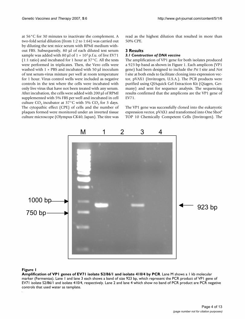
3 Results3.1 Construction of DNA vaccineThe amplification of VP1 gene for both isolates produceda 923 bp band as shown in Figure 1. Each amplicon (VP1gene) had been designed to include the Pst I site and NotI site at both ends to facilitate cloning into expression vec-tor, pVAX1 (Invitrogen, U.S.A.). The PCR products werepurified using QIAquick Gel Extraction Kit (Qiagen, Ger-many) and sent for sequence analysis. The sequencingresults confirmed that the amplicons are the VP1 gene ofEV71.
The VP1 gene was successfully cloned into the eukaryoticexpression vector, pVAX1 and transformed into One Shot®
TOP 10 Chemically Competent Cells (Invitrogen). The
Amplification of VP1 genes of EV71 isolate S2/86/1 and isolate 410/4 by PCRFigure 1Amplification of VP1 genes of EV71 isolate S2/86/1 and isolate 410/4 by PCR. Lane M shows a 1 kb molecular marker (Fermentas). Lane 1 and lane 3 each shows a band of size 923 bp, which represent the PCR product of VP1 gene of EV71 isolate S2/86/1 and isolate 410/4, respectively. Lane 2 and lane 4 which show no band of PCR product are PCR negative controls that used water as template.
1000 bp
M 1 2 3 4
750 bp 923 bp
Page 4 of 13(page number not for citation purposes)
Genetic Vaccines and Therapy 2007, 5:6 http://www.gvt-journal.com/content/5/1/6
positive clones that carried the pVAX1/VP1 DNA vaccinewere screened and selected using PCR and restrictiondigestion analysis. The desired constructs were furtherconfirmed by automated DNA sequencing. The pVAX1vector that was successfully cloned with VP1 gene of eachEV71 isolate S2/86/1 and isolate 410/4, in the correct ori-entation and in frame were designated as pVAX1/VP1-Sand pVAX1/VP1-4, respectively.
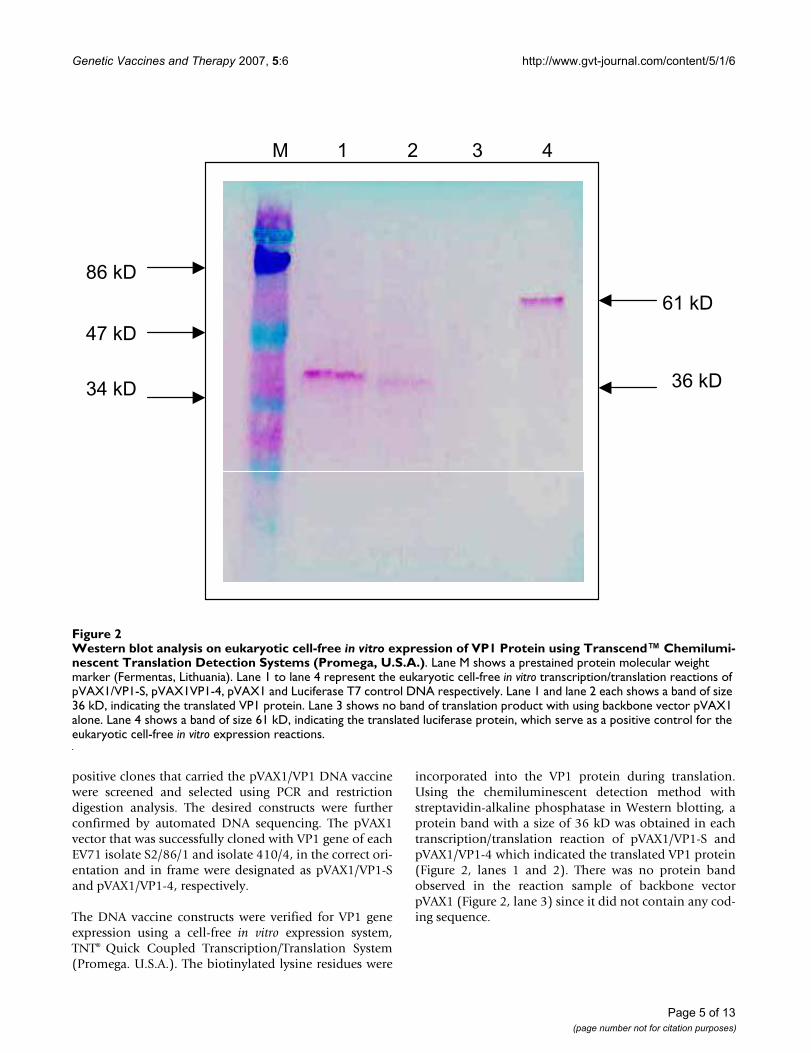
The DNA vaccine constructs were verified for VP1 geneexpression using a cell-free in vitro expression system,TNT® Quick Coupled Transcription/Translation System(Promega. U.S.A.). The biotinylated lysine residues were
incorporated into the VP1 protein during translation.Using the chemiluminescent detection method withstreptavidin-alkaline phosphatase in Western blotting, aprotein band with a size of 36 kD was obtained in eachtranscription/translation reaction of pVAX1/VP1-S andpVAX1/VP1-4 which indicated the translated VP1 protein(Figure 2, lanes 1 and 2). There was no protein bandobserved in the reaction sample of backbone vectorpVAX1 (Figure 2, lane 3) since it did not contain any cod-ing sequence.
Western blot analysis on eukaryotic cell-free in vitro expression of VP1 Protein using Transcend™ Chemiluminescent Transla-tion Detection Systems (Promega, U.S.A.)Figure 2Western blot analysis on eukaryotic cell-free in vitro expression of VP1 Protein using Transcend™ Chemilumi-nescent Translation Detection Systems (Promega, U.S.A.). Lane M shows a prestained protein molecular weight marker (Fermentas, Lithuania). Lane 1 to lane 4 represent the eukaryotic cell-free in vitro transcription/translation reactions of pVAX1/VP1-S, pVAX1VP1-4, pVAX1 and Luciferase T7 control DNA respectively. Lane 1 and lane 2 each shows a band of size 36 kD, indicating the translated VP1 protein. Lane 3 shows no band of translation product with using backbone vector pVAX1 alone. Lane 4 shows a band of size 61 kD, indicating the translated luciferase protein, which serve as a positive control for the eukaryotic cell-free in vitro expression reactions.
61 kD
M 1 2 3 4
86 kD
47 kD
36 kD34 kD
Page 5 of 13(page number not for citation purposes)
Genetic Vaccines and Therapy 2007, 5:6 http://www.gvt-journal.com/content/5/1/6
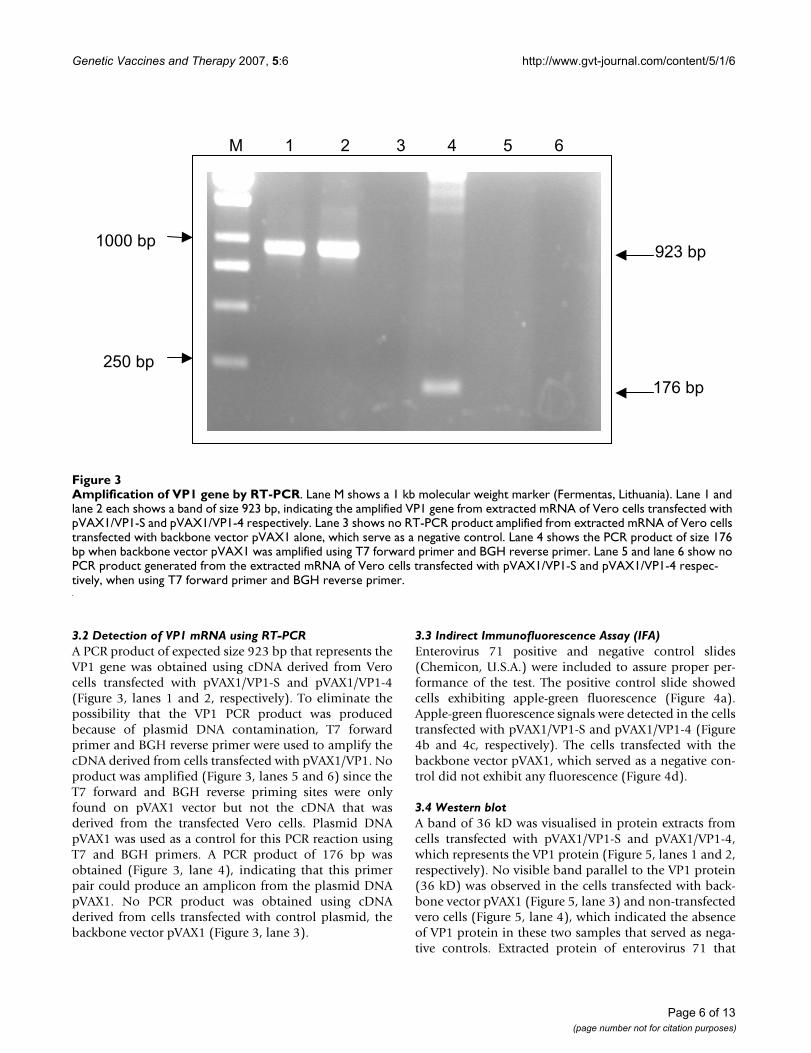
3.2 Detection of VP1 mRNA using RT-PCRA PCR product of expected size 923 bp that represents theVP1 gene was obtained using cDNA derived from Verocells transfected with pVAX1/VP1-S and pVAX1/VP1-4(Figure 3, lanes 1 and 2, respectively). To eliminate thepossibility that the VP1 PCR product was producedbecause of plasmid DNA contamination, T7 forwardprimer and BGH reverse primer were used to amplify thecDNA derived from cells transfected with pVAX1/VP1. Noproduct was amplified (Figure 3, lanes 5 and 6) since theT7 forward and BGH reverse priming sites were onlyfound on pVAX1 vector but not the cDNA that wasderived from the transfected Vero cells. Plasmid DNApVAX1 was used as a control for this PCR reaction usingT7 and BGH primers. A PCR product of 176 bp wasobtained (Figure 3, lane 4), indicating that this primerpair could produce an amplicon from the plasmid DNApVAX1. No PCR product was obtained using cDNAderived from cells transfected with control plasmid, thebackbone vector pVAX1 (Figure 3, lane 3).
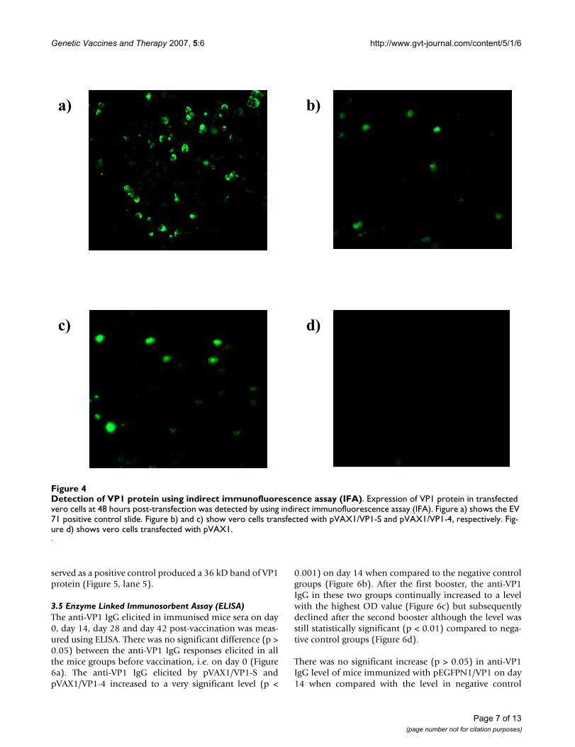
3.3 Indirect Immunofluorescence Assay (IFA)Enterovirus 71 positive and negative control slides(Chemicon, U.S.A.) were included to assure proper per-formance of the test. The positive control slide showedcells exhibiting apple-green fluorescence (Figure 4a).Apple-green fluorescence signals were detected in the cellstransfected with pVAX1/VP1-S and pVAX1/VP1-4 (Figure4b and 4c, respectively). The cells transfected with thebackbone vector pVAX1, which served as a negative con-trol did not exhibit any fluorescence (Figure 4d).
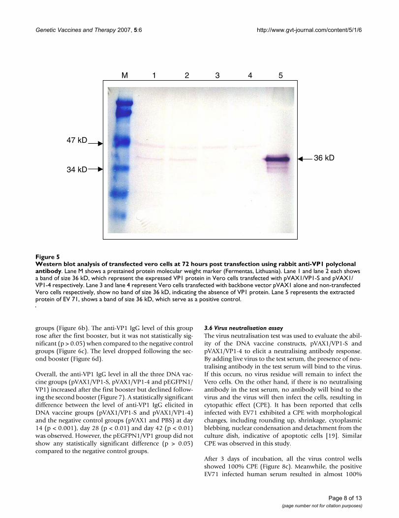
3.4 Western blotA band of 36 kD was visualised in protein extracts fromcells transfected with pVAX1/VP1-S and pVAX1/VP1-4,which represents the VP1 protein (Figure 5, lanes 1 and 2,respectively). No visible band parallel to the VP1 protein(36 kD) was observed in the cells transfected with back-bone vector pVAX1 (Figure 5, lane 3) and non-transfectedvero cells (Figure 5, lane 4), which indicated the absenceof VP1 protein in these two samples that served as nega-tive controls. Extracted protein of enterovirus 71 that
Amplification of VP1 gene by RT-PCRFigure 3Amplification of VP1 gene by RT-PCR. Lane M shows a 1 kb molecular weight marker (Fermentas, Lithuania). Lane 1 and lane 2 each shows a band of size 923 bp, indicating the amplified VP1 gene from extracted mRNA of Vero cells transfected with pVAX1/VP1-S and pVAX1/VP1-4 respectively. Lane 3 shows no RT-PCR product amplified from extracted mRNA of Vero cells transfected with backbone vector pVAX1 alone, which serve as a negative control. Lane 4 shows the PCR product of size 176 bp when backbone vector pVAX1 was amplified using T7 forward primer and BGH reverse primer. Lane 5 and lane 6 show no PCR product generated from the extracted mRNA of Vero cells transfected with pVAX1/VP1-S and pVAX1/VP1-4 respec-tively, when using T7 forward primer and BGH reverse primer.
M 1 2 3 4 5 6
923 bp
176 bp
1000 bp
250 bp
Page 6 of 13(page number not for citation purposes)
Genetic Vaccines and Therapy 2007, 5:6 http://www.gvt-journal.com/content/5/1/6
served as a positive control produced a 36 kD band of VP1protein (Figure 5, lane 5).
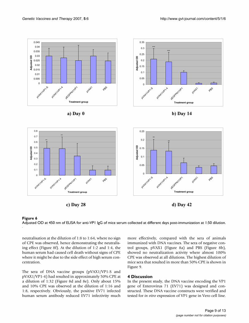
3.5 Enzyme Linked Immunosorbent Assay (ELISA)The anti-VP1 IgG elicited in immunised mice sera on day0, day 14, day 28 and day 42 post-vaccination was meas-ured using ELISA. There was no significant difference (p >0.05) between the anti-VP1 IgG responses elicited in allthe mice groups before vaccination, i.e. on day 0 (Figure6a). The anti-VP1 IgG elicited by pVAX1/VP1-S andpVAX1/VP1-4 increased to a very significant level (p <
0.001) on day 14 when compared to the negative controlgroups (Figure 6b). After the first booster, the anti-VP1IgG in these two groups continually increased to a levelwith the highest OD value (Figure 6c) but subsequentlydeclined after the second booster although the level wasstill statistically significant (p < 0.01) compared to nega-tive control groups (Figure 6d).
There was no significant increase (p > 0.05) in anti-VP1IgG level of mice immunized with pEGFPN1/VP1 on day14 when compared with the level in negative control
Detection of VP1 protein using indirect immunofluorescence assay (IFA)Figure 4Detection of VP1 protein using indirect immunofluorescence assay (IFA). Expression of VP1 protein in transfected vero cells at 48 hours post-transfection was detected by using indirect immunofluorescence assay (IFA). Figure a) shows the EV 71 positive control slide. Figure b) and c) show vero cells transfected with pVAX1/VP1-S and pVAX1/VP1-4, respectively. Fig-ure d) shows vero cells transfected with pVAX1.
d)c)
b)a)
Page 7 of 13(page number not for citation purposes)
Genetic Vaccines and Therapy 2007, 5:6 http://www.gvt-journal.com/content/5/1/6
groups (Figure 6b). The anti-VP1 IgG level of this grouprose after the first booster, but it was not statistically sig-nificant (p > 0.05) when compared to the negative controlgroups (Figure 6c). The level dropped following the sec-ond booster (Figure 6d).
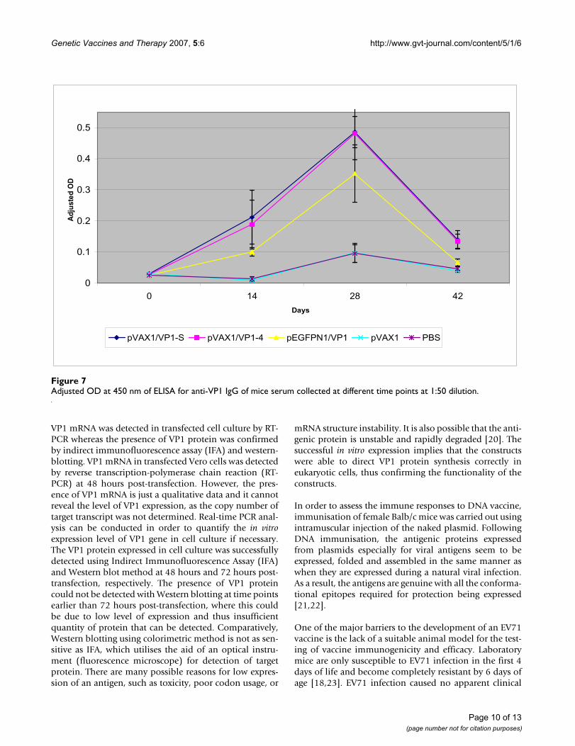
Overall, the anti-VP1 IgG level in all the three DNA vac-cine groups (pVAX1/VP1-S, pVAX1/VP1-4 and pEGFPN1/VP1) increased after the first booster but declined follow-ing the second booster (Figure 7). A statistically significantdifference between the level of anti-VP1 IgG elicited inDNA vaccine groups (pVAX1/VP1-S and pVAX1/VP1-4)and the negative control groups (pVAX1 and PBS) at day14 (p < 0.001), day 28 (p < 0.01) and day 42 (p < 0.01)was observed. However, the pEGFPN1/VP1 group did notshow any statistically significant difference (p > 0.05)compared to the negative control groups.
3.6 Virus neutralisation assayThe virus neutralisation test was used to evaluate the abil-ity of the DNA vaccine constructs, pVAX1/VP1-S andpVAX1/VP1-4 to elicit a neutralising antibody response.By adding live virus to the test serum, the presence of neu-tralising antibody in the test serum will bind to the virus.If this occurs, no virus residue will remain to infect theVero cells. On the other hand, if there is no neutralisingantibody in the test serum, no antibody will bind to thevirus and the virus will then infect the cells, resulting incytopathic effect (CPE). It has been reported that cellsinfected with EV71 exhibited a CPE with morphologicalchanges, including rounding up, shrinkage, cytoplasmicblebbing, nuclear condensation and detachment from theculture dish, indicative of apoptotic cells [19]. SimilarCPE was observed in this study.
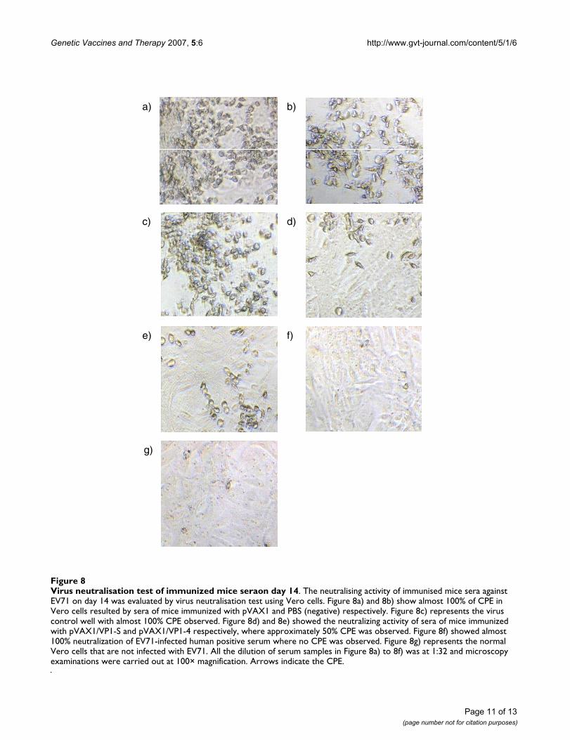
After 3 days of incubation, all the virus control wellsshowed 100% CPE (Figure 8c). Meanwhile, the positiveEV71 infected human serum resulted in almost 100%
Western blot analysis of transfected vero cells at 72 hours post transfection using rabbit anti-VP1 polyclonal antibodyFigure 5Western blot analysis of transfected vero cells at 72 hours post transfection using rabbit anti-VP1 polyclonal antibody. Lane M shows a prestained protein molecular weight marker (Fermentas, Lithuania). Lane 1 and lane 2 each shows a band of size 36 kD, which represent the expressed VP1 protein in Vero cells transfected with pVAX1/VP1-S and pVAX1/VP1-4 respectively. Lane 3 and lane 4 represent Vero cells transfected with backbone vector pVAX1 alone and non-transfected Vero cells respectively, show no band of size 36 kD, indicating the absence of VP1 protein. Lane 5 represents the extracted protein of EV 71, shows a band of size 36 kD, which serve as a positive control.
M 1 2 3 4 5
47 kD
34 kD
36 kD
Page 8 of 13(page number not for citation purposes)
Genetic Vaccines and Therapy 2007, 5:6 http://www.gvt-journal.com/content/5/1/6
neutralisation at the dilution of 1:8 to 1:64, where no signof CPE was observed, hence demonstrating the neutralis-ing effect (Figure 8f). At the dilution of 1:2 and 1:4, thehuman serum had caused cell death without signs of CPEwhere it might be due to the side effect of high serum con-centration.
The sera of DNA vaccine groups (pVAX1/VP1-S andpVAX1/VP1-4) had resulted in approximately 50% CPE ata dilution of 1:32 (Figure 8d and 8e). Only about 15%and 10% CPE was observed at the dilution of 1:16 and1:8, respectively. Obviously, the positive EV71 infectedhuman serum antibody reduced EV71 infectivity much
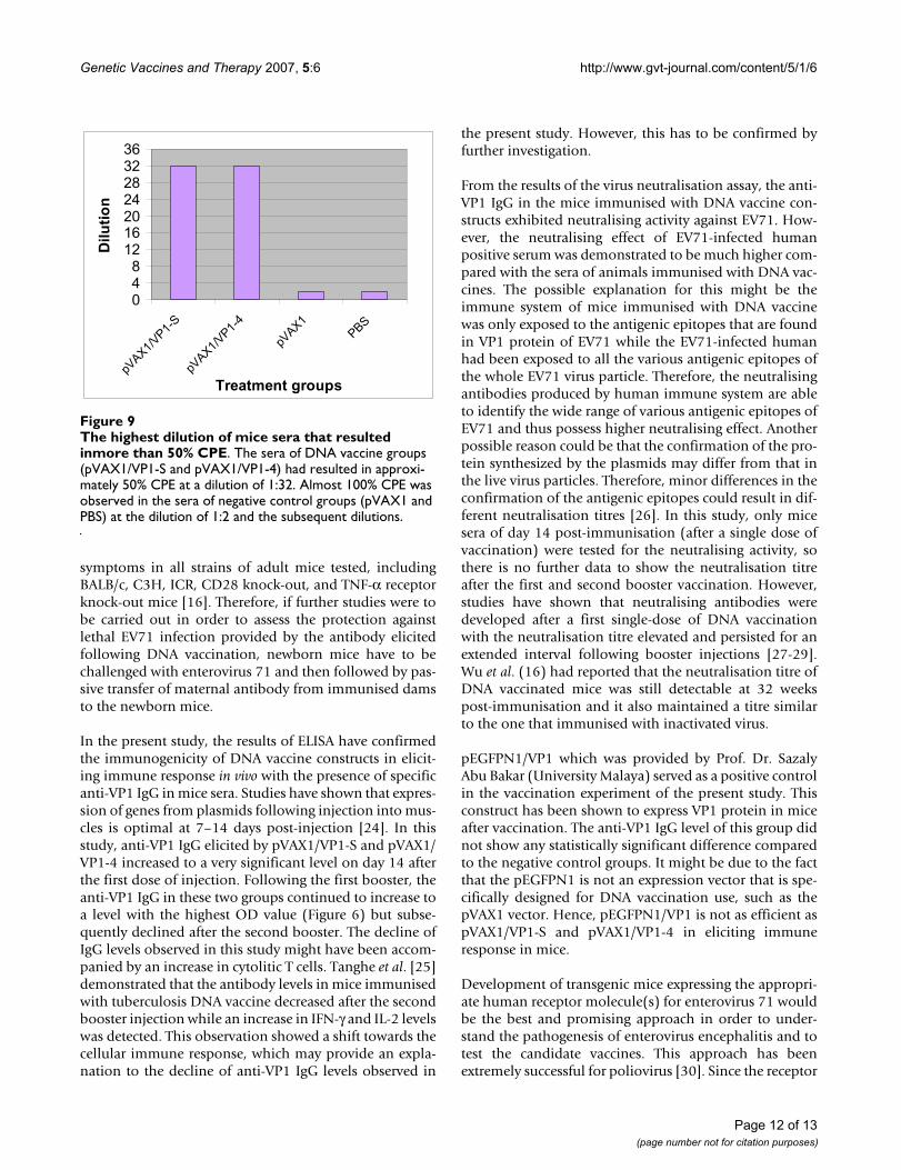
more effectively, compared with the sera of animalsimmunized with DNA vaccines. The sera of negative con-trol groups, pVAX1 (Figure 8a) and PBS (Figure 8b),showed no neutralization activity where almost 100%CPE was observed at all dilutions. The highest dilution ofmice sera that resulted in more than 50% CPE is shown inFigure 9.
4 DiscussionIn the present study, the DNA vaccine encoding the VP1gene of Enterovirus 71 (EV71) was designed and con-structed. These DNA vaccine constructs were verified andtested for in vitro expression of VP1 gene in Vero cell line.
Adjusted OD at 450 nm of ELISA for anti-VP1 IgG of mice serum collected at different days post-immunization at 1:50 dilutionFigure 6Adjusted OD at 450 nm of ELISA for anti-VP1 IgG of mice serum collected at different days post-immunization at 1:50 dilution.
0
0.005
0.01
0.015
0.02
0.025
0.03
0.035
0.04
0.045
pVAX1/
VP1-S
pVAX1/
VP1-4
pEG
FPN1/
VP1
pVAX1
PBS
Treatment group
Ad
juste
d O
D
0
0.05
0.1
0.15
0.2
0.25
0.3
0.35
pVAX1/
VP1-S
pVAX1/
VP1-4
pEGFPN
1/VP1
pVAX1
PBS
Treatment group
Ad
juste
d O
D
***
***
a) Day 0 b) Day 14
0
0.1
0.2
0.3
0.4
0.5
0.6
0.7
0.8
pVAX1/V
P1-S
pVAX1/V
P1-4
pEG
FPN1/
VP1
pVAX1
PBS
Treatment group
Ad
juste
d O
D
**
0
0.05
1
0.15
0.2
0.25
pVAX1/
VP1-S
pVAX1/
VP1-4
pEG
FPN1/
VP1
pVAX1
PBS
Treatment group
juste
d O
D**
0.
Ad
**
**
d) Day 42 c) Day 28
Page 9 of 13(page number not for citation purposes)
Genetic Vaccines and Therapy 2007, 5:6 http://www.gvt-journal.com/content/5/1/6
VP1 mRNA was detected in transfected cell culture by RT-PCR whereas the presence of VP1 protein was confirmedby indirect immunofluorescence assay (IFA) and western-blotting. VP1 mRNA in transfected Vero cells was detectedby reverse transcription-polymerase chain reaction (RT-PCR) at 48 hours post-transfection. However, the pres-ence of VP1 mRNA is just a qualitative data and it cannotreveal the level of VP1 expression, as the copy number oftarget transcript was not determined. Real-time PCR anal-ysis can be conducted in order to quantify the in vitroexpression level of VP1 gene in cell culture if necessary.The VP1 protein expressed in cell culture was successfullydetected using Indirect Immunofluorescence Assay (IFA)and Western blot method at 48 hours and 72 hours post-transfection, respectively. The presence of VP1 proteincould not be detected with Western blotting at time pointsearlier than 72 hours post-transfection, where this couldbe due to low level of expression and thus insufficientquantity of protein that can be detected. Comparatively,Western blotting using colorimetric method is not as sen-sitive as IFA, which utilises the aid of an optical instru-ment (fluorescence microscope) for detection of targetprotein. There are many possible reasons for low expres-sion of an antigen, such as toxicity, poor codon usage, or
mRNA structure instability. It is also possible that the anti-genic protein is unstable and rapidly degraded [20]. Thesuccessful in vitro expression implies that the constructswere able to direct VP1 protein synthesis correctly ineukaryotic cells, thus confirming the functionality of theconstructs.
In order to assess the immune responses to DNA vaccine,immunisation of female Balb/c mice was carried out usingintramuscular injection of the naked plasmid. FollowingDNA immunisation, the antigenic proteins expressedfrom plasmids especially for viral antigens seem to beexpressed, folded and assembled in the same manner aswhen they are expressed during a natural viral infection.As a result, the antigens are genuine with all the conforma-tional epitopes required for protection being expressed[21,22].
One of the major barriers to the development of an EV71vaccine is the lack of a suitable animal model for the test-ing of vaccine immunogenicity and efficacy. Laboratorymice are only susceptible to EV71 infection in the first 4days of life and become completely resistant by 6 days ofage [18,23]. EV71 infection caused no apparent clinical
Adjusted OD at 450 nm of ELISA for anti-VP1 IgG of mice serum collected at different time points at 1:50 dilutionFigure 7Adjusted OD at 450 nm of ELISA for anti-VP1 IgG of mice serum collected at different time points at 1:50 dilution.
0
0.1
0.2
0.3
0.4
0.5
0 14 28 42
Days
Ad
jus
ted
OD
pVAX1/VP1-S pVAX1/VP1-4 pEGFPN1/VP1 pVAX1 PBS
Page 10 of 13(page number not for citation purposes)
Genetic Vaccines and Therapy 2007, 5:6 http://www.gvt-journal.com/content/5/1/6
Page 11 of 13(page number not for citation purposes)
Virus neutralisation test of immunized mice seraon day 14Figure 8Virus neutralisation test of immunized mice seraon day 14. The neutralising activity of immunised mice sera against EV71 on day 14 was evaluated by virus neutralisation test using Vero cells. Figure 8a) and 8b) show almost 100% of CPE in Vero cells resulted by sera of mice immunized with pVAX1 and PBS (negative) respectively. Figure 8c) represents the virus control well with almost 100% CPE observed. Figure 8d) and 8e) showed the neutralizing activity of sera of mice immunized with pVAX1/VP1-S and pVAX1/VP1-4 respectively, where approximately 50% CPE was observed. Figure 8f) showed almost 100% neutralization of EV71-infected human positive serum where no CPE was observed. Figure 8g) represents the normal Vero cells that are not infected with EV71. All the dilution of serum samples in Figure 8a) to 8f) was at 1:32 and microscopy examinations were carried out at 100× magnification. Arrows indicate the CPE.
a)
c)
e)
b)
d)
f)
g)
Genetic Vaccines and Therapy 2007, 5:6 http://www.gvt-journal.com/content/5/1/6
symptoms in all strains of adult mice tested, includingBALB/c, C3H, ICR, CD28 knock-out, and TNF-α receptorknock-out mice [16]. Therefore, if further studies were tobe carried out in order to assess the protection againstlethal EV71 infection provided by the antibody elicitedfollowing DNA vaccination, newborn mice have to bechallenged with enterovirus 71 and then followed by pas-sive transfer of maternal antibody from immunised damsto the newborn mice.
In the present study, the results of ELISA have confirmedthe immunogenicity of DNA vaccine constructs in elicit-ing immune response in vivo with the presence of specificanti-VP1 IgG in mice sera. Studies have shown that expres-sion of genes from plasmids following injection into mus-cles is optimal at 7–14 days post-injection [24]. In thisstudy, anti-VP1 IgG elicited by pVAX1/VP1-S and pVAX1/VP1-4 increased to a very significant level on day 14 afterthe first dose of injection. Following the first booster, theanti-VP1 IgG in these two groups continued to increase toa level with the highest OD value (Figure 6) but subse-quently declined after the second booster. The decline ofIgG levels observed in this study might have been accom-panied by an increase in cytolitic T cells. Tanghe et al. [25]demonstrated that the antibody levels in mice immunisedwith tuberculosis DNA vaccine decreased after the secondbooster injection while an increase in IFN-γ and IL-2 levelswas detected. This observation showed a shift towards thecellular immune response, which may provide an expla-nation to the decline of anti-VP1 IgG levels observed in
the present study. However, this has to be confirmed byfurther investigation.
From the results of the virus neutralisation assay, the anti-VP1 IgG in the mice immunised with DNA vaccine con-structs exhibited neutralising activity against EV71. How-ever, the neutralising effect of EV71-infected humanpositive serum was demonstrated to be much higher com-pared with the sera of animals immunised with DNA vac-cines. The possible explanation for this might be theimmune system of mice immunised with DNA vaccinewas only exposed to the antigenic epitopes that are foundin VP1 protein of EV71 while the EV71-infected humanhad been exposed to all the various antigenic epitopes ofthe whole EV71 virus particle. Therefore, the neutralisingantibodies produced by human immune system are ableto identify the wide range of various antigenic epitopes ofEV71 and thus possess higher neutralising effect. Anotherpossible reason could be that the confirmation of the pro-tein synthesized by the plasmids may differ from that inthe live virus particles. Therefore, minor differences in theconfirmation of the antigenic epitopes could result in dif-ferent neutralisation titres [26]. In this study, only micesera of day 14 post-immunisation (after a single dose ofvaccination) were tested for the neutralising activity, sothere is no further data to show the neutralisation titreafter the first and second booster vaccination. However,studies have shown that neutralising antibodies weredeveloped after a first single-dose of DNA vaccinationwith the neutralisation titre elevated and persisted for anextended interval following booster injections [27-29].Wu et al. (16) had reported that the neutralisation titre ofDNA vaccinated mice was still detectable at 32 weekspost-immunisation and it also maintained a titre similarto the one that immunised with inactivated virus.
pEGFPN1/VP1 which was provided by Prof. Dr. SazalyAbu Bakar (University Malaya) served as a positive controlin the vaccination experiment of the present study. Thisconstruct has been shown to express VP1 protein in miceafter vaccination. The anti-VP1 IgG level of this group didnot show any statistically significant difference comparedto the negative control groups. It might be due to the factthat the pEGFPN1 is not an expression vector that is spe-cifically designed for DNA vaccination use, such as thepVAX1 vector. Hence, pEGFPN1/VP1 is not as efficient aspVAX1/VP1-S and pVAX1/VP1-4 in eliciting immuneresponse in mice.
Development of transgenic mice expressing the appropri-ate human receptor molecule(s) for enterovirus 71 wouldbe the best and promising approach in order to under-stand the pathogenesis of enterovirus encephalitis and totest the candidate vaccines. This approach has beenextremely successful for poliovirus [30]. Since the receptor
The highest dilution of mice sera that resulted inmore than 50% CPEFigure 9The highest dilution of mice sera that resulted inmore than 50% CPE. The sera of DNA vaccine groups (pVAX1/VP1-S and pVAX1/VP1-4) had resulted in approxi-mately 50% CPE at a dilution of 1:32. Almost 100% CPE was observed in the sera of negative control groups (pVAX1 and PBS) at the dilution of 1:2 and the subsequent dilutions.
048
12162024283236
pVAX1/
VP1-S
pVAX1/
VP1-4
pVAX1
PBS
Treatment groups
Dil
uti
on
Page 12 of 13(page number not for citation purposes)
Genetic Vaccines and Therapy 2007, 5:6 http://www.gvt-journal.com/content/5/1/6
for EV71 remains unknown, identification of the EV71receptor is a major priority in vaccine research, as it willallow the development of a transgenic mouse model forstudies of EV71 pathogenesis and vaccine efficacy.
5 ConclusionOverall, this study has given promising results on geneticimmunisation with DNA vaccine encoding VP1 gene ofEV71. With further study and improvement, use of theDNA vaccine might be a potential vaccine strategy againstEV71.
6 AcknowledgementsWe would like to thank Prof. Mary Jane Cardosa from Universiti Malaysia Sarawak (UNIMAS) for kindly providing us the VP1 gene of EV71 isolate 410/4 (genotype B4) and EV71 isolate S2/86/1 (genotype B4) which have been cloned into pCR® Blunt Vector (Zero Blunt™ PCR Cloning Kit, Inv-itrogen). We would also like to thank the Ministry of Science, Technology and Innovation, Malaysia for providing the IRPA Grant (06-02-09-1001-PR001).
References1. Muir P, Kammerer U, Korn K, Mulders MN, Poyry T, Weissbrich B,
Kandolf R, Cleator GM, van Loon AM: Molecular typing of enter-oviruses: current status and future requirements. Clin Micro-biol Rev 1998, 11:202-227.
2. Rueckert RR: Picornaviruses and their replication. In VirologyEdited by: Fields BN, Knipe DM. Lippincott-Raven, New York;1990:507-548.
3. Alexander JP, Baden L, Pallansch MA, Anderson LJ: Enterovirusinfections and neurologic disease – United States, 1977–91. JInfect Dis 1994, 169:905-908.
4. Melnick JL: Enterovirus type 71 infections: a variedclinical pat-tern sometimes mimicking paralytic poliomyelitis. Rev InfectDis 1984, 6(Suppl):S387-S390.
5. Schmidt NJ, Lennette EH, Ho HH: An apparently new enterovi-rus isolated from patients with disease of the central nervoussystem. J Infect Dis 1974, 129:304-309.
6. Chumakov M, Voroshilova M, Shindarov L, Lavrova L, Gracheva L,Koroleva G, Vasilenko S, Brodvarova I, Nikolova M, Gyurova S,Gacheva M, Mitov G, Ninov N, Tsylka E, Robinson I, Frolova M,Bashkirtsev V, Martiyanova L, Rodin V: Enterovirus 71 isolatedfrom cases of epidemic poliomyelitis-like disease in Bulgaria.Arch Virol 1979, 60:329-340.
7. Blomberg J, Lycke E, Ahlfors K, Johnsson T, Wolontis S, von ZeipelG: New enterovirus type associated with aseptic meningitisand/or hand, foot and mouth disease. Lancet 1974, 2:112.
8. Gilbert GL, Dickson KE, Waters MJ, Kennett ML, Land SA, SneddonM: Outbreak of enterovirus 71 in Victoria, Australia, with ahigh incidence of neurologic involvement. Pediatr Infect Dis J1988, 7:484-488.
9. Nagy G, Takatsy S, Kukan E, Mihaly I, Domok I: Virological diagno-sis of enterovirus 71 infections: Experiences gained during anepidemic of acute CNS diseases in Hungary in 1978. Arch Virol1982, 71:217-227.
10. Samuda GM, Chang WK, Yeung CY, Tang PS: Monoplegiacausedby enterovirus 71 infection: an outbreak in Hong Kong. Pedi-atr Infect Dis J 1987, 6:206-208.
11. Lum LCS, Wong KT, Lam SK, Chua KB, Goh AYT: Neurogenic pul-monary oedema and enterovirus 71 encephalomyelitis. Lan-cet 1998, 352:1391.
12. AbuBakar S, Chee HY, Al-Kobaisi MF, Xiaoshan J, Chua KB, Lam SK:Identification of enterovirus 71 isolates from an outbreak ofhand, foot and mouth disease with fatal cases of encephalo-myelitis in Malaysia. Virus Res 1999, 61:1-9.
13. Chang LY, Huang YC, Lin TY: Fulminant neurogenic pulmonaryoedema with hand, foot and mouth disease. Lancet 1998,352:367-368.
14. McMinn PC: An Overview of the evolution of enterovirus71and its clinical and public health significance. FEMS MicrobiolRev 2002, 26:91-107.
15. Shindarov LM, Chumakov MP, Voroshilova MK, Bojinov S, VasilenkoSM, Kirov ID, Kamenov E, Leshchinskaya EV, Mitov G, Robinson IA,Sivchev S, Staikov S: Epidemiological, clinical and pathomor-phological characteristics of epidemic poliomyelitis-like dis-ease caused by enterovirus 71. J Hyg Epidemiol Microbiol Immunol1979, 23:284-295.
16. Wu CN, Lin YC, Cathy Y, Liao NS, Shih SR, Ho MS: Protectionagainst lethal enterovirus 71 infection in newborn mice bypassive immunization with subunit VP1 vaccines and inacti-vated virus. Vaccine 2002, 20:895-904.
17. Liao HT, Hung KL: Neurologic involvement in an outbreakofenterovirus 71 infection: A hospital-based study. Acta PaediatrTaiwan 2001, 42:27-32.
18. Yu CK, Chen CC, Chen CL, Wang JR, Liu CC, Yan JJ, Su IJ: Neutral-izing antibody provided protection against Enterovirus type71 lethal challenge in neonatal mice. J Biomed Sci 2000,7:523-528.
19. Chan YF, AbuBakar S: Enterovirus 71 infection induces apopto-sis in Vero cells. Malaysian J Pathol 2003, 25(1):29-35.
20. Ertl PF, Thomsen LL: Technical issues in construction of nucleicacid vaccines. Methods 2003, 31:199-206.
21. Montgomery DL, Ulmer JB, Donnely JJ, Liu MA: DNAVaccines.Pharmacol Ther 1997, 74(2):195-205.
22. Babiuk LA, Pontarollo R, Babiuk S, Loehr B, van Drunen Littel-vandenHurk S: Induction of immune responses by DNA vaccines inlarge animals. Vaccine 2003, 21:649-658.
23. Roberts GBS, Boyd JF: The histopathology of enterovirus infec-tions in new-born mice. J Infect 1987, 15:45-56.
24. Manthorpe M, Comefert-Jensen F, Hartikka J, Felgner J, Rundell A,Margalith M, Dwarki V: Gene therapy by intramuscular injec-tion of plasmid DNA: studies on firefly luciferase geneexpression in mice. Hum Gene Ther 1993, 4:419-431.
25. Tanghe A, Denis O, Lamvrecht B, Motte V, van den Berg T, HuygenK: Tuberculosis DNA vaccine encoding Ag85A is immuno-genic and protective when administered by intramuscularneedle injection but not by epidermal gene gun bombard-ment. Infect Immun 2000, 68:3854-3860.
26. Kaur R, Sachdeva G, Vrati S: Plasmid DNA immunizationagainstJapanese Encephalitis Virus: Immunogenicity of membrane-anchored and secretory envelope protein. J Infect Dis 2002,185:1-12.
27. Caselli E, Boni M, Di Luca D, Salvatori D, Vita A, Cassai E: A com-bined bovine herpesvirus 1 gB-gD DNA vaccine inducesimmune response in mice. Comp Immunol Microbiol Infect Dis2005, 28:155-166.
28. Guo H, Liu Z, Sun S, Bao H, Chen Y, Liu X, Xie Q: Immuneresponse in guinea pigs vaccinated with DNA vaccine of foot-and-mouth disease virus O/China99. Vaccine 2005,23:3236-3242.
29. Lodmell DL, Parnell MJ, Weyhrich JT, Ewalt LC: Canine rabiesDNA vaccination: a single-dose intradermal injection intoear pinnae elicits elevated and persistent levels of neutraliz-ing antibody. Vaccine 2003, 21:3998-4002.
30. Horie H, Koike S, Kurata T, Sato-Yoshida Y, Ise I, Ota Y, Abe S, HiokeK, Kato H, Taya C, Nomura T, Hashizume S, Yonekawa H, NomotoA: Transgenic mice carrying the human poliovirus receptor:new animal model for the study of poliovirus neurovirulence.J Virol 1994, 68:681-688.
Page 13 of 13(page number not for citation purposes)