Embed Size (px)
Citation preview
REVIEW
Genetics and biology of pancreaticductal adenocarcinomaAram F. Hezel,1,8 Alec C. Kimmelman,1,3,8 Ben Z. Stanger,5 Nabeel Bardeesy,6 andRonald A. DePinho1,2,4,7,9
1Department of Medical Oncology, Dana-Farber Cancer Institute; 2Department of Medicine, Brigham and Women’sHospital; 3Harvard Radiation Oncology Program; 4Department of Genetics; 5Gastrointestinal Unit, 6Massachusetts GeneralHospital Cancer Center, Massachusettes General Hospital; 7Center for Applied Cancer Science and Belfer Institute forInnovative Cancer Science, Dana-Farber Cancer Institute, Harvard Medical School, Boston, Massachusetts 02115, USA
Pancreatic ductal adenocarcinoma (PDAC) is the fourthleading cause of cancer death in the United States with amedian survival of <6 mo and a dismal 5-yr survival rateof 3%–5%. The cancer’s lethal nature stems from itspropensity to rapidly disseminate to the lymphatic sys-tem and distant organs. This aggressive biology and re-sistance to conventional and targeted therapeutic agentsleads to a typical clinical presentation of incurable dis-ease at the time of diagnosis. The well-defined serial his-topathologic picture and accompanying molecular pro-files of PDAC and its precursor lesions have provided theframework for emerging basic and translational research.Recent advances include insights into the cancer’s cel-lular origins, high-resolution genomic profiles pointingto potential new therapeutic targets, and refined mousemodels reflecting both the genetics and histopathologicevolution of human PDAC. This confluence of develop-ments offers the opportunity for accelerated discoveryand the future promise of improved treatment.
Pancreas anatomy and physiology
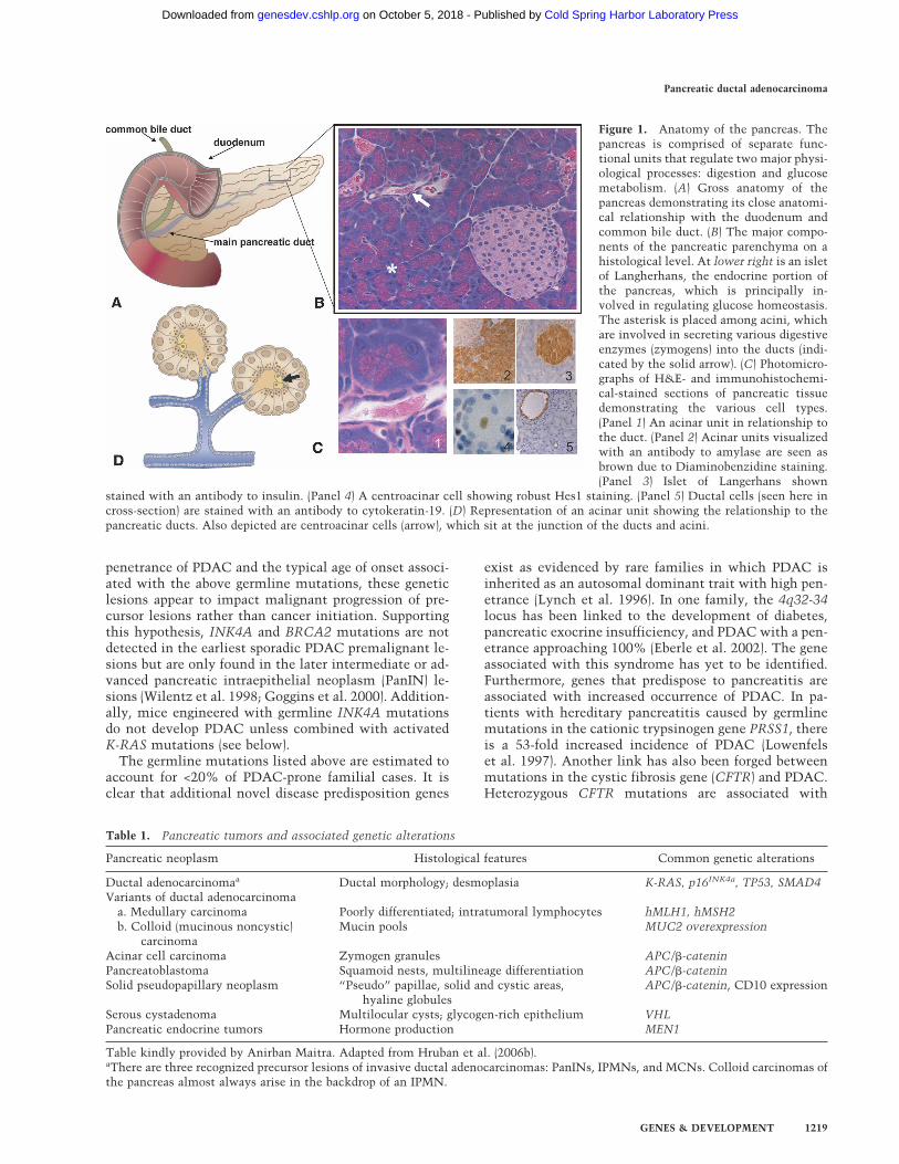
The pancreas, an organ of endodermal derivation, is thekey regulator of protein and carbohydrate digestion andglucose homeostasis (Fig. 1). The exocrine pancreas (80%of the tissue mass of the organ) is composed of a branch-ing network of acinar and duct cells that produce anddeliver digestive zymogens into the gastrointestinaltract. The acinar cells, which are organized in functionalunits along the duct network, synthesize and secrete zy-mogens into the ductal lumen in response to cues fromthe stomach and duodenum. Within the acinar unitsnear the ducts are centroacinar cells. The endocrine pan-creas, which regulates metabolism and glucose homeo-
stasis through the secretion of hormones into the blood-stream, is composed of four specialized endocrine celltypes gathered together into clusters called Islets ofLangerhans.
Mirroring the physiologic and cellular diversity of thepancreas is a spectrum of distinct pancreatic malignan-cies that possess histological and molecular features thatrecall the characteristics of the various normal cellularconstituents. These multiple tumor types and hallmarkfeatures are summarized in Table 1. Pancreatic ductaladenocarcinoma (PDAC), whose nomenclature derivesfrom its histological resemblance to ductal cells, is themost common pancreatic neoplasm and accounts for>85% of pancreatic tumor cases (Warshaw and Fernan-dez-del Castillo 1992; D. Li et al. 2004). PDAC is thefocus of this review, and the reader is directed to thefollowing excellent review covering other pancreas can-cer types (Hruban et al. 2006b).
Epidemiology of PDAC
PDAC is associated with only a few known demographicand environmental risk factors and a handful of autoso-mal dominant genetic conditions. Multiple studies haveestablished advanced age, smoking, and long-standingchronic pancreatitis as clear risk factors; diabetes andobesity also appear to confer increased risk (Everhart andWright 1995; Fuchs et al. 1996; Gapstur et al. 2000; Mi-chaud et al. 2001; Berrington de Gonzalez et al. 2003;Stolzenberg-Solomon et al. 2005). Increased risk has alsobeen documented in relatives of PDAC patients, and it isestimated that 10% of PDAC cases are associated withan inherited predisposition based on familial clustering(Schenk et al. 2001; Petersen and Hruban 2003). Corre-spondingly, germline mutations have been linked to fa-milial PDAC, including those targeting the tumor sup-pressor genes INK4A, BRCA2, and LKB1, the DNA mis-match repair gene MLH1 and the cationic trypsinogengene PRSS1 (Whitcomb et al. 1996; Jaffee et al. 2002).BRCA1 mutation appears to confer increased suscepti-bility to PDAC, albeit with a lower associated risk thanBRCA2 (Thompson and Easton 2002). Given the low
[Keywords: Pancreatic cancer; genetics; pancreas; mouse models; ge-nomic stem cell]8These authors contributed equally to this work.9Corresponding author.E-MAIL [email protected]; FAX (617) 632-6069.Article and publication are at http://www.genesdev.org/cgi/doi/10.1101/gad.1415606.
1218 GENES & DEVELOPMENT 20:1218–1249 © 2006 by Cold Spring Harbor Laboratory Press ISSN 0890-9369/06; www.genesdev.org
Cold Spring Harbor Laboratory Press on October 5, 2018 - Published by genesdev.cshlp.orgDownloaded from
penetrance of PDAC and the typical age of onset associ-ated with the above germline mutations, these geneticlesions appear to impact malignant progression of pre-cursor lesions rather than cancer initiation. Supportingthis hypothesis, INK4A and BRCA2 mutations are notdetected in the earliest sporadic PDAC premalignant le-sions but are only found in the later intermediate or ad-vanced pancreatic intraepithelial neoplasm (PanIN) le-sions (Wilentz et al. 1998; Goggins et al. 2000). Addition-ally, mice engineered with germline INK4A mutationsdo not develop PDAC unless combined with activatedK-RAS mutations (see below).
The germline mutations listed above are estimated toaccount for <20% of PDAC-prone familial cases. It isclear that additional novel disease predisposition genes
exist as evidenced by rare families in which PDAC isinherited as an autosomal dominant trait with high pen-etrance (Lynch et al. 1996). In one family, the 4q32-34locus has been linked to the development of diabetes,pancreatic exocrine insufficiency, and PDAC with a pen-etrance approaching 100% (Eberle et al. 2002). The geneassociated with this syndrome has yet to be identified.Furthermore, genes that predispose to pancreatitis areassociated with increased occurrence of PDAC. In pa-tients with hereditary pancreatitis caused by germlinemutations in the cationic trypsinogen gene PRSS1, thereis a 53-fold increased incidence of PDAC (Lowenfelset al. 1997). Another link has also been forged betweenmutations in the cystic fibrosis gene (CFTR) and PDAC.Heterozygous CFTR mutations are associated with
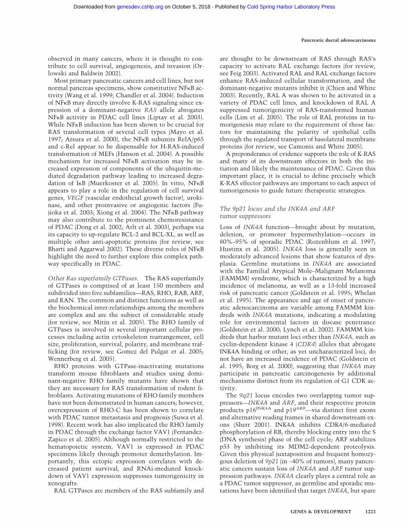
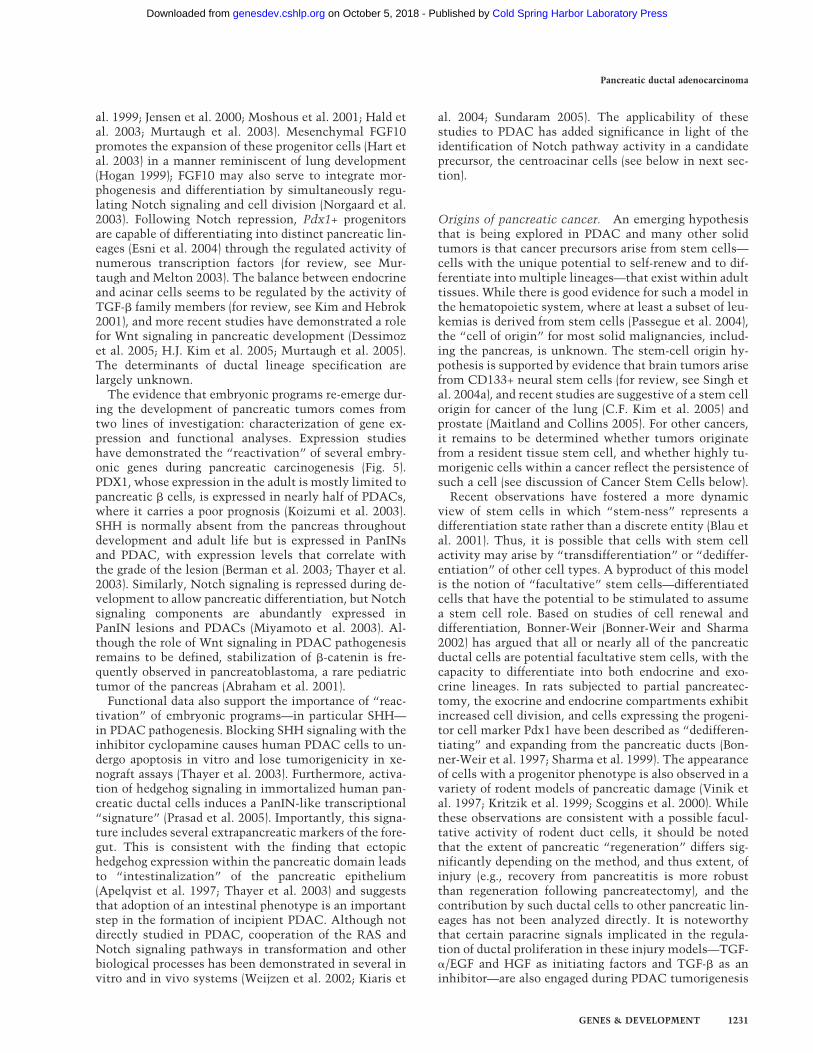
Figure 1. Anatomy of the pancreas. Thepancreas is comprised of separate func-tional units that regulate two major physi-ological processes: digestion and glucosemetabolism. (A) Gross anatomy of thepancreas demonstrating its close anatomi-cal relationship with the duodenum andcommon bile duct. (B) The major compo-nents of the pancreatic parenchyma on ahistological level. At lower right is an isletof Langherhans, the endocrine portion ofthe pancreas, which is principally in-volved in regulating glucose homeostasis.The asterisk is placed among acini, whichare involved in secreting various digestiveenzymes (zymogens) into the ducts (indi-cated by the solid arrow). (C) Photomicro-graphs of H&E- and immunohistochemi-cal-stained sections of pancreatic tissuedemonstrating the various cell types.(Panel 1) An acinar unit in relationship tothe duct. (Panel 2) Acinar units visualizedwith an antibody to amylase are seen asbrown due to Diaminobenzidine staining.(Panel 3) Islet of Langerhans shown
stained with an antibody to insulin. (Panel 4) A centroacinar cell showing robust Hes1 staining. (Panel 5) Ductal cells (seen here incross-section) are stained with an antibody to cytokeratin-19. (D) Representation of an acinar unit showing the relationship to thepancreatic ducts. Also depicted are centroacinar cells (arrow), which sit at the junction of the ducts and acini.
Table 1. Pancreatic tumors and associated genetic alterations
Pancreatic neoplasm Histological features Common genetic alterations
Ductal adenocarcinomaa Ductal morphology; desmoplasia K-RAS, p16INK4a, TP53, SMAD4Variants of ductal adenocarcinoma
a. Medullary carcinoma Poorly differentiated; intratumoral lymphocytes hMLH1, hMSH2b. Colloid (mucinous noncystic)
carcinomaMucin pools MUC2 overexpression
Acinar cell carcinoma Zymogen granules APC/�-cateninPancreatoblastoma Squamoid nests, multilineage differentiation APC/�-cateninSolid pseudopapillary neoplasm “Pseudo” papillae, solid and cystic areas,
hyaline globulesAPC/�-catenin, CD10 expression
Serous cystadenoma Multilocular cysts; glycogen-rich epithelium VHLPancreatic endocrine tumors Hormone production MEN1
Table kindly provided by Anirban Maitra. Adapted from Hruban et al. (2006b).aThere are three recognized precursor lesions of invasive ductal adenocarcinomas: PanINs, IPMNs, and MCNs. Colloid carcinomas ofthe pancreas almost always arise in the backdrop of an IPMN.
Pancreatic ductal adenocarcinoma
GENES & DEVELOPMENT 1219
Cold Spring Harbor Laboratory Press on October 5, 2018 - Published by genesdev.cshlp.orgDownloaded from
chronic pancreatitis, a known risk factor for pancreaticcancer. Recently, a direct link between CFTR mutationand cancer has been posited with the detection of a mu-tant allele in early-onset PDAC cases. Previous studieshad not conclusively identified such a link, but mayhave been limited by smaller numbers of cases and morelimited mutational analysis (Neglia et al. 1995; Sharer etal. 1998; Malats et al. 2001; Matsubayashi et al. 2003;McWilliams et al. 2005).
While the question as to how these separate geneticconditions lead to PDAC remains to be fully understood,the clinical observation of exocrine insufficiency andpancreatitis as a common patho-physiologic processleading to PDAC is compelling. Exocrine organ dysfunc-tion and pancreatitis could promote tumorigenesis inpart by promoting the local release of growth factors,cytokines, and reactive oxygen species (ROS), therebyinducing cell proliferation, disrupting cell differentiationstates, and selecting for oncogenic mutations. The ob-servation that activating K-RAS mutations are detect-able in up to a third of patients with chronic pancreatitisis consistent with this hypothesis (Lohr et al. 2000). Fur-ther evidence from mouse models also suggests the pres-ence of a ductal precursor cell population that undergoesexpansion in response to organ damage (see below insection Origins of Pancreatic Cancer). In states of pan-creatic inflammation or damage, an expanded “stemcell”-like compartment could represent a subpopulationof cells susceptible to oncogenic transformation uponsomatic mutation of key proto-oncogenes and tumorsuppressor genes (Beachy et al. 2004).
Morphological characteristics of PDAC and evolvingpancreatic neoplasms
PDAC commonly arises in the head of the pancreas withinfiltration into surrounding tissues including lymphat-ics, spleen, and peritoneal cavity, and with metastasis tothe liver and lungs. The disease is characterized by thepresence of a dense stroma of fibroblasts and inflamma-tory cells, termed desmoplasia. Pancreatic stellate cells,a subpopulation of cells in the normal pancreas withfibroblast characteristics, have been observed in experi-mental models to respond to pancreatic injury and maycontribute to the desmoplastic response in the setting ofcancer (Jaster 2004). PDAC primarily exhibits a glandu-lar pattern with duct-like structures and varying degreesof cellular atypia and differentiation (Fig. 2). Less com-mon subtypes of PDAC include colloid, adenosquamous,or sarcomatoid histology. Often within an individual tu-mor, there are regional differences in histology, tumorgrade, and degree of differentiation. Even the smallestprimary lesions commonly exhibit perineural and lym-pho-vascular invasion, suggesting a propensity for earlydistant spread.
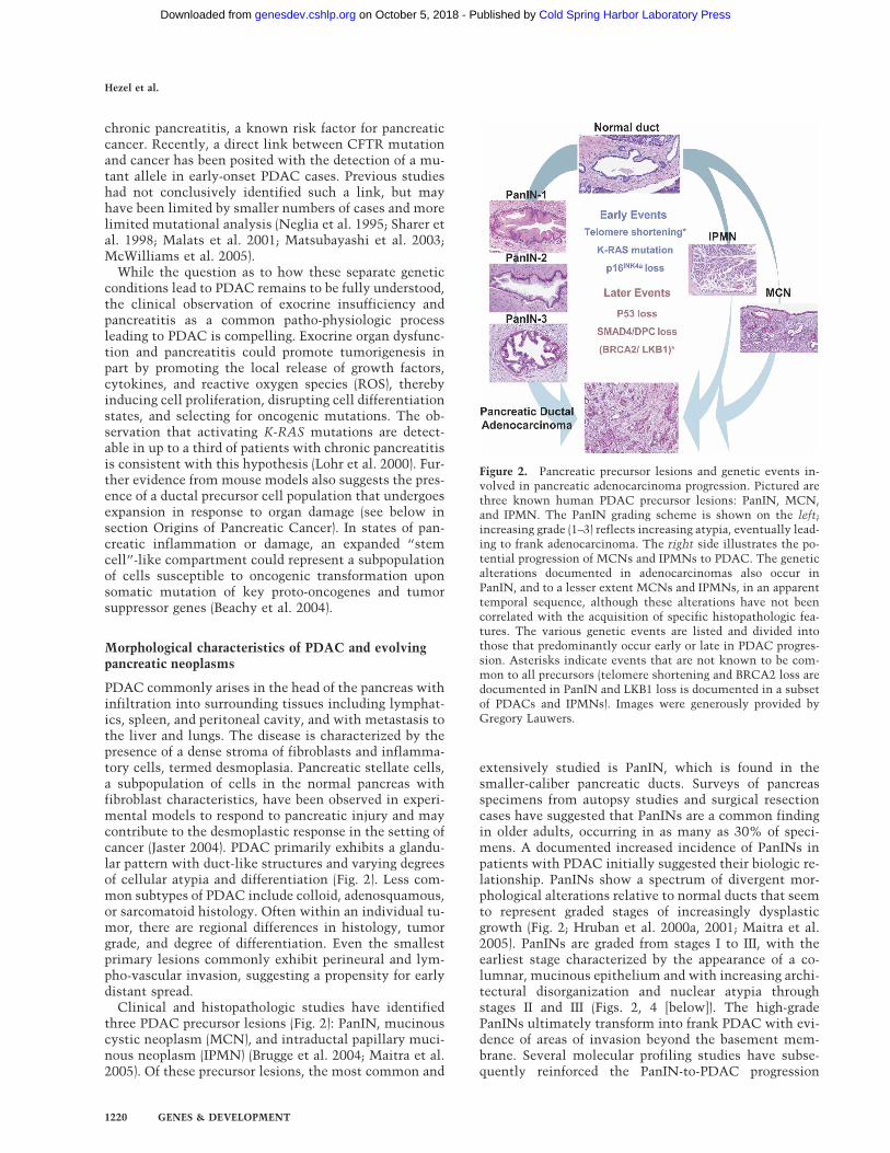
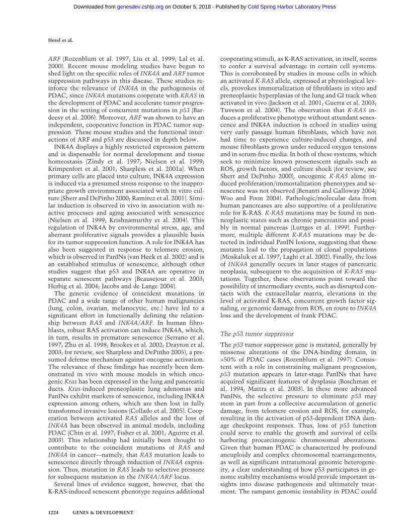
Clinical and histopathologic studies have identifiedthree PDAC precursor lesions (Fig. 2): PanIN, mucinouscystic neoplasm (MCN), and intraductal papillary muci-nous neoplasm (IPMN) (Brugge et al. 2004; Maitra et al.2005). Of these precursor lesions, the most common and
extensively studied is PanIN, which is found in thesmaller-caliber pancreatic ducts. Surveys of pancreasspecimens from autopsy studies and surgical resectioncases have suggested that PanINs are a common findingin older adults, occurring in as many as 30% of speci-mens. A documented increased incidence of PanINs inpatients with PDAC initially suggested their biologic re-lationship. PanINs show a spectrum of divergent mor-phological alterations relative to normal ducts that seemto represent graded stages of increasingly dysplasticgrowth (Fig. 2; Hruban et al. 2000a, 2001; Maitra et al.2005). PanINs are graded from stages I to III, with theearliest stage characterized by the appearance of a co-lumnar, mucinous epithelium and with increasing archi-tectural disorganization and nuclear atypia throughstages II and III (Figs. 2, 4 [below]). The high-gradePanINs ultimately transform into frank PDAC with evi-dence of areas of invasion beyond the basement mem-brane. Several molecular profiling studies have subse-quently reinforced the PanIN-to-PDAC progression
Figure 2. Pancreatic precursor lesions and genetic events in-volved in pancreatic adenocarcinoma progression. Pictured arethree known human PDAC precursor lesions: PanIN, MCN,and IPMN. The PanIN grading scheme is shown on the left;increasing grade (1–3) reflects increasing atypia, eventually lead-ing to frank adenocarcinoma. The right side illustrates the po-tential progression of MCNs and IPMNs to PDAC. The geneticalterations documented in adenocarcinomas also occur inPanIN, and to a lesser extent MCNs and IPMNs, in an apparenttemporal sequence, although these alterations have not beencorrelated with the acquisition of specific histopathologic fea-tures. The various genetic events are listed and divided intothose that predominantly occur early or late in PDAC progres-sion. Asterisks indicate events that are not known to be com-mon to all precursors (telomere shortening and BRCA2 loss aredocumented in PanIN and LKB1 loss is documented in a subsetof PDACs and IPMNs). Images were generously provided byGregory Lauwers.
Hezel et al.
1220 GENES & DEVELOPMENT
Cold Spring Harbor Laboratory Press on October 5, 2018 - Published by genesdev.cshlp.orgDownloaded from
model through documentation of an increasing numberof gene alterations in higher grade PanINs (Fig. 2; Klim-stra and Longnecker 1994; Moskaluk et al. 1997; Hein-moller et al. 2000; Hruban et al. 2000b, 2001; Wilentz etal. 2000; Yamano et al. 2000; Luttges et al. 2001; Maitraet al. 2003).
Less common precursor lesions are MCNs and IPMNs.MCNs are large mucin-producing epithelial cystic le-sions that harbor a distinctive ovarian-type stroma witha variable degree of epithelial dysplasia and focal regionsof invasion. IPMNs resemble PanINs at the cellular levelbut grow into larger cystic structures. Of interest, twosubtypes of invasive cancer have been found in associa-tion with IPMNs; typical PDAC, and a colloid type char-acterized by copious mucin production. Among IPMNsand MCNs, both common and distinct molecular eventsin comparison with PanINs have been described, sug-gesting that each precursor lesion may reflect variationson a common theme of malignant transformation of theduct (Fig. 2; Sato et al. 2001b, 2004a; Adsay et al. 2004).Along these lines, expression profiling has revealed sev-eral up-regulated genes commonly associated withPanINs, IPMNs, and PDAC, pointing toward key sharedmolecular events (Crnogorac-Jurcevic et al. 2002; Sato etal. 2004b; Prasad et al. 2005). How these PDAC precursorlesions relate to each other, their common or separatecellular origins, and whether each type leads to distinctmolecular and biological PDAC subtypes remain to befully explored.
Molecular genetics of PDAC
The molecular analysis of evolving PDAC has provided acompendium of genetic lesions, often implicatingknown cancer genes and classical cancer signaling cas-cades. In many cases, these molecular events have beenlinked with defined histopathologic stages of PDAC pro-gression. While many of these genetic alterations havebeen validated in PDAC pathogenesis, major lingeringquestions center on how these mutations contribute tothe tumor biological features of the neoplasms. In thefollowing subsections, we provide a summary of the cur-rent state of knowledge surrounding the PDAC signaturemutations, their linked pathways, and biological activi-ties.
The K-RAS oncogene and its signaling pathways
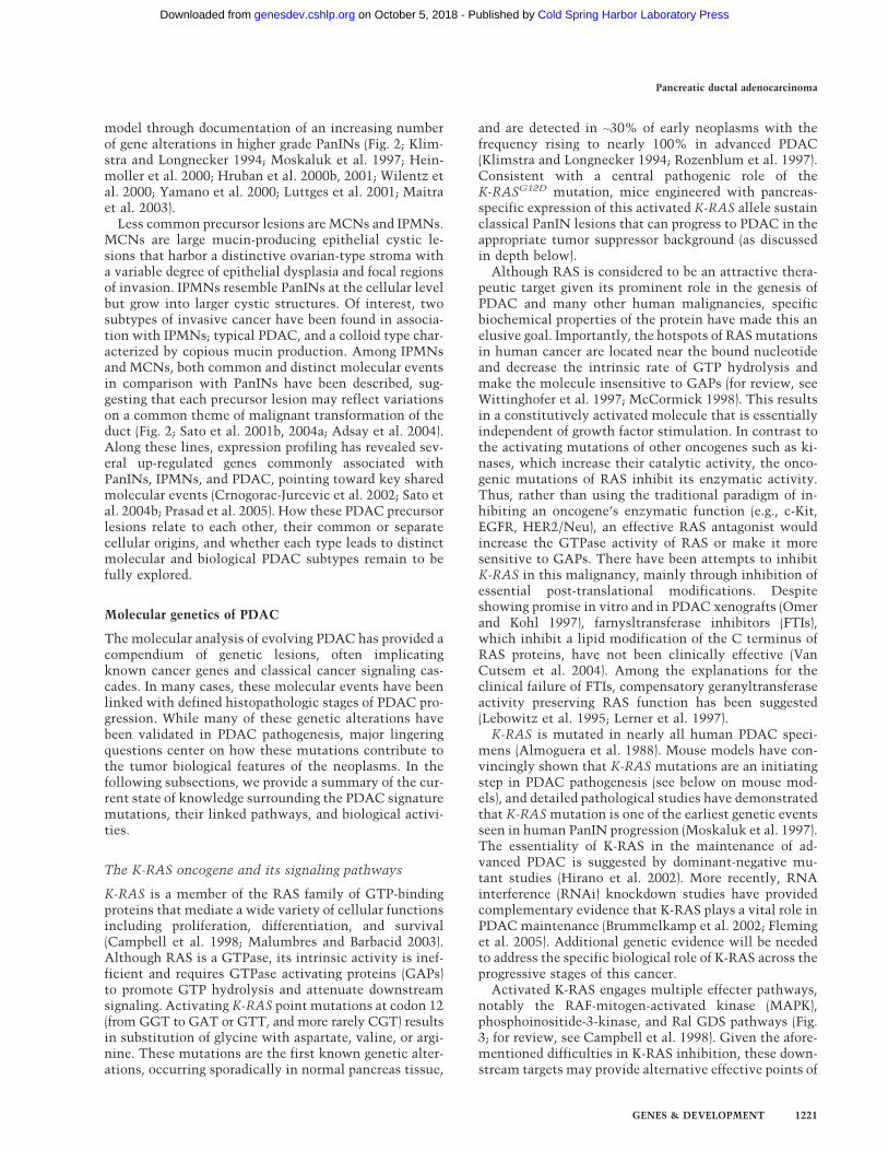
K-RAS is a member of the RAS family of GTP-bindingproteins that mediate a wide variety of cellular functionsincluding proliferation, differentiation, and survival(Campbell et al. 1998; Malumbres and Barbacid 2003).Although RAS is a GTPase, its intrinsic activity is inef-ficient and requires GTPase activating proteins (GAPs)to promote GTP hydrolysis and attenuate downstreamsignaling. Activating K-RAS point mutations at codon 12(from GGT to GAT or GTT, and more rarely CGT) resultsin substitution of glycine with aspartate, valine, or argi-nine. These mutations are the first known genetic alter-ations, occurring sporadically in normal pancreas tissue,
and are detected in ∼30% of early neoplasms with thefrequency rising to nearly 100% in advanced PDAC(Klimstra and Longnecker 1994; Rozenblum et al. 1997).Consistent with a central pathogenic role of theK-RASG12D mutation, mice engineered with pancreas-specific expression of this activated K-RAS allele sustainclassical PanIN lesions that can progress to PDAC in theappropriate tumor suppressor background (as discussedin depth below).
Although RAS is considered to be an attractive thera-peutic target given its prominent role in the genesis ofPDAC and many other human malignancies, specificbiochemical properties of the protein have made this anelusive goal. Importantly, the hotspots of RAS mutationsin human cancer are located near the bound nucleotideand decrease the intrinsic rate of GTP hydrolysis andmake the molecule insensitive to GAPs (for review, seeWittinghofer et al. 1997; McCormick 1998). This resultsin a constitutively activated molecule that is essentiallyindependent of growth factor stimulation. In contrast tothe activating mutations of other oncogenes such as ki-nases, which increase their catalytic activity, the onco-genic mutations of RAS inhibit its enzymatic activity.Thus, rather than using the traditional paradigm of in-hibiting an oncogene’s enzymatic function (e.g., c-Kit,EGFR, HER2/Neu), an effective RAS antagonist wouldincrease the GTPase activity of RAS or make it moresensitive to GAPs. There have been attempts to inhibitK-RAS in this malignancy, mainly through inhibition ofessential post-translational modifications. Despiteshowing promise in vitro and in PDAC xenografts (Omerand Kohl 1997), farnysltransferase inhibitors (FTIs),which inhibit a lipid modification of the C terminus ofRAS proteins, have not been clinically effective (VanCutsem et al. 2004). Among the explanations for theclinical failure of FTIs, compensatory geranyltransferaseactivity preserving RAS function has been suggested(Lebowitz et al. 1995; Lerner et al. 1997).
K-RAS is mutated in nearly all human PDAC speci-mens (Almoguera et al. 1988). Mouse models have con-vincingly shown that K-RAS mutations are an initiatingstep in PDAC pathogenesis (see below on mouse mod-els), and detailed pathological studies have demonstratedthat K-RAS mutation is one of the earliest genetic eventsseen in human PanIN progression (Moskaluk et al. 1997).The essentiality of K-RAS in the maintenance of ad-vanced PDAC is suggested by dominant-negative mu-tant studies (Hirano et al. 2002). More recently, RNAinterference (RNAi) knockdown studies have providedcomplementary evidence that K-RAS plays a vital role inPDAC maintenance (Brummelkamp et al. 2002; Fleminget al. 2005). Additional genetic evidence will be neededto address the specific biological role of K-RAS across theprogressive stages of this cancer.
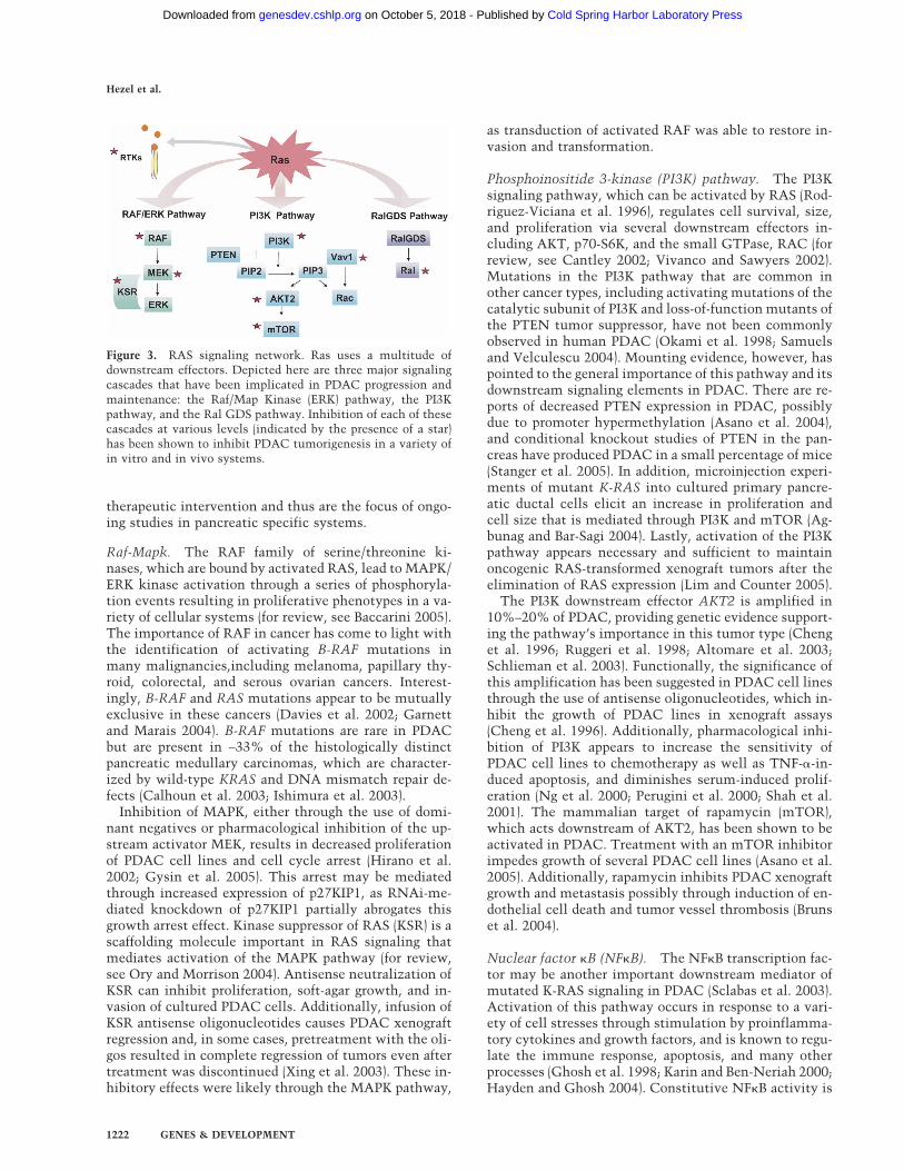
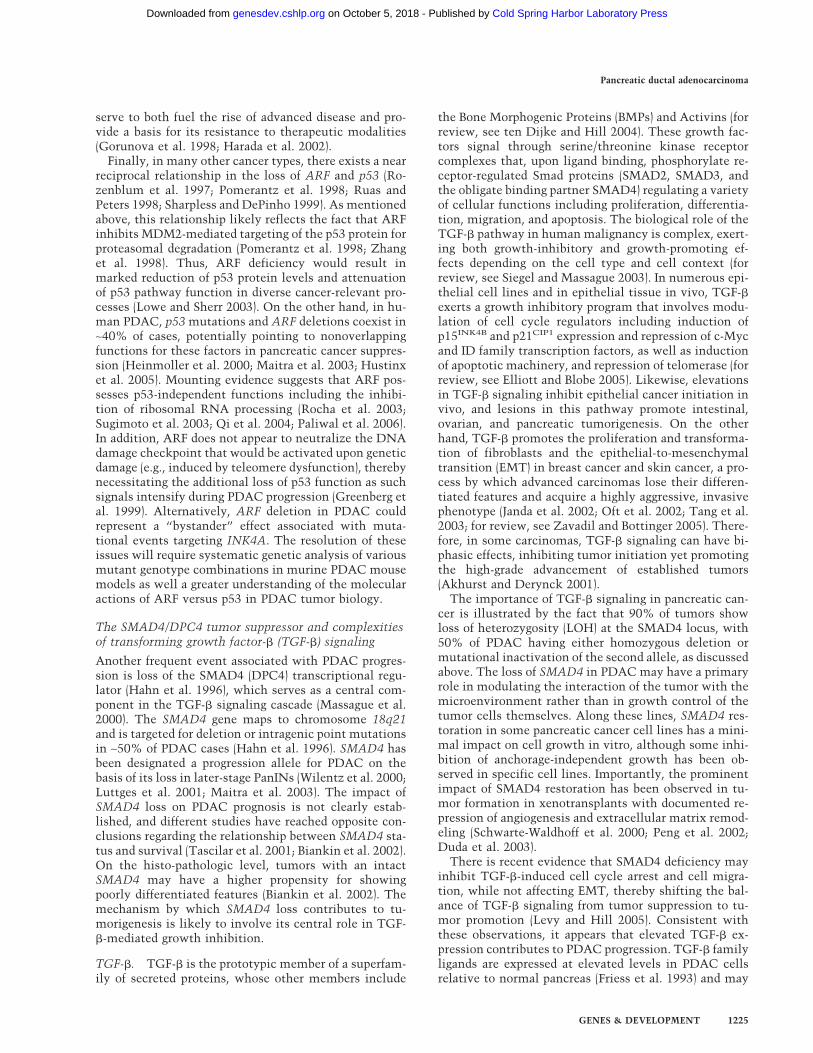
Activated K-RAS engages multiple effecter pathways,notably the RAF-mitogen-activated kinase (MAPK),phosphoinositide-3-kinase, and Ral GDS pathways (Fig.3; for review, see Campbell et al. 1998). Given the afore-mentioned difficulties in K-RAS inhibition, these down-stream targets may provide alternative effective points of
Pancreatic ductal adenocarcinoma
GENES & DEVELOPMENT 1221
Cold Spring Harbor Laboratory Press on October 5, 2018 - Published by genesdev.cshlp.orgDownloaded from
therapeutic intervention and thus are the focus of ongo-ing studies in pancreatic specific systems.
Raf-Mapk. The RAF family of serine/threonine ki-nases, which are bound by activated RAS, lead to MAPK/ERK kinase activation through a series of phosphoryla-tion events resulting in proliferative phenotypes in a va-riety of cellular systems (for review, see Baccarini 2005).The importance of RAF in cancer has come to light withthe identification of activating B-RAF mutations inmany malignancies,including melanoma, papillary thy-roid, colorectal, and serous ovarian cancers. Interest-ingly, B-RAF and RAS mutations appear to be mutuallyexclusive in these cancers (Davies et al. 2002; Garnettand Marais 2004). B-RAF mutations are rare in PDACbut are present in ∼33% of the histologically distinctpancreatic medullary carcinomas, which are character-ized by wild-type KRAS and DNA mismatch repair de-fects (Calhoun et al. 2003; Ishimura et al. 2003).
Inhibition of MAPK, either through the use of domi-nant negatives or pharmacological inhibition of the up-stream activator MEK, results in decreased proliferationof PDAC cell lines and cell cycle arrest (Hirano et al.2002; Gysin et al. 2005). This arrest may be mediatedthrough increased expression of p27KIP1, as RNAi-me-diated knockdown of p27KIP1 partially abrogates thisgrowth arrest effect. Kinase suppressor of RAS (KSR) is ascaffolding molecule important in RAS signaling thatmediates activation of the MAPK pathway (for review,see Ory and Morrison 2004). Antisense neutralization ofKSR can inhibit proliferation, soft-agar growth, and in-vasion of cultured PDAC cells. Additionally, infusion ofKSR antisense oligonucleotides causes PDAC xenograftregression and, in some cases, pretreatment with the oli-gos resulted in complete regression of tumors even aftertreatment was discontinued (Xing et al. 2003). These in-hibitory effects were likely through the MAPK pathway,
as transduction of activated RAF was able to restore in-vasion and transformation.
Phosphoinositide 3-kinase (PI3K) pathway. The PI3Ksignaling pathway, which can be activated by RAS (Rod-riguez-Viciana et al. 1996), regulates cell survival, size,and proliferation via several downstream effectors in-cluding AKT, p70-S6K, and the small GTPase, RAC (forreview, see Cantley 2002; Vivanco and Sawyers 2002).Mutations in the PI3K pathway that are common inother cancer types, including activating mutations of thecatalytic subunit of PI3K and loss-of-function mutants ofthe PTEN tumor suppressor, have not been commonlyobserved in human PDAC (Okami et al. 1998; Samuelsand Velculescu 2004). Mounting evidence, however, haspointed to the general importance of this pathway and itsdownstream signaling elements in PDAC. There are re-ports of decreased PTEN expression in PDAC, possiblydue to promoter hypermethylation (Asano et al. 2004),and conditional knockout studies of PTEN in the pan-creas have produced PDAC in a small percentage of mice(Stanger et al. 2005). In addition, microinjection experi-ments of mutant K-RAS into cultured primary pancre-atic ductal cells elicit an increase in proliferation andcell size that is mediated through PI3K and mTOR (Ag-bunag and Bar-Sagi 2004). Lastly, activation of the PI3Kpathway appears necessary and sufficient to maintainoncogenic RAS-transformed xenograft tumors after theelimination of RAS expression (Lim and Counter 2005).
The PI3K downstream effector AKT2 is amplified in10%–20% of PDAC, providing genetic evidence support-ing the pathway’s importance in this tumor type (Chenget al. 1996; Ruggeri et al. 1998; Altomare et al. 2003;Schlieman et al. 2003). Functionally, the significance ofthis amplification has been suggested in PDAC cell linesthrough the use of antisense oligonucleotides, which in-hibit the growth of PDAC lines in xenograft assays(Cheng et al. 1996). Additionally, pharmacological inhi-bition of PI3K appears to increase the sensitivity ofPDAC cell lines to chemotherapy as well as TNF-�-in-duced apoptosis, and diminishes serum-induced prolif-eration (Ng et al. 2000; Perugini et al. 2000; Shah et al.2001). The mammalian target of rapamycin (mTOR),which acts downstream of AKT2, has been shown to beactivated in PDAC. Treatment with an mTOR inhibitorimpedes growth of several PDAC cell lines (Asano et al.2005). Additionally, rapamycin inhibits PDAC xenograftgrowth and metastasis possibly through induction of en-dothelial cell death and tumor vessel thrombosis (Brunset al. 2004).
Nuclear factor �B (NF�B). The NF�B transcription fac-tor may be another important downstream mediator ofmutated K-RAS signaling in PDAC (Sclabas et al. 2003).Activation of this pathway occurs in response to a vari-ety of cell stresses through stimulation by proinflamma-tory cytokines and growth factors, and is known to regu-late the immune response, apoptosis, and many otherprocesses (Ghosh et al. 1998; Karin and Ben-Neriah 2000;Hayden and Ghosh 2004). Constitutive NF�B activity is
Figure 3. RAS signaling network. Ras uses a multitude ofdownstream effectors. Depicted here are three major signalingcascades that have been implicated in PDAC progression andmaintenance: the Raf/Map Kinase (ERK) pathway, the PI3Kpathway, and the Ral GDS pathway. Inhibition of each of thesecascades at various levels (indicated by the presence of a star)has been shown to inhibit PDAC tumorigenesis in a variety ofin vitro and in vivo systems.
Hezel et al.
1222 GENES & DEVELOPMENT
Cold Spring Harbor Laboratory Press on October 5, 2018 - Published by genesdev.cshlp.orgDownloaded from
observed in many cancers, where it is thought to con-tribute to cell survival, angiogenesis, and invasion (Or-lowski and Baldwin 2002).
Most primary pancreatic cancers and cell lines, but notnormal pancreas specimens, show constitutive NF�B ac-tivity (Wang et al. 1999; Chandler et al. 2004). Inductionof NF�B may directly involve K-RAS signaling since ex-pression of a dominant-negative RAS allele abrogatesNF�B activity in PDAC cell lines (Liptay et al. 2003).While NF�B induction has been shown to be crucial forRAS transformation of several cell types (Mayo et al.1997; Arsura et al. 2000), the NF�B subunits RelA/p65and c-Rel appear to be dispensable for H-RAS-inducedtransformation of MEFs (Hanson et al. 2004). A possiblemechanism for increased NF�B activation may be in-creased expression of components of the ubiquitin-me-diated degradation pathway leading to increased degra-dation of I�B (Muerkoster et al. 2005). In vitro, NF�Bappears to play a role in the regulation of cell survivalgenes, VEGF (vascular endotheial growth factor), uroki-nase, and other proinvasive or angiogenic factors (Fu-jioka et al. 2003; Xiong et al. 2004). The NF�B pathwaymay also contribute to the prominent chemoresistanceof PDAC (Dong et al. 2002; Arlt et al. 2003), perhaps viaits capacity to up-regulate BCL-2 and BCL-XL, as well asmultiple other anti-apoptotic proteins (for review, seeBharti and Aggarwal 2002). These diverse roles of NF�Bhighlight the need to further explore this complex path-way specifically in PDAC.
Other Ras superfamily GTPases. The RAS superfamilyof GTPases is comprised of at least 150 members andsubdivided into five subfamilies—RAS, RHO, RAB, ARF,and RAN. The common and distinct functions as well asthe biochemical inter-relationships among the membersare complex and are the subject of considerable study(for review, see Mitin et al. 2005). The RHO family ofGTPases is involved in several important cellular pro-cesses including actin cytoskeleton rearrangement, cellsize, proliferation, survival, polarity, and membrane traf-ficking (for review, see Gomez del Pulgar et al. 2005;Wennerberg et al. 2005).
RHO proteins with GTPase-inactivating mutationstransform mouse fibroblasts and studies using domi-nant-negative RHO family mutants have shown thatthey are necessary for RAS transformation of rodent fi-broblasts. Activating mutations of RHO family membershave not been demonstrated in human cancers; however,overexpression of RHO-C has been shown to correlatewith PDAC tumor metastasis and prognosis (Suwa et al.1998). Recent work has also implicated the RHO familyin PDAC through the exchange factor VAV1 (Fernandez-Zapico et al. 2005). Although normally restricted to thehematopoietic system, VAV1 is expressed in PDACspecimens likely through promoter demethylation. Im-portantly, this ectopic expression correlates with de-creased patient survival, and RNAi-mediated knock-down of VAV1 expression suppresses tumorigenicity inxenografts.
RAL GTPases are members of the RAS subfamily and
are thought to be downstream of RAS through RAS’scapacity to activate RAL exchange factors (for review,see Feig 2003). Activated RAL and RAL exchange factorsenhance RAS-induced cellular transformation, and thedominant-negative mutants inhibit it (Chien and White2003). Recently, RAL A was shown to be activated in avariety of PDAC cell lines, and knockdown of RAL Asuppressed tumorigenicity of RAS-transformed humancells (Lim et al. 2005). The role of RAL proteins in tu-morigenesis may relate to the requirement of these fac-tors for maintaining the polarity of epithelial cellsthrough the regulated transport of basolateral membraneproteins (for review, see Camonis and White 2005).
A preponderance of evidence supports the role of K-RASand many of its downstream effectors in both the ini-tiation and likely the maintenance of PDAC. Given thisimportant place, it is crucial to define precisely whichK-RAS effector pathways are important to each aspect oftumorigenesis to guide future therapeutic strategies.
The 9p21 locus and the INK4A and ARFtumor suppressors
Loss of INK4A function—brought about by mutation,deletion, or promoter hypermethylation—occurs in80%–95% of sporadic PDAC (Rozenblum et al. 1997;Hustinx et al. 2005). INK4A loss is generally seen inmoderately advanced lesions that show features of dys-plasia. Germline mutations in INK4A are associatedwith the Familial Atypical Mole–Malignant Melanoma(FAMMM) syndrome, which is characterized by a highincidence of melanoma, as well as a 13-fold increasedrisk of pancreatic cancer (Goldstein et al. 1995; Whelanet al. 1995). The appearance and age of onset of pancre-atic adenocarcinoma are variable among FAMMM kin-dreds with INK4A mutations, indicating a modulatingrole for environmental factors in disease penetrance(Goldstein et al. 2000; Lynch et al. 2002). FAMMM kin-dreds that harbor mutant loci other than INK4A, such ascyclin-dependent kinase 4 (CDK4) alleles that abrogateINK4A binding or other, as yet uncharacterized loci, donot have an increased incidence of PDAC (Goldstein etal. 1995; Borg et al. 2000), suggesting that INK4A mayparticipate in pancreatic carcinogenesis by additionalmechanisms distinct from its regulation of G1 CDK ac-tivity.
The 9q21 locus encodes two overlapping tumor sup-pressors—INK4A and ARF, and their respective proteinproducts p16INK4A and p19ARF—via distinct first exonsand alternative reading frames in shared downstream ex-ons (Sherr 2001). INK4A inhibits CDK4/6-mediatedphosphorylation of RB, thereby blocking entry into the S(DNA synthesis) phase of the cell cycle; ARF stabilizesp53 by inhibiting its MDM2-dependent proteolysis.Given this physical juxtaposition and frequent homozy-gous deletion of 9p21 (in ∼40% of tumors), many pancre-atic cancers sustain loss of INK4A and ARF tumor sup-pression pathways. INK4A clearly plays a central role asa PDAC tumor suppressor, as germline and sporadic mu-tations have been identified that target INK4A, but spare
Pancreatic ductal adenocarcinoma
GENES & DEVELOPMENT 1223
Cold Spring Harbor Laboratory Press on October 5, 2018 - Published by genesdev.cshlp.orgDownloaded from
ARF (Rozenblum et al. 1997; Liu et al. 1999; Lal et al.2000). Recent mouse modeling studies have begun toshed light on the specific roles of INK4A and ARF tumorsuppression pathways in this disease. These studies re-inforce the relevance of INK4A in the pathogenesis ofPDAC, since INK4A mutations cooperate with KRAS inthe development of PDAC and accelerate tumor progres-sion in the setting of concurrent mutations in p53 (Bar-deesy et al. 2006). Moreover, ARF was shown to have anindependent, cooperative function in PDAC tumor sup-pression. These mouse studies and the functional inter-actions of ARF and p53 are discussed in depth below.
INK4A displays a highly restricted expression patternand is dispensable for normal development and tissuehomeostasis (Zindy et al. 1997; Nielsen et al. 1999;Krimpenfort et al. 2001; Sharpless et al. 2001a). Whenprimary cells are placed into culture, INK4A expressionis induced via a presumed stress response to the inappro-priate growth environment associated with in vitro cul-ture (Sherr and DePinho 2000; Ramirez et al. 2001). Simi-lar induction is observed in vivo in association with re-active processes and aging associated with senescence(Nielsen et al. 1999; Krishnamurthy et al. 2004). Thisregulation of INK4A by environmental stress, age, andaberrant proliferative signals provides a plausible basisfor its tumor suppression function. A role for INK4A hasalso been suggested in response to telomere erosion,which is observed in PanINs (van Heek et al. 2002) and isan established stimulus of senescence, although otherstudies suggest that p53 and INK4A are operative inseparate senescent pathways (Beausejour et al. 2003;Herbig et al. 2004; Jacobs and de Lange 2004).
The genetic evidence of coincident mutations inPDAC and a wide range of other human malignancies(lung, colon, ovarian, melanocytic, etc.) have led to asignificant effort in functionally defining the relation-ship between RAS and INK4A/ARF. In human fibro-blasts, robust RAS activation can induce INK4A, which,in turn, results in premature senescence (Serrano et al.1997; Zhu et al. 1998; Brookes et al. 2002; Drayton et al.2003; for review, see Sharpless and DePinho 2005), a pre-sumed defense mechanism against oncogene activation.The relevance of these findings has recently been dem-onstrated in vivo with mouse models in which onco-genic Kras has been expressed in the lung and pancreaticducts. Kras-induced preneoplastic lung adenomas andPanINs exhibit markers of senescence, including INK4Aexpression among others, which are then lost in fullytransformed invasive lesions (Collado et al. 2005). Coop-eration between activated RAS alleles and the loss ofINK4A has been observed in animal models, includingPDAC (Chin et al. 1997; Fisher et al. 2001; Aguirre et al.2003). This relationship had initially been thought tocontribute to the coincident mutations of RAS andINK4A in cancer—namely, that RAS mutation leads tosenescence directly through induction of INK4A expres-sion. Thus, mutation in RAS leads to selective pressurefor subsequent mutation in the INK4A/ARF locus.
Several lines of evidence suggest, however, that theK-RAS-induced senescent phenotype requires additional
cooperating stimuli, as K-RAS activation, in itself, seemsto confer a survival advantage in certain cell systems.This is corroborated by studies in mouse cells in whichan activated K-RAS allele, expressed at physiological lev-els, provokes immortalization of fibroblasts in vitro andpreneoplastic hyperplasias of the lung and GI track whenactivated in vivo (Jackson et al. 2001; Guerra et al. 2003;Tuveson et al. 2004). The observation that K-RAS in-duces a proliferative phenotype without attendant senes-cence and INK4A induction is echoed in studies usingvery early passage human fibroblasts, which have nothad time to experience culture-induced changes, andmouse fibroblasts grown under reduced oxygen tensionsand in serum-free media. In both of these systems, whichseek to minimize known prosenescent signals such asROS, growth factors, and culture shock (for review, seeSherr and DePinho 2000), oncogenic K-RAS alone in-duced proliferation/immortalization phenotypes and se-nescence was not observed (Benanti and Galloway 2004;Woo and Poon 2004). Pathologic/molecular data fromhuman pancreases are also supportive of a proliferativerole for K-RAS. K-RAS mutations may be found in non-neoplastic states such as chronic pancreatitis and possi-bly in normal pancreas (Luttges et al. 1999). Further-more, multiple different K-RAS mutations may be de-tected in individual PanIN lesions, suggesting that thesemutants lead to the propagation of clonal populations(Moskaluk et al. 1997; Laghi et al. 2002). Finally, the lossof INK4A generally occurs in later stages of pancreaticneoplasia, subsequent to the acquisition of K-RAS mu-tations. Together, these observations point toward thepossibility of intermediary events, such as disrupted con-tacts with the extracellular matrix, elevations in thelevel of activated K-RAS, concurrent growth factor sig-naling, or genomic damage from ROS, en route to INK4Aloss and the development of frank PDAC.
The p53 tumor suppressor
The p53 tumor suppressor gene is mutated, generally bymissense alterations of the DNA-binding domain, in>50% of PDAC cases (Rozenblum et al. 1997). Consis-tent with a role in constraining malignant progression,p53 mutation appears in later-stage PanINs that haveacquired significant features of dysplasia (Boschman etal. 1994; Maitra et al. 2003). In these more advancedPanINs, the selective pressure to eliminate p53 maystem in part from a collective accumulation of geneticdamage, from telomere erosion and ROS, for example,resulting in the activation of p53-dependent DNA dam-age checkpoint responses. Thus, loss of p53 functioncould serve to enable the growth and survival of cellsharboring procarcinogenic chromosomal aberrations.Given that human PDAC is characterized by profoundaneuploidy and complex chromosomal rearrangements,as well as significant intratumoral genomic heterogene-ity, a clear understanding of how p53 participates in ge-nome stability mechanisms would provide important in-sights into disease pathogenesis and ultimately treat-ment. The rampant genomic instability in PDAC could
Hezel et al.
1224 GENES & DEVELOPMENT
Cold Spring Harbor Laboratory Press on October 5, 2018 - Published by genesdev.cshlp.orgDownloaded from
serve to both fuel the rise of advanced disease and pro-vide a basis for its resistance to therapeutic modalities(Gorunova et al. 1998; Harada et al. 2002).
Finally, in many other cancer types, there exists a nearreciprocal relationship in the loss of ARF and p53 (Ro-zenblum et al. 1997; Pomerantz et al. 1998; Ruas andPeters 1998; Sharpless and DePinho 1999). As mentionedabove, this relationship likely reflects the fact that ARFinhibits MDM2-mediated targeting of the p53 protein forproteasomal degradation (Pomerantz et al. 1998; Zhanget al. 1998). Thus, ARF deficiency would result inmarked reduction of p53 protein levels and attenuationof p53 pathway function in diverse cancer-relevant pro-cesses (Lowe and Sherr 2003). On the other hand, in hu-man PDAC, p53 mutations and ARF deletions coexist in∼40% of cases, potentially pointing to nonoverlappingfunctions for these factors in pancreatic cancer suppres-sion (Heinmoller et al. 2000; Maitra et al. 2003; Hustinxet al. 2005). Mounting evidence suggests that ARF pos-sesses p53-independent functions including the inhibi-tion of ribosomal RNA processing (Rocha et al. 2003;Sugimoto et al. 2003; Qi et al. 2004; Paliwal et al. 2006).In addition, ARF does not appear to neutralize the DNAdamage checkpoint that would be activated upon geneticdamage (e.g., induced by teleomere dysfunction), therebynecessitating the additional loss of p53 function as suchsignals intensify during PDAC progression (Greenberg etal. 1999). Alternatively, ARF deletion in PDAC couldrepresent a “bystander” effect associated with muta-tional events targeting INK4A. The resolution of theseissues will require systematic genetic analysis of variousmutant genotype combinations in murine PDAC mousemodels as well a greater understanding of the molecularactions of ARF versus p53 in PDAC tumor biology.
The SMAD4/DPC4 tumor suppressor and complexitiesof transforming growth factor-� (TGF-�) signaling
Another frequent event associated with PDAC progres-sion is loss of the SMAD4 (DPC4) transcriptional regu-lator (Hahn et al. 1996), which serves as a central com-ponent in the TGF-� signaling cascade (Massague et al.2000). The SMAD4 gene maps to chromosome 18q21and is targeted for deletion or intragenic point mutationsin ∼50% of PDAC cases (Hahn et al. 1996). SMAD4 hasbeen designated a progression allele for PDAC on thebasis of its loss in later-stage PanINs (Wilentz et al. 2000;Luttges et al. 2001; Maitra et al. 2003). The impact ofSMAD4 loss on PDAC prognosis is not clearly estab-lished, and different studies have reached opposite con-clusions regarding the relationship between SMAD4 sta-tus and survival (Tascilar et al. 2001; Biankin et al. 2002).On the histo-pathologic level, tumors with an intactSMAD4 may have a higher propensity for showingpoorly differentiated features (Biankin et al. 2002). Themechanism by which SMAD4 loss contributes to tu-morigenesis is likely to involve its central role in TGF-�-mediated growth inhibition.
TGF-�. TGF-� is the prototypic member of a superfam-ily of secreted proteins, whose other members include
the Bone Morphogenic Proteins (BMPs) and Activins (forreview, see ten Dijke and Hill 2004). These growth fac-tors signal through serine/threonine kinase receptorcomplexes that, upon ligand binding, phosphorylate re-ceptor-regulated Smad proteins (SMAD2, SMAD3, andthe obligate binding partner SMAD4) regulating a varietyof cellular functions including proliferation, differentia-tion, migration, and apoptosis. The biological role of theTGF-� pathway in human malignancy is complex, exert-ing both growth-inhibitory and growth-promoting ef-fects depending on the cell type and cell context (forreview, see Siegel and Massague 2003). In numerous epi-thelial cell lines and in epithelial tissue in vivo, TGF-�exerts a growth inhibitory program that involves modu-lation of cell cycle regulators including induction ofp15INK4B and p21CIP1 expression and repression of c-Mycand ID family transcription factors, as well as inductionof apoptotic machinery, and repression of telomerase (forreview, see Elliott and Blobe 2005). Likewise, elevationsin TGF-� signaling inhibit epithelial cancer initiation invivo, and lesions in this pathway promote intestinal,ovarian, and pancreatic tumorigenesis. On the otherhand, TGF-� promotes the proliferation and transforma-tion of fibroblasts and the epithelial-to-mesenchymaltransition (EMT) in breast cancer and skin cancer, a pro-cess by which advanced carcinomas lose their differen-tiated features and acquire a highly aggressive, invasivephenotype (Janda et al. 2002; Oft et al. 2002; Tang et al.2003; for review, see Zavadil and Bottinger 2005). There-fore, in some carcinomas, TGF-� signaling can have bi-phasic effects, inhibiting tumor initiation yet promotingthe high-grade advancement of established tumors(Akhurst and Derynck 2001).
The importance of TGF-� signaling in pancreatic can-cer is illustrated by the fact that 90% of tumors showloss of heterozygosity (LOH) at the SMAD4 locus, with50% of PDAC having either homozygous deletion ormutational inactivation of the second allele, as discussedabove. The loss of SMAD4 in PDAC may have a primaryrole in modulating the interaction of the tumor with themicroenvironment rather than in growth control of thetumor cells themselves. Along these lines, SMAD4 res-toration in some pancreatic cancer cell lines has a mini-mal impact on cell growth in vitro, although some inhi-bition of anchorage-independent growth has been ob-served in specific cell lines. Importantly, the prominentimpact of SMAD4 restoration has been observed in tu-mor formation in xenotransplants with documented re-pression of angiogenesis and extracellular matrix remod-eling (Schwarte-Waldhoff et al. 2000; Peng et al. 2002;Duda et al. 2003).
There is recent evidence that SMAD4 deficiency mayinhibit TGF-�-induced cell cycle arrest and cell migra-tion, while not affecting EMT, thereby shifting the bal-ance of TGF-� signaling from tumor suppression to tu-mor promotion (Levy and Hill 2005). Consistent withthese observations, it appears that elevated TGF-� ex-pression contributes to PDAC progression. TGF-� familyligands are expressed at elevated levels in PDAC cellsrelative to normal pancreas (Friess et al. 1993) and may
Pancreatic ductal adenocarcinoma
GENES & DEVELOPMENT 1225
Cold Spring Harbor Laboratory Press on October 5, 2018 - Published by genesdev.cshlp.orgDownloaded from
help to promote the characteristic desmoplastic responseof this malignancy as suggested from xenograft studies(Lohr et al. 2001). TGF-� signaling may also contributeto tumorigenesis in an autocrine manner since PDACsoften overexpress the type II TGF-� receptor relative tonormal pancreas (Wagner et al. 1999; for review, seeRane et al. 2006) while experimental blockade of TGF-�signaling by expression of soluble type II TGF-� receptorattenuates tumorigenicity and metastasis of xenografts(Rowland-Goldsmith et al. 2001, 2002). Furthermore, an-tibodies to TGF-� inhibit the invasion of PDAC celllines in vitro, while exogenous addition of this cytokineenhanced invasion and promotes the EMT (Ellenrieder etal. 2001a,b).
The LKB1/STK11 tumor suppressor
The Peutz-Jeghers syndrome (PJS), linked to LKB1/STK11 mutations, is another familial cancer syndromeassociated with an increased incidence of PDAC (Hem-minki et al. 1998; Jenne et al. 1998; Giardiello et al.2000). PJS patients are primarily afflicted with benignintestinal polyposis at a young age (Cooper 1998), al-though advancing age carries increased risk of gastroin-testinal malignancies including a >40-fold increase inPDAC (Giardiello et al. 2000). At the same time, somaticmutation of LKB1 in sporadic PDAC appears to be rare,detected in only 4%–6% of sporadic cases examined (Suet al. 1999), although there is some evidence that therates of inactivation are higher in IPMNs (Sahin et al.2003).
LKB1 encodes a serine/threonine kinase that is in-volved in regulation of diverse processes such as cell po-larity and metabolism, and has been linked to specificsignaling pathways including mTOR, the latter via itscapacity to regulate AMPK (Bardeesy et al. 2002b; Ossi-pova et al. 2003; Baas et al. 2004; Corradetti et al. 2004;Lizcano et al. 2004; Shaw et al. 2004a,b, 2005; Hardie2005). Exactly how LKB1 loss, and through deregulationof which of these pathways/processes, promotes tumori-genesis remains to be established. Control of mTOR sig-naling through AMPK links this gene to a common path-way harboring two other tumor suppressors, PTEN andTSC. The biochemical link to these well-characterizedcancer signaling pathways may provide insights into thebiological mechanisms through which LKB1 suppressestumor formation. At the same time, the role of LKB1 incell polarity, and likely regulation of several less-well-characterized kinases, leaves open several other plau-sible mechanisms of tumor suppression. Current effortsare now directed toward defining additional LKB1 sub-strates and linked biological processes.
The BRCA2 tumor suppressor. Inherited BRCA2 mu-tations are typically associated with familial breast andovarian cancer syndrome, but also carry a significant riskfor the development of pancreatic cancer. One study es-timates that ∼17% of pancreatic cancers occurring in afamilial setting harbor mutations in this gene (Murphyet al. 2002); the del6174T founder mutation is particu-
larly common in familial pancreatic cancers that arise inthe Ashkenazi Jewish population. As is the case for thosewith germline INK4A mutations, the penetrance ofPDAC in BRCA2 mutation carriers is relatively low, andthe age of onset is similar to patients with the sporadicform of the disease. Loss of the wild-type BRCA2 alleleseems to be a late event in those inheriting germlineheterozygous mutations of BRCA2, restricted to severelydysplastic PanINs and PDACs (Goggins et al. 2000). To-gether, these data are consistent with the model that lossof function of BRCA2 promotes the malignant progres-sion of pancreatic neoplasms.
BRCA2 is known to play a critical role in the mainte-nance of genomic stability by regulating homologous re-combination-based DNA repair processes. Conse-quently, BRCA2 deficiency in normal cells results in theaccumulation of procarcinogenic or lethal chromosomalaberrations (Venkitaraman 2002). The fact that BRCA2is selectively mutated late in tumorigenesis likely re-flects the need for DNA damage response pathways(which in a normal cell would lead to senescence or ap-optosis) to be inactivated first—for example, by p53 mu-tation—so that the genetic damage incurred can be tol-erated. Thus, as is the case for telomeres, the carcino-genic role of BRCA2 deficiency may be manifest only inthe appropriate genotypic and cell-type context. BRCA2mutational status may also have therapeutic implica-tions, as it seems to confer susceptibility to DNA cross-linking agents such as Mitomycin C, as is seen in otherrelated Fanconi anemia family genes, particularly FancGand FancC (Taniguchi et al. 2003; van der Heijden et al.2004).
Additional growth factor receptor signaling circuitsin PDAC
Epidermal growth factor. PDAC shows elevated ex-pression of EGF receptors (EGFR and ERBB3) and theirligands (TGF-� and EGF), consistent with the presence ofan autocrine loop (Barton et al. 1991; Korc et al. 1992;Lemoine et al. 1992; Friess et al. 1995, 1999). Impor-tantly, EGFR inhibitors decrease PDAC cell growth andtumorigenesis in vitro (J. Li et al. 2004), as well as inhibitgrowth of orthotopic tumors in combination with cyto-toxic chemotherapy (Bruns et al. 2000). This inhibitionappears to be due to a decrease in tumor vasculaturethrough inhibition of proangiogenic factors, resulting inendothelial apoptosis. In line with these antineoplasticactivities, EGFR inhibitors have been approved for clini-cal use in PDAC patients.
Insulin-like growth factor (IGF). The IGF signalingpathway regulates survival, invasion, and angiogenesisof many human cancers. PDACs show elevated expres-sion of IGF-I in both the tumor cells and the stroma anddisplay aberrant activation of the IGF-I receptor (IGF-IR)in tumor cells (Bergmann et al. 1995; Ouban et al. 2003;Stoeltzing et al. 2003). In vitro, autocrine IGF-I signalingpromotes cell proliferation and growth-factor-indepen-dent survival (Nair et al. 2001). Inhibition of the pathway
Hezel et al.
1226 GENES & DEVELOPMENT
Cold Spring Harbor Laboratory Press on October 5, 2018 - Published by genesdev.cshlp.orgDownloaded from
by anti-IGF-IR antibodies or expression of a dominant-negative form of IGF-IR inhibits the growth of xenograftsand sensitizes tumor cells to chemotherapy (Maloney etal. 2003; Min et al. 2003).
Met and hepatocyte growth factor (HGF). The Met re-ceptor tyrosine kinase and its ligand, HGF/scatter factor,regulate cell motility, invasion, and proliferation and thederegulation of this signaling pathway contributes to theprogression of several malignancies (for review, seeCorso et al. 2005). The Met receptor is expressed at lowlevels in the exocrine pancreas and shows marked up-regulation in PanIN lesions and in PDACs. Additionally,HGF is induced during PDAC progression, present in theepithelium of PanIN lesions and in the stromal cells ofadvanced tumors (Ebert et al. 1994; Di Renzo et al. 1995;Furukawa et al. 1995; Paciucci et al. 1998). HGF pro-motes motility of PDAC cells in vitro, and inhibition ofthis pathway through the administration of blocking an-tibodies or the truncated HGF fragment NK4 inhibitsinvasive growth and angiogenesis of xenografts(Tomioka et al. 2001; Saimura et al. 2002).
Fibroblast growth factor (FGF). FGF signaling (for re-view, see Cross and Claesson-Welsh 2001) appears tocontribute to mitogenesis and angiogenesis of PDAC.Expression of numerous FGF receptors and glypican-1, amembrane heparin sulfate proteoglycan that facilitatesFGF–FGFR interactions, have been detected in primaryPDAC samples (Kobrin et al. 1993; Yamanaka et al.1993a,b; Ohta et al. 1995; Kornmann et al. 1997; Ishi-wata et al. 1998; Kleeff et al. 1998; Kornmann et al.2002). Consistent with a role for FGF signaling in sup-porting PDAC growth, dominant-negative FGFR-1 mu-tants or antisense glypican-1 can inhibit the growth ofpancreatic cancer cell lines in vitro and suppress theirtumorigenic potential in xenografts (Wagner et al. 1998a;Ogawa et al. 2002; Kleeff et al. 2004). FGF signaling mayalso contribute to the desmoplasia associated withPDAC, since elevated bFGF levels are associated withthis phenotype in primary tumors (Kuniyasu et al. 2001).
VEGF. VEGF promotes endothelial cell proliferationand survival by binding to the VEGFR-1 and VEGFR-2endothelial cell transmembrane receptors (for review,see Ferrara et al. 2003). VEGF is overexpressed by PDACcells (Itakura et al. 1997; Seo et al. 2000), whereas dis-ruption of VEGF signaling by expression of soluble VEGFreceptors, VEGF high-affinity binding chimeras, anti-VEGF antibodies, or ribozymes strongly suppresses thetumorigenic growth of pancreatic cancer xenografts (vonMarschall et al. 2000; Hoshida et al. 2002; Tokunaga etal. 2002; Hotz et al. 2003; Fukasawa and Korc 2004).VEGF-C, a regulator of lymphoangiogenesis, is also over-expressed in PDAC and may contribute to lymphaticspread and the lymph node metastasis common in thismalignancy (Tang et al. 2001; Kurahara et al. 2004). Fur-ther study will be required to validate VEGF-C as a drugdevelopment target.
Developmental signaling pathways in PDAC
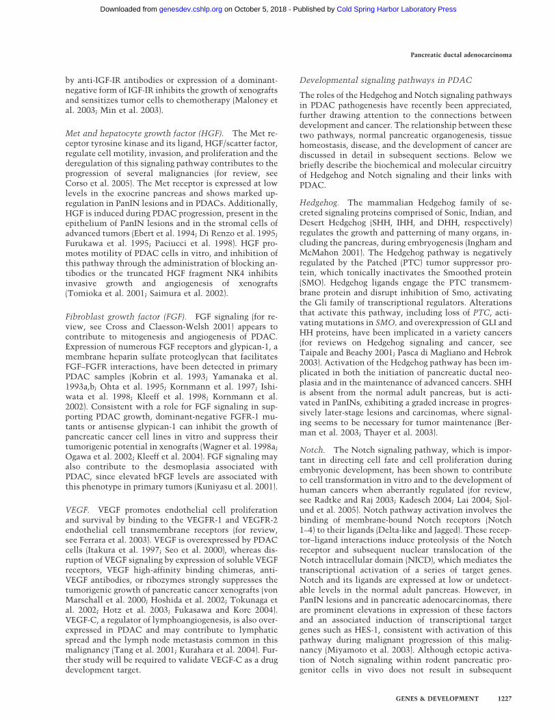
The roles of the Hedgehog and Notch signaling pathwaysin PDAC pathogenesis have recently been appreciated,further drawing attention to the connections betweendevelopment and cancer. The relationship between thesetwo pathways, normal pancreatic organogenesis, tissuehomeostasis, disease, and the development of cancer arediscussed in detail in subsequent sections. Below webriefly describe the biochemical and molecular circuitryof Hedgehog and Notch signaling and their links withPDAC.
Hedgehog. The mammalian Hedgehog family of se-creted signaling proteins comprised of Sonic, Indian, andDesert Hedgehog (SHH, IHH, and DHH, respectively)regulates the growth and patterning of many organs, in-cluding the pancreas, during embryogenesis (Ingham andMcMahon 2001). The Hedgehog pathway is negativelyregulated by the Patched (PTC) tumor suppressor pro-tein, which tonically inactivates the Smoothed protein(SMO). Hedgehog ligands engage the PTC transmem-brane protein and disrupt inhibition of Smo, activatingthe Gli family of transcriptional regulators. Alterationsthat activate this pathway, including loss of PTC, acti-vating mutations in SMO, and overexpression of GLI andHH proteins, have been implicated in a variety cancers(for reviews on Hedgehog signaling and cancer, seeTaipale and Beachy 2001; Pasca di Magliano and Hebrok2003). Activation of the Hedgehog pathway has been im-plicated in both the initiation of pancreatic ductal neo-plasia and in the maintenance of advanced cancers. SHHis absent from the normal adult pancreas, but is acti-vated in PanINs, exhibiting a graded increase in progres-sively later-stage lesions and carcinomas, where signal-ing seems to be necessary for tumor maintenance (Ber-man et al. 2003; Thayer et al. 2003).
Notch. The Notch signaling pathway, which is impor-tant in directing cell fate and cell proliferation duringembryonic development, has been shown to contributeto cell transformation in vitro and to the development ofhuman cancers when aberrantly regulated (for review,see Radtke and Raj 2003; Kadesch 2004; Lai 2004; Sjol-und et al. 2005). Notch pathway activation involves thebinding of membrane-bound Notch receptors (Notch1–4) to their ligands (Delta-like and Jagged). These recep-tor–ligand interactions induce proteolysis of the Notchreceptor and subsequent nuclear translocation of theNotch intracellular domain (NICD), which mediates thetranscriptional activation of a series of target genes.Notch and its ligands are expressed at low or undetect-able levels in the normal adult pancreas. However, inPanIN lesions and in pancreatic adenocarcinomas, thereare prominent elevations in expression of these factorsand an associated induction of transcriptional targetgenes such as HES-1, consistent with activation of thispathway during malignant progression of this malig-nancy (Miyamoto et al. 2003). Although ectopic activa-tion of Notch signaling within rodent pancreatic pro-genitor cells in vivo does not result in subsequent
Pancreatic ductal adenocarcinoma
GENES & DEVELOPMENT 1227
Cold Spring Harbor Laboratory Press on October 5, 2018 - Published by genesdev.cshlp.orgDownloaded from
carcinogenesis (Murtaugh et al. 2003), there is an accu-mulating body of literature demonstrating interactionsof Notch with RAS, both in development as well as intumorigenesis (for review, see Sundaram 2005). In par-ticular, several different cell-based systems have shownthat activated RAS cooperates with Notch to transformcells; however, others have demonstrated that in certainsettings, Notch may suppress transformation. Given thecritical role of K-RAS in PDAC and the context-depen-dant relationship with Notch signaling, it will be im-perative to investigate these interactions on the geneticlevel using pancreatic ductal model systems.
Telomere shortening and dysfunction in PDAC
Telomere dynamics play a central role in shaping thegenomes of many cancer types, particularly epithelialcancers (for review, see Maser and DePinho 2002). Whiletelomerase-mediated preservation of telomere functionhas been shown to promote the development of ad-vanced malignancies (Hahn et al. 1999), there is equallycompelling experimental evidence in both mouse andhuman cancers that the lack of telomerase activity and atransient period of telomere shortening and dysfunctionduring early neoplasia drives cancer initiation. This telo-mere-based mechanism involves generating procarcino-genic chromosomal rearrangements via breakage–fu-sion–bridge BFB cycles (Artandi et al. 2000) that promoteregional amplifications and deletions at the sites of chro-mosomal breakage (O’Hagan et al. 2002). Importantly,the survival of cells with critically short telomeres andongoing BFB events is enhanced by deactivation of p53-dependent DNA damage responses; thus telomere dys-function and p53 loss cooperate to promote the develop-ment of carcinomas in multiple tissues (Chin et al.1999a).
On the basis of these data, telomere erosion mightcontribute to the high incidence of PDAC in the settingof advancing age or inflammatory conditions as occurs inhereditary pancreatitis as a function of epithelial turn-over. Indeed, shortened telomeres and anaphase bridginghave been detected in low-grade PanINs, marking telo-mere erosion as one of the earliest documented geneticevents in the evolution of these ductal neoplasms (vanHeek et al. 2002). Such observations are in line withprevious findings in pancreatic cancer cell lines of thefrequent absence of telomeres at chromosome ends andoccurrence of anaphase bridging indicative of ongoingBFB cycles and persistent genomic instability (Gissels-son et al. 2001). Although reactivation of telomerase ap-pears critical to the emergence of pancreatic cancer cells,it is a late event in PDAC progression and is preceded bya period of telomere shortening and dysfunction thatwould appear likely to promote carcinogenesis by lead-ing to the formation of cancer-relevant chromosomal re-arrangements. In the evolution of human PDAC, telo-mere shortening appears to precede the development ofp53 mutations, which are found in ∼50% of advaned le-sions (Hruban et al. 2000a,b; Luttges et al. 2001; vanHeek et al. 2002). Such observations raise the possibility
that other p53 pathway components involved in the telo-mere-induced checkpoint responses are neutralized in asubset of these neoplasms. Alternatively, the loss of p53-independent responses in some tumors could obviate theneed to inactivate this pathway. These findings under-score the need to define the wiring of the telomerecheckpoint response in evolving PanINs and establishedPDACs. To this end, it will be of interest to specificallycorrelate telomere length, p53 status, and the onset ofgenomic instability in PanINs, and to develop pancreaticcancer models with telomere dysfunction.
Chromosome structural alterations, expressionprofiles, and other cancer loci in PDAC
PDAC is characterized by genomic complexity and in-stability. Telomere shortening, loss of p53, K-RAS mu-tation, and defects in the mitotic spindle apparatus areall likely contributors to this phenotype. Centrosomeabnormalities are detected in 85% of PDAC samples,and there is a correlation between levels of such abnor-malities and the degree of chromosomal aberrations(Sato et al. 1999, 2001a). Overall, the pattern of p53 andBRCA2 mutations and the detection of abnormal mito-sis and nuclear abnormalities in PanIN-2 and PanIN-3lesions suggest that genomic instability is initiated inthese stages of the tumor progression. The known ste-reotypical PDAC mutations described above are likely torepresent only a small fraction of the genetic lesions resi-dent in these cancers. This view is supported by the de-tection of recurrent chromosomal amplifications and de-letions by karyotype analysis, comparative genomic hy-bridization (CGH), and LOH studies. Regions ofconsistent alteration include gains involving 3q, 5p, 7p,8q, 11q, 12p, 17q, and 20q and losses targeting 3p, 4q, 6q,8p, 9p, 10q, 12q, 13q, 17p, 18q, 21q, and 22q (Mahlamakiet al. 1997, 2002; Gorunova et al. 1998; Armengol et al.2000; Schleger et al. 2000; Sirivatanauksorn et al. 2001;Harada et al. 2002; Adsay et al. 2004; Gysin et al. 2005;Nowak et al. 2005).
Several groups have conducted expression profiling ofPDAC cell lines as well as primary tumors, pointing tomany novel markers and targets, some of which havebeen validated by IHC or RT–PCR, including s100P,mapsin, ADAM9, mesothelin, fascin, pleckstrin, 14–3–3,AGR2, IGFBP3 and IGFBP4, and FOXJ1 (Argani et al.2001; Han et al. 2002; Iacobuzio-Donahue et al. 2002,2003; Rosty et al. 2002; Grutzmann et al. 2003). Otherstudies have shown up-regulated expression of knowncancer-relevant genes including ABL2, NOTCH4, andSOD1 or have also sought to determine a metastatic sig-nature within evaluated primary PDACs (Crnogorac-Jurcevic et al. 2002; Missiaglia et al. 2004; Nakamuraet al. 2004). These transcriptional studies have providedinvaluable lists of variably regulated genes in PDAC celllines; which offer several substrates for therapy, futuremodeling studies, and potential prognostic markers(Thomas et al. 2004).
Recent high-resolution array CGH analyses of thePDAC genome have uncovered a large number of recur-
Hezel et al.
1228 GENES & DEVELOPMENT
Cold Spring Harbor Laboratory Press on October 5, 2018 - Published by genesdev.cshlp.orgDownloaded from
rent and highly focal amplifications and deletions, bothnovel and previously described (Aguirre et al. 2004;Heidenblad et al. 2004; Holzmann et al. 2004; Bashyamet al. 2005). The identification of recurrent chromosomalamplifications and deletions indicate that the currentcompendium of known genetic lesions represents a verylimited collection of molecular mechanisms driving thisdisease. In order to identify the target of copy numberalterations intersecting these array-CGH data with ex-pression profiles has proven useful in further delimitingthe candidate cancer gene at each locus. Another filter-ing approach used to further refine genomic profiles hasbeen the comparisons of copy number alterations acrossdifferent cancer types (Signoretti et al. 2000; Adsay et al.2004; Garraway et al. 2005; Tonon et al. 2005).
Tumor biological implications and lessons from PDACgenetics and genomics
These genetic and genomic observations have severalimplications for PDAC pathophysiology. AlthoughK-RAS mutations are an early, and likely necessary,event in the development of PDAC, their absence in aproportion of the earliest lesions suggests that K-RASactivation alone may be insufficient for neoplastic ini-tiation (Klimstra and Longnecker 1994). The onset ofPanIN-like lesions in genetically engineered mousemodels including PTEN loss and elevated Hedgehog andNotch signaling suggests the potential for multiplecoinitiating events. One possibility is that the earliestlesions may be nonclonal areas of aberrant proliferation,representing a population of expanded ductal precursorcells and/or cells exhibiting altered states of differentia-tion that are associated with pancreatic damage or in-flammation. Disruptions in tissue architecture and in-duction of cell proliferation could create conditions thatselect for cells that sustain activating K-RAS mutations.Along these lines, inflammatory stimuli promote the ex-pression of both TGF-� and EGFR in the pancreaticducts, pathways that are known to synergize with acti-vated K-RAS (Barton et al. 1991; Wang et al. 1997).
In addition to the extreme aneuploidy of pancreaticadenocarcinomas, there is a high degree of genetic het-erogeneity within these tumors. For instance, differentK-RAS mutations and 9q, 17p, and 18q LOH patternshave been observed in adjacent PanINs, and multiple K-RAS mutations have been detected in the same adeno-carcinomas (Moskaluk et al. 1997; Yamano et al. 2000;Luttges et al. 2001). Karyotypic analysis identifying mul-tiple clones within early-passage PDAC cell lines (Go-runova et al. 1998) and distinct array-CGH profiles fromseparate regions within a single tumor have further dem-onstrated this heterogeneity (A.F. Hezel and R.A. De-Pinho, unpubl.) and suggested a spatial distribution ofgenetic heterogeneity. Neoplastic foci from adjacent re-gions tend to show similar mutation patterns, whereasincreasing genetic divergence has been documented inmore geographically distant foci (Yamano et al. 2000). Itseems likely that PDAC can develop from the clonalprogression of one of several related but divergent le-
sions. These features may indicate that a key event be-yond the initiation of PanINs is the acquisition of a mu-tator state that allows initiated cells to acquire progres-sion-associated genetic lesions. It is tempting tospeculate that this tremendous degree of heterogeneityand ongoing instability are at the heart of the intenseresistance of pancreatic tumors to chemotherapy and ra-diotherapy.
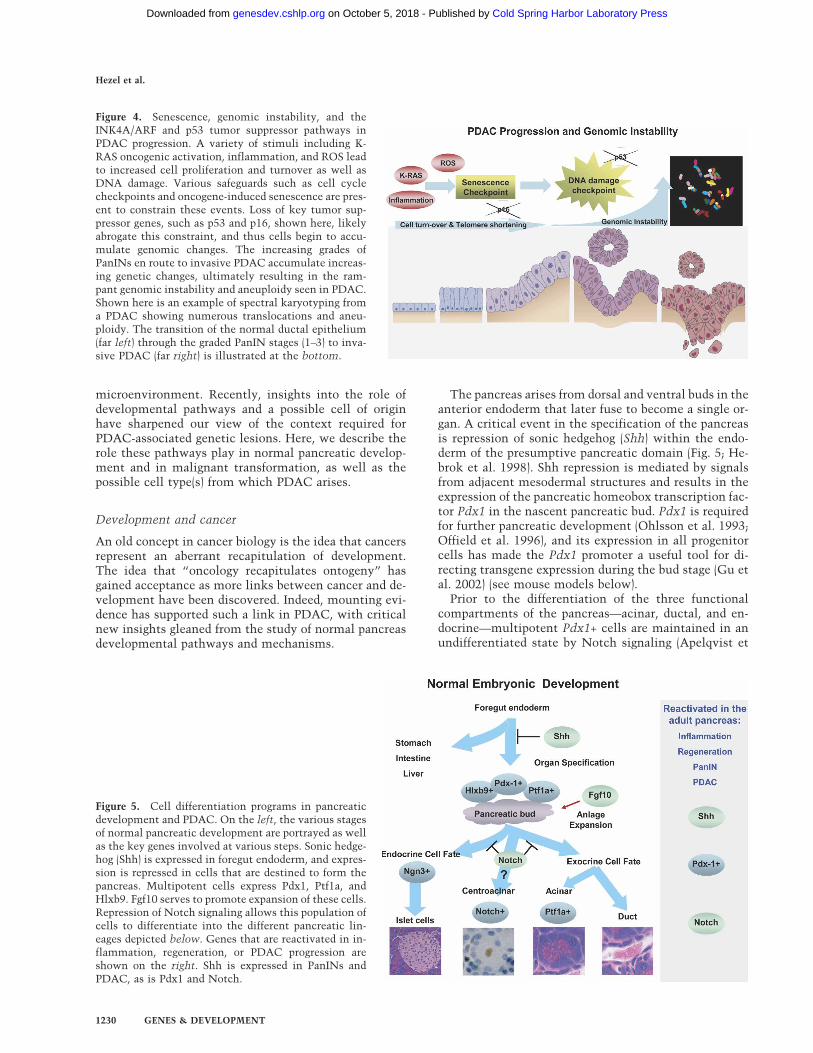
The observation across several tissue types, most no-tably colon and breast, of a histological evolution of nor-mal epithelium, through preneoplastic stages, to cancerin a graded manner has proven to be both clinically andscientifically informative (Kinzler and Vogelstein 1996).These observations have formed the backbone of mostgenetic progression models that have sought to charac-terize molecular profiles at each stage of neoplastic de-velopment. While evidence is suggestive of a dominantpattern of serial mutational events in the evolution to-ward PDAC, this linear tumor progression model willdraw continued scrutiny. Such a model must also takeinto account the altered states of differentiation ofPanINs and other precursor lesions, a potential cell orcells of origin, the role of developmental signaling path-ways, and an emerging knowledge of genomic and tran-scriptional alterations as they relate to each stage of dis-ease. A paradigm of pancreatic carcinogenesis must ac-curately reflect this expanding body of direct evidence.In particular, the acquisition of the genetic mutationsmay be irregular, occurring in fits and starts, rather thana measured process with consecutive mutations occur-ring at intervals in time (Fig. 4). This episodic mutationalactivity could be prompted by key events undermininggenomic integrity such as the loss of DNA damage repairand response checkpoints and the erosion of telomeres(Chin et al. 1999a, 2004; Maser and DePinho 2002). Un-derstanding the relationship between the deregulationand/or loss of these lynchpin cellular processes govern-ing genomic stability and the acquisition of a neoplasticgenetic profile will also be crucial to the development ofaccurate disease progression models. Indeed, such an un-derstanding of disease progression is vital for the rationaland effective implementation of early detection strate-gies and preventive therapies.
The cellular basis of PDAC
Molecular pathology and cancer genetic studies haveprovided an outline of the cellular perturbations that areassociated with PDAC; however, the current picture re-mains static, with only correlative links to underlyingtumor biology. A more direct mechanistic view of howclassical lesions influence pancreatic cancer biology isrequired, and some key questions need to be answered.An important attribute of the signaling pathways acti-vated in pancreatic cancer is their specificity—a permis-sive context is required for the cell-biological impact ofan activated oncogenic pathway to become manifest. Acomprehensive appreciation of PDAC pathogenesismust include consideration of the cell type, developmen-tal stage, the constellation of other genetic lesions, and
Pancreatic ductal adenocarcinoma
GENES & DEVELOPMENT 1229
Cold Spring Harbor Laboratory Press on October 5, 2018 - Published by genesdev.cshlp.orgDownloaded from
microenvironment. Recently, insights into the role ofdevelopmental pathways and a possible cell of originhave sharpened our view of the context required forPDAC-associated genetic lesions. Here, we describe therole these pathways play in normal pancreatic develop-ment and in malignant transformation, as well as thepossible cell type(s) from which PDAC arises.
Development and cancer
An old concept in cancer biology is the idea that cancersrepresent an aberrant recapitulation of development.The idea that “oncology recapitulates ontogeny” hasgained acceptance as more links between cancer and de-velopment have been discovered. Indeed, mounting evi-dence has supported such a link in PDAC, with criticalnew insights gleaned from the study of normal pancreasdevelopmental pathways and mechanisms.
The pancreas arises from dorsal and ventral buds in theanterior endoderm that later fuse to become a single or-gan. A critical event in the specification of the pancreasis repression of sonic hedgehog (Shh) within the endo-derm of the presumptive pancreatic domain (Fig. 5; He-brok et al. 1998). Shh repression is mediated by signalsfrom adjacent mesodermal structures and results in theexpression of the pancreatic homeobox transcription fac-tor Pdx1 in the nascent pancreatic bud. Pdx1 is requiredfor further pancreatic development (Ohlsson et al. 1993;Offield et al. 1996), and its expression in all progenitorcells has made the Pdx1 promoter a useful tool for di-recting transgene expression during the bud stage (Gu etal. 2002) (see mouse models below).
Prior to the differentiation of the three functionalcompartments of the pancreas—acinar, ductal, and en-docrine—multipotent Pdx1+ cells are maintained in anundifferentiated state by Notch signaling (Apelqvist et
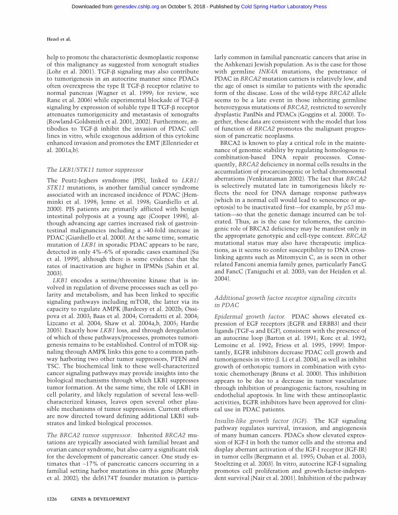
Figure 4. Senescence, genomic instability, and theINK4A/ARF and p53 tumor suppressor pathways inPDAC progression. A variety of stimuli including K-RAS oncogenic activation, inflammation, and ROS leadto increased cell proliferation and turnover as well asDNA damage. Various safeguards such as cell cyclecheckpoints and oncogene-induced senescence are pres-ent to constrain these events. Loss of key tumor sup-pressor genes, such as p53 and p16, shown here, likelyabrogate this constraint, and thus cells begin to accu-mulate genomic changes. The increasing grades ofPanINs en route to invasive PDAC accumulate increas-ing genetic changes, ultimately resulting in the ram-pant genomic instability and aneuploidy seen in PDAC.Shown here is an example of spectral karyotyping froma PDAC showing numerous translocations and aneu-ploidy. The transition of the normal ductal epithelium(far left) through the graded PanIN stages (1–3) to inva-sive PDAC (far right) is illustrated at the bottom.
Figure 5. Cell differentiation programs in pancreaticdevelopment and PDAC. On the left, the various stagesof normal pancreatic development are portrayed as wellas the key genes involved at various steps. Sonic hedge-hog (Shh) is expressed in foregut endoderm, and expres-sion is repressed in cells that are destined to form thepancreas. Multipotent cells express Pdx1, Ptf1a, andHlxb9. Fgf10 serves to promote expansion of these cells.Repression of Notch signaling allows this population ofcells to differentiate into the different pancreatic lin-eages depicted below. Genes that are reactivated in in-flammation, regeneration, or PDAC progression areshown on the right. Shh is expressed in PanINs andPDAC, as is Pdx1 and Notch.
Hezel et al.
1230 GENES & DEVELOPMENT
Cold Spring Harbor Laboratory Press on October 5, 2018 - Published by genesdev.cshlp.orgDownloaded from
al. 1999; Jensen et al. 2000; Moshous et al. 2001; Hald etal. 2003; Murtaugh et al. 2003). Mesenchymal FGF10promotes the expansion of these progenitor cells (Hart etal. 2003) in a manner reminiscent of lung development(Hogan 1999); FGF10 may also serve to integrate mor-phogenesis and differentiation by simultaneously regu-lating Notch signaling and cell division (Norgaard et al.2003). Following Notch repression, Pdx1+ progenitorsare capable of differentiating into distinct pancreatic lin-eages (Esni et al. 2004) through the regulated activity ofnumerous transcription factors (for review, see Mur-taugh and Melton 2003). The balance between endocrineand acinar cells seems to be regulated by the activity ofTGF-� family members (for review, see Kim and Hebrok2001), and more recent studies have demonstrated a rolefor Wnt signaling in pancreatic development (Dessimozet al. 2005; H.J. Kim et al. 2005; Murtaugh et al. 2005).The determinants of ductal lineage specification arelargely unknown.
The evidence that embryonic programs re-emerge dur-ing the development of pancreatic tumors comes fromtwo lines of investigation: characterization of gene ex-pression and functional analyses. Expression studieshave demonstrated the “reactivation” of several embry-onic genes during pancreatic carcinogenesis (Fig. 5).PDX1, whose expression in the adult is mostly limited topancreatic � cells, is expressed in nearly half of PDACs,where it carries a poor prognosis (Koizumi et al. 2003).SHH is normally absent from the pancreas throughoutdevelopment and adult life but is expressed in PanINsand PDAC, with expression levels that correlate withthe grade of the lesion (Berman et al. 2003; Thayer et al.2003). Similarly, Notch signaling is repressed during de-velopment to allow pancreatic differentiation, but Notchsignaling components are abundantly expressed inPanIN lesions and PDACs (Miyamoto et al. 2003). Al-though the role of Wnt signaling in PDAC pathogenesisremains to be defined, stabilization of �-catenin is fre-quently observed in pancreatoblastoma, a rare pediatrictumor of the pancreas (Abraham et al. 2001).
Functional data also support the importance of “reac-tivation” of embryonic programs—in particular SHH—in PDAC pathogenesis. Blocking SHH signaling with theinhibitor cyclopamine causes human PDAC cells to un-dergo apoptosis in vitro and lose tumorigenicity in xe-nograft assays (Thayer et al. 2003). Furthermore, activa-tion of hedgehog signaling in immortalized human pan-creatic ductal cells induces a PanIN-like transcriptional“signature” (Prasad et al. 2005). Importantly, this signa-ture includes several extrapancreatic markers of the fore-gut. This is consistent with the finding that ectopichedgehog expression within the pancreatic domain leadsto “intestinalization” of the pancreatic epithelium(Apelqvist et al. 1997; Thayer et al. 2003) and suggeststhat adoption of an intestinal phenotype is an importantstep in the formation of incipient PDAC. Although notdirectly studied in PDAC, cooperation of the RAS andNotch signaling pathways in transformation and otherbiological processes has been demonstrated in several invitro and in vivo systems (Weijzen et al. 2002; Kiaris et
al. 2004; Sundaram 2005). The applicability of thesestudies to PDAC has added significance in light of theidentification of Notch pathway activity in a candidateprecursor, the centroacinar cells (see below in next sec-tion).
Origins of pancreatic cancer. An emerging hypothesisthat is being explored in PDAC and many other solidtumors is that cancer precursors arise from stem cells—cells with the unique potential to self-renew and to dif-ferentiate into multiple lineages—that exist within adulttissues. While there is good evidence for such a model inthe hematopoietic system, where at least a subset of leu-kemias is derived from stem cells (Passegue et al. 2004),the “cell of origin” for most solid malignancies, includ-ing the pancreas, is unknown. The stem-cell origin hy-pothesis is supported by evidence that brain tumors arisefrom CD133+ neural stem cells (for review, see Singh etal. 2004a), and recent studies are suggestive of a stem cellorigin for cancer of the lung (C.F. Kim et al. 2005) andprostate (Maitland and Collins 2005). For other cancers,it remains to be determined whether tumors originatefrom a resident tissue stem cell, and whether highly tu-morigenic cells within a cancer reflect the persistence ofsuch a cell (see discussion of Cancer Stem Cells below).
Recent observations have fostered a more dynamicview of stem cells in which “stem-ness” represents adifferentiation state rather than a discrete entity (Blau etal. 2001). Thus, it is possible that cells with stem cellactivity may arise by “transdifferentiation” or “dediffer-entiation” of other cell types. A byproduct of this modelis the notion of “facultative” stem cells—differentiatedcells that have the potential to be stimulated to assumea stem cell role. Based on studies of cell renewal anddifferentiation, Bonner-Weir (Bonner-Weir and Sharma2002) has argued that all or nearly all of the pancreaticductal cells are potential facultative stem cells, with thecapacity to differentiate into both endocrine and exo-crine lineages. In rats subjected to partial pancreatec-tomy, the exocrine and endocrine compartments exhibitincreased cell division, and cells expressing the progeni-tor cell marker Pdx1 have been described as “dedifferen-tiating” and expanding from the pancreatic ducts (Bon-ner-Weir et al. 1997; Sharma et al. 1999). The appearanceof cells with a progenitor phenotype is also observed in avariety of rodent models of pancreatic damage (Vinik etal. 1997; Kritzik et al. 1999; Scoggins et al. 2000). Whilethese observations are consistent with a possible facul-tative activity of rodent duct cells, it should be notedthat the extent of pancreatic “regeneration” differs sig-nificantly depending on the method, and thus extent, ofinjury (e.g., recovery from pancreatitis is more robustthan regeneration following pancreatectomy), and thecontribution by such ductal cells to other pancreatic lin-eages has not been analyzed directly. It is noteworthythat certain paracrine signals implicated in the regula-tion of ductal proliferation in these injury models—TGF-�/EGF and HGF as initiating factors and TGF-� as aninhibitor—are also engaged during PDAC tumorigenesis
Pancreatic ductal adenocarcinoma
GENES & DEVELOPMENT 1231
Cold Spring Harbor Laboratory Press on October 5, 2018 - Published by genesdev.cshlp.orgDownloaded from
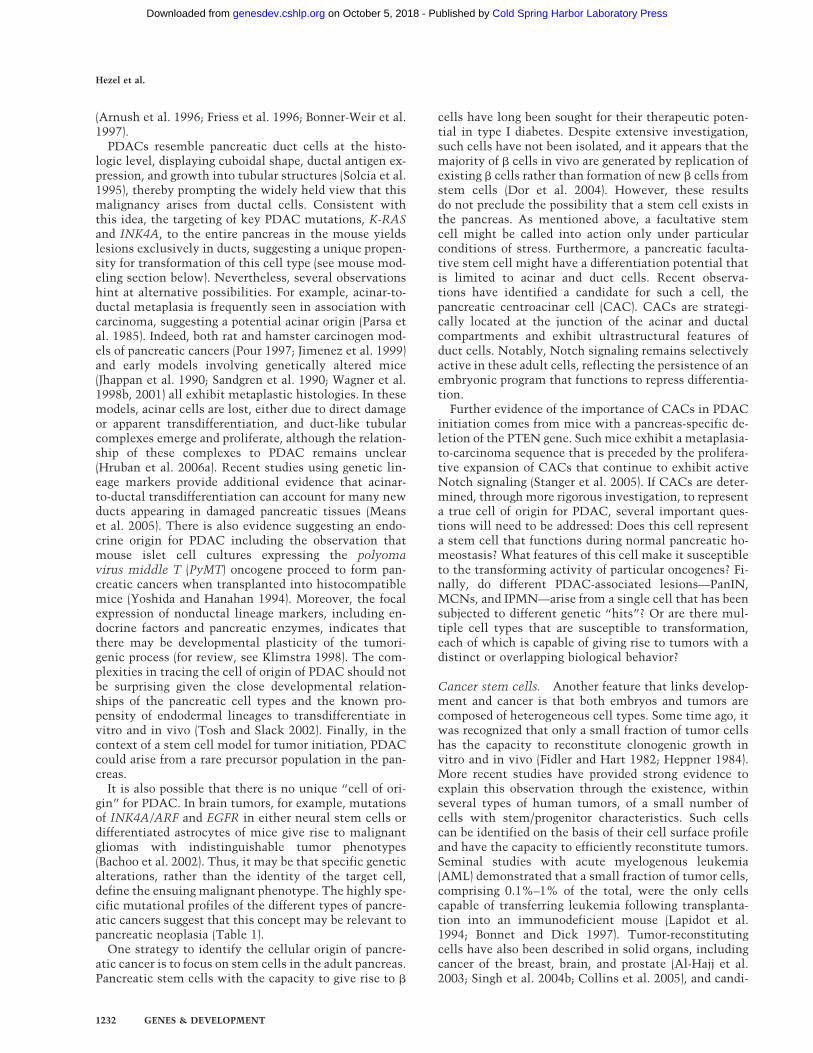
(Arnush et al. 1996; Friess et al. 1996; Bonner-Weir et al.1997).
PDACs resemble pancreatic duct cells at the histo-logic level, displaying cuboidal shape, ductal antigen ex-pression, and growth into tubular structures (Solcia et al.1995), thereby prompting the widely held view that thismalignancy arises from ductal cells. Consistent withthis idea, the targeting of key PDAC mutations, K-RASand INK4A, to the entire pancreas in the mouse yieldslesions exclusively in ducts, suggesting a unique propen-sity for transformation of this cell type (see mouse mod-eling section below). Nevertheless, several observationshint at alternative possibilities. For example, acinar-to-ductal metaplasia is frequently seen in association withcarcinoma, suggesting a potential acinar origin (Parsa etal. 1985). Indeed, both rat and hamster carcinogen mod-els of pancreatic cancers (Pour 1997; Jimenez et al. 1999)and early models involving genetically altered mice(Jhappan et al. 1990; Sandgren et al. 1990; Wagner et al.1998b, 2001) all exhibit metaplastic histologies. In thesemodels, acinar cells are lost, either due to direct damageor apparent transdifferentiation, and duct-like tubularcomplexes emerge and proliferate, although the relation-ship of these complexes to PDAC remains unclear(Hruban et al. 2006a). Recent studies using genetic lin-eage markers provide additional evidence that acinar-to-ductal transdifferentiation can account for many newducts appearing in damaged pancreatic tissues (Meanset al. 2005). There is also evidence suggesting an endo-crine origin for PDAC including the observation thatmouse islet cell cultures expressing the polyomavirus middle T (PyMT) oncogene proceed to form pan-creatic cancers when transplanted into histocompatiblemice (Yoshida and Hanahan 1994). Moreover, the focalexpression of nonductal lineage markers, including en-docrine factors and pancreatic enzymes, indicates thatthere may be developmental plasticity of the tumori-genic process (for review, see Klimstra 1998). The com-plexities in tracing the cell of origin of PDAC should notbe surprising given the close developmental relation-ships of the pancreatic cell types and the known pro-pensity of endodermal lineages to transdifferentiate invitro and in vivo (Tosh and Slack 2002). Finally, in thecontext of a stem cell model for tumor initiation, PDACcould arise from a rare precursor population in the pan-creas.
It is also possible that there is no unique “cell of ori-gin” for PDAC. In brain tumors, for example, mutationsof INK4A/ARF and EGFR in either neural stem cells ordifferentiated astrocytes of mice give rise to malignantgliomas with indistinguishable tumor phenotypes(Bachoo et al. 2002). Thus, it may be that specific geneticalterations, rather than the identity of the target cell,define the ensuing malignant phenotype. The highly spe-cific mutational profiles of the different types of pancre-atic cancers suggest that this concept may be relevant topancreatic neoplasia (Table 1).
One strategy to identify the cellular origin of pancre-atic cancer is to focus on stem cells in the adult pancreas.Pancreatic stem cells with the capacity to give rise to �
cells have long been sought for their therapeutic poten-tial in type I diabetes. Despite extensive investigation,such cells have not been isolated, and it appears that themajority of � cells in vivo are generated by replication ofexisting � cells rather than formation of new � cells fromstem cells (Dor et al. 2004). However, these resultsdo not preclude the possibility that a stem cell exists inthe pancreas. As mentioned above, a facultative stemcell might be called into action only under particularconditions of stress. Furthermore, a pancreatic faculta-tive stem cell might have a differentiation potential thatis limited to acinar and duct cells. Recent observa-tions have identified a candidate for such a cell, thepancreatic centroacinar cell (CAC). CACs are strategi-cally located at the junction of the acinar and ductalcompartments and exhibit ultrastructural features ofduct cells. Notably, Notch signaling remains selectivelyactive in these adult cells, reflecting the persistence of anembryonic program that functions to repress differentia-tion.
Further evidence of the importance of CACs in PDACinitiation comes from mice with a pancreas-specific de-letion of the PTEN gene. Such mice exhibit a metaplasia-to-carcinoma sequence that is preceded by the prolifera-tive expansion of CACs that continue to exhibit activeNotch signaling (Stanger et al. 2005). If CACs are deter-mined, through more rigorous investigation, to representa true cell of origin for PDAC, several important ques-tions will need to be addressed: Does this cell representa stem cell that functions during normal pancreatic ho-meostasis? What features of this cell make it susceptibleto the transforming activity of particular oncogenes? Fi-nally, do different PDAC-associated lesions—PanIN,MCNs, and IPMN—arise from a single cell that has beensubjected to different genetic “hits”? Or are there mul-tiple cell types that are susceptible to transformation,each of which is capable of giving rise to tumors with adistinct or overlapping biological behavior?
Cancer stem cells. Another feature that links develop-ment and cancer is that both embryos and tumors arecomposed of heterogeneous cell types. Some time ago, itwas recognized that only a small fraction of tumor cellshas the capacity to reconstitute clonogenic growth invitro and in vivo (Fidler and Hart 1982; Heppner 1984).More recent studies have provided strong evidence toexplain this observation through the existence, withinseveral types of human tumors, of a small number ofcells with stem/progenitor characteristics. Such cellscan be identified on the basis of their cell surface profileand have the capacity to efficiently reconstitute tumors.Seminal studies with acute myelogenous leukemia(AML) demonstrated that a small fraction of tumor cells,comprising 0.1%–1% of the total, were the only cellscapable of transferring leukemia following transplanta-tion into an immunodeficient mouse (Lapidot et al.1994; Bonnet and Dick 1997). Tumor-reconstitutingcells have also been described in solid organs, includingcancer of the breast, brain, and prostate (Al-Hajj et al.2003; Singh et al. 2004b; Collins et al. 2005), and candi-
Hezel et al.
1232 GENES & DEVELOPMENT
Cold Spring Harbor Laboratory Press on October 5, 2018 - Published by genesdev.cshlp.orgDownloaded from
dates have been found in lung neoplasia and melanoma(Fang et al. 2005; C.F. Kim et al. 2005).
It is currently unknown whether pancreatic cancersalso harbor so-called “cancer stem cells.” But since mostcancer therapies target the bulk of tumor cells, and sincetumor stem cells (like their normal tissue counterparts)may be more resistant to chemotherapy, the question isnot merely an academic one. Indeed, it is possible that amechanism to ensure cancer stem cell renewal is a re-quired aspect of PDAC pathogenesis and maintenance.Specifically, expression of the Bmi proto-oncogene is re-quired for self-renewal of both hematopoietic stem cellsand leukemia stem cells (for review, see Pardal et al.2003). As BMI acts by repressing INK4A/ARF, the invari-able loss of the INK4A/ARF locus in PDAC may reflecta mechanism by which pancreatic cancers ensure self-renewal of a “stem cell” population. Ultimately, if suchtumor-maintaining cells are found to exist in PDAC,comparison with the cell(s) from which PDAC arises(cell of origin) may provide great insight into the molecu-lar and cellular pathogenesis of the disease.
Genetically engineered mouse models of PDAC
The recurrent spectrum and the sequential appearance ofspecific mutational events point toward a defined mo-lecular program for PDAC progression. Genetically en-
gineered mice (GEM) have provided tractable in vivo sys-tems to dissect the biological impact of oncogenic mu-tations in a wide number of malignancies (for review ofother animal models of cancer, see Van Dyke and Jacks2002). Beyond establishing such genotype–phenotype re-lationships, these GEM have the potential to identifyearly markers of disease, pinpoint cooperating geneticalterations, and provide better preclinical models to in-form therapeutic initiatives. A wide range of mousemodels of pancreatic cancer has been built using variedtransgenic and gene targeting approaches. A key consid-eration has been deciding how to target mutant alleles tothe pancreas, or to specific pancreatic cell lineages. Ad-vances in pancreatic developmental biology have en-abled a generation of refined genetically engineeredmouse models that closely mirror many of the geneticand histologic characteristics of the human disease(Table 2). Furthermore, recent histopathologic review ofthese models has led to a consensus view among pancre-atic pathologists, creating a foundation for more accuratecomparisons across the different genetically engineeredmouse models and their relationship to the human dis-ease (Hruban et al. 2006a).
Targeting oncogenes to the acinar cell compart-ment. Early attempts to model exocrine pancreaticcancer used acinar-specific transgene expression, takingadvantage of the wide availability of promoters capable
Table 2. Mouse models of pancreatic cancer
Gene/promoter Phenotype of mouse
Transgenics with predominantly acinar phenotypesT-Ag/elastase Acinar cell carcinomaHras/elastase Acinar cell carcinomaTGF-�/elastase Acinar cell carcinoma
Develop mixed acinar-ductal tumors on a p53+/− backgroundTGF-�/metallothionein Tubular metaplasia. Develop lesions resembling serous cystadenomas on Ink4a/Arf- or p53-null
background.c-myc/elastase Mixed acinar-ductal tumorsKrasG12D/Mist1 Acinar cell carcinoma
Transgenics using the RCAS TVA systemc-myc/elastase Islet cell tumors in Ink4a/Arf-null micePyMT/elastase Mixed acinar-ductal tumors in Ink4a/Arf-null mice.
Activated Kras knock-in GEMKrasG12D Pdx1-Cre Spectrum of PanINs and some mice develop PDAC with long latencyKrasG12D Pdx1-CreInk4a −/−
Develop PDAC with shorter latency than KrasG12D alone.
KrasG12D Pdx1-CreInk4a/Arf−/−
Develop PDAC with high penetrance and short latency. Micrometastatic disease.
KrasG12D Pdx1-Crep53 R273H or p53+/−
Develop PDAC with high penetrance. Gross metastatic disease. LOH of wild-type p53 allele.
KrasG12D Pdx1-CreInk4a/Arf +/−
Develop PDAC with longer latency than Ink/Arf-null mice. Gross metastatic disease. LOH of wild-typeInk4a/Arf allele.
KrasG12D Pdx1-Crep53+/− Ink4a +/-
Develop PDAC with high penetrance and shorter latency than p53+/−. LOH of wild-type p53 allele andloss of Ink4a expression.
Other related GEM with PDAC or precursor phenotypesPten−/− Pdx1-Cre Ductal metaplasia with a fraction of the mice developing PDAC.Pdx-1-Shh Ductal-intestinal metaplasia
Pancreatic ductal adenocarcinoma
GENES & DEVELOPMENT 1233
Cold Spring Harbor Laboratory Press on October 5, 2018 - Published by genesdev.cshlp.orgDownloaded from
of targeting this compartment. Transgenic mice that ex-press SV40 large T antigen (T-Ag) (Ornitz et al. 1987;Glasner et al. 1992), activated H-RAS (Quaife et al. 1987),or c-Myc (Sandgren et al. 1991, 1993) in the acini usingthe Elastase (Ela) promoter develop acinar cell carcino-mas, although Ela-Myc tumors progress to mixed acinar-ductal histology. Transgenic mice expressing TGF-� inthe acinar cells (Ela-TGF-�) on a p53-deficient back-ground develop mixed acinar-ductal tumors or cystic aci-nar tumors (Wagner et al. 2001). Metallothionein-TGF-�(MT-TGF-�) transgenic mice also have acinar TGF-� ex-pression but do not develop PDAC even in the context ofTrp53 and Ink4a/Arf deficiency. Instead, these tumorsuppressor mutations cooperate to promote benign pan-creatic ductal lesions resembling serous cystadenomasin humans (Bardeesy et al. 2002a).
Acinar cells have also been targeted in a more recentreport using the RCAS-TVA system. This refined trans-genesis approach involves the somatic delivery of retro-viruses encoding genes of interest to specific cellularcompartments (Orsulic 2002; Pao et al. 2003). In studiesexploring the differential impact of specific oncogenes,TVA (the receptor for the avian leukosis sarcoma virussubgroup A [ALSV-A]) was placed under the control ofthe elastase promoter that is active in the acinar cells,creating elastase-tva transgenic mice (Lewis et al. 2003).The delivery of ALSV-A-based RCAS vectors encodingeither c-Myc or PyMT antigen to elastase-tva Ink4a/Arf-null mice yielded pancreatic tumors with distinct histo-logical phenotypes. The c-Myc-transduced animals de-veloped only islet cell tumors, whereas PyMT-trans-duced mice developed pancreatic tumors of mixed acinarand ductal features. In the setting of intact Ink4a/Arf,PyMT-transduced mice developed PanIN-like lesions ina subset of cases, suggesting a role for Ink4a/Arf in re-straining PanIN progression. The complex pattern of tu-mors with distinct lineages despite the targeting of theelastase-positive compartment (predominantly acinar)may relate to the introduction of RCAS viruses on post-natal day 2. At this stage, elastase-tva displayed a moreextensive distribution throughout the pancreas com-pared with the acinar-specific expression in the adultpancreas. These studies illustrate how both the specificidentity of an oncogene and the “developmental con-text” may influence the neoplastic phenotype.
Activated Kras GEM. Kras activation is the defininglesion in PDAC, prompting the generation of GEM inwhich activated Kras transgenes have been targeted tospecific lineages or a Kras knock-in allele has been acti-vated throughout the pancreatic epithelium via Cre re-combinase expression. The knock-in studies used aKrasG12D allele (referred to as LSL-KrasG12D) that is ex-pressed at the same level and in the same cell types asthe endogenous gene after Cre-mediated excision of aLoxP-flanked Stopper element. This system mimics theacquisition of such activating point mutations in humancancers.
Pdx1-Cre and Ptf1-p48-Cre deletor strains have beenused to activate Kras and induce mutations in the Ink4a/
Arf and/or p53 tumor suppressor loci in the pancreas.The Pdx1 and Ptf1-p48 promoters are active in the com-mon progenitors of all pancreatic cell types with rela-tively restricted expression outside of the pancreas (Pdx1is also expressed in the developing duodenum and stom-ach, and Ptf1-p48 is expressed in the cerebellum)(Kawaguchi et al. 2002; Gu et al. 2003). Cre-induced ac-tivation of LSL-KrasG12D leads to the rapid developmentof PanIN lesions in the first few weeks of life (Aguirre etal. 2003; Hingorani et al. 2003). A related knock-in stop-per KrasG12V-IRES-lacZ allele produced PanINs whencombined with a CDK4R24C point mutant allele, whichis refractory to INK4A inhibition (Guerra et al. 2003). Inthe KrasG12D-mutant GEM, the resulting neoplasmsshow a gradual, age-dependent progression of lesions re-sembling human PanINs-I–III and the development offrank PDAC after a long latency. Additionally, the pres-ence of normal ducts within these pancreases, all ofwhich harbor the activated KrasG12D allele, conveys thatother events, beyond Kras activation, are required to ini-tiate neoplastic changes. In summary, these studiesclearly illustrate the potent role of KrasG12D in initiatingPanIN development, while also indicating that otherrate-limiting events are likely to constrain progression ofthese KrasG12D-driven neoplasms toward high-gradePanINs and PDAC.
Exploring tumor suppressor function in PDAC progres-
sion. The tumor suppressor roles of p53 and Ink4a/Arfhave been investigated against the backdrop of the Pdx1-Cre LSL-Kras system. Mice with a pancreas-specific de-letion of Ink4a/Arf or p53 do not develop pancreatic neo-plasia. However, when Kras activation is combined withmutations of either tumor suppressor, a rapidly progres-sive and lethal PDAC phenotype emerges. Pdx1-CreLSL-Kras mice, homozygous for a conditional Ink4a/Arfallele, uniformly develop invasive PDAC by 7–11 wk(Aguirre et al. 2003); animals heterozygous for Ink4a/Arfalso developed PDAC but with longer latency (Bardeesyet al. 2006). These PDAC lesions showed histologic andmolecular resemblance to the human disease includingthe association with high-grade PanINs and a proliferat-ing stroma. Similarly, rapidly progressive PDAC occurswith the combination of the KrasG12D allele with a con-ditional null allele of p53 (Bardeesy et al. 2006) or a p53knock-in allele (p53 R273H, a point mutant observed inLi-Fraumeni syndrome) (Hingorani et al. 2005). Overall,these results demonstrate that Ink4a/Arf and p53 do notplay a primary role in the onset of PanIN but, rather,form a critical barrier in blocking progression of PanINinitiated by KrasG12D. The observed contributions ofKras to PanIN initiation and of Ink4a/Arf and p53 toPDAC progression in the mouse fit well with the se-quential appearance of Kras mutations in the earliest-stage PanIN and subsequent Ink4a/Arf and p53 muta-tions in more advanced lesions. It is notable that despitethe activation of Kras and deletion of tumor suppressorsin all pancreatic lineages, no neoplasia of the acini orislets was apparent.
Hezel et al.
1234 GENES & DEVELOPMENT
Cold Spring Harbor Laboratory Press on October 5, 2018 - Published by genesdev.cshlp.orgDownloaded from
As noted above, a significant proportion of humanPDACs show mutational inactivation of each of theInk4a, Arf, and p53 tumor suppressors, and hence a keyquestion is in determining the relative roles of thesegenes. In the Kras Ink4a/Arf model, all PDACs retain p53function, indicating that loss of p53 is not obligate fortumor progression in the mouse (Aguirre et al. 2003; Bar-deesy et al. 2006). In contrast, PDACs in the Krasp53R273H mice—and in mice with a heterozygous p53deletion—retain both Ink4a and Arf while losing expres-sion of the wild-type p53 allele (Hingorani et al. 2005;Bardeesy et al. 2006). One interpretation of these resultsis that Arf and p53 function in a common pathway tosuppress PDAC and that Ink4a loss may not be a criticalevent in murine PDAC progression. On the other hand,studies of Kras mice with combined p53 and Ink4a (butintact Arf) heterozygous deletions reveal loss of Ink4a inmost tumors—via deletion or promoter hypermethyl-ation of the wild-type allele—in addition to loss of wild-type p53 (Bardeesy et al. 2006). Moreover, these animalsdevelop PDAC with shorter latency than mice with het-erozygous p53 deletion and wild-type Ink4a. Finally,germline homozygous deletion of Ink4a accelerates thedevelopment of PDAC in Kras mice, although the la-tency is longer than in mice with deletion of both com-ponents of the Ink4a/Arf locus (Bardeesy et al. 2006).Together, these results suggest that each of these tumorsuppressors contributes to the control of PDAC progres-sion in the mouse; specifically, loss of Arf and p53 ap-pears to have redundant effects on tumorigenesis, andeither of these lesions may cooperate with Ink4a loss.While the retention of Ink4a in the mice engineered tosustain p53 mutations may point to a reduced functionof this tumor suppressor in murine PDAC, it is also pos-sible that engineered pre-existing p53 mutations earlyduring PanIN progression may create a context in whichthere is reduced selective pressure to mutate Ink4a, pos-sibly by facilitating other genetic events that deregulatethe Ink4a–Rb pathway. A notable feature of each of thesemodels is the maintenance of wild-type Smad4 expres-sion in all tumors. Engineering mutant Smad4 alleleswithin the context above described models will be im-portant in uncovering the specific role of this tumor sup-pressor in PDAC progression.
Considering these data from the mouse in relation tothe known genetics of human PanIN progression, itshould be pointed out that p53 mutations are not ob-served in PanIN-I or PanIN-II, whereas these lesionsshow frequent Ink4a loss; therefore, the presence of earlyp53 mutations may create biological conditions not nor-mally encountered in the pathogenesis of PDAC (Hrubanet al. 2000a,b). On the other hand, there is some evidencethat the circuitry regulating induction of Arf, p53, andInk4a in mouse cells may differ from that in humancells; hence there may not be complete overlap of thebiological function of these tumor suppressors acrossspecies (Rangarajan et al. 2004). Specifically, in humansomatic cells experiencing oxidative stress and chronicproliferation, the ensuing shortening of telomeres islikely to activate a DNA damage response pathway lead-
ing to p53 induction (Sharpless and DePinho 2002) (seeabove). In contrast, the long telomeres and constitutivetelomerase activity in the mouse ensure that advancingmurine PanINs do not exhibit telomere erosion and con-sequent activation of p53 (Prowse and Greider 1995).With respect to Arf and Ink4a, murine cells grown invitro show strong activation of Arf by stress stimuli suchas high oxygen tensions, high serum, and oncogene ex-pression, whereas stress stimuli in human cells prefer-entially activate Ink4a (Collins and Sedivy 2003; Brookeset al. 2004). It remains to be determined whether thereare, indeed, cross-species differences in the oncogeniccircuitry of PDAC and how additional perturbations tothe model, such as telomere dysfunction, may affect ge-netic and biological pressures in this disease. Notwith-standing these potential differences, it is clear that thesignature mutations associated with human PDAC alsocontribute to the pathogenesis of the murine tumors.
The different combinations of tumor suppressor genemutations, in conjunction with KrasG12D expression,produce tumors with varying spectra of clinical and his-tological features. While tumors of all genotypes are lo-cally invasive and show micro-metastases, gross metas-tases appear to be restricted to mice engineered to sus-tain heterozygous tumor suppressor deletions (Ink4a/Arflox/+ or p53lox/+ Ink4a lox/+ mice) and do not appear inmice with engineered homozygous deletions (Aguirre etal. 2003; Hingorani et al. 2005; Bardeesy et al. 2006). Thismay reflect the fact that the homozygous models de-velop multifocal tumors resulting in a rapidly lethal tu-mor burden, whereas the longer latency of heterozygousmodels affords the time for clonal maturation, progres-sion, and metastasis. With respect to the impact of geno-type on tumor histology, p53 deficiency is associatedwith a higher prevalence of well-differentiated ductal ad-enocarcinoma compared with the Ink4a/Arf-deficientanimals (Hingorani et al. 2005; Bardeesy et al. 2006).Conversely, undifferentiated sarcomatoid histology, afeature of the Ink/Arf model, is significantly reduced inp53-deficient models. In humans, ductal adenocarci-noma histology predominates, and the sarcomatoid sub-type is an uncommon variant of PDAC with more ag-gressive clinical behavior, although having a comparablespectrum of genetic lesions. These mouse models collec-tively recapitulate these histologic variants, albeit at dif-ferent frequencies from those seen in spontaneous hu-man tumors. Overall, these observations suggest that tu-mor suppressor lesions influence the cell differentiationphenotypes of the resulting tumors.
Mouse models and insights into the PDAC cell of ori-gin. The use of different approaches to express acti-vated Kras in the pancreas or in specific pancreatic lin-eages has begun to provide insights into the PDAC cell oforigin. As indicated above, in the case of the Pdx1-CreLSL-Kras models, all pancreatic cells including islets andacinar cells harbor an activated KrasG12D allele and in-activating tumor suppressor mutations, yet the only neo-plastic phenotype elicited is prominent PanINs andPDAC, while islet and acinar cancers are not observed.
Pancreatic ductal adenocarcinoma
GENES & DEVELOPMENT 1235
Cold Spring Harbor Laboratory Press on October 5, 2018 - Published by genesdev.cshlp.orgDownloaded from
Such observations favor the view that only certain celltypes within the pancreas are susceptible to transform-ing effects of physiological KrasG12D expression. On thesurface, such observations would favor the view of a duc-tal origin or possibly a centroacinar cell origin. At thesame time, while PanINs may appear to arise from dif-ferentiated ducts, it is worth noting that the targeting ofactivated Kras to mature ductal cells using the cytokera-tin-19 promoter failed to produce PanINs or PDAC, re-sulting, instead, in periductal inflammation (Brembecket al. 2003). In a separate set of experiments, elastase-directed activated Kras transgene expression in the aci-nar cells yielded a spectrum of neoplasms including aci-nar carcinomas and ductal lesions with resemblance toPanIN (Brembeck et al. 2003; Grippo et al. 2003). A simi-lar phenotype was observed with the targeting of Kras tothe Mist1 locus, which is expressed at low levels early inpancreatic development and at higher levels in the adultacinar compartment (Tuveson et al. 2006). Taken to-gether, these studies suggest that the ensuing Kras-driven neoplastic phenotypes are determined by the celltype, cellular differentiation state, and/or the level ofKras expression. The close recapitulation in the Pdx1-Cre LSL-Kras models of human PanIN to PDAC progres-sion suggests that these mouse neoplasms may share acommon cellular origin with the human counterpart,and, thus, a detailed analysis of incipient neoplasms inthis model might provide insights into the cellular com-partment(s) susceptible to transformation. It is temptingto speculate that the activation of Kras in all cell typeswithin the pancreas using the Pdx1-driven Cre may tar-get a uniquely susceptible cell compartment such as apancreatic duct precursor cell, a centroacinar cell, or anas-yet-uncharacterized cell type.
Genomic instability profiles in GEM. Widespreadchromosomal instability is a defining characteristic ofhuman PDAC. While the presence of such complexkaryotypes in the vast majority of human carcinomashas long been documented, the pathogenetic significancein driving tumorigenesis and the underlying genome in-stability mechanisms are areas of ongoing study and dis-cussion (Duesberg and Rasnick 2000; Rajagopalan andLengauer 2004). Origins of chromosomal instability incancer are likely to include diverse defects in the mitoticspindle apparatus, various checkpoint pathways such asp53, telomere dysfunction, increased ROS, and DNA re-pair pathways such as nucleotide excision repair (NER)and nonhomologous end-joining (Sharpless et al. 2001b;Maser and DePinho 2002; Woo and Poon 2004; Ciminiand Degrassi 2005; Kops et al. 2005). The observation ofpolyploidy in association with aging and senescence andits observation in premalignant lesions have suggestedthat such an intermediate karyotype may play a role inthe acquisition of aneuploidy (Storchova and Pellman2004). It is likely that a collusion of defects in many ofthe above pathways contribute to the rampant instabil-ity profile of human PDAC.
The recent genomic analyses, using both array-CGHstudies and spectral karyotyping (SKY), have provided
both quantitative and qualitative measures of genomicinstability among several of the above-described mousePDAC models. Importantly, these studies have sug-gested that some of the mechanisms driving genomicinstability are present in these models (Schreiner et al.2003; Hingorani et al. 2005; Bardeesy et al. 2006). In gen-eral, all of the evaluated PDAC models have shown evi-dence of global genomic alteration, and the comparisonof specific models has pointed toward possible influ-ences of genotype on genomic stability. p53 mutant tu-mors demonstrate a modest increase in copy number al-terations in comparison with PDACs arising on anInk4a/Arf mutant background (Bardeesy et al. 2006). Inaddition to these differences in overall genomic com-plexity among cohorts, the patterns of regional genomicchanges fell into groups when analyzed by nonhierarchi-cal clustering algorithms. Moreover, specific regions ofalteration were associated with particular genotypes; tu-mors harboring p53 mutations showed frequent Myc am-plifications, while those associated with Ink4a/Arf mu-tations showed highly recurrent Kras amplifications(Bardeesy et al. 2006).
Defining the anatomy of genomic rearrangements bySKY analysis of mouse PDAC has documented chromo-somal aberrations (e.g., NRTs, tetraploidy, and wholechromosomal gains and losses) reminiscent of the hu-man cytogenetic profiles (Hingorani et al. 2005). At thesame time, it is worth noting that the absolute degree ofchromosomal structural aberrations in the current col-lection of GEM appears less than that of human PDAC.Modeling genomic instability in other mouse cancermodels has been achieved using engineered mutantswith defects in several of these processes including DNArepair genes, checkpoint controls, and telomere mainte-nance. As we have speculated above, engineering telo-mere-based crisis into the various genetically engineeredmouse models may prove useful in driving comparablelevels of NRTs, amplifications, and deletions. Other keyareas that merit further study for potential effects ongenomic stability are the mitotic spindle dynamics,DNA repair mechanisms including mismatch repair andnonhomologous recombination, and the initiation ofPDAC in older adult mice rather than in embryonicstages. The identification of syntenic regions of genomicgain and loss across mouse and human PDAC data setsmay enhance cancer gene discovery efforts. Indeed, theuse of cross-species comparison to discern a Kras onco-genic expression signature has offered support to thisidea, demonstrating the utility of using murine tumorsto uncover patterns of gene expression that are present inhuman cancers (Sweet-Cordero et al. 2005).
Major challenges and opportunities in pancreas cancer
Important insights into PDAC biology have been made.A focus on familial and epidemiologic risk factors,pathologic progression, and molecular characterizationhas pointed toward key processes and pathways govern-ing PDAC genesis and evolution. An increasing numberof the genetic changes have been experimentally verified
Hezel et al.
1236 GENES & DEVELOPMENT
Cold Spring Harbor Laboratory Press on October 5, 2018 - Published by genesdev.cshlp.orgDownloaded from
across a range of systems, and the recent development ofmouse model systems has allowed for functional analy-sis of the various PDAC mutations in vivo. Importantroles of developmental pathways, including Notch andSHH, have been identified and have pointed toward apossible cell of origin for the disease. Overall, the gen-eration of robust model systems and the availability ofcomprehensive molecular profiles have created a strongfoundation for ongoing discovery.
Key outstanding questions remain. In terms of basicbiology, these include definitively establishing the cell(s)of origin, understanding the role of transdifferentiation,and the identification and definition of a cancer stem cellpopulation in PDAC. In addition, the role of the mesen-chyme and stroma, which influence pancreatic develop-ment and are prominent components of PDAC, must bedefined. Progress in reducing the tremendous morbidityand mortality could come with ongoing efforts focusedon understanding the biology of early disease states andidentifying markers of disease. Along these lines, the re-search community should pay particular attention to theevolution of PanIN and other premalignant lesions intoPDAC, focusing on critical shifts in the behavior of theselesions and how they relate to underlying molecular al-terations, checkpoint responses, genomic complexity,and the activation of key signaling cascades. Linkingsuch molecular processes that are indicative of a histo-pathologic stage of disease with a screening test (be it amolecular imaging reagent or proteomic marker presentin serum) could have a profound impact on clinical prac-tice and patient outcome.
The capacity to accurately model PDAC in the mousehas created the opportunity to study the biologic effect ofcancer genes; to characterize genomic instability, angio-genesis, and the tumor microenvironment; and to pro-vide insight into pathways and molecules that couldserve as targets for therapy. Moreover, these modelsserve as a valuable resource to test candidate compoundsfor their therapeutic potential. The development of in-ducible PDAC models is an important priority. Studiesof transgenic melanoma and lung adenocarcinoma mod-els directed by inducible H- and K-ras alleles, respec-tively, have shown that sustained mutant RAS activityis necessary for both the initiation of tumorigenesis andfor maintenance of the transformed state (Chin et al.1999b; Fisher et al. 2001). Existing evidence suggests thatK-RAS is critical for PDAC maintanence. An induciblemodel could serve to validate this in vivo and uncoverprecisely how K-RAS may function to support advancedPDAC. Expression profiling analysis using such an in-ducible system could point toward downstream RAS ef-fectors and potential therapeutic targets (Bardeesy et al.2005). Additionally, inducible model systems could pro-vide serum proteomic profiles specifically regulated byK-RAS activity across the spectrum of disease states,from incipient PanIN, to PDAC, and ultimately meta-static disease. These could lead to the better selection ofdisease markers for future clinical screening tests.
Therapeutic options can be advanced through the de-velopment of drugs targeting key pathways and mol-
ecules. As discussed in detail, the inability to pharma-cologically target K-RAS leaves the research communitywith known downstream pathways, supporting signalingnetworks, and large numbers of oncogene candidates.Presently known RAS effectors (MEK, PI3K components,etc.) (see Fig. 3 for further detail) and other relevantpathways including SHH and Notch have inhibitors invarious stages of development. Multi-agent targetedcombinations are likely to be required. The rational con-struction of such regimens will require consideration ofdisease biology as well as the understanding of how tar-gets function in normal organisms. Mouse model sys-tems may allow us to best address this preclinically, sav-ing valuable resources for the clinical testing of thosecombinations most likely to benefit patients. Given thecollusion of technologies across the spectrum of pro-teomic, molecular-genetic, imaging, and biologic fields,and the recent advances in PDAC research, the promiseof scientific and medical progress appears to be withinreach.
Acknowledgments
We thank Anirban Maitra and Anna Means for their criticalreading of the manuscript and insightful comments, and Gre-gory Lauwers (Department of Pathology, Massachusettes Gen-eral Hospital) for generously contributing his expertise and pro-viding the images used in Figure 2. The original illustrationsused in Figures 1 and 4 were created by Josh Berta. A.F.H. wassupported by the PanCAN/ASCO Samual Stroum young inves-tigator award. B.Z.S. was supported by NIH K08 DK064136.A.C.K. is a recipient of the Leonard B. Holman Research Path-way fellowship. N.B. was supported by NIH K01CA104647A-01A1 and grants from NCI/SAIC Frederick and the HarvardStem Cell Institute. R.A.D. is an American Cancer Society Re-search Professor and an Ellison Medical Foundation SeniorScholar, and is supported by the Robert A. and Renee E. BelferFoundation, Institute for Innovative Cancer Science, and NIHU01CA84313-07 and P01CA117969-01 grants.
References
Abraham, S.C., Wu, T.T., Klimstra, D.S., Finn, L.S., Lee, J.H.,Yeo, C.J., Cameron, J.L., and Hruban, R.H. 2001. Distinctivemolecular genetic alterations in sporadic and familial adeno-matous polyposis-associated pancreatoblastomas: Frequentalterations in the APC/�-catenin pathway and chromosome11p. Am. J. Pathol. 159: 1619–1627.
Adsay, N.V., Merati, K., Basturk, O., Iacobuzio-Donahue, C.,Levi, E., Cheng, J.D., Sarkar, F.H., Hruban, R.H., and Klim-stra, D.S. 2004. Pathologically and biologically distinct typesof epithelium in intraductal papillary mucinous neoplasms:Delineation of an ‘intestinal’ pathway of carcinogenesis inthe pancreas. Am. J. Surg. Pathol. 28: 839–848.
Agbunag, C. and Bar-Sagi, D. 2004. Oncogenic K-ras drives cellcycle progression and phenotypic conversion of primary pan-creatic duct epithelial cells. Cancer Res. 64: 5659–5663.
Aguirre, A.J., Bardeesy, N., Sinha, M., Lopez, L., Tuveson, D.A.,Horner, J., Redston, M.S., and DePinho, R.A. 2003. ActivatedKras and Ink4a/Arf deficiency cooperate to produce meta-static pancreatic ductal adenocarcinoma. Genes & Dev. 17:3112–3126.
Aguirre, A.J., Brennan, C., Bailey, G., Sinha, R., Feng, B., Leo, C.,
Pancreatic ductal adenocarcinoma
GENES & DEVELOPMENT 1237
Cold Spring Harbor Laboratory Press on October 5, 2018 - Published by genesdev.cshlp.orgDownloaded from
Zhang, Y., Zhang, J., Gans, J.D., Bardeesy, N., et al. 2004.High-resolution characterization of the pancreatic adenocar-cinoma genome. Proc. Natl. Acad. Sci. 101: 9067–9072.
Akhurst, R.J. and Derynck, R. 2001. TGF-� signaling in can-cer—A double-edged sword. Trends Cell Biol. 11: S44–S51.
Al-Hajj, M., Wicha, M.S., Benito-Hernandez, A., Morrison, S.J.,and Clarke, M.F. 2003. Prospective identification of tumori-genic breast cancer cells. Proc. Natl. Acad. Sci. 100: 3983–3988.
Almoguera, C., Shibata, D., Forrester, K., Martin, J., Arnheim,N., and Perucho, M. 1988. Most human carcinomas of theexocrine pancreas contain mutant c-K-ras genes. Cell 53:549–554.
Altomare, D.A., Tanno, S., De Rienzo, A., Klein-Szanto, A.J.,Tanno, S., Skele, K.L., Hoffman, J.P., and Testa, J.R. 2003.Frequent activation of AKT2 kinase in human pancreaticcarcinomas. J. Cell. Biochem. 88: 470–476.
Apelqvist, A., Ahlgren, U., and Edlund, H. 1997. Sonic hedgehogdirects specialised mesoderm differentiation in the intestineand pancreas. Curr. Biol. 7: 801–804.
Apelqvist, A., Li, H., Sommer, L., Beatus, P., Anderson, D.J.,Honjo, T., Hrabe de Angelis, M., Lendahl, U., and Edlund, H.1999. Notch signalling controls pancreatic cell differentia-tion. Nature 400: 877–881.
Argani, P., Rosty, C., Reiter, R.E., Wilentz, R.E., Murugesan,S.R., Leach, S.D., Ryu, B., Skinner, H.G., Goggins, M., Jaffee,E.M., et al. 2001. Discovery of new markers of cancerthrough serial analysis of gene expression: Prostate stem cellantigen is overexpressed in pancreatic adenocarcinoma.Cancer Res. 61: 4320–4324.
Arlt, A., Gehrz, A., Muerkoster, S., Vorndamm, J., Kruse, M.L.,Folsch, U.R., and Schafer, H. 2003. Role of NF-�B and Akt/PI3K in the resistance of pancreatic carcinoma cell linesagainst gemcitabine-induced cell death. Oncogene 22: 3243–3251.
Armengol, G., Knuutila, S., Lluis, F., Capella, G., Miro, R., andCaballin, M.R. 2000. DNA copy number changes and evalu-ation of MYC, IGF1R, and FES amplification in xenografts ofpancreatic adenocarcinoma. Cancer Genet. Cytogenet. 116:133–141.
Arnush, M., Gu, D., Baugh, C., Sawyer, S.P., Mroczkowski, B.,Krahl, T., and Sarvetnick, N. 1996. Growth factors in theregenerating pancreas of �-interferon transgenic mice. Lab.Invest. 74: 985–990.
Arsura, M., Mercurio, F., Oliver, A.L., Thorgeirsson, S.S., andSonenshein, G.E. 2000. Role of the I�B kinase complex inoncogenic Ras- and Raf-mediated transformation of rat liverepithelial cells. Mol. Cell. Biol. 20: 5381–5391.
Artandi, S.E., Chang, S., Lee, S.L., Alson, S., Gottlieb, G.J., Chin,L., and DePinho, R.A. 2000. Telomere dysfunction promotesnon-reciprocal translocations and epithelial cancers in mice.Nature 406: 641–645.
Asano, T., Yao, Y., Zhu, J., Li, D., Abbruzzese, J.L., and Reddy,S.A. 2004. The PI 3-kinase/Akt signaling pathway is acti-vated due to aberrant Pten expression and targets transcrip-tion factors NF-�B and c-Myc in pancreatic cancer cells. On-cogene 23: 8571–8580.
———. 2005. The rapamycin analog CCI-779 is a potent inhibi-tor of pancreatic cancer cell proliferation. Biochem. Biophys.Res. Commun. 331: 295–302.
Baas, A.F., Kuipers, J., van der Wel, N.N., Batlle, E., Koerten,H.K., Peters, P.J., and Clevers, H.C. 2004. Complete polar-ization of single intestinal epithelial cells upon activation ofLKB1 by STRAD. Cell 116: 457–466.
Baccarini, M. 2005. Second nature: Biological functions of theRaf-1 ‘kinase.’ FEBS Lett. 579: 3271–3277.
Bachoo, R.M., Maher, E.A., Ligon, K.L., Sharpless, N.E., Chan,S.S., You, M.J., Tang, Y., DeFrances, J., Stover, E., Weiss-leder, R., et al. 2002. Epidermal growth factor receptor andInk4a/Arf. Convergent mechanisms governing terminal dif-ferentiation and transformation along the neural stem cell toastrocyte axis. Cancer Cell 1: 269–277.
Bardeesy, N., Morgan, J., Sinha, M., Signoretti, S., Srivastava, S.,Loda, M., Merlino, G., and DePinho, R.A. 2002a. Obligateroles for p16(Ink4a) and p19(Arf)–p53 in the suppression ofmurine pancreatic neoplasia. Mol. Cell. Biol. 22: 635–643.
Bardeesy, N., Sinha, M., Hezel, A.F., Signoretti, S., Hathaway,N.A., Sharpless, N.E., Loda, M., Carrasco, D.R., and De-Pinho, R.A. 2002b. Loss of the Lkb1 tumour suppressor pro-vokes intestinal polyposis but resistance to transformation.Nature 419: 162–167.
Bardeesy, N., Kim, M., Xu, J., Kim, R.S., Shen, Q., Bosenberg,M.W., Wong, W.H., and Chin, L. 2005. Role of epidermalgrowth factor receptor signaling in RAS-driven melanoma.Mol. Cell. Biol. 25: 4176–4188.
Bardeesy, N., Aguirre, A., Chu, G., Cheng, K., Lopez, L., Hezel,A.F., Feng, B., Brennan, C., Weissleder, R., Mahmood, U., etal. 2006. Both p16Ink4a and the p19Arf–p53 pathway con-strain progression of pancreatic adenocarcinoma in themouse. Proc. Natl. Acad. Sci. 103: 5947–5952.
Barton, C.M., Hall, P.A., Hughes, C.M., Gullick, W.J., andLemoine, N.R. 1991. Transforming growth factor � and epi-dermal growth factor in human pancreatic cancer. J. Pathol.163: 111–116.
Bashyam, M.D., Bair, R., Kim, Y.H., Wang, P., Hernandez-Bous-sard, T., Karikari, C.A., Tibshirani, R., Maitra, A., and Pol-lack, J.R. 2005. Array-based comparative genomic hybridiza-tion identifies localized DNA amplifications and homozy-gous deletions in pancreatic cancer. Neoplasia 7: 556–562.
Beachy, P.A., Karhadkar, S.S., and Berman, D.M. 2004. Tissuerepair and stem cell renewal in carcinogenesis. Nature 432:324–331.
Beausejour, C.M., Krtolica, A., Galimi, F., Narita, M., Lowe,S.W., Yaswen, P., and Campisi, J. 2003. Reversal of humancellular senescence: Roles of the p53 and p16 pathways.EMBO J. 22: 4212–4222.
Benanti, J.A. and Galloway, D.A. 2004. Normal human fibro-blasts are resistant to RAS-induced senescence. Mol. Cell.Biol. 24: 2842–2852.
Bergmann, U., Funatomi, H., Yokoyama, M., Beger, H.G., andKorc, M. 1995. Insulin-like growth factor I overexpression inhuman pancreatic cancer: Evidence for autocrine and para-crine roles. Cancer Res. 55: 2007–2011.
Berman, D.M., Karhadkar, S.S., Maitra, A., Montes De Oca, R.,Gerstenblith, M.R., Briggs, K., Parker, A.R., Shimada, Y.,Eshleman, J.R., Watkins, D.N., et al. 2003. Widespread re-quirement for Hedgehog ligand stimulation in growth of di-gestive tract tumours. Nature 425: 846–851.
Berrington de Gonzalez, A., Sweetland, S., and Spencer, E. 2003.A meta-analysis of obesity and the risk of pancreatic cancer.Br. J. Cancer 89: 519–523.
Bharti, A.C. and Aggarwal, B.B. 2002. Nuclear factor-� B andcancer: Its role in prevention and therapy. Biochem. Phar-macol. 64: 883–888.
Biankin, A.V., Morey, A.L., Lee, C.S., Kench, J.G., Biankin, S.A.,Hook, H.C., Head, D.R., Hugh, T.B., Sutherland, R.L., andHenshall, S.M. 2002. DPC4/Smad4 expression and outcomein pancreatic ductal adenocarcinoma. J. Clin. Oncol. 20:4531–4542.
Blau, H.M., Brazelton, T.R., and Weimann, J.M. 2001. Theevolving concept of a stem cell: Entity or function? Cell 105:829–841.
Hezel et al.
1238 GENES & DEVELOPMENT
Cold Spring Harbor Laboratory Press on October 5, 2018 - Published by genesdev.cshlp.orgDownloaded from
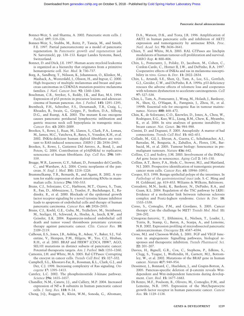
Bonner-Weir, S. and Sharma, A. 2002. Pancreatic stem cells. J.Pathol. 197: 519–526.
Bonner-Weir, S., Stubbs, M., Reitz, P., Taneja, M., and Smith,F.E. 1997. Partial pancreatectomy as a model of pancreaticregeneration. In Pancreatic growth and regeneration (ed.N. Sarvetnick), pp. 138–153. Karger Landes Systems, Basel,Switzerland.
Bonnet, D. and Dick, J.E. 1997. Human acute myeloid leukemiais organized as a hierarchy that originates from a primitivehematopoietic cell. Nat. Med. 3: 730–737.
Borg, A., Sandberg, T., Nilsson, K., Johannsson, O., Klinker, M.,Masback, A., Westerdahl, J., Olsson, H., and Ingvar, C. 2000.High frequency of multiple melanomas and breast and pan-creas carcinomas in CDKN2A mutation-positive melanomafamilies. J. Natl. Cancer Inst. 92: 1260–1266.
Boschman, C.R., Stryker, S., Reddy, J.K., and Rao, M.S. 1994.Expression of p53 protein in precursor lesions and adenocar-cinoma of human pancreas. Am. J. Pathol. 145: 1291–1295.
Brembeck, F.H., Schreiber, F.S., Deramaudt, T.B., Craig, L.,Rhoades, B., Swain, G., Grippo, P., Stoffers, D.A., Silberg,D.G., and Rustgi, A.K. 2003. The mutant K-ras oncogenecauses pancreatic periductal lymphocytic infiltration andgastric mucous neck cell hyperplasia in transgenic mice.Cancer Res. 63: 2005–2009.
Brookes, S., Rowe, J., Ruas, M., Llanos, S., Clark, P.A., Lomax,M., James, M.C., Vatcheva, R., Bates, S., Vousden, K.H., et al.2002. INK4a-deficient human diploid fibroblasts are resis-tant to RAS-induced senescence. EMBO J. 21: 2936–2945.
Brookes, S., Rowe, J., Gutierrez Del Arroyo, A., Bond, J., andPeters, G. 2004. Contribution of p16(INK4a) to replicativesenescence of human fibroblasts. Exp. Cell Res. 298: 549–559.
Brugge, W.R., Lauwers, G.Y., Sahani, D., Fernandez-del Castillo,C., and Warshaw, A.L. 2004. Cystic neoplasms of the pan-creas. N. Engl. J. Med. 351: 1218–1226.
Brummelkamp, T.R., Bernards, R., and Agami, R. 2002. A sys-tem for stable expression of short interfering RNAs in mam-malian cells. Science 296: 550–553.
Bruns, C.J., Solorzano, C.C., Harbison, M.T., Ozawa, S., Tsan,R., Fan, D., Abbruzzese, J., Traxler, P., Buchdunger, E., Ra-dinsky, R., et al. 2000. Blockade of the epidermal growthfactor receptor signaling by a novel tyrosine kinase inhibitorleads to apoptosis of endothelial cells and therapy of humanpancreatic carcinoma. Cancer Res. 60: 2926–2935.
Bruns, C.J., Koehl, G.E., Guba, M., Yezhelyev, M., Steinbauer,M., Seeliger, H., Schwend, A., Hoehn, A., Jauch, K.W., andGeissler, E.K. 2004. Rapamycin-induced endothelial celldeath and tumor vessel thrombosis potentiate cytotoxictherapy against pancreatic cancer. Clin. Cancer Res. 10:2109–2119.
Calhoun, E.S., Jones, J.B., Ashfaq, R., Adsay, V., Baker, S.J., Val-entine, V., Hempen, P.M., Hilgers, W., Yeo, C.J., Hruban,R.H., et al. 2003. BRAF and FBXW7 (CDC4, FBW7, AGO,SEL10) mutations in distinct subsets of pancreatic cancer:Potential therapeutic targets. Am. J. Pathol. 163: 1255–1260.
Camonis, J.H. and White, M.A. 2005. Ral GTPases: Corruptingthe exocyst in cancer cells. Trends Cell Biol. 15: 327–332.
Campbell, S.L., Khosravi-Far, R., Rossman, K.L., Clark, G.J., andDer, C.J. 1998. Increasing complexity of Ras signaling. On-cogene 17: 1395–1413.
Cantley, L.C. 2002. The phosphoinositide 3-kinase pathway.Science 296: 1655–1657.
Chandler, N.M., Canete, J.J., and Callery, M.P. 2004. Increasedexpression of NF-� B subunits in human pancreatic cancercells. J. Surg. Res. 118: 9–14.
Cheng, J.Q., Ruggeri, B., Klein, W.M., Sonoda, G., Altomare,
D.A., Watson, D.K., and Testa, J.R. 1996. Amplification ofAKT2 in human pancreatic cells and inhibition of AKT2expression and tumorigenicity by antisense RNA. Proc.Natl. Acad. Sci. 93: 3636–3641.
Chien, Y. and White, M.A. 2003. RAL GTPases are linchpinmodulators of human tumour-cell proliferation and survival.EMBO Rep. 4: 800–806.
Chin, L., Pomerantz, J., Polsky, D., Jacobson, M., Cohen, C.,Cordon-Cardo, C., Horner II, J.W., and DePinho, R.A. 1997.Cooperative effects of INK4a and ras in melanoma suscepti-bility in vivo. Genes & Dev. 11: 2822–2834.
Chin, L., Artandi, S.E., Shen, Q., Tam, A., Lee, S.L., Gottlieb,G.J., Greider, C.W., and DePinho, R.A. 1999a. p53 deficiencyrescues the adverse effects of telomere loss and cooperateswith telomere dysfunction to accelerate carcinogenesis. Cell97: 527–538.
Chin, L., Tam, A., Pomerantz, J., Wong, M., Holash, J., Bardeesy,N., Shen, Q., O’Hagan, R., Pantginis, J., Zhou, H., et al.1999b. Essential role for oncogenic Ras in tumour mainte-nance. Nature 400: 468–472.
Chin, K., de Solorzano, C.O., Knowles, D., Jones, A., Chou, W.,Rodriguez, E.G., Kuo, W.L., Ljung, B.M., Chew, K., Myambo,K., et al. 2004. In situ analyses of genome instability inbreast cancer. Nat. Genet. 36: 984–988.
Cimini, D. and Degrassi, F. 2005. Aneuploidy: A matter of badconnections. Trends Cell Biol. 15: 442–451.
Collado, M., Gil, J., Efeyan, A., Guerra, C., Schuhmacher, A.J.,Barradas, M., Benguria, A., Zaballos, A., Flores, J.M., Bar-bacid, M., et al. 2005. Tumour biology: Senescence in pre-malignant tumours. Nature 436: 642.
Collins, C.J. and Sedivy, J.M. 2003. Involvement of the INK4a/Arf gene locus in senescence. Aging Cell 2: 145–150.
Collins, A.T., Berry, P.A., Hyde, C., Stower, M.J., and Maitland,N.J. 2005. Prospective identification of tumorigenic prostatecancer stem cells. Cancer Res. 65: 10946–10951.
Cooper, H.S. 1998. Benign epithelial polyps of the intestines. InPathology of the gastrointestinal tract (eds. S.-C. Ming andH. Goldman), pp. 819–853. Wiliams & Wilkens, Baltimore.
Corradetti, M.N., Inoki, K., Bardeesy, N., DePinho, R.A., andGuan, K.L. 2004. Regulation of the TSC pathway by LKB1:Evidence of a molecular link between tuberous sclerosiscomplex and Peutz-Jeghers syndrome. Genes & Dev. 18:1533–1538.
Corso, S., Comoglio, P.M., and Giordano, S. 2005. Cancertherapy: Can the challenge be MET? Trends Mol. Med. 11:284–292.
Crnogorac-Jurcevic, T., Efthimiou, E., Nielsen, T., Loader, J.,Terris, B., Stamp, G., Baron, A., Scarpa, A., and Lemoine,N.R. 2002. Expression profiling of microdissected pancreaticadenocarcinomas. Oncogene 21: 4587–4594.
Cross, M.J. and Claesson-Welsh, L. 2001. FGF and VEGF func-tion in angiogenesis: Signalling pathways, biological re-sponses and therapeutic inhibition. Trends Pharmacol. Sci.22: 201–207.
Davies, H., Bignell, G.R., Cox, C., Stephens, P., Edkins, S.,Clegg, S., Teague, J., Woffendin, H., Garnett, M.J., Bottom-ley, W., et al. 2002. Mutations of the BRAF gene in humancancer. Nature 417: 949–954.
Dessimoz, J., Bonnard, C., Huelsken, J., and Grapin-Botton, A.2005. Pancreas-specific deletion of �-catenin reveals Wnt-dependent and Wnt-independent functions during develop-ment. Curr. Biol. 15: 1677–1683.
Di Renzo, M.F., Poulsom, R., Olivero, M., Comoglio, P.M., andLemoine, N.R. 1995. Expression of the Met/hepatocytegrowth factor receptor in human pancreatic cancer. CancerRes. 55: 1129–1138.
Pancreatic ductal adenocarcinoma
GENES & DEVELOPMENT 1239
Cold Spring Harbor Laboratory Press on October 5, 2018 - Published by genesdev.cshlp.orgDownloaded from
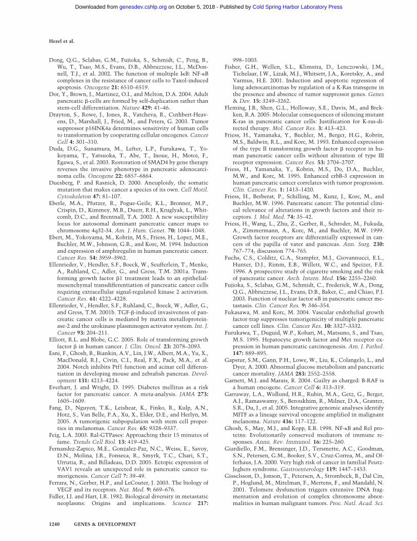
Dong, Q.G., Sclabas, G.M., Fujioka, S., Schmidt, C., Peng, B.,Wu, T., Tsao, M.S., Evans, D.B., Abbruzzese, J.L., McDon-nell, T.J., et al. 2002. The function of multiple I�B: NF-�Bcomplexes in the resistance of cancer cells to Taxol-inducedapoptosis. Oncogene 21: 6510–6519.
Dor, Y., Brown, J., Martinez, O.I., and Melton, D.A. 2004. Adultpancreatic �-cells are formed by self-duplication rather thanstem-cell differentiation. Nature 429: 41–46.
Drayton, S., Rowe, J., Jones, R., Vatcheva, R., Cuthbert-Heav-ens, D., Marshall, J., Fried, M., and Peters, G. 2003. Tumorsuppressor p16INK4a determines sensitivity of human cellsto transformation by cooperating cellular oncogenes. CancerCell 4: 301–310.
Duda, D.G., Sunamura, M., Lefter, L.P., Furukawa, T., Yo-koyama, T., Yatsuoka, T., Abe, T., Inoue, H., Motoi, F.,Egawa, S., et al. 2003. Restoration of SMAD4 by gene therapyreverses the invasive phenotype in pancreatic adenocarci-noma cells. Oncogene 22: 6857–6864.
Duesberg, P. and Rasnick, D. 2000. Aneuploidy, the somaticmutation that makes cancer a species of its own. Cell Motil.Cytoskeleton 47: 81–107.
Eberle, M.A., Pfutzer, R., Pogue-Geile, K.L., Bronner, M.P.,Crispin, D., Kimmey, M.B., Duerr, R.H., Kruglyak, L., Whit-comb, D.C., and Brentnall, T.A. 2002. A new susceptibilitylocus for autosomal dominant pancreatic cancer maps tochromosome 4q32-34. Am. J. Hum. Genet. 70: 1044–1048.
Ebert, M., Yokoyama, M., Kobrin, M.S., Friess, H., Lopez, M.E.,Buchler, M.W., Johnson, G.R., and Korc, M. 1994. Inductionand expression of amphiregulin in human pancreatic cancer.Cancer Res. 54: 3959–3962.
Ellenrieder, V., Hendler, S.F., Boeck, W., Seufferlein, T., Menke,A., Ruhland, C., Adler, G., and Gress, T.M. 2001a. Trans-forming growth factor �1 treatment leads to an epithelial-mesenchymal transdifferentiation of pancreatic cancer cellsrequiring extracellular signal-regulated kinase 2 activation.Cancer Res. 61: 4222–4228.
Ellenrieder, V., Hendler, S.F., Ruhland, C., Boeck, W., Adler, G.,and Gress, T.M. 2001b. TGF-�-induced invasiveness of pan-creatic cancer cells is mediated by matrix metalloprotein-ase-2 and the urokinase plasminogen activator system. Int. J.Cancer 93: 204–211.
Elliott, R.L. and Blobe, G.C. 2005. Role of transforming growthfactor � in human cancer. J. Clin. Oncol. 23: 2078–2093.
Esni, F., Ghosh, B., Biankin, A.V., Lin, J.W., Albert, M.A., Yu, X.,MacDonald, R.J., Civin, C.I., Real, F.X., Pack, M.A., et al.2004. Notch inhibits Ptf1 function and acinar cell differen-tiation in developing mouse and zebrafish pancreas. Devel-opment 131: 4213–4224.
Everhart, J. and Wright, D. 1995. Diabetes mellitus as a riskfactor for pancreatic cancer. A meta-analysis. JAMA 273:1605–1609.
Fang, D., Nguyen, T.K., Leishear, K., Finko, R., Kulp, A.N.,Hotz, S., Van Belle, P.A., Xu, X., Elder, D.E., and Herlyn, M.2005. A tumorigenic subpopulation with stem cell proper-ties in melanomas. Cancer Res. 65: 9328–9337.
Feig, L.A. 2003. Ral-GTPases: Approaching their 15 minutes offame. Trends Cell Biol. 13: 419–425.
Fernandez-Zapico, M.E., Gonzalez-Paz, N.C., Weiss, E., Savoy,D.N., Molina, J.R., Fonseca, R., Smyrk, T.C., Chari, S.T.,Urrutia, R., and Billadeau, D.D. 2005. Ectopic expression ofVAV1 reveals an unexpected role in pancreatic cancer tu-morigenesis. Cancer Cell 7: 39–49.
Ferrara, N., Gerber, H.P., and LeCouter, J. 2003. The biology ofVEGF and its receptors. Nat. Med. 9: 669–676.
Fidler, I.J. and Hart, I.R. 1982. Biological diversity in metastaticneoplasms: Origins and implications. Science 217:
998–1003.Fisher, G.H., Wellen, S.L., Klimstra, D., Lenczowski, J.M.,
Tichelaar, J.W., Lizak, M.J., Whitsett, J.A., Koretsky, A., andVarmus, H.E. 2001. Induction and apoptotic regression oflung adenocarcinomas by regulation of a K-Ras transgene inthe presence and absence of tumor suppressor genes. Genes& Dev. 15: 3249–3262.
Fleming, J.B., Shen, G.L., Holloway, S.E., Davis, M., and Brek-ken, R.A. 2005. Molecular consequences of silencing mutantK-ras in pancreatic cancer cells: Justification for K-ras-di-rected therapy. Mol. Cancer Res. 3: 413–423.
Friess, H., Yamanaka, Y., Buchler, M., Berger, H.G., Kobrin,M.S., Baldwin, R.L., and Korc, M. 1993. Enhanced expressionof the type II transforming growth factor � receptor in hu-man pancreatic cancer cells without alteration of type IIIreceptor expression. Cancer Res. 53: 2704–2707.
Friess, H., Yamanaka, Y., Kobrin, M.S., Do, D.A., Buchler,M.W., and Korc, M. 1995. Enhanced erbB-3 expression inhuman pancreatic cancer correlates with tumor progression.Clin. Cancer Res. 1: 1413–1420.
Friess, H., Berberat, P., Schilling, M., Kunz, J., Korc, M., andBuchler, M.W. 1996. Pancreatic cancer: The potential clini-cal relevance of alterations in growth factors and their re-ceptors. J. Mol. Med. 74: 35–42.
Friess, H., Wang, L., Zhu, Z., Gerber, R., Schroder, M., Fukuda,A., Zimmermann, A., Korc, M., and Buchler, M.W. 1999.Growth factor receptors are differentially expressed in can-cers of the papilla of vater and pancreas. Ann. Surg. 230:767–774; discussion 774–765.
Fuchs, C.S., Colditz, G.A., Stampfer, M.J., Giovannucci, E.L.,Hunter, D.J., Rimm, E.B., Willett, W.C., and Speizer, F.E.1996. A prospective study of cigarette smoking and the riskof pancreatic cancer. Arch. Intern. Med. 156: 2255–2260.
Fujioka, S., Sclabas, G.M., Schmidt, C., Frederick, W.A., Dong,Q.G., Abbruzzese, J.L., Evans, D.B., Baker, C., and Chiao, P.J.2003. Function of nuclear factor �B in pancreatic cancer me-tastasis. Clin. Cancer Res. 9: 346–354.
Fukasawa, M. and Korc, M. 2004. Vascular endothelial growthfactor-trap suppresses tumorigenicity of multiple pancreaticcancer cell lines. Clin. Cancer Res. 10: 3327–3332.
Furukawa, T., Duguid, W.P., Kobari, M., Matsuno, S., and Tsao,M.S. 1995. Hepatocyte growth factor and Met receptor ex-pression in human pancreatic carcinogenesis. Am. J. Pathol.147: 889–895.
Gapstur, S.M., Gann, P.H., Lowe, W., Liu, K., Colangelo, L., andDyer, A. 2000. Abnormal glucose metabolism and pancreaticcancer mortality. JAMA 283: 2552–2558.
Garnett, M.J. and Marais, R. 2004. Guilty as charged: B-RAF isa human oncogene. Cancer Cell 6: 313–319.
Garraway, L.A., Widlund, H.R., Rubin, M.A., Getz, G., Berger,A.J., Ramaswamy, S., Beroukhim, R., Milner, D.A., Granter,S.R., Du, J., et al. 2005. Integrative genomic analyses identifyMITF as a lineage survival oncogene amplified in malignantmelanoma. Nature 436: 117–122.
Ghosh, S., May, M.J., and Kopp, E.B. 1998. NF-�B and Rel pro-teins: Evolutionarily conserved mediators of immune re-sponses. Annu. Rev. Immunol. 16: 225–260.
Giardiello, F.M., Brensinger, J.D., Tersmette, A.C., Goodman,S.N., Petersen, G.M., Booker, S.V., Cruz-Correa, M., and Of-ferhaus, J.A. 2000. Very high risk of cancer in familial Peutz-Jeghers syndrome. Gastroenterology 119: 1447–1453.
Gisselsson, D., Jonson, T., Petersen, A., Strombeck, B., Dal Cin,P., Hoglund, M., Mitelman, F., Mertens, F., and Mandahl, N.2001. Telomere dysfunction triggers extensive DNA frag-mentation and evolution of complex chromosome abnor-malities in human malignant tumors. Proc. Natl. Acad. Sci.
Hezel et al.
1240 GENES & DEVELOPMENT
Cold Spring Harbor Laboratory Press on October 5, 2018 - Published by genesdev.cshlp.orgDownloaded from
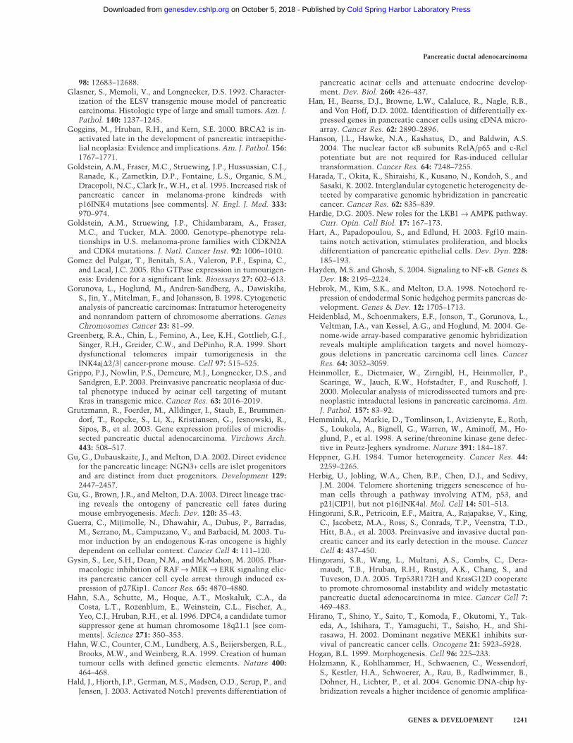
98: 12683–12688.Glasner, S., Memoli, V., and Longnecker, D.S. 1992. Character-
ization of the ELSV transgenic mouse model of pancreaticcarcinoma. Histologic type of large and small tumors. Am. J.Pathol. 140: 1237–1245.
Goggins, M., Hruban, R.H., and Kern, S.E. 2000. BRCA2 is in-activated late in the development of pancreatic intraepithe-lial neoplasia: Evidence and implications. Am. J. Pathol. 156:1767–1771.
Goldstein, A.M., Fraser, M.C., Struewing, J.P., Hussussian, C.J.,Ranade, K., Zametkin, D.P., Fontaine, L.S., Organic, S.M.,Dracopoli, N.C., Clark Jr., W.H., et al. 1995. Increased risk ofpancreatic cancer in melanoma-prone kindreds withp16INK4 mutations [see comments]. N. Engl. J. Med. 333:970–974.
Goldstein, A.M., Struewing, J.P., Chidambaram, A., Fraser,M.C., and Tucker, M.A. 2000. Genotype–phenotype rela-tionships in U.S. melanoma-prone families with CDKN2Aand CDK4 mutations. J. Natl. Cancer Inst. 92: 1006–1010.
Gomez del Pulgar, T., Benitah, S.A., Valeron, P.F., Espina, C.,and Lacal, J.C. 2005. Rho GTPase expression in tumourigen-esis: Evidence for a significant link. Bioessays 27: 602–613.
Gorunova, L., Hoglund, M., Andren-Sandberg, A., Dawiskiba,S., Jin, Y., Mitelman, F., and Johansson, B. 1998. Cytogeneticanalysis of pancreatic carcinomas: Intratumor heterogeneityand nonrandom pattern of chromosome aberrations. GenesChromosomes Cancer 23: 81–99.
Greenberg, R.A., Chin, L., Femino, A., Lee, K.H., Gottlieb, G.J.,Singer, R.H., Greider, C.W., and DePinho, R.A. 1999. Shortdysfunctional telomeres impair tumorigenesis in theINK4a(�2/3) cancer-prone mouse. Cell 97: 515–525.
Grippo, P.J., Nowlin, P.S., Demeure, M.J., Longnecker, D.S., andSandgren, E.P. 2003. Preinvasive pancreatic neoplasia of duc-tal phenotype induced by acinar cell targeting of mutantKras in transgenic mice. Cancer Res. 63: 2016–2019.
Grutzmann, R., Foerder, M., Alldinger, I., Staub, E., Brummen-dorf, T., Ropcke, S., Li, X., Kristiansen, G., Jesnowski, R.,Sipos, B., et al. 2003. Gene expression profiles of microdis-sected pancreatic ductal adenocarcinoma. Virchows Arch.443: 508–517.
Gu, G., Dubauskaite, J., and Melton, D.A. 2002. Direct evidencefor the pancreatic lineage: NGN3+ cells are islet progenitorsand are distinct from duct progenitors. Development 129:2447–2457.
Gu, G., Brown, J.R., and Melton, D.A. 2003. Direct lineage trac-ing reveals the ontogeny of pancreatic cell fates duringmouse embryogenesis. Mech. Dev. 120: 35–43.
Guerra, C., Mijimolle, N., Dhawahir, A., Dubus, P., Barradas,M., Serrano, M., Campuzano, V., and Barbacid, M. 2003. Tu-mor induction by an endogenous K-ras oncogene is highlydependent on cellular context. Cancer Cell 4: 111–120.
Gysin, S., Lee, S.H., Dean, N.M., and McMahon, M. 2005. Phar-macologic inhibition of RAF → MEK → ERK signaling elic-its pancreatic cancer cell cycle arrest through induced ex-pression of p27Kip1. Cancer Res. 65: 4870–4880.
Hahn, S.A., Schutte, M., Hoque, A.T., Moskaluk, C.A., daCosta, L.T., Rozenblum, E., Weinstein, C.L., Fischer, A.,Yeo, C.J., Hruban, R.H., et al. 1996. DPC4, a candidate tumorsuppressor gene at human chromosome 18q21.1 [see com-ments]. Science 271: 350–353.
Hahn, W.C., Counter, C.M., Lundberg, A.S., Beijersbergen, R.L.,Brooks, M.W., and Weinberg, R.A. 1999. Creation of humantumour cells with defined genetic elements. Nature 400:464–468.
Hald, J., Hjorth, J.P., German, M.S., Madsen, O.D., Serup, P., andJensen, J. 2003. Activated Notch1 prevents differentiation of
pancreatic acinar cells and attenuate endocrine develop-ment. Dev. Biol. 260: 426–437.
Han, H., Bearss, D.J., Browne, L.W., Calaluce, R., Nagle, R.B.,and Von Hoff, D.D. 2002. Identification of differentially ex-pressed genes in pancreatic cancer cells using cDNA micro-array. Cancer Res. 62: 2890–2896.
Hanson, J.L., Hawke, N.A., Kashatus, D., and Baldwin, A.S.2004. The nuclear factor �B subunits RelA/p65 and c-Relpotentiate but are not required for Ras-induced cellulartransformation. Cancer Res. 64: 7248–7255.
Harada, T., Okita, K., Shiraishi, K., Kusano, N., Kondoh, S., andSasaki, K. 2002. Interglandular cytogenetic heterogeneity de-tected by comparative genomic hybridization in pancreaticcancer. Cancer Res. 62: 835–839.
Hardie, D.G. 2005. New roles for the LKB1 → AMPK pathway.Curr. Opin. Cell Biol. 17: 167–173.
Hart, A., Papadopoulou, S., and Edlund, H. 2003. Fgf10 main-tains notch activation, stimulates proliferation, and blocksdifferentiation of pancreatic epithelial cells. Dev. Dyn. 228:185–193.
Hayden, M.S. and Ghosh, S. 2004. Signaling to NF-�B. Genes &Dev. 18: 2195–2224.
Hebrok, M., Kim, S.K., and Melton, D.A. 1998. Notochord re-pression of endodermal Sonic hedgehog permits pancreas de-velopment. Genes & Dev. 12: 1705–1713.
Heidenblad, M., Schoenmakers, E.F., Jonson, T., Gorunova, L.,Veltman, J.A., van Kessel, A.G., and Hoglund, M. 2004. Ge-nome-wide array-based comparative genomic hybridizationreveals multiple amplification targets and novel homozy-gous deletions in pancreatic carcinoma cell lines. CancerRes. 64: 3052–3059.
Heinmoller, E., Dietmaier, W., Zirngibl, H., Heinmoller, P.,Scaringe, W., Jauch, K.W., Hofstadter, F., and Ruschoff, J.2000. Molecular analysis of microdissected tumors and pre-neoplastic intraductal lesions in pancreatic carcinoma. Am.J. Pathol. 157: 83–92.
Hemminki, A., Markie, D., Tomlinson, I., Avizienyte, E., Roth,S., Loukola, A., Bignell, G., Warren, W., Aminoff, M., Ho-glund, P., et al. 1998. A serine/threonine kinase gene defec-tive in Peutz-Jeghers syndrome. Nature 391: 184–187.
Heppner, G.H. 1984. Tumor heterogeneity. Cancer Res. 44:2259–2265.
Herbig, U., Jobling, W.A., Chen, B.P., Chen, D.J., and Sedivy,J.M. 2004. Telomere shortening triggers senescence of hu-man cells through a pathway involving ATM, p53, andp21(CIP1), but not p16(INK4a). Mol. Cell 14: 501–513.
Hingorani, S.R., Petricoin, E.F., Maitra, A., Rajapakse, V., King,C., Jacobetz, M.A., Ross, S., Conrads, T.P., Veenstra, T.D.,Hitt, B.A., et al. 2003. Preinvasive and invasive ductal pan-creatic cancer and its early detection in the mouse. CancerCell 4: 437–450.
Hingorani, S.R., Wang, L., Multani, A.S., Combs, C., Dera-maudt, T.B., Hruban, R.H., Rustgi, A.K., Chang, S., andTuveson, D.A. 2005. Trp53R172H and KrasG12D cooperateto promote chromosomal instability and widely metastaticpancreatic ductal adenocarcinoma in mice. Cancer Cell 7:469–483.
Hirano, T., Shino, Y., Saito, T., Komoda, F., Okutomi, Y., Tak-eda, A., Ishihara, T., Yamaguchi, T., Saisho, H., and Shi-rasawa, H. 2002. Dominant negative MEKK1 inhibits sur-vival of pancreatic cancer cells. Oncogene 21: 5923–5928.
Hogan, B.L. 1999. Morphogenesis. Cell 96: 225–233.Holzmann, K., Kohlhammer, H., Schwaenen, C., Wessendorf,
S., Kestler, H.A., Schwoerer, A., Rau, B., Radlwimmer, B.,Dohner, H., Lichter, P., et al. 2004. Genomic DNA-chip hy-bridization reveals a higher incidence of genomic amplifica-
Pancreatic ductal adenocarcinoma
GENES & DEVELOPMENT 1241
Cold Spring Harbor Laboratory Press on October 5, 2018 - Published by genesdev.cshlp.orgDownloaded from
tions in pancreatic cancer than conventional comparativegenomic hybridization and leads to the identification ofnovel candidate genes. Cancer Res. 64: 4428–4433.
Hoshida, T., Sunamura, M., Duda, D.G., Egawa, S., Miyazaki, S.,Shineha, R., Hamada, H., Ohtani, H., Satomi, S., and Mat-suno, S. 2002. Gene therapy for pancreatic cancer using anadenovirus vector encoding soluble flt-1 vascular endothe-lial growth factor receptor. Pancreas 25: 111–121.
Hotz, H.G., Hines, O.J., Hotz, B., Foitzik, T., Buhr, H.J., andReber, H.A. 2003. Evaluation of vascular endothelial growthfactor blockade and matrix metalloproteinase inhibition as acombination therapy for experimental human pancreaticcancer. J. Gastrointest. Surg. 7: 220–227; discussion 227–228.
Hruban, R.H., Goggins, M., Parsons, J., and Kern, S.E. 2000a.Progression model for pancreatic cancer. Clin. Cancer Res.6: 2969–2972.
Hruban, R.H., Wilentz, R.E., and Kern, S.E. 2000b. Genetic pro-gression in the pancreatic ducts. Am. J. Pathol. 156: 1821–1825.
Hruban, R.H., Adsay, N.V., Albores-Saavedra, J., Compton, C.,Garrett, E.S., Goodman, S.N., Kern, S.E., Klimstra, D.S.,Kloppel, G., Longnecker, D.S., et al. 2001. Pancreatic intra-epithelial neoplasia: A new nomenclature and classificationsystem for pancreatic duct lesions. Am. J. Surg. Pathol. 25:579–586.
Hruban, R.H., Adsay, N.V., Albores-Saavedra, J., Anver, M.R.,Biankin, A.V., Boivin, G.P., Furth, E.E., Furukawa, T., Klein,A., Klimstra, D.S., et al. 2006a. Pathology of genetically en-gineered mouse models of pancreatic exocrine cancer: Con-sensus report and recommendations. Cancer Res. 66: 95–106.
Hruban, R.H., Bishop Pitman, M., and Klimstra, D.S. 2006b At-las of tumor pathology: Tumors of the pancreas. Fourth Se-ries (series eds. L.H. Sobin and J. Albores-Saavedra) ARPPress, Washington, DC.
Hustinx, S.R., Leoni, L.M., Yeo, C.J., Brown, P.N., Goggins, M.,Kern, S.E., Hruban, R.H., and Maitra, A. 2005. Concordantloss of MTAP and p16/CDKN2A expression in pancreaticintraepithelial neoplasia: Evidence of homozygous deletionin a noninvasive precursor lesion. Mod. Pathol. 18: 959–963.
Iacobuzio-Donahue, C.A., Maitra, A., Shen-Ong, G.L., vanHeek, T., Ashfaq, R., Meyer, R., Walter, K., Berg, K., Holl-ingsworth, M.A., Cameron, J.L., et al. 2002. Discovery ofnovel tumor markers of pancreatic cancer using global geneexpression technology. Am. J. Pathol. 160: 1239–1249.
Iacobuzio-Donahue, C.A., Maitra, A., Olsen, M., Lowe, A.W.,van Heek, N.T., Rosty, C., Walter, K., Sato, N., Parker, A.,Ashfaq, R., et al. 2003. Exploration of global gene expressionpatterns in pancreatic adenocarcinoma using cDNA micro-arrays. Am. J. Pathol. 162: 1151–1162.
Ingham, P.W. and McMahon, A.P. 2001. Hedgehog signaling inanimal development: Paradigms and principles. Genes &Dev. 15: 3059–3087.
Ishimura, N., Yamasawa, K., Karim Rumi, M.A., Kadowaki, Y.,Ishihara, S., Amano, Y., Nio, Y., Higami, T., and Kinoshita,Y. 2003. BRAF and K-ras gene mutations in human pancre-atic cancers. Cancer Lett. 199: 169–173.
Ishiwata, T., Kornmann, M., Beger, H.G., and Korc, M. 1998.Enhanced fibroblast growth factor 5 expression in stromaland exocrine elements of the pancreas in chronic pancreati-tis. Gut 43: 134–139.
Itakura, J., Ishiwata, T., Friess, H., Fujii, H., Matsumoto, Y.,Buchler, M.W., and Korc, M. 1997. Enhanced expression ofvascular endothelial growth factor in human pancreatic can-cer correlates with local disease progression. Clin. Cancer
Res. 3: 1309–1316.Jackson, E.L., Willis, N., Mercer, K., Bronson, R.T., Crowley, D.,
Montoya, R., Jacks, T., and Tuveson, D.A. 2001. Analysis oflung tumor initiation and progression using conditional ex-pression of oncogenic K-ras. Genes & Dev. 15: 3243–3248.
Jacobs, J.J. and de Lange, T. 2004. Significant role for p16 inp53-independent telomere-directed senescence. Curr. Biol.14: 2302–2308.
Jaffee, E.M., Hruban, R.H., Canto, M., and Kern, S.E. 2002. Fo-cus on pancreas cancer. Cancer Cell 2: 25–28.
Janda, E., Lehmann, K., Killisch, I., Jechlinger, M., Herzig, M.,Downward, J., Beug, H., and Grunert, S. 2002. Ras and TGF�
cooperatively regulate epithelial cell plasticity and metasta-sis: Dissection of Ras signaling pathways. J. Cell Biol. 156:299–313.
Jaster, R. 2004. Molecular regulation of pancreatic stellate cellfunction. Mol. Cancer 3: 26.
Jenne, D.E., Reimann, H., Nezu, J., Friedel, W., Loff, S., Jeschke,R., Muller, O., Back, W., and Zimmer, M. 1998. Peutz-Jegh-ers syndrome is caused by mutations in a novel serine threo-nine kinase. Nat. Genet. 18: 38–43.
Jensen, J., Pedersen, E.E., Galante, P., Hald, J., Heller, R.S., Ishi-bashi, M., Kageyama, R., Guillemot, F., Serup, P., and Mad-sen, O.D. 2000. Control of endodermal endocrine develop-ment by Hes-1. Nat. Genet. 24: 36–44.
Jhappan, C., Stahle, C., Harkins, R.N., Fausto, N., Smith, G.H.,and Merlino, G.T. 1990. TGF � overexpression in transgenicmice induces liver neoplasia and abnormal development ofthe mammary gland and pancreas. Cell 61: 1137–1146.
Jimenez, R.E., Z’Graggen, K., Hartwig, W., Graeme-Cook, F.,Warshaw, A.L., and Fernandez-del Castillo, C. 1999. Immu-nohistochemical characterization of pancreatic tumors in-duced by dimethylbenzanthracene in rats. Am. J. Pathol.154: 1223–1229.
Kadesch, T. 2004. Notch signaling: The demise of elegant sim-plicity. Curr. Opin. Genet. Dev. 14: 506–512.
Karin, M. and Ben-Neriah, Y. 2000. Phosphorylation meetsubiquitination: The control of NF-�B activity. Annu. Rev.Immunol. 18: 621–663.
Kawaguchi, Y., Cooper, B., Gannon, M., Ray, M., MacDonald,R.J., and Wright, C.V. 2002. The role of the transcriptionalregulator Ptf1a in converting intestinal to pancreatic pro-genitors. Nat. Genet. 32: 128–134.
Kiaris, H., Politi, K., Grimm, L.M., Szabolcs, M., Fisher, P., Ef-stratiadis, A., and Artavanis-Tsakonas, S. 2004. Modulationof Notch signaling elicits signature tumors and inhibitshras1-induced oncogenesis in the mouse mammary epithe-lium. Am. J. Pathol. 165: 695–705.
Kim, S.K. and Hebrok, M. 2001. Intercellular signals regulatingpancreas development and function. Genes & Dev. 15: 111–127.
Kim, C.F., Jackson, E.L., Woolfenden, A.E., Lawrence, S., Babar,I., Vogel, S., Crowley, D., Bronson, R.T., and Jacks, T. 2005.Identification of bronchioalveolar stem cells in normal lungand lung cancer. Cell 121: 823–835.
Kim, H.J., Schleiffarth, J.R., Jessurun, J., Sumanas, S., Petryk, A.,Lin, S., and Ekker, S.C. 2005. Wnt5 signaling in vertebratepancreas development. BMC Biol. 3: 23.
Kinzler, K.W. and Vogelstein, B. 1996. Lessons from hereditarycolorectal cancer. Cell 87: 159–170.
Kleeff, J., Ishiwata, T., Kumbasar, A., Friess, H., Buchler, M.W.,Lander, A.D., and Korc, M. 1998. The cell-surface heparansulfate proteoglycan glypican-1 regulates growth factor ac-tion in pancreatic carcinoma cells and is overexpressed inhuman pancreatic cancer. J. Clin. Invest. 102: 1662–1673.
Kleeff, J., Kothari, N.H., Friess, H., Fan, H., and Korc, M. 2004.
Hezel et al.
1242 GENES & DEVELOPMENT
Cold Spring Harbor Laboratory Press on October 5, 2018 - Published by genesdev.cshlp.orgDownloaded from
Adenovirus-mediated transfer of a truncated fibroblastgrowth factor (FGF) type I receptor blocks FGF-2 signaling inmultiple pancreatic cancer cell lines. Pancreas 28: 25–30.
Klimstra, D.S. 1998. Cell lineage in pancreatic neoplasms. InPancreatic cancer: Advances in molecular pathology, diag-nosis & clinical management (eds. F.S. Sarkar and M.C.Duggan), pp. 21–48. Eaton Publishing, Natick, MA.
Klimstra, D.S. and Longnecker, D.S. 1994. K-ras mutations inpancreatic ductal proliferative lesions. Am. J. Pathol. 145:1547–1550.
Kobrin, M.S., Yamanaka, Y., Friess, H., Lopez, M.E., and Korc,M. 1993. Aberrant expression of type I fibroblast growth fac-tor receptor in human pancreatic adenocarcinomas. CancerRes. 53: 4741–4744.
Koizumi, M., Doi, R., Toyoda, E., Masui, T., Tulachan, S.S.,Kawaguchi, Y., Fujimoto, K., Gittes, G.K., and Imamura, M.2003. Increased PDX-1 expression is associated with out-come in patients with pancreatic cancer. Surgery 134: 260–266.
Kops, G.J., Weaver, B.A., and Cleveland, D.W. 2005. On the roadto cancer: Aneuploidy and the mitotic checkpoint. Nat. Rev.Cancer 5: 773–785.
Korc, M., Chandrasekar, B., Yamanaka, Y., Friess, H., Buchier,M., and Beger, H.G. 1992. Overexpression of the epidermalgrowth factor receptor in human pancreatic cancer is asso-ciated with concomitant increases in the levels of epidermalgrowth factor and transforming growth factor �. J. Clin. In-vest. 90: 1352–1360.
Kornmann, M., Ishiwata, T., Beger, H.G., and Korc, M. 1997.Fibroblast growth factor-5 stimulates mitogenic signalingand is overexpressed in human pancreatic cancer: Evidencefor autocrine and paracrine actions. Oncogene 15:1417–1424.
Kornmann, M., Ishiwata, T., Matsuda, K., Lopez, M.E., Fukahi,K., Asano, G., Beger, H.G., and Korc, M. 2002. IIIc isoform offibroblast growth factor receptor 1 is overexpressed in hu-man pancreatic cancer and enhances tumorigenicity of ham-ster ductal cells. Gastroenterology 123: 301–313.
Krimpenfort, P., Quon, K.C., Mooi, W.J., Loonstra, A., andBerns, A. 2001. Loss of p16Ink4a confers susceptibility tometastatic melanoma in mice. Nature 413: 83–86.
Krishnamurthy, J., Torrice, C., Ramsey, M.R., Kovalev, G.I.,Al-Regaiey, K., Su, L., and Sharpless, N.E. 2004. Ink4a/Arfexpression is a biomarker of aging. J. Clin. Invest. 114: 1299–1307.
Kritzik, M.R., Jones, E., Chen, Z., Krakowski, M., Krahl, T.,Good, A., Wright, C., Fox, H., and Sarvetnick, N. 1999.PDX-1 and Msx-2 expression in the regenerating and devel-oping pancreas. J. Endocrinol. 163: 523–530.
Kuniyasu, H., Abbruzzese, J.L., Cleary, K.R., and Fidler, I.J.2001. Induction of ductal and stromal hyperplasia by basicfibroblast growth factor produced by human pancreatic car-cinoma. Int. J. Oncol. 19: 681–685.
Kurahara, H., Takao, S., Maemura, K., Shinchi, H., Natsugoe, S.,and Aikou, T. 2004. Impact of vascular endothelial growthfactor-C and -D expression in human pancreatic cancer: Itsrelationship to lymph node metastasis. Clin. Cancer Res. 10:8413–8420.
Laghi, L., Orbetegli, O., Bianchi, P., Zerbi, A., Di Carlo, V.,Boland, C.R., and Malesci, A. 2002. Common occurrence ofmultiple K-RAS mutations in pancreatic cancers with asso-ciated precursor lesions and in biliary cancers. Oncogene 21:4301–4306.
Lai, E.C. 2004. Notch signaling: Control of cell communicationand cell fate. Development 131: 965–973.
Lal, G., Liu, L., Hogg, D., Lassam, N.J., Redston, M.S., and Gall-
inger, S. 2000. Patients with both pancreatic adenocarci-noma and melanoma may harbor germline CDKN2A muta-tions. Genes Chromosomes Cancer 27: 358–361.
Lapidot, T., Sirard, C., Vormoor, J., Murdoch, B., Hoang, T.,Caceres-Cortes, J., Minden, M., Paterson, B., Caligiuri, M.A.,and Dick, J.E. 1994. A cell initiating human acute myeloidleukaemia after transplantation into SCID mice. Nature367: 645–648.
Lebowitz, P.F., Davide, J.P., and Prendergast, G.C. 1995. Evi-dence that farnesyltransferase inhibitors suppress Ras trans-formation by interfering with Rho activity. Mol. Cell. Biol.15: 6613–6622.
Lemoine, N.R., Jain, S., Hughes, C.M., Staddon, S.L., Maillet, B.,Hall, P.A., and Kloppel, G. 1992. Ki-ras oncogene activationin preinvasive pancreatic cancer. Gastroenterology 102:230–236.
Lerner, E.C., Zhang, T.T., Knowles, D.B., Qian, Y., Hamilton,A.D., and Sebti, S.M. 1997. Inhibition of the prenylation ofK-Ras, but not H- or N-Ras, is highly resistant to CAAXpeptidomimetics and requires both a farnesyltransferase anda geranylgeranyltransferase I inhibitor in human tumor celllines. Oncogene 15: 1283–1288.
Levy, L. and Hill, C.S. 2005. Smad4 dependency defines twoclasses of transforming growth factor � (TGF-�) target genesand distinguishes TGF-�-induced epithelial–mesenchymaltransition from its antiproliferative and migratory responses.Mol. Cell. Biol. 25: 8108–8125.
Lewis, B.C., Klimstra, D.S., and Varmus, H.E. 2003. The c-mycand PyMT oncogenes induce different tumor types in a so-matic mouse model for pancreatic cancer. Genes & Dev. 17:3127–3138.
Li, D., Xie, K., Wolff, R., and Abbruzzese, J.L. 2004. Pancreaticcancer. Lancet 363: 1049–1057.
Li, J., Kleeff, J., Giese, N., Buchler, M.W., Korc, M., and Friess,H. 2004. Gefitinib (‘Iressa,’ ZD1839), a selective epidermalgrowth factor receptor tyrosine kinase inhibitor, inhibitspancreatic cancer cell growth, invasion, and colony forma-tion. Int. J. Oncol. 25: 203–210.
Lim, K.H. and Counter, C.M. 2005. Reduction in the require-ment of oncogenic Ras signaling to activation of PI3K/AKTpathway during tumor maintenance. Cancer Cell 8: 381–392.
Lim, K.H., Baines, A.T., Fiordalisi, J.J., Shipitsin, M., Feig, L.A.,Cox, A.D., Der, C.J., and Counter, C.M. 2005. Activation ofRalA is critical for Ras-induced tumorigenesis of humancells. Cancer Cell 7: 533–545.
Liptay, S., Weber, C.K., Ludwig, L., Wagner, M., Adler, G., andSchmid, R.M. 2003. Mitogenic and antiapoptotic role of con-stitutive NF-�B/Rel activity in pancreatic cancer. Int. J. Can-cer 105: 735–746.
Liu, L., Dilworth, D., Gao, L., Monzon, J., Summers, A., Lassam,N., and Hogg, D. 1999. Mutation of the CDKN2A 5� UTRcreates an aberrant initiation codon and predisposes to mela-noma. Nat. Genet. 21: 128–132.
Lizcano, J.M., Goransson, O., Toth, R., Deak, M., Morrice, N.A.,Boudeau, J., Hawley, S.A., Udd, L., Makela, T.P., Hardie,D.G., et al. 2004. LKB1 is a master kinase that activates 13kinases of the AMPK subfamily, including MARK/PAR-1.EMBO J. 23: 833–843.
Lohr, M., Maisonneuve, P., and Lowenfels, A.B. 2000. K-Rasmutations and benign pancreatic disease. Int. J. Pancreatol.27: 93–103.
Lohr, M., Schmidt, C., Ringel, J., Kluth, M., Muller, P., Nizze,H., and Jesnowski, R. 2001. Transforming growth factor-�1induces desmoplasia in an experimental model of humanpancreatic carcinoma. Cancer Res. 61: 550–555.
Pancreatic ductal adenocarcinoma
GENES & DEVELOPMENT 1243
Cold Spring Harbor Laboratory Press on October 5, 2018 - Published by genesdev.cshlp.orgDownloaded from
Lowe, S.W. and Sherr, C.J. 2003. Tumor suppression by Ink4a-Arf: Progress and puzzles. Curr. Opin. Genet. Dev. 13: 77–83.
Lowenfels, A.B., Maisonneuve, P., DiMagno, E.P., Elitsur, Y.,Gates Jr., L.K., Perrault, J., and Whitcomb, D.C. 1997. He-reditary pancreatitis and the risk of pancreatic cancer. Inter-national Hereditary Pancreatitis Study Group. [see com-ments].J. Natl. Cancer Inst. 89: 442–446
Luttges, J., Schlehe, B., Menke, M.A., Vogel, I., Henne-Bruns, D.,and Kloppel, G. 1999. The K-ras mutation pattern in pancre-atic ductal adenocarcinoma usually is identical to that inassociated normal, hyperplastic, and metaplastic ductal epi-thelium. Cancer 85: 1703–1710.
Luttges, J., Galehdari, H., Brocker, V., Schwarte-Waldhoff, I.,Henne-Bruns, D., Kloppel, G., Schmiegel, W., and Hahn, S.A.2001. Allelic loss is often the first hit in the biallelic inacti-vation of the p53 and DPC4 genes during pancreatic carci-nogenesis. Am. J. Pathol. 158: 1677–1683.
Lynch, H.T., Smyrk, T., Kern, S.E., Hruban, R.H., Lightdale,C.J., Lemon, S.J., Lynch, J.F., Fusaro, L.R., Fusaro, R.M., andGhadirian, P. 1996. Familial pancreatic cancer: A review.Semin. Oncol. 23: 251–275.
Lynch, H.T., Brand, R.E., Hogg, D., Deters, C.A., Fusaro, R.M.,Lynch, J.F., Liu, L., Knezetic, J., Lassam, N.J., Goggins, M., etal. 2002. Phenotypic variation in eight extended CDKN2Agermline mutation familial atypical multiple mole melano-ma–pancreatic carcinoma-prone families: The familial atypi-cal mole melanoma–pancreatic carcinoma syndrome. Can-cer 94: 84–96.
Mahlamaki, E.H., Hoglund, M., Gorunova, L., Karhu, R.,Dawiskiba, S., Andren-Sandberg, A., Kallioniemi, O.P., andJohansson, B. 1997. Comparative genomic hybridization re-veals frequent gains of 20q, 8q, 11q, 12p, and 17q, and lossesof 18q, 9p, and 15q in pancreatic cancer. Genes Chromo-somes Cancer 20: 383–391.
Mahlamaki, E.H., Barlund, M., Tanner, M., Gorunova, L., Ho-glund, M., Karhu, R., and Kallioniemi, A. 2002. Frequentamplification of 8q24, 11q, 17q, and 20q-specific genes inpancreatic cancer. Genes Chromosomes Cancer 35:353–358.
Maitland, N.J. and Collins, A. 2005. A tumour stem cell hy-pothesis for the origins of prostate cancer. BJU Int. 96: 1219–1223.
Maitra, A., Adsay, N.V., Argani, P., Iacobuzio-Donahue, C., DeMarzo, A., Cameron, J.L., Yeo, C.J., and Hruban, R.H. 2003.Multicomponent analysis of the pancreatic adenocarcinomaprogression model using a pancreatic intraepithelial neopla-sia tissue microarray. Mod. Pathol. 16: 902–912.
Maitra, A., Fukushima, N., Takaori, K., and Hruban, R.H. 2005.Precursors to invasive pancreatic cancer. Adv. Anat. Pathol.12: 81–91.
Malats, N., Casals, T., Porta, M., Guarner, L., Estivill, X., andReal, F.X. 2001. Cystic fibrosis transmembrane regulator(CFTR) DeltaF508 mutation and 5T allele in patients withchronic pancreatitis and exocrine pancreatic cancer.PANKRAS II Study Group. Gut 48: 70–74.
Maloney, E.K., McLaughlin, J.L., Dagdigian, N.E., Garrett, L.M.,Connors, K.M., Zhou, X.M., Blattler, W.A., Chittenden, T.,and Singh, R. 2003. An anti-insulin-like growth factor I re-ceptor antibody that is a potent inhibitor of cancer cell pro-liferation. Cancer Res. 63: 5073–5083.
Malumbres, M. and Barbacid, M. 2003. RAS oncogenes: Thefirst 30 years. Nat. Rev. Cancer 3: 459–465.
Maser, R.S. and DePinho, R.A. 2002. Connecting chromosomes,crisis, and cancer. Science 297: 565–569.
Massague, J., Blain, S.W., and Lo, R.S. 2000. TGF� signaling in
growth control, cancer, and heritable disorders. Cell 103:295–309.
Matsubayashi, H., Fukushima, N., Sato, N., Brune, K., Canto,M., Yeo, C.J., Hruban, R.H., Kern, S.E., and Goggins, M.2003. Polymorphisms of SPINK1 N34S and CFTR in patientswith sporadic and familial pancreatic cancer. Cancer Biol.Ther. 2: 652–655.
Mayo, M.W., Wang, C.Y., Cogswell, P.C., Rogers-Graham, K.S.,Lowe, S.W., Der, C.J., and Baldwin Jr., A.S. 1997. Require-ment of NF-�B activation to suppress p53-independent apo-ptosis induced by oncogenic Ras. Science 278: 1812–1815.
McCormick, F. 1998. Going for the GAP. Curr. Biol. 8: R673–R674.
McWilliams, R., Highsmith, W.E., Rabe, K.G., de Andrade, M.,Tordsen, L.A., Holtegaard, L.M., and Petersen, G.M. 2005.Cystic fibrosis transmembrane regulator gene carrier statusis a risk factor for young onset pancreatic adenocarcinoma.Gut 54: 1661–1662.
Means, A.L., Meszoely, I.M., Suzuki, K., Miyamoto, Y., Rustgi,A.K., Coffey Jr., R.J., Wright, C.V., Stoffers, D.A., and Leach,S.D. 2005. Pancreatic epithelial plasticity mediated by aci-nar cell transdifferentiation and generation of nestin-posi-tive intermediates. Development 132: 3767–3776.
Michaud, D.S., Giovannucci, E., Willett, W.C., Colditz, G.A.,Stampfer, M.J., and Fuchs, C.S. 2001. Physical activity, obe-sity, height, and the risk of pancreatic cancer. JAMA 286:921–929.
Min, Y., Adachi, Y., Yamamoto, H., Ito, H., Itoh, F., Lee, C.T.,Nadaf, S., Carbone, D.P., and Imai, K. 2003. Genetic block-ade of the insulin-like growth factor-I receptor: A promisingstrategy for human pancreatic cancer. Cancer Res. 63: 6432–6441.
Missiaglia, E., Blaveri, E., Terris, B., Wang, Y.H., Costello, E.,Neoptolemos, J.P., Crnogorac-Jurcevic, T., and Lemoine,N.R. 2004. Analysis of gene expression in cancer cell linesidentifies candidate markers for pancreatic tumorigenesisand metastasis. Int. J. Cancer 112: 100–112.
Mitin, N., Rossman, K.L., and Der, C.J. 2005. Signaling inter-play in Ras superfamily function. Curr. Biol. 15: R563–R574.
Miyamoto, Y., Maitra, A., Ghosh, B., Zechner, U., Argani, P.,Iacobuzio-Donahue, C.A., Sriuranpong, V., Iso, T., Meszoely,I.M., Wolfe, M.S., et al. 2003. Notch mediates TGF �-in-duced changes in epithelial differentiation during pancreatictumorigenesis. Cancer Cell 3: 565–576.
Moshous, D., Callebaut, I., de Chasseval, R., Corneo, B., Cavaz-zana-Calvo, M., Le Deist, F., Tezcan, I., Sanal, O., Bertrand,Y., Philippe, N., et al. 2001. Artemis, a novel DNA double-strand break repair/V(D)J recombination protein, is mutatedin human severe combined immune deficiency. Cell 105:177–186.
Moskaluk, C.A., Hruban, R.H., and Kern, S.E. 1997. p16 andK-ras gene mutations in the intraductal precursors of humanpancreatic adenocarcinoma. Cancer Res. 57: 2140–2143.
Muerkoster, S., Arlt, A., Sipos, B., Witt, M., Grossmann, M.,Kloppel, G., Kalthoff, H., Folsch, U.R., and Schafer, H. 2005.Increased expression of the E3-ubiquitin ligase receptor sub-unit �TRCP1 relates to constitutive nuclear factor-�B acti-vation and chemoresistance in pancreatic carcinoma cells.Cancer Res. 65: 1316–1324.
Murphy, K.M., Brune, K.A., Griffin, C., Sollenberger, J.E., Pe-tersen, G.M., Bansal, R., Hruban, R.H., and Kern, S.E. 2002.Evaluation of candidate genes MAP2K4, MADH4, ACVR1B,and BRCA2 in familial pancreatic cancer: DeleteriousBRCA2 mutations in 17%. Cancer Res. 62: 3789–3793.
Murtaugh, L.C. and Melton, D.A. 2003. Genes, signals, and lin-eages in pancreas development. Annu. Rev. Cell Dev. Biol.
Hezel et al.
1244 GENES & DEVELOPMENT
Cold Spring Harbor Laboratory Press on October 5, 2018 - Published by genesdev.cshlp.orgDownloaded from
19: 71–89.Murtaugh, L.C., Stanger, B.Z., Kwan, K.M., and Melton, D.A.
2003. Notch signaling controls multiple steps of pancreaticdifferentiation. Proc. Natl. Acad. Sci. 100: 14920–14925.
Murtaugh, L.C., Law, A.C., Dor, Y., and Melton, D.A. 2005.�-Catenin is essential for pancreatic acinar but not islet de-velopment. Development 132: 4663–4674.
Nair, P.N., De Armond, D.T., Adamo, M.L., Strodel, W.E., andFreeman, J.W. 2001. Aberrant expression and activation ofinsulin-like growth factor-1 receptor (IGF-1R) are mediatedby an induction of IGF-1R promoter activity and stabiliza-tion of IGF-1R mRNA and contributes to growth factor in-dependence and increased survival of the pancreatic cancercell line MIA PaCa-2. Oncogene 20: 8203–8214.
Nakamura, T., Furukawa, Y., Nakagawa, H., Tsunoda, T., Ohi-gashi, H., Murata, K., Ishikawa, O., Ohgaki, K., Kashimura,N., Miyamoto, M., et al. 2004. Genome-wide cDNA micro-array analysis of gene expression profiles in pancreatic can-cers using populations of tumor cells and normal ductal epi-thelial cells selected for purity by laser microdissection. On-cogene 23: 2385–2400.
Neglia, J.P., FitzSimmons, S.C., Maisonneuve, P., Schoni, M.H.,Schoni-Affolter, F., Corey, M., and Lowenfels, A.B. 1995.The risk of cancer among patients with cystic fibrosis. Cys-tic Fibrosis and Cancer Study Group. N. Engl. J. Med. 332:494–499.
Ng, S.S.W., Tsao, M.S., Chow, S., and Hedley, D.W. 2000. Inhi-bition of phosphatidylinositide 3-kinase enhances gemcita-bine-induced apoptosis in human pancreatic cancer cells.Cancer Res. 60: 5451–5455.
Nielsen, G.P., Stemmer-Rachamimov, A.O., Shaw, J., Roy, J.E.,Koh, J., and Louis, D.N. 1999. Immunohistochemical surveyof p16INK4A expression in normal human adult and infanttissues. Lab. Invest. 79: 1137–1143.
Norgaard, G.A., Jensen, J.N., and Jensen, J. 2003. FGF10 signal-ing maintains the pancreatic progenitor cell state revealing anovel role of Notch in organ development. Dev. Biol. 264:323–338.
Nowak, N.J., Gaile, D., Conroy, J.M., McQuaid, D., Cowell, J.,Carter, R., Goggins, M.G., Hruban, R.H., and Maitra, A.2005. Genome-wide aberrations in pancreatic adenocarci-noma. Cancer Genet. Cytogenet. 161: 36–50.
Offield, M.F., Jetton, T.L., Labosky, P.A., Ray, M., Stein, R.W.,Magnuson, M.A., Hogan, B.L., and Wright, C.V. 1996. PDX-1is required for pancreatic outgrowth and differentiation ofthe rostral duodenum. Development 122: 983–995.
Oft, M., Akhurst, R.J., and Balmain, A. 2002. Metastasis isdriven by sequential elevation of H-ras and Smad2 levels.Nat. Cell Biol. 4: 487–494.
Ogawa, T., Takayama, K., Takakura, N., Kitano, S., and Ueno,H. 2002. Anti-tumor angiogenesis therapy using soluble re-ceptors: Enhanced inhibition of tumor growth when solublefibroblast growth factor receptor-1 is used with soluble vas-cular endothelial growth factor receptor. Cancer Gene Ther.9: 633–640.
O’Hagan, R.C., Chang, S., Maser, R.S., Mohan, R., Artandi, S.E.,Chin, L., and DePinho, R.A. 2002. Telomere dysfunctionprovokes regional amplification and deletion in cancer ge-nomes. Cancer Cell 2: 149–155.
Ohlsson, H., Karlsson, K., and Edlund, T. 1993. IPF1, a ho-meodomain-containing transactivator of the insulin gene.EMBO J. 12: 4251–4259.
Ohta, T., Yamamoto, M., Numata, M., Iseki, S., Tsukioka, Y.,Miyashita, T., Kayahara, M., Nagakawa, T., Miyazaki, I.,Nishikawa, K., et al. 1995. Expression of basic fibroblastgrowth factor and its receptor in human pancreatic carcino-
mas. Br. J. Cancer 72: 824–831.Okami, K., Wu, L., Riggins, G., Cairns, P., Goggins, M., Evron,
E., Halachmi, N., Ahrendt, S.A., Reed, A.L., Hilgers, W., etal. 1998. Analysis of PTEN/MMAC1 alterations in aerodi-gestive tract tumors. Cancer Res. 58: 509–511.
Omer, C.A. and Kohl, N.E. 1997. CA1A2X-competitive inhibi-tors of farnesyltransferase as anti-cancer agents. TrendsPharmacol. Sci. 18: 437–444.
Orlowski, R.Z. and Baldwin Jr., A.S. 2002. NF-�B as a therapeu-tic target in cancer. Trends Mol. Med. 8: 385–389.
Ornitz, D.M., Hammer, R.E., Messing, A., Palmiter, R.D., andBrinster, R.L. 1987. Pancreatic neoplasia induced by SV40T-antigen expression in acinar cells of transgenic mice. Sci-ence 238: 188–193.
Orsulic, S. 2002. An RCAS-TVA-based approach to designermouse models. Mamm. Genome 13: 543–547.
Ory, S. and Morrison, D.K. 2004. Signal transduction: Implica-tions for Ras-dependent ERK signaling. Curr. Biol. 14: R277–R278.
Ossipova, O., Bardeesy, N., DePinho, R.A., and Green, J.B. 2003.LKB1 (XEEK1) regulates Wnt signalling in vertebrate devel-opment. Nat. Cell Biol. 5: 889–894.
Ouban, A., Muraca, P., Yeatman, T., and Coppola, D. 2003.Expression and distribution of insulin-like growth factor-1receptor in human carcinomas. Hum. Pathol. 34: 803–808.
Paciucci, R., Vila, M.R., Adell, T., Diaz, V.M., Tora, M., Naka-mura, T., and Real, F.X. 1998. Activation of the urokinaseplasminogen activator/urokinase plasminogen activator re-ceptor system and redistribution of E-cadherin are associatedwith hepatocyte growth factor-induced motility of pancreastumor cells overexpressing Met. Am. J. Pathol. 153: 201–212.
Paliwal, S., Pande, S., Kovi, R.C., Sharpless, N.E., Bardeesy, N.,and Grossman, S.R. 2006. Targeting of C-terminal bindingprotein (CtBP) by ARF results in p53-independent apoptosis.Mol. Cell. Biol. 26: 2360–2372.
Pao, W., Klimstra, D.S., Fisher, G.H., and Varmus, H.E. 2003.Use of avian retroviral vectors to introduce transcriptionalregulators into mammalian cells for analyses of tumor main-tenance. Proc. Natl. Acad. Sci. 100: 8764–8769.
Pardal, R., Clarke, M.F., and Morrison, S.J. 2003. Applying theprinciples of stem-cell biology to cancer. Nat. Rev. Cancer 3:895–902.
Parsa, I., Longnecker, D.S., Scarpelli, D.G., Pour, P., Reddy, J.K.,and Lefkowitz, M. 1985. Ductal metaplasia of human exo-crine pancreas and its association with carcinoma. CancerRes. 45: 1285–1290.
Pasca di Magliano, M., and Hebrok, M. 2003. Hedgehog signal-ling in cancer formation and maintenance. Nat. Rev. Cancer3: 903–911.
Passegue, E., Wagner, E.F., and Weissman, I.L. 2004. JunB defi-ciency leads to a myeloproliferative disorder arising fromhematopoietic stem cells. Cell 119: 431–443.
Peng, B., Fleming, J.B., Breslin, T., Grau, A.M., Fojioka, S., Ab-bruzzese, J.L., Evans, D.B., Ayers, D., Wathen, K., Wu, T., etal. 2002. Suppression of tumorigenesis and induction ofp15(ink4b) by Smad4/DPC4 in human pancreatic cancercells. Clin. Cancer Res. 8: 3628–3638.
Perugini, R.A., McDade, T.P., Vittimberga Jr., F.J., and Callery,M.P. 2000. Pancreatic cancer cell proliferation is phosphati-dylinositol 3-kinase dependent. J. Surg. Res. 90: 39–44.
Petersen, G.M. and Hruban, R.H. 2003. Familial pancreatic can-cer: Where are we in 2003? J. Natl. Cancer Inst. 95: 180–181.
Pomerantz, J., Schreiber-Agus, N., Liegeois, N.J., Silverman, A.,Alland, L., Chin, L., Potes, J., Chen, K., Orlow, I., Lee, H.W.,et al. 1998. The Ink4a tumor suppressor gene product,
Pancreatic ductal adenocarcinoma
GENES & DEVELOPMENT 1245
Cold Spring Harbor Laboratory Press on October 5, 2018 - Published by genesdev.cshlp.orgDownloaded from
p19Arf, interacts with MDM2 and neutralizes MDM2’s in-hibition of p53. Cell 92: 713–723.
Pour, P.M. 1997. The role of Langerhans islets in pancreaticductal adenocarcinoma. Front. Biosci. 2: D271–D282.
Prasad, N.B., Biankin, A.V., Fukushima, N., Maitra, A., Dhara,S., Elkahloun, A.G., Hruban, R.H., Goggins, M., and Leach,S.D. 2005. Gene expression profiles in pancreatic intraepi-thelial neoplasia reflect the effects of Hedgehog signaling onpancreatic ductal epithelial cells. Cancer Res. 65: 1619–1626.
Prowse, K.R. and Greider, C.W. 1995. Developmental and tis-sue-specific regulation of mouse telomerase and telomerelength. Proc. Natl. Acad. Sci. 92: 4818–4822.
Qi, Y., Gregory, M.A., Li, Z., Brousal, J.P., West, K., and Hann,S.R. 2004. p19ARF directly and differentially controls thefunctions of c-Myc independently of p53. Nature 431: 712–717.
Quaife, C.J., Pinkert, C.A., Ornitz, D.M., Palmiter, R.D., andBrinster, R.L. 1987. Pancreatic neoplasia induced by ras ex-pression in acinar cells of transgenic mice. Cell 48: 1023–1034.
Radtke, F. and Raj, K. 2003. The role of Notch in tumorigenesis:Oncogene or tumour suppressor? Nat. Rev. Cancer 3: 756–767.
Rajagopalan, H. and Lengauer, C. 2004. Aneuploidy and cancer.Nature 432: 338–341.
Ramirez, R.D., Morales, C.P., Herbert, B.S., Rohde, J.M., Pas-sons, C., Shay, J.W., and Wright, W.E. 2001. Putative telo-mere-independent mechanisms of replicative aging reflectinadequate growth conditions. Genes & Dev. 15: 398–403.
Rane, S.G., Lee, J.H., and Lin, H.M. 2006. Transforming growthfactor-� pathway: Role in pancreas development and pancre-atic disease. Cytokine Growth Factor Rev. 17: 107–119.
Rangarajan, A., Hong, S.J., Gifford, A., and Weinberg, R.A. 2004.Species- and cell type-specific requirements for cellulartransformation. Cancer Cell 6: 171–183.
Rocha, S., Campbell, K.J., and Perkins, N.D. 2003. p53- andMdm2-independent repression of NF-� B transactivation bythe ARF tumor suppressor. Mol. Cell 12: 15–25.
Rodriguez-Viciana, P., Warne, P.H., Vanhaesebroeck, B., Water-field, M.D., and Downward, J. 1996. Activation of phospho-inositide 3-kinase by interaction with Ras and by point mu-tation. EMBO J. 15: 2442–2451.
Rosty, C., Christa, L., Kuzdzal, S., Baldwin, W.M., Zahurak,M.L., Carnot, F., Chan, D.W., Canto, M., Lillemoe, K.D.,Cameron, J.L., et al. 2002. Identification of hepatocarci-noma-intestine–pancreas/pancreatitis-associated protein I asa biomarker for pancreatic ductal adenocarcinoma by pro-tein biochip technology. Cancer Res. 62: 1868–1875.
Rowland-Goldsmith, M.A., Maruyama, H., Kusama, T., Ralli,S., and Korc, M. 2001. Soluble type II transforming growthfactor-� (TGF-�) receptor inhibits TGF-� signaling in COLO-357 pancreatic cancer cells in vitro and attenuates tumorformation. Clin. Cancer Res. 7: 2931–2940.
Rowland-Goldsmith, M.A., Maruyama, H., Matsuda, K.,Idezawa, T., Ralli, M., Ralli, S., and Korc, M. 2002. Solubletype II transforming growth factor-� receptor attenuates ex-pression of metastasis-associated genes and suppresses pan-creatic cancer cell metastasis. Mol. Cancer Ther. 1: 161–167.
Rozenblum, E., Schutte, M., Goggins, M., Hahn, S.A., Panzer,S., Zahurak, M., Goodman, S.N., Sohn, T.A., Hruban, R.H.,Yeo, C.J., et al. 1997. Tumor-suppressive pathways in pan-creatic carcinoma. Cancer Res. 57: 1731–1734.
Ruas, M. and Peters, G. 1998. The p16INK4a/CDKN2A tumorsuppressor and its relatives. Biochim. Biophys. Acta 1378:F115–F177.
Ruggeri, B.A., Huang, L., Wood, M., Cheng, J.Q., and Testa, J.R.
1998. Amplification and overexpression of the AKT2 onco-gene in a subset of human pancreatic ductal adenocarcino-mas. Mol. Carcinog. 21: 81–86.
Sahin, F., Maitra, A., Argani, P., Sato, N., Maehara, N., Mont-gomery, E., Goggins, M., Hruban, R.H., and Su, G.H. 2003.Loss of Stk11/Lkb1 expression in pancreatic and biliary neo-plasms. Mod. Pathol. 16: 686–691.
Saimura, M., Nagai, E., Mizumoto, K., Maehara, N., Mina-mishima, Y.A., Katano, M., Matsumoto, K., Nakamura, T.,and Tanaka, M. 2002. Tumor suppression through angiogen-esis inhibition by SUIT-2 pancreatic cancer cells geneticallyengineered to secrete NK4. Clin. Cancer Res. 8: 3243–3249.
Samuels, Y. and Velculescu, V.E. 2004. Oncogenic mutations ofPIK3CA in human cancers. Cell Cycle 3: 1221–1224.
Sandgren, E.P., Luetteke, N.C., Palmiter, R.D., Brinster, R.L.,and Lee, D.C. 1990. Overexpression of TGF � in transgenicmice: Induction of epithelial hyperplasia, pancreatic meta-plasia, and carcinoma of the breast. Cell 61: 1121–1135.
Sandgren, E.P., Quaife, C.J., Paulovich, A.G., Palmiter, R.D.,and Brinster, R.L. 1991. Pancreatic tumor pathogenesis re-flects the causative genetic lesion. Proc. Natl. Acad. Sci. 88:93–97.
Sandgren, E.P., Luetteke, N.C., Qiu, T.H., Palmiter, R.D., Brin-ster, R.L., and Lee, D.C. 1993. Transforming growth factor �
dramatically enhances oncogene-induced carcinogenesis intransgenic mouse pancreas and liver. Mol. Cell. Biol. 13:320–330.
Sato, N., Mizumoto, K., Nakamura, M., Nakamura, K., Kusu-moto, M., Niiyama, H., Ogawa, T., and Tanaka, M. 1999.Centrosome abnormalities in pancreatic ductal carcinoma.Clin. Cancer Res. 5: 963–970.
Sato, N., Mizumoto, K., Nakamura, M., Maehara, N., Mina-mishima, Y.A., Nishio, S., Nagai, E., and Tanaka, M. 2001a.Correlation between centrosome abnormalities and chromo-somal instability in human pancreatic cancer cells. CancerGenet. Cytogenet. 126: 13–19.
Sato, N., Rosty, C., Jansen, M., Fukushima, N., Ueki, T., Yeo,C.J., Cameron, J.L., Iacobuzio-Donahue, C.A., Hruban, R.H.,and Goggins, M. 2001b. STK11/LKB1 Peutz-Jeghers gene in-activation in intraductal papillary-mucinous neoplasms ofthe pancreas. Am. J. Pathol. 159: 2017–2022.
Sato, N., Fukushima, N., Maitra, A., Iacobuzio-Donahue, C.A.,van Heek, N.T., Cameron, J.L., Yeo, C.J., Hruban, R.H., andGoggins, M. 2004a. Gene expression profiling identifiesgenes associated with invasive intraductal papillary muci-nous neoplasms of the pancreas. Am. J. Pathol. 164: 903–914.
Sato, N., Fukushima, N., Matsubayashi, H., and Goggins, M.2004b. Identification of maspin and S100P as novel hypo-methylation targets in pancreatic cancer using global geneexpression profiling. Oncogene 23: 1531–1538.
Schenk, M., Schwartz, A.G., O’Neal, E., Kinnard, M., Greenson,J.K., Fryzek, J.P., Ying, G.S., and Garabrant, D.H. 2001. Fa-milial risk of pancreatic cancer. J. Natl. Cancer Inst. 93:640–644.
Schleger, C., Arens, N., Zentgraf, H., Bleyl, U., and Verbeke, C.2000. Identification of frequent chromosomal aberrations inductal adenocarcinoma of the pancreas by comparative ge-nomic hybridization (CGH). J. Pathol. 191: 27–32.
Schlieman, M.G., Fahy, B.N., Ramsamooj, R., Beckett, L., andBold, R.J. 2003. Incidence, mechanism and prognostic valueof activated AKT in pancreas cancer. Br. J. Cancer 89: 2110–2115.
Schreiner, B., Baur, D.M., Fingerle, A.A., Zechner, U., Greten,F.R., Adler, G., Sipos, B., Kloppel, G., Hameister, H., andSchmid, R.M. 2003. Pattern of secondary genomic changesin pancreatic tumors of Tgf�/Trp53+/− transgenic mice.
Hezel et al.
1246 GENES & DEVELOPMENT
Cold Spring Harbor Laboratory Press on October 5, 2018 - Published by genesdev.cshlp.orgDownloaded from
Genes Chromosomes Cancer 38: 240–248.Schwarte-Waldhoff, I., Volpert, O.V., Bouck, N.P., Sipos, B.,
Hahn, S.A., Klein-Scory, S., Luttges, J., Kloppel, G., Graeven,U., Eilert-Micus, C., et al. 2000. Smad4/DPC4-mediated tu-mor suppression through suppression of angiogenesis. Proc.Natl. Acad. Sci. 97: 9624–9629.
Sclabas, G.M., Fujioka, S., Schmidt, C., Evans, D.B., and Chiao,P.J. 2003. NF-�B in pancreatic cancer. Int. J. Gastrointest.Cancer 33: 15–26.
Scoggins, C.R., Meszoely, I.M., Wada, M., Means, A.L., Yang, L.,and Leach, S.D. 2000. p53-dependent acinar cell apoptosistriggers epithelial proliferation in duct-ligated murine pan-creas. Am. J. Physiol. Gastrointest. Liver Physiol. 279:G827–G836.
Seo, Y., Baba, H., Fukuda, T., Takashima, M., and Sugimachi, K.2000. High expression of vascular endothelial growth factoris associated with liver metastasis and a poor prognosis forpatients with ductal pancreatic adenocarcinoma. Cancer 88:2239–2245.
Serrano, M., Lin, A.W., McCurrach, M.E., Beach, D., and Lowe,S.W. 1997. Oncogenic ras provokes premature cell senes-cence associated with accumulation of p53 and p16INK4a.Cell 88: 593–602.
Shah, S.A., Potter, M.W., Hedeshian, M.H., Kim, R.D., Chari,R.S., and Callery, M.P. 2001. PI-3� kinase and NF-�B cross-signaling in human pancreatic cancer cells. J. Gastrointest.Surg. 5: 603–612; discussion 612–613.
Sharer, N., Schwarz, M., Malone, G., Howarth, A., Painter, J.,Super, M., and Braganza, J. 1998. Mutations of the cysticfibrosis gene in patients with chronic pancreatitis. N. Engl. J.Med. 339: 645–652.
Sharma, A., Zangen, D.H., Reitz, P., Taneja, M., Lissauer, M.E.,Miller, C.P., Weir, G.C., Habener, J.F., and Bonner-Weir, S.1999. The homeodomain protein IDX-1 increases after anearly burst of proliferation during pancreatic regeneration.Diabetes 48: 507–513.
Sharpless, N.E. and DePinho, R.A. 1999. The INK4A/ARF locusand its two gene products. Curr. Opin. Genet. Dev. 9: 22–30.
———. 2002. p53: Good cop/bad cop. Cell 110: 9–12.———. 2005. Cancer: Crime and punishment. Nature 436: 636–
637.Sharpless, N.E., Bardeesy, N., Lee, K.H., Carrasco, D., Castril-
lon, D.H., Aguirre, A.J., Wu, E.A., Horner, J.W., and De-Pinho, R.A. 2001a. Loss of p16Ink4a with retention of p19Arfpredisposes mice to tumorigenesis. Nature 413: 86–91.
Sharpless, N.E., Ferguson, D.O., O’Hagan, R.C., Castrillon,D.H., Lee, C., Farazi, P.A., Alson, S., Fleming, J., Morton,C.C., Frank, K., et al. 2001b. Impaired nonhomologous end-joining provokes soft tissue sarcomas harboring chromo-somal translocations, amplifications, and deletions. Mol.Cell 8: 1187–1196.
Shaw, R.J., Bardeesy, N., Manning, B.D., Lopez, L., Kosmatka,M., DePinho, R.A., and Cantley, L.C. 2004a. The LKB1 tu-mor suppressor negatively regulates mTOR signaling. Can-cer Cell 6: 91–99.
Shaw, R.J., Kosmatka, M., Bardeesy, N., Hurley, R.L., Witters,L.A., DePinho, R.A., and Cantley, L.C. 2004b. The tumorsuppressor LKB1 kinase directly activates AMP-activated ki-nase and regulates apoptosis in response to energy stress.Proc. Natl. Acad. Sci. 101: 3329–3335.
Shaw, R.J., Lamia, K.A., Vasquez, D., Koo, S.H., Bardeesy, N.,Depinho, R.A., Montminy, M., and Cantley, L.C. 2005. Thekinase LKB1 mediates glucose homeostasis in liver andtherapeutic effects of metformin. Science 310: 1642–1646.
Sherr, C.J. 2001. The INK4a/ARF network in tumour suppres-sion. Nat. Rev. Mol. Cell Biol. 2: 731–737.
Sherr, C.J. and DePinho, R.A. 2000. Cellular senescence: Mi-totic clock or culture shock? Cell 102: 407–410.
Siegel, P.M. and Massague, J. 2003. Cytostatic and apoptoticactions of TGF-� in homeostasis and cancer. Nat. Rev. Can-cer 3: 807–821.
Signoretti, S., Waltregny, D., Dilks, J., Isaac, B., Lin, D., Garr-away, L., Yang, A., Montironi, R., McKeon, F., and Loda, M.2000. p63 is a prostate basal cell marker and is required forprostate development. Am. J. Pathol. 157: 1769–1775.
Singh, S.K., Clarke, I.D., Hide, T., and Dirks, P.B. 2004a. Cancerstem cells in nervous system tumors. Oncogene 23: 7267–7273.
Singh, S.K., Hawkins, C., Clarke, I.D., Squire, J.A., Bayani, J.,Hide, T., Henkelman, R.M., Cusimano, M.D., and Dirks,P.B. 2004b. Identification of human brain tumour initiatingcells. Nature 432: 396–401.
Sirivatanauksorn, V., Sirivatanauksorn, Y., Gorman, P.A., Dav-idson, J.M., Sheer, D., Moore, P.S., Scarpa, A., Edwards, P.A.,and Lemoine, N.R. 2001. Non-random chromosomal rear-rangements in pancreatic cancer cell lines identified by spec-tral karyotyping. Int. J. Cancer 91: 350–358.
Sjolund, J., Manetopoulos, C., Stockhausen, M.T., and Axelson,H. 2005. The Notch pathway in cancer: Differentiation goneawry. Eur. J. Cancer 41: 2620–2629.
Solcia, E., Capella, C., and Kloppel, G. 1995. Tumors of thepancreas. Armed Forces Institute for Pathology, Washing-ton, DC.
Stanger, B.Z., Stiles, B., Lauwers, G.Y., Bardeesy, N., Mendoza,M., Wang, Y., Greenwood, A., Cheng, K.H., McLaughlin, M.,Brown, D., et al. 2005. Pten constrains centroacinar cell ex-pansion and malignant transformation in the pancreas. Can-cer Cell 8: 185–195.
Stoeltzing, O., Liu, W., Reinmuth, N., Fan, F., Parikh, A.A.,Bucana, C.D., Evans, D.B., Semenza, G.L., and Ellis, L.M.2003. Regulation of hypoxia-inducible factor-1�, vascularendothelial growth factor, and angiogenesis by an insulin-like growth factor-I receptor autocrine loop in human pan-creatic cancer. Am. J. Pathol. 163: 1001–1011.
Stolzenberg-Solomon, R.Z., Graubard, B.I., Chari, S., Limburg,P., Taylor, P.R., Virtamo, J., and Albanes, D. 2005. Insulin,glucose, insulin resistance, and pancreatic cancer in malesmokers. JAMA 294: 2872–2878.
Storchova, Z. and Pellman, D. 2004. From polyploidy to aneu-ploidy, genome instability and cancer. Nat. Rev. Mol. CellBiol. 5: 45–54.
Su, G.H., Hruban, R.H., Bansal, R.K., Bova, G.S., Tang, D.J.,Shekher, M.C., Westerman, A.M., Entius, M.M., Goggins,M., Yeo, C.J., et al. 1999. Germline and somatic mutations ofthe STK11/LKB1 Peutz-Jeghers gene in pancreatic and biliarycancers. Am. J. Pathol. 154: 1835–1840.
Sugimoto, M., Kuo, M.L., Roussel, M.F., and Sherr, C.J. 2003.Nucleolar Arf tumor suppressor inhibits ribosomal RNAprocessing. Mol. Cell 11: 415–424.
Sundaram, M.V. 2005. The love–hate relationship between Rasand Notch. Genes & Dev. 19: 1825–1839.
Suwa, H., Ohshio, G., Imamura, T., Watanabe, G., Arii, S.,Imamura, M., Narumiya, S., Hiai, H., and Fukumoto, M.1998. Overexpression of the rhoC gene correlates with pro-gression of ductal adenocarcinoma of the pancreas. Br. J.Cancer 77: 147–152.
Sweet-Cordero, A., Mukherjee, S., Subramanian, A., You, H.,Roix, J.J., Ladd-Acosta, C., Mesirov, J., Golub, T.R., andJacks, T. 2005. An oncogenic KRAS2 expression signatureidentified by cross-species gene-expression analysis. Nat.Genet. 37: 48–55.
Taipale, J. and Beachy, P.A. 2001. The Hedgehog and Wnt sig-
Pancreatic ductal adenocarcinoma
GENES & DEVELOPMENT 1247
Cold Spring Harbor Laboratory Press on October 5, 2018 - Published by genesdev.cshlp.orgDownloaded from
nalling pathways in cancer. Nature 411: 349–354.Tang, R.F., Itakura, J., Aikawa, T., Matsuda, K., Fujii, H., Korc,
M., and Matsumoto, Y. 2001. Overexpression of lymphan-giogenic growth factor VEGF-C in human pancreatic cancer.Pancreas 22: 285–292.
Tang, B., Vu, M., Booker, T., Santner, S.J., Miller, F.R., Anver,M.R., and Wakefield, L.M. 2003. TGF-� switches from tu-mor suppressor to prometastatic factor in a model of breastcancer progression. J. Clin. Invest. 112: 1116–1124.
Taniguchi, T., Tischkowitz, M., Ameziane, N., Hodgson, S.V.,Mathew, C.G., Joenje, H., Mok, S.C., and D’Andrea, A.D.2003. Disruption of the Fanconi anemia–BRCA pathway incisplatin-sensitive ovarian tumors. Nat. Med. 9: 568–574.
Tascilar, M., Skinner, H.G., Rosty, C., Sohn, T., Wilentz, R.E.,Offerhaus, G.J., Adsay, V., Abrams, R.A., Cameron, J.L.,Kern, S.E., et al. 2001. The SMAD4 protein and prognosis ofpancreatic ductal adenocarcinoma. Clin. Cancer Res. 7:4115–4121.
ten Dijke, P. and Hill, C.S. 2004. New insights into TGF-�–Smad signalling. Trends Biochem. Sci. 29: 265–273.
Thayer, S.P., di Magliano, M.P., Heiser, P.W., Nielsen, C.M.,Roberts, D.J., Lauwers, G.Y., Qi, Y.P., Gysin, S., Fernandez-del Castillo, C., Yajnik, V., et al. 2003. Hedgehog is an earlyand late mediator of pancreatic cancer tumorigenesis. Na-ture 425: 851–856.
Thomas, A.M., Santarsiero, L.M., Lutz, E.R., Armstrong, T.D.,Chen, Y.C., Huang, L.Q., Laheru, D.A., Goggins, M., Hru-ban, R.H., and Jaffee, E.M. 2004. Mesothelin-specific CD8+ Tcell responses provide evidence of in vivo cross-priming byantigen-presenting cells in vaccinated pancreatic cancer pa-tients. J. Exp. Med. 200: 297–306.
Thompson, D. and Easton, D.F. 2002. Cancer incidence inBRCA1 mutation carriers. J. Natl. Cancer Inst. 94: 1358–1365.
Tokunaga, T., Abe, Y., Tsuchida, T., Hatanaka, H., Oshika, Y.,Tomisawa, M., Yoshimura, M., Ohnishi, Y., Kijima, H.,Yamazaki, H., et al. 2002. Ribozyme mediated cleavage ofcell-associated isoform of vascular endothelial growth factorinhibits liver metastasis of a pancreatic cancer cell line. Int.J. Oncol. 21: 1027–1032.
Tomioka, D., Maehara, N., Kuba, K., Mizumoto, K., Tanaka, M.,Matsumoto, K., and Nakamura, T. 2001. Inhibition ofgrowth, invasion, and metastasis of human pancreatic carci-noma cells by NK4 in an orthotopic mouse model. CancerRes. 61: 7518–7524.
Tonon, G., Wong, K.K., Maulik, G., Brennan, C., Feng, B.,Zhang, Y., Khatry, D.B., Protopopov, A., You, M.J., Aguirre,A.J., et al. 2005. High-resolution genomic profiles of humanlung cancer. Proc. Natl. Acad. Sci. 102: 9625–9630.
Tosh, D. and Slack, J.M. 2002. How cells change their pheno-type. Nat. Rev. Mol. Cell Biol. 3: 187–194.
Tuveson, D.A., Shaw, A.T., Willis, N.A., Silver, D.P., Jackson,E.L., Chang, S., Mercer, K.L., Grochow, R., Hock, H., Crow-ley, D., et al. 2004. Endogenous oncogenic K-ras(G12D)stimulates proliferation and widespread neoplastic and de-velopmental defects. Cancer Cell 5: 375–387.
Tuveson, D.A., Zhu, L., Gopinathan, A., Willis, N.A., Kacha-trian, L., Grochow, R., Pin, C.L., Mitin, N.Y., Taparowsky,E.J., Gimotty, P.A., et al. 2006. Mist1-KrasG12D knock-inmice develop mixed differentiation metastatic exocrine pan-creatic carcinoma and hepatocellular carcinoma. CancerRes. 66: 242–247.
Van Cutsem, E., van de Velde, H., Karasek, P., Oettle, H., Ver-venne, W.L., Szawlowski, A., Schoffski, P., Post, S., Ver-slype, C., Neumann, H., et al. 2004. Phase III trial of gem-citabine plus tipifarnib compared with gemcitabine plus pla-
cebo in advanced pancreatic cancer. J. Clin. Oncol. 22: 1430–1438.
van der Heijden, M.S., Brody, J.R., Gallmeier, E., Cunningham,S.C., Dezentje, D.A., Shen, D., Hruban, R.H., and Kern, S.E.2004. Functional defects in the Fanconi anemia pathway inpancreatic cancer cells. Am. J. Pathol. 165: 651–657.
Van Dyke, T. and Jacks, T. 2002. Cancer modeling in the mod-ern era: Progress and challenges. Cell 108: 135–144.
van Heek, N.T., Meeker, A.K., Kern, S.E., Yeo, C.J., Lillemoe,K.D., Cameron, J.L., Offerhaus, G.J., Hicks, J.L., Wilentz,R.E., Goggins, M.G., et al. 2002. Telomere shortening isnearly universal in pancreatic intraepithelial neoplasia. Am.J. Pathol. 161: 1541–1547.
Venkitaraman, A.R. 2002. Cancer susceptibility and the func-tions of BRCA1 and BRCA2. Cell 108: 171–182.
Vinik, A.I., Pittenger, G.L., Rafaeloff, R., Rosenberg, L., andDuguid, W. 1997. Determinants of pancreatic iselt cell neo-genesis—The cellophane wrap model. In Pancreatic growthand regeneration (ed. N. Sarvetnick), pp. 183–217. KargerLandes Systems, Basel, Switzerland.
Vivanco, I. and Sawyers, C.L. 2002. The phosphatidylinositol3-kinase AKT pathway in human cancer. Nat. Rev. Cancer2: 489–501.
von Marschall, Z., Cramer, T., Hocker, M., Burde, R., Plath, T.,Schirner, M., Heidenreich, R., Breier, G., Riecken, E.O.,Wiedenmann, B., et al. 2000. De novo expression of vascularendothelial growth factor in human pancreatic cancer: Evi-dence for an autocrine mitogenic loop. Gastroenterology119: 1358–1372.
Wagner, M., Lopez, M.E., Cahn, M., and Korc, M. 1998a. Sup-pression of fibroblast growth factor receptor signaling inhib-its pancreatic cancer growth in vitro and in vivo. Gastroen-terology 114: 798–807.
Wagner, M., Luhrs, H., Kloppel, G., Adler, G., and Schmid, R.M.1998b. Malignant transformation of duct-like cells originat-ing from acini in transforming growth factor transgenicmice. Gastroenterology 115: 1254–1262.
Wagner, M., Kleeff, J., Friess, H., Buchler, M.W., and Korc, M.1999. Enhanced expression of the type II transforminggrowth factor-� receptor is associated with decreased sur-vival in human pancreatic cancer. Pancreas 19: 370–376.
Wagner, M., Greten, F.R., Weber, C.K., Koschnick, S., Mattfeldt,T., Deppert, W., Kern, H., Adler, G., and Schmid, R.M. 2001.A murine tumor progression model for pancreatic cancerrecapitulating the genetic alterations of the human disease.Genes & Dev. 15: 286–293.
Wang, R.N., Rehfeld, J.F., Nielsen, F.C., and Kloppel, G. 1997.Expression of gastrin and transforming growth factor-� dur-ing duct to islet cell differentiation in the pancreas of duct-ligated adult rats. Diabetologia 40: 887–893.
Wang, W., Abbruzzese, J.L., Evans, D.B., Larry, L., Cleary, K.R.,and Chiao, P.J. 1999. The nuclear factor-� B RelA transcrip-tion factor is constitutively activated in human pancreaticadenocarcinoma cells. Clin. Cancer Res. 5: 119–127.
Warshaw, A.L. and Fernandez-del Castillo, C. 1992. Pancreaticcarcinoma. N. Engl. J. Med. 326: 455–465.
Weijzen, S., Rizzo, P., Braid, M., Vaishnav, R., Jonkheer, S.M.,Zlobin, A., Osborne, B.A., Gottipati, S., Aster, J.C., Hahn,W.C., et al. 2002. Activation of Notch-1 signaling maintainsthe neoplastic phenotype in human Ras-transformed cells.Nat. Med. 8: 979–986.
Wennerberg, K., Rossman, K.L., and Der, C.J. 2005. The Rassuperfamily at a glance. J. Cell Sci. 118: 843–846.
Whelan, A.J., Bartsch, D., and Goodfellow, P.J. 1995. Brief re-port: A familial syndrome of pancreatic cancer and mela-noma with a mutation in the CDKN2 tumor-suppressor
Hezel et al.
1248 GENES & DEVELOPMENT
Cold Spring Harbor Laboratory Press on October 5, 2018 - Published by genesdev.cshlp.orgDownloaded from
gene [see comments]. N. Engl. J. Med. 333: 975–977.Whitcomb, D.C., Gorry, M.C., Preston, R.A., Furey, W., Sossen-
heimer, M.J., Ulrich, C.D., Martin, S.P., Gates Jr., L.K.,Amann, S.T., Toskes, P.P., et al. 1996. Hereditary pancreati-tis is caused by a mutation in the cationic trypsinogen gene[see comments]. Nat. Genet. 14: 141–145.
Wilentz, R.E., Geradts, J., Maynard, R., Offerhaus, G.J., Kang,M., Goggins, M., Yeo, C.J., Kern, S.E., and Hruban, R.H.1998. Inactivation of the p16(INK4A) tumor-suppressor genein pancreatic duct lesions: Loss of intranuclear expression.Cancer Res. 58: 4740–4744.
Wilentz, R.E., Iacobuzio-Donahue, C.A., Argani, P., McCarthy,D.M., Parsons, J.L., Yeo, C.J., Kern, S.E., and Hruban, R.H.2000. Loss of expression of Dpc4 in pancreatic intraepithelialneoplasia: Evidence that DPC4 inactivation occurs late inneoplastic progression. Cancer Res. 60: 2002–2006.
Wittinghofer, A., Scheffzek, K., and Ahmadian, M.R. 1997. Theinteraction of Ras with GTPase-activating proteins. FEBSLett. 410: 63–67.
Woo, R.A. and Poon, R.Y. 2004. Activated oncogenes promoteand cooperate with chromosomal instability for neoplastictransformation. Genes & Dev. 18: 1317–1330.
Xing, H.R., Cordon-Cardo, C., Deng, X., Tong, W., Campodo-nico, L., Fuks, Z., and Kolesnick, R. 2003. Pharmacologicinactivation of kinase suppressor of ras-1 abrogates Ras-me-diated pancreatic cancer. Nat. Med. 9: 1266–1268.
Xiong, H.Q., Abbruzzese, J.L., Lin, E., Wang, L., Zheng, L., andXie, K. 2004. NF-�B activity blockade impairs the angiogenicpotential of human pancreatic cancer cells. Int. J. Cancer108: 181–188.
Yamanaka, Y., Friess, H., Buchler, M., Beger, H.G., Uchida, E.,Onda, M., Kobrin, M.S., and Korc, M. 1993a. Overexpressionof acidic and basic fibroblast growth factors in human pan-creatic cancer correlates with advanced tumor stage. CancerRes. 53: 5289–5296.
Yamanaka, Y., Friess, H., Kobrin, M.S., Buchler, M., Beger, H.G.,and Korc, M. 1993b. Coexpression of epidermal growth fac-tor receptor and ligands in human pancreatic cancer is asso-ciated with enhanced tumor aggressiveness. Anticancer Res.13: 565–569.
Yamano, M., Fujii, H., Takagaki, T., Kadowaki, N., Watanabe,H., and Shirai, T. 2000. Genetic progression and divergencein pancreatic carcinoma. Am. J. Pathol. 156: 2123–2133.
Yoshida, T. and Hanahan, D. 1994. Murine pancreatic ductaladenocarcinoma produced by in vitro transduction of poly-oma middle T oncogene into the islets of Langerhans. Am. J.Pathol. 145: 671–684.
Zavadil, J. and Bottinger, E.P. 2005. TGF-� and epithelial-to-mesenchymal transitions. Oncogene 24: 5764–5774.
Zhang, Y., Xiong, Y., and Yarbrough, W.G. 1998. ARF promotesMDM2 degradation and stabilizes p53: ARF-INK4a locus de-letion impairs both the Rb and p53 tumor suppression path-ways. Cell 92: 725–734.
Zhu, J., Woods, D., McMahon, M., and Bishop, J.M. 1998. Se-nescence of human fibroblasts induced by oncogenic Raf.Genes & Dev. 12: 2997–3007.
Zindy, F., Quelle, D.E., Roussel, M.F., and Sherr, C.J. 1997. Ex-pression of the p16INK4a tumor suppressor versus otherINK4 family members during mouse development and aging.Oncogene 15: 203–211.
Pancreatic ductal adenocarcinoma
GENES & DEVELOPMENT 1249
Cold Spring Harbor Laboratory Press on October 5, 2018 - Published by genesdev.cshlp.orgDownloaded from
10.1101/gad.1415606Access the most recent version at doi: 20:2006, Genes Dev.
Aram F. Hezel, Alec C. Kimmelman, Ben Z. Stanger, et al. Genetics and biology of pancreatic ductal adenocarcinoma
References
http://genesdev.cshlp.org/content/20/10/1218.full.html#ref-list-1
This article cites 376 articles, 116 of which can be accessed free at:
License
ServiceEmail Alerting
click here.right corner of the article or
Receive free email alerts when new articles cite this article - sign up in the box at the top
Copyright © 2006, Cold Spring Harbor Laboratory Press
Cold Spring Harbor Laboratory Press on October 5, 2018 - Published by genesdev.cshlp.orgDownloaded from