Embed Size (px)
Citation preview

Smith et al., page
1
Maintenance of interphase chromosome compaction and homolog pairing in Drosophila
is regulated by the condensin Cap-H2 and its partner Mrg15
Authors: Helen F Smith*§, Meredith A Roberts*§, Huy Q Nguyen†§, Maureen Peterson†§,
Tom A Hartl*, Xiao-Jun Wang*, Joseph E Klebba**, Gregory C Rogers**, Giovanni
Bosco†,*
†Department of Genetics, Geisel School of Medicine at Dartmouth, Hanover, NH
*Department of Molecular and Cellular Biology, University of Arizona, Tucson, AZ
**Department of Cellular and Molecular Medicine, University of Arizona, Tucson, AZ
§These authors contributed equally to this work
Genetics: Early Online, published on July 2, 2013 as 10.1534/genetics.113.153544
Copyright 2013.

Smith et al., page
2
Running Title: Chromosome compaction by Mrg15 and Cap-H2
Keywords: Condensin, homolog pairing, Mrg15, chromosome structure, transvection
Corresponding author: Giovanni Bosco, Geisel School of Medicine at Dartmouth
Department of Genetics, 7400 Remsen, Hanover, NH 03755

Smith et al., page
3
Abstract
Dynamic regulation of chromosome structure and organization is critical for
fundamental cellular processes such as gene expression and chromosome segregation.
Condensins are conserved chromosome associated proteins that regulate a variety of
chromosome dynamics, including axial shortening, lateral compaction and homolog
pairing. However, how the in vivo activities of condensins are regulated and how
functional interactors that may target condensins to chromatin are not well understood. To
better understand how Drosophila melanogaster condensin is regulated we performed a
yeast two-hybrid screen and identified the chromo-barrel domain protein Mrg15 to
interact with the Cap-H2 condensin subunit. Genetic interactions demonstrate that Mrg15
function is required for Cap-H2 mediated unpairing of polytene chromosomes in ovarian
nurse cells and salivary gland cells. In diploid tissues, transvection assays demonstrate
that Mrg15 inhibits transvection at Ubx and cooperates with Cap-H2 to antagonize
transvection at yellow. In cultured cells we show that levels of chromatin bound Cap-H2
protein are partially dependent on Mrg15 and Cap-H2 mediated homolog unpairing is
suppressed by RNAi depletion of Mrg15. Thus, maintenance of interphase chromosome
compaction and homolog pairing status requires both Mrg15 and Cap-H2. We propose a
model where the Mrg15 and Cap-H2 protein-protein interaction may serve to recruit Cap-
H2 to chromatin and facilitates compaction of interphase chromatin.

Smith et al., page
4
Introduction
Chromosome structure is highly dynamic in proliferating cells as chromatin states must
accommodate repeated rounds of replication, condensation, segregation, and
decondensation. Although dramatic changes in chromosome morphology are usually
associated with condensation of chromosomes in mitosis, dynamic 3-dimensional (3D)
spatial organization of interphase chromosomes is also thought to be important for gene
regulation(BELMONT 2006; JACKSON 2010; RAJAPAKSE and GROUDINE 2011). In some
cell types, interphase chromosomes can maintain a Rabl conformation, while others
arrange interphase chromosomes into discrete subnuclear compartments known as
chromosome territories(CREMER and CREMER 2006; LIEBERMAN-AIDEN et al. 2009;
RAJAPAKSE et al. 2009). Recent evidence suggests that compaction of interphase
chromosomes is sufficient to drive chromosome territories in Drosophila polyploid cells,
and this is achieved through the activities of condensin II (BAUER et al. 2012; HARTL et
al. 2008b). Condensin II activity is similarly required for axial compaction of mitotic
chromosomes in a variety of systems(GREEN et al. 2012; SHINTOMI and HIRANO 2011),
and the regulation of mitotic condensin activity has been extensively studied (BAZILE et
al. 2010; CUYLEN and HAERING 2011). How condensin activity is regulated in interphase
cells to modulate global chromosome organization remains unclear.
A characteristic of interphase chromosome organization is that there are extensive
interactions between different chromosomes despite being organized into globular
territories (SANYAL et al. 2011). These trans-interactions can occur between homologous
or non-homologous sequences in order to form local functional compartments or nuclear
bodies(DUNDR 2012; SUTHERLAND and BICKMORE 2009). In Drosophila, allelic

Smith et al., page
5
interactions are favored due to extensive pairing of homologs in somatic cells, and the
mechanisms through which this pairing is regulated is only recently being revealed
(BOSCO 2012). In addition to its chromosome compaction activity, condensin II also has
been shown to be a potent antagonist of homologous chromosome pairing in somatic
cells and in male meiosis (BATEMAN et al. 2012; BAUER et al. 2012; HARTL et al. 2008a;
HARTL et al. 2008b; JOYCE et al. 2012). That condensin II, and in particular the Cap-H2
condensin subunit, is important for functional trans-interactions is evidenced by
Drosophila mutants that enhance transvection(HARTL et al. 2008a). Transvection is a
specific type of pairing sensitive process in interphase cells, and was first described by
Ed Lewis in the 1950’s (LEWIS 1954). Transvection occurs when a regulatory site on one
allele activates or represses the transcriptional state of its homologous allele (KENNISON
and SOUTHWORTH 2002). This process is thought to be dependent on the proximity of the
two homologous chromosomes in 3D space and therefore can be affected by
chromosomal movements altering homologs’ proximity to each other. Trans-activation is
presumed to occur by the productive interactions of enhancers and promoters on two
different homologous chromosomes (KENNISON and SOUTHWORTH 2002; WU and
MORRIS 1999). Trans-repression has also been observed in Drosophila. In the case of the
Drosophila bwD mutation a 2Mb insertion of heterochromatic repeats functions to
physically move the normally euchromatic bw+ allele to a heterochromatic environment
via trans-chromosomal interactions between the two bw allelic chromosomal regions
(HENIKOFF and DREESEN 1989). Both trans-activation and trans-repression are similar in
that both require homolog pairing to mediate regulation of gene expression of one allele
by a second allele. Similarly, both trans-activation and trans-repression can also be

Smith et al., page
6
hindered by chromosomal rearrangements that are thought to inhibit long/contiguous
stretches of DNA homology along homologs. In mammalian systems, interchromosomal
interactions are associated with gene regulation (LOMVARDAS et al. 2006; SPILIANAKIS et
al. 2005; TAKIZAWA et al. 2008), and in some cases may explain sporadic reoccurring
chromosomal translocations (ROIX et al. 2003; SOUTOGLOU et al. 2007). The underlying
molecular mechanisms of these and other examples of chromosomal structural
reorganization and movements in interphase cells are not well understood. In the
Drosophila system, it has been proposed that the condensin II subunit, Cap-H2, provides
a strong anti-pairing activity that normally antagonizes transvection (HARTL et al. 2008a).
This condensin anti-pairing activity has also been shown in cultured Drosophila cells
(BATEMAN et al. 2012; BUSTER et al. 2013; JOYCE et al. 2012). A recent study showed
that high levels of homolog pairing is maintained in interphase by active destruction of
the Cap-H2 protein through the SCFSlimb ubiquitin E3-ligase (BUSTER et al. 2013).
Because RNAi depletion or mutations of Cap-H2 lead to increased homolog pairing, it
has been proposed that low levels of Cap-H2 protein in interphase nuclei must be
important for modulating pairing status (BATEMAN et al. 2012; BAUER et al. 2012;
BUSTER et al. 2013; HARTL et al. 2008a; JOYCE et al. 2012). However, how Cap-H2 is
activated in interphase cells to oppose homolog pairing has not been studied. Moreover,
whether condensins play any anti-pairing function in systems other than Drosophila is not
known. It has been proposed that the axial compaction activity provided by condensin II
is sufficient for its anti-pairing activity by sequestering sequences into inter-chromosomal
globules and thus indirectly antagonizing trans-interactions between homologs and
heterologous chromosomes (BAUER et al. 2012; HARTL et al. 2008a).

Smith et al., page
7
Condensin protein complexes were originally identified as having mitotic
chromosome condensation activity in vitro (HIRANO et al. 1997). Subsequent work has
shown that condensins also play diverse roles on interphase chromosomes (WOOD et al.
2010; ZAIDI et al. 2010). Both condensin I and II contain two structural maintenance of
chromosomes subunits, SMC2 and SMC4, that are highly conserved and contain ATPase
domains (HIRANO and HIRANO 2006; HIRANO 2006). Mammalian condensin I contains
Cap-H, Cap-D2 and Cap-G while condensin II contains Cap-H2, Cap-D3 and Cap-G2
(ONO et al. 2003; YEONG et al. 2003). Interestingly, a Drosophila Cap-G2 encoding
gene has not been identified. Condensin I and II do not completely overlap in function as
it has been shown that condensin II contributes to axial shortening of chromosomes
whereas condensin I promotes lateral compaction (GREEN et al. 2012; SHINTOMI and
HIRANO 2011). Similarly, Drosophila condensin II has recently been shown to drive axial
shortening and unpairing of interphase polyploid chromosomes (BAUER et al. 2012). In
cultured Drosophila cells this anti-pairing activity has been shown to be dependent on
condensin II specific subunits but not condensin I specific subunits(BUSTER et al. 2013;
JOYCE et al. 2012).
To better understand how Cap-H2 may be targeted to chromatin and its activity
regulated, we wished to take a non-genetic approach to uncover as yet unidentified Cap-
H2 interacting proteins. Such novel interacting proteins may serve to modulate in vivo
condensin activities and/or recruit condensin activity to local regions of the genome. We
first performed a yeast two-hybrid screen to identify candidates that physically interacted
with the Drosophila Cap-H2 protein. We show that the Drosophila homolog of the
human Mortality Factor 4 (Morf4), Mrg15, was identified to physically and genetically

Smith et al., page
8
interact with Cap-H2.
Methods and Materials
Yeast two-hybrid cDNA expression library screening:
Total RNA was extracted from ovaries of Drosophila melanogaster strain cn bw
sp (Bloomington stock #4455) using Trizol reagent (Invitrogen). Poly-A+ RNA was
enriched using Poly-ATtract mRNA Isolation System (Promega). Subsequent cDNA
library construction and screening was performed using BD Matchmaker Library
Construction and Screening Kits (BD Biosciences-Clontech). Briefly, poly-A RNA was
used to synthesize first-stranded cDNA with CDS III oligo (dT) primer and the BD
SMART III primer and the synthesized first-strand cDNA molecules were flanked at 5’
and 3’ ends by BD SMART III and CDS III anchors, respectively. The cDNA was
amplified by PCR with primers of BD SMART III and CDS III anchors to generate a
double-stranded cDNA library. The cDNA library was transformed into yeast strain
AH109 together with linear vector pGADT7-Rec and Cap-H2 bait construct in vector
pGBKT7 to screen the library according to the kit manual. The transformed yeast cells
were selected on standard defined medium lacking adenine, histidine, leucine and
tryptophan. The number of total possible transformants was calculated by plating
dilution aliquots of each transformation reaction on double dropout plates lacking leucine
and tryptophan, which merely selected for the presence of the bait (pGBKT7) and prey
(pGADT7-Rec) plasmids; in this case growth was not dependent on two-hybrid
interacting proteins. Individual colonies that supported growth on quadruple dropout

Smith et al., page
9
plates were collected, restreaked on selective media, and putative cDNA inserts were
PCR amplified with primers of BD SMART III and CDS III anchors, as above. These
PCR products were transformed back into AH109 together with linear vector pGADT7-
Rec and Cap-H2 bait construct in vector pGBKT7, as above. PCR products that gave
high levels of transformants on quadruple dropout media were analyzed further by PCR
amplification of three different clones to confirm single insert size and by DNA
sequencing. Clones with more than one insert were re-transformed into yeast as above,
and clones that could not be sequenced were discarded. Sequences mapping to
Drosophila open reading frames were identified by BLAST searches.
S2 Cell transfection, protein extraction and immunoprecipitations.
Mrg15 and Cap-H2 cDNAs were cloned in pMT/V5-His-TOPO (Invitrogen),
which has metal-inducible Drosophila methallothionein (MT) promoter to drive gene
expression. To make a C-terminal tagged pMT>Cap-H2-EGFP, first EGFP was cloned
into pMT using EcoRI to NotI restriction sites. Cap-H2 cDNA was PCR amplified using
primers CGGGGTACCatggagcgggttttgcc and CCGGAATTCcttcaggcgggctgtcg, where
the lower case nucleotides are Cap-H2 sequences, digested with KpnI and EcoRI and
cloned in frame to EGFP. Drosophila S2 cells were transfected using an Amaxa
Nucleofector 2b (Lonza) with 2µg total plasmid DNA. For immunoprecipitations 0.25mg
of mouse monoclonal anti-V5 antibody (Invitrogen) was bound to equilibrated ProteinG-
coupled Sepharose (Sigma-Aldrich) by gently rocking overnight at 4o in 0.2 M sodium
borate. For GFP immunoprecipitations, GFP-binding protein (GFPBP) (ROTHBAUER et
al. 2008) was fused to the Fc domain of human IgG (pIg-Tail) (R&D Systems, MN),

Smith et al., page
10
tagged with His6 in pET28a (EMD Biosciences), expressed in E. coli and purified on
Talon resin (Clontech) according to manufacturer’s instructions. GFPBP was bound to
ProteinA-coupled Sepharose, cross-linked to the resin using dimethylpimelimidate,
rocked for 1 h at 22o, and the coupling reaction quenched in 0.2 M ethanolamine (pH 8.0)
and rocked for 2 h at 22o. Antibody or GFPBP-coated beads were washed 3x with 1.5ml
of cell lysis buffer (CLB; 100 mM Tris, pH 7.2, 125 mM NaCl, 1 mM DTT, 0.1% Triton
X-100, 0.1 mM PMSF). Transfected S2 cells were induced to express EGFP, V5,
Mrg15-V5 or Cap-H2-EGFP with 0.35 mM CuSO4. After 24 h, cells were lysed in 0.5
ml of CLB, clarified by centrifugation, and then diluted to 2-5 mg/ml in CLB. Antibody-
coated beads or GFPBP crosslinked beads were mixed with lysate for 40 min at 4o,
washed 3x with CLB, and then boiled in Laemmli sample buffer. Cap-H2-EGFP was
detected on immunoblots with mouse monoclonal anti-GFP (JL-8 Clontech). In all cases
25µl of ProteinG-beads with either saturating amount of crosslinked GFPBP or
preconjugated with 0.25mg of anti-V5 was used. For immunoblots anti-V5 was used at
1:500 dilution.
S2 cell RNAi and chromatin fractionation
Double stranded RNA (dsRNA) was made to a noncoding region of the pBlueScript SK
plasmid (control) and to Mrg15 sequences as follows: control pBlueScript SK DNA was
PCR amplified using primers 5’-TAATACGACTCACTATAGGG
ATGGATAAGTTGTCGATCG-3’ and 5’- TAATACGACTCACTATAGGG
ACCAGGTTCACATGCTTGCG -3’. Mrg15 DNA was amplified from Oregon R
genomic DNA using primers 5’- TAATACGACTCACTATAGGG

Smith et al., page
11
ATCTCGTGCCTCGACGC -3’ and 5- TAATACGACTCACTATAGGG
CACTGGTCGATTTCACGG -3’. The underlined nucleotides denote the T7 RNA
polymerase promoter sequence, and dsRNA was made using Ribomax Large Scale RNA
Production System (Promega) according to manufacturer instructions. S2 cells were
transfected by Amaxa nucleofection (Lonza) according to manufacturer instructions with
2 µg pMT-Cap-H2-V5 and treated every other day for a total of 6 days with 10 µg of
dsRNA directed against pBlueScript SK as a control or against Mrg15 mRNA sequence.
Induction of Cap-H2-V5 was done on day 5 using 1 mM final concentration of CuSO4.
Cells were harvested and 20 µg of total cell lysate protein was analyzed by western, using
mouse anti-V5 (Invitrogen) at 1:500, rabbit anti-Mrg15 at 1:500 (KUSCH et al. 2004), and
mouse anti-Lamin Dm0 at 1:1000 (ADL84.12-c, Developmental Studies Hybridoma
Bank); each antibody detects only one major band (Figure S5). Cells treated with dsRNA
targeting Mrg15 exhibit >75% depletion of endogenous Mrg15 protein, relative to
nuclear Lamin (Figure S5). This was consistent through four biological replicates. Cells
were prepared into cytoplasmic, nuclear and chromatin fractions as previously described
(WYSOCKA et al. 2001). Total protein recovered from each fraction was quantified by
Bradford’s assay, 20 µg of protein from each fraction was immunoblotted for Cap-H2-V5
or Cap-H2-EGFP and Lamin, and specific bands were quantitated by densitometry after
background subtraction using ImageJ. The levels of chromatin bound epitope-tagged
Cap-H2-V5 or Cap-H2-EGFP for control RNAi and Mrg15 RNAi were calculated with
the following formula: [(chromatin fraction Cap-H2/chromatin fraction Lamin)/whole
cell lysate Cap-H2/whole cell lysate Lamin)for Mrg15 dsRNA treated cells]/[(chromatin
fraction Cap-H2/chromatin fraction Lamin)/whole cell lysate Cap-H2/whole cell lysate
Smith et al., page
12
Lamin)for control dsRNA treated cells]. Four biological replicates were done and an
average fold enrichment was calculated. P-value was calculated by two tailed T-test
assuming unequal variance in Microsoft Excel.
Chromosome length measurements in Kc167 cells
Double stranded RNA depletion of Mrg15 and Cap-H2 was done as described
above for S2 cells. Primers for Cap-H2 RNAi were as follows and underlined DNA are
T7 RNA polymerase sequences: 5’-TAATACGACTCACTATAGGG
ACCGGAGAAAAACGAGCGCAGGCC and 5'-TAATACGACTCACTATAGGG
GGGATCCACTCTCGTGC. Image z-stacks were acquired with a Nikon A1RSi
confocal microscope with Plan Apo 100x oil immersion objective lens. Three-
dimensional distances between pair-wise probe sets were made using the 3D distance
measurement tool in Nikon elements 4.0 software package. Pairwise distance
measurements were made by marking the centroid of each respective FISH
focus. Statistical analyses were performed using a Student’s T-test in Microsoft Excel.
BAC clones (CHORI BACPAC Resources) for FISH were as follows: X1, BACR30C13
and BACR18F10; X2, BACR20K01 and BACR35A18; X3, BACR11C13 and
BACR07F15; 2L(1) BACR30M19 and BACR29P12; 2L(2), BACR15P08 and
BACR14I17. Table S4 reports number of nuclei sampled and other details for each
experimental treatment.
Salivary Gland Suppression
Smith et al., page
13
We used two Drosophila stocks in which we can observe LacI-GFP spot
localization: One in which we refer to the “spots” line is homozygous for 256x LacO
arrays at cytological location 60F, hs83>LacI-GFP on the second chromosome and Cap-
H2EY09979 (Bloomington #17627), Hsp70>Gal4 in which both the LacI-GFP and Gal4 are
under heatshock control. The second line which we refer to as “donut” is identical to the
“spots” line except it does not contain the Cap-H2EY09979 and thus Cap-H2 cannot be
overexpressed. The UAS>Mrg15-RNAi line is VDRC #v43802. The Mrg15j6A3
(Bloomington #10290) is a P-element insertion. Crosses were done on
yeast/molasses/cornmeal media and kept at 25°. Five to six days after the cross larvae
were heat shocked at 32° overnight. Third instar larvae were dissected in PBS with 0.1%
Triton-X (PBT) and glands were fixed 10 minutes in PBS, 4% formaldehyde. Glands
were rinsed three times with PBT and stained 10min with DAPI (0.1µg/µL final) in 1mL
of PBS with 0.005% Triton-X followed by two 10min washes in PBT. Initial
quantification of percent polytene was done visually by DAPI staining using a Nikon
Eclipse E800 40X objective (Figure S1). Further imaging was done using a Zeiss LSM
510 Meta Confocal at 63x with 1µm slices and measurements of the number of GFP
spots and distance were done using the LSM image browser software (release 4.0). At
least three different glands (biological replicates) were imaged for the GFP spot counting.
In cases where the number of GFP spots and distance between spots was measured, the
same nuclei were used for both calculations. All Student T-tests were done using two-
tailed and two samples with equal variance as the parameters in Microsoft-Excel.
Quantitative PCR for Mrg15 mRNA levels was done by extracting RNA with
Qiagen RNeasy Mini Kit from heat shocked larvae resulting from “spots” crossed to

Smith et al., page
14
UAS>Mrg15-RNAi line (VDRC #43802) or “spots” crossed to Oregon-R (control).
Primers for Mrg15 were 5’-GAAAATAAAATGGGAGAAGTAAAACC-3’ and 5’-
GGTTTTGTTTTCAGAACCTTGG-3’. First strand cDNA was made with Verso cDNA
synthesis kit (Thermo Scientific cat. # AB-1453/B). mRNA levels were amplified using
iQ SYBR Green Supermix (Bio-Rad) and Stratagene Mx3005P (Agilent Technologies)
along with MxPro qPCR software. Primers for RpL32 were 5’-
CCTTCCAGCTTCAAGATGACC-3’and 5’-ACTCTGTTGTCGATACCCTTGG-3.’
Levels of Mrg15 mRNA for each sample were calculated relative to RpL32 as follows: (1
+ Mrg15 efficiency)(ΔCT Mrg15 (control – treated))/ (1 + RpL32 efficiency)(ΔCt Rpl32 (control – treated)),
as recommended by the manufacturer (Bio-Rad).
Nurse cell FISH and spot quantitation
Strains used: P-element hypomorph for Smc4 (gluon) Smc4k08819/CyO
(Bloomington #10831, and in the text we refer to this allele as Smc4k08819) and the
Smc488-82/CyO have been described (COBBE et al. 2006); the Cap-H2 mutation Cap-H2Z3-
0019 and Df(3R)Exel6195/TM6 B deficiency deleting the Cap-H2 locus was described
(HARTL et al. 2008a); the deficiency deleting Mrg15 was Df(3R)BSC741 (Bloomington
#26839) which has breakpoints at 88E8 and 88F1.
FISH probe preparation: BAC clones (CHORI BACPAC Resources) were used
for the detection of the 34D region (RP98-16P12, RP98-48E2, and RP98-30I21), the Ubx
region (RP98-24L18, and RP98-28H1) and the CapH2 region (RP98-29B06). DNA was
labeled and tissues were prepared for hybridizations as previously described (BAUER et
al. 2012; HARTL et al. 2008a).

Smith et al., page
15
Microscopy and spot analysis: Stage 6/7 egg chambers were imaged on a Zeiss
LSM 510 Meta Confocal with an objective of 40X with z-slices of 1µm. Spots were
manually counted for each probe.
Nurse Cell RNAi-depletion of Mrg15 and polytene unpairing
The maternal alpha-Tubulin67C driver was used to express female germline
specific Gal4VP16 (Matα4-GAL-VP16; Bloomington stocks 7062, 7063)(HACKER and
PERRIMON 1998). Virgin Matα4-GAL-VP16 females were crossed to males from a TRiP
line capable of expressing an RNA-hairpin targeting the Mrg15 transcript in the germline
(Bloomington stock 35241: y[1] sc[*] v[1]; P[y[+t7.7]v[+t1.8]=TRiP.GL00128]
attP2/TM3, Sb[1]). One-to-three day-old Sb+ F1 females were collected and fattened on
yeast paste for two days at 25o. Ovaries were dissected as described above for FISH and
processed for DNA FISH and anti-Mrg15 immunofluorescence. As controls, F1 progeny
from Matα4-GAL-VP16 females crossed to Oregon-R or a different TRiP line targeting
the Set2 gene (Bloomington 33076): y[1] sc[*] v[1]; P[y[+t7.7]
v[+t1.8]=TRiP.HMS00583]attP2). Endogenous Mrg15 protein germline-specific
depletion was confirmed by whole-mount immunofluorescence of egg chambers as
follows: Mrg15-TRiP and control ovaries were incubated with rabbit polyclonal anti-
Mrg15 serum at 1:100, goat anti-rabbit Alexa Fluor 488 (Invitrogen) at 1:200, image z-
stacks (0.5µm z-step size) were acquired with a Nikon A1RSi confocal microscope with
Plan Apo 60x oil immersion objective lens, and relative amounts of protein were
measured by drawing a line across a 2-dimensional confocal z-slice of an entire stage 5/6
egg chamber. Gray scale pixel intensity values across two follicle cells and two nurse

Smith et al., page
16
cells (approximately 50-60µm total distance) for DAPI and anti-Mrg15 were acquired
with the NIH ImageJ analysis tool "Plot Profile". Relative protein levels in RNA-hairpin
expressing and control egg chambers were normalized to somatic follicle cell Mrg15
immunofluorescence signal (the Matα4-GAL-VP16 driver is germline specific and
therefore protein levels in somatic follicle cells is unaffected in both control and TRiP
lines).
Transvection Assays
These assays were done as previously described in (HARTL et al. 2008a) and
stocks were a gift from Chao-Ting Wu. Scoring of Ubx phenotypes and yellow
pigmentation transvection phenotypes were done blind where multiple individuals were
not aware of the genotypes. Briefly, for Ubx crosses were done at 25° and brooded twice.
The UbxCbx-1 Ubx1/TM6B Tu, Hu, e was crossed to Oregon-R or Mrg15j6A3/TM6B Tu,
Hu, e. The rearrangement of UbxCbx-1 Ubx1 (BTD in Figure S3) was #800.7 BTD24/MRS,
Sb T(2;3) 50C1/C4; 81F. Wings were chosen from females with no balancers with
genotypes: UbxCbx-1 Ubx1/+ and UbxCbx-1 Ubx1/Mrg15j6A3. Class A wings had loss of
posterior tissue, typical of the UbxCbx-1 Ubx1/+ background but clearly not wild type;
Class B had a large loss of posterior tissue and blistering of the wing. See Figure 7B and
7C for representative class A and B wing images. For transvection at the yellow gene,
methods and alleles were as previously described (GEYER et al. 1990; HARTL et al.
2008a; MORRIS et al. 1999). The following crosses were done: First, y82f29/y82f29; Cap-
H2Z3-0019/TM6B, Hu females were crossed to w1118/Y; Mrg15j6A3/TM3 Sb males and F1
y82f29/Y; Mrg15j6A3/TM6B, Hu males were collected. These y82f29/Y; Mrg15j6A3/TM6B, Hu

Smith et al., page
17
males were crossed to females from three different lines: (1) y1#8/y1#8; Cap-H2Z3-
0019/TM6B, (2) y1#8/y1#8; Df(3R)Exel6159/TM6B, and (3) y1#8/y1#8. For additional controls,
y1#8/y1#8; Cap-H2Z3-0019/TM6B and y1#8/y1#8; Df(3R)Exel6159/TM6B were crossed to
y82f29/Y males. For each genotype, between 10 and 15 independent crosses were
established, each with two females and two males. Each cross was brooded three times
each for 3 days, after which parental flies were then discarded. Adult F1 female progeny
were collected daily (over a period of 15 days), held in another vial for 3 days at 25°, then
the abdominal stripe pigmentation of flies from each genotype that eclosed on the same
day and arose from the same brood were scored relative to one another. To prevent
biasing the scorer, genotypes were concealed until after scoring completion, and females
were scored blind by two individuals, as previously described (HARTL et al. 2008a). All
female progeny with relevant genotypes were scored and placed into four body
pigmentation classes. Note that only abdominal stripes were considered in pigmentation
scoring and not interstripe abdominal cuticle or the thoracic cuticle. This pigmentation
scale was developed independently of others and should not be compared to those
previously described. Class 1 had no/little detectable pigmentation equal to y1#8/y1#8 and
y82f29/y82f29 homozygotes(MORRIS et al. 1999). Class 2 had moderate pigmentation; class
3 had moderate-to-darkly pigmented posterior-most stripes only; class 4 had darkly
pigmented posterior stripes and some dark pigmentation of more anterior stripes (for
examples see Figure 8 and details in figure legend). Subtle variation within each class
was noted. Transvection experiments at the yellow locus were done with an Mrg15j6A3
line (a P-w+ insertion) that was crossed to a w1118 female and w+ females were
backcrossed to w1118 males for seven generations, selecting for w+ female progeny, thus
Smith et al., page
18
allowing female meiotic recombination to occur on all chromosomes.
Results
Cap-H2 and Mrg15 proteins interact
Cap-H2 is required for polytene chromosome unpairing as well as unpairing of
homologous sequences in cultured Drosophila cells(BAUER et al. 2012; BUSTER et al.
2013; HARTL et al. 2008a; JOYCE et al. 2012). Although this activity is dependent on
other condensin genes, such as SMC4 and Cap-D3, it is not known what other factors
may function to recruit and/or activate this condensin activity on chromatin. To identify
proteins that interact with Cap-H2 protein, a yeast two-hybrid screen was done using
Cap-H2 cDNA fused to the GAL4 DNA binding domain (GAL4BD) as bait protein.
Because the primary phenotypes observed in Cap-H2 mutants are in the ovary we
reasoned that potential regulators of Cap-H2 would be most abundant in the ovarian
transcriptome. Therefore, a cDNA library from Drosophila ovarian mRNA was
constructed as GAL4-activation domain (GAL4AD) fusions and was used as prey
protein. Of approximately 105 total putative cDNA clones that were plated 124 were able
to grow under selection that required a bait-prey interaction. Clone inserts were PCR
amplified using universal vector primers and re-cloned into the prey vector by
homologous recombination in yeast. Of these, 84 clones retested as positive and inserts
were sequenced (Table S1). We found that a disproportionate number of clones encoded
known or predicted ribosomal proteins (21 clones) and translation regulatory factors
(seven clones). In addition, yolk protein 1 (CG2985) was represented by seven

Smith et al., page
19
independent clones. We speculate that these interactions may be non-specific, and they
are over-represented because of the vast abundance of ovarian transcripts encoding
protein synthesis, chorion and yolk proteins.
Two additional genes were represented by three or more Cap-H2 interacting
clones: Rack1 (CG7111) was recovered in seven clones while Mrg15 (CG6363) was
recovered in three clones (Table S1). In an in vitro protein translation system, Rack1
protein exhibited only weak protein-protein interactions with Cap-H2, and three different
Rack1 mutants (Rack11.8, Rack1EY00128, Rack1EE) failed to modify nurse cell polytene
phenotypes caused by Cap-H2 and Smc4 mutations (data not shown). Therefore, we did
not pursue Rack1 as an interactor of condensin function. An interaction between Cap-H2
and Mrg15 proteins previously has been identified by a large scale protein interaction
screen (GIOT et al. 2003). Here, we report further analysis of the Cap-H2 interaction with
Mrg15.
The Mrg15 gene is found in all metazoans. The Mrg15 homolog in the budding
yeast is Eaf3 and the human homolog is Morf4 (BERTRAM et al. 1999; BERTRAM and
PEREIRA-SMITH 2001; CHEN et al. 2010). The yeast Eaf3 protein can bind histone H3
monomethyl, dimethyl and trimethyl lysine-36 as well as H3 trimethyl lysine-4 (JOSHI
and STRUHL 2005). The human Morf4 chromodomain binds to H3 dimethyl and
trimethyl lysine-36 (ZHANG et al. 2006). The Drosophila Mrg15 protein is enriched at
sites of active gene expression (FILION et al. 2010), it has been reported to bind H3
trimethyl lysine-36 and monomethyl and dimethyl lysine-20 on histone H4 (MOORE et al.
2010). The MRG-domain is less well characterized, but it has been reported to contain
HLH and leucine-zipper motifs, suggesting the MRG-domain may mediate interactions

Smith et al., page
20
with other proteins (BERTRAM and PEREIRA-SMITH 2001; CHEN et al. 2010). A previously
described Mrg15 protein binding consensus sequence (FxLP) has been determined
through MRG-domain structure-function studies(XIE et al. 2012). Interestingly, a FKLP
sequence exists within all four reported Drosophila Cap-H2 protein isoforms.
A protein domain illustration in Figure 1A shows the full-length 424-amino acid
protein and portions of Mrg15 that were recovered in three independent clones from the
yeast two-hybrid screen. All three of the interacting Mrg15 cDNA inserts contain the C-
terminal MRG domain while one clone is lacking the entire N-terminal chromodomain,
suggesting that the MRG domain is sufficient for the interaction with Cap-H2. C-terminal
deletions were constructed and tested, and these did not exhibit any interaction with the
GAL4BD-Cap-H2 bait (Figure 1B, and data not shown), further demonstrating that the
MRG domain is necessary for a two-hybrid interaction with Cap-H2. These data suggest
that amino acids 256-424 are necessary for the Mrg15 protein interaction with Cap-H2.
The interaction between Mrg15 and Cap-H2 was tested in cell culture using S2
cells. Both Mrg15 and Cap-H2 cDNA clones were inserted into pMT expression vectors
under the control of the metallothionein promoter. pMT-Mrg15-V5 contains a V5-
epitope C-terminal tag while the pMT-Cap-H2-EGFP contains a C-terminal enhanced
green fluorescent protein (EGFP or GFP). Co-transfection of pMT-Cap-H2-EGFP and an
empty vector control (pMT with no insert) resulted in a Cap-H2-EGFP band to be
immunoprecipitated (IP) with recombinant GFP binding protein, hereafter referred to as
GFPBP(ROTHBAUER et al. 2008) while EGFP was detected on an immunoblot (IB) with
anti-GFP antibody (Figure 1C). The same IP reactions were immunoblotted with anti-V5
antibodies and did not yield detectable Cap-H2-EGFP bands (Figure 1C, lower panel).

Smith et al., page
21
When pMT-EGFP was co-transfected with pMT-Mrg15-V5, GFPBP was able to IP
EGFP but not Mrg15-V5 (Figure 1D). In these same extracts, anti-V5 antibodies were
able to IP Mrg15-V5 but not EGFP alone (Figure 1D, right panel). These data show that
in S2 cell extracts anti-V5 antibodies cannot IP EGFP and GFPBP cannot IP
overexpressed Mrg15-V5 protein. Next we co-transfected pMT-Cap-H2-EGFP with
pMT-Mrg15-V5. In these extracts expressing Cap-H2-EGFP and Mrg15-V5, GFPBP was
able to IP both Cap-H2-EGFP and Mrg15-V5 (Figure 1E, top panel). In the reciprocal
experiment, anti-V5 antibodies were able to IP both Mrg15-V5 and Cap-H2-EGFP
(Figure 1E, lower panel).
Because proteins are likely overexpressed from the pMT-plasmid, we tested
whether endogenous Mrg15 protein could IP with overexpressed Cap-H2-EGFP. First we
transfected S2 cells with either pMT-EGFP alone or pMT-Cap-H2-EGFP alone. GFP
binding protein (GFPBP) immunoprecipitations did not detect any protein bands at
~175kD in the pMT-EGFP only cells (Figure 1F, top-left panel), whereas GFPBP could
IP a ~175kD protein band from cells transfected with pMT-Cap-H2-EGFP (Figure 1F,
top-right panel). In cells expressing pMT-EGFP alone, anti-GFP immunoblots (IB:GFP)
specifically recognize EGFP at ~27kD from a GFPBP IP, and does not detect a
polypeptide at the predicted ~175kD size for Cap-H2-EGFP (Figure 1F, left panels). This
again demonstrates that anti-GFP immunoblots specifically recognize GFP-tagged
proteins only. In cells transfected with pMT-EGFP alone GFPBP was not able to IP
endogenous Mrg15 detected at ~50kD, even though 10% of total input extract used has
abundant levels of Mrg15 protein (Figure 1F, bottom-left panel). In cells transfected with

Smith et al., page
22
pMT-Cap-H2-EGFP, GFPBP was able to IP both Cap-H2-EGFP and endogenous Mrg15
(Figure 1F, bottom-right panel).
Taken together with the yeast 2-hybrid interaction these co-immunoprecipitation
experiments suggest that Cap-H2 and Mrg15 proteins interact. This observation is
consistent with a previously reported large-scale Drosophila yeast 2-hybrid screen
demonstrating a Cap-H2-Mrg15 protein interaction (GIOT et al. 2003).
Mrg15 is required for Cap-H2 mediated salivary gland polytene dispersal.
To ask if Mrg15 and Cap-H2 interact in vivo, a functional genetic test was done
using salivary gland cells. Wildtype larval salivary gland nuclei are polyploid and
contain one to two thousand copies of each chromosome arranged in register to form a
structure called polytene chromosomes where chromatids and homologs are paired in a
homology dependent manner. We previously have shown that Cap-H2 overexpression in
the larval salivary gland was sufficient to drive unpairing of polytene chromosomes, and
this unpairing activity was completely suppressed by Cap-D3 loss-of-function mutations
(HARTL et al. 2008a). We tested whether Mrg15 function was also necessary for this
Cap-H2 mediated disassembly of salivary gland chromosomes. The LacO/LacI-GFP
system (VAZQUEZ et al. 2002) allows visualization of the pairing status of a single locus
(the exogenous LacO array) in the genome. In salivary gland cells expressing wildtype
levels of Cap-H2 there is one GFP spot or band because polytene chromosomes have all
homologous sequences paired (HARTL et al. 2008a), and transgenic lines carrying
UAS>Mrg15-RNAi similarly exhibit one large LacI-GFP spot or polytene band (Figure
2A-C). In larvae containing Hs>Gal4, UAS>Cap-H2 chromosomes become non-
Smith et al., page
23
polytene, homologs and sister chromatids unpair and result in multiple GFP spots after
heatshock induction (Figure 2D-F). We previously demonstrated that this Cap-H2
overexpression phenotype could be suppressed by mutations in Cap-D3, suggesting that
this polytene unpairing phenotype could be modulated by altering genetic factors (HARTL
et al. 2008a). Here, we extend this observation by demonstrating that in vivo RNAi to
Cap-D3, Cap-H2 or Smc4 also suppress the salivary gland Cap-H2 overexpression
phenotype (Figure S1). To test whether suppression could also be achieved by RNAi we
used UAS>RNAi to other condensins and condensin interacting genes: RNAi targeting of
Cap-G, Cap-H (barren), Smc3, Smc5 and Polo kinase. These transgenic lines gave
minimal to partial rescue of the polytene dispersal phenotype (Figure S1), although at
present it is unclear how RNAi depletion of Smc3 or Smc5 results in rescue. UAS>RNAi
transgenes targeting Cap-D2, Smc1, trithorax (trx), enhancer of zest (E(z)) and Tip60 had
little or no significant suppression of the polytene dispersal phenotype (Figure S1).
Together, these data support a model where Cap-H2 most likely functions in the context
of condensin complex to disperse polytene chromosomes since knock-down of other
condensin subunits produces partial or complete suppression, consistent with previous
observations suggesting that this is a condensin complex function (BAUER et al. 2012;
BUSTER et al. 2013; HARTL et al. 2008a; JOYCE et al. 2012). More importantly, this
demonstrates that double stranded RNA expression in the salivary gland cells can be an
effective assay for Cap-H2 interacting genes. We note that not all UAS-driven hairpin
transgenes target efficient depletion of mRNA, and therefore for those genes where little
or no effects were observed we cannot exclude the possibility that these genes were not
sufficiently depleted.

Smith et al., page
24
We used this Hs>Gal4/UAS>RNAi system to test whether Mrg15 interacts with
Cap-H2. When UAS>Mrg15 RNAi alone is crossed to the heat shock driven Gal4, the
polytene structure is maintained after heat shock, demonstrating that Mrg15 depletion
alone does not impact polytene structure (Figure 2A-C). This was apparent by qualitative
DAPI polytene visualization as well as by LacI-GFP tethering to LacO arrays (Figure
2A-C). By contrast, Cap-H2 overexpression produced a non-polytene phenotype as
indicated by an average of 15 GFP spots (Figure 2D-E, 2M). The introduction of
UAS>Mrg15 RNAi into the Cap-H2 overexpression line suppressed the non-polytene
phenotype as indicated by one GFP signal (Figure 2G-I). Using this LacI-GFP system,
the number of distinct spots and the maximum distance between the two furthest spots
was calculated (Figure 2M and N). In larvae with Mrg15 RNAi Cap-H2 overexpression,
both the number of GFP spots and the maximum distance between spots was reduced
significantly compared to Cap-H2 overexpression alone (p <10-8 and <10-5,
respectively). To confirm that this Mrg15 RNAi line effectively depleted Mrg15, we
measured Mrg15 mRNA levels by quantitative RT-PCR. We found that the Mrg15 RNAi
larvae had Mrg15 mRNA levels that were 50 ±6% (p=0.032) that of control larvae not
containing the Mrg15 RNAi transgene (relative to RpL32 mRNA levels, see methods).
This is consistent with a previous report showing that this very same RNAi line can
deplete Mrg15 mRNA by 10-36% in whole larvae (ZHANG et al. 2010). It is also
noteworthy that an Mrg15 overlapping gene, l(3)neo43 (CG14865), shares no sequence
homology to the 332Bp predicted RNA hairpin produced by the Mrg15-RNAi line.
Given that a 50% RNAi depletion of Mrg15 mRNA was sufficient to suppress the
Cap-H2 overexpression phenotype (Figure 2G-I), this predicted that reducing the Mrg15

Smith et al., page
25
gene dosage to half could also suppress the salivary polytene phenotype. An Mrg15
mutant, Mrg15j6A3, was also crossed into the LacO/LacI-GFP Hs>Gal4, UAS>Cap-H2
overexpression line. As visualized by qualitative polytene DAPI staining we observed an
intermediate degree of suppression (Figure 2J-L); however the number of spots was
significantly reduced (p<10-4) as was the maximum distance between GFP foci (p=0.02).
This suggests that the Cap-H2 overexpression phenotype is very sensitive to Mrg15
dosage, and 50% reduction by RNAi or by a mutation in one allele can lead to
suppression. Additionally, because the RNAi-transgene and the loss-of-function mutant
both independently produce similar suppression of the Cap-H2 gain-of-function
phenotype it is unlikely that this suppression is caused by genetic background effects.
Therefore, this suggests that Mrg15 function is required in vivo for Cap-H2 mediated
salivary gland polytene unpairing.
Mrg15 mutants enhance condensin II partial loss-of-function in ovarian nurse cells.
In wildtype egg chambers, polyploid nurse cells undergo a transition from the
polytene structure to non-polytene during stage 5 (DEJ 1999; DEJ and SPRADLING 1999).
This developmentally regulated chromosome reorganization is dependent on Cap-H2,
Smc4 and Cap-D3, and thus we used this polytene to non-polytene transition to test
whether Mrg15 was also required. We used fluorescent in situ hybridization (FISH) with
probes spanning approximately 350Kb to each of three different genomic locations at the
Ubx, Cap-H2 and 34D loci. Confocal 3-dimensional images of stage 6/7 egg chamber
nurse cells were acquired, and the number of discrete FISH signals in each nurse cell
nucleus for each probe is a direct measure of chromatid and homolog pairing within the

Smith et al., page
26
polytene structure (BAUER et al. 2012; DEJ 1999; HARTL et al. 2008a). In Oregon-R
stage 6/7 egg chambers we detected 15 to 17 spots per wildtype nurse cell, depending on
the probe used (Figure S2). Homozygous mutant Cap-H2 nurse cells show complete
polytene structure, have only one large detectable FISH spot and therefore were not
useful for testing enhancers of nurse cell polytene structure (HARTL et al. 2008a). To
assay for a genetic interaction we desired a sensitized background that is not completely
polytene. Double heterozygous mutations in the condensin II complex (SMC4k08819/+;
Cap-H2Z3-0019/+) give an intermediate phenotype in nurse cells(HARTL et al. 2008a) and
therefore were ideal for use in our assay. Furthermore, this double heterozygous
phenotype can be enhanced by an additional heterozygous mutation in the Cap-D3 gene
and suppressed by a heterozygous mutation in the Slimb E3-ubiquitin ligase (BUSTER et
al. 2013; HARTL et al. 2008a). Therefore, the SMC4k08819/+; Cap-H2Z3-0019/+double
heterozygous line is ideal for testing genetic interactors. We used this sensitized genetic
background to test for interactions between Cap-H2 and Mrg15 in ovarian nurse cells.
If Mrg15 is working with Cap-H2 to unpair polytenes, then a mutation in Mrg15
should further limit condensin function and therefore enhance the SMC4k08819/+; Cap-
H2Z3-0019/+ intermediate phenotype. The heterozygous Mrg15j6A3 alone does not affect
nurse cell polytene unpairing (Figure 3A-C) since these nurse cells exhibit the same
number of FISH spots as wild-type Oregon-R nurse cells (Figure S2). In the sensitized
SMC4k08819/+; Cap-H2Z3-0019/+ condensin mutant line, nurse cells exhibit 5 to 8 spots per
nucleus (Figure 3D-F). By contrast, when the Mrg15j6A3 mutant was crossed into the
double heterozygous condensin mutant, there was a decrease in the number of spots,
indicating a more polytene-like structure (Figure 3G-I). For each of the three FISH

Smith et al., page
27
probes, there was a significant difference between the double heterozygote (Smc4k08819/+;
Cap-H2Z3-0019/+) and the Smc4k08819/+; Cap-H2Z3-0019/Mrg15 j6A3 (p<10-11 for Ubx probe,
p<10-7 for 34D probe, p<10-12 for Cap-H2 probe). A deficiency (Df(3R)BSC741) that
deletes the entire Mrg15 locus crossed into the sensitized background also decreased the
number of FISH spots (Figure 3J-L). The Mrg15 deficiency (Df(3R)BSC741) alone did
not change the number of FISH spots, as compared to wildtype cells, and exhibited no
other obvious nurse cell phenotypes (Figure S2). To ensure that this was not a condensin
allele specific interaction, we used a different Smc4 allele combined with a small
deficiency that deletes the Cap-H2 locus. This condensin Smc488-82/+; Df(3R)Exel6195/+
double heterozygote exhibited 7-9 FISH spots, similar to Smc4k08819/+; Cap-H2Z3-0019/+
background (Figure S2). By contrast, the Smc488-82/+; Df(3R)Exel6195/Mrg15 j6A3
exhibited an average of 4-5 FISH spots (p<10-12 for Ubx, p<10-11 for 34D, p<10-5 for
Cap-H2, Figure S2). This suggests that Mrg15 genetically interacts with Smc4 and Cap-
H2 in nurse cells. This interaction is not specific to Smc4 or Cap-H2 mutant alleles, and
therefore also unlikely to be due to non-specific genetic background effects in the
condensin mutant lines.
RNAi depletion of Mrg15 in nurse cells leads to polytene unpairing defects
To further test the function of Mrg15 in nurse cell polytene unpairing, we used a
TRiP RNAi line capable of expressing an RNA-hairpin in the female germline. We used
this Mrg15-TRiP in combination with a germline specific Matα4-GAL-VP16 driver
(HACKER and PERRIMON 1998; JANUSCHKE et al. 2002). First, we confirmed that
endogenous Mrg15 protein was expressed in somatic and germline cells of control

Smith et al., page
28
ovaries from Matα4-GAL-VP16 transgenic females (Figure 4A-C) (KUSCH et al. 2004).
Second, we validated RNAi depletion of Mrg15 in ovaries from germline specific RNAi
(Figure 4D-F). The first detectable Mrg15 depletion was found in stage 4 nurse cells and
by stage 5/6 protein levels were barely detectable (Figure 4A, F). Note that stage 5/6 and
stage 7 nurse cell DNA has blob-like chromosomes (compare Figure 4A to 4D, and 4G to
4J), typical of polytene chromosomes that have failed to unpair (DEJ 1999). This
suggested that RNAi depletion of endogenous Mrg15 in the ovary could be
accomplished.
Because the Gal4 driver is germline specific the somatic follicle cells that are not
affected by the RNAi serve to normalize the Mrg15 protein within control and RNAi
treated egg chambers (Figure 5). We used this to further asses Mrg15 protein levels. A z-
slice confocal image of a stage 5/6 egg chamber was taken from Figure 4 and pixel
intensities were acquired along a single 50-60µm line that crossed exactly two follicle
cells (fc1, fc2) and two nurse cells (nc1 and nc2) (Figure 5A-B, F-G). Intensities along
these lines for both DAPI and anti-Mrg15 signal were plotted, and we observed that
follicle cell Mrg15 levels were approximately equal in control and RNAi tissues (Figure
5C & H). Nuclear localized Mrg15 protein was also apparent in follicle cells of later
stage egg chambers of both control and Mrg15 RNAi ovaries (Figure 4). However, nurse
cells from control egg chambers exhibit high levels of Mrg15 while RNAi expressing
nurse cells have vastly reduced Mrg15 staining (Figure 5 A-C, F-H). The position at
which nurse cell nuclei (nc1 and nc2) are found within each egg chamber, relative to the
Mrg15-positive follicle cells, is noted by the DAPI signal (Figure 5C, H). Nurse cell
DNA in the control egg chamber had the expected unpaired polyploid nuclei with DNA

Smith et al., page
29
distributed throughout the nucleus (Figure 5D-E). Nurse cell DNA in Mrg15 RNAi
depleted egg chamber had non-dispersed DNA, with blob-like features (Figure 5I-J).
The level of polytene pairing in nurse cells expressing Mrg15-targeting dsRNA
was further evaluated by DNA FISH. In control stage 10 nurse cells chromosomes were
dispersed, as visualized by probes on the X-chromosome and chromosome-II (Figure 6A-
C). In contrast, 100% (n>100 nuclei) of stage 10 nurse cell nuclei from Mrg15 RNAi
expressing tissue had partially paired or completely paired polytene chromosomes
(Figure 6D-I). The failure to unpair was so severe in nurse cells earlier than stage 6 that it
was not possible to count individual FISH spots. In stage 10 egg chambers where
individual FISH spots could be counted we observed that control nurse cells had an
average of 42±3 2L-chromosome spots and 40±3 X-chromosome spots (Figure S3). Stage
10 nurse cells from Mrg15 RNAi ovaries had 14±1 2L-chromosome spots and 19±2 X-
chromosome spots per nucleus (Figure S3). This represents a significant defect in nurse
cell polytene chromosome unpairing (2L-probe, p<10-8; X-probe, p<10-6, Figure S3). We
conclude that Mrg15 protein function is important for normal nurse cell polytene
unpairing regardless of condensin status. That both Mrg15 and condensin gene functions
are required to normally drive unpairing of polytene chromosomes in ovarian nurse cells
is consistent with our observations in the salivary gland where Cap-H2 and Mrg15 are
polytene pairing antagonists (Figure 2).
We note that a previous report has shown that a second site lethal mutation is also
present on the Mrg15 j6A3 chromosome (QI et al. 2006). Therefore, with experiments
where the Mrg15 j6A3 chromosome was used we cannot exclude the possibility that this
undefined lethal mutation may interact with mutations in Smc4 and Cap-H2. However,

Smith et al., page
30
because the RNAi depletion of Mrg15 in the salivary gland and in nurse cells (using two
different RNAi-transgene systems) give similar results as does the Mrg15 j6A3 mutation,
we suggest that it is highly unlikely that a second site mutation on this chromosome is
responsible for suppression of condensin phenotypes in the salivary gland and
enhancement of condensin phenotypes in nurse cells.
Mrg15 antagonizes transvection at Ubx and yellow.
Our observations indicated that Mrg15, like Cap-H2, inhibits inter-chromosomal
interactions in polyploid cells, and we wished to determine whether this was also true in
diploid cells. Previously, we showed that in diploid cells Cap-H2 mutations enhance
trans-activation (transvection) at the yellow and Ubx loci, while overexpression can
suppress transvection at Ubx (HARTL et al. 2008a). This trans-activation of one mutant
allele by the second allele is thought to require extensive physical interactions between
the two homologous chromosomes, and chromosomal rearrangements that disrupt diploid
homolog pairing also suppress transvection (LEWIS 1954). The UbxCbx-1 Ubx1/++ is a
genetic assay for homologous chromosome interactions (LEWIS 1954). Ubx is normally
expressed in non-wing tissues to repress wing-specific gene expression. However, in the
UbxCbx-1 Ubx1/++ genetic background the Cbx enhancer can express Ubx in the wing and
disrupt development of the ventral side of the wing. This is true despite the fact that the
Ubx1 mutation, in cis with the Cbx enhancer, is a Ubx null (Figure 7A-B). Lewis
proposed that the Cbx enhancer activates, in trans, the wild-type copy of Ubx on the
homologous chromosome (LEWIS 1954). If Mrg15 inhibits these trans interactions, like
Cap-H2, then Mrg15 mutants are predicted to enhance transvection. We observed that a

Smith et al., page
31
heterozygous Mrg15 mutation enhances the UbxCbx-1 Ubx1 transvection phenotype (Figure
7C-D) similar to the Cap-H2 mutant (HARTL et al. 2008a). Transvection tests done at the
same time with a Set21 loss-of-function mutation in the dSet2 histone methyl transferase
did not enhance transvection at Ubx (Figure S4). We considered the possibility that
Mrg15 is a general repressor of Ubx transcription, and this could explain the increase in
wing phenotype. One control for this is the transposition of the wildtype allele of Ubx to
a non-allelic position, by a chromosomal rearrangement (BTD), which would be expected
to have no effect on the ability of the Mrg15 mutation to increase Ubx expression in the
wing. However, if the enhancement of transvection caused by the Mrg15 mutation is
homolog-pairing dependent, then the transposed Ubx should not show a phenotype. We
found that enhancement of transvection at Ubx by a mutation in Mrg15 does not occur in
the translocation background (Figure S4).
Cap-H2 mutations also have been shown to be recessive enhancers of transvection
at the yellow locus (HARTL et al. 2008a). We asked whether a Mrg15 mutation (alone or
in combination with Cap-H2 mutations) could also modify the y1#8/y82f29 transvecting
alleles. We scored all female progeny from different crosses into four classes based on
darkly pigmented abdominal stripes (see methods and Figure 8), as previously described
(HARTL et al. 2008a). We observed that approximately 20% of all y1#8/y82f29 ; Mrg15
j6A3/+ females were darkly pigmented (class 3&4), and this was not significantly different
from y1#8/y82f29 ; Cap-H2Z3-0019/+ and y1#8/y82f29 ; Df(3R)Exel6195/+ females (Figure 8).
Thus, these three heterozygous controls do not differ from each other in the proportion of
darkly pigmented females (pigmentation class 3&4). By contrast, we observed that 60-
65% of all y1#8/y82f29;Cap-H2Z3-0019/Mrg15 j6A3 and y1#8/y82f29;Df(3R)Exel6195/Mrg15 j6A3
Smith et al., page
32
females were darkly pigmented (Figure 8). This represents a significant enhancement of
transvection at the yellow locus when both Cap-H2 and Mrg15 are heterozygous.
Because Mrg15 is an essential gene (KUSCH et al. 2004) it is not possible to test
homozygous effects on transvection. Furthermore, because the Mrg15 j6A3 chromosome
has previously been shown to contain a second lethal mutation (QI et al. 2006) these
studies were done in a line where Mrg15 j6A3 was allowed to recombine over seven
generations in a w1118 background. These data suggest that Mrg15, like Cap-H2, normally
functions to inhibit transvection at Ubx and yellow and/or interacts with Cap-H2 to
regulate the expression of Ubx and yellow. However, we cannot exclude the possibility
that Mrg15 could alter expression of both the Ubx and yellow genes in a manner that is
independent of chromosomal interactions leading to transvection.
Cap-H2 levels on chromatin is dependent on Mrg15 levels.
The physical interaction between Cap-H2 and Mrg15 maps to the Mrg-domain
that is not overlapping with the chromodomain (Figure 1). These results suggest that the
MRG-domain of Mrg15 could act as a region to bind to Cap-H2, while the
chromodomain serves to bind Mrg15 to chromatin; therefore, Mrg15 may serve as a
tether to recruit the Cap-H2 protein to chromatin. To test this possibility, we measured
levels of Cap-H2 bound to chromatin in cultured Drosophila S2 cells as previously done
for Cap-H2 chromatin-fractionation studies (BUSTER et al. 2013). We compared control
RNAi chromatin-bound Cap-H2 levels in S2 cells to those depleted of Mrg15 by RNAi.
To accomplish this, pMT-Cap-H2-EGFP or pMT-Cap-H2-V5 were transfected into S2
cells, and we used anti-GFP or anti-V5 antibody to determine levels of Cap-H2 in the

Smith et al., page
33
whole cell lysate (WCL), cytosolic (Cyto), soluble nuclear (Nuc) and chromatin bound
(Chr) fractions in control RNAi treated cells and cells RNAi-depleted of Mrg15. First,
whole cell lysates show a single major band when probed with either anti-Mrg15, anti-
V5, or anti-lamin (Figure 9 and Figure S5). Treatment of cells with dsRNA directed to
Mrg15 sequences depleted endogenous Mrg15 protein by approximately 75% relative to
extracts from dsRNA to a control sequence within the pBlueScript SK plasmid (Figure
S5), and this level of depletion was consistent in four biological replicates. Upon cell
fractionation followed by immunoblot analysis, Cap-H2 chromatin bound levels were
normalized to lamin chromatin bound levels for each treatment (mock RNAi control and
Mrg15 RNAi); the same calculation was performed for whole cell lysate levels of Cap-
H2 and lamin in each treatment (see methods, and as previously described (BUSTER et al.
2013)). Subsequently, the chromatin bound Cap-H2/lamin ratio was divided by the Cap-
H2/lamin whole cell lysate ratio to calculate chromatin bound enrichment. We then
compared the chromatin bound enrichment value between the control RNAi and Mrg15
RNAi treatments. We found that Cap-H2-EGFP (or Cap-H2-V5) bound to chromatin in
Mrg15 RNAi treated cells was reduced by more than 50% (p<0.005) compared to that in
cells treated with a control RNAi (Figure 9). By normalizing to whole cell lysate lamin
levels, we also determined that levels of Cap-H2 expression did not significantly differ
between control RNAi and Mrg15 RNAi treatments. These data suggest that in S2 cells
Mrg15 protein facilitates Cap-H2 tethering to chromatin. Although anti-Cap-H2
antibodies have been reported we found that this reagent is not of sufficient quality to
detect endogenous protein in whole cell lysates, and thus the effects of Mrg15 on
endogenous chromatin bound Cap-H2 could not be ascertained.
Smith et al., page
34
Mrg15 and Cap-H2 are required to maintain chromosome compaction and function
as anti-pairing factors in cultured cells
The loss of condensin II activity results in chromosome length increases in
interphase polyploid cells, coincident with a failure to unpair polytene chromosomes
(BAUER et al. 2012). This suggests that condensins are necessary for maintaining some
level of interphase chromosome compaction. In addition, Cap-H2, Cap-D3 and Smc2
condensin subunits have recently been identified as heterochromatin anti-pairing factors
in cultured Kc167 cells (JOYCE et al. 2012). To determine whether the Cap-H2 axial
compaction and anti-pairing functions was Mrg15 dependent, we used 3D DNA FISH in
Kc cells to measure chromosome compaction and homolog pairing. Note that because
homologs are normally paired at high frequency in most Drosophila cells, including Kc
cells, each unique FISH probe is expected to give only one spot in most nuclei (Figure
10A-F, arrow). In some cells two or more FISH spots can be seen for each of the probes
(Figure 10A-F, arrowhead), and multiple probes on the same chromosome allows for an
indirect measure of axial compaction (Figure 10G-H). Using three different probes on the
X-chromosome (all in euchromatin) we found that RNAi depletion of Mrg15 (validated
by immunoblots, Figure S5) gave a slight decrease in the average number of FISH spots
per nucleus, relative to a control RNAi treatment (Figure 10I). Overexpression (OE) of
Cap-H2, however, resulted in a dramatic increase in the average number of FISH foci per
nucleus (Figure 10I). Cap-H2 overexpression also led to a decrease in the proportion of
nuclei with paired homologs from 80% of cells with paired homologs in control cells
compared to 35% (p<0.005) paired in Cap-H2 overexpressing cells (Figure 10J). This

Smith et al., page
35
suggests that Cap-H2 overexpression is sufficient to drive unpairing of homologs,
consistent with its previously reported anti-pairing function. By contrast, overexpression
of Cap-H2 in combination with RNAi depletion of Mrg15 rescued both the average
number of FISH spots per nucleus and increased the percent of nuclei with paired
homologs to 59% (p<0.04) (Figure 10I-J).
To measure axial compaction we used pairs of probes on the same chromosome.
Using three different probes on the X-chromosome and two different probes on the
second chromosome (all in euchromatin) we found that RNAi depletion of Cap-H2 or
Mrg15 increased the 3D-distance between these sequences by approximately 0.2µm (17-
to-23%) for each pairwise probe distance, when compared to a control RNAi treatment
(p<0.03, Figure 10K). RNAi depletion of both Cap-H2 and Mrg15 resulted in an even
greater increase in distance between probes, ranging from 0.31µm-0.51µm (24-to-63%)
longer than the control RNAi (**p<0.03, Figure 10K). By contrast, overexpression (OE)
of Cap-H2 led to axial shortening of chromosomes by 0.18µm-0.41µm (20-to-61%),
relative to control cells (***p<10-4, Figure 10K). This Cap-H2 induced axial shortening
was almost completely rescued by RNAi depletion of Mrg15, where probe distances
differed by 0.03µm-0.04µm compared to the control RNAi treated cells (p=0.6, Figure
10K).
Although our previous studies were on non-mitotic polyploid cells, here we
considered the possibility that altering the Cap-H2 levels could change the number of
cells in mitosis, thus skewing chromosome compaction measurements. To determine
whether the fraction of cells in mitosis was altered by depletion or overexpression of
Cap-H2 we stained cells with anti-phospho-histone H3. We found that 0.01% of control

Smith et al., page
36
cells were phospho-H3 positive, while 0.014% of Cap-H2 RNAi cells and 0.01% of Cap-
H2 overexpression cells were phospho-H3 positive (n>395 cells for each treatment,
p>0.2). These observations are consistent with previous findings that altering Cap-H2
levels result in changes in interphase chromosome length in Drosophila (BAUER et al.
2012). Mrg15 RNAi depletion did not lead to mitotic arrest or delay as 0.009% of cells
were phospho-H3 positive, while 0.01% of control RNAi treated cells were phospho-H3
positive (n>600 cells, p=0.21). In addition, nuclear size for cells after any RNAi
treatment or Cap-H2 overexpression were not significantly different from control RNAi
treated cells. We conclude that both Cap-H2 and Mrg15 are required to maintain
interphase chromosome compaction in cultured Kc cells, consistent with a condensin II
axial shortening function (BAUER et al. 2012; SHINTOMI and HIRANO 2011).
Discussion
A previously reported large scale yeast two-hybrid screen found Drosophila Cap-
H2 and Mrg15 proteins to interact (GIOT et al. 2003). Here, we have replicated this result,
and show Cap-H2 to interact with the MRG domain of Mrg15 while the chromo-domain
is not sufficient for an interaction with Cap-H2 (Figure 1A). Immunoprecipitation with
S2 cell extracts further supports this interaction (Figure 1). In ovarian nurse cells, where
condensin is normally required for polytene unpairing, two different Mrg15 heterozygous
mutants enhanced condensin mutant chromosome unpairing defects (Figure 3). Similarly,
Mrg15 RNAi knock-down or decreasing its gene dosage by 50% in the salivary glands
suppressed Cap-H2 induced polytene dispersal (Figure 2). Mutations in Mrg15 showed
similar enhancement of transvection at the Ubx locus and interact with Cap-H2 mutations

Smith et al., page
37
to enhance transvection at the yellow locus (Figures 7 and 8). We cannot exclude the
possibility that both Mrg15 and Cap-H2 proteins cooperate to directly regulate Ubx and
yellow transcription, as opposed to productive homolog pairing that leads to transvection.
However, our observations reported here and previous studies in polyploid, diploid and
male meiotic chromosomes suggesting that Cap-H2 is required for chromosome
unpairing (HARTL et al. 2008a; HARTL et al. 2008b) supports a model in which Cap-H2
and Mrg15 antagonize transvection by inhibiting trans-interactions. That Cap-H2
provides a homolog anti-pairing activity has recently been shown for euchromatic and
heterochromatic sequences in cultured Drosophila cells (BUSTER et al. 2013; JOYCE et al.
2012), also suggests a more direct function in antagonizing transvection. Interestingly,
the Mrg15 homolog in C. elegans (Mrg-1), has also been suggested to antagonize ectopic
pairing in meiosis (DOMBECKI et al. 2011). Additionally, using cellular fractionation we
observed that RNAi depletion of endogenous Mrg15 protein in S2 cells results in
approximately 2-fold decrease of chromatin bound Cap-H2 protein (Figure 9). RNAi
depletion of Mrg15 in cultured Kc cells leads to axial lengthening of interphase
chromosomes (Figure 10K), consistent with a chromatin compaction function thought to
be provided by condensins (BAUER et al. 2012; SHINTOMI and HIRANO 2011).
Importantly, axial compaction driven by overexpression of Cap-H2 can be suppressed by
RNAi depletion of Mrg15 (Figure 10K). This suggests that interphase Cap-H2 mediated
compaction in polyploid cells in vivo and in cultured cells is Mrg15 dependent.
These data raise the possibility that a protein-protein interaction between the Cap-
H2 and Mrg15 is important for Cap-H2 activity in vivo. Because chromatin bound Cap-
H2 levels are partially Mrg15 dependent (Figure 9), and because Cap-H2 interacts with

Smith et al., page
38
the MRG-domain we speculate that this interaction allows the chromodomain of Mrg15
to recruit Cap-H2 to chromatin. These data do not exclude the possibility that Mrg15 also
functions to regulate Cap-H2 activity in a manner that is independent of a possible
chromatin tethering role. A model in which Mrg15 allows tethering or enrichment of
Cap-H2 to chromatin through direct protein-protein interactions with the MRG-domain of
Mrg15 is attractive because the Mrg15 chromodomain is known to bind to methylated
histones (FILION et al. 2010; JOSHI and STRUHL 2005; KUSCH et al. 2004; MOORE et al.
2010; ZHANG et al. 2006). We speculate that such a mechanism could be utilized in
interphase cells to recruit condensin activity to specific genomic regions marked by
histone methylation. This idea is similar to what has been proposed for the Cap-D3
condensin II subunit that interacts with pRB, and pRB is proposed to tether Cap-D3 to
centromeric chromatin to effect compaction during mitosis (LONGWORTH et al. 2008;
MANNING et al. 2010). Currently it is not clear if histone methylation or histone methyl
transferases, like Set2, play any role in Cap-H2 tethering to chromatin (Figure S2, S3).
Future studies that systematically examine histone methyl-transferases will be necessary.
Mrg15 can also associate with chromatin through interactions with the Tip60
complex (KUSCH et al. 2004), and potentially provide an indirect mechanism of recruiting
condensins to chromatin. We tested reptin and Tip60 mutants in both the salivary glands
and the ovary. RNAi to reptin led to high larval lethality and those few larvae that were
alive had very small salivary glands (data not shown). In the ovary, two different reptin
mutants showed similar levels of enhancement when crossed to the Smc4k08819/+; Cap-
H2Z3-0019/+ double heterozygote (Figure S2, top panel). The level of enhancement was
equal if not greater than that of the Mrg15 mutants (Figure S2 and Table S3). This is

Smith et al., page
39
consistent with the idea of the two proteins, reptin and Mrg15, working in the same
complex. In Drosophila, Mrg15 and reptin proteins have been co-purified and also show
similar genetic interactions in position effect variegation (KUSCH et al. 2004; QI et al.
2006). Interestingly, the reptin protein and its binding partner pontin, also known as
RVB2/RVB1, may have DNA/RNA binding functions and participate in a variety of
chromatin remodeling complexes (JHA and DUTTA 2009), further linking condensins and
Mrg15 to chromatin and histone modifying activities. When we performed RNAi to
Tip60 in conjunction with Cap-H2 overexpression in the salivary glands, there was no
significant suppression observed (Figure S1). However, when a Tip60 mutant
("Tip60G", Tip60GG01739, Table S3) was crossed into the SMC4k08819/+; Cap-H2Z3-0019/+
double heterozygote we did observe significant suppression of the polytene unpairing
defect (Figure S2, top panel). This means that the ovarian nurse cells became less
polytene and looked more like wildtype, which is contrary to the enhancing effect of
Mrg15 and reptin mutants (Figure 3 and Figure S2). A different transposon insertion at
Tip60 ("Tip60E", Tip60e02395) did not modify the condensin polytene nurse cell
phenotype (Figure S2, top panel). Although intriguing, the exact role of Tip60 in polytene
structure and Cap-H2 activity is not clear, and further studies will be required to
determine whether Tip60 directly contributes to these observed phenotypes.
In both mammalian and Drosophila cells the retinoblastoma (Rb) transcriptional
repressor has been shown to directly interact with the Cap-D3 condensin II subunit
(LONGWORTH et al. 2008). The Rb protein also forms a complex with Mrg15, and Mrg15
blocks the Rb transcriptional repressor activity (LEUNG et al. 2001). Together, these
observations suggest that chromatin factors, such as Mrg15 and Rb, facilitate condensin

Smith et al., page
40
activities in vivo, perhaps by recruitment of condensins to specific genomic regions. The
existence of these protein complexes also raises the interesting possibilities that in
proliferating cells, condensins interact with chromatin remodeling complexes to
coordinate cell cycle progression and/or transcriptional regulation to changes in global
chromosome compaction states. Coordination of chromosome organization, condensation
and transcriptional “bookmarking” is likely to be important for maintenance of epigenetic
expression states, and local inactivation of condensin activity has been posited to serve
such a bookmarking function (XING et al. 2008; XING et al. 2005). Our observations that
mutations in Cap-H2 and Mrg15 result in enhancement of transvection at two different
loci is consistent with the idea that condensin inactivation may be required for inheritance
of active transcriptional states, possibly by regulating trans-interactions such as those
observed at transvecting loci. Recently it has been shown that trans-splicing of mRNA
can occur in Drosophila, and that homolog pairing status was proposed to play a role in
facilitating trans-splicing between paired alleles (MCMANUS et al. 2010). Interestingly, in
mammalian cells alternative RNA splicing is thought to be influenced by histone H3K36
methylation of intron/exon boundaries (LUCO et al. 2011; LUCO et al. 2010). It will be of
great interest to determine whether Mrg15 and Cap-H2 modulation of allelic interactions
use histone methylation patterns to also regulate pre-mRNA trans-splicing in Drosophila.
That interphase chromosome length may be actively regulated is a novel
observation (Figure 10K) (BAUER et al. 2012). Recent reports have proposed that local
and global condensin mediated chromatin compaction is important for mammalian T-cell
quiescence and erythroid maturation (RAWLINGS et al. 2011; XU et al. 2006). Thus, it is
intriguing that both Cap-H2 and Mrg15 are necessary for interphase chromosome length

Smith et al., page
41
maintenance in Drosophila cells (Figure 10K), as the Cap-H2 mouse homolog has been
shown to be required for chromatin compaction necessary for maintenance of T-cell
quiescence (RAWLINGS et al. 2011). Whether Cap-H2, and its interaction with Mrg15,
have a function in coordinating global interphase chromosome structure with
transcriptional processes remains to be determined. It is also unclear whether histone
methylation states serve as docking sites for the Mrg15-Cap-H2 complexes or whether
other factors serve to recruit Cap-H2 activity to specific genomic regions. It will be of
interest to reveal genome-wide condensin localization patterns and how these relate to
local functions such as interphase chromatin compaction and gene activity, propensity for
transvection, RNA trans-splicing and general chromatin states.
Acknowledgements
This work was funded by NIH R01GM69462 to GB. We thank M Heck, JL Workman
and T Kusch for antibodies. Thanks to all of the Bosco lab members for helpful
discussion. We are grateful for the essential community resources provided by FlyBase,
the UCSC Genome Browser, the Bloomington and Vienna VDRC Drosophila stock
centers, the Harvard TRiP collection, the University of Iowa Developmental Studies
Hybridoma Bank for antibodies and the DGRC for cell lines. We also thank the
anonymous reviewer for extensive comments making this third revision possible.
Smith et al., page
42
References
BATEMAN, J. R., E. LARSCHAN, R. D'SOUZA, L. S. MARSHALL, K. E. DEMPSEY et al.,
2012 A genome-wide screen identifies genes that affect somatic homolog pairing
in Drosophila. G3 2: 731-740.
BAUER, C. R., T. A. HARTL and G. BOSCO, 2012 Condensin II Promotes the Formation of
Chromosome Territories by Inducing Axial Compaction of Polyploid Interphase
Chromosomes. PLoS genetics 8: e1002873.
BAZILE, F., J. ST-PIERRE and D. D'AMOURS, 2010 Three-step model for condensin
activation during mitotic chromosome condensation. Cell Cycle 9: 3243-3255.
BELMONT, A. S., 2006 Mitotic chromosome structure and condensation. Curr Opin Cell
Biol 18: 632-638.
BERTRAM, M. J., N. G. BERUBE, X. HANG-SWANSON, Q. RAN, J. K. LEUNG et al., 1999
Identification of a gene that reverses the immortal phenotype of a subset of cells
and is a member of a novel family of transcription factor-like genes. Molecular
and cellular biology 19: 1479-1485.
BERTRAM, M. J., and O. M. PEREIRA-SMITH, 2001 Conservation of the MORF4 related
gene family: identification of a new chromo domain subfamily and novel protein
motif. Gene 266: 111-121.
BOSCO, G., 2012 Chromosome pairing: a hidden treasure no more. PLoS genetics 8:
e1002737.
BUSTER, D. W., S. G. DANIEL, H. Q. NGUYEN, S. L. WINDLER, L. C. SKWAREK et al.,
2013 SCFSlimb ubiquitin ligase suppresses condensin II-mediated nuclear
reorganization by degrading Cap-H2. The Journal of cell biology.

Smith et al., page
43
CHEN, M., K. TOMINAGA and O. M. PEREIRA-SMITH, 2010 Emerging role of the
MORF/MRG gene family in various biological processes, including aging.
Annals of the New York Academy of Sciences 1197: 134-141.
COBBE, N., E. SAVVIDOU and M. M. HECK, 2006 Diverse mitotic and interphase
functions of condensins in Drosophila. Genetics 172: 991-1008.
CREMER, T., and C. CREMER, 2006 Rise, fall and resurrection of chromosome territories:
a historical perspective. Part II. Fall and resurrection of chromosome territories
during the 1950s to 1980s. Part III. Chromosome territories and the functional
nuclear architecture: experiments and models from the 1990s to the present. Eur J
Histochem 50: 223-272.
CUYLEN, S., and C. H. HAERING, 2011 Deciphering condensin action during chromosome
segregation. Trends in cell biology 21: 552-559.
DEJ, K. J., 1999 The structure and function of the nurse cell chromosomes during
oogenesis in Drosophila melanogaster, pp. in Biology. The Johns Hopkins
Unversity, Baltimore.
DEJ, K. J., and A. C. SPRADLING, 1999 The endocycle controls nurse cell polytene
chromosome structure during Drosophila oogenesis. Development 126: 293-303.
DOMBECKI, C. R., A. C. CHIANG, H. J. KANG, C. BILGIR, N. A. STEFANSKI et al., 2011
The chromodomain protein MRG-1 facilitates SC-independent homologous
pairing during meiosis in Caenorhabditis elegans. Developmental cell 21: 1092-
1103.
DUNDR, M., 2012 Nuclear bodies: multifunctional companions of the genome. Current
opinion in cell biology 24: 415-422.

Smith et al., page
44
FILION, G. J., J. G. VAN BEMMEL, U. BRAUNSCHWEIG, W. TALHOUT, J. KIND et al., 2010
Systematic protein location mapping reveals five principal chromatin types in
Drosophila cells. Cell 143: 212-224.
GEYER, P. K., M. M. GREEN and V. G. CORCES, 1990 Tissue-specific transcriptional
enhancers may act in trans on the gene located in the homologous chromosome:
the molecular basis of transvection in Drosophila. The EMBO journal 9: 2247-
2256.
GIOT, L., J. S. BADER, C. BROUWER, A. CHAUDHURI, B. KUANG et al., 2003 A protein
interaction map of Drosophila melanogaster. Science 302: 1727-1736.
GREEN, L. C., P. KALITSIS, T. M. CHANG, M. CIPETIC, J. H. KIM et al., 2012 Contrasting
roles of condensin I and condensin II in mitotic chromosome formation. Journal
of cell science 125: 1591-1604.
HACKER, U., and N. PERRIMON, 1998 DRhoGEF2 encodes a member of the Dbl family of
oncogenes and controls cell shape changes during gastrulation in Drosophila.
Genes & development 12: 274-284.
HARTL, T. A., H. F. SMITH and G. BOSCO, 2008a Chromosome alignment and
transvection are antagonized by condensin II. Science 322: 1384-1387.
HARTL, T. A., S. J. SWEENEY, P. J. KNEPLER and G. BOSCO, 2008b Condensin II
Resolves Chromosomal Associations to Enable Anaphase I Segregation in
Drosophila Male Meiosis. PLoS Genet 4: e1000228.
HENIKOFF, S., and T. D. DREESEN, 1989 Trans-inactivation of the Drosophila brown
gene: evidence for transcriptional repression and somatic pairing dependence.
Proc Natl Acad Sci U S A 86: 6704-6708.

Smith et al., page
45
HIRANO, M., and T. HIRANO, 2006 Opening closed arms: long-distance activation of
SMC ATPase by hinge-DNA interactions. Mol Cell 21: 175-186.
HIRANO, T., 2006 At the heart of the chromosome: SMC proteins in action. Nat Rev Mol
Cell Biol 7: 311-322.
HIRANO, T., R. KOBAYASHI and M. HIRANO, 1997 Condensins, chromosome
condensation protein complexes containing XCAP-C, XCAP-E and a Xenopus
Homolog of the Drosophila Barren Protein. Cell 89: 511-521.
JACKSON, D. A., 2010 Spatial epigenetics: linking nuclear structure and function in higher
eukaryotes. Essays in biochemistry 48: 25-43.
JANUSCHKE, J., L. GERVAIS, S. DASS, J. A. KALTSCHMIDT, H. LOPEZ-SCHIER et al., 2002
Polar transport in the Drosophila oocyte requires Dynein and Kinesin I
cooperation. Current biology : CB 12: 1971-1981.
JHA, S., and A. DUTTA, 2009 RVB1/RVB2: running rings around molecular biology.
Molecular cell 34: 521-533.
JOSHI, A. A., and K. STRUHL, 2005 Eaf3 chromodomain interaction with methylated H3-
K36 links histone deacetylation to Pol II elongation. Molecular cell 20: 971-978.
JOYCE, E. F., B. R. WILLIAMS, T. XIE and C. T. WU, 2012 Identification of Genes That
Promote or Antagonize Somatic Homolog Pairing Using a High-Throughput
FISH-Based Screen. PLoS genetics 8: e1002667.
KENNISON, J. A., and J. W. SOUTHWORTH, 2002 Transvection in Drosophila. Adv Genet
46: 399-420.
Smith et al., page
46
KUSCH, T., L. FLORENS, W. H. MACDONALD, S. K. SWANSON, R. L. GLASER et al., 2004
Acetylation by Tip60 is required for selective histone variant exchange at DNA
lesions. Science 306: 2084-2087.
LEUNG, J. K., N. BERUBE, S. VENABLE, S. AHMED, N. TIMCHENKO et al., 2001 MRG15
activates the B-myb promoter through formation of a nuclear complex with the
retinoblastoma protein and the novel protein PAM14. The Journal of biological
chemistry 276: 39171-39178.
LEWIS, E. B., 1954 The theory and application of a new method of detecting
chromosomal rearrangements in Drosophila melanogaster. The American
Naturalist 88: 225-239.
LIEBERMAN-AIDEN, E., N. L. VAN BERKUM, L. WILLIAMS, M. IMAKAEV, T. RAGOCZY et
al., 2009 Comprehensive mapping of long-range interactions reveals folding
principles of the human genome. Science 326: 289-293.
LOMVARDAS, S., G. BARNEA, D. J. PISAPIA, M. MENDELSOHN, J. KIRKLAND et al., 2006
Interchromosomal interactions and olfactory receptor choice. Cell 126: 403-413.
LONGWORTH, M. S., A. HERR, J. Y. JI and N. J. DYSON, 2008 RBF1 promotes chromatin
condensation through a conserved interaction with the Condensin II protein
dCAP-D3. Genes Dev 22: 1011-1024.
LUCO, R. F., M. ALLO, I. E. SCHOR, A. R. KORNBLIHTT and T. MISTELI, 2011 Epigenetics
in alternative pre-mRNA splicing. Cell 144: 16-26.
LUCO, R. F., Q. PAN, K. TOMINAGA, B. J. BLENCOWE, O. M. PEREIRA-SMITH et al., 2010
Regulation of alternative splicing by histone modifications. Science 327: 996-
1000.

Smith et al., page
47
MANNING, A. L., M. S. LONGWORTH and N. J. DYSON, 2010 Loss of pRB causes
centromere dysfunction and chromosomal instability. Genes & development 24:
1364-1376.
MCMANUS, C. J., M. O. DUFF, J. EIPPER-MAINS and B. R. GRAVELEY, 2010 Global
analysis of trans-splicing in Drosophila. Proceedings of the National Academy of
Sciences of the United States of America 107: 12975-12979.
MOORE, S. A., Y. FERHATOGLU, Y. JIA, R. A. AL-JIAB and M. J. SCOTT, 2010 Structural
and biochemical studies on the chromo-barrel domain of male specific lethal 3
(MSL3) reveal a binding preference for mono- or dimethyllysine 20 on histone
H4. The Journal of biological chemistry 285: 40879-40890.
MORRIS, J. R., J. CHEN, S. T. FILANDRINOS, R. C. DUNN, R. FISK et al., 1999 An analysis
of transvection at the yellow locus of Drosophila melanogaster. Genetics 151:
633-651.
ONO, T., A. LOSADA, M. HIRANO, M. P. MYERS, A. F. NEUWALD et al., 2003 Differential
contributions of condensin I and condensin II to mitotic chromosome architecture
in vertebrate cells. Cell 115: 109-121.
QI, D., H. JIN, T. LILJA and M. MANNERVIK, 2006 Drosophila Reptin and other TIP60
complex components promote generation of silent chromatin. Genetics 174: 241-
251.
RAJAPAKSE, I., and M. GROUDINE, 2011 On emerging nuclear order. The Journal of cell
biology 192: 711-721.

Smith et al., page
48
RAJAPAKSE, I., M. D. PERLMAN, D. SCALZO, C. KOOPERBERG, M. GROUDINE et al., 2009
The emergence of lineage-specific chromosomal topologies from coordinate gene
regulation. Proc Natl Acad Sci U S A 106: 6679-6684.
RAWLINGS, J. S., M. GATZKA, P. G. THOMAS and J. N. IHLE, 2011 Chromatin
condensation via the condensin II complex is required for peripheral T-cell
quiescence. The EMBO journal 30: 263-276.
ROIX, J. J., P. G. MCQUEEN, P. J. MUNSON, L. A. PARADA and T. MISTELI, 2003 Spatial
proximity of translocation-prone gene loci in human lymphomas. Nat Genet 34:
287-291.
ROTHBAUER, U., K. ZOLGHADR, S. MUYLDERMANS, A. SCHEPERS, M. C. CARDOSO et al.,
2008 A versatile nanotrap for biochemical and functional studies with fluorescent
fusion proteins. Molecular & cellular proteomics : MCP 7: 282-289.
SANYAL, A., D. BAU, M. A. MARTI-RENOM and J. DEKKER, 2011 Chromatin globules: a
common motif of higher order chromosome structure? Current opinion in cell
biology 23: 325-331.
SHINTOMI, K., and T. HIRANO, 2011 The relative ratio of condensin I to II determines
chromosome shapes. Genes & development 25: 1464-1469.
SOUTOGLOU, E., J. F. DORN, K. SENGUPTA, M. JASIN, A. NUSSENZWEIG et al., 2007
Positional stability of single double-strand breaks in mammalian cells. Nat Cell
Biol 9: 675-682.
SPILIANAKIS, C. G., M. D. LALIOTI, T. TOWN, G. R. LEE and R. A. FLAVELL, 2005
Interchromosomal associations between alternatively expressed loci. Nature 435:
637-645.

Smith et al., page
49
SUTHERLAND, H., and W. A. BICKMORE, 2009 Transcription factories: gene expression in
unions? Nature reviews. Genetics 10: 457-466.
TAKIZAWA, T., P. R. GUDLA, L. GUO, S. LOCKETT and T. MISTELI, 2008 Allele-specific
nuclear positioning of the monoallelically expressed astrocyte marker GFAP.
Genes Dev 22: 489-498.
VAZQUEZ, J., A. S. BELMONT and J. W. SEDAT, 2002 The dynamics of homologous
chromosome pairing during male Drosophila meiosis. Curr Biol 12: 1473-1483.
WOOD, A. J., A. F. SEVERSON and B. J. MEYER, 2010 Condensin and cohesin complexity:
the expanding repertoire of functions. Nature reviews. Genetics 11: 391-404.
WU, C. T., and J. R. MORRIS, 1999 Transvection and other homology effects. Curr Opin
Genet Dev 9: 237-246.
WYSOCKA, J., P. T. REILLY and W. HERR, 2001 Loss of HCF-1-chromatin association
precedes temperature-induced growth arrest of tsBN67 cells. Molecular and
cellular biology 21: 3820-3829.
XIE, T., R. GRAVELINE, G. S. KUMAR, Y. ZHANG, A. KRISHNAN et al., 2012 Structural
basis for molecular interactions involving MRG domains: implications in
chromatin biology. Structure 20: 151-160.
XING, H., N. L. VANDERFORD and K. D. SARGE, 2008 The TBP-PP2A mitotic complex
bookmarks genes by preventing condensin action. Nature cell biology 10: 1318-
1323.
XING, H., D. C. WILKERSON, C. N. MAYHEW, E. J. LUBERT, H. S. SKAGGS et al., 2005
Mechanism of hsp70i gene bookmarking. Science 307: 421-423.

Smith et al., page
50
XU, Y., C. G. LEUNG, D. C. LEE, B. K. KENNEDY and J. D. CRISPINO, 2006 MTB, the
murine homolog of condensin II subunit CAP-G2, represses transcription and
promotes erythroid cell differentiation. Leukemia : official journal of the
Leukemia Society of America, Leukemia Research Fund, U.K 20: 1261-1269.
YEONG, F. M., H. HOMBAUER, K. S. WENDT, T. HIROTA, I. MUDRAK et al., 2003
Identification of a subunit of a novel Kleisin-beta/SMC complex as a potential
substrate of protein phosphatase 2A. Current biology : CB 13: 2058-2064.
ZAIDI, S. K., D. W. YOUNG, M. MONTECINO, J. B. LIAN, J. L. STEIN et al., 2010
Architectural epigenetics: mitotic retention of mammalian transcriptional
regulatory information. Molecular and cellular biology 30: 4758-4766.
ZHANG, H., Y. LI, J. YANG, K. TOMINAGA, O. M. PEREIRA-SMITH et al., 2010
Conditional inactivation of MRG15 gene function limits survival during larval
and adult stages of Drosophila melanogaster. Experimental gerontology 45: 825-
833.
ZHANG, P., J. DU, B. SUN, X. DONG, G. XU et al., 2006 Structure of human MRG15
chromo domain and its binding to Lys36-methylated histone H3. Nucleic Acids
Research 34: 6621-6628.

Smith et al., page
51
Figure Legends
Figure 1: Mrg15 protein interacts with Cap-H2.
(A) Linear protein map showing Mrg15 not to scale. The red line shows the chromo-
barrel domain and the blue line shows the MRG domain. Numbers represent amino acid
positions relative to the full length 424 amino acid predicted protein (GenBank accession
AAF55161). Three Mrg15 clones were recovered from a yeast 2-hybrid screen (asterisk)
and the full length protein were found to interact. (B) Linear depictions of the constructs
that failed to interact with Cap-H2 in the yeast 2-hybrid assay. (C-E)
Immunoprecipitations (IP) were done with GFP binding protein (IP:GFP) or anti-V5
(IP:V5), and 10% of IP supernatant from S2 cell extracts were immunoblotted (IB) with
anti-GFP (IB:GFP) or anti-V5 (IB:V5). (C) S2 cells co-transfected with pMT-Cap-H2-
EGFP and pMT(empty vector). (D) Cells co-transfected with pMT-EGFP and pMT-
Mrg15-V5. (E) Cells co-transfected with pMT-Cap-H2-EGFP and pMT-Mrg15-V5. (F)
Top Panel: Cells transfected with pMT-Cap-H2-EGFP (right) produce a protein band at
~175kD that is immunoprecipitated (IP) by GFP binding protein and detected by anti-
GFP (IB:GFP). Cells transfected with pMT-EGFP (left) do not produce a protein band at
~175kD on immunoblots probed with anti-GFP. Middle panel: Cells transfected with
pMT-EGFP (left) produce a GFP band at ~27kD, while cells transfected with pMT-Cap-
H2-EGFP (right) do not produce a ~27kD protein band. Bottom panel: Cells transfected
with pMT-EGFP (left) have endogenous Mrg15 protein that does not IP with GFP
binding protein. GFP binding protein can IP endogenous Mrg15 from cells transfected
with pMT-Cap-H2-EGFP (right).

Smith et al., page
52
Figure 2: Mrg15-RNAi suppresses Cap-H2 overexpression phenotypes in salivary
glands.
(A-C) Single salivary gland nucleus of a control UAS>Mrg15-RNAi/+, Hs>LacI-
GFP/+, LacO (60F)/+; Hs>Gal4/+. (D-F) Cap-H2 overexpression (OE) line crossed to
Oregon-R resulting in larvae as follows: Hs>LacI-GFP/+, LacO (60F)/+; Hs>Gal4/+,
UAS>Cap-H2EY09979/+. (G-I) Cap-H2 overexpression line crossed to a Mrg15-RNAi line
resulting in larvae as follows: UAS>Mrg15-RNAi/+, Hs>LacI-GFP/+, LacO (60F)/+;
Hs>Gal4/+, UAS>Cap-H2EY09979/+. (J-L) Cap-H2 overexpression in an Mrg15j6A3
heterozygous background resulted in larvae as follows: Hs>LacI-GFP/+, LacO (60F)/+;
Hs>Gal4/+, UAS>Cap-H2EY09979/+, Mrg15j6A3/+. The left column (A, D, G and J) is the
DAPI channel. The middle column (B, E, H and K) is the GFP channel. The right
column (C, F, I and L) is a merge of the two channels with DAPI in white and GFP in
green. The white scale bar in (L) indicates 5µm. (M) The average number of GFP spots
per nucleus and standard error are plotted. One asterisk indicates a p-value of 1.9x10⁻⁴
and two asterisks indicates a p-value of 3.9x10⁻⁹ using a two-tailed, equal variance
student T-test. (N) The maximum distance between two GFP signals were calculated and
the average distance with standard error were plotted. Using the student T-test, one
asterisk indicates p= 0.02 and two asterisks is p= 1.1x10⁻⁶. For each genotype n=10
nuclei.
Figure 3: Mrg15 mutants enhance condensin II partial loss of function in ovarian nurse
cells.
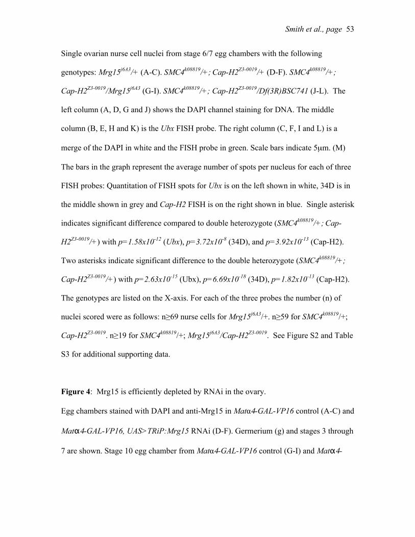
Smith et al., page
53
Single ovarian nurse cell nuclei from stage 6/7 egg chambers with the following
genotypes: Mrg15j6A3/+ (A-C). SMC4k08819/+; Cap-H2Z3-0019/+ (D-F). SMC4k08819/+;
Cap-H2Z3-0019/Mrg15j6A3 (G-I). SMC4k08819/+; Cap-H2Z3-0019/Df(3R)BSC741 (J-L). The
left column (A, D, G and J) shows the DAPI channel staining for DNA. The middle
column (B, E, H and K) is the Ubx FISH probe. The right column (C, F, I and L) is a
merge of the DAPI in white and the FISH probe in green. Scale bars indicate 5µm. (M)
The bars in the graph represent the average number of spots per nucleus for each of three
FISH probes: Quantitation of FISH spots for Ubx is on the left shown in white, 34D is in
the middle shown in grey and Cap-H2 FISH is on the right shown in blue. Single asterisk
indicates significant difference compared to double heterozygote (SMC4k08819/+; Cap-
H2Z3-0019/+) with p=1.58x10⁻¹² (Ubx), p=3.72x10⁻⁸ (34D), and p=3.92x10⁻¹³ (Cap-H2).
Two asterisks indicate significant difference to the double heterozygote (SMC4k08819/+;
Cap-H2Z3-0019/+) with p=2.63x10⁻¹⁵ (Ubx), p=6.69x10⁻¹⁸ (34D), p=1.82x10⁻¹³ (Cap-H2).
The genotypes are listed on the X-axis. For each of the three probes the number (n) of
nuclei scored were as follows: n≥69 nurse cells for Mrg15j6A3/+. n≥59 for SMC4k08819/+;
Cap-H2Z3-0019. n≥19 for SMC4k08819/+; Mrg15j6A3/Cap-H2Z3-0019. See Figure S2 and Table
S3 for additional supporting data.
Figure 4: Mrg15 is efficiently depleted by RNAi in the ovary.
Egg chambers stained with DAPI and anti-Mrg15 in Matα4-GAL-VP16 control (A-C) and
Matα4-GAL-VP16, UAS>TRiP:Mrg15 RNAi (D-F). Germerium (g) and stages 3 through
7 are shown. Stage 10 egg chamber from Matα4-GAL-VP16 control (G-I) and Matα4-

Smith et al., page
54
GAL-VP16, UAS>TRiP:Mrg15 RNAi (J-L). Note decreases Mrg15 protein levels in
nurse cells but not in follicle cells in stage 4 to stage 10 egg chambers. Bar is 50µm.
Figure 5: Mrg15 RNAi depleted nurse cells have altered chromosome organization.
Confocal slice of a stage 5/6 egg chamber from control Matα4-GAL-VP16 stained with
DAPI and Mrg15 (A-B). Pixel intensity for DAPI (blue) and Mrg15 (red) was determined
along a 50µm line through the egg chamber. Two follicle cells (fc1, fc2) and two nurse
cells (nc1, nc2) were included in the quantitation (C). Single DAPI stained nucleus from
control (D) egg chamber and topographical map of signal intensity showing DNA
distribution (E). Confocal slice of a stage 5/6 egg chamber from Matα4-GAL-VP16,
TRiP:Mrg15 stained with DAPI and Mrg15 (F-G). Pixel intensity for DAPI (blue) and
Mrg15 (red) was determined along a 58µm line through the egg chamber. Two follicle
cells (fc1, fc2) and two nurse cells (nc1, nc2) were included in the quantitation (H). Note
that the signal intensity for the follicle cell nuclei in the TRiP:Mrg15 sample is
comparable to that of the follicle cell nuclei in the control shown in C. Signal for the
Mrg15 protein is vastly diminished in the nurse cells (nc1, nc2). Single DAPI stained
nucleus from control (I) egg chamber and topographical map of signal intensity showing
DNA distribution (J). Note the uneven blob-like distribution of DAPI signal in nurse cells
from RNAi egg chambers.
Figure 6: Mrg15 is required for nurse cell polytene unpairing.
Confocal projection of a stage 10 single nurse cell nucleus from control Matα4-GAL-VP16
stained with DAPI and DNA FISH to 2L (green) and X-chromosome (red) (B) and the

Smith et al., page
55
merged image (C). Confocal projection of a stage 10 single nurse cell nucleus from
Matα4-GAL-VP16, TRiP:Mrg15 stained with DAPI (D, G), DNA FISH (E, H) and the
merged images (F, I). Note the clustering and fewer FISH spots in the TRiP:Mrg15 nuclei
(E, H) compared to the control in (B). Bar is 20µm. See Figure S3 for additional images
and quantitation of FISH spots.
Figure 7: Mrg15 antagonizes transvection at Ubx.
(A) Wild-type wing from Oregon-R. (B) Wing of class A typical of UbxCbx-1 Ubx1/++.
(C) Wing of class B typical of UbxCbx-1 Ubx1/Mrg15j6A3. (D) Quantification of phenotypic
classes for UbxCbx-1 Ubx1/++ and UbxCbx-1 Ubx1/Mrg15j6A3. n>50 for both genotypes.
p<10-3 for either class using Chi-squared. See Figure S3 for additional supporting data.
Figure 8: Mrg15 cooperates with Cap-H2 to antagonize transvection at yellow.
(A) Similarly aged females with the following genotypes from left to right: Oregon-R,
y1#8/y82f29; Cap-H2Z3-0019/+, y1#8/y82f29;Mrg15j6A3/+ and y1#8/y82f29; Cap-H2Z3-0019/
Mrg15j6A3. Arrows point to darkly pigmented posterior stripes. (B) Percent of all female
progeny exhibiting darkly pigmented posterior stripes per brood for the following
genotypes left to right: y1#8/y82f29; Cap-H2Z3-0019/+ (n=53), y1#8/y82f29;Mrg15j6A3/+ (n=77),
y1#8/y82f29;Df(3R)Exel6195/+ (n=45), y1#8/y82f29; Cap-H2Z3-0019/Mrg15j6A3 (n=96) and
y1#8/y82f29;Df(3R)Exel6195/Mrg15j6A3 (n=91). Asterisks denote p<0.001, two tailed T-test
assuming unequal variance.
Figure 9: Chromatin bound Cap-H2 is partially dependent on Mrg15 levels.

Smith et al., page
56
S2 cells transfected with pMT-Cap-H2-EGFP: (A) Cells were treated with control RNAi
and RNAi targeting Mrg15. Cells expressing Cap-H2-EGFP were fractionated and
extracts were immunoblotted with anti-GFP and anti-Lamin. Anti-Mrg15 immunoblots
were used to confirm RNAi depletion of Mrg15 (see Figure S5). Each lane represents
20µg of whole cell lysates (WCL), cytosolic (Cyto), soluble nuclear (Nuc) and chromatin
bound (Chr). (B) The fold-enrichment of Cap-H2-EGFP in the chromatin fraction was
calculated from four biological replicates as described in methods and p<0.005, two
tailed T-test assuming unequal variance. Control RNAi shown with the black bar and
Mrg15 RNAi shown with gray bar. (See Figure S5 for supporting data.)
Figure 10: Cap-H2 and Mrg15 are required to maintain chromosome length and control
axial compaction.
(A-F) Cultured Kc cell nuclei stained with DAPI (blue), X-chromosome probe 1 (X1
probe, green), X-chromosome probe 2 (X2 probe, red). Arrows indicate nuclei with a
single FISH spot for each probe showing paired homologs. Arrowheads indicate nuclei
with multiple spots of unpaired homologs. (A) Cells treated with control RNAi. (B) Cells
treated with RNAi targeting Cap-H2. (C) Cells treated with RNAi targeting Mrg15. (D)
Cells treated with RNAi targeting both Cap-H2 and Mrg15. (E) Cells with Cap-H2
overexpression (OE) from pMT-Cap-H2. (F) Cells with Cap-H2 overexpression plus
RNAi targeting Mrg15. (G) Single nucleus of control RNAi and (H) Mrg15 RNAi probed
with two different probes on the X-chromosome. (I) The average number of FISH spots
per probe per cell is indicated as a measure of homolog pairing. Spots were counted as
one spot if they were merged or their centers were <0.2µm apart. (J) The proportion of

Smith et al., page
57
nuclei with paired homologs is shown. (K) Pairwise distances were measured in three-
dimensional image reconstructions for three different FISH probe pairs: X1-X2
(~2.05Mb apart), X1-X3 (~3.6Mb apart) probes were on the X-chromosomes and 2L
(~2Mb apart) probes were on the left arm of the second chromosome (see methods and
Table S4). Significance levels were calculated by T-test, assuming equal variance in
MS Excel, where one asterisk is p=0.6, two asterisks is p<0.03 and three asterisks is
p<10-4. All p-values are calculated relative to control RNAi samples. Table S4 contains
further details including number of nuclei measured, average distances, standard error
and p-values. All bars are 5µm.


![Modular Connected [2MB]](https://img.pdfslide.net/doc/110x75/58a2f86c1a28ab8e608c1e28/modular-connected-2mb.jpg)


![Reportable Claims [PDF, 2MB]](https://img.pdfslide.net/doc/110x75/5868cb291a28ab3f7c8b6c73/reportable-claims-pdf-2mb.jpg)
![Wedding Ceremony Readings [2Mb]](https://img.pdfslide.net/doc/110x75/589eee441a28abf84d8b8813/wedding-ceremony-readings-2mb.jpg)