Embed Size (px)
Citation preview
Genetics, molecular and cell biology of cell death
Ágnes Czibula, Roberta Fajka-BojaInstitute of GeneticsDecember 14, 2016
„Practice-oriented, student-friendly modernization of the biomedical education for strengthening the international competitiveness of the rural Hungarian universities”TÁMOP-4.1.1.C-13/1/KONV-2014-0001
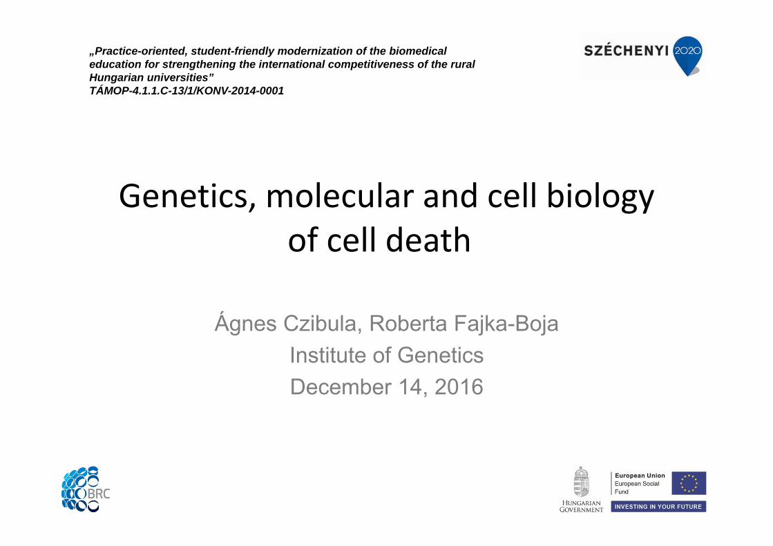
Morphological classification of cell deaths:apoptosis and necrosis
Apoptosis:- Membrane blebbing- Cytosolic condensation, cell shrinkage- Protein degradation - Nuclear condensation - Fragmentation of nuclear DNA- Formation of apoptotic bodies - Genetically regulated- The apoptotic bodies are phagocytosed,no inflammatory response!
Necrosis:- Cell swelling, round up- Rapid loss of membrane integrity- No nuclear condensation - No DNA degradation - Spilling of cellular content- Accidental, tissue injury- The release of cytoplasmic contents triggers inflammatory response.
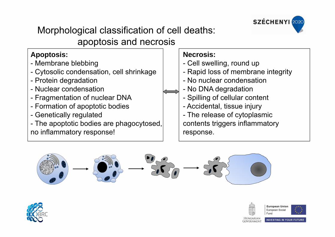
• Kerr JF, Wyllie AH, Currie AR. Apoptosis: a basic biological phenomenon with wide-ranging implications in tissue kinetics. Br J Cancer. 1972, 26(4):239-57
• Apoptosis occurs in all tissues as part of normal cellular turnover.
• Apoptosis also occurs during embryogenesis in which particular cells are ‘programmed’ to die, and hence the term ‘programmed cell death’ is used to describe this process.
• Intrinsic program for removal of damaged cells.
• Examples:- Embryogenesis, development - Regulation of organ size and morphology - Immunohomeostasis - DNS-damage, stress, hypoxia
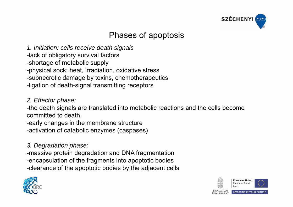
1. Initiation: cells receive death signals-lack of obligatory survival factors-shortage of metabolic supply-physical sock: heat, irradiation, oxidative stress-subnecrotic damage by toxins, chemotherapeutics-ligation of death-signal transmitting receptors
2. Effector phase:-the death signals are translated into metabolic reactions and the cells become committed to death.-early changes in the membrane structure-activation of catabolic enzymes (caspases)
3. Degradation phase:-massive protein degradation and DNA fragmentation -encapsulation of the fragments into apoptotic bodies-clearance of the apoptotic bodies by the adjacent cells
Phases of apoptosis
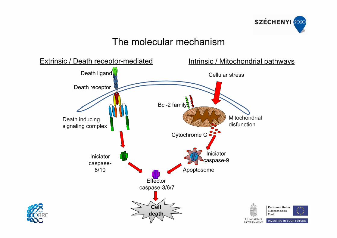
The molecular mechanism
Death receptor
Death ligand
Death inducing signaling complex
Iniciator caspase-
8/10
Effectorcaspase-3/6/7
Extrinsic / Death receptor-mediated Intrinsic / Mitochondrial pathways
CelldeathCell
death
Cellular stress
Mitochondrial disfunction
Cytochrome C
Bcl-2 family
Iniciator caspase-9
Apoptosome
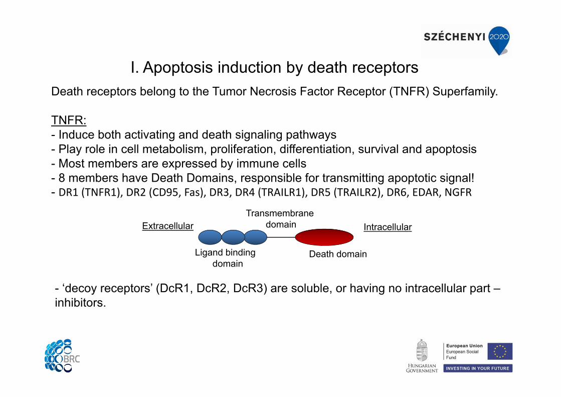
Death receptors belong to the Tumor Necrosis Factor Receptor (TNFR) Superfamily.
TNFR:- Induce both activating and death signaling pathways- Play role in cell metabolism, proliferation, differentiation, survival and apoptosis- Most members are expressed by immune cells- 8 members have Death Domains, responsible for transmitting apoptotic signal! - DR1 (TNFR1), DR2 (CD95, Fas), DR3, DR4 (TRAILR1), DR5 (TRAILR2), DR6, EDAR, NGFR
I. Apoptosis induction by death receptors
- ‘decoy receptors’ (DcR1, DcR2, DcR3) are soluble, or having no intracellular part –inhibitors.
Death domain
Transmembrane domain
Ligand binding domain
Extracellular Intracellular
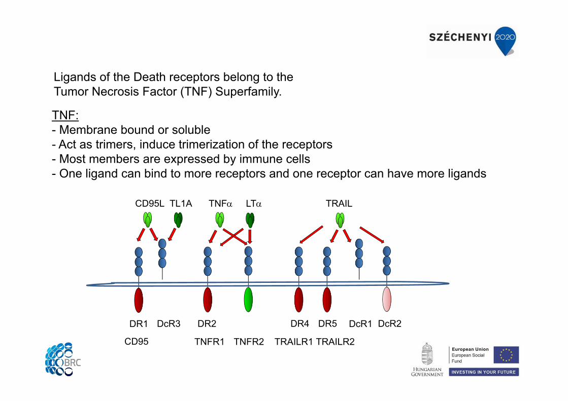
TNF:- Membrane bound or soluble- Act as trimers, induce trimerization of the receptors - Most members are expressed by immune cells- One ligand can bind to more receptors and one receptor can have more ligands
Ligands of the Death receptors belong to the Tumor Necrosis Factor (TNF) Superfamily.
CD95
DcR3
TNFR1 TNFR2
DR4 DR5 DcR1 DcR2
CD95L TNF TRAILTL1A LT
DR1 DR2
TRAILR1 TRAILR2
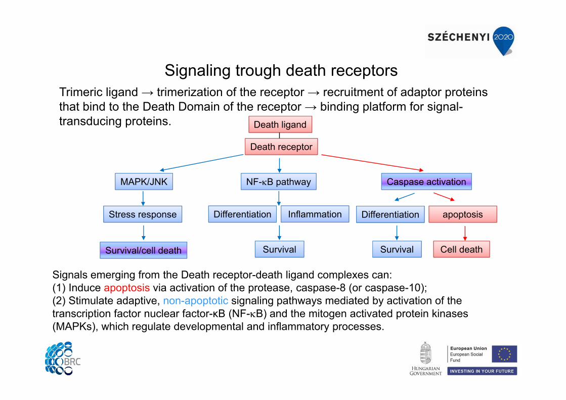
Signals emerging from the Death receptor-death ligand complexes can: (1) Induce apoptosis via activation of the protease, caspase-8 (or caspase-10);(2) Stimulate adaptive, non-apoptotic signaling pathways mediated by activation of the transcription factor nuclear factor-κB (NF-B) and the mitogen activated protein kinases (MAPKs), which regulate developmental and inflammatory processes.
Signaling trough death receptorsTrimeric ligand → trimerization of the receptor → recruitment of adaptor proteins that bind to the Death Domain of the receptor → binding platform for signal-transducing proteins. Death ligand
Death receptor
NF-B pathwayMAPK/JNK
Stress response
Survival/cell death
Differentiation Inflammation
Survival
Caspase activation
Differentiation apoptosis
Survival Cell death
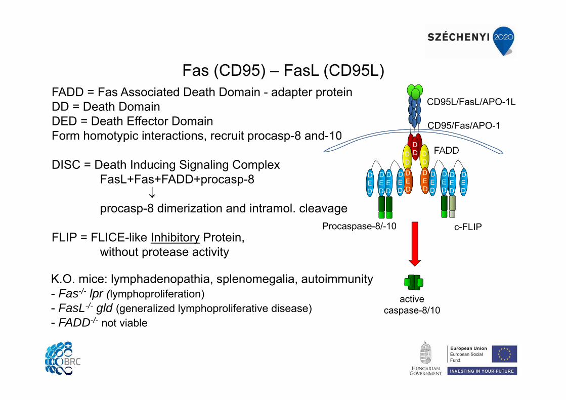
Fas (CD95) – FasL (CD95L)
K.O. mice: lymphadenopathia, splenomegalia, autoimmunity- Fas-/- lpr (lymphoproliferation)- FasL-/- gld (generalized lymphoproliferative disease)- FADD-/- not viable
FADD = Fas Associated Death Domain - adapter proteinDD = Death DomainDED = Death Effector DomainForm homotypic interactions, recruit procasp-8 and-10
DISC = Death Inducing Signaling ComplexFasL+Fas+FADD+procasp-8
procasp-8 dimerization and intramol. cleavage
FLIP = FLICE-like Inhibitory Protein, without protease activity
CD95L/FasL/APO-1L
activecaspase-8/10
CD95/Fas/APO-1
FADD
Procaspase-8/-10 c-FLIP
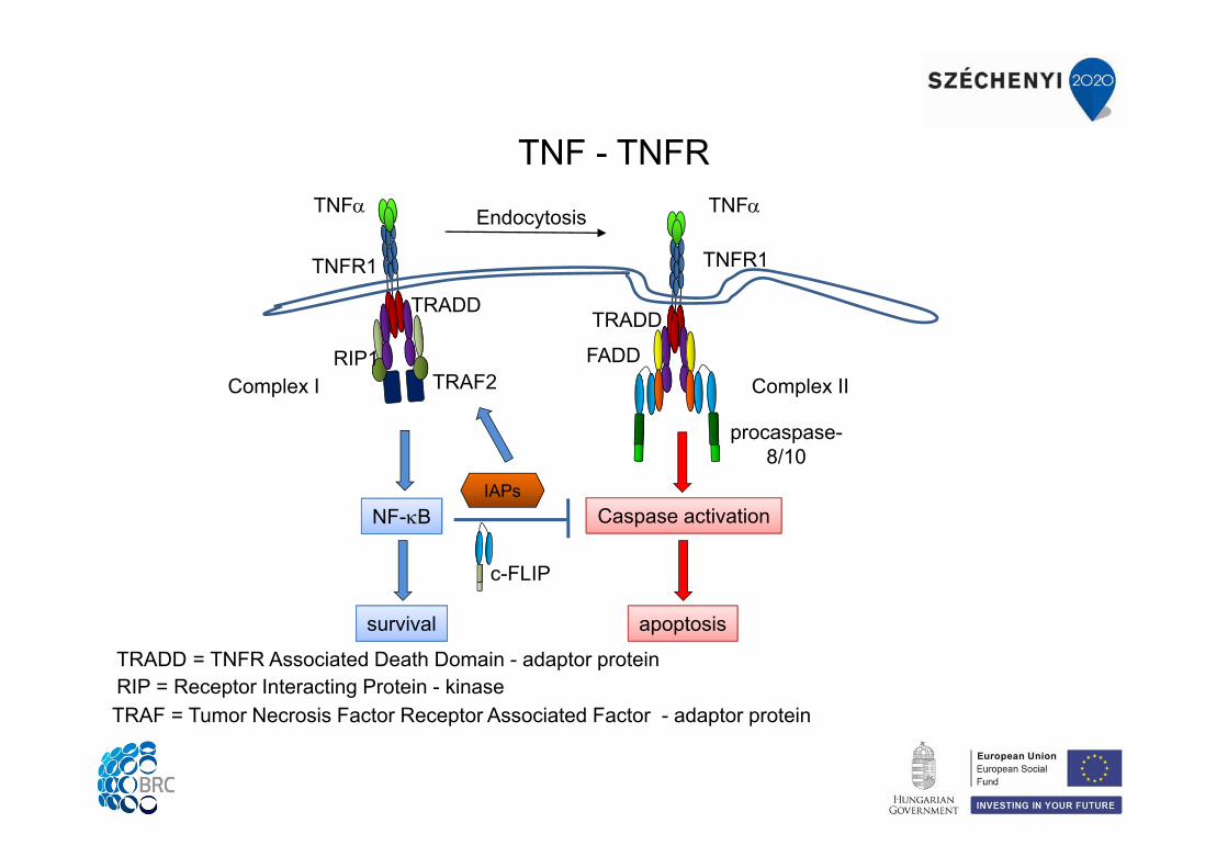
TRAF = Tumor Necrosis Factor Receptor Associated Factor - adaptor proteinRIP = Receptor Interacting Protein - kinaseTRADD = TNFR Associated Death Domain - adaptor protein
TNF - TNFR
TNFR1
TNF
Complex I
procaspase-8/10
TRADD
TRAF2RIP1
NF-B
survival
Endocytosis
TRADD
Caspase activation
apoptosis
TNFR1
TNF
Complex IIFADD
IAPs
c-FLIP
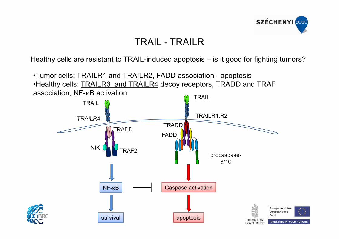
TRAIL - TRAILR
•Tumor cells: TRAILR1 and TRAILR2, FADD association - apoptosis•Healthy cells: TRAILR3 and TRAILR4 decoy receptors, TRADD and TRAF association, NF-B activation
Healthy cells are resistant to TRAIL-induced apoptosis – is it good for fighting tumors?
TRAILR4
TRAIL
procaspase-8/10
TRADD
TRAF2NIK
NF-B
survival
TRADD
FADD
Caspase activation
apoptosis
TRAIL
TRAILR1,R2
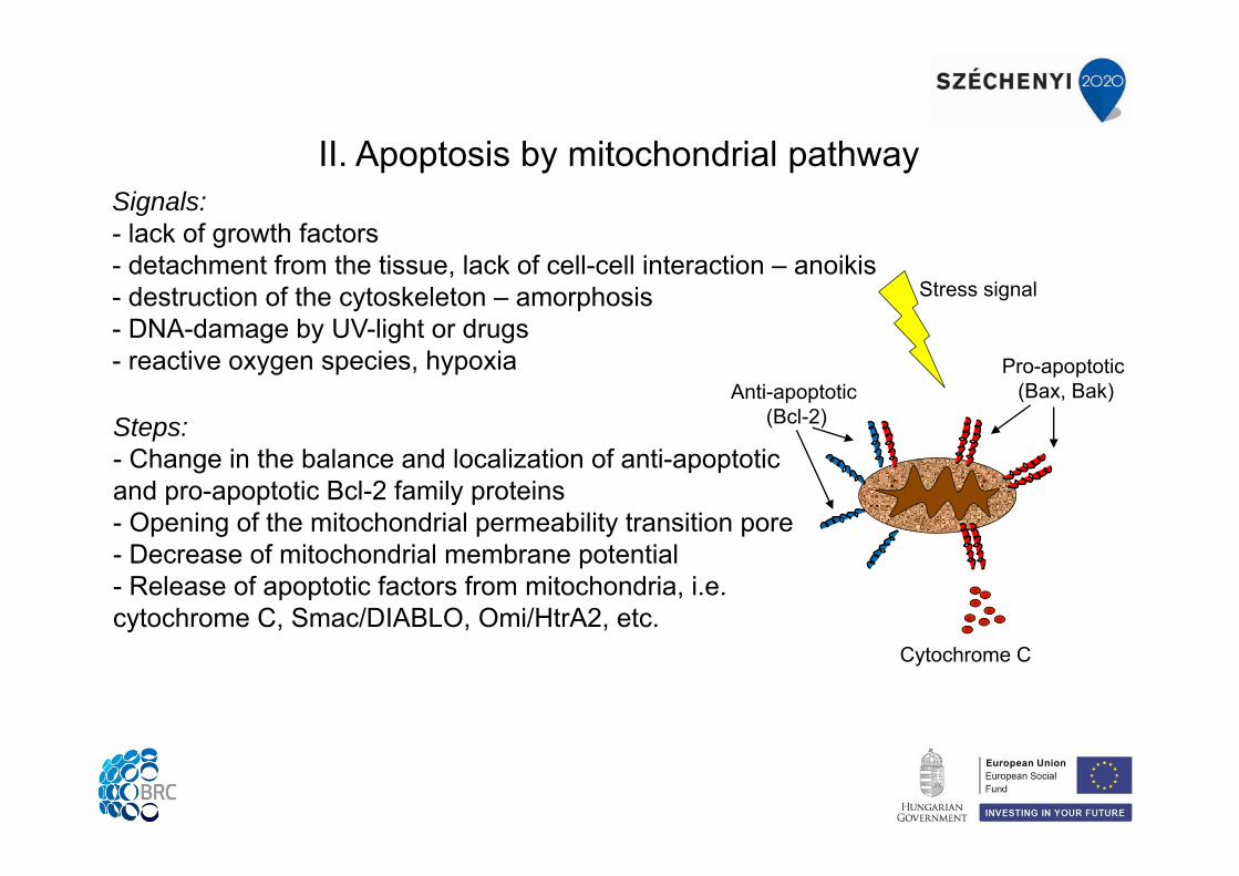
II. Apoptosis by mitochondrial pathwaySignals:- lack of growth factors- detachment from the tissue, lack of cell-cell interaction – anoikis- destruction of the cytoskeleton – amorphosis- DNA-damage by UV-light or drugs- reactive oxygen species, hypoxia
Steps:- Change in the balance and localization of anti-apoptotic and pro-apoptotic Bcl-2 family proteins- Opening of the mitochondrial permeability transition pore- Decrease of mitochondrial membrane potential- Release of apoptotic factors from mitochondria, i.e. cytochrome C, Smac/DIABLO, Omi/HtrA2, etc.
Cytochrome C
Anti-apoptotic (Bcl-2)
Pro-apoptotic (Bax, Bak)
Stress signal
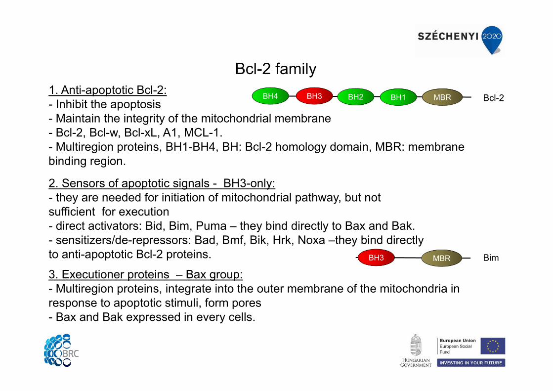
Bcl-2 family1. Anti-apoptotic Bcl-2:- Inhibit the apoptosis- Maintain the integrity of the mitochondrial membrane- Bcl-2, Bcl-w, Bcl-xL, A1, MCL-1.- Multiregion proteins, BH1-BH4, BH: Bcl-2 homology domain, MBR: membrane binding region.
3. Executioner proteins – Bax group:- Multiregion proteins, integrate into the outer membrane of the mitochondria in response to apoptotic stimuli, form pores - Bax and Bak expressed in every cells.
2. Sensors of apoptotic signals - BH3-only:- they are needed for initiation of mitochondrial pathway, but not sufficient for execution- direct activators: Bid, Bim, Puma – they bind directly to Bax and Bak.- sensitizers/de-repressors: Bad, Bmf, Bik, Hrk, Noxa –they bind directly to anti-apoptotic Bcl-2 proteins.
MBRBH1BH4 BH2BH3 Bcl-2
MBRBH3 Bim
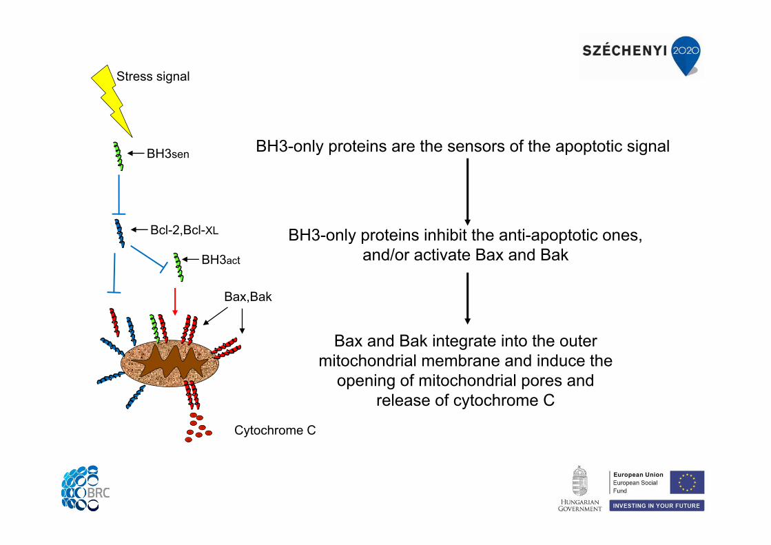
BH3-only proteins are the sensors of the apoptotic signal
BH3-only proteins inhibit the anti-apoptotic ones, and/or activate Bax and Bak
Bax and Bak integrate into the outer mitochondrial membrane and induce the
opening of mitochondrial pores and release of cytochrome C
Cytochrome C
Bax,Bak
Stress signal
BH3sen
BH3act
Bcl-2,Bcl-XL
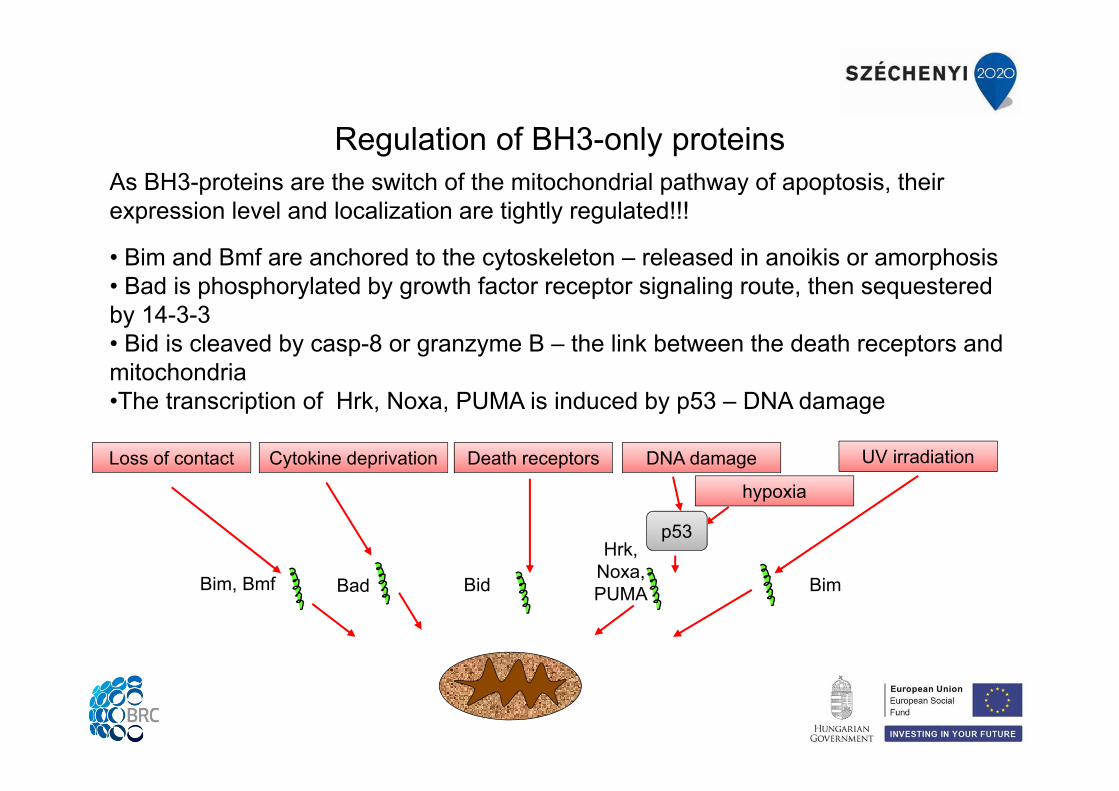
Regulation of BH3-only proteinsAs BH3-proteins are the switch of the mitochondrial pathway of apoptosis, their expression level and localization are tightly regulated!!!
• Bim and Bmf are anchored to the cytoskeleton – released in anoikis or amorphosis• Bad is phosphorylated by growth factor receptor signaling route, then sequestered by 14-3-3 • Bid is cleaved by casp-8 or granzyme B – the link between the death receptors and mitochondria•The transcription of Hrk, Noxa, PUMA is induced by p53 – DNA damage
Loss of contact Cytokine deprivation DNA damage UV irradiation
hypoxia
Death receptors
p53
Bim, Bmf Bad Bid
Hrk,Noxa, PUMA Bim
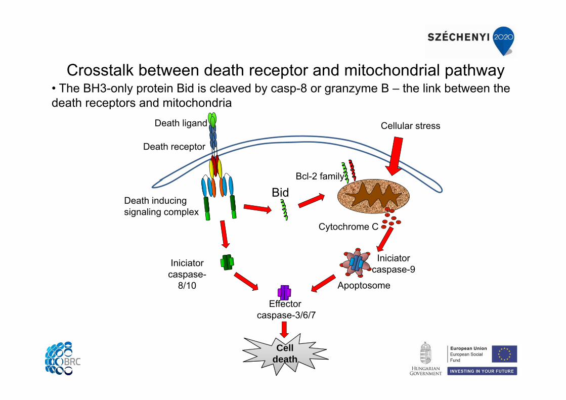
Crosstalk between death receptor and mitochondrial pathway
Death receptor
Death ligand
Death inducing signaling complex
Iniciator caspase-
8/10
Effectorcaspase-3/6/7
CelldeathCell
death
Cellular stress
Cytochrome C
Bcl-2 family
Iniciator caspase-9
Apoptosome
Bid
• The BH3-only protein Bid is cleaved by casp-8 or granzyme B – the link between the death receptors and mitochondria
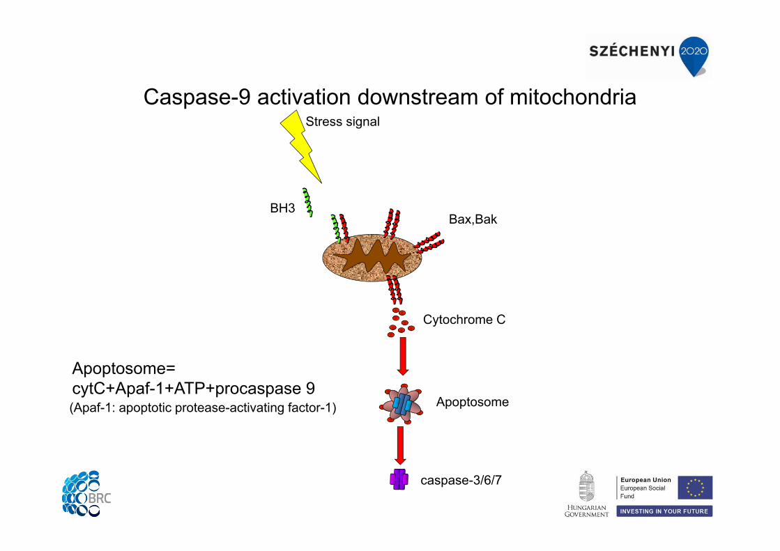
Caspase-9 activation downstream of mitochondria
(Apaf-1: apoptotic protease-activating factor-1)
Apoptosome=cytC+Apaf-1+ATP+procaspase 9
Cytochrome C
Bax,Bak
Stress signal
BH3
caspase-3/6/7
Apoptosome
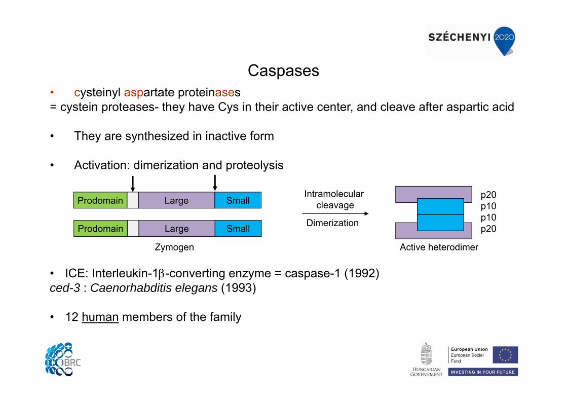
Caspases• cysteinyl aspartate proteinases= cystein proteases- they have Cys in their active center, and cleave after aspartic acid
• They are synthesized in inactive form
• Activation: dimerization and proteolysis
• ICE: Interleukin-1-converting enzyme = caspase-1 (1992)ced-3 : Caenorhabditis elegans (1993)
• 12 human members of the family
Active heterodimer
Intramolecular cleavage
p20p10p10p20
Large SmallProdomain
Zymogen
Large SmallProdomain Dimerization
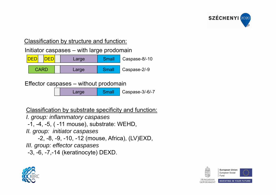
Classification by structure and function:
Classification by substrate specificity and function:I. group: inflammatory caspases-1, -4, -5, ( -11 mouse), substrate: WEHD,II. group: initiator caspases
-2, -8, -9, -10, -12 (mouse, Africa), (LV)EXD,III. group: effector caspases-3, -6, -7,-14 (keratinocyte) DEXD.
Large SmallCARD
Large SmallDEDDED Caspase-8/-10
Caspase-2/-9
Large Small Caspase-3/-6/-7
Initiator caspases – with large prodomain
Effector caspases – without prodomain
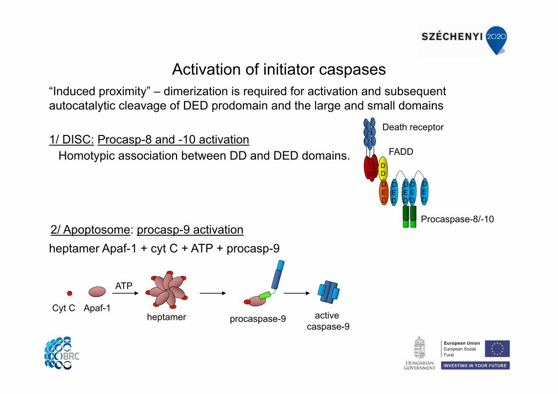
Activation of initiator caspases
1/ DISC: Procasp-8 and -10 activationHomotypic association between DD and DED domains.
“Induced proximity” – dimerization is required for activation and subsequent autocatalytic cleavage of DED prodomain and the large and small domains
2/ Apoptosome: procasp-9 activationheptamer Apaf-1 + cyt C + ATP + procasp-9
Death receptor
FADD
Procaspase-8/-10
ATP
heptamerCyt C Apaf-1
procaspase-9 active caspase-9
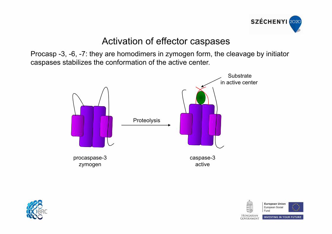
Activation of effector caspasesProcasp -3, -6, -7: they are homodimers in zymogen form, the cleavage by initiator caspases stabilizes the conformation of the active center.
procaspase-3zymogen
Proteolysis
caspase-3active
Substratein active center
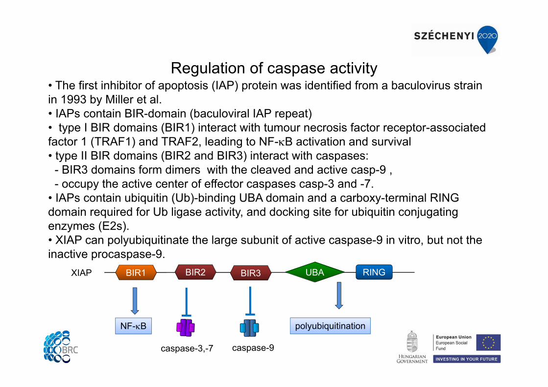
Regulation of caspase activity• The first inhibitor of apoptosis (IAP) protein was identified from a baculovirus strain in 1993 by Miller et al. • IAPs contain BIR-domain (baculoviral IAP repeat)• type I BIR domains (BIR1) interact with tumour necrosis factor receptor-associated factor 1 (TRAF1) and TRAF2, leading to NF-B activation and survival• type II BIR domains (BIR2 and BIR3) interact with caspases:
- BIR3 domains form dimers with the cleaved and active casp-9 ,- occupy the active center of effector caspases casp-3 and -7.
• IAPs contain ubiquitin (Ub)-binding UBA domain and a carboxy-terminal RING domain required for Ub ligase activity, and docking site for ubiquitin conjugating enzymes (E2s).• XIAP can polyubiquitinate the large subunit of active caspase-9 in vitro, but not the inactive procaspase-9.
RINGBIR2BIR1 BIR3 UBAXIAP
NF-B polyubiquitination
caspase-3,-7 caspase-9
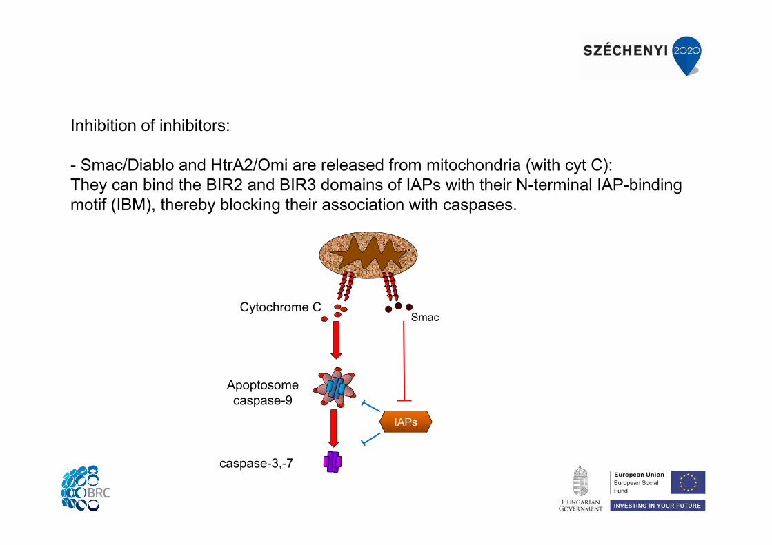
Inhibition of inhibitors:
- Smac/Diablo and HtrA2/Omi are released from mitochondria (with cyt C):They can bind the BIR2 and BIR3 domains of IAPs with their N-terminal IAP-binding motif (IBM), thereby blocking their association with caspases.
Cytochrome CSmac
caspase-3,-7
Apoptosomecaspase-9
IAPs
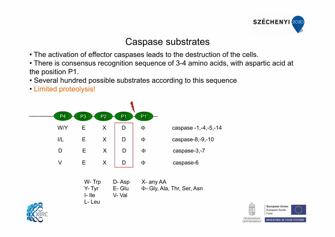
• The activation of effector caspases leads to the destruction of the cells. • There is consensus recognition sequence of 3-4 amino acids, with aspartic acid at the position P1. • Several hundred possible substrates according to this sequence• Limited proteolysis!
Caspase substrates
W- Trp D- Asp X- any AAY- Tyr E- Glu - Gly, Ala, Thr, Ser, AsnI- Ile V- ValL- Leu
P4 P3 P2 P1 P1’
W/Y E X D caspase -1,-4,-5,-14
I/L E X D caspase-8,-9,-10
D E X D caspase-3,-7
V E X D caspase-6
- Inactivation or activation of signaling proteins, enzymes (kinases, phosphatases, phospholipases, Bcl-2 family proteins) – amplification of apoptotic signaling.
- Desintegration of cytoskeletal elements (actin, gelsolin, lamin A,B) – morphological changes.
- Cleavage of nuclear proteins (DNA polimerases, inhibitor of DNase) – stopping DNA replication and repair, internucleosomal fragmentation of DNA (see the practical demonstration).
Consequences of caspase activation:
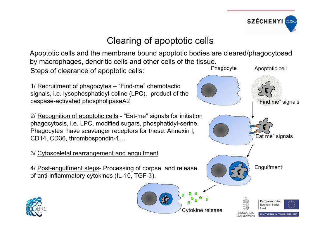
Steps of clearance of apoptotic cells:
1/ Recruitment of phagocytes – “Find-me” chemotactic signals, i.e. lysophosphatidyl-coline (LPC), product of the caspase-activated phospholipaseA2
2/ Recognition of apoptotic cells - “Eat-me” signals for initiation phagocytosis, i.e. LPC, modified sugars, phosphatidyl-serine. Phagocytes have scavenger receptors for these: Annexin I, CD14, CD36, thrombospondin-1…
3/ Cytosceletal rearrangement and engulfment
4/ Post-engulfment steps- Processing of corpse and release of anti-inflammatory cytokines (IL-10, TGF-).
Clearing of apoptotic cellsApoptotic cells and the membrane bound apoptotic bodies are cleared/phagocytosed by macrophages, dendritic cells and other cells of the tissue.
Phagocyte Apoptotic cell
“Find me” signals
“Eat me” signals
Cytokine release
Engulfment
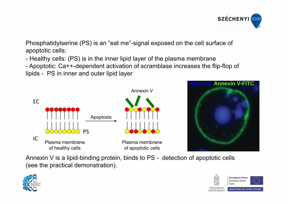
Phosphatidylserine (PS) is an “eat me”-signal exposed on the cell surface of apoptotic cells:- Healthy cells: (PS) is in the inner lipid layer of the plasma membrane- Apoptotic: Ca++-dependent activation of scramblase increases the flip-flop of lipids - PS in inner and outer lipid layer
Hoechst Annexin V-FITC
Plasma membrane of healthy cells
Apoptosis
Plasma membrane of apoptotic cells
EC
ICPS
Annexin V
Annexin V is a lipid-binding protein, binds to PS - detection of apoptotic cells (see the practical demonstration).
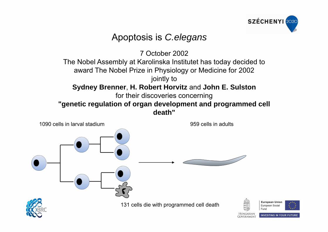
7 October 2002The Nobel Assembly at Karolinska Institutet has today decided to
award The Nobel Prize in Physiology or Medicine for 2002jointly to
Sydney Brenner, H. Robert Horvitz and John E. Sulstonfor their discoveries concerning
"genetic regulation of organ development and programmed cell death"
Apoptosis is C.elegans
959 cells in adults 1090 cells in larval stadium
131 cells die with programmed cell death
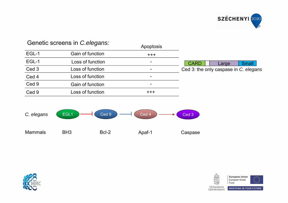
Genetic screens in C.elegans:
EGL-1EGL-1Ced 3Ced 4Ced 9
Ced 9
Gain of function
Gain of functionLoss of function
Loss of functionLoss of functionLoss of function
+++--
-
Apoptosis
+++
-Ced 3: the only caspase in C. elegans
Large SmallCARD
EGL1
BH3
Ced 9
Bcl-2
Ced 4
Apaf-1
Ced 3
Caspase
C. elegans
Mammals
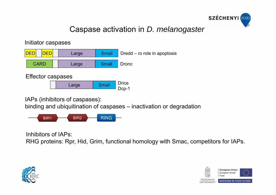
Caspase activation in D. melanogaster
IAPs (inhibitors of caspases): binding and ubiquitination of caspases – inactivation or degradation
Inhibitors of IAPs:RHG proteins: Rpr, Hid, Grim, functional homology with Smac, competitors for IAPs.
Large SmallCARD
Large SmallDEDDED Dredd – ro role in apoptosis
Dronc
Large Small DriceDcp-1
Effector caspases
Initiator caspases
RINGBIR2BIR1
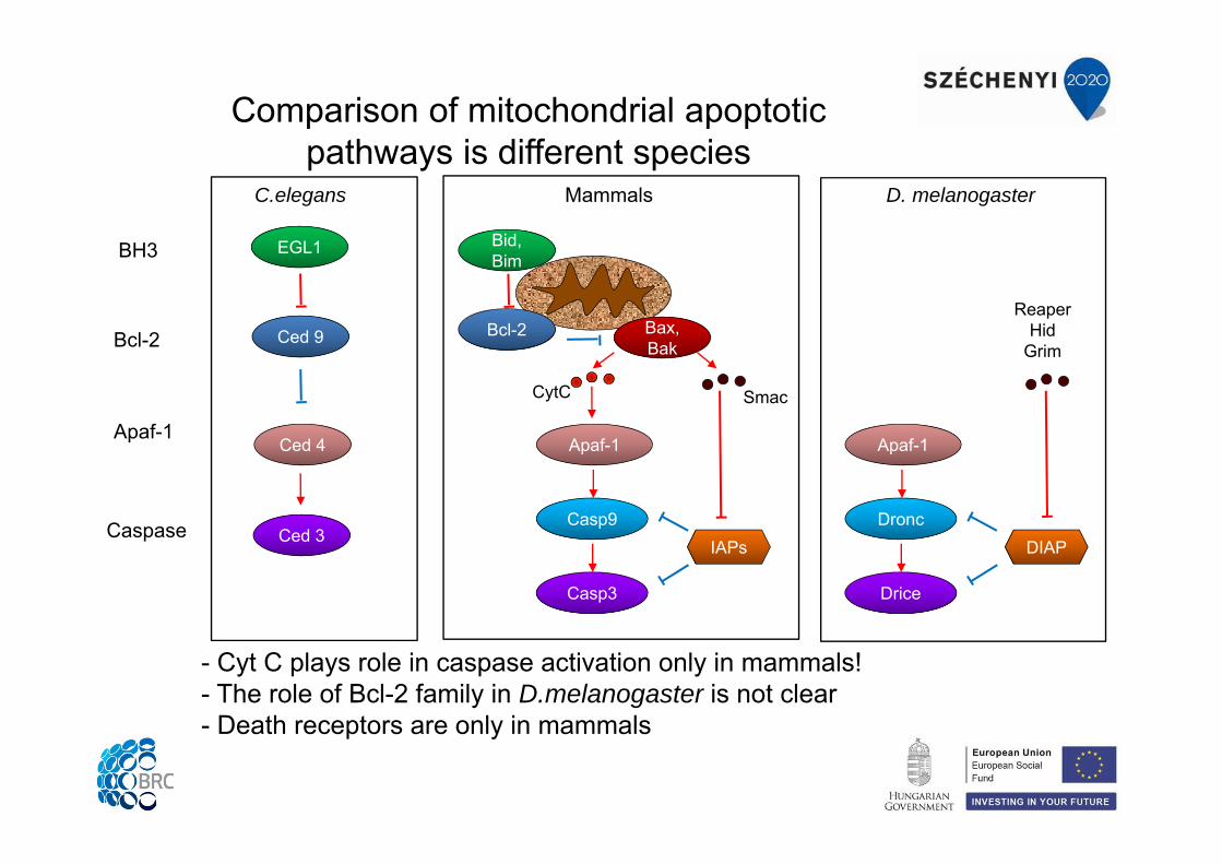
Comparison of mitochondrial apoptotic pathways is different species
- Cyt C plays role in caspase activation only in mammals!- The role of Bcl-2 family in D.melanogaster is not clear- Death receptors are only in mammals
EGL1
Ced 9
Ced 4
Ced 3
BH3
Bcl-2
Apaf-1
Caspase
C.elegans Mammals D. melanogaster
Bid, Bim
Bcl-2
Apaf-1
Casp3
Bax, Bak
CytC
Casp9
Smac
IAPs
Apaf-1
Drice
Dronc
ReaperHid
Grim
DIAP
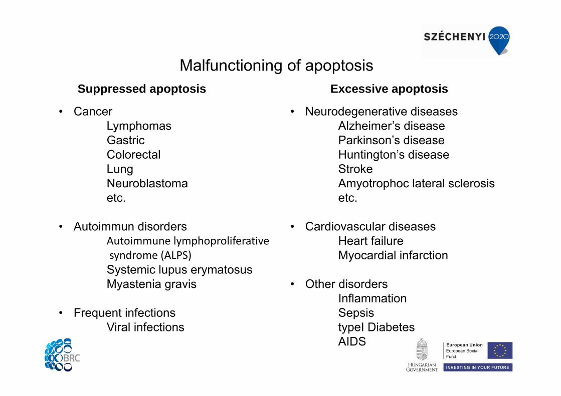
Malfunctioning of apoptosisSuppressed apoptosis Excessive apoptosis
• CancerLymphomasGastricColorectalLungNeuroblastomaetc.
• Autoimmun disordersAutoimmune lymphoproliferativesyndrome (ALPS)Systemic lupus erymatosusMyastenia gravis
• Frequent infectionsViral infections
• Neurodegenerative diseasesAlzheimer’s diseaseParkinson’s diseaseHuntington’s diseaseStrokeAmyotrophoc lateral sclerosisetc.
• Cardiovascular diseasesHeart failureMyocardial infarction
• Other disordersInflammationSepsistypeI DiabetesAIDS
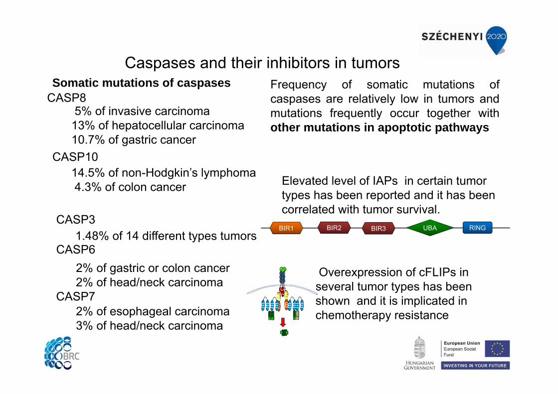
CASP6 1.48% of 14 different types tumors
CASP3
2% of esophageal carcinoma3% of head/neck carcinoma
CASP7
2% of gastric or colon cancer2% of head/neck carcinoma
Somatic mutations of caspases
5% of invasive carcinoma13% of hepatocellular carcinoma10.7% of gastric cancer
CASP8
CASP10 14.5% of non-Hodgkin’s lymphoma4.3% of colon cancer
Frequency of somatic mutations ofcaspases are relatively low in tumors andmutations frequently occur together withother mutations in apoptotic pathways
Overexpression of cFLIPs in several tumor types has been shown and it is implicated in chemotherapy resistance
Elevated level of IAPs in certain tumor types has been reported and it has been correlated with tumor survival.
RINGBIR2BIR1 BIR3 UBA
Caspases and their inhibitors in tumors
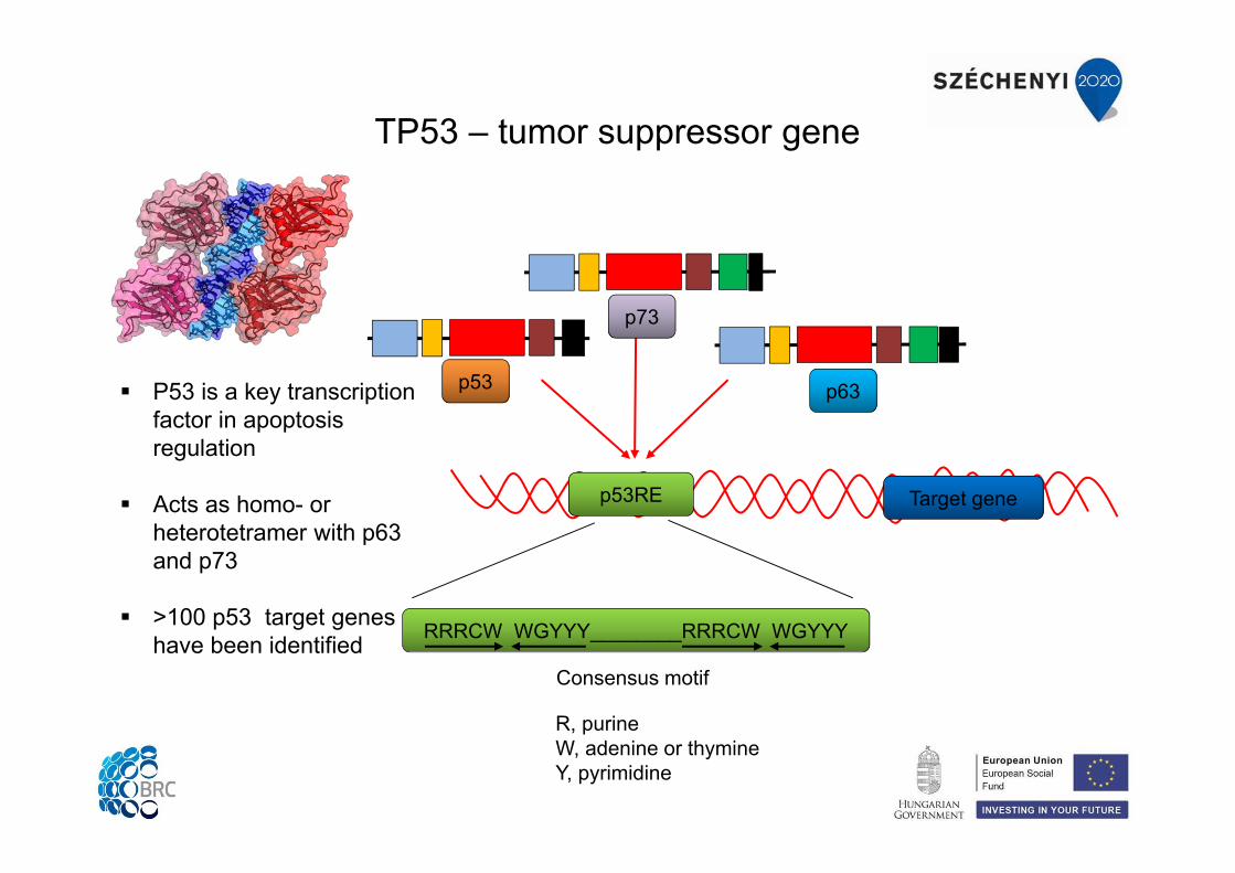
TP53 – tumor suppressor gene
p53RE Target gene
p53
p73
p63
RRRCW WGYYY________RRRCW WGYYY
Consensus motif
R, purineW, adenine or thymineY, pyrimidine
P53 is a key transcription factor in apoptosis regulation
Acts as homo- or heterotetramer with p63 and p73
>100 p53 target genes have been identified
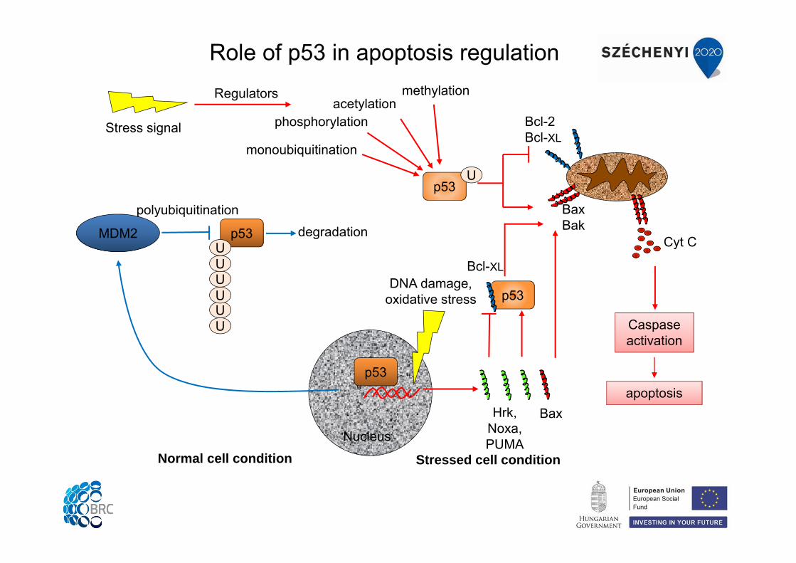
Role of p53 in apoptosis regulation
Nucleus
p53
DNA damage,oxidative stress
Hrk,Noxa, PUMA
p53
Bcl-XL
p53U
Cyt C
Bcl-2Bcl-XL
BaxBak
apoptosis
Caspaseactivation
Bax
MDM2 p53
UUUUUU
degradationpolyubiquitination
monoubiquitination
phosphorylationacetylation
methylation
Normal cell condition Stressed cell condition
Stress signal
Regulators
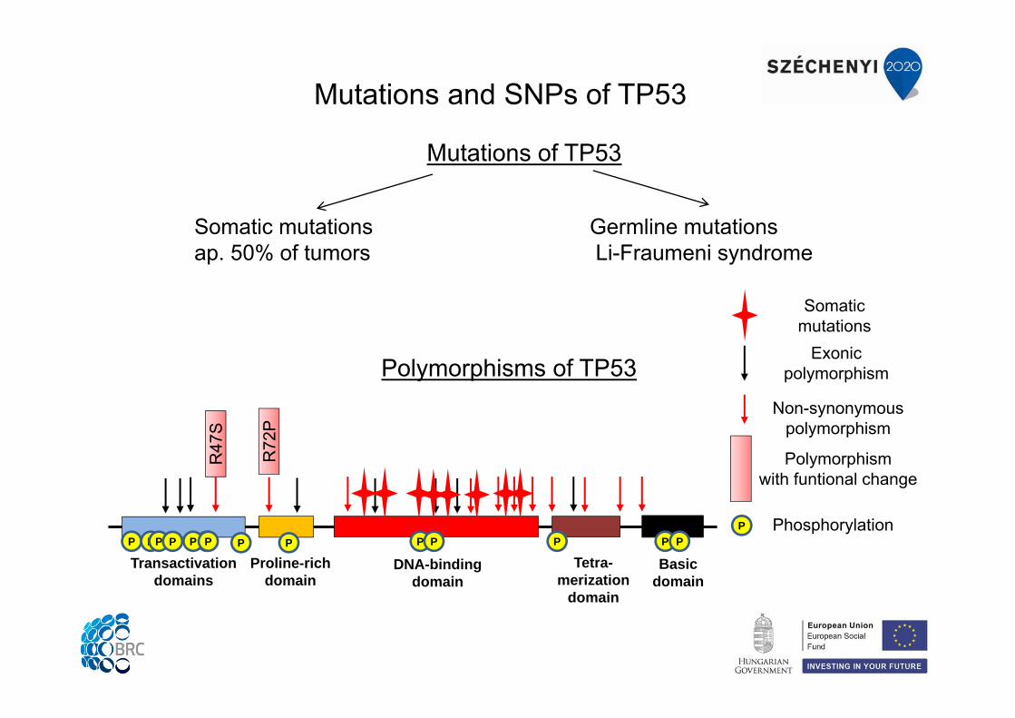
Mutations and SNPs of TP53
Mutations of TP53
Somatic mutations ap. 50% of tumors
Germline mutationsLi-Fraumeni syndrome
Polymorphisms of TP53
Transactivationdomains
Proline-richdomain
DNA-bindingdomain
Tetra-merization
domain
Basicdomain
R47
S
R72
P
Exonic polymorphism
Non-synonymouspolymorphism
Polymorphismwith funtional change
P PP P PP PP PP P P PP
Somatic mutations
P Phosphorylation
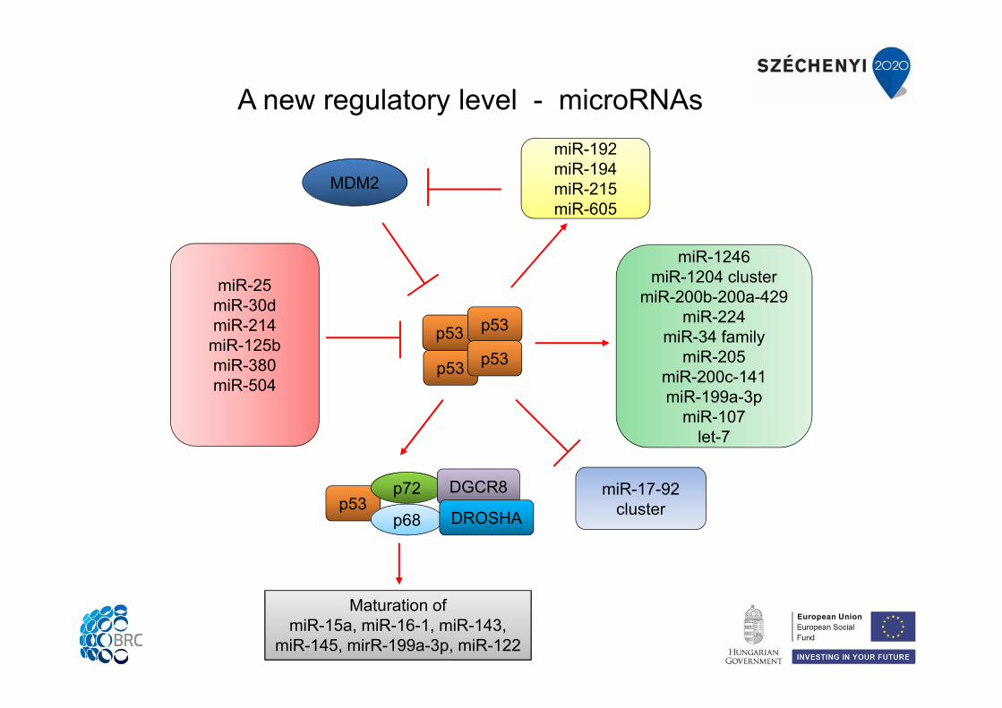
A new regulatory level - microRNAs
p53
p53 p53
p53
miR-25miR-30dmiR-214
miR-125bmiR-380miR-504
miR-1246miR-1204 cluster
miR-200b-200a-429miR-224
miR-34 familymiR-205
miR-200c-141miR-199a-3p
miR-107let-7
miR-17-92 clusterp53
p72
p68
DGCR8
DROSHA
Maturation of miR-15a, miR-16-1, miR-143,
miR-145, mirR-199a-3p, miR-122
MDM2
miR-192miR-194miR-215miR-605
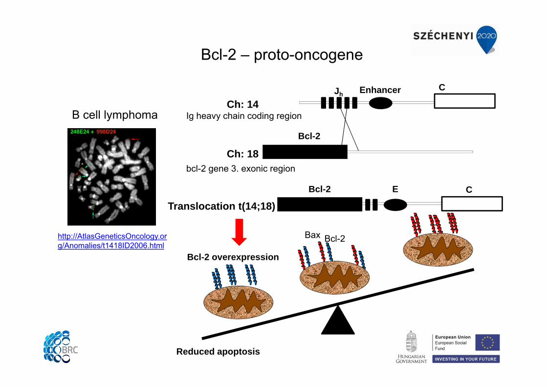
http://AtlasGeneticsOncology.org/Anomalies/t1418ID2006.html
Bcl-2 – proto-oncogene
B cell lymphoma
Enhancer CJh
Bcl-2
Bcl-2 E C
Translocation t(14;18)
Ch: 18
Ch: 14
Bcl-2 overexpression
Reduced apoptosis
Bax Bcl-2
Ig heavy chain coding region
bcl-2 gene 3. exonic region
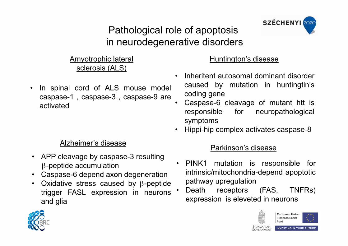
Pathological role of apoptosisin neurodegenerative disorders
• Inheritent autosomal dominant disordercaused by mutation in huntingtin’scoding gene
• Caspase-6 cleavage of mutant htt isresponsible for neuropathologicalsymptoms
• Hippi-hip complex activates caspase-8
Alzheimer’s disease
Huntington’s disease
• APP cleavage by caspase-3 resulting-peptide accumulation
• Caspase-6 depend axon degeneration• Oxidative stress caused by -peptide
trigger FASL expression in neuronsand glia
Parkinson’s disease
• PINK1 mutation is responsible forintrinsic/mitochondria-depend apoptoticpathway upregulation
• Death receptors (FAS, TNFRs)expression is eleveted in neurons
Amyotrophic lateralsclerosis (ALS)
• In spinal cord of ALS mouse modelcaspase-1 , caspase-3 , caspase-9 areactivated
References
2/ Chipuk et al. The BCL-2 family reunion. Mol Cell. 2010, 37(3):299-310.
4/ Crawford ED, Wells JA. Caspase substrates and cellular remodeling. Annu Rev Biochem. 2011, 80:1055-87.
7/ Kepp et al. Cell death assays for drug discovery. Nat Rev Drug Discov. 2011, 10(3):221-37.
5/ Gyrd-Hansen M, Meier P. IAPs: from caspase inhibitors to modulators of NF-kappaB, inflammation and cancer. Nat Rev Cancer. 2010, 10(8):561-74.
6/ Ravichandran KS. Find-me and eat-me signals in apoptotic cell clearance: progress and conundrums. J Exp Med. 2010, 207(9):1807-17.
1/ Sessler et al. Structural determinants of DISC function: new insights into death receptor-mediated apoptosis signalling. Pharmacol Ther. 2013, 140(2):186-99.
3/ Riedl SJ, Shi Y. Molecular mechanisms of caspase regulation during apoptosis. Nat Rev Mol Cell Biol. 2004, 5(11):897-907.
8/ Favaloro B et al. Role of apoptosis in disease. Aging (Albany NY). 2012, 4(5):330-49.
9/ Whibley C, Pharoah PD, Hollstein M. p53 polymorphisms: cancer implications. Nat Rev Cancer. 2009, 9(2):95-107.
Thank you for your attention!
This work is supported by the European Union, co-financed by the European Social Fund, within the framework of " Practice-
oriented, student-friendly modernization of the biomedical education for strengthening the international
competitiveness of the rural Hungarian universities " TÁMOP-4.1.1.C-13/1/KONV-2014-0001 project.