Embed Size (px)
Citation preview
Genetics of Cancer
Lecture 34
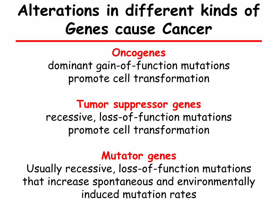
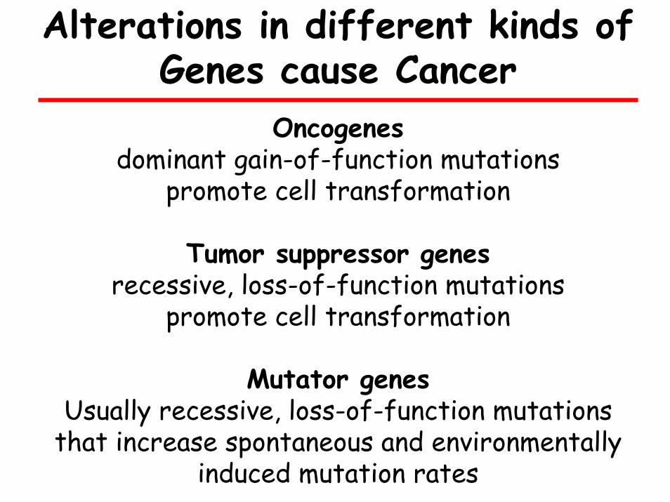
Alterations in different kinds of Genes cause Cancer
Oncogenesdominant gain-of-function mutations
promote cell transformation
Tumor suppressor genes recessive, loss-of-function mutations
promote cell transformation
Mutator genes Usually recessive, loss-of-function mutations
that increase spontaneous and environmentally induced mutation rates
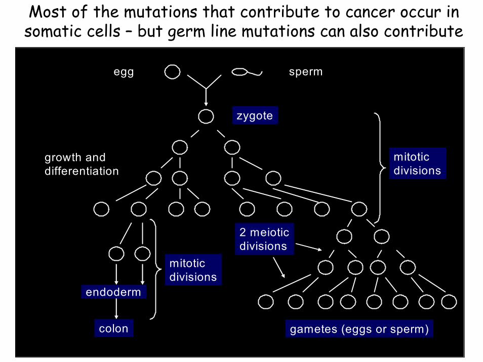
Most of the mutations that contribute to cancer occur in somatic cells – but germ line mutations can also contribute
egg sperm
zygote
endoderm
colon
growth anddifferentiation
mitoticdivisions
mitoticdivisions
2 meioticdivisions
gametes (eggs or sperm)
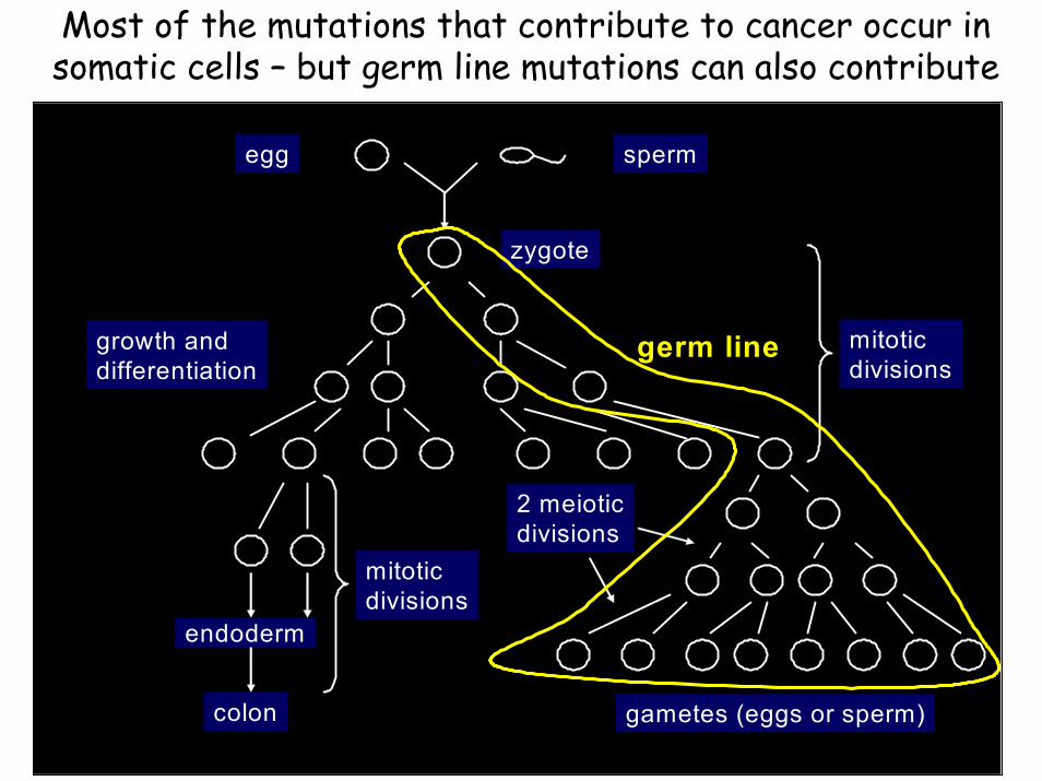
Most of the mutations that contribute to cancer occur in somatic cells – but germ line mutations can also contribute
egg sperm
zygote
endoderm
colon
growth anddifferentiation
mitoticdivisions
mitoticdivisions
2 meioticdivisions
gametes (eggs or sperm)
germ line
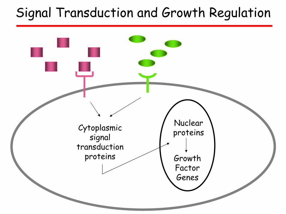
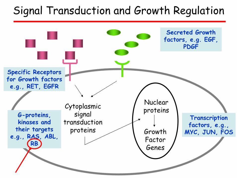
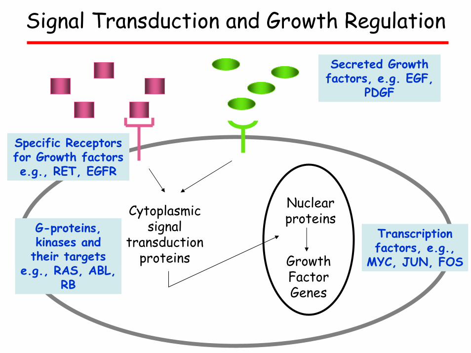
Cytoplasmicsignal
transduction proteins
Nuclear proteins
Growth Factor Genes
Signal Transduction and Growth Regulation
Cytoplasmicsignal
transduction proteins
Nuclear proteins
Growth Factor Genes
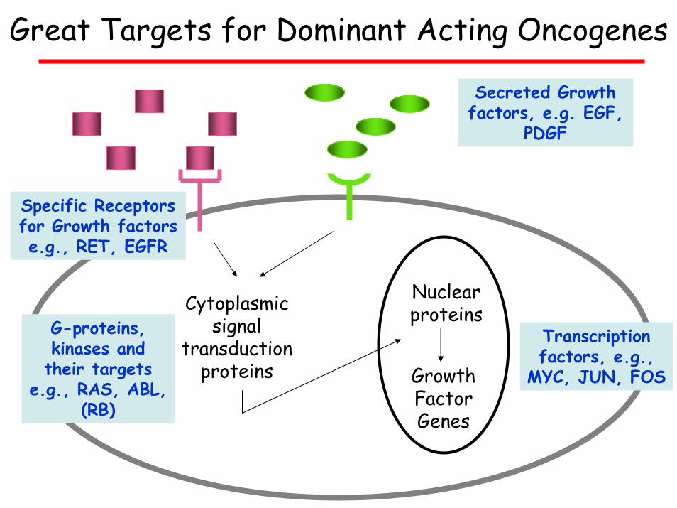
Great Targets for Dominant Acting Oncogenes
Secreted Growth factors, e.g. EGF,
PDGF
Specific Receptors for Growth factors e.g., RET, EGFR
G-proteins, kinases and their targets
e.g., RAS, ABL, (RB)
Transcription factors, e.g.,
MYC, JUN, FOS
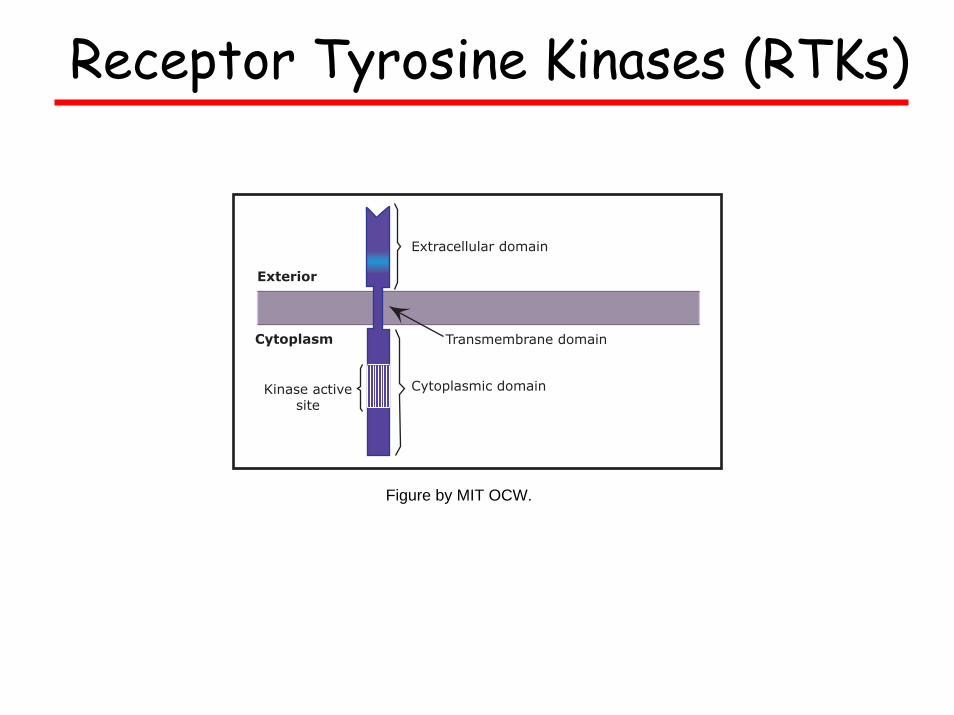
Receptor Tyrosine Kinases (RTKs)
Extracellular domain
Cytoplasmic domain
Exterior
Cytoplasm
Kinase activesite
Transmembrane domain
Figure by MIT OCW.
Receptor Tyrosine Kinases (RTKs)
Images removed due to copyright reasons.
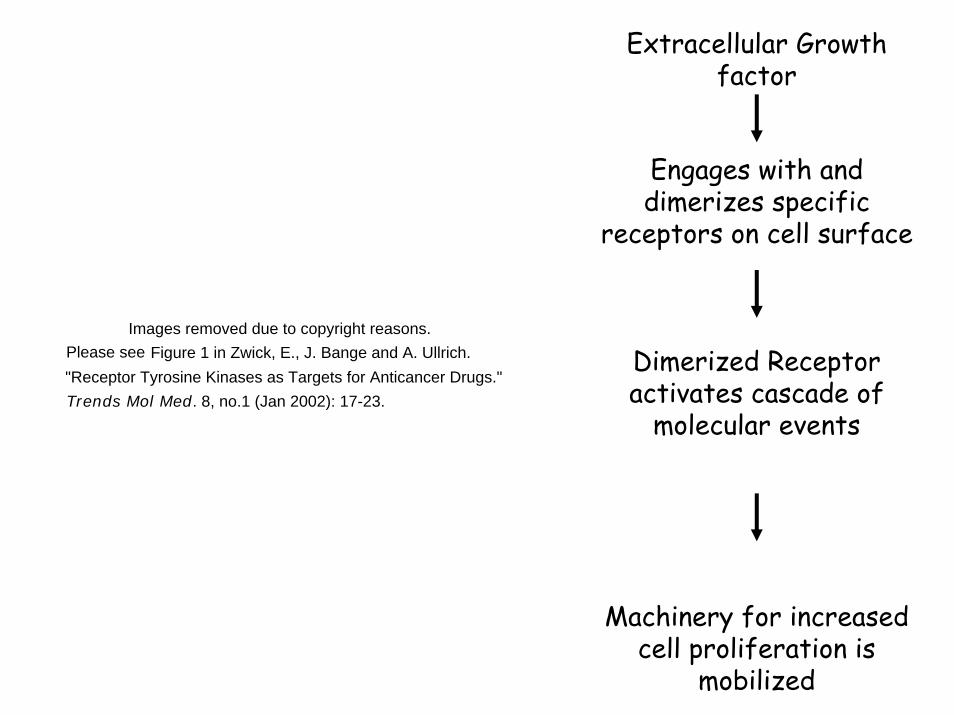
Extracellular Growth factor
Engages with and dimerizes specific
receptors on cell surface
Dimerized Receptor activates cascade of
molecular events
Machinery for increased cell proliferation is
mobilized
Images removed due to copyright reasons. Please see Figure 1 in Zwick, E., J. Bange and A. Ullrich.
Trends Mol Med. 8, no.1 (Jan 2002): 17-23. "Receptor Tyrosine Kinases as Targets for Anticancer Drugs."
Receptor Tyrosine Kinases (RTKs)
Kinases
Trans-criptionFactors
Images removed due to copyright reasons.
Constitutive Activation converts RTKsto Dominant Acting Oncogenes
Images removed due to copyright reasons.
Please see Figure 2 in Zwick, E., J. Bange and A. Ullrich.
Trends Mol Med. 8, no. 1 (Jan 2002):17-23. "Receptor Tyrosine Kinases as Targets for Anticancer Drugs."
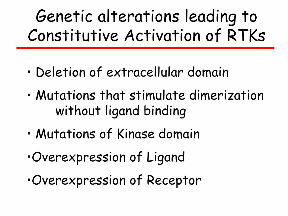
Genetic alterations leading to Constitutive Activation of RTKs
• Deletion of extracellular domain
• Mutations that stimulate dimerizationwithout ligand binding
• Mutations of Kinase domain
•Overexpression of Ligand
•Overexpression of Receptor
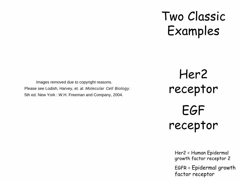
Two Classic Examples
Her2 receptor
EGF receptor
Her2 = Human Epidermal growth factor receptor 2
EGFR = Epidermal growth factor receptor
Images removed due to copyright reasons. Please see Lodish, Harvey, et. al. Molecular Cell Biology.
5th ed. New York : W.H. Freeman and Company, 2004.
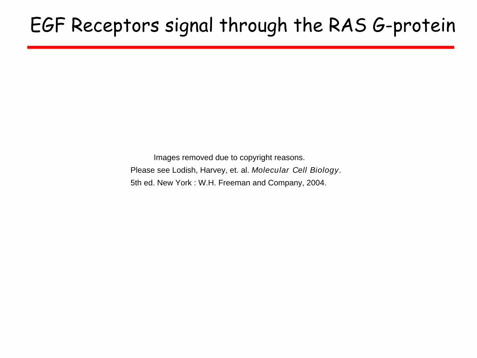
EGF Receptors signal through the RAS G-protein
Images removed due to copyright reasons. Please see Lodish, Harvey, et. al. Molecular Cell Biology.
5th ed. New York : W.H. Freeman and Company, 2004.

Cytoplasmicsignal
transduction proteins
Nuclear proteins
Growth Factor Genes
Signal Transduction and Growth Regulation
Secreted Growth factors, e.g. EGF,
PDGF
Specific Receptors for Growth factors e.g., RET, EGFR
G-proteins and kinases, e.g., RAS, ABL, RB
Transcription factors, e.g.,
MYC, JUN, FOS
G-proteins, kinases and their targets
e.g., RAS, ABL, RB

cABL – A non-receptor, cytoplasmic tyrosine kinase that can be converted into an
oncoprotein
• cABL proto-oncogene product signals to many of the same
molecules as the RTKs
• Signals cell cycle progression and cell proliferation
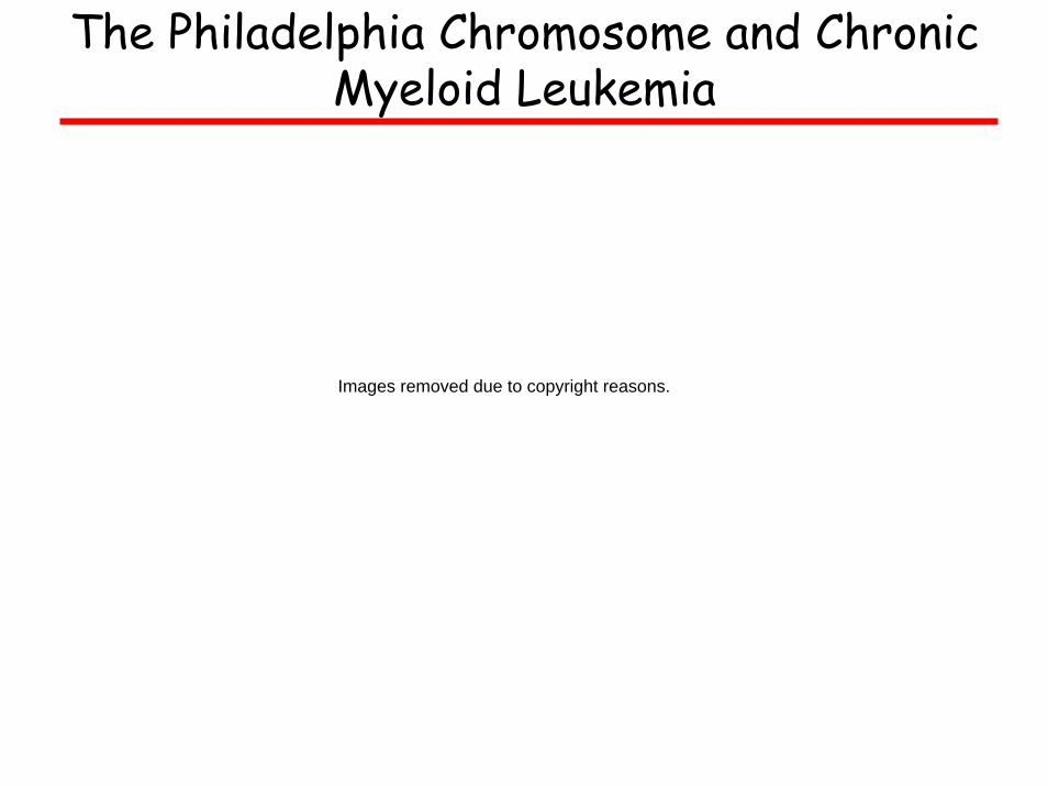
The Philadelphia Chromosome and Chronic Myeloid Leukemia
Images removed due to copyright reasons.
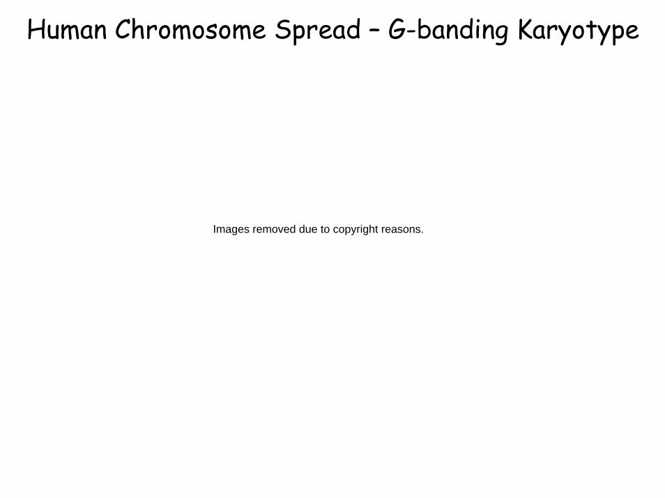
Human Chromosome Spread – G-banding Karyotype
Images removed due to copyright reasons.
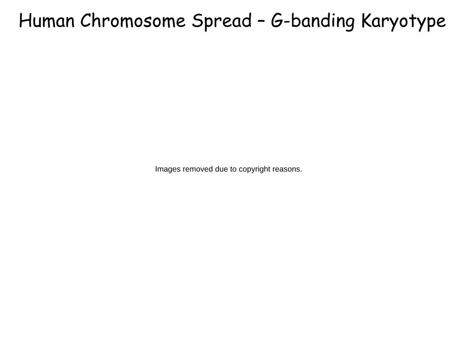
Human Chromosome Spread – G-banding Karyotype
Images removed due to copyright reasons.
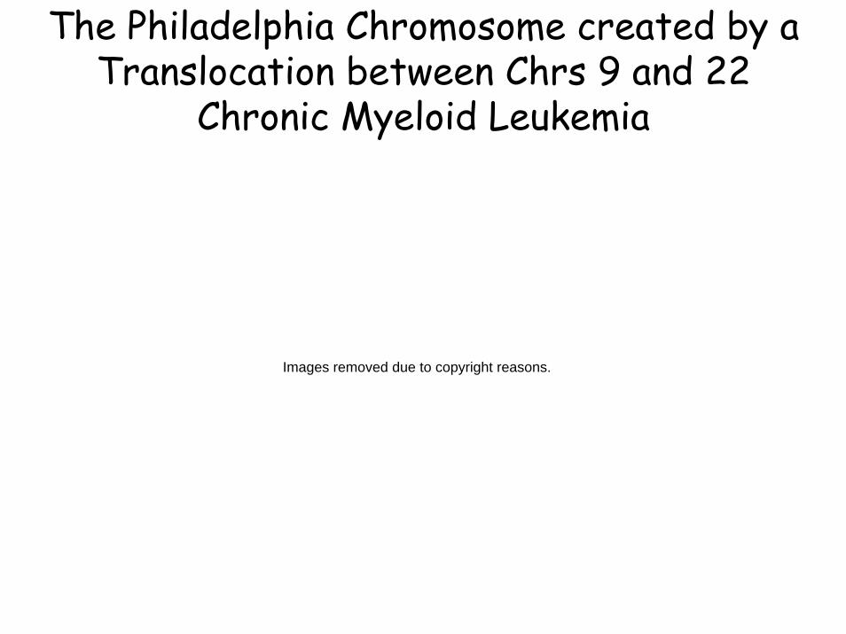
The Philadelphia Chromosome created by a Translocation between Chrs 9 and 22
Chronic Myeloid Leukemia
Images removed due to copyright reasons.
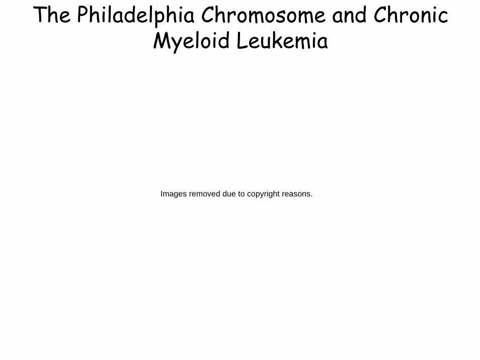
The Philadelphia Chromosome and Chronic Myeloid Leukemia
Images removed due to copyright reasons.
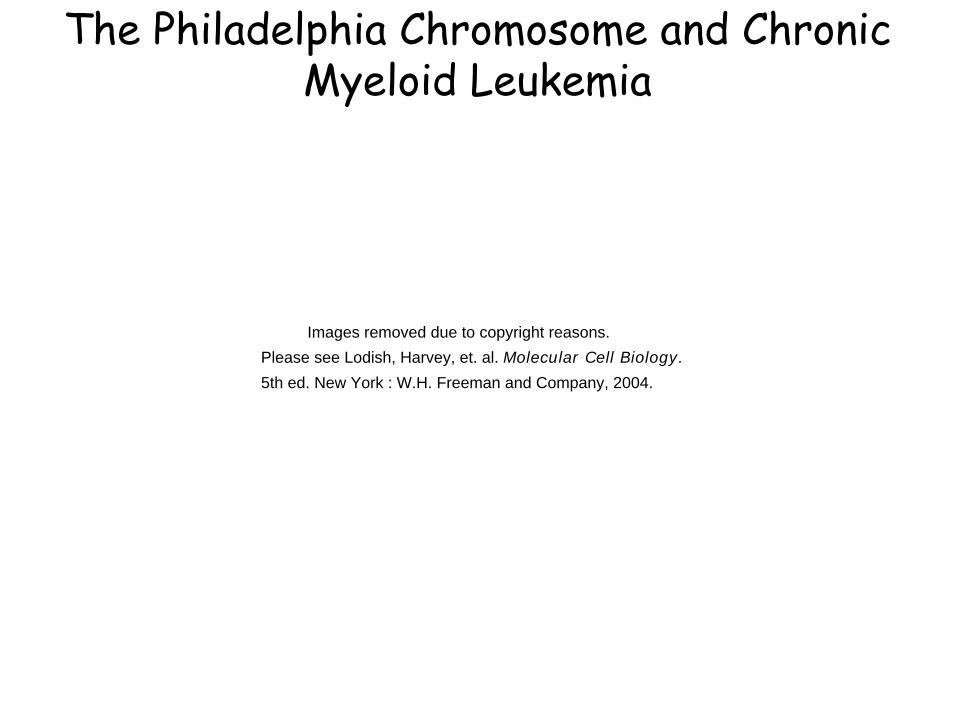
The Philadelphia Chromosome and Chronic Myeloid Leukemia
Images removed due to copyright reasons. Please see Lodish, Harvey, et. al. Molecular Cell Biology.
5th ed. New York : W.H. Freeman and Company, 2004.
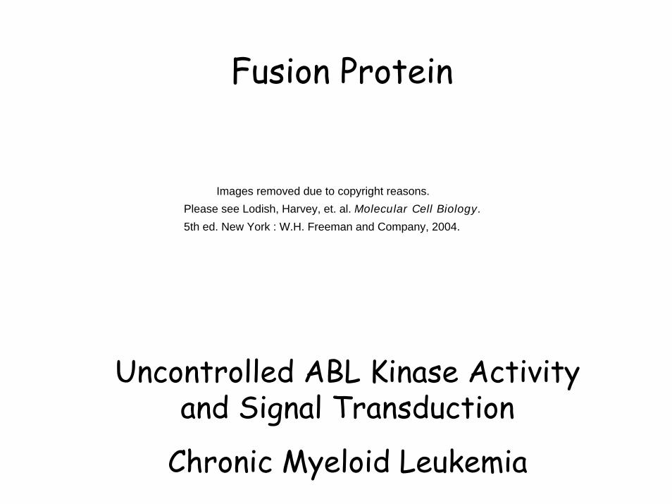
Fusion Protein
Uncontrolled ABL Kinase Activity and Signal Transduction
Chronic Myeloid Leukemia
Images removed due to copyright reasons. Please see Lodish, Harvey, et. al. Molecular Cell Biology.
5th ed. New York : W.H. Freeman and Company, 2004.

Cytoplasmicsignal
transduction proteins
Nuclear proteins
Growth Factor Genes
Signal Transduction and Growth Regulation
Secreted Growth factors, e.g. EGF,
PDGF
Specific Receptors for Growth factors e.g., RET, EGFR
G-proteins and kinases, e.g., RAS, ABL, RB
Transcription factors, e.g.,
MYC, JUN, FOS
G-proteins, kinases and their targets
e.g., RAS, ABL, RB
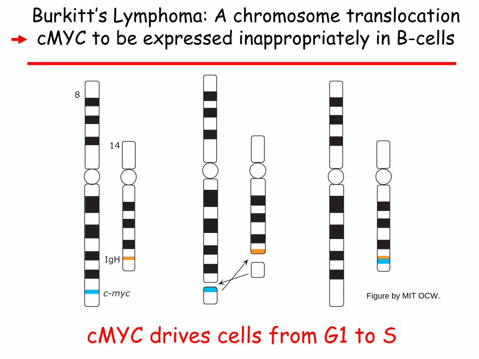
Burkitt’s Lymphoma: A chromosome translocation cMYC to be expressed inappropriately in B-cells
cMYC drives cells from G1 to S
c-myc
IgH
14
8
Figure by MIT OCW.
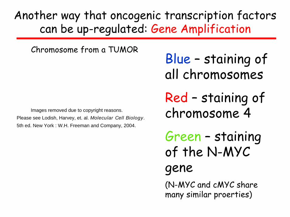
Another way that oncogenic transcription factors can be up-regulated: Gene Amplification
Chromosome from a TUMORBlue – staining of all chromosomes
Red – staining of chromosome 4
Green – staining of the N-MYC gene(N-MYC and cMYC share many similar proerties)
Images removed due to copyright reasons. Please see Lodish, Harvey, et. al. Molecular Cell Biology.
5th ed. New York : W.H. Freeman and Company, 2004.

One more example – with an interesting twistA translocation between Chr 14 and Chr 18 to put
the BCL2 gene under the strong IgH promoter
The BCL2 protein PREVENTS programmed cell death, B cells live longer than normal leading to B-cell Lymphomas
lgH enhancer
Chromosome 14
Chromosome 18
Translocation 4;18
Breakpoint
BreakpointRejoining ofbreakpoints
Immunoglobulin heavy chain gene (lgH)
Not active in B lymphocytes
bcl2 gene
Active in B lymphocytes
Figure by MIT OCW.
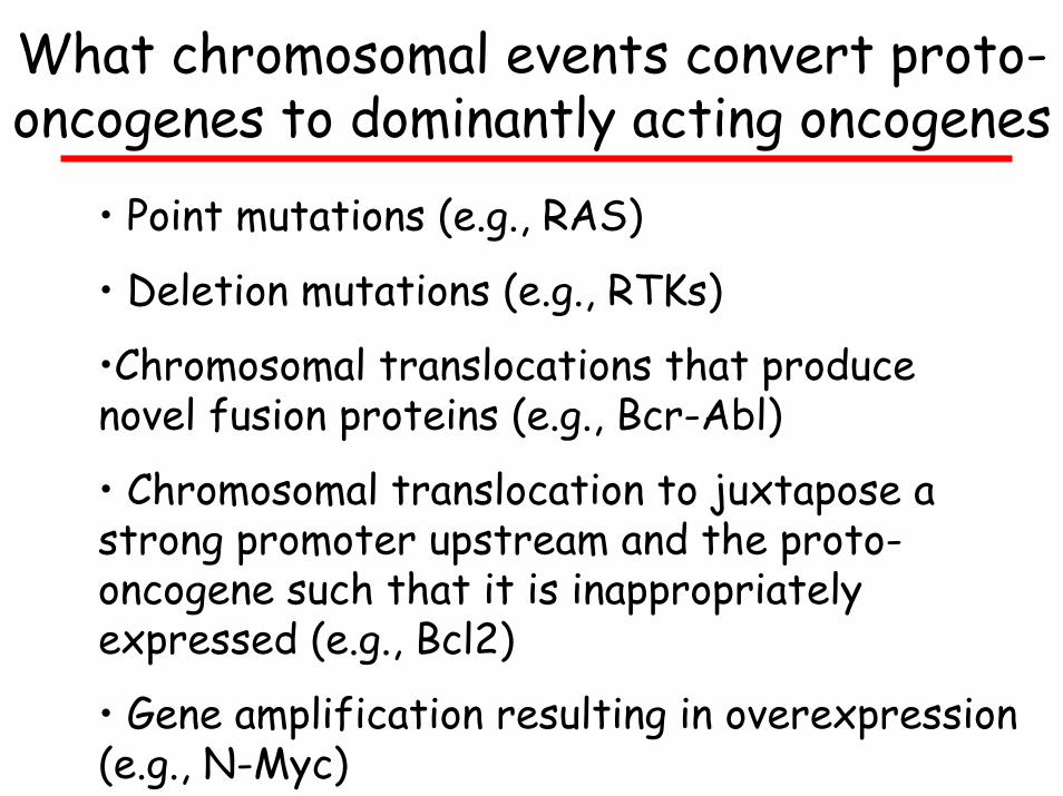
What chromosomal events convert proto-oncogenes to dominantly acting oncogenes
• Point mutations (e.g., RAS)
• Deletion mutations (e.g., RTKs)
•Chromosomal translocations that produce novel fusion proteins (e.g., Bcr-Abl)
• Chromosomal translocation to juxtapose a strong promoter upstream and the proto-oncogene such that it is inappropriately expressed (e.g., Bcl2)
• Gene amplification resulting in overexpression(e.g., N-Myc)
Cytoplasmicsignal
transduction proteins
Nuclear proteins
Growth Factor Genes
Signal Transduction and Growth Regulation
Secreted Growth factors, e.g. EGF,
PDGF
Specific Receptors for Growth factors e.g., RET, EGFR
Transcription factors, e.g.,
MYC, JUN, FOS
G-proteins, kinases and their targets
e.g., RAS, ABL, RB
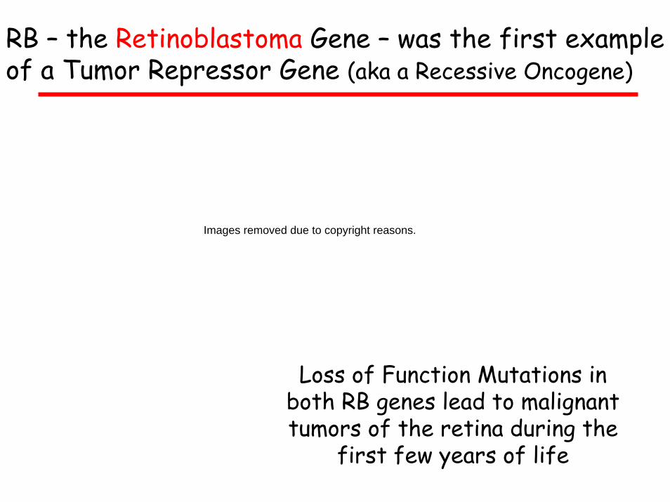
RB – the Retinoblastoma Gene – was the first example of a Tumor Repressor Gene (aka a Recessive Oncogene)
Loss of Function Mutations in both RB genes lead to malignant tumors of the retina during the
first few years of life
Images removed due to copyright reasons.
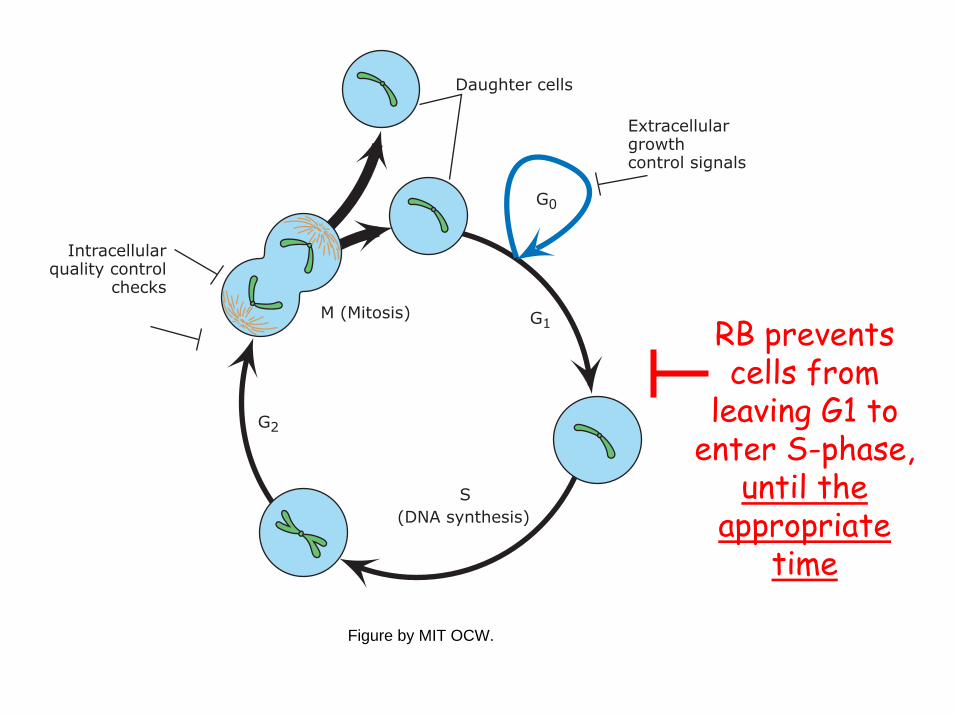
RB prevents cells from
leaving G1 to enter S-phase,
until the appropriate
time
Extracellular growthcontrol signals
Intracellularquality control
checks
(DNA synthesis)S
Daughter cells
M (Mitosis) G1
G0
G2
Figure by MIT OCW.
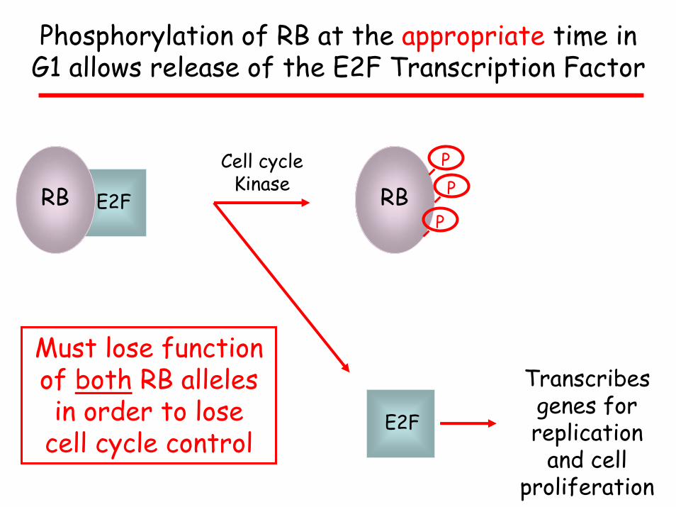
Phosphorylation of RB at the appropriate time in G1 allows release of the E2F Transcription Factor
E2FRB RB
E2F
PP
P
Cell cycle Kinase
Must lose function of both RB alleles in order to lose
cell cycle control
Transcribes genes for replication
and cell proliferation
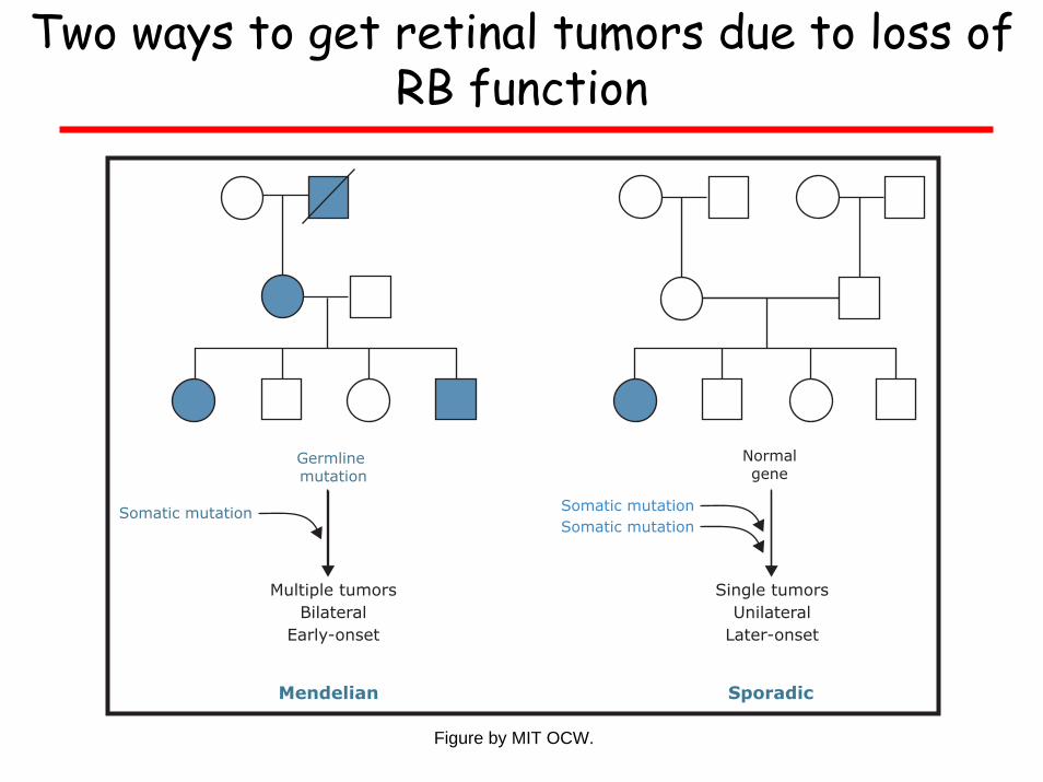
Two ways to get retinal tumors due to loss of RB function
Germline mutation
Mendelian Sporadic
Normalgene
Somatic mutation
Multiple tumorsBilateral
Early-onset
Single tumorsUnilateral
Later-onset
Somatic mutationSomatic mutation
Figure by MIT OCW.
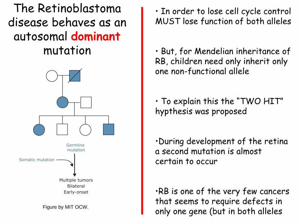
• In order to lose cell cycle control MUST lose function of both alleles
• But, for Mendelian inheritance of RB, children need only inherit only one non-functional allele
• To explain this the “TWO HIT” hypthesis was proposed
•During development of the retina a second mutation is almost certain to occur
•RB is one of the very few cancers that seems to require defects in only one gene (but in both alleles
The Retinoblastoma disease behaves as an autosomal dominant
mutation
Germline mutation
Somatic mutation
Multiple tumorsBilateral
Early-onset
Figure by MIT OCW.
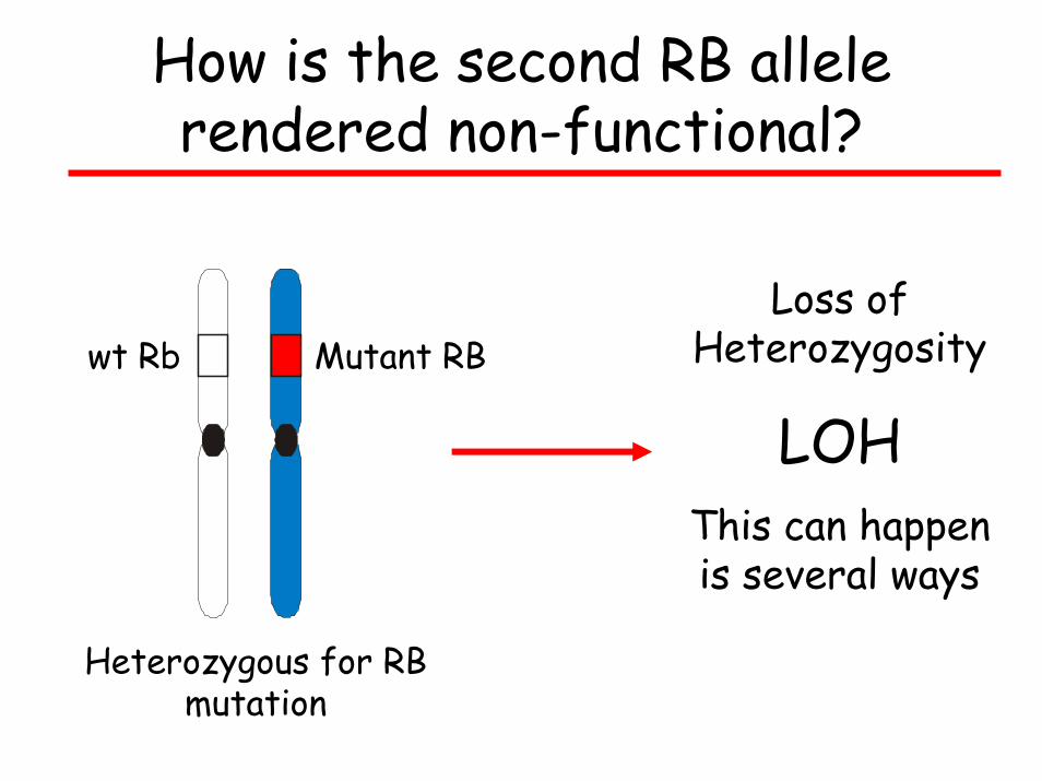
How is the second RB allele rendered non-functional?
Loss of Heterozygosity
LOHThis can happen is several ways
Mutant RBwt Rb
Heterozygous for RB mutation
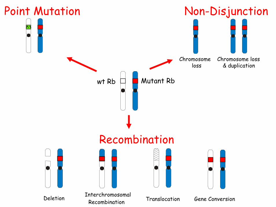
Point Mutation Non-Disjunction
Chromosome loss& duplication
Chromosomeloss
Recombination
Deletion InterchromosomalRecombination Gene ConversionTranslocation
Mutant Rbwt Rb
Cytoplasmicsignal
transduction proteins
Nuclear proteins
Growth Factor Genes
Signal Transduction and Growth Regulation
Secreted Growth factors, e.g. EGF,
PDGF
Specific Receptors for Growth factors e.g., RET, EGFR
G-proteins and kinases, e.g., RAS, ABL, RB
Transcription factors, e.g.,
MYC, JUN, FOS
G-proteins, kinases and their targets
e.g., RAS, ABL, RB
Alterations in different kinds of Genes cause Cancer
Oncogenesdominant gain-of-function mutations
promote cell transformation
Tumor suppressor genes recessive, loss-of-function mutations
promote cell transformation
Mutator genes Usually recessive, loss-of-function mutations
that increase spontaneous and environmentally induced mutation rates