Embed Size (px)
Citation preview
Prospects & Overviews
Genome analyses substantiate malemutation bias in many species
Melissa A. Wilson Sayres1)2) and Kateryna D. Makova1)�
In many species the mutation rate is higher in males
than in females, a phenomenon denoted as male
mutation bias. This is often observed in animals where
males produce many more sperm than females produce
eggs, and is thought to result from differences in the
number of replication-associated mutations accumulated
in each sex. Thus, studies of male mutation bias have
the capacity to reveal information about the replication-
dependent or replication-independent nature of different
mutations. The availability of whole genome sequences
for many species, as well as for multiple individuals
within a species, has opened the door to studying fac-
tors, both sequence-specific and those acting on the
genome globally, that affect differences in mutation rates
between males and females. Here, we assess the advan-
tages that genomic sequences provide for studies of
male mutation bias and general mutation mechanisms,
discuss major challenges left unresolved, and speculate
about the direction of future studies.
Keywords:.life history traits; male mutation bias; mutation rates
Introduction
Mutations are the primary cause of human genetic diseases.Currently, there are over 100,000 mutations associated withdisease phenotypes described in the Human Gene MutationDatabase (HGMD) [1]. Sixty-eight percent of entries are nucleo-tide substitutions and 31% are small insertions/deletions, withan additional approximately 300 disease-causing mutationsrelated to repetitive elements [1]. Moreover, many mutationshave been implicated in a variety of human cancers [2, 3].Thus, it is crucial to unravel the mechanisms of mutagenesisfor these common, and frequently disease-causing, mutations.The phenomenon of male mutation bias, in particular, canbe utilized to address the question of whether mutationsare driven by replication-dependent or by replication-independent factors.
In many species the number of germline cell divisions (andthus of DNA replications) prior to reproduction is higher inmales than in females (Table 1) [4, 5]. When more mutationsoriginate in the male germline (sperm and their precursors)than the female germline (eggs and their precursors) [6], this isreferred to as a male mutation bias. One way to determinewhether male mutation bias occurs is to compute a, the ratioof male-to-female mutation rates. If a equals one, there is nobias; a less than one indicates more mutations coming fromthe female germline, whereas a greater than one suggests amale mutation bias, also called male-driven evolution (Fig. 1).The difference in the number of germline cell divisionsbetween males and females provides an opportunity to testthe hypothesis that mutations result from errors in DNA rep-lication. If mutations are primarily replication-driven, then a
should be similar to c, the male-to-female ratio in the numberof germline cell divisions (Fig. 2). If, on the other hand a issmaller than c, then the role of replication-independentfactors (e.g. free radicals present in cells) in generatingmutations is significant [7, 8]. Estimates of c are only availablefor a limited number of species (humans [9, 10], mice and rats[11], flies [12], and an estimate across birds [13]) and are highlydependent on paternal age. For instance, in humans, c is�6 ifthe father’s age is, on average, 20 years (Fig. 2) [9, 10], whereasthis value will increase rapidly as paternal age increases.Alternatively in rodents, c is �2, assuming that the
DOI 10.1002/bies.201100091
1) Center for Comparative Genomics and Bioinformatics, The PennsylvaniaState University, University Park, PA, USA
2) Current address: Statistics Department and Integrative BiologyDepartment, University of California-Berkeley, Berkeley, CA, USA
*Corresponding author:Kateryna D. MakovaE-mail: [email protected]
Abbreviation:TE, transposable element.
938 www.bioessays-journal.com Bioessays 33: 938–945,� 2011 WILEY Periodicals, Inc.
Review
essays
average age of males at reproduction in the wild isfive months [11].
Comparisons of species with a variety of sex determinationmechanisms, such as fish [14], birds [13, 15], and mammals [16],find evidence of male mutation bias, indicating that differencesin the number of germline cell divisions between malesand females are important for shaping genome evolution. Todate, the vast majority of mammals and birds studied haveshown evidence of a male mutation bias. In mammals, withmale heterogamety (XX females and XY males), estimates of avary greatly, including a low of approximately 2 in rodents [11],an intermediate a of about 4 in Perissodactyls (horses and
rhinos) [17], and even higher estimates in primates [17, 18].Sequence comparisons between many species of birds (withZW females and ZZ males) indicate that, as expected, malemutation bias occurs independently of male or femaleheterogamety, with estimates of a in birds ranging from 3.1to 6.5 [13, 15, 19].
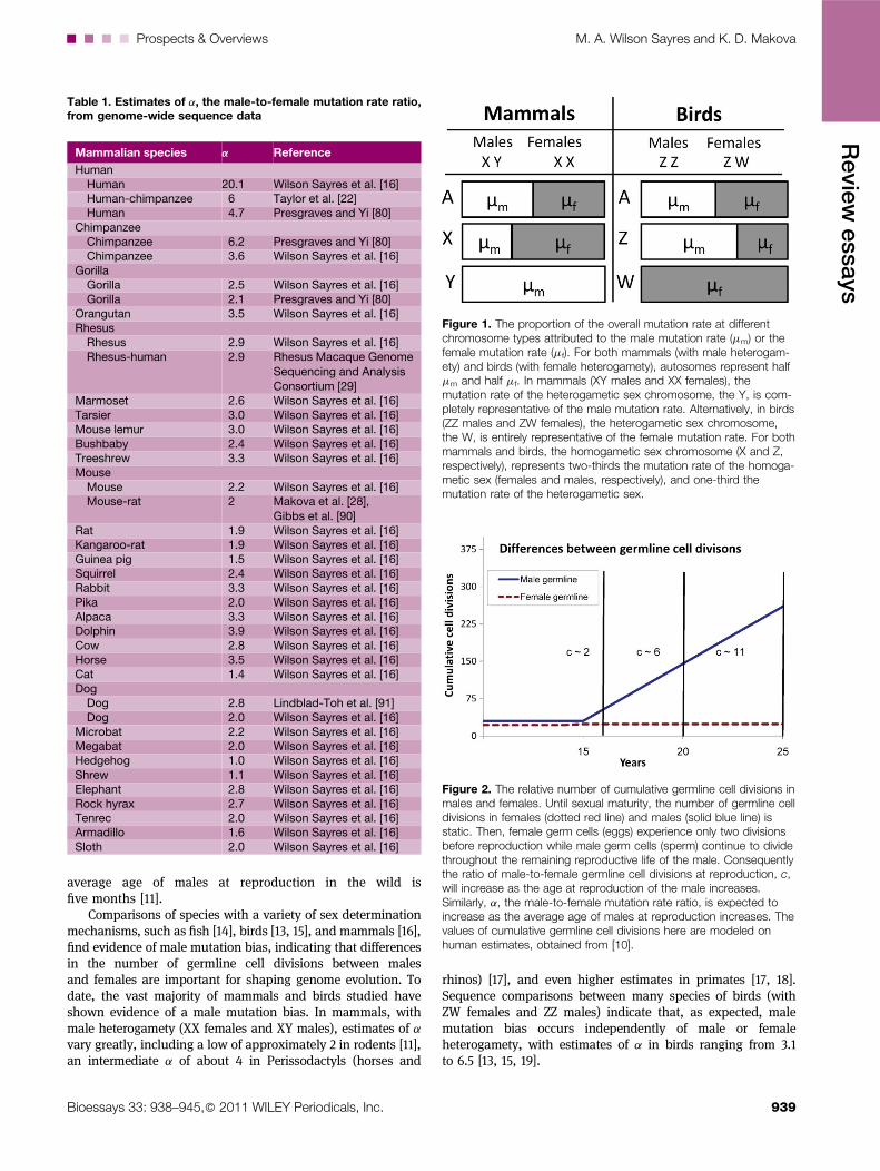
Table 1. Estimates of a, the male-to-female mutation rate ratio,from genome-wide sequence data
Mammalian species a Reference
HumanHuman 20.1 Wilson Sayres et al. [16]Human-chimpanzee 6 Taylor et al. [22]Human 4.7 Presgraves and Yi [80]
ChimpanzeeChimpanzee 6.2 Presgraves and Yi [80]Chimpanzee 3.6 Wilson Sayres et al. [16]
GorillaGorilla 2.5 Wilson Sayres et al. [16]Gorilla 2.1 Presgraves and Yi [80]
Orangutan 3.5 Wilson Sayres et al. [16]Rhesus
Rhesus 2.9 Wilson Sayres et al. [16]Rhesus-human 2.9 Rhesus Macaque Genome
Sequencing and AnalysisConsortium [29]
Marmoset 2.6 Wilson Sayres et al. [16]Tarsier 3.0 Wilson Sayres et al. [16]Mouse lemur 3.0 Wilson Sayres et al. [16]Bushbaby 2.4 Wilson Sayres et al. [16]Treeshrew 3.3 Wilson Sayres et al. [16]Mouse
Mouse 2.2 Wilson Sayres et al. [16]Mouse-rat 2 Makova et al. [28],
Gibbs et al. [90]Rat 1.9 Wilson Sayres et al. [16]Kangaroo-rat 1.9 Wilson Sayres et al. [16]Guinea pig 1.5 Wilson Sayres et al. [16]Squirrel 2.4 Wilson Sayres et al. [16]Rabbit 3.3 Wilson Sayres et al. [16]Pika 2.0 Wilson Sayres et al. [16]Alpaca 3.3 Wilson Sayres et al. [16]Dolphin 3.9 Wilson Sayres et al. [16]Cow 2.8 Wilson Sayres et al. [16]Horse 3.5 Wilson Sayres et al. [16]Cat 1.4 Wilson Sayres et al. [16]Dog
Dog 2.8 Lindblad-Toh et al. [91]Dog 2.0 Wilson Sayres et al. [16]
Microbat 2.2 Wilson Sayres et al. [16]Megabat 2.0 Wilson Sayres et al. [16]Hedgehog 1.0 Wilson Sayres et al. [16]Shrew 1.1 Wilson Sayres et al. [16]Elephant 2.8 Wilson Sayres et al. [16]Rock hyrax 2.7 Wilson Sayres et al. [16]Tenrec 2.0 Wilson Sayres et al. [16]Armadillo 1.6 Wilson Sayres et al. [16]Sloth 2.0 Wilson Sayres et al. [16]
Figure 1. The proportion of the overall mutation rate at differentchromosome types attributed to the male mutation rate (mm) or thefemale mutation rate (mf). For both mammals (with male heterogam-ety) and birds (with female heterogamety), autosomes represent halfmm and half mf. In mammals (XY males and XX females), themutation rate of the heterogametic sex chromosome, the Y, is com-pletely representative of the male mutation rate. Alternatively, in birds(ZZ males and ZW females), the heterogametic sex chromosome,the W, is entirely representative of the female mutation rate. For bothmammals and birds, the homogametic sex chromosome (X and Z,respectively), represents two-thirds the mutation rate of the homoga-metic sex (females and males, respectively), and one-third themutation rate of the heterogametic sex.
Figure 2. The relative number of cumulative germline cell divisions inmales and females. Until sexual maturity, the number of germline celldivisions in females (dotted red line) and males (solid blue line) isstatic. Then, female germ cells (eggs) experience only two divisionsbefore reproduction while male germ cells (sperm) continue to dividethroughout the remaining reproductive life of the male. Consequentlythe ratio of male-to-female germline cell divisions at reproduction, c,will increase as the age at reproduction of the male increases.Similarly, a, the male-to-female mutation rate ratio, is expected toincrease as the average age of males at reproduction increases. Thevalues of cumulative germline cell divisions here are modeled onhuman estimates, obtained from [10].
....Prospects & Overviews M. A. Wilson Sayres and K. D. Makova
Bioessays 33: 938–945,� 2011 WILEY Periodicals, Inc. 939
Review
essays
Not all species exhibit clear evidence for a male mutation bias.In flies, both males and females produce many gametes lead-ing to the expectation of no male mutation bias [20]. Indeed,initial studies in Drosophila melanogaster and D. simulansestimated that there should be a slight female mutation biaswhen flies mated early (3 days after hatching c � 0.77), and alow male mutation bias when they mated later (c � 1.33,21 days after hatching), but did not observe significant differ-ences between substitution rates on the X and autosomes,implying that, on average, flies do not exhibit sex-biasedmutation rates [12]. However, a more recent genomic compari-son of 205 homologous neo-X- and neo-Y-linked sequences(vs. the smaller set of non-homologous loci compared pre-viously [12]) indicated the presence of male mutation bias inD. miranda, with a � 2 [21]. Given that a estimates vary greatlyacross mammals (see below), it is reasonable that malemutation bias may also differ between fly species, and sothe aforementioned studies may not be incompatible.
Genome-wide data are particularly useful for advancingstudies of male mutation bias, and, subsequently, elucidatingthe roles that replication-dependent and -independent mech-anisms play in mutation accumulation. Indeed, the abun-dance of information from genome sequences allows forthe study of mutation mechanisms of context-dependent sub-stitutions, as well as of transposable elements (TEs), simplerepeats, and insertions and deletions. Finally, genomesequences from a variety of species now make it possible toinvestigate branch-specific mutation rates, whereas mostprevious studies were only able to analyze pairwise mutationrates. Estimates of male mutation bias can vary depending onwhich mutation types are included in the analysis [22], whichchromosomes are compared [23], and whether ancestral poly-morphism for closely related species is corrected for [24].Further, differences in life history traits may cause variationsin a between species [16]. Here we will address recent advan-ces in the study of male mutation bias brought about by thegenomic era, suggest potential avenues of research, andspeculate about future prospects in the field.
Utilizing comparative genomicsto measure male mutation bias:An updated evolutionary approach
One way to study the differences in mutation rates betweenmales and females in species with chromosomal sex determi-nation is to compare the mutation rates between the two sexchromosomes, or between a sex chromosome and autosomes[25]. Usually, neutrally evolving regions of the genome (eithernon-coding and non-repetitive, or those that fall within ances-tral interspersed repeats [22, 26]) are utilized, to avoid theconfounding effects of selection. Substitution rates are used asa proxy for mutation rates. Because we know how much timeeach chromosome type spends in the male versus femalegermline – in mammals, autosomes spend equal amountsof time in both germlines, X spends 2/3 of its time in thefemale germline and 1/3 in the male germline, and Y is foundexclusively in the male germline (Fig. 1) – substitution ratesbetween any two chromosome types can be compared to
estimate the male-to-female mutation rate ratio, a. In specieswith female heterogamety, like birds, the W-specific substi-tution rate represents the female-specific mutation rate, whilethe Z spends 2/3 of its time in males and only 1/3 of its time infemales, and autosomes are again split 50:50 with respect totime spent in the male and female germlines (Fig. 1). Theevolutionary approach has the advantage of comparingmutation rates over a large number of sites, and it does notsuffer from disease-associated ascertainment bias [27].Moreover, although earlier studies employing this approachwere affected by regional variation in substitution rates (e.g.ref. [17]), the availability of genome-wide sequences circum-vents this limitation [16, 22, 28, 29] because regional variationwill be averaged out.
However, there are some drawbacks to the evolutionaryapproach. First, it requires at least two alignable genomes tocompute evolutionary rates and at least three to polarizemutations to specific branches. In a modification of the evol-utionary method, divergence in the nucleotide sequences ofTEs on the X, Y, and autosomes with a single species can becompared to evaluate the magnitude of male mutation bias[30, 31]. Another pitfall of the evolutionary method is that itmay be affected by ancestral polymorphism when very closelyrelated species are compared, which can inflate or deflateestimates of a, depending on which chromosomes are com-pared [24]. Furthermore, although genome-wide sequenceshave recently become available, allowing for global estimatesof a, the complete Y chromosome has not been sequenced formost mammals (with the exception of human [32] and chim-panzee [33]), limiting, for now, the ability of researchers toexplore how comparisons among different chromosome typesaffect estimates of male mutation bias.
Novel mutations can be identified directly
Historically, instead of being estimated from sequencechanges between or within species, a was computed by infer-ring the parental origin (paternal vs. maternal) of mutationsleading to human genetic diseases (X-linked or autosomaldominant disorders). Specifically, after the origins of manymutations for a particular disorder have been inferred for avariety of individuals, a can be calculated as the ratio of thenumber of paternal mutations to the number of maternalmutations (reviewed in ref. [27]). However, mutations leadingto human genetic diseases usually affect gene function and, asa result, are likely to be affected by selection, confoundingestimates of male mutation bias.
In contrast to observing mutations through disease associ-ations, another way to directly study mutation rates – avail-able now with genomic sequences – is to analyze novelmutations in families including a mother, father, and one[34, 35] or several children [36]. As a pilot study of the1,000 genomes project, whole-genome sequence data fortwo sets of trios were used to detect 49 and 35 de novomutations occurring in the germlines of the two families,respectively. Initially no differentiation was made regardingparental origin of mutations [37], but a subsequent studypartitioned the new mutations into those coming from thepaternal or maternal germlines [35]. Curiously, results from
M. A. Wilson Sayres and K. D. Makova Prospects & Overviews....
940 Bioessays 33: 938–945,� 2011 WILEY Periodicals, Inc.
Review
essays
one of the two families suggested a strong male mutation biaswhile the other indicated a female mutation bias [35]. Giventhat newmutations in the germ cells (that may be passed on tothe next generation) are likely to be rare, including more thanone child may greatly improve the power of a study to deter-mine the magnitude of male mutation bias [36].
Even with the sequencing of complete genomes, thesequence of the Y chromosome is often difficult to capturebecause it is small, highly heterochromatic and shares manyhighly similar sequences with the X that can complicate align-ments. However, flow sorting Y chromosomes (choosing just theY chromosome out of the set of all autosomes and sex chromo-somes) gives researchers an edge by reducing the chance ofmisalignment. In fact, the direct sequencing of flow-sorted Ychromosomes from two human males, with knowledge of thepedigree (relationship between individuals), resulted in a directestimate of the mutation rate on the human Y chromosome(3.0 � 10�8 mutations per nucleotide per generation) [38]. Astudy of the autosomal mutation rate from a family includingboth parents and two children found that the autosomal sub-stitution rate is approximately 1.1 � 10�8 mutations per nucleo-tide per generation [36], which is lower than the substitutionrate reported for the Y chromosome [38] (as would be expectedin the presence of male mutation bias), but not significantly so[36]. Because only a few mutations get passed on each gener-ation, variation due to the small sample sizes in each studymaylimit the power to determine significant differences.
Using families with parents and offspring to study de novogermline mutations has the advantage of avoiding germlinecell collection (which is especially invasive for females), butthe challenge is that each novel mutation must be validated.Single-molecule sequencing holds the promise of fast andreliable sequencing [39] of individual genomes that will makeit feasible and affordable to study the male and femalemutation rates directly within many families. Studies ofmutation rates from families should include a large numberof individuals, to account for the small number of novelmutations, and stochastic fluctuations [35]. Further, familiesfrom diverse ethnic backgrounds should be included, to takeinto account the effects of population structure and matingpatterns. In addition to the non-uniform mating of males andfemales in different cultures, whichmight lead to variance in a
estimates due to increased or decreased diversity on the X or Ychromosomes (see ref. [40] for a more detailed discussion),individuals in some populations may have a history of repro-ducing earlier, on average, leading to a lower estimate of a,while individuals in others may delay reproduction, increas-ing the difference in germline cell divisions betweenmales andfemales. In addition, one must be careful, to distinguish germ-line mutations from somatic mutations (which will not bepassed on to the next generation) and from mutations arisingduring the propagation of cultured cell lines.
Estimates of male mutation bias varyacross nucleotide sites
Most autosomal dominant disorders caused by nucleotidesubstitutions show an excess of new mutations originatingin males (reviewed in refs. [27, 41]). Male mutation bias theory
assumes that the majority of new mutations are due to errorsduring replication, but some nucleotide substitutions aremorelikely to occur through replication-independent mechanisms,which may skew estimates of a. For example, a methylatedcytosine followed immediately by a guanine (CpG), has anincreased likelihood of being spontaneously deaminated to auracil, which is then ‘‘repaired’’ as a thymine, resulting in aC-to-T transition that is independent of replication [42]. Thislargely replication-independent nucleotide substitution has amutation rate that is 10–50 times higher than that at non-CpGsites [43], and is very high in the male and female germlines,effectively lowering estimates of a at CpG sites and maskingthe effects of male mutation bias [22].
Other, non-CpG, neighbor-dependent substitution proc-esses are still poorly understood, but analyzing suchmutations in the context of male mutation bias has a potentialto determine whether they are associated with errors madeduring replication. For example, substitution rates in pseudo-genes were found to be highest in purines surrounded bypyrimidines and in pyrimidines surrounded by purines, andslowest within purine and pyrimidine tracts [44–46]. It washypothesized that an increase in dNTP misinsertion or tran-sient misalignment during replication is associated with struc-tural alterations of the DNA backbone in alternating purine-pyrimidine sequences [47], thus dNTP misinsertions in alter-nating purine-pyrimidine tracks should exhibit a strong malemutation bias. Another substitution type, the instability ofthymines occurring in tandem (TpT) was proposed to bedue to thymine photodimer formation in DNA during exposureto UV light [48]. Since exposure to UV should occur independ-ent of replication (and likely affects germline cells only min-imally), observations of thymine dimers are expected to showweak or no male mutation bias. Several models of sequenceevolution have been developed to investigate neighbor-dependent substitution biases genome-wide from intraspecific[46] and interspecific comparisons [49–51]. These models holdgreat promise for investigating context-dependent biaseswithin the framework and hypotheses formed by malemutation bias.
Insertions and deletions are subjectto male mutation bias
Studies of male bias for indels (insertions and deletions) mayprovide a unique opportunity to disentangle the effects ofrecombination and replication on the formation of indels.Although large (>1 kb) indels, which include copy numbervariants, have not yet been explored genome-wide in thecontext of male mutation bias, small (<50 bp) indels havebeen studied across chromosome types, leading to some inter-esting conclusions [52, 53]. When replication-dependent andrecombination-dependent genomic landscape predictors wereanalyzed, the results suggested that insertions are more oftenassociated with recombination while deletions result mostlyfrom errors during replication [52, 53]. Even with these differ-ent forces primarily associated with formation of insertionsversus deletions, the incidence of both insertions anddeletions is reduced on the X chromosome, relative to theautosomes (i.e. there is male bias for small indels), tentatively
....Prospects & Overviews M. A. Wilson Sayres and K. D. Makova
Bioessays 33: 938–945,� 2011 WILEY Periodicals, Inc. 941
Review
essays
suggesting that replication may play a strong role in determin-ing indel rates [28, 52]. Comparisons with the Y chromosomewill need to be made to rule out significant effects of recombi-nation. Future studies will also need to assess if the samepatterns hold true for larger indels. Given the implied femalebias of large indels in human genetic diseases (see above), thiswill be of great interest to evolutionary biologists and clini-cians alike. Indeed, while some small indels (<50 bp) thatcause human genetic diseases originate more frequently inmales (e.g. in neurofibromatosis type 2 and Rett syndrome[54, 55]), there is evidence that some large indels (>1 kb orlarger) exhibit a maternal mutation bias (e.g. the ones causingneurofibromatosis type 1 and hemophilia B [56, 57]).
Variations in microsatellite repeats areprone to replication-dependent changes
Microsatellites – repeats of short (1-6 bp) DNA motifs wherethe length of the repeated sequence can vary dramaticallyfrom person to person – have been implicated in a variety ofhuman cancers [3] and are widely used in forensics, so under-standing how these quickly changing sequences mutate,especially in males versus females can have far-reachingimplications. Microsatellite mutation rates are predicted tooccur primarily through misalignment and strand slippageof the repetitive microsatellite DNA during replication [58].Thus, microsatellite mutations are expected to originatepredominately in males, who experience a greater numberof germline cell divisions, as compared with females.However, the data on the parental origin of trinucleotidemicrosatellite expansions leading to human neurological dis-eases have been contradictory. Some exhibit a clear paternalbias (e.g. dentatorubral-pallidoluysian atrophy, and spinocer-ebellar ataxia types 1 through 3 [59]), while others exhibit amaternal bias (e.g. myotonic dystrophy [60], FRAXE mentalretardation, Friedrich’s ataxia, and spinocerebellar ataxiatype 8 [59]).
An evolutionary study of microsatellite mutability shows amale mutation bias where mutability is highest on the Ychromosome, intermediate on the autosomes and lowest onthe X, likely due to the amount of time each chromosome typespends in the male and female germline [61]. Note that in thisstudy [61] mutability in microsatellite repeats was observed tobe significantly different between the two sex chromosomesonly for mononucleotide repeats, either due to the lack of datafor larger motifs or, potentially, due to real differences in theimportance of replication for microsatellites with differentmotif sizes [62]. Future studies of variation in microsatellitemutability betweenmales and females at all motif lengths (e.g.di-, tri-, and tetranucleotide microsatellites) and compositions(e.g. A/T vs. C/G repeats for mononucleotide microsatellites)are likely to find many similarities supporting the relativeimportance of replication slippage in the formation of allmicrosatellites, but may also unveil differences that willsuggest alternative processes affecting mutability. In addition,comparisons of mutability at microsatellites with mutability atother types of repetitive elements, such as minisatellites andlarger-scale copy number variants, may discover some globalmutation mechanisms at repetitive regions.
Strong male mutation bias is observed forsome but not all transposable elements
Transposable elements – elements that replicate and integratethemselves selfishly in our genomes – contribute significantlyto both genetic diseases and cancer [63, 64], with over 65separate human diseases caused by the de novo insertionof mobile elements [65, 66]. Male mutation bias provides aunique perspective for studying TEs, such as short and longinterspersed repetitive elements (SINEs and LINEs), because itcan be utilized to investigate both how and when duringdevelopment TEs integrate, as well as in which germlines theyintegrate. Different families of TEs may have different prefer-ences for integration – either into the male or female germline– or they may not show a preference. For instance, if integ-ration occurs preferentially in the male germline, as suggestedfor Alus [67] and for human endogenous retroviruses [68], weexpect highest, intermediate, and lowest TE densities at Y,autosomes, and X, respectively. No differences in the TEdensities among chromosomal types are expected if integ-ration occurs during early embryogenesis, as experimentalevidence suggests for L1s [69] (but, others suggested integ-ration of L1s in oocytes [70]). Largely consistent with thesewet-lab observations, a recent computational study observed astrongmalemutation bias forAlu elements, while no such biaswas observed for L1 elements [71]. Potential male mutationbias at other interspersed repeat types (e.g. DNA transposons)has yet to be investigated. Knowing roughly when and howTEs integrate in the genome may, in the future, assist clinicaltests (particularly in the egg and sperm before conception) indetecting TE-related genetic diseases.
Can life history traits account for variationsin male mutation bias between species?
Estimates of male mutation bias vary greatly between mam-mals, even when entire genome sequences are used to computea [16]. Life history traits, which also differ from species tospecies, might influence male mutation bias by affecting eitherthe relative number of germline cell divisions, or differences inDNA damage/repair between males and females. First, if thenumber of cell divisions in oogenesis is roughly constant acrossspecies, then species with longer generation times should alsoexhibit stronger male mutation bias. This is because, as inhumans, and presumably all other mammals, sperm are pro-duced continuously during a male’s reproductive life, so thetotal number of DNA replications accumulates with age. Forinstance, a sperm of a 20-year-old man has completed �160chromosome replications, while a sperm of a 40-year-old manhas completed �610 chromosome replications [9, 41]. A recentanalysis of 32 mammalian genomes [16] corroborated the domi-nant role of generation time in determining the variation in themagnitude of male mutation bias for nucleotide substitutions,finding it explained over 30% of the variation in a estimatesacross mammals. Second, metabolic rate, specifically its associ-ation with oxidative damage of the DNA through reactiveoxygen species, may act in a replication-independent way toaffect germline mutations. Oxidative damage may increase the
M. A. Wilson Sayres and K. D. Makova Prospects & Overviews....
942 Bioessays 33: 938–945,� 2011 WILEY Periodicals, Inc.
Review
essays
mutation rate in males over females if, as postulated, eggs aremore efficient at avoiding such damage than sperm because theformer have a denser cell membrane and the latter exist in amore reactive oxygen rich environment [72]. Consistent withthese expectations, genome-wide comparisons indicate thatmetabolic rate is a significant positive predictor of malemutation bias; however, it loses its significance when includedin a multiple regression model alongside predictors of gener-ation time [16]. These results suggest that although metabolicrate appears to affect male mutation bias, generation time, areplication-dependent factor, is still the driving cause of differ-ences between the male and female mutation rates. Finally,increased sperm competition, which occurs when sperm frommultiple males compete to fertilize the egg(s) from a singlefemale, is associated with increased testes size [73, 74] andsperm quantity [75], and more rounds of reproduction per unitof time [76], and thus is expected to lead to more mutations inthe male germline, increasing the magnitude of male mutationbias. Although this expected positive trend was observed quali-tatively for a few bird species [77], results from recent regressionanalyses suggest that available predictors of sperm competitionare not significant in explaining variation inmalemutation biasacross 32 mammalian species [16]. If sperm competition canaffect substitution rates on a short time scale, as suggested forflies [74], rapid changes betweenmating patternsmaymask theeffects of sperm competition on male mutation bias, especiallywhen comparing divergent species.
Male mutation bias in humans
Given the variation in estimates of male mutation bias acrossmutation types, along with the general interest in human-specific evolution, it is unsurprising that there has been somecontroversy surrounding a estimates in humans, and itsimplications are directly relevant to understanding mutationmechanisms. Many earlier estimates were based on one or a fewloci, leading to drastically different estimates (due to regionalvariation in mutation rates) with wide confidence intervals[7, 24, 78]. Some estimates of a in humans were much lowerthan 6 [78, 79], which suggested that amay be roughly constantacross species, and casted doubt upon the theory that replica-tion errors were an important mutational mechanism. However,more recent studies, accounting for ancestral polymorphismand using genome-wide data [16, 22, 80], showed that malemutation bias in humans and other primates likely reflects areplication-dependent nature of the majority of mutations, witha estimates consistent with, or sometimes even larger than,estimates of c. Future avenues of research into human-specificmale mutation bias using population genetics can addresswhether large a values are due to reduced purifying selection,increases in the average age at reproduction, or changes insome other demographic parameters.
Factors that limit studies of malemutation bias
Several external features may affect actual and estimatedvalues of male mutation bias. First, although the current
literature supports the explanation that the male mutationbias is due to differences between the number of germline celldivisions between males and females within species, c hasbeen measured in only a few model organisms [10, 20]. Thispaucity of c values hinders researchers’ ability to investigatewhether a deviates from c across species, and why. Such datawill need to be generated if genomic data are to be useful,especially given recent studies that suggest oocyte productionmay not be finished at birth (mouse ovaries likely containsome mitotically active germ cells [81]), and that the length oftime needed to complete a spermatogenesis cycle may varyeven between closely related species (as observed in shrews[76]). Second, if replication were the only cause of mutationrate differences between males and females, then the magni-tude of male mutation bias, measured by a, should be thesame whether comparing X/Y, X/A, or Y/A (or alternativelyZ/W, Z/A, or W/A in female-heterogametic systems). However,there are differences in the estimates of a, depending onwhichratio it is computed from [16, 22, 23], indicating that, unsur-prisingly, replication-independent factors (e.g. recombina-tion) might also differentially influence substitution rateson different chromosomal types. Initial studies have suggestedthat recombination may be mutagenic at chromosomal re-arrangements and copy number variants [82], and also fornucleotide substitutions [83]. However, more research isneeded because the latest genomic data from humans ques-tion the mutagenicity of recombination [37], implying thatreplication-independent processes other than recombinationmay be at play. For example, although sperm do not showincreased susceptibility to induced DNA damage comparedto somatic tissues [84], it is not yet known whether eggs aremore or less robust to such damage, which would affectestimates of a. Another possibility is that sperm accumulatereplication-independent mutations as a result of the reducedDNA-repair fidelity in late spermatogenesis [85]. Third, mostgenomic analyses of male mutation bias study putativelyneutrally evolving sequences to avoid the confounding effectsof selection. But, one of the most striking conclusions ofstudies of mutations in sperm is that selection may play animportant role in affecting which disease-related mutationsare maintained in the genome [86, 87]. However, becauseselection acts differently on the autosomes, and the sexchromosomes [88], it likely affects all studies of male mutationbias, even those of presumed neutral regions [16, 22, 23].Currently there is not a systematic way to globally correctfor the effects of selection, but it is an open challenge tothe field.
Conclusion and future prospects
Genome-wide analyses have shown that there is, indeed amale mutation bias in many species, with estimates of a
consistent with the ratio of expected germline cell divisionsin males and females, suggesting this bias is largely driven byreplication-dependent mutational processes. Such studiesalso observed variations in a across species and betweenmutation types. The growing abundance of completelysequenced genomes from a variety of species [89], as wellas from many individuals within a species (particularly across
....Prospects & Overviews M. A. Wilson Sayres and K. D. Makova
Bioessays 33: 938–945,� 2011 WILEY Periodicals, Inc. 943
Review
essays
families and populations of humans), will open up possibil-ities for future studies of male mutation bias. Perhaps the mostexciting prospect that we only touched upon here is the abilityto study mutation rates in males and females directly, fromparents to offspring using sequenced families consisting of afather, mother, and child (or even better, many children)[35, 36]. Further, computational studies of male mutation biashave, and will continue to inform upon the general mechan-isms for a variety of mutation types including substitutions,insertions, deletions, microsatellites and TEs, and guide futurewet-lab biochemical experiments. All of these results willilluminate how male mutation bias varies across individualhuman families and across species, and, when taken togetherwith life history trait data, will allow researchers to elucidatethe mechanisms that cause differences between male andfemale germline mutation rates. The wealth of genomic datais, unfortunately, paralleled by a dearth of similar, abundant,and high quality, references about species life history traits.This imbalance emphasizes the need for experimental advan-ces in collecting such information, especially on the number ofgermline cell divisions per unit of time in both males andfemales. Importantly, understanding the factors affectingmale mutation bias will contribute to studies of age-relateddiseases, and can assist in the development and timing of pre-and post-conception genetic counseling.
AcknowledgmentsThis study was supported by the NIH grant R01-GM072264.
References
1. Stenson PD, Ball EV, Howells K, Phillips AD, et al. 2009. The HumanGene Mutation Database: providing a comprehensive central mutationdatabase for molecular diagnostics and personalized genomics. HumGenomics 4: 69–72.
2. Stratton MR. 2011. Exploring the genomes of cancer cells: progress andpromise. Science 331: 1553–8.
3. Oda S, Maehara Y, Sumiyoshi Y, Sugimachi K. 2002. Microsatelliteinstability in cancer: what problems remain unanswered? Surgery 131:S55–62.
4. Haldane J. 1935. The rate of spontaneous mutation of a human gene. JGenet 31: 317–26.
5. Haldane J. 1947. The mutation rate of the gene for haemophilia, and itssegregation ratios in males and females. Ann Eugen 13: 262–71.
6. Miyata T, Hayashida H, Kuma K, Mitsuyasu K, et al. 1987. Male-drivenmolecular evolution: a model of nucleotide sequence analysis. ColdSpring Harb Symp Quant Biol 52: 863–7.
7. Li WH, Ellsworth DL, Krushkal J, Chang BH-J, et al. 1996. Rates ofnucleotide substitution in primates and rodents and the generation-timeeffect hypothesis. Mol Phylogenet Evol 5: 182–7.
8. Shimmin LC, Chang BH-J, Hewett-Emmett D, Li W-H. 1993. Potentialproblems in estimating the male-to-female mutation rate ratio from DNAsequence data. J Mol Evol 37: 160–6.
9. Hurst LD, Ellegren H. 1998. Sex biases in the mutation rate. TrendsGenet 14: 446–52.
10. Vogel F,Motulsky A. 1997.HumanGenetics: Problems and Applications.3rd edition. Berlin, Heidelberg, New York: Springer, Verlag.
11. Chang BH-J, Shimmin LC, Shyue S, Hewett-Emmett D, et al. 1994.Weak male-driven molecular evolution in rodents. Proc Natl Acad Sci USA91: 827–31.
12. Bauer VL, Aquadro CF. 1997. Rates of DNA sequence evolution are notsex-biased in Drosophila melanogaster and D. simulans. Mol Biol Evol 14:1252–7.
13. Kahn NW, Quinn TW. 1999. Male-driven evolution among Eoaves? A testof the replicative division hypothesis in a heterogametic female (ZW)system. J Mol Evol 49: 750–9.
14. Ellegren H, Fridolfsson A-K. 2003. Sex-specific mutation rates in sal-monid fish. J Mol Evol 56: 458–63.
15. Ellegren H, Fridolfsson A-K. 1997. Male-driven evolution of DNAsequences in birds. Nat Genet 17: 182–4.
16. Wilson Sayres MA, Venditti C, Pagel M, Makova KD. 2011. Dovariations in substitution rates and male mutation bias correlate withlife history traits? A study of 32 mammalian genomes. Evolution 65:2800–15.
17. Goetting-Minesky MP, Makova KD. 2006. Mammalian male mutationbias; impacts of generation time and regional variation in substitutionrates. J Mol Evol 63: 537–44.
18. Elango N, Lee J, Peng Z, Loh YH, et al. 2009. Evolutionary rate variationin Old World monkeys. Biol Lett 5: 405–8.
19. Carmichael AN, Fridolfsson AK, Halverson J, Ellegren H. 2000. Male-biased mutation rates revealed from Z and W chromosome-linkedATP synthase alpha-subunit (ATP5A1) sequences in birds. J Mol Evol50: 443–7.
20. Drost JB, Lee WR. 1995. Biological basis of germline mutation: com-parisons of spontaneous germline mutation rates among Drosophila,mouse, and human. Environ Mol Mutagen 25: 48–64.
21. Bachtrog D. 2008. Evidence for male-driven evolution in Drosophila. MolBiol Evol 25: 617–9.
22. Taylor J, Tyekucheva S, Zody M, Chiaromonte F, et al. 2006.Strong and weak male mutation bias at different sites in the primategenomes: insights from the human-chimpanzee comparison. Mol BiolEvol 23: 565–73.
23. Pink CJ, Swaminathan SK, Dunham I, Rogers J, et al. 2009. Evidencethat replication-associated mutation alone does not explain between-chromosome differences in substitution rates. Genome Biol Evol 30:13–22.
24. Makova KD, Li W-H. 2002. Strong male-driven evolution of DNA sequen-ces in humans and apes. Nature 416: 624–6.
25. Miyata T, Hayashida H, Kuma K, Mitsuyasu K, et al. 1987. Male-drivenmolecular evolution: a model and nucleotide sequence analysis. ColdSpring Harb Symp Quant Biol 52: 863–7.
26. Hardison RC, Roskin KM, Yang S, Diekhans M, et al. 2003. Covariationin frequencies of substitution, deletion, transposition, and recombinationduring eutherian evolution. Genome Res 13: 13–26.
27. Li W-H, Yi S, Makova K. 2002. Male-driven evolution. Curr Opin GenetDev 12: 650–6.
28. Makova KD, Yang S, Chiaromonte F. 2004. Insertions and deletions aremale biased too: a whole-genome analysis in rodents. Genome Res 14:567–73.
29. Rhesus Macaque Genome Sequencing and Analysis Consortium. 2007.Evolutionary and biomedical insights from the rhesus macaque genome.Science 316: 222–34.
30. Lander ES, Linton LM, Birren B, Nusbaum C, et al. 2001. Initialsequencing and analysis of the human genome. Nature 409: 860–921.
31. Erlandsson R, Wilson JF, Paabo S. 2000. Sex chromosomal transpos-able element accumulation and male-driven substitutional evolution inhumans. Mol Biol Evol 17: 804–12.
32. Skaletsky H, Kuroda-Kawaguchi T, Minx PJ, Cordum HS, et al. 2003.The male-specific region of the human Y chromosome is a mosaic ofdiscrete sequence classes. Nature 423: 825–37.
33. Hughes JF, Skaletsky H, Pyntikova T, Graves TA, et al. 2010.Chimpanzee and human Y chromosomes are remarkably divergent instructure and gene content. Nature 463: 536–9.
34. Vicard P,Dawid AP,Mortera J, Lauritzen SL. 2008. Estimating mutationrates from paternity casework. Foren Sci Int Genet 2: 9–18.
35. Conrad DF, Keebler JE, Depristo MA, Lindsay SJ, et al. 2011. Variationin genome-wide mutation rates within and between human families. NatGenet 43: 712–4.
36. Roach JC, GlusmanG, Smit AF, Huff CD, et al. 2010. Analysis of geneticinheritance in a family quartet by whole-genome sequencing. Science328: 636–9.
37. Durbin RM, Abecasis GR, Altshuler DL, Auton A, et al. 2010. A map ofhuman genome variation from population-scale sequencing. Nature 467:1061–73.
38. Xue Y, Wang Q, Long Q, Ng BL, et al. 2009. Human Y chromosomebase-substitution mutation rate measured by direct sequencing in adeep-rooting pedigree. Curr Biol 19: 1453–7.
39. Pushkarev D, Neff NF, Quake SR. 2009. Single-molecule sequencing ofan individual human genome. Nat Biotechnol 27: 847–52.
40. Bustamante CD, Ramachandran S. 2009. Evaluating signatures of sex-specific processes in the human genome. Nat Genet 41: 8–10.
41. Crow JF. 2000. The origins, patterns and implications of human spon-taneous mutation. Nat Rev Genet 1: 40–7.
M. A. Wilson Sayres and K. D. Makova Prospects & Overviews....
944 Bioessays 33: 938–945,� 2011 WILEY Periodicals, Inc.
Review
essays
42. EhrlichM, Wang RY. 1981. 5-Methylcytosine in eukaryotic DNA. Science212: 1350–7.
43. Walser JC, Furano AV. 2010. The mutational spectrum of non-CpG DNAvaries with CpG content. Genome Res 20: 875–82.
44. Morton BR, Oberholzer VM, Clegg MT. 1997. The influence of specificneighboring bases on substitution bias in noncoding regions of the plantchloroplast genome. J Mol Evol 45: 227–31.
45. Yang YW, Chen Y, Li WH. 2002. The influence of adjacent nucleotides onthe pattern of nucleotide substitution in mitochondrial introns of angio-sperms. J Mol Evol 55: 111–5.
46. Zhao Z, Boerwinkle E. 2002. Neighboring-nucleotide effects on singlenucleotide polymorphisms: a study of 2.6 million polymorphisms acrossthe human genome. Genome Res 12: 1679–86.
47. Hess ST, Blake JD, Blake RD. 1994. Wide variations in neighbor-dependent substitution rates. J Mol Biol 236: 1022–33.
48. Arndt PF, Hwa T. 2005. Identification and measurement of neighbor-dependent nucleotide substitution processes. Bioinformatics 21: 2322–8.
49. Siepel A, Haussler D. 2004. Phylogenetic estimation of context-depend-ent substitution rates by maximum likelihood. Mol Bio Evol 21: 468–88.
50. Hwang DG, Green P. 2004. Bayesian Markov chain Monte Carlosequence analysis reveals varying neutral substitution patterns in mam-malian evolution. Proc Natl Acad Sci USA 101: 13994–4001.
51. Duret L, Arndt PF. 2008. The impact of recombination on nucleotidesubstitutions in the human genome. PLoS Genet 4: e1000071.
52. Kvikstad EM, Tyekucheva S, Chiaromonte F, Makova KD. 2007. Amacaque’s-eye view of human insertions and deletions: differences inmechanisms. PLoS Comput Biol 3: 1772–82.
53. Kvikstad EM, Chiaromonte F, Makova KD. 2009. Ride the wavelet: amultiscale analysis of genomic contexts flanking small insertions anddeletions. Genome Res 19: 1153–64.
54. Kluwe L, Mautner V, Parry DM, Jacoby LB, et al. 2000. The parentalorigin of new mutations in neurofibromatosis 2. Neurogenetics 3: 17–24.
55. Trappe R, Laccone F, Cobilanschi J, Meins M, et al. 2001. MECP2mutations in sporadic cases of Rett syndrome are almost exclusively ofpaternal origin. Am J Hum Genet 68: 1093–101.
56. LazaroC,Gaona A,Ainsworth P, Tenconi R, et al. 1996. Sex differencesin mutational rate and mutational mechanism in the NF1 gene in neuro-fibromatosis type 1 patients. Hum Genet 98: 696–9.
57. Sommer SS, Scaringe WA, Hill KA. 2001. Human germline mutation inthe factor IX gene. Mutat Res 487: 1–17.
58. Levinson G, Gutman GA. 1987. Slipped-strand mispairing: a majormechanism for DNA sequence evolution. Mol Biol Evol 4: 203–21.
59. Usdin K, Grabczyk E. 2000. DNA repeat expansions and human disease.Cell Mol Life Sci 57: 914–31.
60. Tome S, Panigrahi GB, Lopez Castel A, Foiry L, et al. 2011. Maternalgermline-specific effect of DNA ligase I on CTG/CAG instability. HumMolGenet 20: 2131–43.
61. Kelkar YD, Tyekucheva S, Chiaromonte F, Makova KD. 2008. Thegenome-wide determinants of human and chimpanzee microsatelliteevolution. Genome Res 18: 30–8.
62. Schlotterer C, Tautz D. 1992. Slippage synthesis of simple sequenceDNA. Nucleic Acids Res 20: 211–5.
63. Batzer MA, Deininger PL. 2002. Alu repeats and human genomic diver-sity. Nat Rev Genet 3: 370–9.
64. Chen JM, Stenson PD, Cooper DN, Ferec C. 2005. A systematicanalysis of LINE-1 endonuclease-dependent retrotranspositional eventscausing human genetic disease. Hum Genet 117: 411–27.
65. Cordaux R, Batzer MA. 2009. The impact of retrotransposons on humangenome evolution. Nat Rev Genet 10: 691–703.
66. Belancio VP, Hedges DJ, Deininger P. 2008. Mammalian non-LTRretrotransposons: for better or worse, in sickness and in health.Genome Res 18: 343–58.
67. Jurka J, Kohany O, Pavlicek A, Kapitonov VV, et al. 2004. Duplication,coclustering, and selection of human Alu retrotransposons. Proc NatlAcad Sci USA 101: 1268–72.
68. Katzourakis A, Pereira V, Tristem M. 2007. Effects of recombinationrate on human endogenous retrovirus fixation and persistence. J Virol 81:10712–7.
69. Kano H, Godoy I, Courtney C, Vetter MR, et al. 2009. L1 retrotranspo-sition occurs mainly in embryogenesis and creates somatic mosaicism.Genes Dev 23: 1303–12.
70. Georgiou I, Noutsopoulos D, Dimitriadou E, Markopoulos G, et al.2009. Retrotransposon RNA expression and evidence for retrotranspo-sition events in human oocytes. Hum Mol Genet 18: 1221–8.
71. Kvikstad EM, Makova KD. 2010. The (r)evolution of SINE versus LINEdistributions in primate genomes: sex chromosomes are important.Genome Res 20: 600–13.
72. Velando A, Torres R, Alonso-Alvarez C. 2008. Avoiding bad genes:oxidatively damaged DNA in germ line and mate choice. BioEssays 30:1212–9.
73. Ramm SA, Stockley P. 2010. Sperm competition and spermlength influence the rate of mammalian spermatogenesis. Biol Lett 6:219–21.
74. Hosken DJ, Ward PI. 2001. Experimental evidence for testis size evol-ution via sperm competition. Ecol Lett 4: 10–3.
75. Luepold S, Linz GM, Rivers JW, Westneat DF, et al. 2009. Spermcompetition selects beyond relative testes size in birds. Evolution 63:391–402.
76. Parapanov R, Nussle S, Vogel P. 2007. Cycle length of spermatogenesisin shrews (Mammalia: Soricidae) with high and low metabolic rates anddifferent mating systems. Biol Reprod 76: 833–40.
77. Bartosch-Harlid A, Berlin S, Smith NG, Møller A, et al. 2003. Life historyand the male mutation bias. Evolution 57: 2398–406.
78. Bohossian HB, Skaletsky H, Page DC. 2000. Unexpectedly similar ratesof nucleotide substitution found in male and female hominids. Nature 406:622–5.
79. de Jong P, Catanese JJ, Osoegawa K, Shizuya H, et al. 2001. Initialsequencing and analysis of the human genome. Nature 412: 565–6.
80. Presgraves DC, Yi SV. 2009. Doubts about complex speciation betweenhumans and chimpanzees. Trends Ecol Evol 24: 533–40.
81. Johnson J, Canning J, Kaneko T, Pru JK, et al. 2004. Germline stemcells and follicular renewal in the postnatal mammalian ovary. Nature 428:145–50.
82. Volker M, Backstrom N, Skinner BM, Langley EJ, et al. 2010. Copynumber variation, chromosome rearrangement, and their association withrecombination during avian evolution. Genome Res 20: 503–11.
83. Strathern JN, Shafer BK, McGill CB. 1995. DNA synthesis errors associ-ated with double-strand-break repair. Genetics 140: 965–72.
84. Sawyer DE, Mercer BG, Wiklendt AM, Aitken RJ. 2003. Quantitativeanalysis of gene-specific DNA damage in human spermatozoa. Mutat Res529: 21–34.
85. Olsen AK, Lindeman B, Wiger R, Duale N, et al. 2005. How domale germ cells handle DNA damage? Toxicol Appl Pharmacol 207:521–31.
86. Yoon SR, Qin J, Glaser RL, Jabs EW, et al. 2009. The ups and downs ofmutation frequencies during aging can account for the Apert syndromepaternal age effect. PLoS Genet 5: e1000558.
87. Choi SK, Yoon SR, Calabrese P, Arnheim N. 2008. A germ-line-selective advantage rather than an increased mutation rate can explainsome unexpectedly common human disease mutations. Proc Natl AcadSci USA 105: 10143–8.
88. Charlesworth B, Charlesworth D. 2000. The degeneration of Y chromo-somes. Philos Trans Royal Soc Biol Sci 355: 1563–72.
89. Check Hayden E. 2009. 10,000 genomes to come. Nature 462: 21.90. Gibbs RA, Weinstock GM, Metzker ML, Muzny DM, et al. 2004.
Genome sequence of the Brown Norway rat yields insights into mamma-lian evolution. Nature 428: 493–521.
91. Lindblad-Toh K, Wade CM, Mikkelsen TS, Karlsson EK, et al. 2005.Genome sequence, comparative analysis and haplotype structure of thedomestic dog. Nature 438: 803–19.
....Prospects & Overviews M. A. Wilson Sayres and K. D. Makova
Bioessays 33: 938–945,� 2011 WILEY Periodicals, Inc. 945
Review
essays