Embed Size (px)
Citation preview
APPLIED AND ENVIRONMENTAL MICROBIOLOGY, Jan. 2008, p. 424–436 Vol. 74, No. 20099-2240/08/$08.00�0 doi:10.1128/AEM.01850-07Copyright © 2008, American Society for Microbiology. All Rights Reserved.
Genome-Scale Genotype-Phenotype Matching of Two Lactococcus lactisIsolates from Plants Identifies Mechanisms of
Adaptation to the Plant Niche�†Roland J. Siezen,1,2,3* Marjo J. C. Starrenburg,1 Jos Boekhorst,3‡ Bernadet Renckens,1,3
Douwe Molenaar,1,2 and Johan E. T. van Hylckama Vlieg1,2
NIZO food research, Kluyver Centre for Genomics of Industrial Fermentation, P.O. Box 20, 6710 BA Ede, The Netherlands1;TI Food and Nutrition, P.O. Box 557, 6700 AN Wageningen, The Netherlands2; and Centre for Molecular and
Biomolecular Informatics (CMBI 260), Nijmegen Centre for Molecular Life Sciences, Radboud University Medical Centre,P.O. Box 9101, 6500 HB Nijmegen, The Netherlands3
Received 9 August 2007/Accepted 12 November 2007
Lactococcus lactis is a primary constituent of many starter cultures used for the manufacturing of fermenteddairy products, but the species also occurs in various nondairy niches such as (fermented) plant material.Three genome sequences of L. lactis dairy strains (IL-1403, SK11, and MG1363) are publicly available. Anextensive molecular and phenotypic diversity analysis was now performed on two L. lactis plant isolates.Diagnostic sequencing of their genomes resulted in over 2.5 Mb of sequence for each strain. A high synteny wasfound with the genome of L. lactis IL-1403, which was used as a template for contig mapping and locatingdeletions and insertions in the plant L. lactis genomes. Numerous genes were identified that do not havehomologs in the published genome sequences of dairy L. lactis strains. Adaptation to growth on substratesderived from plant cell walls is evident from the presence of gene sets for the degradation of complex plantpolymers such as xylan, arabinan, glucans, and fructans but also for the uptake and conversion of typical plantcell wall degradation products such as �-galactosides, �-glucosides, arabinose, xylose, galacturonate, gluc-uronate, and gluconate. Further niche-specific differences are found in genes for defense (nisin biosynthesis),stress response (nonribosomal peptide synthesis and various transporters), and exopolysaccharide biosynthe-sis, as well as the expected differences in various mobile elements such as prophages, plasmids, restriction-modification systems, and insertion sequence elements. Many of these genes were identified for the first timein Lactococcus lactis. In most cases good correspondence was found with the phenotypic characteristics of thesetwo strains.
Lactococcus lactis is a primary constituent of many startercultures used for the manufacturing of fermented dairy prod-ucts. Because of its tremendous industrial importance, numer-ous studies have been dedicated to the elucidation of thephysiology and molecular biology of many traits relevant forindustrial applications, including the production of flavor com-pounds, vitamins and other nutraceuticals, and exopolysaccha-rides relevant for texture development (33, 60, 68). Moreover,due to the availability of a vast molecular toolbox for geneticengineering and recent genome sequencing efforts, L. lactis hasgained a strong position as a model organism for low-GCgram-positive bacteria and lactic acid bacteria in particular (7,39, 43). Much of the biochemical and genetic research hasbeen conducted with a limited number of strains, mainly theplasmid-cured strains IL-1403 and MG1363, which originatefrom dairy fermentations.
(Fermenting) plant material is a second important ecosys-
tem occupied by L. lactis, where it typically occurs as an earlycolonizer that is later replaced by species that are more toler-ant of low pH values (30, 31). Most plant-associated strainsbelong to Lactococcus lactis subsp. lactis, whereas Lactococcuslactis subsp. cremoris is typically found in dairy fermentations(30, 31). Fermenting plant material comprises a broad array ofhighly variable niches with respect to chemical composition, asfor instance the availability of carbohydrates other than lactoseas growth substrates. Moreover, protein concentrations aretypically much lower than those observed in the dairy environ-ment. As a result, strains isolated from fermenting plant ma-terial do not harvest amino acids through proteolysis but de-pend on amino acid biosynthesis and consequently exhibitfewer amino acid auxotrophies than do dairy isolates (2).Therefore, it can be anticipated that strains adapted to theplant ecological niche will exhibit large metabolic differencesand their metabolic diversity will most certainly exceed that ofdairy strains. Recently it has been shown that strains isolatedfrom a nondairy environment exhibit flavor-forming activitiesthat may be beneficial to dairy fermentation, as exemplified bythe production of the key flavor fusel aldehydes as a result ofa unique �-keto acid decarboxylase activity (58, 59). Moreover,it was shown that some nondairy L. lactis strains produce theenzyme glutamate dehydrogenase, which converts glutamate to�-ketoglutarate (62). This compound, �-ketoglutarate, is theacceptor of the amino group in aminotransferase reactions, the
* Corresponding author. Mailing address: NIZO food research, P.O.Box 20, 6710 BA Ede, The Netherlands. Phone: 31-318659511. Fax:31-318650400. E-mail: [email protected].
‡ Present address: Department of Biology, Utrecht University,Utrecht, The Netherlands.
† Supplemental material for this article may be found at http://aem.asm.org/.
� Published ahead of print on 26 November 2007.
424
on April 5, 2019 by guest
http://aem.asm
.org/D
ownloaded from
first step in the production of flavor compounds from aminoacid, and present at rate-limiting concentrations in cheese (62).
Several studies have addressed the biodiversity of L. lactisusing a variety of molecular approaches as well as extendedphenotyping, and this has revealed an unusual populationstructure (30, 46, 50, 68). A recent extensive genotypic andphenotypic diversity analysis of a large strain collection of dairyand nondairy origins confirmed the existence of two majorgenomic lineages (50). Most nondairy isolates belong to thelineage containing strains of L. lactis subsp. lactis, and it wasshown that nondairy isolates represent molecular diversity notfound within the dairy strains. Therefore, our present view ongenomic and metabolic diversity of L. lactis may be limited andis likely to be highly biased toward dairy isolates, as only thegenomes of strains L. lactis subsp. lactis IL-1403 (7), L. lactissubsp. cremoris SK11 (39), and L. lactis subsp. cremorisMG1363 (71) of dairy origin have been sequenced. In thecurrent study we report the low-coverage (threefold) diagnos-tic sequencing of two L. lactis subsp. lactis strains isolated from(fermenting) plant material. Unique genes and gene clustersfound within these isolates are discussed, and predicted phe-notypes are validated experimentally. The results provide afirst view of the gene pool present within the species and yieldinsight into the molecular basis of adaptation to their propa-gation in fermenting plant material.
(A preliminary report of this work appeared in reference 68.)
MATERIALS AND METHODS
Bacterial isolates and media. Lactococcus lactis subsp. lactis strains KF147 andKF282 were obtained from W. Kelly (31); KF147 was isolated from mung beansprouts, and KF282 was isolated from mustard and cress. Strains were main-tained in M17 broth (Oxoid Ltd., Basingstoke, Hampshire, England) with 0.5%(wt/vol) glucose as a carbon source (GM17). Serial transfer was minimized toprevent the occurrence of mutations as a result of adaptation to laboratory mediaand conditions.
Fermentation tests. The ability to ferment citrate was measured with wheypermeate calcium citrate Castione agar as described previously (22) but withmilk replaced by whey permeate. The fermentation substrate range with respectto mono/oligosaccharide range was determined with the API 50 CHL assay(bioMerieux, Marcy l’Etoile, France) as described in reference 50. In the currentpaper, these data were used for experimental validation of the phenotypespredicted from comparative genome analysis. The ability to ferment other car-bohydrates and complex polysaccharides was tested by growing the strains at30°C in 96-well microplates filled with 250 �l GM17 medium per well. Subse-quently, these cultures were used to inoculate 250 �l M17 containing 0.5% or 1%of a carbohydrate carbon source. All incubations were carried out at 30°C and inquadruplicate. Growth was analyzed by determining the turbidity at 600 nm andscored as negative (�), weak (�/�), or positive (�), compared with GM17.
Nisin assay. Nisin concentrations in supernatants of overnight culture weredetermined by a plate diffusion method (65) using Micrococcus flavus DSM1790as the assay organism. Standards were prepared from nisin (Sigma; 2.5% purenisin).
DNA isolation and sequencing. Total DNA of the isolates KF147 and KF282was isolated as described previously (55). The total DNA was sequenced using aminimal shotgun sequencing approach. Four different genomic libraries wereconstructed: three plasmid libraries with average insert sizes of 0.8, 1.8, and 5 kb,respectively, and a fosmid library with inserts of 30 to 40 kb. Over 8 Mb wassequenced from these libraries, corresponding to approximately threefold cov-erage, assuming a genome size of 2.75 Mb (GATC Biotech, Konstanz, Ger-many). Contigs were assembled using the Seqman Genome Software of DNAstar(Madison, WI). Contigs were mapped to the template genome of L. lactisIL-1403 using Projector 2 (http://bioinformatics.biol.rug.nl/websoftware/projector2/) (67). Projector 2 settings were as follows: BLAST analysis; size ofchopped fragments, 250 nucleotides (nt); minimum size contig, 1,000 nt; mini-mum size remaining fragment, 250 nt; cutoff for repeats, 1E-20; maximum lengthdeviation mapped versus original template, 20%. Repeats (e.g., rRNA and in-
sertion sequence [IS] elements) are filtered out before template matching. Sta-tistics of sequencing and template mapping are summarized in Table 1. Inter-esting novel genes/gene clusters from strain KF147 contigs were resequenced byprimer walking and PCR on fosmid clones, to close gaps, improve sequencequality, merge contigs, and correct frameshifts (GATC Biotech AG, Konstanz,Germany).
Sequence analysis. Automatic open reading frame calling was performed withGlimmer (15) and Pedant-Pro (Biomax Informatics AG, Martinsreid, Germany).Annotation was first performed automatically using the Pedant-Pro integratedannotation package, which includes BLASTP/BLASTN analysis (1) and homol-ogy analysis against the protein family database PFAM (3) and the orthologousgene database COG (63). Protein and nucleotide sequences of the L. lactisstrains KF147 and KF282 were compared to each other, to the published ge-nomes of L. lactis IL-1403 (7) and L. lactis SK11 (39), to the ERGO genomedatabase (http://ergo.integratedgenomics.com/ERGO), and to the GenBanknonredundant database of NCBI (ftp://ftp.ncbi.nih.gov/BLAST/db/FASTA/), us-ing different types of homology tools such as BLAST and CD-hit from the Entrezpackage of NCBI (http://www.ncbi.nlm.nih.gov/entrez/). Sequence similarity wasdetected with BLAST, while multiple sequence alignments were made withClustal W (64). Improved manual annotation was performed using PFAM (3,21), InterproScan (49), and the ERGO Bioinformatics Suite (48). Carbohydrate-active enzymes were identified using the CAZy database (11) (http://www.cazy.org). Transmembrane helices were predicted with TMHMM 2 (35) and signalpeptides with SignalP 3.0 (5, 45). Sortase-dependent LPxTG-type peptidoglycananchors were searched for using a hidden Markov model (6).
Nucleotide sequence accession numbers. The complete nucleotide sequencesof selected novel regions of L. lactis KF147 (about 250 kb; summarized in TableS1 in the supplemental material) have been submitted to the EMBL/GenBank/DDBJ databases and are available under accession numbers EU255902 toEU255918. Detailed annotation of the contigs is available in Table S2 in thesupplemental material.
RESULTS AND DISCUSSION
Phenotypic analysis. A variety of phenotypic properties of L.lactis strains IL-1403 (dairy), SK11 (cheese), KF147 (mungbean), and KF282 (mustard and cress) were analyzed andcompared. Growth tests on various mono- and oligosaccha-rides showed that the plant isolates grew on a wider range ofsugar substrates than either IL-1403 or SK11 did (Table 2). Forinstance, only the two plant isolates grew on L-arabinose, D-xylose, mannitol, sucrose, gluconate, or glucuronate, while
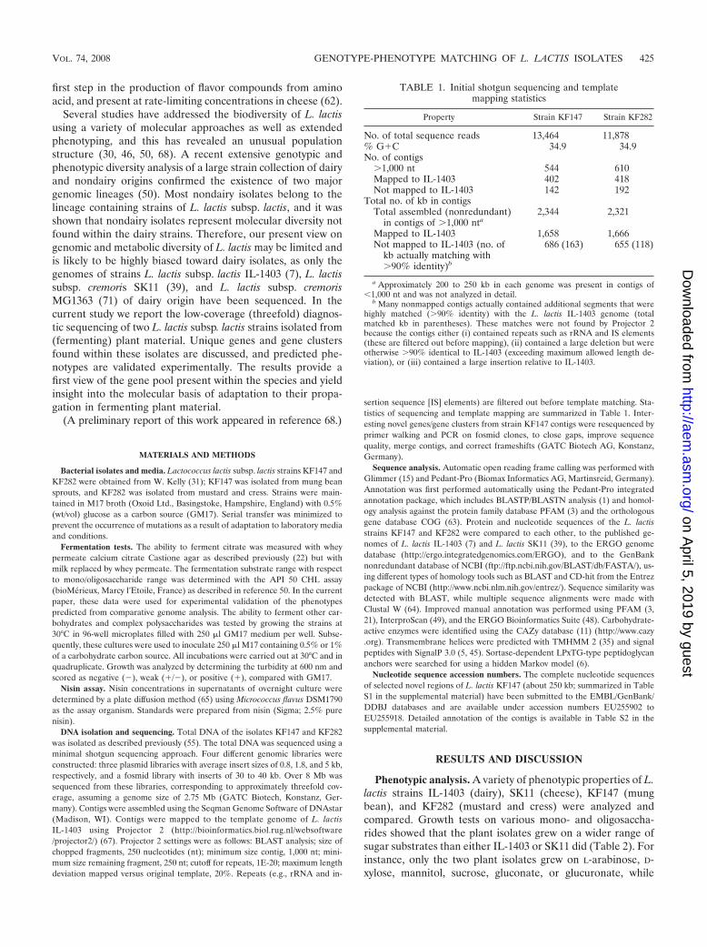
TABLE 1. Initial shotgun sequencing and templatemapping statistics
Property Strain KF147 Strain KF282
No. of total sequence reads 13,464 11,878% G�C 34.9 34.9No. of contigs
�1,000 nt 544 610Mapped to IL-1403 402 418Not mapped to IL-1403 142 192
Total no. of kb in contigsTotal assembled (nonredundant)
in contigs of �1,000 nta2,344 2,321
Mapped to IL-1403 1,658 1,666Not mapped to IL-1403 (no. of
kb actually matching with�90% identity)b
686 (163) 655 (118)
a Approximately 200 to 250 kb in each genome was present in contigs of�1,000 nt and was not analyzed in detail.
b Many nonmapped contigs actually contained additional segments that werehighly matched (�90% identity) with the L. lactis IL-1403 genome (totalmatched kb in parentheses). These matches were not found by Projector 2because the contigs either (i) contained repeats such as rRNA and IS elements(these are filtered out before mapping), (ii) contained a large deletion but wereotherwise �90% identical to IL-1403 (exceeding maximum allowed length de-viation), or (iii) contained a large insertion relative to IL-1403.
VOL. 74, 2008 GENOTYPE-PHENOTYPE MATCHING OF L. LACTIS ISOLATES 425
on April 5, 2019 by guest
http://aem.asm
.org/D
ownloaded from
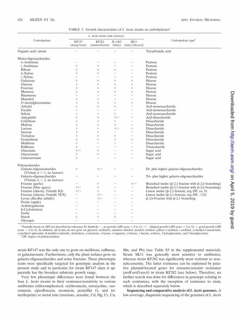
strain KF147 was the only one to grow on melibiose, raffinose,or galacturonate. Furthermore, only the plant isolates grew ongalacto-oligosaccharides and some fructans. These phenotypictraits were specifically targeted for genotypic analysis in thepresent study and in particular for strain KF147 since it ap-parently has the broadest substrate growth range.
Very few phenotypic differences were found between thefour L. lactis strains in their resistance/sensitivity to variousantibiotics (chloramphenicol, erythromycin, tetracycline, van-comycin, ciprofloxacin, neomycin, penicillin G, and tri-methoprim) or metal ions (arsenate, arsenite, Cd, Hg, Cr, Cu,
Mn, and Pb) (see Table S3 in the supplemental material).Strain SK11 was generally most sensitive to antibiotics,whereas strain KF282 was significantly most resistant to arse-nate/arsenite. The latter resistance can be explained by puta-tive plasmid-located genes for arsenate/arsenite resistance(arsR-arsD-arsA) in strain KF282 (see below). Therefore, nofurther search was done for differences in genotype relating tosuch resistances, with the exception of resistance to nisin,which is described separately below.
Sequencing and comparative analysis of L. lactis genomes. Alow-coverage, diagnostic sequencing of the genomes of L. lactis
TABLE 2. Growth characteristics of L. lactis strains on carbohydratesa
Carbohydrate
L. lactis strain code (source)
Carbohydrate typebKF147
(mung bean)KF282
(mustard/cress)IL-1403(dairy)
SK11(dairy �cheese�)
Organic acid: citrate � � � � Tricarboxylic acid
Mono/oligosaccharidesD-Arabinose � � � � PentoseL-Arabinose � � � � PentoseRibose � � � � PentoseD-Xylose � � � � PentoseL-Xylose � � � � PentoseGalactose � � � � HexoseGlucose � � � � HexoseFructose � � � � HexoseMannose � � � � HexoseRhamnose � � � � HexoseMannitol � � � � HexoseN-Acetylglucosamine � � � � HexoseArbutin � � � � Aryl-monosaccharideEsculin � � � � Aryl-monosaccharideSalicin � � � � Aryl-monosaccharideAmygdalin � � �/� � Aryl-disaccharideCellobiose � � � � DisaccharideMaltose � � � � DisaccharideLactose � � �/� � DisaccharideSucrose � � � � DisaccharideTrehalose � � � � DisaccharideGentiobiose � � � � DisaccharideMelibiose � � � � DisaccharideRaffinose � � � � TrisaccharideGluconate �/� � � � Sugar acidGlucuronate � � � � Sugar acidGalacturonate � � � � Sugar acid
PolysaccharidesGalacto-oligosaccharides
(Vivinal, n � 1, no lactose)� �/� � � Di- plus higher galacto-oligosaccharides
Galacto-oligosaccharides(Vivinal, n � 2, no lactose)
� � � � Tri- plus higher galacto-oligosaccharides
Fructan (garlic) � � � �/� Branched inulin (-2,1-fructan with -2,6 branching)Fructan (blue agave) �/� � � � Branched inulin (-2,1-fructan with -2,6 branching)Fructan (chicory, Frutafit IQ) �/� � � � Linear inulin (-2,1-fructan; avg DP, ca. 9)Fructan (chicory, Frutafit TEX) � �/� � � Linear inulin (-2,1-fructan; avg DP, �23)Levan (Bacillus subtilis) � �/� � � -2,6-Fructan with -2,1 branchingPectin (apple) � � � �Arabinogalactan � � � �-Cyclodextran � � � �Inulin � � � �Starch � � � �Glycogen � � � �
a Partially based on API test described in reference 50. Symbols: �, no growth (API score 0 to 1); �/�, limited growth (API score 2 to 3); �, good growth (APIscore 4 to 6). In addition, all strains do not grow on glycerol, erythritol, adonitol, dulcitol, inositol, sorbitol, xylitol, D-arabitol, L-arabitol, �-methyl-D-mannoside,�-methyl-D-glucoside, -methyl-D-xyloside, melezitose, D-turanose, D-lyxose, D-tagatose, D-fucose, L-fucose, sorbose, 2-ketogluconate, and 5-ketogluconate.
b DP, degree of polymerization.
426 SIEZEN ET AL. APPL. ENVIRON. MICROBIOL.
on April 5, 2019 by guest
http://aem.asm
.org/D
ownloaded from
strains KF147 and KF282 was performed (Table 1). The as-sembled contigs suggest that the sizes and G�C contents of thetwo genomes are similar (�2.5 to 2.6 Mb and 34.9%, respec-tively) and comparable to those of L. lactis strains IL-1403(2.365 Mb and 35.4%, respectively) (7), SK11 (2.438 Mb and35.8%, respectively) (39), and MG1363 (2.529 Mb and 35.8%,respectively) (71). The initial BLAST analysis showed that thelarge majority of contigs of both L. lactis plant isolates werehighly similar to one another (�95% nucleotide sequenceidentity) and to the IL-1403 sequence (�95% identity) andthat the genes showed conserved gene order. Therefore, con-tigs larger than 1,000 nt were first mapped onto the IL-1403template genome using Projector 2.0 (for examples, see Fig.S1A and S1B for KF147 and KF282, respectively, in the sup-plemental material). This mapping provided a fast first esti-mate of genome coverage, contig order, gap sizes, and possiblepositions of large insertions and deletions relative to IL-1403.
Closer inspection showed that many unmatched contigs ac-tually contained additional large segments that were highlymatched (�90% identity) to the IL-1403 genome. Thesematches were not found by Projector because the contigs ei-ther (i) contained repeats (these are filtered out before map-ping), (ii) contained a large deletion but were otherwise �90%identical to IL-1403 (exceeding maximum allowed length de-viation in Projector), or (iii) contained a large insertion relativeto IL-1403. These partially matching contigs provided furtherclues for positions of insertions and deletions. Together, atleast 1.8 Mb (�75%) of the genome of each plant isolate wasfound to match the IL-1403 genome, and the majority of thesematches were found in both plant isolates.
To characterize genotypic differences between plant anddairy strains in more detail, we subsequently focused on largeregions/clusters that were absent on the contigs from strainKF147 or KF282 compared to strain IL-1403 and on functionsencoded on contigs that did not map to IL-1403 (Table 3).Since these regions were highly similar in strains KF147 andKF282, we performed high-quality resequencing only of therelevant regions of the KF147 genome.
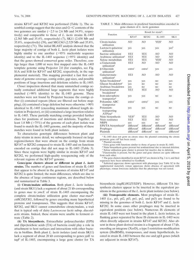
Genes/gene clusters absent or different in plant L. lactisstrains. The number of genes and functions of strain IL-1403that appear to be absent in the genomes of strains KF147 andKF282 is quite limited; the main differences, which are due tothe absence of large continuous regions, are described belowand summarized in Table 3.
(i) Citrate/malate utilization. Both plant L. lactis isolates(and strain SK11) lack a segment of about 21 kb correspondingto genes mae to ymcE of strain IL-1403 encompassing thecomplete citrate/malate utilization gene cluster mae-maeP-citRCDEFXG, followed by genes encoding many hypotheticalproteins and transposases. This suggests that strains KF147,KF282, and SK11 cannot cometabolize citrate/malate, a traitthat is typical only of dairy Lactococcus lactis subsp. diacetyl-actis strains. Indeed, these strains were unable to ferment ci-trate (Table 2).
(ii) TA biosynthesis. Extracellular polysaccharides (EPS)and (lipo)teichoic acids (TA) of bacteria could play a role inattachment to host surfaces and interactions with other bacte-ria in biofilms. Both plant L. lactis isolates (and strain SK11)lack a segment of about 20 kb corresponding to genes yjdE totagF of IL-1403, encompassing a large gene cluster for TA
biosynthesis (tagRLHGZYXDF). However, different TA bio-synthesis clusters appear to be inserted in the equivalent po-sitions in the genomes of the L. lactis plant isolates (see below).
(iii) Transposable elements. Most prophages of strain IL-1403 (i.e., pi1, pi2, pi3, ps1, ps2, and ps3) are found to bemissing in the genomes of both L. lactis KF147 and L. lactisKF282. In some cases other prophages may be inserted inequivalent positions (see below). Numerous IS elements ofstrain IL-1403 were not found in the plant L. lactis isolates, asflanking genes separated by these IS elements in IL-1403 wereoften directly adjacent in strains KF147 and KF282. Also ab-sent in these plant-derived strains is a fragment of about 17 kb,encoding an integrase (XerD), a type I restriction-modificationsystem (HsdRMS), transposases, and many hypotheticals, lo-cated in strain IL-1403 between the eno and ygfA genes (whichare adjacent in strain KF147).
TABLE 3. Main differences in predicted functionalities encoded ingene clusters of L. lactis genomes
FunctionResult for strainh:
KF147 KF282 IL-1403 SK11
Citrate/malateutilization
NO NO YESa NO
Lactose/D-galactoseutilization
yes yes yes yesb
Sucrose utilization YES YES NO NOArabinose utilizationg YES YES NO NOXylose metabolism YES YES YESe NO�-Galactoside
utilizationgYES NO NO NO
-Glucosideutilizationg
extrac extrac yes yes
Galacturonateutilizationg
YES NO NO NO
Xylan breakdowng yes yes no yesGlucan breakdowng yes no no noArabinan breakdown yes no no noFructan/mannan
breakdowngYES YES NO NO
Iron-siderophoretransporterg
yes yes no no
Polyamine ABCtransporter
extrac extrac yes yes
Potassium-transportingATPaseg
yes yes no no
Nisin biosynthesis YESd YES NO NONisin resistance YES YES NO NONRPS yes yes no noEPS/TA biosynthesisg differentf differentf differentf differentf
Prophages differentf differentf differentf differentf
Plasmids differentf differentf no differentf
a IL-1403 does not grow on citrate since the plasmid containing the citratepermease gene citP has been cured.
b Plasmid encoded.c Extra genes with functions similar to those of genes in strain IL-1403.d Nisin biosynthesis genes present but nonfunctional due to internal deletion.e Xylose utilization gene cluster is present but inactive due to deletion.f Functionality is present, but the genes and gene clusters are quite different
from each other.g The gene clusters identified in strain KF147 are shown in Fig. 1 to 4, and their
sequences have been submitted to GenBank.h Italicized uppercase shows agreement with phenotype (see Table S2 in the
supplemental material); roman uppercase shows absence of agreement withphenotype; roman lowercase indicates that the phenotype was not tested.
VOL. 74, 2008 GENOTYPE-PHENOTYPE MATCHING OF L. LACTIS ISOLATES 427
on April 5, 2019 by guest
http://aem.asm
.org/D
ownloaded from
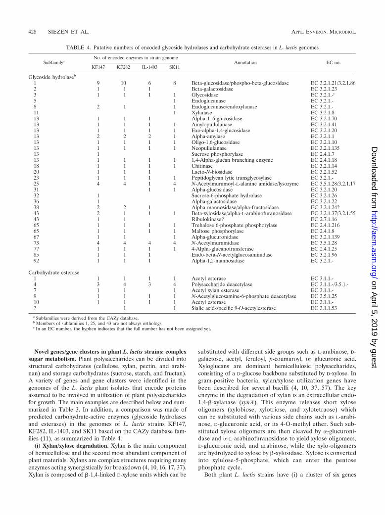
Novel genes/gene clusters in plant L. lactis strains: complexsugar metabolism. Plant polysaccharides can be divided intostructural carbohydrates (cellulose, xylan, pectin, and arabi-nan) and storage carbohydrates (sucrose, starch, and fructan).A variety of genes and gene clusters were identified in thegenomes of the L. lactis plant isolates that encode proteinsassumed to be involved in utilization of plant polysaccharidesfor growth. The main examples are described below and sum-marized in Table 3. In addition, a comparison was made ofpredicted carbohydrate-active enzymes (glycoside hydrolasesand esterases) in the genomes of L. lactis strains KF147,KF282, IL-1403, and SK11 based on the CAZy database fam-ilies (11), as summarized in Table 4.
(i) Xylan/xylose degradation. Xylan is the main componentof hemicellulose and the second most abundant component ofplant materials. Xylans are complex structures requiring manyenzymes acting synergistically for breakdown (4, 10, 16, 17, 37).Xylan is composed of -1,4-linked D-xylose units which can be
substituted with different side groups such as L-arabinose, D-galactose, acetyl, feruloyl, p-coumaroyl, or glucuronic acid.Xyloglucans are dominant hemicellulosic polysaccharides,consisting of a D-glucose backbone substituted by D-xylose. Ingram-positive bacteria, xylan/xylose utilization genes havebeen described for several bacilli (4, 10, 37, 57). The keyenzyme in the degradation of xylan is an extracellular endo-1,4--xylanase (xynA). This enzyme releases short xyloseoligomers (xylobiose, xylotriose, and xylotetraose) whichcan be substituted with various side chains such as L-arabi-nose, D-glucuronic acid, or its 4-O-methyl ether. Such sub-stituted xylose oligomers are then cleaved by �-glucuroni-dase and �-L-arabinofuranosidase to yield xylose oligomers,D-glucuronic acid, and arabinose, while the xylo-oligomersare hydrolyzed to xylose by -xylosidase. Xylose is convertedinto xylulose-5-phosphate, which can enter the pentosephosphate cycle.
Both plant L. lactis strains have (i) a cluster of six genes
TABLE 4. Putative numbers of encoded glycoside hydrolases and carbohydrate esterases in L. lactis genomes
SubfamilyaNo. of encoded enzymes in strain genome
Annotation EC no.KF147 KF282 IL-1403 SK11
Glycoside hydrolaseb
1 9 10 6 8 Beta-glucosidase/phospho-beta-glucosidase EC 3.2.1.21/3.2.1.862 1 1 1 Beta-galactosidase EC 3.2.1.233 1 1 1 1 Glycosidase EC 3.2.1.-c
5 1 Endoglucanase EC 3.2.1.-8 2 1 1 Endoglucanase/endoxylanase EC 3.2.1.-11 1 Xylanase EC 3.2.1.813 1 1 1 Alpha-1–6-glucosidase EC 3.2.1.7013 1 1 1 1 Amylopullulanase EC 3.2.1.4113 1 1 1 1 Exo-alpha-1,4-glucosidase EC 3.2.1.2013 2 2 2 1 Alpha-amylase EC 3.2.1.113 1 1 1 1 Oligo-1,6-glucosidase EC 3.2.1.1013 1 1 1 1 Neopullulanase EC 3.2.1.13513 1 Sucrose phosphorylase EC 2.4.1.713 1 1 1 1 1,4-Alpha-glucan branching enzyme EC 2.4.1.1818 1 1 1 1 Chitinase EC 3.2.1.1420 1 1 1 Lacto-N-biosidase EC 3.2.1.5223 1 1 1 1 Peptidoglycan lytic transglycosylase EC 3.2.1.-25 4 4 1 4 N-Acetylmuramoyl-L-alanine amidase/lysozyme EC 3.5.1.28/3.2.1.1731 1 1 Alpha-glucosidase EC 3.2.1.2032 1 Sucrose-6-phosphate hydrolase EC 3.2.1.2636 1 Alpha-galactosidase EC 3.2.1.2238 2 2 1 Alpha mannosidase/alpha-fructosidase EC 3.2.1.24?43 2 1 1 1 Beta-xylosidase/alpha-L-arabinofuranosidase EC 3.2.1.37/3.2.1.5543 1 1 Ribulokinase? EC 2.7.1.1665 1 1 1 1 Trehalose 6-phosphate phosphorylase EC 2.4.1.21665 1 1 1 1 Maltose phosphorylase EC 2.4.1.867 1 1 1 Alpha-glucuronidase EC 3.2.1.13973 4 4 4 4 N-Acetylmuramidase EC 3.5.1.2877 1 1 1 1 4-Alpha-glucanotransferase EC 2.4.1.2585 1 1 1 Endo-beta-N-acetylglucosaminidase EC 3.2.1.9692 1 1 1 Alpha-1,2-mannosidase EC 3.2.1.-
Carbohydrate esterase1 1 1 1 1 Acetyl esterase EC 3.1.1.-4 3 4 3 4 Polysaccharide deacetylase EC 3.1.1.-/3.5.1.-7 1 1 1 Acetyl xylan esterase EC 3.1.1.-9 1 1 1 1 N-Acetylglucosamine-6-phosphate deacetylase EC 3.5.1.2510 1 1 1 1 Acetyl esterase EC 3.1.1.-? 1 1 Sialic acid-specific 9-O-acetylesterase EC 3.1.1.53
a Subfamilies were derived from the CAZy database.b Members of subfamilies 1, 25, and 43 are not always orthologs.c In an EC number, the hyphen indicates that the full number has not been assigned yet.
428 SIEZEN ET AL. APPL. ENVIRON. MICROBIOL.
on April 5, 2019 by guest
http://aem.asm
.org/D
ownloaded from
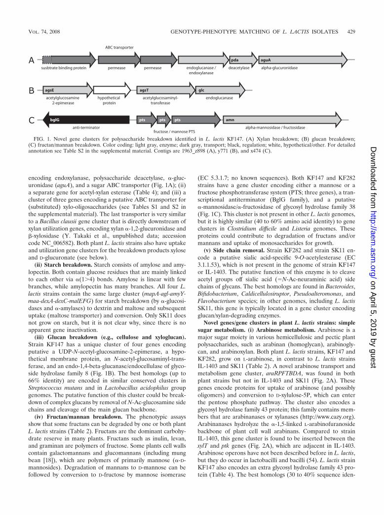
encoding endoxylanase, polysaccharide deacetylase, �-gluc-uronidase (aguA), and a sugar ABC transporter (Fig. 1A); (ii)a separate gene for acetyl-xylan esterase (Table 4); and (iii) acluster of three genes encoding a putative ABC transporter for(substituted) xylo-oligosaccharides (see Tables S1 and S2 inthe supplemental material). The last transporter is very similarto a Bacillus clausii gene cluster that is directly downstream ofxylan utilization genes, encoding xylan �-1,2-glucuronidase and-xylosidase (Y. Takaki et al., unpublished data; accessioncode NC_006582). Both plant L. lactis strains also have uptakeand utilization gene clusters for the breakdown products xyloseand D-glucuronate (see below).
(ii) Starch breakdown. Starch consists of amylose and amy-lopectin. Both contain glucose residues that are mainly linkedto each other via �(1�4) bonds. Amylose is linear with fewbranches, while amylopectin has many branches. All four L.lactis strains contain the same large cluster (mapA-agl-amyY-maa-dexA-dexC-malEFG) for starch breakdown (by �-glucosi-dases and �-amylases) to dextrin and maltose and subsequentuptake (maltose transporter) and conversion. Only SK11 doesnot grow on starch, but it is not clear why, since there is noapparent gene inactivation.
(iii) Glucan breakdown (e.g., cellulose and xyloglucan).Strain KF147 has a unique cluster of four genes encodingputative a UDP-N-acetyl-glucosamine-2-epimerase, a hypo-thetical membrane protein, an N-acetyl-glucosaminyl-trans-ferase, and an endo-1,4-beta-glucanase/endocellulase of glyco-side hydrolase family 8 (Fig. 1B). The best homologs (up to66% identity) are encoded in similar conserved clusters inStreptococcus mutans and in Lactobacillus acidophilus groupgenomes. The putative function of this cluster could be break-down of complex glucans by removal of N-Ac-glucosamine sidechains and cleavage of the main glucan backbone.
(iv) Fructan/mannan breakdown. The phenotypic assaysshow that some fructans can be degraded by one or both plantL. lactis strains (Table 2). Fructans are the dominant carbohy-drate reserve in many plants. Fructans such as inulin, levan,and graminan are polymers of fructose. Some plants cell wallscontain galactomannans and glucomannans (including mungbean [18]), which are polymers of primarily mannose (�-D-mannosides). Degradation of mannans to D-mannose can befollowed by conversion to D-fructose by mannose isomerase
(EC 5.3.1.7; no known sequences). Both KF147 and KF282strains have a gene cluster encoding either a mannose or afructose phosphotransferase system (PTS; three genes), a tran-scriptional antiterminator (BglG family), and a putative�-mannosidase/�-fructosidase of glycosyl hydrolase family 38(Fig. 1C). This cluster is not present in other L. lactis genomes,but it is highly similar (40 to 60% amino acid identity) to geneclusters in Clostridium difficile and Listeria genomes. Theseproteins could contribute to degradation of fructans and/ormannans and uptake of monosaccharides for growth.
(v) Side chain removal. Strain KF282 and strain SK11 en-code a putative sialic acid-specific 9-O-acetylesterase (EC3.1.1.53), which is not present in the genome of strain KF147or IL-1403. The putative function of this enzyme is to cleaveacetyl groups off sialic acid (N-Ac-neuraminic acid) sidechains of glycans. The best homologs are found in Bacteroides,Bifidobacterium, Caldicellulosiruptor, Pseudoalteromonas, andFlavobacterium species; in other genomes, including L. lactisSK11, this gene is typically located in a gene cluster encodingglucan/xylan-degrading enzymes.
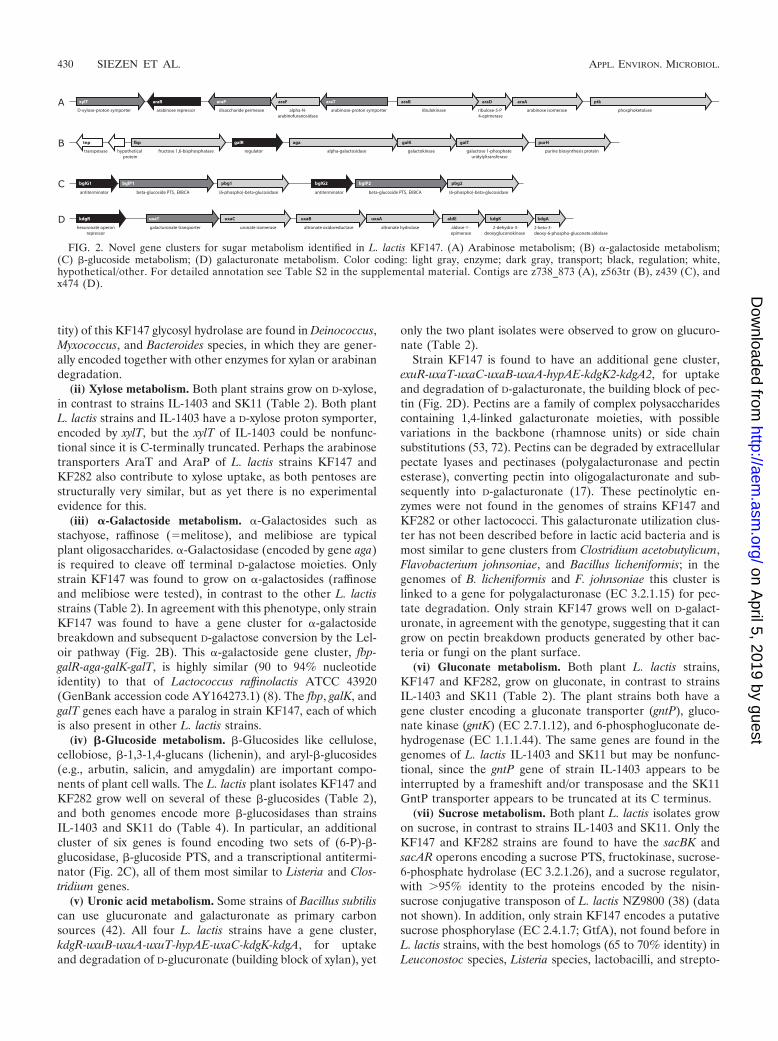
Novel genes/gene clusters in plant L. lactis strains: simplesugar metabolism. (i) Arabinose metabolism. Arabinose is amajor sugar moiety in various hemicellulosic and pectic plantpolysaccharides, such as arabinan (homoglycan), arabinogly-can, and arabinoxylan. Both plant L. lactis strains, KF147 andKF282, grow on L-arabinose, in contrast to L. lactis strainsIL-1403 and SK11 (Table 2). A novel arabinose transport andmetabolism gene cluster, araRPFTBDA, was found in bothplant strains but not in IL-1403 and SK11 (Fig. 2A). Thesegenes encode proteins for uptake of arabinose (and possiblyoligomers) and conversion to D-xylulose-5P, which can enterthe pentose phosphate pathway. The cluster also encodes aglycosyl hydrolase family 43 protein; this family contains mem-bers that are arabinanases or xylanases (http://www.cazy.org).Arabinanases hydrolyze the �-1,5-linked L-arabinofuranosidebackbone of plant cell wall arabinans. Compared to strainIL-1403, this gene cluster is found to be inserted between thexylT and ptk genes (Fig. 2A), which are adjacent in IL-1403.Arabinose operons have not been described before in L. lactis,but they do occur in lactobacilli and bacilli (54). L. lactis strainKF147 also encodes an extra glycosyl hydrolase family 43 pro-tein (Table 4). The best homologs (30 to 40% sequence iden-
FIG. 1. Novel gene clusters for polysaccharide breakdown identified in L. lactis KF147. (A) Xylan breakdown; (B) glucan breakdown;(C) fructan/mannan breakdown. Color coding: light gray, enzyme; dark gray, transport; black, regulation; white, hypothetical/other. For detailedannotation see Table S2 in the supplemental material. Contigs are 1963_z898 (A), y771 (B), and x474 (C).
VOL. 74, 2008 GENOTYPE-PHENOTYPE MATCHING OF L. LACTIS ISOLATES 429
on April 5, 2019 by guest
http://aem.asm
.org/D
ownloaded from
tity) of this KF147 glycosyl hydrolase are found in Deinococcus,Myxococcus, and Bacteroides species, in which they are gener-ally encoded together with other enzymes for xylan or arabinandegradation.
(ii) Xylose metabolism. Both plant strains grow on D-xylose,in contrast to strains IL-1403 and SK11 (Table 2). Both plantL. lactis strains and IL-1403 have a D-xylose proton symporter,encoded by xylT, but the xylT of IL-1403 could be nonfunc-tional since it is C-terminally truncated. Perhaps the arabinosetransporters AraT and AraP of L. lactis strains KF147 andKF282 also contribute to xylose uptake, as both pentoses arestructurally very similar, but as yet there is no experimentalevidence for this.
(iii) �-Galactoside metabolism. �-Galactosides such asstachyose, raffinose (melitose), and melibiose are typicalplant oligosaccharides. �-Galactosidase (encoded by gene aga)is required to cleave off terminal D-galactose moieties. Onlystrain KF147 was found to grow on �-galactosides (raffinoseand melibiose were tested), in contrast to the other L. lactisstrains (Table 2). In agreement with this phenotype, only strainKF147 was found to have a gene cluster for �-galactosidebreakdown and subsequent D-galactose conversion by the Lel-oir pathway (Fig. 2B). This �-galactoside gene cluster, fbp-galR-aga-galK-galT, is highly similar (90 to 94% nucleotideidentity) to that of Lactococcus raffinolactis ATCC 43920(GenBank accession code AY164273.1) (8). The fbp, galK, andgalT genes each have a paralog in strain KF147, each of whichis also present in other L. lactis strains.
(iv) �-Glucoside metabolism. -Glucosides like cellulose,cellobiose, -1,3-1,4-glucans (lichenin), and aryl--glucosides(e.g., arbutin, salicin, and amygdalin) are important compo-nents of plant cell walls. The L. lactis plant isolates KF147 andKF282 grow well on several of these -glucosides (Table 2),and both genomes encode more -glucosidases than strainsIL-1403 and SK11 do (Table 4). In particular, an additionalcluster of six genes is found encoding two sets of (6-P)--glucosidase, -glucoside PTS, and a transcriptional antitermi-nator (Fig. 2C), all of them most similar to Listeria and Clos-tridium genes.
(v) Uronic acid metabolism. Some strains of Bacillus subtiliscan use glucuronate and galacturonate as primary carbonsources (42). All four L. lactis strains have a gene cluster,kdgR-uxuB-uxuA-uxuT-hypAE-uxaC-kdgK-kdgA, for uptakeand degradation of D-glucuronate (building block of xylan), yet
only the two plant isolates were observed to grow on glucuro-nate (Table 2).
Strain KF147 is found to have an additional gene cluster,exuR-uxaT-uxaC-uxaB-uxaA-hypAE-kdgK2-kdgA2, for uptakeand degradation of D-galacturonate, the building block of pec-tin (Fig. 2D). Pectins are a family of complex polysaccharidescontaining 1,4-linked galacturonate moieties, with possiblevariations in the backbone (rhamnose units) or side chainsubstitutions (53, 72). Pectins can be degraded by extracellularpectate lyases and pectinases (polygalacturonase and pectinesterase), converting pectin into oligogalacturonate and sub-sequently into D-galacturonate (17). These pectinolytic en-zymes were not found in the genomes of strains KF147 andKF282 or other lactococci. This galacturonate utilization clus-ter has not been described before in lactic acid bacteria and ismost similar to gene clusters from Clostridium acetobutylicum,Flavobacterium johnsoniae, and Bacillus licheniformis; in thegenomes of B. licheniformis and F. johnsoniae this cluster islinked to a gene for polygalacturonase (EC 3.2.1.15) for pec-tate degradation. Only strain KF147 grows well on D-galact-uronate, in agreement with the genotype, suggesting that it cangrow on pectin breakdown products generated by other bac-teria or fungi on the plant surface.
(vi) Gluconate metabolism. Both plant L. lactis strains,KF147 and KF282, grow on gluconate, in contrast to strainsIL-1403 and SK11 (Table 2). The plant strains both have agene cluster encoding a gluconate transporter (gntP), gluco-nate kinase (gntK) (EC 2.7.1.12), and 6-phosphogluconate de-hydrogenase (EC 1.1.1.44). The same genes are found in thegenomes of L. lactis IL-1403 and SK11 but may be nonfunc-tional, since the gntP gene of strain IL-1403 appears to beinterrupted by a frameshift and/or transposase and the SK11GntP transporter appears to be truncated at its C terminus.
(vii) Sucrose metabolism. Both plant L. lactis isolates growon sucrose, in contrast to strains IL-1403 and SK11. Only theKF147 and KF282 strains are found to have the sacBK andsacAR operons encoding a sucrose PTS, fructokinase, sucrose-6-phosphate hydrolase (EC 3.2.1.26), and a sucrose regulator,with �95% identity to the proteins encoded by the nisin-sucrose conjugative transposon of L. lactis NZ9800 (38) (datanot shown). In addition, only strain KF147 encodes a putativesucrose phosphorylase (EC 2.4.1.7; GtfA), not found before inL. lactis strains, with the best homologs (65 to 70% identity) inLeuconostoc species, Listeria species, lactobacilli, and strepto-
FIG. 2. Novel gene clusters for sugar metabolism identified in L. lactis KF147. (A) Arabinose metabolism; (B) �-galactoside metabolism;(C) -glucoside metabolism; (D) galacturonate metabolism. Color coding: light gray, enzyme; dark gray, transport; black, regulation; white,hypothetical/other. For detailed annotation see Table S2 in the supplemental material. Contigs are z738_873 (A), z563tr (B), z439 (C), andx474 (D).
430 SIEZEN ET AL. APPL. ENVIRON. MICROBIOL.
on April 5, 2019 by guest
http://aem.asm
.org/D
ownloaded from
cocci. The presence of this sucrose phosphorylase suggests thatthere should also be a permease for uptake of sucrose.
Nisin production/resistance. Both plant strains are muchmore resistant than strains IL-1403 and SK11 to the lantibioticpeptide nisin; strains KF147 and KF282 survived �2,000 ng/mlnisin, while IL-1403 and SK11 survived at maximally 125 and25 ng/ml, respectively. In a nisin biosynthesis assay, only strainKF282 showed a high growth inhibition of the indicator strain.The gene cluster nisZABTCIPRKFEG for biosynthesis of nisinZ and immunity to nisin Z (19, 28, 36) is found to be presentin both KF147 and KF282, in contrast to strains IL-1403 andSK11. However, strain KF147 clearly has an internal deletionin parts of the nisB and nisC genes, which should lead toinactivation of the encoded nisin biosynthetic enzymes. Hence,the genotype predicts that both plant strains should be resis-tant to nisin (presence of immunity genes) but that only strainKF282 should be a nisin producer. This genotype agrees com-pletely with the phenotypes found by us and Kelly et al. (31).
Survival/stress response. (i) NRPS. Several contigs wereidentified in both L. lactis strain KF147 (total of �40 kb; datanot shown) and L. lactis strain KF282 encoding enzymes whichare characteristic of nonribosomal peptide or polyketide bio-synthesis, such as phospho-pantetheine protein transferase,thioesterase, and multimodular peptide synthetases with ad-enylation, condensation, and acyl carrier protein domains (20,70). Such systems are commonly found in environmental bac-teria such as Bacillus, Pseudomonas, and Streptomyces spp.,where they can play a role in survival, defense, signaling, oradhesion (20). This is the first identification of such a system inLactococcus lactis. Studies are under way to sequence the com-plete gene clusters and identify their nonribosomal peptidesynthesis (NRPS)/polyketide synthesis products.
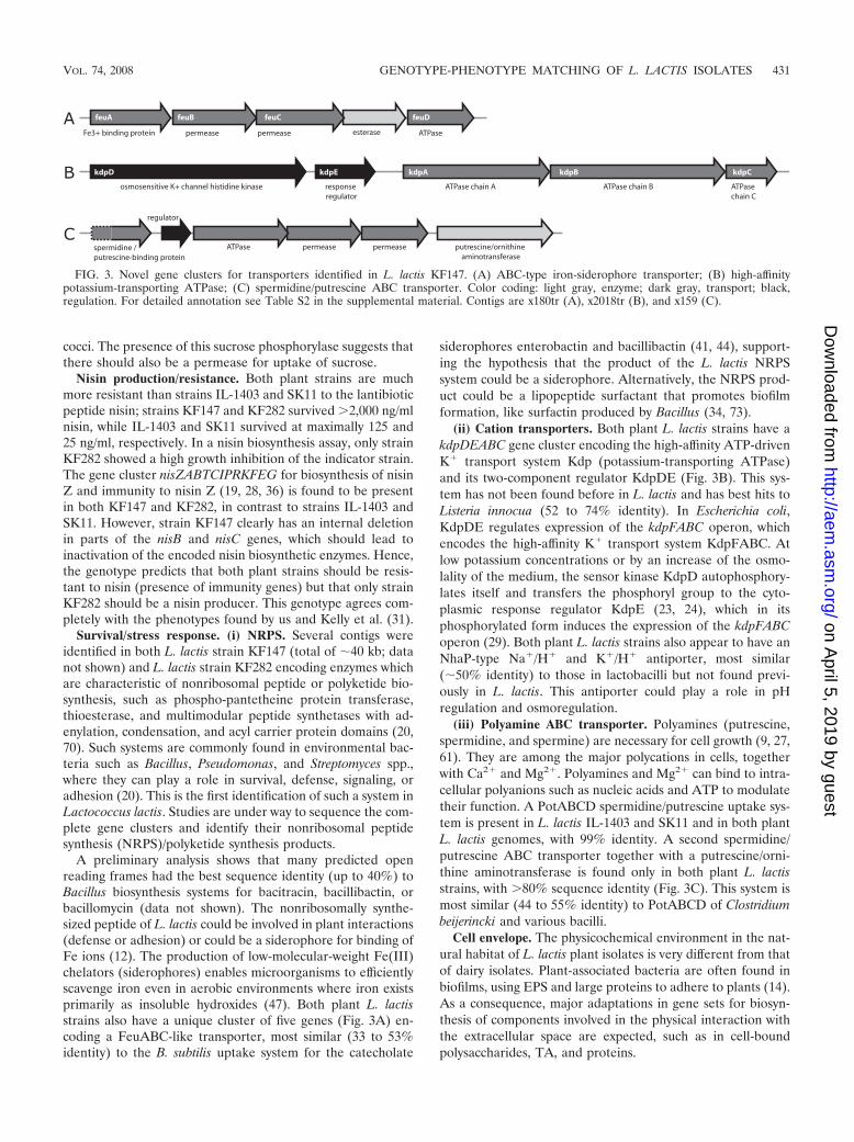
A preliminary analysis shows that many predicted openreading frames had the best sequence identity (up to 40%) toBacillus biosynthesis systems for bacitracin, bacillibactin, orbacillomycin (data not shown). The nonribosomally synthe-sized peptide of L. lactis could be involved in plant interactions(defense or adhesion) or could be a siderophore for binding ofFe ions (12). The production of low-molecular-weight Fe(III)chelators (siderophores) enables microorganisms to efficientlyscavenge iron even in aerobic environments where iron existsprimarily as insoluble hydroxides (47). Both plant L. lactisstrains also have a unique cluster of five genes (Fig. 3A) en-coding a FeuABC-like transporter, most similar (33 to 53%identity) to the B. subtilis uptake system for the catecholate
siderophores enterobactin and bacillibactin (41, 44), support-ing the hypothesis that the product of the L. lactis NRPSsystem could be a siderophore. Alternatively, the NRPS prod-uct could be a lipopeptide surfactant that promotes biofilmformation, like surfactin produced by Bacillus (34, 73).
(ii) Cation transporters. Both plant L. lactis strains have akdpDEABC gene cluster encoding the high-affinity ATP-drivenK� transport system Kdp (potassium-transporting ATPase)and its two-component regulator KdpDE (Fig. 3B). This sys-tem has not been found before in L. lactis and has best hits toListeria innocua (52 to 74% identity). In Escherichia coli,KdpDE regulates expression of the kdpFABC operon, whichencodes the high-affinity K� transport system KdpFABC. Atlow potassium concentrations or by an increase of the osmo-lality of the medium, the sensor kinase KdpD autophosphory-lates itself and transfers the phosphoryl group to the cyto-plasmic response regulator KdpE (23, 24), which in itsphosphorylated form induces the expression of the kdpFABCoperon (29). Both plant L. lactis strains also appear to have anNhaP-type Na�/H� and K�/H� antiporter, most similar(�50% identity) to those in lactobacilli but not found previ-ously in L. lactis. This antiporter could play a role in pHregulation and osmoregulation.
(iii) Polyamine ABC transporter. Polyamines (putrescine,spermidine, and spermine) are necessary for cell growth (9, 27,61). They are among the major polycations in cells, togetherwith Ca2� and Mg2�. Polyamines and Mg2� can bind to intra-cellular polyanions such as nucleic acids and ATP to modulatetheir function. A PotABCD spermidine/putrescine uptake sys-tem is present in L. lactis IL-1403 and SK11 and in both plantL. lactis genomes, with 99% identity. A second spermidine/putrescine ABC transporter together with a putrescine/orni-thine aminotransferase is found only in both plant L. lactisstrains, with �80% sequence identity (Fig. 3C). This system ismost similar (44 to 55% identity) to PotABCD of Clostridiumbeijerincki and various bacilli.
Cell envelope. The physicochemical environment in the nat-ural habitat of L. lactis plant isolates is very different from thatof dairy isolates. Plant-associated bacteria are often found inbiofilms, using EPS and large proteins to adhere to plants (14).As a consequence, major adaptations in gene sets for biosyn-thesis of components involved in the physical interaction withthe extracellular space are expected, such as in cell-boundpolysaccharides, TA, and proteins.
FIG. 3. Novel gene clusters for transporters identified in L. lactis KF147. (A) ABC-type iron-siderophore transporter; (B) high-affinitypotassium-transporting ATPase; (C) spermidine/putrescine ABC transporter. Color coding: light gray, enzyme; dark gray, transport; black,regulation. For detailed annotation see Table S2 in the supplemental material. Contigs are x180tr (A), x2018tr (B), and x159 (C).
VOL. 74, 2008 GENOTYPE-PHENOTYPE MATCHING OF L. LACTIS ISOLATES 431
on April 5, 2019 by guest
http://aem.asm
.org/D
ownloaded from
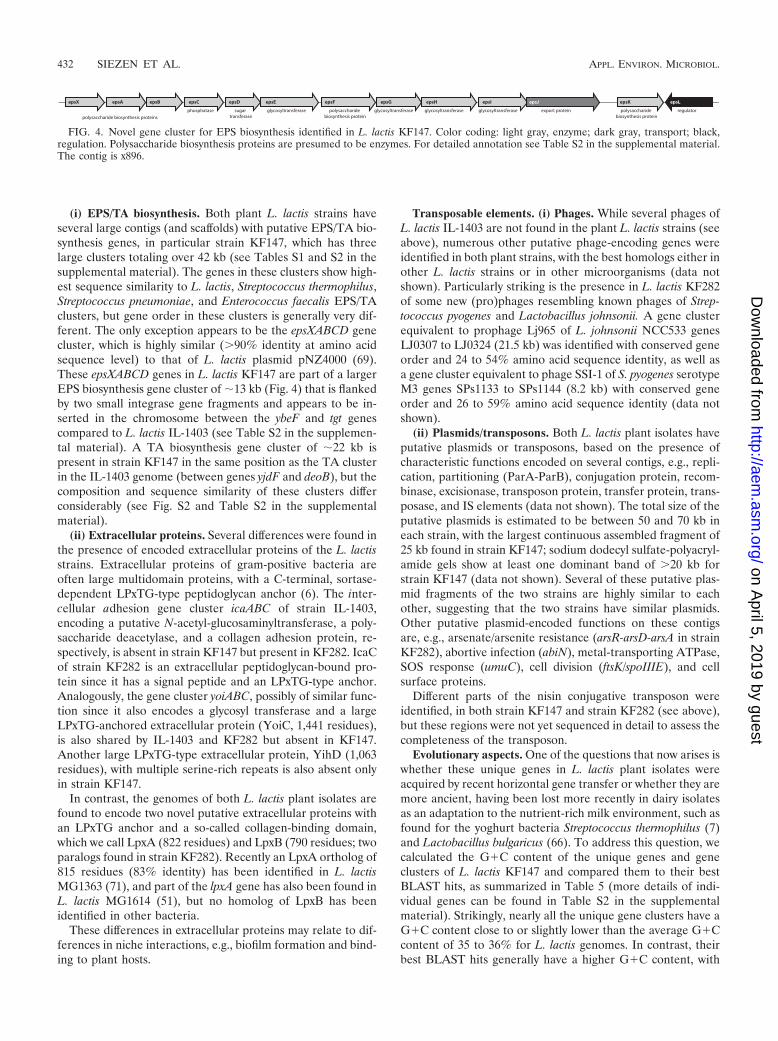
(i) EPS/TA biosynthesis. Both plant L. lactis strains haveseveral large contigs (and scaffolds) with putative EPS/TA bio-synthesis genes, in particular strain KF147, which has threelarge clusters totaling over 42 kb (see Tables S1 and S2 in thesupplemental material). The genes in these clusters show high-est sequence similarity to L. lactis, Streptococcus thermophilus,Streptococcus pneumoniae, and Enterococcus faecalis EPS/TAclusters, but gene order in these clusters is generally very dif-ferent. The only exception appears to be the epsXABCD genecluster, which is highly similar (�90% identity at amino acidsequence level) to that of L. lactis plasmid pNZ4000 (69).These epsXABCD genes in L. lactis KF147 are part of a largerEPS biosynthesis gene cluster of �13 kb (Fig. 4) that is flankedby two small integrase gene fragments and appears to be in-serted in the chromosome between the ybeF and tgt genescompared to L. lactis IL-1403 (see Table S2 in the supplemen-tal material). A TA biosynthesis gene cluster of �22 kb ispresent in strain KF147 in the same position as the TA clusterin the IL-1403 genome (between genes yjdF and deoB), but thecomposition and sequence similarity of these clusters differconsiderably (see Fig. S2 and Table S2 in the supplementalmaterial).
(ii) Extracellular proteins. Several differences were found inthe presence of encoded extracellular proteins of the L. lactisstrains. Extracellular proteins of gram-positive bacteria areoften large multidomain proteins, with a C-terminal, sortase-dependent LPxTG-type peptidoglycan anchor (6). The inter-cellular adhesion gene cluster icaABC of strain IL-1403,encoding a putative N-acetyl-glucosaminyltransferase, a poly-saccharide deacetylase, and a collagen adhesion protein, re-spectively, is absent in strain KF147 but present in KF282. IcaCof strain KF282 is an extracellular peptidoglycan-bound pro-tein since it has a signal peptide and an LPxTG-type anchor.Analogously, the gene cluster yoiABC, possibly of similar func-tion since it also encodes a glycosyl transferase and a largeLPxTG-anchored extracellular protein (YoiC, 1,441 residues),is also shared by IL-1403 and KF282 but absent in KF147.Another large LPxTG-type extracellular protein, YihD (1,063residues), with multiple serine-rich repeats is also absent onlyin strain KF147.
In contrast, the genomes of both L. lactis plant isolates arefound to encode two novel putative extracellular proteins withan LPxTG anchor and a so-called collagen-binding domain,which we call LpxA (822 residues) and LpxB (790 residues; twoparalogs found in strain KF282). Recently an LpxA ortholog of815 residues (83% identity) has been identified in L. lactisMG1363 (71), and part of the lpxA gene has also been found inL. lactis MG1614 (51), but no homolog of LpxB has beenidentified in other bacteria.
These differences in extracellular proteins may relate to dif-ferences in niche interactions, e.g., biofilm formation and bind-ing to plant hosts.
Transposable elements. (i) Phages. While several phages ofL. lactis IL-1403 are not found in the plant L. lactis strains (seeabove), numerous other putative phage-encoding genes wereidentified in both plant strains, with the best homologs either inother L. lactis strains or in other microorganisms (data notshown). Particularly striking is the presence in L. lactis KF282of some new (pro)phages resembling known phages of Strep-tococcus pyogenes and Lactobacillus johnsonii. A gene clusterequivalent to prophage Lj965 of L. johnsonii NCC533 genesLJ0307 to LJ0324 (21.5 kb) was identified with conserved geneorder and 24 to 54% amino acid sequence identity, as well asa gene cluster equivalent to phage SSI-1 of S. pyogenes serotypeM3 genes SPs1133 to SPs1144 (8.2 kb) with conserved geneorder and 26 to 59% amino acid sequence identity (data notshown).
(ii) Plasmids/transposons. Both L. lactis plant isolates haveputative plasmids or transposons, based on the presence ofcharacteristic functions encoded on several contigs, e.g., repli-cation, partitioning (ParA-ParB), conjugation protein, recom-binase, excisionase, transposon protein, transfer protein, trans-posase, and IS elements (data not shown). The total size of theputative plasmids is estimated to be between 50 and 70 kb ineach strain, with the largest continuous assembled fragment of25 kb found in strain KF147; sodium dodecyl sulfate-polyacryl-amide gels show at least one dominant band of �20 kb forstrain KF147 (data not shown). Several of these putative plas-mid fragments of the two strains are highly similar to eachother, suggesting that the two strains have similar plasmids.Other putative plasmid-encoded functions on these contigsare, e.g., arsenate/arsenite resistance (arsR-arsD-arsA in strainKF282), abortive infection (abiN), metal-transporting ATPase,SOS response (umuC), cell division (ftsK/spoIIIE), and cellsurface proteins.
Different parts of the nisin conjugative transposon wereidentified, in both strain KF147 and strain KF282 (see above),but these regions were not yet sequenced in detail to assess thecompleteness of the transposon.
Evolutionary aspects. One of the questions that now arises iswhether these unique genes in L. lactis plant isolates wereacquired by recent horizontal gene transfer or whether they aremore ancient, having been lost more recently in dairy isolatesas an adaptation to the nutrient-rich milk environment, such asfound for the yoghurt bacteria Streptococcus thermophilus (7)and Lactobacillus bulgaricus (66). To address this question, wecalculated the G�C content of the unique genes and geneclusters of L. lactis KF147 and compared them to their bestBLAST hits, as summarized in Table 5 (more details of indi-vidual genes can be found in Table S2 in the supplementalmaterial). Strikingly, nearly all the unique gene clusters have aG�C content close to or slightly lower than the average G�Ccontent of 35 to 36% for L. lactis genomes. In contrast, theirbest BLAST hits generally have a higher G�C content, with
FIG. 4. Novel gene cluster for EPS biosynthesis identified in L. lactis KF147. Color coding: light gray, enzyme; dark gray, transport; black,regulation. Polysaccharide biosynthesis proteins are presumed to be enzymes. For detailed annotation see Table S2 in the supplemental material.The contig is x896.
432 SIEZEN ET AL. APPL. ENVIRON. MICROBIOL.
on April 5, 2019 by guest
http://aem.asm
.org/D
ownloaded from
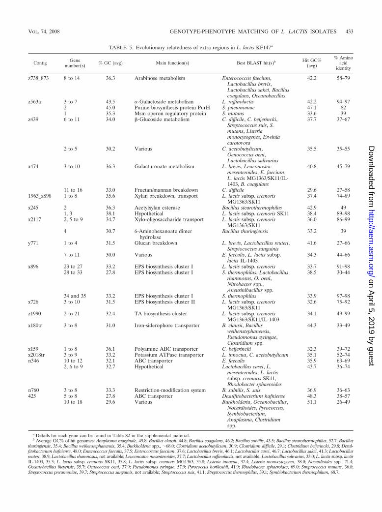
TABLE 5. Evolutionary relatedness of extra regions in L. lactis KF147a
Contig Genenumber(s) % GC (avg) Main function(s) Best BLAST hit(s)b Hit GC%
(avg)
% Aminoacid
identity
z738_873 8 to 14 36.3 Arabinose metabolism Enterococcus faecium,Lactobacillus brevis,Lactobacillus sakei, Bacilluscoagulans, Oceanobacillus
42.2 58–79
z563tr 3 to 7 43.5 �-Galactoside metabolism L. raffinolactis 42.2 94–972 45.0 Purine biosynthesis protein PurH S. pneumoniae 47.1 821 35.3 Msm operon regulatory protein S. mutans 33.6 39
z439 6 to 11 34.0 -Glucoside metabolism C. difficile, C. beijerincki,Streptococcus suis, S.mutans, Listeriamonocytogenes, Erwiniacarotovora
37.7 37–67
2 to 5 30.2 Various C. acetobutylicum,Oenococcus oeni,Lactobacillus salivarius
35.5 35–55
x474 3 to 10 36.3 Galacturonate metabolism L. brevis, Leuconostocmesenteroides, E. faecium,L. lactis MG1363/SK11/IL-1403, B. coagulans
40.8 45–79
11 to 16 33.0 Fructan/mannan breakdown C. difficile 29.6 27–581963_z898 1 to 8 35.6 Xylan breakdown, transport L. lactis subsp. cremoris
MG1363/SK1137.4 74–89
x245 2 36.3 Acetylxylan esterase Bacillus stearothermophilus 42.9 491, 3 38.1 Hypothetical L. lactis subsp. cremoris SK11 38.4 89–98
x2117 2, 5 to 9 34.7 Xylo-oligosaccharide transport L. lactis subsp. cremorisMG1363/SK11
36.0 86–99
4 30.7 6-Aminohexanoate dimerhydrolase
Bacillus thuringiensis 33.2 39
y771 1 to 4 31.5 Glucan breakdown L. brevis, Lactobacillus reuteri,Streptococcus sanguinis
41.6 27–66
7 to 11 30.0 Various E. faecalis, L. lactis subsp.lactis IL-1403
34.3 44–66
x896 23 to 27 33.2 EPS biosynthesis cluster I L. lactis subsp. cremoris 33.7 91–9828 to 33 27.8 EPS biosynthesis cluster I S. thermophilus, Lactobacillus
rhamnosus, O. oeni,Nitrobacter spp.,Aneurinibacillus spp.
38.5 30–44
34 and 35 33.2 EPS biosynthesis cluster I S. thermophilus 33.9 97–98x726 3 to 10 31.5 EPS biosynthesis cluster II L. lactis subsp. cremoris
MG1363/SK1132.6 75–92
z1990 2 to 21 32.4 TA biosynthesis cluster L. lactis subsp. cremorisMG1363/SK11/IL-1403
34.1 49–99
x180tr 3 to 8 31.0 Iron-siderophore transporter B. clausii, Bacillusweihenstephanensis,Pseudomonas syringae,Clostridium spp.
44.3 33–49
x159 1 to 8 36.1 Polyamine ABC transporter C. beijerincki 32.3 39–72x2018tr 3 to 9 33.2 Potassium ATPase transporter L. innocua, C. acetobutylicum 35.1 52–74n346 10 to 12 32.1 ABC transporter E. faecalis 35.9 63–69
2, 6 to 9 32.7 Hypothetical Lactobacillus casei, L.mesenteroides, L. lactissubsp. cremoris SK11,Rhodobacter sphaeroides
43.7 36–74
n760 3 to 8 33.3 Restriction-modification system B. subtilis, S. suis 36.9 36–63425 5 to 8 27.8 ABC transporter Desulfitobacterium hafniense 48.3 38–57
10 to 18 29.6 Various Burkholderia, Oceanobacillus,Nocardioides, Pyrococcus,Symbiobacterium,Anaplasma, Clostridiumspp.
51.1 26–49
a Details for each gene can be found in Table S2 in the supplemental material.b Average GC% of hit genomes: Anaplasma marginale, 49.8; Bacillus clausii, 44.8; Bacillus coagulans, 46.2; Bacillus subtilis, 43.5; Bacillus stearothermophilus, 52.7; Bacillus
thuringiensis, 35.4; Bacillus weihenstephanensis, 35.4; Burkholderia spp., �68.0; Clostridium acetobutylicum, 30.9; Clostridium difficile, 29.1; Clostridium beijerincki, 29.8; Desul-fitobacterium hafniense, 48.0; Enterococcus faecalis, 37.5; Enterococcus faecium, 37.6; Lactobacillus brevis, 46.1; Lactobacillus casei, 46.7; Lactobacillus sakei, 41.3; Lactobacillusreuteri, 38.9; Lactobacillus rhamnosus, not available; Leuconostoc mesenteroides, 37.7; Lactobacillus raffinolactis, not available; Lactobacillus salivarius, 33.0; L. lactis subsp. lactisIL-1403, 35.3; L. lactis subsp. cremoris SK11, 35.8; L. lactis subsp. cremoris MG1363, 35.8; Listeria innocua, 37.4; Listeria monocytogenes, 38.0; Nocardioides spp., 71.4;Oceanobacillus iheyensis, 35.7; Oenococcus oeni, 37.9; Pseudomonas syringae, 57.9; Pyrococcus horikoshii, 41.9; Rhodobacter sphaeroides, 69.0; Streptococcus mutans, 36.8;Streptococcus pneumoniae, 39.7; Streptococcus sanguinis, not available; Streptococcus suis, 41.1; Streptococcus thermophilus, 39.1; Symbiobacterium thermophilum, 68.7.
VOL. 74, 2008 GENOTYPE-PHENOTYPE MATCHING OF L. LACTIS ISOLATES 433
on April 5, 2019 by guest
http://aem.asm
.org/D
ownloaded from
the exception of the clostridial best hits, which have a loweraverage G�C content, in all cases closer to the average G�Ccontent for their own genomes (Table 5 footnote). Thisstrongly suggests that most of these “unique” gene clusters ofplant isolates are actually more ancient and were lost in manydairy isolates. This hypothesis is supported both by the fact thatmost of the best BLAST hits are from phylogenetically relatedLactobacillales (see, for instance, Fig. 5 of reference 40), in-dicative of presence in a common ancestor, and by the fact thatsome gene clusters are still found in L. lactis subsp. cremorisstrain SK11 or MG1363, but not in L. lactis subsp. lactis IL-1403 (Table 5).
However, one clear candidate of horizontal gene transfer isthe gene cluster for �-galactoside metabolism (contig z563tr),which is highly similar to that in Lactococcus raffinolactis (8),but in both bacteria the G�C content is much higher thanexpected for Lactococcus strains.
Conclusions and outlook. The L. lactis strains KF147 andKF282 isolated from mung bean sprouts and mustard andcress, respectively, are found to have many adaptations to theplant environment, particularly for growth on plant carbohy-drates. Mung bean (Phaseolus vulgaris) cell walls consist mainlyof arabinose (most dominant), uronic acids, galactose, xylose,mannose, and glucose (25, 56). White mustard (Sinapsis alba)cell walls (mucilage) consist mainly of glucose (most domi-nant), galactose, mannose, rhamnose, arabinose, galacturonicacid, and xylose (13, 26, 52); their polysaccharides are mainly1,4-linked -D-glucan (branched cellulose), complex pectin,and xyloglucan. The adaptation to growth on substrates de-rived from these plant cell walls is evident from the presence ofgene sets for the degradation of complex plant polymers suchas xylan, arabinan, glucans, and fructans but also for the uptakeand conversion of typical plant cell wall degradation productssuch as �-galactosides, -glucosides, arabinose, xylose, galact-uronate, glucuronate, and gluconate. Lactococci growing onplants generally live in synergy with other microbes in biofilms(14), including various bacteria and fungi, which could havesimilar and complementary enzymes (e.g., pectinases), allow-ing lactococci to grow on the plant cell wall breakdown prod-ucts generated by other microbes.
Other plant niche adaptations include genes for defense(such as nisin biosynthesis and immunity) and stress response.The latter involves several extra putative transport systems foruptake of iron (possibly involving a siderophore), potassium,and polyamines. In most cases these genotypes agree well withthe observed phenotypes (Table 3). Many of these genes andgene clusters have been identified for the first time in L. lactis.For instance, in addition to the new genes for plant sugarmetabolism, the nonribosomal peptide biosynthesis gene clus-ter is new for lactococci, and the only other lactic acid bacte-rium with a known but unrelated NRPS cluster is Lactobacillusplantarum WCFS1 (32).
Our approach of low-cost diagnostic sequencing provides afirst quick view of the gene pool present within lactococci fromplant environments and yields insight into their molecular basisof adaptation. It also indicates that, to achieve insight into thepangenome of Lactococcus lactis, or any other microbe for thatmatter, it is essential to isolate and sequence strains from awide variety of environments to allow for inclusion of all pos-sible adaptation mechanisms. Based on this newly identified
repertoire of genes in L. lactis, we have constructed a first-generation L. lactis pangenome microarray, using ultra-high-density DNA arrays (Nimblegen technology) for the assess-ment of genomic diversity within a large collection ofLactococcus lactis strains from dairy and nondairy origins, withthe aim of correlating genomic makeup with phenotypic traitsand better defining evolution of the Lactococcus lactis branch(G. Felis et al., unpublished results).
ACKNOWLEDGMENTS
We thank Bill Kelly for the L. lactis strains, Iris van Swam for DNAisolation, Sacha van Hijum for assistance with Projector 2 (67), Bern-hard Henrissat for CAZy analysis (11), and Ronald de Vries andChristof Francke for stimulating discussions.
This research was partly funded by the Kluyver Centre for Genomicsof Industrial Fermentation, a Centre of Excellence of The NetherlandsGenomics Initiative.
REFERENCES
1. Altschul, S. F., T. L. Madden, A. A. Schaffer, J. Zhang, Z. Zhang, W. Miller,and D. J. Lipman. 1997. Gapped BLAST and PSI-BLAST: a new generationof protein database search programs. Nucleic Acids Res. 25:3389–3402.
2. Ayad, E. H., A. Verheul, C. De Jong, J. T. Wouters, and G. Smit. 1999.Flavour forming abilities and amino acid requirements of Lactococcus lactisstrains isolated from artisanal and non-dairy origin. Int. Dairy J. 9:725–735.
3. Bateman, A., L. Coin, R. Durbin, R. D. Finn, V. Hollich, S. Griffiths-Jones,A. Khanna, M. Marshall, S. Moxon, E. L. Sonnhammer, D. J. Studholme, C.Yeats, and S. R. Eddy. 2004. The Pfam protein families database. NucleicAcids Res. 32:D138–D141.
4. Beg, Q. K., M. Kapoor, L. Mahajan, and G. S. Hoondal. 2001. Microbialxylanases and their industrial applications: a review. Appl. Microbiol. Bio-technol. 56:326–338.
5. Bendtsen, J. D., H. Nielsen, G. von Heijne, and S. Brunak. 2004. Improvedprediction of signal peptides: SignalP 3.0. J. Mol. Biol. 340:783–795.
6. Boekhorst, J., M. W. de Been, M. Kleerebezem, and R. J. Siezen. 2005.Genome-wide detection and analysis of cell wall-bound proteins withLPxTG-like sorting motifs. J. Bacteriol. 187:4928–4934.
7. Bolotin, A., P. Wincker, S. Mauger, O. Jaillon, K. Malarme, J. Weissenbach,S. D. Ehrlich, and A. Sorokin. 2001. The complete genome sequence of thelactic acid bacterium Lactococcus lactis ssp. lactis IL1403. Genome Res.11:731–753.
8. Boucher, I., C. Vadeboncoeur, and S. Moineau. 2003. Characterization ofgenes involved in the metabolism of alpha-galactosides by Lactococcus raf-finolactis. Appl. Environ. Microbiol. 69:4049–4056.
9. Cohen, S. 1997. A guide to the polyamines. Oxford University Press, Oxford,United Kingdom.
10. Collins, T., C. Gerday, and G. Feller. 2005. Xylanases, xylanase families andextremophilic xylanases. FEMS Microbiol. Rev. 29:3–23.
11. Coutinho, P. M., and B. Henrissat. 1999. Carbohydrate-active enzymes: anintegrated database approach, p. 3–12. In H. J. Gilbert, G. Davies, B. Hen-rissat, and B. Svensson (ed.), Recent advances in carbohydrate bioengineer-ing. The Royal Society of Chemistry, Cambridge, United Kingdom.
12. Crosa, J. H., and C. T. Walsh. 2002. Genetics and assembly line enzymologyof siderophore biosynthesis in bacteria. Microbiol. Mol. Biol. Rev. 66:223–249.
13. Cui, S. W., M. A. Eskin, Y. Wu, and S. Ding. 2006. Synergisms betweenyellow mustard mucilage and galactomannans and applications in food prod-ucts—a mini review. Adv. Colloid Interface Sci. 128–130:249–256.
14. Danhorn, T., and C. Fuqua. 2007. Biofilm formation by plant-associatedbacteria. Annu. Rev. Microbiol. 61:401–422.
15. Delcher, A. L., D. Harmon, S. Kasif, O. White, and S. L. Salzberg. 1999.Improved microbial gene identification with GLIMMER. Nucleic Acids Res.27:4636–4641.
16. de Vries, R. P., H. C. Kester, C. H. Poulsen, J. A. Benen, and J. Visser. 2000.Synergy between enzymes from Aspergillus involved in the degradation ofplant cell wall polysaccharides. Carbohydr. Res. 327:401–410.
17. de Vries, R. P., and J. Visser. 2001. Aspergillus enzymes involved in degra-dation of plant cell wall polysaccharides. Microbiol. Mol. Biol. Rev. 65:497–522.
18. Elbein, A. D. 1969. Biosynthesis of a cell wall glucomannan in mung beanseedlings. J. Biol. Chem. 244:1608–1616.
19. Entian, K. D., and W. M. de Vos. 1996. Genetics of subtilin and nisinbiosyntheses: biosynthesis of lantibiotics. Antonie Leeuwenhoek 69:109–117.
20. Finking, R., and M. A. Marahiel. 2004. Biosynthesis of nonribosomal pep-tides. Annu. Rev. Microbiol. 58:453–488.
21. Finn, R. D., J. Mistry, B. Schuster-Bockler, S. Griffiths-Jones, V. Hollich, T.
434 SIEZEN ET AL. APPL. ENVIRON. MICROBIOL.
on April 5, 2019 by guest
http://aem.asm
.org/D
ownloaded from
Lassmann, S. Moxon, M. Marshall, A. Khanna, R. Durbin, S. R. Eddy, E. L.Sonnhammer, and A. Bateman. 2006. Pfam: clans, web tools and services.Nucleic Acids Res. 34:D247–D251.
22. Galesloot, T. E., I. Hassing, and J. Stadhouders. 1961. Agar media for theisolation and enumeration of aroma bacteria in starters. Netherlands MilkDairy J. 15:145–150.
23. Gassel, M., and K. Altendorf. 2001. Analysis of KdpC of the K�-transportingKdpFABC complex of Escherichia coli. Eur. J. Biochem. 268:1772–1781.
24. Gassel, M., T. Mollenkamp, W. Puppe, and K. Altendorf. 1999. The KdpFsubunit is part of the K�-translocating Kdp complex of Escherichia coli andis responsible for stabilization of the complex in vitro. J. Biol. Chem. 274:37901–37907.
25. Gooneratne, J., P. W. Needs, P. Ryden, and R. R. Selvendran. 1994. Struc-tural features of cell wall polysaccharides from the cotyledons of mung beanVigna radiata. Carbohydr. Res. 265:61–77.
26. Gould, S. E., D. A. Rees, and N. J. Wight. 1971. Polysaccharides in germi-nation. Xyloglucans (�amyloids’) from the cotyledons of white mustard. Bio-chem. J. 124:47–53.
27. Igarashi, K., and K. Kashiwagi. 1999. Polyamine transport in bacteria andyeast. Biochem. J. 344:633–642.
28. Immonen, T., and P. E. Saris. 1998. Characterization of the nisFEG operonof the nisin Z producing Lactococcus lactis subsp. lactis N8 strain. DNA Seq.9:263–274.
29. Jung, K., and K. Altendorf. 2002. Towards an understanding of the molec-ular mechanisms of stimulus perception and signal transduction by theKdpD/KdpE system of Escherichia coli. J. Mol. Microbiol. Biotechnol.4:223–228.
30. Kelly, W., and L. Ward. 2002. Genotypic vs. phenotypic biodiversity inLactococcus lactis. Microbiology 148:3332–3333.
31. Kelly, W. J., G. P. Davey, and L. J. Ward. 1998. Characterization of lacto-cocci isolated from minimally processed fresh fruit and vegetables. Int. J.Food Microbiol. 45:85–92.
32. Kleerebezem, M., J. Boekhorst, R. van Kranenburg, D. Molenaar, O. P.Kuipers, R. Leer, R. Tarchini, S. A. Peters, H. M. Sandbrink, M. W. Fiers,W. Stiekema, R. M. Lankhorst, P. A. Bron, S. M. Hoffer, M. N. Groot, R.Kerkhoven, M. de Vries, B. Ursing, W. M. de Vos, and R. J. Siezen. 2003.Complete genome sequence of Lactobacillus plantarum WCFS1. Proc. Natl.Acad. Sci. USA 100:1990–1995.
33. Kok, J., G. Buist, A. L. Zomer, S. A. van Hijum, and O. P. Kuipers. 2005.Comparative and functional genomics of lactococci. FEMS Microbiol. Rev.29:411–433.
34. Koumoutsi, A., X. H. Chen, A. Henne, H. Liesegang, G. Hitzeroth, P. Franke,J. Vater, and R. Borriss. 2004. Structural and functional characterization ofgene clusters directing nonribosomal synthesis of bioactive cyclic lipopep-tides in Bacillus amyloliquefaciens strain FZB42. J. Bacteriol. 186:1084–1096.
35. Krogh, A., B. Larsson, G. von Heijne, and E. L. Sonnhammer. 2001. Pre-dicting transmembrane protein topology with a hidden Markov model: ap-plication to complete genomes. J. Mol. Biol. 305:567–580.
36. Kuipers, O. P., M. M. Beerthuyzen, R. J. Siezen, and W. M. De Vos. 1993.Characterization of the nisin gene cluster nisABTCIPR of Lactococcus lactis.Requirement of expression of the nisA and nisI genes for development ofimmunity. Eur. J. Biochem. 216:281–291.
37. Kulkarni, N., A. Shendye, and M. Rao. 1999. Molecular and biotechnologicalaspects of xylanases. FEMS Microbiol. Rev. 23:411–456.
38. Luesink, E. J., J. D. Marugg, O. P. Kuipers, and W. M. de Vos. 1999.Characterization of the divergent sacBK and sacAR operons, involved insucrose utilization by Lactococcus lactis. J. Bacteriol. 181:1924–1926.
39. Makarova, K., A. Slesarev, Y. Wolf, A. Sorokin, B. Mirkin, E. Koonin, A.Pavlov, N. Pavlova, V. Karamychev, N. Polouchine, V. Shakhova, I. Grigoriev, Y.Lou, D. Rohksar, S. Lucas, K. Huang, D. M. Goodstein, T. Hawkins, V.Plengvidhya, D. Welker, J. Hughes, Y. Goh, A. Benson, K. Baldwin, J. H. Lee,I. Diaz-Muniz, B. Dosti, V. Smeianov, W. Wechter, R. Barabote, G. Lorca, E.Altermann, R. Barrangou, B. Ganesan, Y. Xie, H. Rawsthorne, D. Tamir, C.Parker, F. Breidt, J. Broadbent, R. Hutkins, D. O’Sullivan, J. Steele, G.Unlu, M. Saier, T. Klaenhammer, P. Richardson, S. Kozyavkin, B. Weimer,and D. Mills. 2006. Comparative genomics of the lactic acid bacteria. Proc.Natl. Acad. Sci. USA 103:15611–15616.
40. Makarova, K. S., and E. V. Koonin. 2007. Evolutionary genomics of lacticacid bacteria. J. Bacteriol. 189:1199–1208.
41. May, J. J., T. M. Wendrich, and M. A. Marahiel. 2001. The dhb operon ofBacillus subtilis encodes the biosynthetic template for the catecholic sid-erophore 2,3-dihydroxybenzoate-glycine-threonine trimeric ester bacillibac-tin. J. Biol. Chem. 276:7209–7217.
42. Mekjian, K. R., E. M. Bryan, B. W. Beall, and C. P. Moran, Jr. 1999.Regulation of hexuronate utilization in Bacillus subtilis. J. Bacteriol. 181:426–433.
43. Mierau, I., and M. Kleerebezem. 2005. 10 years of the nisin-controlled geneexpression system (NICE) in Lactococcus lactis. Appl. Microbiol. Biotech-nol. 68:705–717.
44. Miethke, M., O. Klotz, U. Linne, J. J. May, C. L. Beckering, and M. A.Marahiel. 2006. Ferri-bacillibactin uptake and hydrolysis in Bacillus subtilis.Mol. Microbiol. 61:1413–1427.
45. Nielsen, H., J. Engelbrecht, S. Brunak, and G. von Heijne. 1997. A neuralnetwork method for identification of prokaryotic and eukaryotic signal pep-tides and prediction of their cleavage sites. Int. J. Neural Syst. 8:581–599.
46. Nomura, M., M. Kobayashi, T. Narita, H. Kimoto-Nira, and T. Okamoto.2006. Phenotypic and molecular characterization of Lactococcus lactis frommilk and plants. J. Appl. Microbiol. 101:396–405.
47. Ollinger, J., K. B. Song, H. Antelmann, M. Hecker, and J. D. Helmann. 2006.Role of the Fur regulon in iron transport in Bacillus subtilis. J. Bacteriol.188:3664–3673.
48. Overbeek, R., N. Larsen, T. Walunas, M. D’Souza, G. Pusch, E. Selkov, Jr., K.Liolios, V. Joukov, D. Kaznadzey, I. Anderson, A. Bhattacharyya, H. Burd, W.Gardner, P. Hanke, V. Kapatral, N. Mikhailova, O. Vasieva, A. Osterman, V.Vonstein, M. Fonstein, N. Ivanova, and N. Kyrpides. 2003. The ERGO genomeanalysis and discovery system. Nucleic Acids Res. 31:164–171.
49. Quevillon, E., V. Silventoinen, S. Pillai, N. Harte, N. Mulder, R. Apweiler,and R. Lopez. 2005. InterProScan: protein domains identifier. Nucleic AcidsRes. 33:W116–W120.
50. Rademaker, J., H. Herbet, M. Starrenburg, S. Naser, D. Gevers, W. Kelly, J.Hugenholtz, J. Swings, and J. van Hylckama Vlieg. 2007. Diversity analysisof dairy and nondairy Lactococcus lactis isolates, using a novel multilocussequence analysis scheme and (GTG)5-PCR fingerprinting. Appl. Environ.Microbiol. 73:7128–7137.
51. Ravn, P., J. Arnau, S. M. Madsen, A. Vrang, and H. Israelsen. 2000. Thedevelopment of TnNuc and its use for the isolation of novel secretion signalsin Lactococcus lactis. Gene 242:347–356.
52. Rees, D. A., and N. J. Wight. 1969. Molecular cohesion in plant cell walls.Methylation analysis of pectic polysaccharides from the cotyledons of whitemustard. Biochem. J. 115:431–439.
53. Ridley, B. L., M. A. O’Neill, and D. Mohnen. 2001. Pectins: structure, bio-synthesis, and oligogalacturonide-related signaling. Phytochemistry 57:929–967.
54. Sa-Nogueira, I., T. V. Nogueira, S. Soares, and H. de Lencastre. 1997. TheBacillus subtilis L-arabinose (ara) operon: nucleotide sequence, genetic or-ganization and expression. Microbiology 143:957–969.
55. Sambrook, J., E. F. Fritsch, and T. Maniatis. 1989. Molecular cloning: alaboratory manual, 2nd ed. Cold Spring Harbor Laboratory Press, ColdSpring Harbor, NY.
56. Shiga, T. M., F. M. Lajolo, and T. M. C. C. Filisetti. 2003. Cell wall poly-saccharides of common beans (Phaseolus vulgaris L.). Cienc. Tecnol. Ali-ment. Campinas 23:141–148.
57. Shulami, S., O. Gat, A. L. Sonenshein, and Y. Shoham. 1999. The glucuronicacid utilization gene cluster from Bacillus stearothermophilus T-6. J. Bacte-riol. 181:3695–3704.
58. Smit, B. A., W. J. Engels, J. T. Wouters, and G. Smit. 2004. Diversity ofL-leucine catabolism in various microorganisms involved in dairy fermenta-tions, and identification of the rate-controlling step in the formation of thepotent flavour component 3-methylbutanal. Appl. Microbiol. Biotechnol.64:396–402.
59. Smit, B. A., J. E. van Hylckama Vlieg, W. J. Engels, L. Meijer, J. T. Wouters,and G. Smit. 2005. Identification, cloning, and characterization of a Lacto-coccus lactis branched-chain �-keto acid decarboxylase involved in flavorformation. Appl. Environ. Microbiol. 71:303–311.
60. Smit, G., B. A. Smit, and W. J. Engels. 2005. Flavour formation by lactic acidbacteria and biochemical flavour profiling of cheese products. FEMS Micro-biol. Rev. 29:591–610.
61. Tabor, C. W., and H. Tabor. 1984. Polyamines. Annu. Rev. Biochem. 53:749–790.
62. Tanous, C., A. Kieronczyk, S. Helinck, E. Chambellon, and M. Yvon. 2002.Glutamate dehydrogenase activity: a major criterion for the selection offlavour-producing lactic acid bacteria strains. Antonie Leeuwenhoek 82:271–278.
63. Tatusov, R. L., N. D. Fedorova, J. D. Jackson, A. R. Jacobs, B. Kiryutin, E. V.Koonin, D. M. Krylov, R. Mazumder, S. L. Mekhedov, A. N. Nikolskaya,B. S. Rao, S. Smirnov, A. V. Sverdlov, S. Vasudevan, Y. I. Wolf, J. J. Yin, andD. A. Natale. 2003. The COG database: an updated version includes eu-karyotes. BMC Bioinformatics 4:41.
64. Thompson, J. D., D. G. Higgins, and T. J. Gibson. 1994. CLUSTAL W:improving the sensitivity of progressive multiple sequence alignment throughsequence weighting, position-specific gap penalties and weight matrix choice.Nucleic Acids Res. 22:4673–4680.
65. Tramer, J., and G. G. Fowler. 1964. Estimation of nisin in foods. J. Sci. FoodAgric. 15:522–528.
66. van de Guchte, M., S. Penaud, C. Grimaldi, V. Barbe, K. Bryson, P. Nicolas,C. Robert, S. Oztas, S. Mangenot, A. Couloux, V. Loux, R. Dervyn, R. Bossy,A. Bolotin, J. M. Batto, T. Walunas, J. F. Gibrat, P. Bessieres, J. Weissen-bach, S. D. Ehrlich, and E. Maguin. 2006. The complete genome sequenceof Lactobacillus bulgaricus reveals extensive and ongoing reductive evolution.Proc. Natl. Acad. Sci. USA 103:9274–9279.
67. van Hijum, S. A., A. L. Zomer, O. P. Kuipers, and J. Kok. 2005. Projector 2:contig mapping for efficient gap-closure of prokaryotic genome sequenceassemblies. Nucleic Acids Res. 33:W560–W566.
68. van Hylckama Vlieg, J. E., J. L. Rademaker, H. Bachmann, D. Molenaar,
VOL. 74, 2008 GENOTYPE-PHENOTYPE MATCHING OF L. LACTIS ISOLATES 435
on April 5, 2019 by guest
http://aem.asm
.org/D
ownloaded from
W. J. Kelly, and R. J. Siezen. 2006. Natural diversity and adaptive responsesof Lactococcus lactis. Curr. Opin. Biotechnol. 17:183–190.
69. van Kranenburg, R., M. Kleerebezem, and W. M. de Vos. 2000. Nucleotidesequence analysis of the lactococcal EPS plasmid pNZ4000. Plasmid 43:130–136.
70. Weber, T., and M. A. Marahiel. 2001. Exploring the domain structure ofmodular nonribosomal peptide synthetases. Structure 9:R3–R9.
71. Wegmann, U., M. O’Connell-Motherway, A. Zomer, G. Buist, C. Shearman,C. Canchaya, M. Ventura, A. Goesmann, M. J. Gasson, O. P. Kuipers, D. van
Sinderen, and J. Kok. 2007. Complete genome sequence of the prototypelactic acid bacterium Lactococcus lactis subsp. cremoris MG1363. J. Bacte-riol. 189:3256–3270.
72. Willats, W. G., L. McCartney, W. Mackie, and J. P. Knox. 2001. Pectin: cellbiology and prospects for functional analysis. Plant Mol. Biol. 47:9–27.
73. Yao, S., X. Gao, N. Fuchsbauer, W. Hillen, J. Vater, and J. Wang. 2003.Cloning, sequencing, and characterization of the genetic region relevant tobiosynthesis of the lipopeptides iturin A and surfactin in Bacillus subtilis.Curr. Microbiol. 47:272–277.
436 SIEZEN ET AL. APPL. ENVIRON. MICROBIOL.
on April 5, 2019 by guest
http://aem.asm
.org/D
ownloaded from