Embed Size (px)
Citation preview

JOURNAL OF BACTERIOLOGY, Feb. 2010, p. 841–860 Vol. 192, No. 30021-9193/10/$12.00 doi:10.1128/JB.01254-09Copyright © 2010, American Society for Microbiology. All Rights Reserved.
Genome Sequence of the Fleming Strain of Micrococcus luteus, aSimple Free-Living Actinobacterium�†‡
Michael Young,1* Vladislav Artsatbanov,2 Harry R. Beller,3 Govind Chandra,4 Keith F. Chater,4Lynn G. Dover,5 Ee-Been Goh,3 Tamar Kahan,6 Arseny S. Kaprelyants,2 Nikos Kyrpides,7Alla Lapidus,7 Stephen R. Lowry,7 Athanasios Lykidis,7 Jacques Mahillon,8 Victor Markowitz,9
Konstantinos Mavromatis,7 Galina V. Mukamolova,10 Aharon Oren,11 J. Stefan Rokem,12
Margaret C. M. Smith,13 Danielle I. Young,1 and Charles L. Greenblatt12
Institute of Biological Environmental and Rural Sciences, Aberystwyth University, Aberystwyth, Ceredigion SY23 3DD, United Kingdom1;Bah Institute of Biochemistry, Leninsky Pr. 33, Moscow 119071, Russia2; Joint BioEnergy Institute, Lawrence Berkeley National
Laboratory, Berkeley, California3; John Innes Centre, Norwich NR4 7UH, United Kingdom4; Biomolecular and BiomedicalResearch Centre, School of Applied Sciences, Northumbria University, Newcastle upon Tyne NE1 8ST, United Kingdom5;
Bioinformatics Unit, Faculty of Medicine, The Hebrew University of Jerusalem, Ein Kerem, Jerusalem, Israel6; DOE-Joint GenomeInstitute, 2800 Mitchell Drive, Walnut Creek, California 949587; Laboratory of Food and Environmental Microbiology, Universite
Catholique de Louvain, B-1348 Louvain-la-Neuve, Belgium8; Biological Data Management and Technology Center, LawrenceBerkeley National Laboratory, Berkeley, California9; Department of Infection, Immunity and Inflammation, University of Leicester,
P.O. Box 138, Leicester LE1 9HN, United Kingdom10; Department of Plant and Environmental Sciences, The Institute of LifeSciences, and the Moshe Shilo Minerva Center for Marine Biogeochemistry, The Hebrew University of Jerusalem, 91904 Jerusalem,Israel11; Department of Microbiology and Molecular Genetics, IMRIC, Hebrew University Hadassah Medical School, P.O.B, 12272,
IL-91120 Jerusalem, Israel12; and Institute of Medical Sciences, University of Aberdeen, Foresterhill,Aberdeen AB252ZD, United Kingdom13
Received 18 September 2009/Accepted 15 November 2009
Micrococcus luteus (NCTC2665, “Fleming strain”) has one of the smallest genomes of free-living actinobac-teria sequenced to date, comprising a single circular chromosome of 2,501,097 bp (G�C content, 73%)predicted to encode 2,403 proteins. The genome shows extensive synteny with that of the closely relatedorganism, Kocuria rhizophila, from which it was taxonomically separated relatively recently. Despite its smallsize, the genome harbors 73 insertion sequence (IS) elements, almost all of which are closely related toelements found in other actinobacteria. An IS element is inserted into the rrs gene of one of only two rrnoperons found in M. luteus. The genome encodes only four sigma factors and 14 response regulators, a findingindicative of adaptation to a rather strict ecological niche (mammalian skin). The high sensitivity of M. luteusto �-lactam antibiotics may result from the presence of a reduced set of penicillin-binding proteins and theabsence of a wblC gene, which plays an important role in the antibiotic resistance in other actinobacteria.Consistent with the restricted range of compounds it can use as a sole source of carbon for energy and growth,M. luteus has a minimal complement of genes concerned with carbohydrate transport and metabolism and itsinability to utilize glucose as a sole carbon source may be due to the apparent absence of a gene encodingglucokinase. Uniquely among characterized bacteria, M. luteus appears to be able to metabolize glycogen onlyvia trehalose and to make trehalose only via glycogen. It has very few genes associated with secondarymetabolism. In contrast to most other actinobacteria, M. luteus encodes only one resuscitation-promotingfactor (Rpf) required for emergence from dormancy, and its complement of other dormancy-related proteinsis also much reduced. M. luteus is capable of long-chain alkene biosynthesis, which is of interest for advancedbiofuel production; a three-gene cluster essential for this metabolism has been identified in the genome.
Micrococcus luteus, the type species of the genus Micrococ-cus (family Micrococcaceae, order Actinomycetales) (117), isan obligate aerobe. Three biovars have been distinguished(138). Its simple, coccoid morphology delayed the recognitionof its relationship to actinomycetes, which are typically mor-
phologically more complex. In the currently accepted phyloge-netic tree of the actinobacteria, Micrococcus clusters withArthrobacter and Renibacterium. Some other coccoid acti-nobacteria originally also called Micrococcus, but reclassifiedinto four new genera (Kocuria, Nesterenkonia, Kytococcus, andDermacoccus), are more distant relatives (121). The genusMicrococcus now includes only five species: M. luteus, M. lylae,M. antarcticus, M. endophyticus, and M. flavus (20, 69, 70, 121).
We report here the genome sequence of Micrococcus luteusNCTC2665 (DSM 20030T), a strain of historical interest, sinceFleming used it to demonstrate bacteriolytic activity (due tolysozyme) in a variety of body tissues and secretions (29, 30),leading to its designation as Micrococcus lysodeikticus until itstaxonomic status was clarified in 1972 (59). M. luteus has been
* Corresponding author. Mailing address: Institute of Biological, En-vironmental and Rural Sciences, Aberystwyth University, Penglais Cam-pus, Aberystwyth, Ceredigion SY23 3DD, United Kingdom. Phone: 44-1970-622348. Fax: 44-1970-622354. E-mail: [email protected].
� Published ahead of print on 30 November 2009.† Supplemental material for this article may be found at http://jb
.asm.org/.‡ The authors have paid a fee to allow immediate free access to
this article.
841
on March 28, 2020 by guest
http://jb.asm.org/
Dow
nloaded from

used in a number of scientific contexts. The ease with which itscell wall could be removed made it a favored source of bacte-rial cell membranes and protoplasts for investigations in bioen-ergetics (28, 34, 89, 93). Because of the exceptionally high GCcontent of its DNA, M. luteus was used to investigate therelationship between codon usage and tRNA representation inbacterial genomes (51, 52, 61). Although it does not formendospores, M. luteus can enter a profoundly dormant state,which could explain why it may routinely be isolated fromamber (39). Dormancy has been convincingly demonstratedunder laboratory conditions (53–55, 83), and a secreted protein(Rpf) with muralytic activity is involved in the process of re-suscitation (81, 82, 84, 85, 87, 125, 133).
Micrococci are also of biotechnological interest. In additionto the extensive exploitation of these and related organisms bythe pharmaceutical industry for testing and assaying com-pounds for antibacterial activity, micrococci can synthesizelong-chain alkenes (1, 2, 127). They are also potentially usefulfor ore dressing and bioremediation applications, since theyare able to concentrate heavy metals from low-grade ores (26,66, 67, 116).
Given its intrinsic historical and biological importance, andits biotechnological potential, it is perhaps surprising that thegenome sequence of M. luteus was not determined previously(130). We consider here the strikingly small genome sequencein these contexts and also in relation to the morphologicalsimplicity of M. luteus compared to many of its actinobacterialrelatives, which include important pathogens as well as devel-opmentally complex, antibiotic-producing bacteria with someof the largest bacterial genomes.
MATERIALS AND METHODS
Genome sequencing, assembly, and gap closure. The genome of M. luteus wassequenced at the Joint Genome Institute (JGI) by using a combination of 8-kband fosmid (40-kb) libraries and 454 pyrosequencing. All general aspects oflibrary construction and sequencing performed at the JGI can be found athttp://www.jgi.doe.gov/. Pyrosequencing reads were assembled by using Newblerassembler (Roche http://www.454.com/downloads/protocols/1_paired_end.pdf).Large Newbler contigs were chopped into 2,793 overlapping fragments of 1,000bp and entered into assembly as pseudoreads. The sequences were assignedquality scores based on Newbler consensus q-scores with modifications to ac-count for overlap redundancy and adjust-inflated q-scores. Hybrid 454/Sangerassembly was performed with Arachne assembler (8). Possible mis-assemblieswere corrected and gaps between contigs were closed by custom primer walksfrom subclones or PCR products. The error rate of the completed genomesequence of M. luteus is less than 1 in 50,000.
Genome annotation was performed with the Integrated Microbial GenomesExpert Review (IMG-ER) annotation pipeline (72). Predicted coding sequences(CDSs) were additionally manually evaluated by using JGI’s Quality Assurancepipeline (http://geneprimp.jgi-psf.org/).
Genome analysis. Comparative analysis of M. luteus with related organismswas performed mainly using a set of tools available in IMG. The cutoff for theminimal size of an open reading frame (ORF) was set to 30 residues. Unique andhomologous M. luteus genes were identified by using BLASTp (cutoff scores ofE � 10�2). Reciprocal hits were calculated based on these values. Comparisonsbetween genes for the identification of common genes were conducted usingBLAST similarities of e value 10�5 and similarity � 20% (3). For the determi-nation of orthologs in selected other genomes, BLASTp (3) was used with anexpect value threshold of 1e�4. Two proteins were considered orthologous onlyif they were reciprocal best hits of each other in BLAST databases consisting ofall proteins encoded by their respective genomes. In some situations wheredetailed “manual” evaluation was made, apparent orthologs which showed lessthan 50% amino acid identity or covered less than 80% of the longer proteinwere flagged as doubtful and rejected unless they were part of a syntenous
segment. Signal peptides were identified by using SignalP 3.0 (10) and TMHMM(62) at default values.
Following automated annotation of the completed genome sequence, a work-shop was held in Jerusalem (13 to 18 April 2008), as part of the IMG expertreview process, to compare M. luteus with other closely related actinobacteriaand illuminate its relationship to Arthrobacter and Kocuria rhizophila (formerlySarcina lutea) (123), which are two of its closest relatives. Comparative genomicstudies were performed in the context of the Integrated Microbial Genomessystem (IMG), v.2.2 (73). Habitat information was retrieved from the GenomesOn Line Database (GOLD) (68).
Nucleotide sequence accession numbers. The sequence data described herehave been deposited in GenBank (NC_012803) and GOLD (accession numberGc01033).
RESULTS AND DISCUSSION
General features and architecture of the genome. The ge-nome consists of one circular chromosome of 2,501,097 bp witha 73% GC content. It is one of the smallest actinobacterialgenomes sequenced to date. The origin of replication wasidentified in a region upstream of dnaA, where a significantshift in the GC skew value was observed (Fig. 1). In commonwith K. rhizophila, there is no evidence of the global GC skew(preference for G on the leading strand) that is commonlyobserved in many other bacterial genomes, including those ofsome of its close relatives, including Arthrobacter aurescens andRenibacterium salmoninarum (78, 123, 137). Of 2,458 predictedgenes, 2,403 encode proteins. Putative functions were assignedto 74.2% of genes, while the remaining 25.8% were annotatedas hypothetical proteins. The ATG start codon is used mostfrequently (1,718 times), followed by GTG and TTG (665 and20 times, respectively). There is an even stronger bias to theuse of TGA as stop codon (2,238 times), with TAG and TAAbeing used only 115 and 50 times, respectively.
The properties and statistics of the M. luteus genome aresummarized in Table 1. There are two rRNA operons with thetypical order of 16S, 23S, and 5S RNA genes. However, in oneof them, the rrs gene, Mlut_14290, encoding the 16S rRNA, isinterrupted by an ISL3 family transposase (see below). Theother rrs gene, Mlut_03860, has an additional “A” residue atposition 1437 that is absent in Mlut_14290. A BLAST search ofthe nucleotide database with Mlut_03860 showed that of thetop 100 hits (classified as M. luteus, Micrococcus sp., or uncul-tured bacterium) 95 clearly do not have the A at position 1437.The transposon insertion has therefore inactivated the rrs genethat mostly closely resembles that of the species. Possibly, thepresence of both functional rrs genes was detrimental to thefunctionality of the ribosome and the transposon insertion inMlut_14290 rescued fitness. Forty-eight tRNA genes wereidentified encoding tRNAs theoretically capable of translatingall codons including AGA, which had previously been found tobe untranslatable using an in vitro system derived from a M.luteus strain (61). Eighteen additional RNA genes were pre-dicted by using models from the RNA families database(Rfam) (40). The substantially reduced number of paralogousgenes present (9.2%) compared to Arthrobacter spp. (57.6%for Arthrobacter aurescens TC1 and 61.3% for Arthrobacter sp.strain FB24) correlates with the reduced size of the M. luteusgenome. This indicates that most of the gene reduction orexpansion that postdates the last common ancestor of Micro-coccus and Arthrobacter has affected paralogous gene families.
Genes found in a 0.9-Mbp segment, including the presumed
842 YOUNG ET AL. J. BACTERIOL.
on March 28, 2020 by guest
http://jb.asm.org/
Dow
nloaded from

origin of replication, show no particular strand bias, whereasgenes located in the remainder of the genome (0.5 to 2.1 Mbp)are more abundant on the presumed leading strand (Fig. 1).RNA genes are more abundant on one chromosome arm (0 to1.25 Mbp) than on the other (1.25 to 2.5 Mbp).
In order to evaluate the functional content of the genomesof various members of the Micrococcaceae, the numbers ofgenes assigned to different COG functional categories arecompared in Table 2 and Fig. 2. Generally speaking, the rela-tive proportions of genes in different functional categories are
in line with expectation, based on its small genome size, asdescribed previously (60). M. luteus devotes a greater propor-tion of its genome to core processes of translation, replication,and repair than do the other members of the Micrococcaceaeexcept K. rhizophila, which has a reduced complement of genesconcerned with replication and repair. On the other hand,Arthrobacter spp. and Renibacterium salmoninarum devote agreater proportion of their genomes (between 10 and 11%) totranscription and its regulation than do either M. luteus or K.rhizophila (6% and 7%, respectively) and have a greater rep-
FIG. 1. Circular representation of the M. luteus chromosome. Genome coordinates are given in Mbp. From outside to inside, the various circlesrepresent genes on the forward strand, genes on the reverse strand, RNA genes (tRNAs green, rRNAs red, other RNAs black), GC content, andGC skew. Genes are color coded according to their COG category. The color code of function category for top COG hit is shown in Table S4 inthe supplemental material.
VOL. 192, 2010 GENOME SEQUENCE OF M. LUTEUS FLEMING STRAIN 843
on March 28, 2020 by guest
http://jb.asm.org/
Dow
nloaded from

ertoire of sigma factors and associated regulatory proteins.Other prominent differences are the reduced number and pro-portion of genes concerned with carbohydrate metabolism inM. luteus (and K. rhizophila) compared to other members ofthe Micrococcaceae and also the presence of a very large num-ber of genes encoding transposases in the M. luteus genome(see below). In line with their small genome sizes, both M.luteus and K. rhizophila have fewer genes concerned with sec-ondary metabolism than R. salmoninarum and the twoArthrobacter spp. Genes within the other COG functional catego-ries shown in Table 2 (amino acid metabolism, lipid metabolism,energy production, and ion transport) increase in number inproportion to genome size.
Dot plots comparing the positions of genes in M. luteus withtheir putative orthologs in other actinobacteria reveal exten-sive synteny with other members of the Micrococcaceae, withevidence for one and two inversions about the presumed rep-lication origins in the comparisons with K. rhizophila andArthrobacter sp. strain FB24, respectively (Fig. 3). Synteny,although interrupted by many more inversions about the ori-gin, was also evident with more distantly related organisms,such as Clavibacter michiganensis, Renibacterium salmonina-rum, and Mycobacterium tuberculosis, and even with Streptomy-
ces coelicolor A3(2), despite the linearity and �3-fold largersize of this streptomycete genome (11).
TTA-containing genes are exceptionally rare in M. luteus.The high G�C content of actinobacterial genomes is corre-lated with a paucity of A�T-rich codons. This is particularlymarked for the TTA codon, one of six encoding leucine, to theextent that in streptomycetes the codon is found only in genesthat are nonessential for growth. For example, S. coelicolor hasonly 145 TTA-containing genes, and the determinant for thecognate tRNA can be deleted without impairing vegetativegrowth. It has been proposed that the translation of UUA-containing mRNA is subject to checkpoint control by regula-tion of the availability of charged cognate tRNA (130). Re-markably, an even smaller proportion of M. luteus genes, just24 of 2,403, contain a TTA codon. It would be of considerableinterest to find out whether the elimination of the relevanttRNA determinant has any phenotypic consequences.
Mobile genetic elements. Although M. luteus has one of thesmallest actinobacterial genomes, it harbors more than 70 ISelements, or their remnants. Thirty distinct IS elements arepresent, representing 8 of the 23 well-characterized IS families,i.e., IS3, IS5, IS21, IS30, IS110, IS256, IS481, and ISL3 (Table3). No elements related to the class II transposon, Tn3, were
TABLE 1. Genome statistics for members of the Micrococcaceae
Characteristic
Organism
Micrococcusluteus Kocuria rhizophila Arthrobacter
aurescens TC1Arthrobacter sp.
strain FB24Renibacteriumsalmoninarum
Genome size in bp 2,501,097 2,697,540 5,226,648 5,070,478 3,155,210Coding region size in bp (%) 2,296,689 (91.8) 2,408,673 (89.29) 4,705,572 (90) 4,573,776 (90.2) 2,863,187 (90.7)G�C content (%) 73 71 62 65 56
No. (%) of:Plasmids 0a 0 2 3 0Total genes 2,348 2,413 4,793 4,622 3,558RNA genes 60 (2.6) 56 (2.4) 94 (2.0%) 86 (1.9) 51 (1.4)Protein-coding genes 2,288 (97.4) 2,357 (97.7) 4,699 (98.0) 4,536 (98.1) 3,507 (98.6)Genes with function 1,742 (74.2) 1,478 (61.3) 3,419 (71.33) 3,279 (70.9) 2,679 (75.3)Genes in ortholog clusters 4,316 (90.4) 4,216 (91.5)Genes in paralog clusters 217 (9.2) 967 (40.1) 2,749 (57.6) 2,824 (61.3)Genes assigned to COGs 1,717 (73.1) 1,799 (74.6) 3,307 (69.0) 3,361 (72.7) 2,389 (67.1)Genes assigned to Pfam 1,731 (73.7) 1,822 (75.5) 3,525 (73.5) 3,426 (74.1) 2,478 (69.7)Genes with signal peptides 487 (20.7) 653 (27.1) 1,442 (30.1) 1,454 (31.5) 1,030 (29.0)Genes with transmembrane helices 543 (23.1) 569 (23.6) 1,187 (24.9) 1,168 (25.3) 836 (23.5)Fused genes 195 (8.3) 135 (5.6) 292 (6.1) 320 (6.9) 125 (3.5)
a Although a plasmid denoted pMLU1 has previously been reported from the NCTC 2665 strain of M. luteus (81), there was no evidence of it in the DNA providedfor the genome sequencing project.
TABLE 2. Genes in selected COG functional categories
OrganismNo. ofCOGGenes
No. (%) of genes involved in:
Amino acidmetabolism
Carbohydratemetabolism Energy Ion
transportLipid
metabolismReplication
repairSecondary
metabolism Transcription Translation
Micrococcus luteus (Fleming)NCTC 2665
1,768 195 (11.0) 97 (5.5) 118 (6.7) 113 (6.4) 92 (5.2) 172 (9.7) 39 (2.2) 106 (6.0) 147 (8.3)
Kocuria rhizophila DC2201 1,799 212 (11.8) 125 (7.0) 123 (6.8) 110 (6.1) 101 (5.6) 107 (6.0) 44 (2.5) 129 (7.2) 150 (8.3)Arthrobacter aurescens TC1 3,307 370 (11.2) 441 (13.3) 213 (6.4) 198 (6.0) 153 (4.6) 157 (4.8) 108 (3.3) 364 (11.0) 165 (5.0)Arthrobacter sp. strain FB24 3,361 364 (10.8) 436 (13.0) 239 (7.1) 208 (6.2) 157 (4.7) 164 (4.9) 112 (3.3) 363 (10.8) 162 (4.8)Renibacterium salmoninarum
ATCC 332092,389 288 (12.1) 250 (10.5) 155 (6.5) 142 (5.9) 141 (5.9) 183 (7.7) 84 (3.5) 238 (10.0) 158 (6.6)
844 YOUNG ET AL. J. BACTERIOL.
on March 28, 2020 by guest
http://jb.asm.org/
Dow
nloaded from

found. Although the IS3 family elements show the greatestdiversity (eight distinct elements are present), only one of themis intact. There are five distinct types of IS256 family elements,most of which (19 of 24 copies) are intact. Some regionscontain several elements, including examples of one IS beinginserted into another. For example, there are three IS3 ele-ments into which IS256 family members (ISMlu1, ISMlu2, orISMlu11) have inserted. With only one exception (Burkhold-eria mallei), all ISMlu transposases have their closest relativesin other actinomycetes, i.e., Brevibacterium linens, Corynebac-terium diphtheriae, Corynebacterium jeikeium, Corynebacteriumstriatum, Leifsonia xyli, Mycobacterium avium, Mycobacteriumbranderi, Mycobacterium marinum, Mycobacterium smegmatis,Rhodococcus aetherivorans, Rhodococcus erythropolis, S. coeli-color, and Terrabacter sp. One copy of ISMlu4 (ISL3 family)has inserted into one of the two M. luteus rrs genes (Mlut_14290)(see above). There is evidence from Escherichia coli that suchinsertions are not necessarily polar, so the genes encoding the23S and 5S rRNA downstream of Mlut_14290 may be ex-pressed (15, 79). Another element, ISMlu9 (IS481 family), islocated just downstream of the corresponding 5S rRNA.
A complete compilation of the “ISome” of M. luteus is givenin the ISfinder database (115). The plethora of IS elements inM. luteus may be responsible, at least in part, for the intraspe-cies heterogeneity that has been described previously (90, 138).
A search for integrated genetic elements associated withintegrase/recombinase proteins revealed three serine recombi-nases, Mlut_16170, Mlut_06210, and Mlut_00100. Mlut_00100and Mlut_06210 are members of the family of large serinerecombinases which are common in the actinomycetes andlow-GC% Gram-positive bacteria (118). These recombinasescan be phage-encoded or present on integrating conjugativeelements (ICEs), where they excise an element from the chro-mosome to form a circle of DNA that then undergoes conju-gation to a new host (16, 88). In the recipient the circularmolecule integrates, often site specifically via the action of therecombinase. Mlut_00100 lies at one end of 59 genes, generallyof low GC%, that interrupt a region with synteny to
Arthrobacter (Fig. 4). This element, IEMlut1, may be con-jugative since it carries a putative relaxase and DNA pri-mase that could act to initiate conjugal transfer of an excisedcircle of DNA. Paralogues of dnaK, grpE, dnaJ, clpB, andthioredoxin metabolism genes encoded on IEMlut1 mayhave a role in overcoming environmental stress.
A second element, IEMlut2, appears to have integrated intoa flavin-dependent oxidoreductase represented by the genefragments Mlut_06220 and Mlut_06130. When these two frag-ments are spliced together and used to search the proteindatabase, they align well with close homologues, e.g., fromArthrobacter. The large serine recombinase, Mlut_06210, is atone end of this element and is probably responsible for theintegration in the ORF. There is a putative relaxase gene,albeit annotated as a pseudogene, suggesting that this elementmight once have been conjugative. IEMlut2 encodes a putativemercuric reductase and a MerR-like regulator.
IEMlut3 and IEMlut4 were probably mobilized by a con-served gene triplet acting together. An alignment of the XerDhomologues (which are members of the tyrosine recombinasefamily of site-specific recombinases) shows that Mlut_06590and Mlut_20700 are almost identical, as are Mlut_06600 andMlut_20690. In addition, Mlut_06610 is 83% identical toMlut_20680. These six genes therefore represent two examplesof a conserved triplet of genes that can also be seen in Myco-bacterium sp. strain KMS, M. smegmatis, Arthrobacter, an or-ganism denoted Vibrio angustum S14 in the GOLD (http://genomesonline.org/index2.htm) and others. In M. luteus thetriplets are located at each end of two low GC-rich regions,IEMlut3 and IEMlut4 (Fig. 4). IEMlut3 encodes one of thefew sigma factors in M. luteus so acquisition of this elementmay have had a global effect on gene expression. The extent ofthis element is currently defined only by the lower thanaverage GC% since it lies in a region with little synteny withArthrobacter. IEMlut4 contains putative arsenic and cad-mium resistance determinants (Mlut_20620 and Mlut_20660, re-spectively).
Two of the four putative integrated elements may therefore
FIG. 2. Percentage of genes assigned to different COG categories in M. luteus and related organisms.
VOL. 192, 2010 GENOME SEQUENCE OF M. LUTEUS FLEMING STRAIN 845
on March 28, 2020 by guest
http://jb.asm.org/
Dow
nloaded from

FIG. 3. Synteny between actinobacterial genomes. For each genome the first gene is dnaA, except in the case of the linear S. coelicolor genome,in which dnaA is located centrally. Each dot represents a reciprocal best match (BLASTp) between proteins in the genomes being compared. Dotsare positioned according to their genome locations. See Materials and Methods for further details. Abbreviations: Mlu, Micrococcus luteus; Krh,Kocuria rhizophila (123); Art, Arthrobacter sp. strain FB24; Cmm, Clavibacter michiganensis subsp. michiganensis (32); Mtb, Mycobacteriumtuberculosis (22); Sco, Streptomyces coelicolor (11); Rsa, Renibacterium salmoninarum (137).
846 YOUNG ET AL. J. BACTERIOL.
on March 28, 2020 by guest
http://jb.asm.org/
Dow
nloaded from

have been the vehicles for introducing mercury, arsenic, andcadmium resistance into M. luteus. In addition, Mlut_18330 (aDDE family transposase; no other paralogues in M. luteus) isadjacent to a putative copper resistance protein (68% GC;Mlut_18340), suggesting the possible presence of a small, fifthelement within M. luteus.
Regulation and signal transduction. Detailed analysis ofindividual genes revealed that only 103 M. luteus genes, i.e.,4.4%, including its four sigma factors (see below), encodelikely DNA-interacting regulatory proteins, while 27 others(1.1%) have roles in signal transduction. One might expect thatas the genome size approaches some minimal level for a fullyfunctional organism, the proportion of regulatory genes thathave orthologs in related genomes of larger size would ap-proach (though not reach) 100%. However, the suite of M.luteus regulatory genes includes many that do not appear to beconserved in the genomes of other members of the Micrococ-caceae (K. rhizophila and Arthrobacter sp. strain FB24). Toevaluate this more closely, we carried out a limited “manual”analysis of apparent orthologs, based on the relatively stringentcriteria of reciprocal BLASTp hits plus at least 50% aminoacid identity over at least 80% of the length of the longerprotein. On this basis, 69 of the 99 genes (excluding sigmafactors) were considered to be orthologous with genes in oneor more of the other genomes (see Table S1 in the supplemen-tal material).
Of the four genes likely to encode sigma factors, Mlut_13280encodes the principal sigma factor (RpoD). It is very similar tothe principal sigma factor (HrdB) of S. coelicolor (84% identityover the C-terminal 344 residues; 35% identity over the N-terminal 153 amino acids). The other three appear to encodeECF sigma factors, which are usually involved in responses toextra-cytoplasmic stresses or stimuli. One of these, Mlut_07700, iswidely conserved and corresponds to SCO5216 of S. coelicolor,whose product (SigR) plays a key role in responses to disulfidestress. Mlut_07700 is located immediately upstream of an or-tholog of rsrA, which in other actinomycetes encodes an anti-sigma that antagonizes SigR. The micrococcal RsrA retains theconserved cysteine residues involved in sensing disulfide stress.Another sigma factor gene, Mlut_06410, is species specific andis located within IEMlut3 (see above and Fig. 4), and the third,
Mlut_14900, appears to be present in Arthrobacter and possiblyother actinobacteria. The absence of any class III sigma genes(i.e., related to sigma B of Bacillus subtilis) and of any genesencoding the relevant classes of anti-sigmas or anti anti-sigmasis unusual among Gram-positive bacteria. In general, membersof that class of sigmas are involved either in nonnutritionalstress responses or sporulation. There was no gene identifiedfor a sigma-54: this class of sigmas appears to be confined toGram-negative bacteria. Since sigma factors underpin nearlyall cellular developmental programs in bacteria, the simplicityof the sigma factor profile suggests that there is no undiscov-ered developmental program in M. luteus and that its range ofstress responses may be exceptionally narrow.
The genome encodes 14 response regulators, accounting foran unusually high proportion (14%) of the total suite of reg-ulatory proteins (only ca. 6% of Streptomyces coelicolor regu-latory proteins are response regulators). Eleven of them formclear two-component systems with an adjacent gene encodingthe cognate histidine kinase, two of which are widely con-served. PhoRP (Mlut_03740 and Mlut_03750) probably re-spond to phosphate limitation and MtrAB (Mlut_14770 andMlut_14760) probably play a role in cell cycle progression (buthere without the widely conserved adjacent gene lpqB, whichappears to play an accessory role in the action of MtrAB inother actinobacteria). In addition, the Mlut_14100 andMlut_14110 genes are probably involved in the regulation ofcitrate/malate metabolism, whereas the Mlut_04120-Mlut_04130gene pair, of unknown function, is present in many simpleactinobacteria but absent from S. coelicolor. Three responseregulator genes are “orphans” not located very close to genesfor histidine protein kinases, while just two of the thirteengenes for histidine protein kinases are located away from anyresponse regulator gene. All but one (Mlut_03350) of the “or-phan” response regulators retain the highly conserved aspar-tate expected to be phosphorylated, as well as other conservedresidues in the typical “phosphorylation pocket,” suggestingthat their regulation is via phosphorylation. Atypical responseregulators that seem unlikely to be regulated by conventionalphosphorylation are found in other organisms, e.g., S. coeli-color (45). Among the three orphans, one is widespread amongthe actinobacteria (Mlut_11030), but there appears to be noinformation about the functions of the orthologs, at leastamong streptomycetes. One is confined to the Micrococcineae(Mlut_03350), and one (Mlut_21850) is genome specific.
M. luteus has three pkn genes encoding serine/threonine pro-tein kinases (STPKs): Mlut_00760, Mlut_00750, and Mlut_13750,which correspond to pknA, pknB, and pknL found in M. leprae,C. glutamicum, and M. tuberculosis (which has 11 pkn genes) (6,22, 23, 96). The three STPK genes (see also below, under celldivision, morphogenesis, and peptidoglycan biosynthesis), to-gether with a gene encoding a partial STPK sequence appar-ently fused to a protein of unknown function, represent ca. 4%of the “regulatory” genome (a similar proportion as in S. coeli-color).
Unusual small iron-sulfur cluster-containing regulatory pro-teins resembling the archetypal WhiB sporulation protein ofstreptomycetes (hence the term Wbl, for WhiB-like) have beenfound in all actinobacteria and in no other bacteria. M. luteushas an unusually small number (two) of them. In general,orthologs of some of these small genes are widely conserved,
TABLE 3. Distribution of M. luteus IS elements among thedifferent families
Familya Chemistryb No. of distinctelements
Total no. ofcopies (partial)
IS3 DDE 8 8 (7)IS5 DDE 4 12 (0)IS21 DDE 1 1 (0)IS30 DDE 1 2 (0)IS110 DDE? 1 2 (0)IS256 DDE 5 24 (5)IS481 DDE 6 7 (4)
Total 31 73 (19)
a The various IS families have been described and documented by Mahillonand Chandler (19, 71). IS1, IS4, IS6, IS66, IS91, IS200/IS605, IS607, IS630, IS701,IS982, IS1380, IS1634, ISAs1, ISH3, ISL3, and Tn3 family elements were notfound.
b Details of the transposase chemistry are given on the ISfinder website (http://www-is.biotoul.fr/).
VOL. 192, 2010 GENOME SEQUENCE OF M. LUTEUS FLEMING STRAIN 847
on March 28, 2020 by guest
http://jb.asm.org/
Dow
nloaded from

although their small size makes it difficult to be confidentabout orthology. One of the two (Mlut_05270) appears to beorthologous with the near-ubiquitous archetypal whiB, which isimportant not only for development in streptomycetes but alsofor cell division in mycobacteria (38, 119). The other is similarto wblE, which is also nearly ubiquitous but whose role is lessclear, although it has been implicated in the oxidative stressresponse in corynebacteria (58). The absence of a wblC genemay account for the sensitivity of M. luteus to many antibiotics.In mycobacteria and streptomycetes WblC is a pleiotropic reg-ulator that plays an important role in resistance to diverseantibiotics and other inhibitors (80).
The presence of two crp-like genes (Mlut_09560 and Mlut_18280),and a putative adenylate cyclase gene (Mlut_05920), indicates thatcyclic AMP (cAMP) plays a signaling role in M. luteus, as inmost other actinobacteria. Mlut_18280 is conserved amongother actinobacteria, so it probably plays the major role insensing cAMP.
The M. luteus genome contains a strikingly high representa-tion (11) of genes related to merR, whose products typicallyexert their regulatory effects by compensating for aberrantspacing between the �10 and �35 regions of the promotersthey regulate (95). There are two putative hspR genes for theregulation of the heat shock response (Mlut_00590 andMlut_18780), both located next to dnaJ-like genes, and threegenes for ArsR-like regulators. The arsR-like genes are adja-cent to genes encoding a cation (zinc, cobalt, and cadmium)diffusion facilitator (Mlut_13910), an arsenic resistance protein(Mlut_20620), and a cadmium transporter (Mlut_20660), andthey are all located in regions mainly comprising M. luteus-specific genes (see above). One of the merR-like genes(Mlut_06140) is close to genes involved in mercury resistance(e.g., mercuric reductase, Mlut_06150), and another (Mlut_20770) isrelated to certain excisionases. At least four of the MerR/ArsRproteins are likely to play a role in metal resistance, which is ofinterest since M. luteus may have potential utility for goldrecovery from low-grade ores (66, 67).
Cell division, morphogenesis, and peptidoglycan biosynthe-sis. Many of the known genes dedicated to peptidoglycan syn-thesis, cell division, and morphogenesis are conserved betweenM. luteus and M. tuberculosis. M. luteus peptidoglycan is ofsubgroup A2, in which an L-Ala-D-Glu-L-Lys-D-Ala stem pep-tide (with a glycyl modification of the d-Glu component) iscross-linked to the D-Ala residue of its counterpart by an iden-
tical tetrapeptide (107). All but one of the expected genesrequired for production of a UDP-N-acetylmuramate-pen-tapeptide-N-acetylglucosamine precursor were readily identi-fied (Table 4), the exception being that for the enzyme respon-sible for the glycinyl modification of the D-glutamate residue ofthe stem peptide.
Schleifer and Kandler (106, 107) proposed that the use of astem peptide as a functional interpeptide, which is a feature ofM. luteus peptidoglycan, could involve cleavage between theN-acetylmuramate moiety of one PG monomer and the L-Alaof its peptide component after that peptide had been directlycross-linked to the L-Lys of a neighboring chain. Thereafter,the terminal D-Ala would be linked to the ε-amino group of theL-Lys of a third stem peptide. Either a single transpeptidase(TP) with broad acceptor specificity, or two specific TPs arerequired. The Mlut_16840 product, which is probably a mem-ber of the amidase_2 superfamily (pfam 01510, 9e�15 BITscore of 53.7), appears to be a strong candidate for the N-acetylmuramoyl-l-alanine amidase. It bears a twin-argininetransporter type N-terminal signal sequence in conjunctionwith a Cys residue occupying position 33, suggesting that it is alipoprotein, as required for this proposed function.
According to Ghuysen (35), class A high-molecular-mass-penicillin-binding proteins (HMM-PBPs) can perform all ofthe basic functions required for PG polymerization. Many bac-teria possess several class A HMM-PBPs (37) that may func-
FIG. 4. Proposed integrated elements (IEs) in M. luteus Fleming. Block arrows containing numbers represent ORFs and their %GC values. Theproposed function of the gene products is shown where predictions from database searches are informative. All four of the proposed IEs are withinregions of lower than average %GC for M. luteus, and three of the elements (IEMlut1, IEMlut2, and IEMlut4) interrupt regions with good syntenywith Arthrobacter. The ORFs are colored as follows: brown indicates synteny of gene order with Arthrobacter; gray indicates that the gene productmight be involved in plasmid replication or transfer; green is a transposase or fragment thereof; red is used to highlight the putative metalresistance genes. A and B. IEMlut1 (approximate coordinates 11840 to 72798) and IEMlut2 (approximate coordinates 672329 to 680904) may havebeen integrated via the serine integrases, Mlut_00100 and Mlut_06210, respectively. The putative DnaK, GrpE, and DnaJ and the ClpB-likechaperone genes (Mlut_00560 to Mlut00580, Mlut_00600) have been included in IEMlut1 since they appear to have been acquired horizontally.Their closest relatives are not the paralogous genes on the M. luteus chromosome (Mlut_11810, Mlut_11800, Mlut_11790, and Mlut_18660) butgenes from other actinomycetes such as Streptomyces sp., Catenulispora, and Gordonia. On the other hand, the closest relatives of Mlut_11790,Mlut_11800, and Mlut_18660 are from the phylogenetically close Arthrobacter and Kocuria. IEMlut2 appears to have inserted into a putativeoxidoreductase to yield two gene fragments, Mlut_06130 and Mlut_06220 (yellow). (C and D) IEMlut3 (approximate coordinates 695571 to717542) and IEMlut4 (approximate coordinates 2223379 to 2238868) may have integrated via the action of the conserved triplet of genes thatincludes two tyrosine recombinases (the closest homologues are either purple [Mlut_06600 and Mlut_20690] or light blue [Mlut_06590 andMlut_20700]) and a conserved hypothetical (CH; colored blue-green) (Mlut_06610 and Mlut_20680).
TABLE 4. M. luteus genes concerned with production of polyprenyllipid-linked peptidoglycan monomer precursors
Gene Mlutidentifier Product function
murA Mlut_08760 UDP-GlcNAc carboxyvinyltransferasemurB Mlut_17500 UDP-MurNAc dehydrogenasemurC Mlut_13590 UDP-MurNAc-L-Ala ligasemurD Mlut_13620 UDP-MurNAc-L-Ala-D-glutamate ligasemurE Mlut_13650 UDP-MurNAc-L-Ala-D-Glu-L-Lys ligasealr Mlut_13550 Alanine racemaseddl Mlut_08790 D-Ala-D-Ala ligasemurF Mlut_13640 UDP-MurNAc-tripeptide D-Ala-D-Ala ligasemraY Mlut_13630 Phospho-MurNac-pentapeptide transferasemurG Mlut_13600 Polyprenyl diphospho-MurNAc-pentapeptide
GlcNAc transferasemurI ? UDP-MurNAc penatapeptide (D-Glu)
glycinyltransferase
VOL. 192, 2010 GENOME SEQUENCE OF M. LUTEUS FLEMING STRAIN 849
on March 28, 2020 by guest
http://jb.asm.org/
Dow
nloaded from
tionally substitute for each other (57), but M. luteus possessesonly one, encoded by Mlut_18460.
Class B HMM-PBPs possess transpeptidase activity and con-tain additional modules that may mediate protein-protein in-teractions (44) or assist with protein folding (37). They areinvolved in septation, lateral wall expansion and shape main-tenance (48, 100, 120, 136). M. luteus and M. tuberculosis sharesimilar complements of class B HMM-PBPs. As is seen withthe cognate M. tuberculosis genes, Mlut_13660 (ftsI) is associ-ated with the division/cell wall (DCW) cluster and Mlut_00770is clustered with other genes encoding cell division and cellshape-determining factors such as FtsW and the regulatoryelements PknAB (37). M. luteus lacks an ortholog of the thirdM. tuberculosis PBP, Rv2864c, which may contribute to itswell-known �-lactam sensitivity.
The products of Mlut_01190 and Mlut_16800, potentiallyD-alanyl-D-alanine carboxypeptidases, may be involved in cellwall remodeling or provide the extra TP potentially required toincorporate the interpeptide unit of PG. Mlut_16800 is close toMlut_16840, encoding the putative MurNAc-L-alanine ami-dase that may also be involved in this process. Similarly, theputative soluble murein transglycosylase encoded by Mlut_13740probably participates in cell wall remodeling.
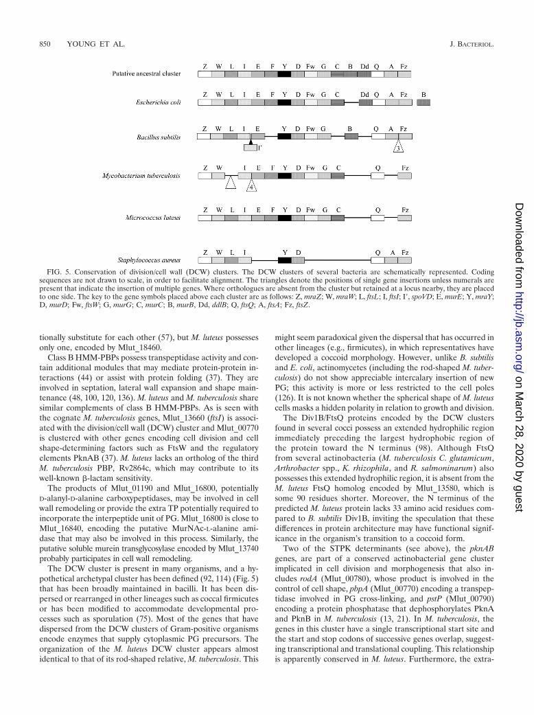
The DCW cluster is present in many organisms, and a hy-pothetical archetypal cluster has been defined (92, 114) (Fig. 5)that has been broadly maintained in bacilli. It has been dis-persed or rearranged in other lineages such as coccal firmicutesor has been modified to accommodate developmental pro-cesses such as sporulation (75). Most of the genes that havedispersed from the DCW clusters of Gram-positive organismsencode enzymes that supply cytoplasmic PG precursors. Theorganization of the M. luteus DCW cluster appears almostidentical to that of its rod-shaped relative, M. tuberculosis. This
might seem paradoxical given the dispersal that has occurred inother lineages (e.g., firmicutes), in which representatives havedeveloped a coccoid morphology. However, unlike B. subtilisand E. coli, actinomycetes (including the rod-shaped M. tuber-culosis) do not show appreciable intercalary insertion of newPG; this activity is more or less restricted to the cell poles(126). It is not known whether the spherical shape of M. luteuscells masks a hidden polarity in relation to growth and division.
The Div1B/FtsQ proteins encoded by the DCW clustersfound in several cocci possess an extended hydrophilic regionimmediately preceding the largest hydrophobic region ofthe protein toward the N terminus (98). Although FtsQfrom several actinobacteria (M. tuberculosis C. glutamicum,Arthrobacter spp., K. rhizophila, and R. salmoninarum) alsopossesses this extended hydrophilic region, it is absent from theM. luteus FtsQ homolog encoded by Mlut_13580, which issome 90 residues shorter. Moreover, the N terminus of thepredicted M. luteus protein lacks 33 amino acid residues com-pared to B. subtilis Div1B, inviting the speculation that thesedifferences in protein architecture may have functional signif-icance in the organism’s transition to a coccoid form.
Two of the STPK determinants (see above), the pknABgenes, are part of a conserved actinobacterial gene clusterimplicated in cell division and morphogenesis that also in-cludes rodA (Mlut_00780), whose product is involved in thecontrol of cell shape, pbpA (Mlut_00770) encoding a transpep-tidase involved in PG cross-linking, and pstP (Mlut_00790)encoding a protein phosphatase that dephosphorylates PknAand PknB in M. tuberculosis (13, 21). In M. tuberculosis, thegenes in this cluster have a single transcriptional start site andthe start and stop codons of successive genes overlap, suggest-ing transcriptional and translational coupling. This relationshipis apparently conserved in M. luteus. Furthermore, the extra-
FIG. 5. Conservation of division/cell wall (DCW) clusters. The DCW clusters of several bacteria are schematically represented. Codingsequences are not drawn to scale, in order to facilitate alignment. The triangles denote the positions of single gene insertions unless numerals arepresent that indicate the insertion of multiple genes. Where orthologues are absent from the cluster but retained at a locus nearby, they are placedto one side. The key to the gene symbols placed above each cluster are as follows: Z, mraZ; W, mraW; L, ftsL; I, ftsI; I�, spoVD; E, murE; Y, mraY;D, murD; Fw, ftsW; G, murG; C, murC; B, murB, Dd, ddlB; Q, ftsQ; A, ftsA; Fz, ftsZ.
850 YOUNG ET AL. J. BACTERIOL.
on March 28, 2020 by guest
http://jb.asm.org/
Dow
nloaded from

cellular domain of PknB has been described as a penicillin-binding and Ser/Thr kinase-associated (PASTA) domain thatis also found in the bifunctional HMM-PBPs involved in PGsynthesis. This domain may bind both penicillins and PG-re-lated analogues (141), as well as muropeptides, effectively cou-pling cell envelope synthesis to other core processes includingtranscription and translation (111). One of the phosphoryla-tion targets of M. tuberculosis PknA is the product of wag31(50). This essential gene (104), also found in M. luteus(Mlut_13520) with a conserved genetic context and neighbor-ing the DCW cluster, encodes a homolog of DivIVA thatcontrols placement of the division septum in B. subtilis (18) butappears to differ functionally in actinomycetes. Recent studiesusing C. glutamicum and other actinobacteria suggest its role inpolar peptidoglycan synthesis is more significant than its in-volvement in septation (65, 105).
Anionic wall polysaccharides. The cytoplasmic membrane ofM. luteus bears a �-D-mannosyl-(133)-�-D-mannosyl-(133)-diacylglycerol (Man2-DAG) glycolipid and a succinylated lipo-mannan (sucLM) based on it (64, 94, 97). The lipomannancomponents of Corynebacterium glutamicum and M. luteushave structural similarities (76, 77, 124), and the genes encod-ing sucLM biosynthesis in M. luteus were identified by com-parison with the cognate genes from C. glutamicum. The prod-uct of Mlut_04450 is a strong candidate for one of themannosyltransferases that forms Man2-DAG. Genes encodinghomologs of MptA (Mlut_09700) and MptB (Mlut_09690)form a cluster with another gene (Mlut_09710) that encodes aGT-C family glycosyltransferase, suggesting that Mlut_09710 isalso involved in sucLM biosynthesis. These three genes arecotranscribed and probably translationally coupled, since eachoverlaps its predecessor by four nucleotides, suggesting coor-dinate regulation through a polycistronic mRNA, whereastheir homologs in corynebacteria and mycobacteria are widelydispersed. Assuming Mlut_09690 and Mlut_09700 are MptBAorthologues, they would, in concert, provide an �-(136) linkedmannosyl backbone, a common feature in other lipoglycans.Mlut_09710 might then provide either the 2- or 3-linked man-nose residues reported in early characterizations (108). How-ever, it is also possible that each of these three GT-C glyco-syltransferases produces a distinct linkage and that this operonprovides all of the biosynthetic capability to produce the bulkof the sucLM mannan. A homolog of mycobacterial and coryne-bacterial polyprenyl monophosphomannose synthases, neces-sary to provide mannosyl donors to MptAB, is encoded byMlut_12000. Interestingly, a gene encoding a C-N hydrolasecommonly found immediately downstream or, in the case of M.tuberculosis, fused in a continuous reading frame, is absentfrom M. luteus (36, 41).
The M. luteus cell wall is decorated with a teichuronic acid(TUA) consisting of repeating disaccharide units of N-acetyl-mannosaminuronic acid (ManNAcU) and glucose (Glc) (42,47). The polymer is attached to PG via the phosphate group ofa reducing terminal trisaccharide consisting of two ManNAcUresidues and an N-acetylglucosamine (GlcNAc) phosphate res-idue (33). The TUA operon of B. subtilis provides few usefulsearch queries, and the genes concerned with TUA biosynthe-sis in most other organisms have not been well characterized.M. luteus contains three homologs of UDP-ManNAcU dehy-drogenase (Mlut_05630, Mlut_08960, and Mlut_08980), which
might produce the ManNAcU nucleotide donor. Mlut_05630 isclustered with a single putative glycosyltransferase (Mlut_05650) andtwo other genes of unknown function, while Mlut_08960 andMlut_08980 lie within a large cluster encoding several putativeGTs, as well as other functions necessary for TUA biosynthesisand export. The individual glycosyl residues of the repeatingpolymer unit are both derived from UDP-glycosyl donors (43).A likely biosynthetic route to UDP-ManNAcU, predominantlybased in this cluster, is apparent. UDP-GlcNAc may be formedby the Mlut_05450 product, a homolog of E. coli GlmU (abifunctional enzyme with glucosamine-1-phosphate N-acetyl-transferase and GlcNAc-1-phosphate uridyltransferase activi-ties). UDP-GlcNAc could then be transformed to UDP-ManNAcvia a putative UDP-GlcNAc 2-epimerase encoded by Mlut_09080.The oxidation of this precursor to UDP-ManNAcU could beachieved by either of the UDP-N-acetyl-D-mannosaminur-onate dehydrogenase homologs encoded by Mlut_08960 orMlut_08980. Mlut_08720 appears to encode a UDP-Glc 6-de-hydrogenase; together with the apparent aminosugar prefer-ence of both Mlut_08960 and Mlut_08980, this suggests thatthere may be greater heterogeneity in TUA biosynthesis thanis currently recognized. Since both Mlut_08960 and Mlut_08980appear to be twinned with GT genes as immediate upstreamneighbors, these might reflect potentially interchangeable func-tional units for the introduction of aminosugar-derived glycuronicacid residues into a TUA polymer.
The complement of GTs necessary for TUA biosynthesis canbe estimated from biochemical data. A polyprenyl pyrophos-phate-GlcNAc-(ManNAc)2 acceptor is synthesized initially andthen extended by separable Glc and ManNAcU transferasesthat intervene alternately in polymer elongation (43, 122). Ac-ceptor synthesis will require TagO (Mlut_08100), a polyprenylphosphate-dependent GlcNAc phosphate transferase that isimplicated universally in the attachment of polymers to PGtogether with (probably) two ManNAcU transferases. The onlypartially characterized GT involved in M. luteus TUA biosyn-thesis is the glucosyltransferase responsible for the depositionof the �-D-Glc residues within the polymer; two apparent sub-unit types of mass 54 kDa and 52.5 kDa with an estimated pIof �5 for the octameric active enzyme were described previ-ously (25). These parameters show a good match with thepredicted GT product of Mlut_09020 (molecular mass 56.8kDa, pI 5.94). Finally, the secretion of the TUA may beaccomplished by the products of Mlut_09060 and Mlut_09070,which together constitute a predicted ABC family polysaccha-ride export system.
The Mlut_08990, Mlut_09000, and Mlut_09010 genes thatcomplete this cluster encode products related to aminosugar-N-acetyltransferases, a pyridoxal phosphate-dependent amino-transferase and a dehydrogenase, respectively. These activitiesare consistent with the provision of a further N-acetylated aminosugar for glycuronic acid formation. Interestingly, Mlut_09000 isa pseudogene; analysis of the in silico-translated sequence re-veals homology with several predicted UDP-4-amino-4-deoxy-L-arabinose-oxoglutarate aminotransferases over the full se-quence length with the incorporation of a stop codon througha nucleotide substitution in codon 121. Thus, the structure ofthis gene cluster is entirely consistent with the biosynthesis ofthe TUA described for M. luteus and taken together with theapparent redundancy in N-acetylglycosaminuronate provision
VOL. 192, 2010 GENOME SEQUENCE OF M. LUTEUS FLEMING STRAIN 851
on March 28, 2020 by guest
http://jb.asm.org/
Dow
nloaded from

and transfer, the apparent loss of Mlut_09000 function sug-gests a previously more diverse TUA profile.
Energy metabolism. The M. luteus genome contains genesencoding the various respiratory chain components, includingcomplex I (NADH-quinone oxidoreductase: Mlut_18970 toMlut_18940, Mlut_11050, Mlut_11060, and Mlut_05010),complex II (succinate dehydrogenase with cytochrome b anda Fe-S cluster: Mlut_04850 to Mlut_04820), and complexesIII and IV (quinol-cytochrome c oxidoreductase with cyto-chrome b and a Fe-S center and the cytochrome c-cyto-chrome aa3 oxidase complex: Mlut_12150 to Mlut_12120,Mlut_12210, and Mlut_12220). The M. luteus respiratorychain also contains the quinol-oxidase complex with cyto-chrome bd (Mlut_13220 and Mlut_13210), which is usuallyresponsible for the growth of bacteria under low-oxygen con-ditions (128). Genes encoding a membrane-bound malate-mena-quinone oxidoreductase (Mlut_08440) and transmembrane L-lac-tate–menaquinone oxidoreductase (Mlut_21510) are present,and the corresponding activities have been demonstrated ex-perimentally in isolated membrane particles (5). The M. luteusrespiratory chain may also contain a membrane-bound pyru-vate dehydrogenase (cytochrome; Mlut_02710), glycerol-3-phosphate dehydrogenase (Mlut_23030), and L-proline dehy-drogenase (Mlut_19430) as in C. glutamicum (14).
Central carbon metabolism. Genes encoding a complete setof enzymes of the citric acid cycle are present. The genes forthe � and � subunits of succinyl coenzyme A (CoA) synthetaselie adjacent to each other (Mlut_04280 and Mlut_04270) andgenes for the succinate dehydrogenase subunits A to D form acluster (Mlut_04820 to Mlut_04850). The other genes encod-ing citrate synthase (Mlut_15490), aconitase (Mlut_13040),isocitrate dehydrogenase (Mlut_04530), the E1 and E2 com-ponents of 2-oxoglutarate dehydrogenase (Mlut_07730 andMlut_13330), fumarase (Mlut_05170), and malate dehydroge-nase (Mlut_00950) are located separately.
M. luteus can use acetate as a sole source of carbon forenergy and growth (V. Artsatbanov, unpublished results), so itmust contain the isocitrate lyase and malate synthase A com-ponents of the glyoxylate shunt (and, despite its small genome,must have all of the necessary pathways to synthesize aminoacids, nucleotides, carbohydrates, and lipids from acetate). Theisocitrate lyase and malate synthase A components are func-tionally active (E. Salina, unpublished results), and they areencoded by adjacent genes (Mlut_02080 and Mlut_02090). In-terestingly, the isocitrate lyase is considered a “persistencefactor” in M. tuberculosis, where it “allows net carbon gain bydiverting acetyl-CoA from �-oxidation of fatty acids into theglyoxylate shunt pathway” (74, 112).
All of the main enzymes of glycolysis are present other thanthe first enzyme, glucokinase, responsible for the phosphory-lation of glucose. This is consistent with the observed inabilityof M. luteus to grow with glucose as sole carbon source (140),but it may not serve to explain it, since the product of Mlut_13470 ispredicted to be a polyphosphate glucokinase. Other closely relatedspecies, e.g., R. salmoninarum, Arthrobacter spp., and Rhodococcussp., do have glucokinase genes. M. luteus is able to synthesizeall glycolytic intermediates (presumably by gluconeogenesis)from phosphoenolpyruvate or pyruvate obtained from oxalo-acetate through the agency of phosphoenolpyruvate carboxyki-nase (Mlut_03380) or pyruvate carboxylase (Mlut_13810). It
has a complete complement of enzymes concerned with triosemetabolism, and genes for all of the pentose phosphate path-way enzymes are annotated except for 6-phosphogluconolac-tonase. Although this open cycle could produce all intermedi-ates, pentoses would presumably be synthesized via thenonoxidative pentose phosphate pathway that is not coupled tothe reduction of NADP�.
Does glycogen serve mainly as a biosynthetic intermediatefor trehalose in M. luteus? One remarkable feature of someactinobacteria is the importance of the glucose-related storagemetabolites, trehalose and glycogen, and the complexity ofassociated metabolic processes: in some organisms, this area ofmetabolism appears to be essential for viability (9, 17). Thereare at least three ways in which mycobacteria and streptomy-cetes can make trehalose (17) and some interconnections be-tween trehalose and glycogen metabolism (110).
M. luteus has strikingly fewer genes concerned with carbo-hydrate metabolism than its close relatives (Table 2). All of thegenes for conventional glycogen biosynthesis from central me-tabolism are present: pgm (Mlut_01060), glgA (Mlut_11690),glgB (Mlut_03850), glgC (Mlut_11680), together with a de-branching enzyme gene, glgX (Mlut_16760), and the incom-pletely characterized, glycogen-related gene glgE (Mlut_03840). Re-markably, however, there is no obvious candidate gene forglycogen phosphorylase, a highly conserved enzyme that isalmost universal and which provides the major route for gly-cogen breakdown. This raises the question of whether M. luteuscan metabolize glycogen.
Equally surprising is the absence of the otsAB genes for theconventional synthesis of trehalose. However, in the last 10years, another pathway (present in many actinobacteria) hasbeen discovered, in which the terminal reducing glucosyl res-idue on chains of �-1,4-linked glucose polymers is “flipped” bythe TreY trehalose malto-oligosyl trehalose synthase enzymeso that it is now �-1,1-linked, and then this terminal trehalosyldisaccharide is cleaved off by the TreZ malto-oligosyltrehalosetrehalohydrolase enzyme. This pathway is present in M. luteus(Mlut_03980, treY; Mlut_03990, treZ) and could therefore pro-vide a route for glycogen breakdown to give trehalose. In otherMicrococcaceae, greater numbers of trehalose biosyntheticgenes have been annotated (four in Arthrobacter sp. strainFB24, six in K. rhizophila, and eight in R. salmoninarum and A.aurescens TC1), and in more distantly related organisms suchas C. glutamicum, multiple trehalose biosynthetic pathwaysmay be present (139). The degradation of trehalose to yieldglucose is catalyzed by trehalase. Although it is generally dif-ficult to recognize trehalases by homology, Mlut_17860 en-codes a protein with 35% identity to a trehalase recently char-acterized in M. smegmatis (17). It seems possible that thebreakdown of glycogen in M. luteus may well take place viatrehalose.
Another pathway for trehalose biosynthesis, found in manyactinobacteria, uses the trehalose synthase enzyme to intercon-vert maltose and trehalose (see reference (110 and referencestherein). There is no close homolog of treS in M. luteus. Thus,the only obvious route for trehalose synthesis appears to be viaglycogen. In this respect, it is relevant to note that the treYZgenes of M. luteus are separated from glgE and glgB only by agroup of genes peculiar to M. luteus that were therefore prob-ably acquired by lateral gene transfer. The glg and tre genes
852 YOUNG ET AL. J. BACTERIOL.
on March 28, 2020 by guest
http://jb.asm.org/
Dow
nloaded from

may previously have been adjacent, which is consistent with theidea of a combined glycogen-trehalose biosynthetic pathway.We are not aware of a comparable system in any otherorganism.
M. luteus has relatively little capacity for secondary metab-olism. The genome of M. luteus includes only 39 genes (2.2%)annotated as being concerned with secondary metabolism (Ta-ble 2). Among these are 11 clustered genes (Mlut_21170–21270) implicated in carotenoid synthesis (Table 5). Yellowpigmentation has long been important for the identification ofM. luteus, suggesting that the genes concerned with pigmentproduction would show a restricted distribution. The phytoenesynthetase (Mlut_21230), phytoene desaturase (Mlut_21220),and polyprenyl transferase (Mlut_21210) genes lie in the cen-ter of the cluster and have homologs in many different organ-isms, including the photosynthetic bacteria (Synechococcus),where the carotenoids might function to protect against radi-ation damage, as has been proposed in M. luteus (4). Other
genes toward the extremities of the cluster show a more re-stricted distribution (Table 6). Although homologs of most ofthese genes are present in other high G�C Gram-positivebacteria, a high level of synteny is restricted to only a feworganisms, e.g., L. xyli, Corynebacterium efficiens, C. glutami-cum, and C. michiganensis.
There is little evidence from the genome annotation for thepresence of other secondary metabolic functions. There is acluster of genes involved in siderophore transport (Mlut_22080to Mlut_22120), as well as single genes that, according to theannotation, might be involved in nonribosomal peptide synthe-sis (COG1020) and benzoate catabolism (protocatechuate 3,4-dioxygenase [COG3485]). The genome is among the minorityof actinobacterial genomes (including C. diphtheriae and Tro-pheryma whipplei) that encode no obvious cytochromes P450;for example, free-living mycobacteria and streptomycetes gen-erally encode more than 10 (130). There is no evidence for asignificant repertoire of genes that might be involved in
TABLE 5. Genes involved in carotenoid production
Mlut Aminoacid Annotation Commenta EC or COG no.
21270 143 Thioredoxin Ubiquitous oxidoreductase 1.8.7.1.21260 209 Isopentenyl diphosphate delta isomerase 3-Isopentenyl pyrophosphate 3 dimethylallyl
pyrophosphate2.5.1.1 (?)
21250 182 Hypothetical No matches21240 Geranylgeranyl pyrophosphate synthase trans, trans-Farnesyl diphosphate � isopentenyl
diphosphate 3 diphosphate �geranylgeranyl diphosphate
2.5.1.29
21230 298 Squalene/phytoene synthetase Probably phytoene synthetase 2 geranylgeranyldiphosphate 3 diphosphate � prephytoenediphosphate
2.5.1.32
21220 566 Phytoene desaturase Carotene desaturation, a step in carotenoidbiosynthesis
COG1233
21210 294 4-Hydroxybenzoate polyprenyltransferaseand related prenyltransferases
crtEB lycopene elongation 2.5.1.39
21200 129 Putative C50 carotenoid epsilon cyclase crtYe ring closure21190 117 Hypothetical Matches short segments of lycopene e-cyclase
isoprenoid and putative C50 carotenoidepsilon cyclase (crtYf)
a Notations for the genes crtEB, crtYe, and crtYf are as cited for Corynebacterium glutamicum (63).
TABLE 6. Distribution of genes involved in carotenoid synthesis
Organism
Distribution of genea:
Mlut_21190(partial carotenoid
cyclase)
Mlut_21200(C50 carotenoidepsilon cyclase)
Mlut_21210(polyprenyltransferase)
Mlut_21220(phytoene
desaturase)
Mlut_21230(phytoenesynthetase)
Mlut_21240(geranylgeranyl
synthase)
Mlut_21250(hypothetical)
Mlut_21260(isopentenylisomerase)
Actinobacteria � � � � � � �Alphaproteobacteria � � �Betaproteobacteria �Gammaproteobacteria �Deltaproteobacteria � � � �Firmicutes � �Archaea � � � � �Green nonsulfur � �Green sulfur � �Cyanobacteria � �Planctomycetes � � �Verrucomicrobiae �Basidiomycetes �Ascomycetes �
a The gene type or description is given in parentheses.
VOL. 192, 2010 GENOME SEQUENCE OF M. LUTEUS FLEMING STRAIN 853
on March 28, 2020 by guest
http://jb.asm.org/
Dow
nloaded from

polyketide production (only Mlut_20260 and Mlut_22550) orxenobiotic catabolism. Finally, M. luteus uses the nonmeval-onate pathway for C5 isoprenoid biosynthesis (Table 7). Thegenes are dispersed about the bacterial chromosome withMlut_03780 encoding a bifunctional enzyme comprising IspD(2-C-methyl-D-erythritol 4-phosphate cytidylyltransferase [EC2.7.7.60]) and IspF (2-C-methyl-D-erythritol 2,4-cyclodiphos-phate synthase [EC 4.6.1.12]), as is also the case in severalother actinobacteria, e.g., Kytococcus sedentarius, C. michi-ganensis, L. xyli, and T. whipplei (58, 48, 46, and 42% identities,respectively).
Osmotolerance. M. luteus is salt tolerant and grows in richmedium, such as nutrient broth, containing 10% NaCl, al-though pigment production is abolished at concentrationsgreater than 5% NaCl (G. Price and M. Young, unpublisheddata). Heterotrophic bacteria that are salt requiring or salttolerant generally use organic solutes such as amino acids(glutamate, proline), glycine betaine, ectoine, and trehalose forosmotic balance.
When grown in rich medium, most heterotrophic bacteriaaccumulate glycine betaine (46). However, very few hetero-trophs are capable of de novo synthesis of betaine; they tend toaccumulate the compound from the medium (yeast extract is agood source of betaine). Accordingly, M. luteus encodes anABC transporter system for proline/glycine betaine with fourcomponents clustered in the genome (Mlut_15720 to Mlut_15750).In addition, two choline/carnitine/betaine transporters have alsobeen annotated (Mlut_01900 and Mlut_16530). All of these geneshave homologs throughout the actinobacteria.
Although an ectoine synthetase gene is annotated (Mlut_02920),the remainder of the operon required for production of thismolecule is apparently absent. The M. luteus gene has greatestsimilarity to genes found in Stigmatella aurantiaca and Burk-holderia ambifaria (55 and 54% identities, respectively), inwhich the remaining genes required for ectoine synthesis arealso apparently absent. Complete ectoine operons are found inthe genomes of many actinobacteria including Brevibacterium lin-ens BL2 and Streptomyces avermitilis.
As noted above, M. luteus has two genes potentially con-cerned with trehalose production from glycogen: a trehalosemalto-oligosyl trehalose synthase (Mlut_03980) and a malto-oligosyltrehalose trehalohydrolase (Mlut_03990). This suggeststhat of the common compatible solutes, M. luteus can onlyproduce glutamate, proline, and trehalose (the last of these
only via glycogen) although, like many heterotrophic bacteria,it does have transporters for other compatible osmopro-tectants.
Long-chain alkene biosynthesis by M. luteus. Four decadesago, two research groups studying a close relative of M. luteus,Sarcina lutea ATCC 533 (now Kocuria rhizophila), reported thebiosynthesis of iso- and anteiso-branched, long-chain (primarilyC25 to C29) alkenes (1, 2, 127). Although the biosyntheticpathway was postulated to involve decarboxylation and con-densation of fatty acids, the underlying biochemistry and ge-netics of alkene biosynthesis have remained unknown untilvery recently. M. luteus strains have also been shown to pro-duce primarily branched C29 monoalkenes; this includes thesequenced strain (based upon analysis by gas chromatography-chemical ionization-time of flight mass spectrometry [9a]) andstrain ATCC 27141 (31).
Recently, Friedman and Rude (international patent applica-tion WO 2008/113041) reported that heterologous expression ofoleACD from a range of bacteria (including Stenotrophomonasmaltophilia, Xanthomonas axonopodis, and Chloroflexus ag-gregans) resulted in long-chain alkene biosynthesis, and in vitrostudies indicated that the alkenes indeed result from fattyacyl-CoA or fatty acyl-ACP precursors. Beller and coworkershave heterologously expressed the three-gene M. luteus clustercomprising oleA (Mlut_13230), oleBC (a gene fusion;Mlut_13240), and oleD (Mlut_13250) in E. coli and observedC27 and C29 alkenes that were not detectable in negative con-trols with a plasmid lacking any M. luteus genes (9a). In vitrostudies with M. luteus OleA indicated that it catalyzes decar-boxylation and condensation of activated fatty acids (9a).
Some close relatives of M. luteus that also produce long-chain alkenes, such as K. rhizophila and A. aurescens TC1, havesimilar oleABCD gene organization (Fig. 6) and a relativelyhigh degree of sequence identity. To illustrate, the OleA,OleBC, and OleD sequences of K. rhizophila and A. aurescensTC1 share 45 to 57% identity with those of M. luteus. Incontrast, Arthrobacter sp. strain FB24, which does not producealkenes (31), appears to lack an oleB gene and has OleA andOleD sequences that share only 25 to 27% sequence identitywith those of M. luteus. It is possible that the divergence of olesequences in strain FB24 from those in the other strains dis-cussed here explains its inability to produce long-chain alkenes.In the Gram-negative, alkene-producing bacterium S. malto-philia, oleB and oleC are separate genes, in contrast to the
TABLE 7. Comparison of the nonmevalonate pathway for isoprenoid biosynthesis in M. luteus and My. tuberculosis
Enzyme (EC no.) M. tuberculosis gene M. luteus homologa % Identity
1-Deoxy-D-xylulose-5-phosphate synthase (2.2.1.7) Rv2682c, dxs1 Mlut_13030 57.51-Deoxy-D-xylulose 5-phosphate reductoisomerase (1.1.1.267) Rv2870c, dxr Mlut_06920 56.32-C-methyl-D-erythritol 4-phosphate cytidylyltransferase
(2.7.7.60)Rv3582c, ispD Mlut_03780* 42.6
4-Diphosphocytidyl-2-C-methyl-D-erythritol kinase (2.7.1.148) Rv1011, ispE Mlut_05400 44.72-C-methyl-D-erythritol 2,4-cyclodiphosphate synthase
(4.6.1.12)Rv3581c, ispF Mlut_03780* 60.5
4-Hydroxy-3-methylbut-2-en-1-yl diphosphate synthase(1.17.4.3)
Rv2868c, ispG (gcpE) Mlut_06940 77.23
4-Hydroxy-3-methylbut-2-enyl diphosphate reductase(1.17.1.2)
Rv1110, ispH, lytB Mlut_16300 62.7
Isopentenyl-diphosphate delta-isomerase (5.3.3.2) Rv1745c, idi Mlut_21260 46.4
a �, Mlut_03780 encodes a bifunctional enzyme, as also occurs in some other actinobacteria (see the text).
854 YOUNG ET AL. J. BACTERIOL.
on March 28, 2020 by guest
http://jb.asm.org/
Dow
nloaded from

Gram-positive species represented in Fig. 6, but there is stillrelatively strong similarity between the S. maltophilia and M.luteus sequences (e.g., OleA and OleD both share 39% se-quence identity between these two organisms).
Although BLASTp searches with M. luteus OleA (Mlut_13230)showed best hits to �-ketoacyl-ACP-synthase III (FabH), a keyenzyme involved in fatty acid biosynthesis, the true fabH inthe M. luteus genome, Mlut_09310, falls in a cluster of genescritical to the biosynthesis of branched-chain fatty acids,including a putative branched-chain �-keto acid decarbox-ylase (Mlut_09340), malonyl-CoA:ACP transacylase (fabD;Mlut_09320), acyl carrier protein (ACP; Mlut_09300), and�-ketoacyl-ACP-synthase II (fabF; Mlut_09290).
Dormancy. M. luteus can enter a profoundly dormant statefrom which it can be resuscitated by a secreted protein calledresuscitation-promoting factor (Rpf) (85). Among its closestrelatives, Arthrobacter sp. strain FB24, R. salmoninarum, and K.rhizophila, all encode a secreted protein with an N-terminaltransglycosylase-like domain and a C-terminal LysM domainthat closely resembles M. luteus Rpf (123, 137). A secondprotein belonging to the highly conserved RpfB family (99) isencoded by genes found in Arthrobacter sp. strain FB24 and R.salmoninarum. RpfB is also present in Arthrobacter aurescensTC1, which does not encode a protein with a domain structuresimilar to that of M. luteus Rpf.
Dormancy has been extensively studied in M. tuberculosis,which has five rpf genes involved in controlling culturabilityand resuscitation (12, 27, 86, 102, 113, 129), and the remainder
of this section will focus on a comparison with this organism. InM. tuberculosis, a state of growth arrest often referred to asdormancy is occasioned by hypoxia in vitro (135). This leads tothe expression of a cohort of ca. 50 genes (the Dos regulon)under the control of the devRS (dosRS) two-component system(101). Microarray studies have been accomplished using fivedifferent dormancy models and many genes belonging to theDos regulon are upregulated in these datasets (see Table S2 inthe supplemental material). Throughout these datasets, be-tween 45 and 50% of the genes that are significantly upregu-lated have homologs in M. luteus (Table 8). Several of the M.luteus genes in these lists have multiple paralogs in M. tuber-culosis, the most striking example being Mlut_01830. This geneencodes the universal stress protein UspA. It is representedonce in the M. luteus genome, whereas there are six homologsin M. tuberculosis (Rv1996, Rv2005c, Rv2028c, Rv2623,Rv2624c, and Rv3134c). Out of a cohort of 17 genes that areupregulated in all five M. tuberculosis dormancy models inTable S2 in the supplemental material, nine have homologs inM. luteus (Table 9). Notable among these are genes encodingthe universal stress protein UspA (Mlut_01830), ferredoxin(Mlut_15510), an erythromycin esterase homolog (Mlut_05460), anHsp20 family heat shock protein (Mlut_16210) and a zincmetalloprotease (Mlut_11840) that lies within a cluster includ-ing putative proteasome components. This cluster shows a highdegree of synteny with Arthrobacter sp. strain FB24.
Recent studies of the enduring hypoxic response of M. tu-berculosis (103) revealed that more than 200 genes are upregu-
FIG. 6. Organization of the ole (olefin synthesis) genes in M. luteus and other bacteria.
VOL. 192, 2010 GENOME SEQUENCE OF M. LUTEUS FLEMING STRAIN 855
on March 28, 2020 by guest
http://jb.asm.org/
Dow
nloaded from

lated, only five of which belong to the Dos regulon, and onlytwo of these five were upregulated by more than threefold(103). However, comparison of the 47 genes upregulated morethan threefold in the enduring hypoxic response with upregu-lated genes from the other five dormancy models (Table 8)showed that 22 of them (47%) were upregulated by more thanthreefold in at least one of those models. 62% of these 47genes have homologs in the M. luteus genome (Table S3 in thesupplemental material).
M. luteus therefore contains many genes similar to genes
upregulated in different M. tuberculosis dormancy models.Among them are members of the Dos regulon, including thedevRS (dosRS) genes encoding the sensory histidine kinaseand response regulator that control the regulon (possiblyMlut_18530 and Mlut_18540, although other gene pairs, suchas Mlut_16250 and Mlut_16240 or Mlut_21850 and Mlut_21860,might fulfill this role). The total number of M. luteus dormancy-related genes revealed by these various comparisons is roughlyproportional to the twofold difference in genome size betweenM. luteus and M. tuberculosis. The elevated number of dorman-
TABLE 8. Number of genes upregulated (�3-fold induction) in different models of M. tuberculosis dormancy and their M. luteus homologsa
Parameter Allgenes
Genes seen in: Nonreplicating persistenceChemostat
model
Seen in bothNRP1 and
NRP2
Macrophagesactivated for
48 h�2
modelsAll 5
modelsNRP1, 8
daysNRP2, 20
days
Reference 132 132 7 92 109Total no. 247 72 17 55 94 52 90 111No. of homologs in
M. luteus137 36 9 24 53 30 44 61
% with M. luteushomolog
55.5 50.0 53 43.6 56.4 57.7 48.9 55.0
a Minimum 20% identity; maximum e value 1e�2. A low stringency was used to identify as many genes as possible in M. luteus that might be homologs of thedormancy-related genes of M. tuberculosis. Even so, comparatively few candidates emerged from the analysis. Despite a significant reduction in the number of“dormancy-related” genes, M. luteus can readily adopt a dormant state.
TABLE 9. Many of the genes upregulated in all 5 My. tuberculosis dormancy models have M. luteus homologs
M. tuberculosislocus taga Product name and/or assignment M. luteus
homolog Product name and/or assignment E-value % Identity/BIT score
Rv0079 Hypothetical proteinRv0080 Conserved hypothetical protein Mlut_04380 Predicted flavin-nucleotide-
binding protein3e–08 30.4/50
Rv1733c Possible membrane proteinRv1738 Conserved hypothetical proteinRv1996 COG0589: universal stress protein
UspA and related nucleotide-binding proteins
Mlut_01830 Universal stress protein UspAand related nucleotide-binding proteins
9e–29 31.6/120
Rv2007c Ferredoxin Mlut_15510 Ferredoxin 5e–36 58.0/142Rv2030c COG2312: erythromycin esterase
homolog COG1926: predictedphosphoribosyltransferases
Mlut_18600b Amidophosphoribosyltransferase(EC 2.4.2.14)
1e–03 27.9/38
Rv2031c 14-kDa antigen, heat shock proteinHsp20 family
Mlut_16210 Heat shock protein Hsp20(IMGterm)
6e–09 36.9/52
Rv2032 Conserved hypothetical protein AcgRv2623 COG0589: universal stress protein
UspA and related nucleotide-binding proteins
Mlut_01830c Universal stress protein UspAand related nucleotide-binding proteins
4e–34 36.8/137
Rv2624c COG0589: universal stress proteinUspA and related nucleotide-binding proteins
Mlut_01830 Universal stress protein UspAand related nucleotide-binding proteins
7e–22 32.4/97
Rv2625c COG1994: Zn-dependent proteases(probable conservedtransmembrane alanine andleucine-rich protein)
Mlut_11840 Zn-dependent proteases 7e–31 30.1/127
Rv2626c Conserved hypothetical proteinRv2627c Conserved hypothetical proteinRv3127 Conserved hypothetical proteinRv3130c IGR02946 acyltransferase,
WS/DGAT/MGATRv3134c COG0589: universal stress protein
UspA and related nucleotide-binding proteins
Mlut_01830 Universal stress protein UspAand related nucleotide-binding proteins
2e–16 36.4/78
a All genes except Rv1733c belong to the DOS regulon.b Three more homologs.c One more homolog.
856 YOUNG ET AL. J. BACTERIOL.
on March 28, 2020 by guest
http://jb.asm.org/
Dow
nloaded from

cy-related genes in M. tuberculosis is accounted for, in part atleast, by the presence of multiple paralogs that do not exist inM. luteus, indicating that the dormancy machinery of M. luteusis highly minimized compared to that of M. tuberculosis, al-though it clearly remains fully functional (24, 27, 49, 54, 56,113, 131, 132, 134).
Conclusion. The M. luteus genome is very small compared tothose of other free-living actinobacteria, raising speculationthat this may be connected with both its simple morphologyand a restricted ecology. Soil-dwelling organisms typically havea substantial capability for environmental responses mediatedthrough two-component systems and sigma factors, but the M.luteus genome encodes only 14 response regulators and foursigma factors, a finding indicative of adaptation to a ratherstrict ecological niche. We therefore speculate that its primaryadaptation is to (mammalian?) skin, where it is often found,and that its occasional presence elsewhere (water and soil)might possibly arise from contamination by skin flakes. Thesomewhat minimized nature of the genome may also provideopportunities to evaluate the roles of conserved genes that, inother actinobacteria, are members of substantial paralogousfamilies.
Despite its small size, the M. luteus genome contains anexceptionally high number of transposable elements. These donot seem to have resulted in large-scale genome rearrange-ments, since the genome sequences for the phylogeneticallyclosest actinobacteria available show only one or two multi-gene inversions spanning the oriC region, and none that do notinclude oriC, compared to M. luteus. The possibility remains,however, that some of the transposable elements could haveplayed a part in the contraction of the M. luteus genome froma larger ancestral version. Such elements are present at anumber of points of discontinuity between the genomes of M.luteus and its closest characterized relatives (not shown).
The simple morphology of M. luteus is reflected in the ab-sence of nearly all genes known to be concerned with devel-opmental decisions in more complex actinobacteria, and theconfinement of genes for cell division and cell wall biosynthesisto a minimal set. The only obvious exception to this is thepresence of orthologues of the Streptomyces sporulation geneswhiA and whiB, but this is not surprising since whiA ortho-logues are found in virtually all Gram-positive bacteria, bothfirmicutes and actinobacteria, and whiB orthologues are nearlyuniversal among actinobacteria. The roles of these two genesin such a simple organism as M. luteus merit exploration. Thepresence of only a single class A PBP, and only two of class B,may be connected with the adoption of spherical morphology,and with high sensitivity to �-lactams and lysozyme. The well-known sensitivity of M. luteus to diverse other antibiotics maypossibly be due to its lack of a wblC gene: this gene confersincreased resistance to a wide range of antibiotics and otherinhibitors on streptomycetes and mycobacteria, apparently byaffecting the expression of a large number of genes that includemany predicted to affect resistance (80).
ACKNOWLEDGMENTS
We are grateful to the British Council, the Israeli Ministry of Sci-ence and Technology, and the Hebrew University for funding a work-shop on the M. luteus genome held on 13 to 18 April 2008 in Jerusalem.For H.R.B. and E.B.G., this study was part of the DOE Joint Bio-
Energy Institute (http://www.jbei.org) supported by the U.S. Depart-ment of Energy, Office of Science, Office of Biological and Environ-mental Research, through contract DE-AC02-05CH11231 betweenLawrence Berkeley National Laboratory and the U.S. Department ofEnergy. M.Y. thanks the UK BBSRC for financial support, and V.A.and A.S.K. thank the MCB RAS program for financial support.
REFERENCES
1. Albro, P. W. 1971. Confirmation of the identification of the major C-29hydrocarbons of Sarcina lutea. J. Bacteriol. 108:213–218.
2. Albro, P. W., and J. C. Dittmer. 1969. The biochemistry of long-chain,nonisoprenoid hydrocarbons. I. Characterization of the hydrocarbons ofSarcina lutea and the isolation of possible intermediates of biosynthesis.Biochemistry 8:394–404.
3. Altschul, S. F., T. L. Madden, A. A. Schaffer, J. Zhang, Z. Zhang, W. Miller,and D. J. Lipman. 1997. Gapped BLAST and PSI-BLAST: a new genera-tion of protein database search programs. Nucleic Acids Res. 25:3389–3402.
4. Anwar, M., T. H. Khan, J. Prebble, and P. F. Zagalsky. 1977. Membrane-bound carotenoid in Micrococcus luteus protects naphthoquinone fromphotodynamic action. Nature 270:538–540.
5. Artsatbanov, V., G. V. Tikhonova, and D. N. Ostrovskii. 1983. Generationof membrane potential by aerobic bacteria Micrococcus lysodeikticus. Cor-relation between coupled and uncoupled respiration. Biokhimiia 48:1568–1579. (In Russian.)
6. Av-Gay, Y., and M. Everett. 2000. The eukaryotic-like Ser/Thr proteinkinases of Mycobacterium tuberculosis. Trends Microbiol. 8:238–244.
7. Bacon, J., B. W. James, L. Wernisch, A. Williams, K. A. Morley, G. J.Hatch, J. A. Mangan, J. Hinds, N. G. Stoker, P. D. Butcher, and P. D.Marsh. 2004. The influence of reduced oxygen availability on pathogenicityand gene expression in Mycobacterium tuberculosis. Tuberculosis (Edinb.)84:205–217.
8. Batzoglou, S., D. B. Jaffe, K. Stanley, J. Butler, S. Gnerre, E. Mauceli, B.Berger, J. P. Mesirov, and E. S. Lander. 2002. ARACHNE: a whole-genome shotgun assembler. Genome Res. 12:177–189.
9. Belanger, A. E., and G. F. Hatfull. 1999. Exponential-phase glycogen recy-cling is essential for growth of Mycobacterium smegmatis. J. Bacteriol. 181:6670–6678.
9a.Beller, H. R., E.-B. Goh, and J. D. Keasling. Genes involved in long-chainalkene biosynthesis in Micrococcus luteus. Appl. Environ. Microbiol., inpress.
10. Bendtsen, J. D., H. Nielsen, G. von Heijne, and S. Brunak. 2004. Improvedprediction of signal peptides: SignalP 3.0. J. Mol. Biol. 340:783–795.
11. Bentley, S. D., K. F. Chater, A.-M. Cerdeno-Tarraga, G. L. Challis, N. R.Thomson, K. D. James, D. E. Harris, M. A. Quail, H. Kieser, D. Harper, A.Bateman, S. Brown, G. Chandra, C. W. Chen, M. Collins, A. Cronin, A.Fraser, A. Goble, J. Hidalgo, T. Hornsby, S. Howarth, C.-H. Huang, T.Kieser, L. Larke, L. Murphey, K. Oliver, S. O’Neil, E. Rabbinowitsch, M.-A.Rajandream, K. Rutherford, S. Rutter, K. Seeger, D. Saunders, S. Sharp,R. Squares, S. Squares, K. Taylor, T. Warren, A. Wietzorrek, J. Woodward,B. G. Barrell, J. Parkhill, and D. A. Hopwood. 2002. Complete genomesequence of the model actinomycete Streptomyces coelicolor A3(2). Nature417:141–147.
12. Biketov, S., V. Potapov, E. Ganina, K. Downing, B. D. Kana, and A. Ka-prelyants. 2007. The role of resuscitation promoting factors in pathogenesisand reactivation of Mycobacterium tuberculosis during intra-peritoneal in-fection in mice. BMC Infect. Dis. 7:146.
13. Boitel, B., M. Ortiz-Lombardia, R. Duran, F. Pompeo, S. T. Cole, C.Cervenansky, and P. M. Alzari. 2003. PknB kinase activity is regulated byphosphorylation in two Thr residues and dephosphorylation by PstP, thecognate phospho-Ser/Thr phosphatase, in Mycobacterium tuberculosis. Mol.Microbiol. 49:1493–1508.
14. Bott, M., and A. Niebisch. 2003. The respiratory chain of Corynebacteriumglutamicum. J. Biotechnol. 104:129–153.
15. Brewster, J. M., and E. A. Morgan. 1981. Tn9 and IS1 inserts in a ribosomalribonucleic acid operon of Escherichia coli are incompletely polar. J. Bac-teriol. 148:897–903.
16. Burrus, V., J. Marrero, and M. K. Waldor. 2006. The current ICE age:biology and evolution of SXT-related integrating conjugative elements.Plasmid 55:173–183.
17. Carroll, J. D., I. Pastuszak, V. K. Edavana, Y. T. Pan, and A. D. Elbein.2007. A novel trehalase from Mycobacterium smegmatis: purification, prop-erties, requirements. FEBS J. 274:1701–1714.
18. Cha, J. H., and G. C. Stewart. 1997. The divIVA minicell locus of Bacillussubtilis. J. Bacteriol. 179:1671–1683.
19. Chandler, M., and J. Mahillon. 2002. Insertion sequences revisited, p.305–366. In N. L. Craig, R. Craigie, M. Gellert, and A. M. Lambowitz (ed.),Mobile DNA II. ASM Press, Washington, DC.
20. Chen, H. H., G. Z. Zhao, D. J. Park, Y. Q. Zhang, L. H. Xu, J. C. Lee, C. J.Kim, and W. J. Li. 2009. Micrococcus endophyticus sp. nov., isolated fromsurface-sterilized Aquilaria sinensis roots. Int. J. Syst. Evol. Microbiol. 59:1070–1075.
VOL. 192, 2010 GENOME SEQUENCE OF M. LUTEUS FLEMING STRAIN 857
on March 28, 2020 by guest
http://jb.asm.org/
Dow
nloaded from

21. Chopra, P., B. Singh, R. Singh, R. Vohra, A. Koul, L. S. Meena, H. Koduri,M. Ghildiyal, P. Deol, T. K. Das, A. K. Tyagi, and Y. Singh. 2003. Phos-phoprotein phosphatase of Mycobacterium tuberculosis dephosphorylatesserine-threonine kinases PknA and PknB. Biochem. Biophys. Res. Com-mun. 311:112–120.
22. Cole, S. T., R. Brosch, J. Parkhill, T. Garnier, C. Churcher, D. Harris, S. V.Gordon, K. Eiglmeier, S. Gas, C. E. Barry, F. Tekaia, K. Badcock, D.Basham, D. Brown, T. Chillingworth, R. Connor, R. Davies, K. Devlin, T.Feltwell, S. Gentles, N. Hamlin, S. Holroyd, T. Hornby, K. Jagels, A. Krogh,J. McLean, S. Moule, L. Murphy, K. Oliver, J. Osborne, M. A. Quail, M. A.Rajandream, J. Rogers, S. Rutter, K. Seeger, J. Skelton, R. Squares, S.Squares, J. E. Sulston, K. Taylor, S. Whitehead, and B. G. Barrell. 1998.Deciphering the biology of Mycobacterium tuberculosis from the completegenome sequence. Nature 393:537–544.
23. Cole, S. T., K. Eiglmeier, J. Parkhill, K. D. James, N. R. Thomson, P. R.Wheeler, N. Honore, T. Garnier, C. Churcher, D. Harris, K. Mungall, D.Basham, D. Brown, T. Chillingworth, R. Connor, R. M. Davies, K. Devlin,S. Duthoy, T. Feltwell, A. Fraser, N. Hamlin, S. Holroyd, T. Hornsby, K.Jagels, C. Lacroix, J. Maclean, S. Moule, L. Murphy, K. Oliver, M. A.Quail, M. A. Rajandream, K. M. Rutherford, S. Rutter, K. Seeger, S.Simon, M. Simmonds, J. Skelton, R. Squares, S. Squares, K. Stevens, K.Taylor, S. Whitehead, J. R. Woodward, and B. G. Barrell. 2001. Massivegene decay in the leprosy bacillus. Nature 409:1007–1011.
24. Daniel, J., C. Deb, V. S. Dubey, T. D. Sirakova, B. Abomoelak, H. R.Moribondi, and P. E. Kolattukudy. 2004. Induction of a novel class of diacylglycerol transferases and triacylglycerol accumulation in Mycobacteriumtuberculosis as it goes into a dormancy-like state in culture. J. Bacteriol.186:5017–5030.
25. Deng, L., and J. S. Anderson. 1997. Biosynthesis of teichuronic acid in thebacterial cell wall: purification and characterization of the glucosyltrans-ferase of Micrococcus luteus. J. Biol. Chem. 272:479–485.
26. Doddamani, H. P., and H. Z. Ninnekar. 2001. Biodegradation of carbaryl bya Micrococcus species. Curr. Microbiol. 43:69–73.
27. Downing, K. J., V. V. Mischenko, M. O. Shleeva, D. I. Young, M. Young,A. S. Kaprelyants, A. S. Apt, and V. Mizrahi. 2005. Mutants of Mycobac-terium tuberculosis lacking three of the five rpf-like genes are defective forgrowth in vivo and for resuscitation in vitro. Infect. Immun. 73:3038–3043.
28. Erickson, S. K., and G. L. Parker. 1969. The electron-transport system ofMicrococcus lutea (Sarcina lutea). Biochim. Biophys. Acta 180:56–62.
29. Fleming, A. 1922. On a remarkable bacteriolytic substance found in secre-tions and tissues. Proc. R. Soc. Lond. Ser. B 93:306–317.
30. Fleming, A., and V. D. Allison. 1922. Further observations on a bacteriolyticelement found in tissues and secretions. Proc. R. Soc. Lond. Ser. B 94:142–151.
31. Frias, J. A., J. E. Richman, and L. P. Wackett. 2009. C29 olefinic hydro-carbons biosynthesized by Arthrobacter species. Appl. Environ. Microbiol.75:1774–1777.
32. Gartemann, K. H., B. Abt, T. Bekel, A. Burger, J. Engemann, M. Flugel, L.Gaigalat, A. Goesmann, I. Grafen, J. Kalinowski, O. Kaup, O. Kirchner, L.Krause, B. Linke, A. McHardy, F. Meyer, S. Pohle, C. Ruckert, S.Schneiker, E. M. Zellermann, A. Puhler, R. Eichenlaub, O. Kaiser, and D.Bartels. 2008. The genome sequence of the tomato-pathogenic actinomy-cete Clavibacter michiganensis subsp. michiganensis NCPPB382 reveals alarge island involved in pathogenicity. J. Bacteriol. 190:2138–2149.
33. Gassner, G. T., J. P. Dickie, D. A. Hamerski, J. K. Magnuson, and J. S.Anderson. 1990. Teichuronic acid reducing terminal N-acetylglucosamineresidue linked by phosphodiester to peptidoglycan of Micrococcus luteus. J.Bacteriol. 172:2273–2279.
34. Gel’man, N. S., G. V. Tikhonova, I. M. Simakova, M. A. Lukoyanova, S. D.Taptykova, and H. M. Mikelsaar. 1970. Fragmentation by detergents of therespiratory chain of Micrococcus lysodeikticus membranes. Biochim. Bio-phys. Acta 223:321–331.
35. Ghuysen, J. M. 1991. Serine beta-lactamases and penicillin-binding pro-teins. Annu. Rev. Microbiol. 45:37–67.
36. Gibson, K. J., L. Eggeling, W. N. Maughan, K. Krumbach, S. S. Gurcha, J.Nigou, G. Puzo, H. Sahm, and G. S. Besra. 2003. Disruption of Cg-Ppm1,a polyprenyl monophosphomannose synthase, and the generation of lipo-glycan-less mutants in Corynebacterium glutamicum. J. Biol. Chem. 278:40842–40850.
37. Goffin, C., and J. M. Ghuysen. 1998. Multimodular penicillin-binding pro-teins: an enigmatic family of orthologs and paralogs. Microbiol. Mol. Biol.Rev. 62:1079–1093.
38. Gomez, J. E., and W. R. Bishai. 2000. whmD is an essential mycobacterialgene required for proper septation and cell division. Proc. Natl. Acad. Sci.U. S. A. 97:8554–8559.
39. Greenblatt, C. L., J. Baum, B. Y. Klein, S. Nachshon, V. Koltunov, and R. J.Cano. 2004. Micrococcus luteus: survival in amber. Microb. Ecol. 48:120–127.
40. Griffiths-Jones, S., S. Moxon, M. Marshall, A. Khanna, S. R. Eddy, and A.Bateman. 2005. Rfam: annotating non-coding RNAs in complete genomes.Nucleic Acids Res. 33:D121–D124.
41. Gurcha, S. S., A. R. Baulard, L. Kremer, C. Locht, D. B. Moody, W.
Muhlecker, C. E. Costello, D. C. Crick, P. J. Brennan, and G. S. Besra.2002. Ppm1, a novel polyprenol monophosphomannose synthase from My-cobacterium tuberculosis. Biochem. J. 365:441–450.
42. Hase, S., and Y. Matsushima. 1972. Structural studies on a glucose-con-taining polysaccharide obtained from cell walls of Micrococcus lysodeikticus.3. Determination of the structure. J. Biochem. 72:1117–1128.
43. Hildebrandt, K. M., and J. S. Anderson. 1990. Biosynthetic elongation ofisolated teichuronic acid polymers via glucosylsyltransferase and N-acetyl-mannosaminuronosyltransferases from solubilized cytoplasmic membranefragments of Micrococcus luteus. J. Bacteriol. 172:5160–5164.
44. Holtje, J. V. 1996. A hypothetical holoenzyme involved in the replication ofthe murein sacculus of Escherichia coli. Microbiology 142:1911–1918.
45. Hutchings, M. I. 2007. Unusual two-component signal transduction path-ways in the actinobacteria. Adv. Appl. Microbiol. 61:1–26.
46. Imhoff, J. F., and F. Rodriguez-Valera. 1984. Betaine is the main compat-ible solute of halophilic eubacteria. J. Bacteriol. 160:478–479.
47. Johnson, S. D., K. P. Lacher, and J. S. Anderson. 1981. Carbon-13 nuclearmagnetic resonance spectroscopic study of teichuronic acid from Micrococ-cus luteus cell walls. Comparison of the polysaccharide isolated from cellswith that synthesized in vitro. Biochemistry 20:4781–4785.
48. Joseleau-Petit, D., D. Thevenet, and R. D’Ari. 1994. ppGpp concentration,growth without PBP2 activity, and growth-rate control in Escherichia coli.Mol. Microbiol. 13:911–917.
49. Kana, B. D., B. G. Gordhan, K. J. Downing, N. Sung, G. Vostroktunova,E. E. Machowski, L. Tsenova, M. Young, A. Kaprelyants, G. Kaplan, and V.Mizrahi. 2008. The resuscitation-promoting factors of Mycobacterium tu-berculosis are required for virulence and resuscitation from dormancy butare collectively dispensable for growth in vitro. Mol. Microbiol. 67:672–684.
50. Kang, C. M., D. W. Abbott, S. T. Park, C. C. Dascher, L. C. Cantley, andR. N. Husson. 2005. The Mycobacterium tuberculosis serine/threonine ki-nases PknA and PknB: substrate identification and regulation of cell shape.Genes Dev. 19:1692–1704.
51. Kano, A., Y. Andachi, T. Ohama, and S. Osawa. 1991. Novel anticodoncomposition of transfer RNAs in Micrococcus luteus, a bacterium with ahigh genomic G�C content. Correlation with codon usage. J. Mol. Biol.221:387–401.
52. Kano, A., T. Ohama, R. Abe, and S. Osawa. 1993. Unassigned or nonsensecodons in Micrococcus luteus. J. Mol. Biol. 230:51–56.
53. Kaprelyants, A. S., J. C. Gottschal, and D. B. Kell. 1993. Dormancy innon-sporulating bacteria. FEMS Microbiol. Rev. 104:271–286.
54. Kaprelyants, A. S., and D. B. Kell. 1993. Dormancy in stationary-phasecultures of Micrococcus luteus: flow cytometric analysis of starvation andresuscitation. Appl. Environ. Microbiol. 59:3187–3196.
55. Kaprelyants, A. S., G. V. Mukamolova, and D. B. Kell. 1994. Estimation ofdormant Micrococcus luteus cells by penicillin lysis and by resuscitation incell-free spent medium at high dilution. FEMS Microbiol. Lett. 115:347–352.
56. Karakousis, P. C., T. Yoshimatsu, G. Lamichhane, S. C. Woolwine, E. L.Nuermberger, J. Grosset, and W. R. Bishai. 2004. Dormancy phenotypedisplayed by extracellular Mycobacterium tuberculosis within artificial gran-ulomas in mice. J. Exp. Med. 200:647–657.
57. Kato, J., H. Suzuki, and Y. Hirota. 1985. Dispensability of either penicillin-binding protein-1a or -1b involved in the essential process for cell elonga-tion in Escherichia coli. Mol. Gen. Genet. 200:272–277.
58. Kim, T. H., J. S. Park, H. J. Kim, Y. Kim, P. Kim, and H. S. Lee. 2005. ThewhcE gene of Corynebacterium glutamicum is important for survival follow-ing heat and oxidative stress. Biochem. Biophys. Res. Commun. 337:757–764.
59. Kocur, M., Z. Paova, and T. Martinec. 1972. Taxonomic status of Micro-coccus luteus (Schroeter 1872) Cohn 1872, and designation of the neotypestrain. Int. J. Syst. Bacteriol. 22:218–223.
60. Konstantinidis, K. T., and J. M. Tiedje. 2004. Trends between gene contentand genome size in prokaryotic species with larger genomes. Proc. Natl.Acad. Sci. U. S. A. 101:3160–3165.
61. Kowal, A. K., and J. S. Oliver. 1997. Exploiting unassigned codons inMicrococcus luteus for tRNA-based amino acid mutagenesis. Nucleic AcidsRes. 25:4685–4689.
62. Krogh, A., B. Larsson, G. von Heijne, and E. L. Sonnhammer. 2001. Pre-dicting transmembrane protein topology with a hidden Markov model:application to complete genomes. J. Mol. Biol. 305:567–580.
63. Krubasik, P., M. Kobayashi, and G. Sandmann. 2001. Expression andfunctional analysis of a gene cluster involved in the synthesis of decaprenox-anthin reveals the mechanisms for C50 carotenoid formation. Eur. J. Bio-chem. 268:3702–3708.
64. Lennarz, W. J., and B. Talamo. 1966. The chemical characterization andenzymatic synthesis of mannolipids in Micrococcus lysodeikticus. J. Biol.Chem. 241:2707–2719.
65. Letek, M., E. Ordonez, J. Vaquera, W. Margolin, K. Flardh, L. M. Mateos,and J. A. Gil. 2008. DivIVA is required for polar growth in the MreB-lacking rod-shaped actinomycete Corynebacterium glutamicum. J. Bacteriol.190:3283–3292.
66. Levchenko, L. A., A. P. Sadkov, N. V. Lariontseva, E. M. Koldasheva, A. K.
858 YOUNG ET AL. J. BACTERIOL.
on March 28, 2020 by guest
http://jb.asm.org/
Dow
nloaded from

Shilova, and A. E. Shilov. 2002. Gold helps bacteria to oxidize methane.J. Inorg. Biochem. 88:251–253.
67. Levchenko, L. A., A. P. Sadkov, S. A. Marakushev, and N. V. Lariontseva.1997. Participation of biological membranes in colloidal gold transforma-tion by Micrococcus luteus cells. Membr. Cell Biol. 11:131–135.
68. Liolios, K., K. Mavromatis, N. Tavernarakis, and N. C. Kyrpides. 2008. TheGenomes On Line Database (GOLD) in 2007: status of genomic andmetagenomic projects and their associated metadata. Nucleic Acids Res.36:D475–D479.
69. Liu, H., Y. Xu, Y. Ma, and P. Zhou. 2000. Characterization of Micrococcusantarcticus sp. nov., a psychrophilic bacterium from Antarctica. Int. J. Syst.Evol. Microbiol. 50:715–719.
70. Liu, X. Y., B. J. Wang, C. Y. Jiang, and S. J. Liu. 2007. Micrococcus flavussp. nov., isolated from activated sludge in a bioreactor. Int. J. Syst. Evol.Microbiol. 57:66–69.
71. Mahillon, J., and M. Chandler. 1998. Insertion sequences. Microbiol. Mol.Biol. Rev. 62:725–774.
72. Markowitz, V. M., K. Mavromatis, N. N. Ivanova, I. M. Chen, K. Chu, andN. C. Kyrpides. 2009. IMG ER: a system for microbial genome annotationexpert review and curation. Bioinformatics 25:2271–2278.
73. Markowitz, V. M., E. Szeto, K. Palaniappan, Y. Grechkin, K. Chu, I. M.Chen, I. Dubchak, I. Anderson, A. Lykidis, K. Mavromatis, N. N. Ivanova,and N. C. Kyrpides. 2008. The integrated microbial genomes (IMG) systemin 2007: data content and analysis tool extensions. Nucleic Acids Res.36:D528–D533.
74. McKinney, J. D., K. H. zu Bentrup, E. J. Munoz-Elias, A. Miczak, B. Chen,W. T. Chan, D. Swenson, J. C. Sacchettini, W. R. Jacobs, and D. G. Russell.2000. Persistence of Mycobacterium tuberculosis in macrophages and micerequires the glyoxylate shunt enzyme isocitrate lyase. Nature 406:735–738.
75. Mingorance, J., J. Tamames, and M. Vicente. 2004. Genomic channeling inbacterial cell division. J. Mol. Recognit. 17:481–487.
76. Mishra, A. K., L. J. Alderwick, D. Rittmann, R. V. Tatituri, J. Nigou, M.Gilleron, L. Eggeling, and G. S. Besra. 2007. Identification of an �(136)mannopyranosyltransferase (MptA), involved in Corynebacterium glutami-cum lipomanann biosynthesis, and identification of its orthologue in Myco-bacterium tuberculosis. Mol. Microbiol. 65:1503–1517.
77. Mishra, A. K., L. J. Alderwick, D. Rittmann, C. Wang, A. Bhatt, W. R.Jacobs, Jr., K. Takayama, L. Eggeling, and G. S. Besra. 2008. Identificationof a novel �(136) mannopyranosyltransferase MptB from Corynebacteriumglutamicum by deletion of a conserved gene, NCgl1505, affords a lipoman-nan- and lipoarabinomannan-deficient mutant. Mol. Microbiol. 68:1595–1613.
78. Mongodin, E. F., N. Shapir, S. C. Daugherty, R. T. DeBoy, J. B. Emerson,A. Shvartzbeyn, D. Radune, J. Vamathevan, F. Riggs, V. Grinberg, H.Khouri, L. P. Wackett, K. E. Nelson, and M. J. Sadowsky. 2006. Secrets ofsoil survival revealed by the genome sequence of Arthrobacter aurescensTC1. PLoS Genet. 2:e214.
79. Morgan, E. A. 1980. Insertions of Tn10 into an E. coli rRNA operon areincompletely polar. Cell 21:257–265.
80. Morris, R. P., L. Nguyen, J. Gatfield, K. Visconti, K. Nguyen, D. Schnap-pinger, S. Ehrt, Y. Liu, L. Heifets, J. Pieters, G. Schoolnik, and C. J.Thompson. 2005. Ancestral antibiotic resistance in Mycobacterium tubercu-losis. Proc. Natl. Acad. Sci. U. S. A. 102:12200–12205.
81. Mukamolova, G. V., A. S. Kaprelyants, D. I. Young, M. Young, and D. B.Kell. 1998. A bacterial cytokine. Proc. Natl. Acad. Sci. U. S. A. 95:8916–8921.
82. Mukamolova, G. V., S. S. Kormer, D. B. Kell, and A. S. Kaprelyants. 1999.Stimulation of the multiplication of Micrococcus luteus by an autocrinegrowth factor. Arch. Microbiol. 172:9–14.
83. Mukamolova, G. V., S. S. Kormer, N. D. Yanopolskaya, and A. S. Kapre-lyants. 1995. Properties of dormant cells in stationary-phase cultures ofMicrococcus luteus during prolonged incubation. Mikrobiologia 64:284–288.
84. Mukamolova, G. V., A. G. Murzin, E. G. Salina, G. R. Demina, D. B. Kell,A. S. Kaprelyants, and M. Young. 2006. Muralytic activity of Micrococcusluteus Rpf and its relationship to physiological activity in promoting bacte-rial growth and resuscitation. Mol. Microbiol. 59:84–98.
85. Mukamolova, G. V., O. A. Turapov, K. Kazaryan, M. Telkov, A. S. Kapre-lyants, D. B. Kell, and M. Young. 2002. The rpf gene of Micrococcus luteusencodes an essential secreted growth factor. Mol. Microbiol. 46:611–621.
86. Mukamolova, G. V., O. A. Turapov, D. I. Young, A. S. Kaprelyants, D. B.Kell, and M. Young. 2002. A family of autocrine growth factors in Myco-bacterium tuberculosis. Mol. Microbiol. 46:623–635.
87. Mukamolova, G. V., N. D. Yanopolskaya, D. B. Kell, and A. S. Kaprelyants.1998. On resuscitation from the dormant state of Micrococcus luteus. An-tonie Van Leeuwenhoek 73:237–243.
88. Mullany, P., A. P. Roberts, and H. Wang. 2002. Mechanism of integrationand excision in conjugative transposons. Cell Mol. Life Sci. 59:2017–2022.
89. Munoz, E., J. H. Freer, D. J. Ellar, and M. R. Salton. 1968. Membrane-associated ATPase activity from Micrococcus lysodeikticus. Biochim. Bio-phys. Acta 150:531–533.
90. Murayama, O., M. Matsuda, and J. E. Moore. 2003. Studies on the genomic
heterogeneity of Micrococcus luteus strains by macro-restriction analysisusing pulsed-field gel electrophoresis. J. Basic Microbiol. 43:337–340.
91. Muttucumaru, D. G., G. Roberts, J. Hinds, R. A. Stabler, and T. Parish.2004. Gene expression profile of Mycobacterium tuberculosis in a non-replicating state. Tuberculosis (Edinb.) 84:239–246.
92. Nikolaichik, Y. A., and W. D. Donachie. 2000. Conservation of gene orderamongst cell wall and cell division genes in Eubacteria, and ribosomal genesin Eubacteria and eukaryotic organelles. Genetica 108:1–7.
93. Ostrovskii, D. N., N. A. Pereverzev, I. G. Zhukova, S. M. Trutko, and N. S.Gel’man. 1968. Some physico-chemical characteristics of the complex ofNAD-H2 and malate dehydrogenases in Micrococcus lysodeikticus mem-branes. Biokhimiia 33:319–325. (In Russian.)
94. Pakkiri, L. S., and C. J. Waechter. 2005. Dimannosyldiacylglycerol serves asa lipid anchor precursor in the assembly of the membrane-associated lipo-mannan in Micrococcus luteus. Glycobiology 15:291–302.
95. Parkhill, J., and N. L. Brown. 1990. Site-specific insertion and deletionmutants in the mer promoter-operator region of Tn501; the nineteen base-pair spacer is essential for normal induction of the promoter by MerR.Nucleic Acids Res. 18:5157–5162.
96. Peirs, P., L. De Wit, M. Braibant, K. Huygen, and J. Content. 1997. Aserine/threonine protein kinase from Mycobacterium tuberculosis. Eur.J. Biochem. 244:604–612.
97. Pless, D. D., A. S. Schmit, and W. J. Lennarz. 1975. The characterization ofmannan of Micrococcus lysodeikticus as an acidic lipopolysaccharide. J. Biol.Chem. 250:1319–1327.
98. Pucci, M. J., J. A. Thanassi, L. F. Discotto, R. E. Kessler, and T. J.Dougherty. 1997. Identification and characterization of cell wall-cell divi-sion gene clusters in pathogenic gram-positive cocci. J. Bacteriol. 179:5632–5635.
99. Ravagnani, A., C. L. Finan, and M. Young. 2005. A novel firmicute proteinfamily related to the actinobacterial resuscitation-promoting factors by non-orthologous domain displacement. BMC Genomics 6:39.
100. Reddy, P. S., A. Raghavan, and D. Chatterji. 1995. Evidence for a ppGpp-binding site on Escherichia coli RNA polymerase: proximity relationshipwith the rifampicin-binding domain. Mol. Microbiol. 15:255–265.
101. Roberts, D. M., R. P. Liao, G. Wisedchaisri, W. G. Hol, and D. R. Sherman.2004. Two sensor kinases contribute to the hypoxic response of Mycobac-terium tuberculosis. J. Biol. Chem. 279:23082–23087.
102. Russell-Goldman, E., J. Xu, X. Wang, J. Chan, and J. M. Tufariello. 2008.A double Rpf knockout Mycobacterium tuberculosis strain exhibits profounddefects in reactivation from chronic tuberculosis and innate immunity phe-notypes. Infect. Immun. 76:4269–4281.
103. Rustad, T. R., M. I. Harrell, R. Liao, and D. R. Sherman. 2008. Theenduring hypoxic response of Mycobacterium tuberculosis. PLoS One3:e1502.
104. Sassetti, C. M., D. H. Boyd, and E. J. Rubin. 2003. Genes required formycobacterial growth defined by high density mutagenesis. Mol. Microbiol.48:77–84.
105. Scherr, N., and L. Nguyen. 2009. Mycobacterium versus Streptomyces: we aredifferent, we are the same. Curr. Opin. Microbiol. 12:699–707.
106. Schleifer, K. H., and O. Kandler. 1967. Micrococcus lysodeikticus: a newtype of cross-linkage of the murein. Biochem. Biophys. Res. Commun.28:965–972.
107. Schleifer, K. H., and O. Kandler. 1972. Peptidoglycan types of bacterial cellwalls and their taxonomic implications. Bacteriol. Rev. 36:407–477.
108. Schmit, A. S., D. D. Pless, and W. J. Lennarz. 1974. Some aspects of thechemistry and biochemistry of membranes of gram-positive bacteria. Ann.N. Y. Acad. Sci. 235:91–104.
109. Schnappinger, D., S. Ehrt, M. I. Voskuil, Y. Liu, J. A. Mangan, I. M.Monahan, G. Dolganov, B. Efron, P. D. Butcher, C. Nathan, and G. K.Schoolnik. 2003. Transcriptional adaptation of Mycobacterium tuberculosiswithin macrophages: insights into the phagosomal environment. J. Exp.Med. 198:693–704.
110. Schneider, D., C. J. Bruton, and K. F. Chater. 2000. Duplicated geneclusters suggest an interplay of glycogen and trehalose metabolism duringsequential stages of aerial mycelium development in Streptomyces coelicolorA3(2). Mol. Gen. Genet. 263:543–553.
111. Shah, I. M., M. H. Laaberki, D. L. Popham, and J. Dworkin. 2008. Aeukaryotic-like Ser/Thr kinase signals bacteria to exit dormancy in responseto peptidoglycan fragments. Cell 135:486–496.
112. Sharma, V., S. Sharma, K. Hoener zu Bentrup, J. D. McKinney, D. G.Russell, W. R. Jacobs, Jr., and J. C. Sacchettini. 2000. Structure of isoci-trate lyase, a persistence factor of Mycobacterium tuberculosis. Nat. Struct.Biol. 7:663–668.
113. Shleeva, M. O., K. Bagramyan, M. V. Telkov, G. V. Mukamolova, M.Young, D. B. Kell, and A. S. Kaprelyants. 2002. Formation and resuscita-tion of “non-culturable” cells of Rhodococcus rhodochrous and Mycobacte-rium tuberculosis in prolonged stationary phase. Microbiology 148:1581–1591.
114. Siefert, J. L., and G. E. Fox. 1998. Phylogenetic mapping of bacterialmorphology. Microbiology 144:2803–2808.
115. Siguier, P., J. Perochon, L. Lestrade, J. Mahillon, and M. Chandler. 2006.
VOL. 192, 2010 GENOME SEQUENCE OF M. LUTEUS FLEMING STRAIN 859
on March 28, 2020 by guest
http://jb.asm.org/
Dow
nloaded from

ISfinder: the reference centre for bacterial insertion sequences. NucleicAcids Res. 34:D32–D36.
116. Sims, G. K., L. E. Sommers, and A. Konopka. 1986. Degradation of pyri-dine by Micrococcus luteus isolated from soil. Appl. Environ. Microbiol.51:963–968.
117. Skerman, V. B. D., V. McGowan, P. H. A. Sneath, and (ed.). 1980. Approvedlists of bacterial names. Int. J. Syst. Bacteriol. 30:225–420.
118. Smith, M. C., and H. M. Thorpe. 2002. Diversity in the serine recombinases.Mol. Microbiol. 44:299–307.
119. Soliveri, J. A., J. Gomez, W. R. Bishai, and K. F. Chater. 2000. Multipleparalogous genes related to the Streptomyces coelicolor developmental reg-ulatory gene whiB are present in Streptomyces and other actinomycetes.Microbiology 146:333–343.
120. Spratt, B. G. 1977. Properties of the penicillin-binding proteins of Esche-richia coli K12. Eur. J. Biochem. 72:341–352.
121. Stackebrandt, E., C. Koch, O. Gvozdiak, and P. Schumann. 1995. Taxo-nomic dissection of the genus Micrococcus: Kocuria gen. nov., Nesterenkoniagen. nov., Kytococcus gen. nov., Dermacoccus gen. nov., and MicrococcusCohn 1872 gen. emend. Int. J. Syst. Bacteriol. 45:682–692.
122. Stark, N. J., G. N. Levy, T. E. Rohr, and J. S. Anderson. 1977. Reactions ofsecond stage of biosynthesis of teichuronic acid of Micrococcus lysodeikticuscell walls. J. Biol. Chem. 252:3466–3472.
123. Takarada, H., M. Sekine, H. Kosugi, Y. Matsuo, T. Fujisawa, S. Omata, E.Kishi, A. Shimizu, N. Tsukatani, S. Tanikawa, N. Fujita, and S. Harayama.2008. Complete genome sequence of the soil actinomycete Kocuria rhizo-phila. J. Bacteriol. 190:4139–4146.
124. Tatituri, R. V., P. A. Illarionov, L. G. Dover, J. Nigou, M. Gilleron, P.Hitchen, K. Krumbach, H. R. Morris, N. Spencer, A. Dell, L. Eggeling, andG. S. Besra. 2007. Inactivation of Corynebacterium glutamicum NCgl0452and the role of MgtA in the biosynthesis of a novel mannosylated glycolipidinvolved in lipomannan biosynthesis. J. Biol. Chem. 282:4561–4572.
125. Telkov, M. V., G. R. Demina, S. A. Voloshin, E. G. Salina, T. V. Dudik, T. N.Stekhanova, G. V. Mukamolova, K. A. Kazaryan, A. V. Goncharenko, M.Young, and A. S. Kaprelyants. 2006. Proteins of the Rpf (resuscitationpromoting factor) family are peptidoglycan hydrolases. Biochemistry(Mosc.) 71:414–422.
126. Thanky, N. R., D. B. Young, and B. D. Robertson. 2007. Unusual featuresof the cell cycle in mycobacteria: polar-restricted growth and the snapping-model of cell division. Tuberculosis (Edinb.) 87:231–236.
127. Tornabene, T. G., E. Gelpi, and J. Oro. 1967. Identification of fatty acidsand aliphatic hydrocarbons in Sarcina lutea by gas chromatography andcombined gas chromatography-mass spectrometry. J. Bacteriol. 94:333–343.
128. Trutko, S. M., L. I. Evtushenko, L. V. Dorofeeva, M. G. Shliapnikov, E.Gavrish, N. E. Suzina, and V. K. Akimenko. 2003. Terminal oxidases indifferent genera of the family Microbacteriaceae. Mikrobiologiia 72:301–307. (In Russian.)
129. Tufariello, J. M., K. Mi, J. Xu, Y. C. Manabe, A. K. Kesavan, J. Drumm, K.Tanaka, W. R. Jacobs, Jr., and J. Chan. 2006. Deletion of the Mycobacte-rium tuberculosis resuscitation-promoting factor Rv1009 gene results indelayed reactivation from chronic tuberculosis. Infect. Immun. 74:2985–2995.
130. Ventura, M., C. Canchaya, A. Tauch, G. Chandra, G. F. Fitzgerald, K. F.Chater, and D. van Sinderen. 2007. Genomics of Actinobacteria: tracing theevolutionary history of an ancient phylum. Microbiol. Mol. Biol. Rev. 71:495–548.
131. Voskuil, M. I., D. Schnappinger, K. C. Visconti, M. I. Harrell, G. M.Dolganov, D. R. Sherman, and G. K. Schoolnik. 2003. Inhibition of respi-ration by nitric oxide induces a Mycobacterium tuberculosis dormancy pro-gram. J. Exp. Med. 198:705–713.
132. Voskuil, M. I., K. C. Visconti, and G. K. Schoolnik. 2004. Mycobacteriumtuberculosis gene expression during adaptation to stationary phase andlow-oxygen dormancy. Tuberculosis (Edinb.) 84:218–227.
133. Votyakova, T. V., A. S. Kaprelyants, and D. B. Kell. 1994. Influence ofviable cells on the resuscitation of dormant cells in Micrococcus luteuscultures held in an extended stationary phase: the population effect. Appl.Environ. Microbiol. 60:3284–3291.
134. Wayne, L. G. 1994. Dormancy of Mycobacterium tuberculosis and latency ofdisease. Eur. J. Clin. Microbiol. Infect. Dis. 13:908–914.
135. Wayne, L. G., and C. D. Sohaskey. 2001. Nonreplicating persistence ofMycobacterium tuberculosis. Annu. Rev. Microbiol. 55:139–163.
136. Weiss, D. S., K. Pogliano, M. Carson, L. M. Guzman, C. Fraipont, M.Nguyen-Disteche, R. Losick, and J. Beckwith. 1997. Localization of theEscherichia coli cell division protein Ftsl (PBP3) to the division site and cellpole. Mol. Microbiol. 25:671–681.
137. Wiens, G. D., D. D. Rockey, Z. Wu, J. Chang, R. Levy, S. Crane, D. S. Chen,G. R. Capri, J. R. Burnett, P. S. Sudheesh, M. J. Schipma, H. Burd, A.Bhattacharyya, L. D. Rhodes, R. Kaul, and M. S. Strom. 2008. Genomesequence of the fish pathogen Renibacterium salmoninarum suggests reduc-tive evolution away from an environmental Arthrobacter ancestor. J. Bac-teriol. 190:6970–6982.
138. Wieser, M., E. B. Denner, P. Kampfer, P. Schumann, B. Tindall, U. Steiner,D. Vybiral, W. Lubitz, A. M. Maszenan, B. K. Patel, R. J. Seviour, C. Radax,and H. J. Busse. 2002. Emended descriptions of the genus Micrococcus,Micrococcus luteus (Cohn 1872), and Micrococcus lylae (Kloos et al.). Int. J.Syst. Evol. Microbiol. 52:629–637.
139. Wolf, A., R. Kramer, and S. Morbach. 2003. Three pathways for trehalosemetabolism in Corynebacterium glutamicum ATCC 13032 and their signif-icance in response to osmotic stress. Mol. Microbiol. 49:1119–1134.
140. Wolin, H. L., and H. B. Naylor. 1957. Basic nutritional requirements ofMicrococcus lysodeikticus. J. Bacteriol. 74:163–167.
141. Yeats, C., R. D. Finn, and A. Bateman. 2002. The PASTA domain: abeta-lactam-binding domain. Trends Biochem. Sci. 27:438.
860 YOUNG ET AL. J. BACTERIOL.
on March 28, 2020 by guest
http://jb.asm.org/
Dow
nloaded from