Embed Size (px)
Citation preview
Genome-Wide Effects of Selenium and Translational Uncoupling onTranscription in the Termite Gut Symbiont Treponema primitia
Eric G. Matson, Adam Z. Rosenthal, Xinning Zhang, Jared R. Leadbetter
Ronald and Maxine Linde Center for Global Environmental Science, California Institute of Technology, Pasadena, California, USA
E.G.M. and A.Z.R. contributed equally to this work..
ABSTRACT When prokaryotic cells acquire mutations, encounter translation-inhibiting substances, or experience adverse environ-mental conditions that limit their ability to synthesize proteins, transcription can become uncoupled from translation. Such uncou-pling is known to suppress transcription of protein-encoding genes in bacteria. Here we show that the trace element selenium controlstranscription of the gene for the selenocysteine-utilizing enzyme formate dehydrogenase (fdhFSec) through a translation-coupledmechanism in the termite gut symbiont Treponema primitia, a member of the bacterial phylum Spirochaetes. We also evaluatedchanges in genome-wide transcriptional patterns caused by selenium limitation and by generally uncoupling translation from tran-scription via antibiotic-mediated inhibition of protein synthesis. We observed that inhibiting protein synthesis in T. primitia influ-ences transcriptional patterns in unexpected ways. In addition to suppressing transcription of certain genes, the expected consequenceof inhibiting protein synthesis, we found numerous examples in which transcription of genes and operons is truncated far downstreamfrom putative promoters, is unchanged, or is even stimulated overall. These results indicate that gene regulation in bacteria allows forspecific post-initiation transcriptional responses during periods of limited protein synthesis, which may depend both on translationalcoupling and on unclassified intrinsic elements of protein-encoding genes.
IMPORTANCE A large body of literature demonstrates that the coupling of transcription and translation is a general and essentialmethod by which bacteria regulate gene expression levels. However, the potential role of noncanonical amino acids in regulatingtranscriptional output via translational control remains, for the most part, undefined. Furthermore, the genome-wide transcrip-tional state in response to translational decoupling is not well quantified. The results presented here suggest that the noncanoni-cal amino acid selenocysteine is able to tune transcription of an important metabolic gene via translational coupling. Further-more, a genome-wide analysis reveals that transcriptional decoupling produces a wide-ranging effect and that this effect is notuniform. These results exemplify how growth conditions that impact translational processivity can rapidly feed back on tran-scriptional productivity of prespecified groups of genes, providing bacteria with an efficient response to environmental changes.
Received 10 October 2013 Accepted 15 October 2013 Published 12 November 2013
Citation Matson EG, Rosenthal AZ, Zhang X, Leadbetter JR. 2013. Genome-wide effects of selenium and translational uncoupling on transcription in the termite gut symbiontTreponema primitia. mBio 4(6):e00869-13. doi:10.1128/mBio.00869-13.
Editor Margaret McFall-Ngai, University of Wisconsin
Copyright © 2013 Matson et al. This is an open-access article distributed under the terms of the Creative Commons Attribution-Noncommercial-ShareAlike 3.0 Unportedlicense, which permits unrestricted noncommercial use, distribution, and reproduction in any medium, provided the original author and source are credited.
Address correspondence to Eric G. Matson, [email protected], or Adam Z. Rosenthal, [email protected].
The direct coupling of transcription and translation is a charac-teristic unique to prokaryotes because both processes occur
simultaneously in the same compartment. The coupling and un-coupling of these processes are responsible for the well-characterized regulatory mechanism known as attenuation, ex-emplified by the tryptophan biosynthesis (trp) operon ofEscherichia coli (1). In this mechanism of gene regulation, com-mon in several amino acid biosynthesis pathways and in manybacteria, alternative stem-loop mRNA secondary structures canbe formed or resolved as a result of stalled or actively translatingribosomes. These alternative RNA structures regulate the tran-scription of downstream genes by inhibiting or allowing proces-sive transcription by RNA polymerase (RNAP) (1). The relation-ship between transcription and translation also gives rise to polareffects in bacteria, first observed by Zipser in 1969 (2). In thisphenomenon, point mutations and reading frame shifts that in-troduce translational stop codons in protein-coding genes signal
the end of translation and thus prematurely uncouple translationfrom transcription, inhibiting transcription of genes downstreamof the site of an introduced lesion within an operon.
Interactions between translational machinery and transcriptionalmachinery can also govern the rate of transcription of bacterial genes.In bacterial cells, RNAP functions via a Brownian ratchet mechanismwherein forward progression and backtracking both occur in anATP-independent manner (3). In highly transcribed but nontrans-lated genes, such as rRNAs, multiple RNAPs operating in successionmechanically limit backtracking and contribute to high transcrip-tional throughput (4). The capacity of ribosomes to influence tran-scriptional rates has also been documented. The mechanics of ribo-some and RNAP interactions can involve direct contact or indirectcontact through other protein factors, including NusG and NusE (5)and the transcriptional termination factor Rho. Proshkin and col-leagues (6) have demonstrated the finely tuned coupling of transla-tion and transcription by comparing genes containing frequently
RESEARCH ARTICLE
November/December 2013 Volume 4 Issue 6 e00869-13 ® mbio.asm.org 1
on April 21, 2020 by guest
http://mbio.asm
.org/D
ownloaded from
used codons with genes containing infrequently used codons, whichare translated at a lower rate and are consequently transcribed at alower rate, matching that of translation. The authors thus concludedthat macromolecular interactions between translational and tran-scriptional machinery are the fundamental mechanism of post-initiation gene regulation and adaptation to environmental changesin bacteria (6).
The synthesis of some proteins inevitably depends on the abil-ity of the translational machinery to decode extremely rarecodons. This is the case for proteins that utilize the noncanonical,twenty-first amino acid, selenocysteine, found in the catalytic ac-tive site of a variety of enzymes. Selenium substitutes for the sulfurmoiety in the thiol R group of cysteine in the formation of seleno-cysteine (7). The incorporation of selenocysteine into proteinsrequires dedicated cellular machinery because it is encoded byUGA, a codon normally used to signal a translational stop. A spe-cialized translation elongation factor (SelB) recognizes a stem-loop mRNA structure known as a selenocysteine incorporationsequence (SECIS) element located downstream from selenocys-teine codons and directs ribosomes to incorporate selenocysteineinto the nascent polypeptide chain (8, 9). A lack of selenium (andthus selenocysteine) should prevent ribosomes from correctlytranslating UGA codons as selenocysteine and instead pause orterminate translation.
The homoacetogenic spirochete Treponema primitia has twogenes that encode formate dehydrogenase isoenzymes, allowingthis organism to grow lithotrophically on H2 plus CO2 (10, 11).One version of the gene (fdhFCys) encodes an enzyme that utilizescysteine in the active site, while the other (fdhFSec) encodes anenzyme that uses the noncanonical amino acid selenocysteine.Studies have shown that selenocysteine-containing enzymes canbe orders of magnitude more active than cysteine-containing ho-mologs (12–16). Consistent with the hypothesis that theselenocysteine-containing enzyme is preferred in T. primitia be-cause it may be more active, we previously showed that transcrip-tion of the selenocysteine version of the gene is favored over thecysteine version when a source of selenium is available. Althoughselenium levels influenced the transcription of fdhFCys and fdhFSec,it appeared to specifically prevent full-length transcription offdhFSec. The transcriptional pattern of fdhFSec suggested that sele-nium regulation did not affect transcriptional initiation but in-stead influenced transcriptional elongation downstream from thepromoter such that production of the full-length transcript is de-pendent on the presence of the trace element in the medium. Areport by Liu et al. (17) in which the authors synthetically engi-neered an attenuation mechanism based on the ability of bacterialcells to incorporate a rare (unnatural) amino acid provides anelegant example of how a transcription/translation couplingmechanism might function within the open reading frame of agene, but such a system has not been reported to occur in nature.In this study, we demonstrate that the naturally occurring sele-nium regulation of fdhFSec transcription proceeds via a similarmechanism of uncoupling of transcription from translation,which explains our previous observation of the transcriptionalpatterns of this gene.
To examine the influence of translation on transcription ofprotein-encoding genes in T. primitia, we included the first appli-cation, to our knowledge, of high-throughput sequencing to pre-cisely track genome-wide transcriptional effects of uncouplingtranslation from transcription. Our results demonstrate that the
availability of the trace element selenium influences transcriptionof fdhFSec in T. primitia through a translational-coupled mecha-nism involving the incorporation of the noncanonical selenocys-teine amino acid and provide additional details on the effect.Moreover, our genome-wide transcriptional data show that whiletranslation does indeed broadly influence transcription of func-tional genes, consistent with the results of Proshkin et al. (6),translation alone does not necessarily or uniformly govern tran-scription. Specific post-initiation transcriptional responses dur-ing periods of limited protein synthesis may therefore depend ontranslational coupling (an environmentally determined condi-tion) as well as on intrinsic sequence features within protein-encoding genes (a selectable and heritable trait). This could pro-vide bacterial cells a means to specifically govern transcription ofcertain genes under nonpermissive or weakly permissive condi-tions for protein synthesis.
RESULTS AND DISCUSSIONTranslation through the SECIS element increases transcriptionalelongation of fdhFSec. Homoacetogens, like the spirochete T. primi-tia, use formate dehydrogenase enzymes to perform a vital functionin the gut microbial communities of termites. These bacteria use theacetyl coenzyme A (acetyl-CoA) pathway for lithotrophic metabo-lism via the reduction of CO2 with H2, producing acetate as the endproduct of this anaerobic respiration (18–21). In some termites, ace-tate generated through the acetyl-CoA pathway meets ca. one-fifth toone-third of the insect’s energy demand (22). In return, homoaceto-genic bacteria enjoy near-saturating levels of hydrogen in the termitegut, which fuels their metabolism (23). T. primitia possesses twoparalogous isozymes of formate dehydrogenase (encoded by fdhFSec
and fdhFCys). fdhFSec encodes an enzyme that uses a catalytic seleno-cysteine, whereas fdhFCys encodes a selenium-independent enzymethat instead uses a catalytic cysteine, presumably for periods of sele-nium scarcity (11). Homologs of both isozymes are pervasive in thegut communities of a wide variety of termite species, suggesting a rolefor selenium dynamics in shaping the evolution of termite gut ho-moacetogens (11, 24–26).
We previously investigated the influence of selenium on thetranscription of fdhFCys and fdhFSec in T. primitia (11). Thosestudies showed that selenium limitation increases overall tran-script levels of fdhFCys but decreases transcript levels of fdhFSec
only near the 3= end of the gene. The results are consistent withfdhFSec transcription being initiated but prematurely terminatedor paused under growth conditions in which selenium is scarce.Here we used quantitative reverse transcription-PCR (qRT-PCR)to map the locations in fdhFSec where premature termination/pausing may occur. Seven forward and reverse primer pairs weredesigned to generate a “sliding window” of ca. 100-bp amplicons(Fig. 1A) to measure transcript levels over a range of nucleotidepositions near the beginning of fdhFSec in T. primitia culturesamended with selenium and in control cultures that were notamended with selenium. To eliminate amplification bias amongthe different primer sets, the signal of each amplicon in controlcultures was divided by the signal from the corresponding ampli-con in selenium-amended cultures. A plot of these fractions as afunction of nucleotide position produces a curve in which ordi-nate values of �1 indicate that the level of transcript is higher inselenium-amended cultures than in control cultures (Fig. 1B).These results show that fdhFSec mRNA is prematurely terminated/paused with greater frequency in control cultures than in
Matson et al.
2 ® mbio.asm.org November/December 2013 Volume 4 Issue 6 e00869-13
on April 21, 2020 by guest
http://mbio.asm
.org/D
ownloaded from
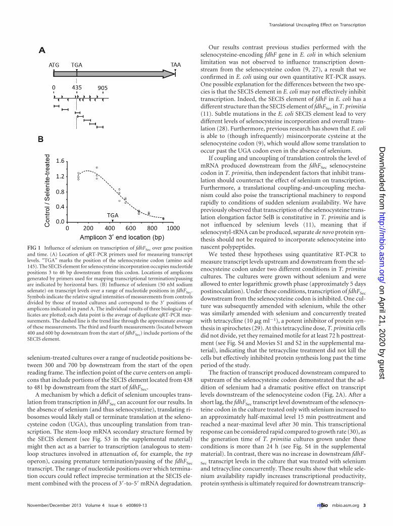
selenium-treated cultures over a range of nucleotide positions be-tween 300 and 700 bp downstream from the start of the openreading frame. The inflection point of the curve centers on ampli-cons that include portions of the SECIS element located from 438to 481 bp downstream from the start of fdhFSec.
A mechanism by which a deficit of selenium uncouples trans-lation from transcription in fdhFSec can account for our results. Inthe absence of selenium (and thus selenocysteine), translating ri-bosomes would likely stall or terminate translation at the seleno-cysteine codon (UGA), thus uncoupling translation from tran-scription. The stem-loop mRNA secondary structure formed bythe SECIS element (see Fig. S3 in the supplemental material)might then act as a barrier to transcription (analogous to stem-loop structures involved in attenuation of, for example, the trpoperon), causing premature termination/pausing of the fdhFSec
transcript. The range of nucleotide positions over which termina-tion occurs could reflect imprecise termination at the SECIS ele-ment combined with the process of 3=-to-5=mRNA degradation.
Our results contrast previous studies performed with theselenocysteine-encoding fdhF gene in E. coli in which seleniumlimitation was not observed to influence transcription down-stream from the selenocysteine codon (9, 27), a result that weconfirmed in E. coli using our own quantitative RT-PCR assays.One possible explanation for the differences between the two spe-cies is that the SECIS element in E. coli may not effectively inhibittranscription. Indeed, the SECIS element of fdhF in E. coli has adifferent structure than the SECIS element of fdhFSec in T. primitia(11). Subtle mutations in the E. coli SECIS element lead to verydifferent levels of selenocysteine incorporation and overall trans-lation (28). Furthermore, previous research has shown that E. coliis able to (though infrequently) misincorporate cysteine at theselenocysteine codon (9), which would allow some translation tooccur past the UGA codon even in the absence of selenium.
If coupling and uncoupling of translation controls the level ofmRNA produced downstream from the fdhFSec selenocysteinecodon in T. primitia, then independent factors that inhibit trans-lation should counteract the effect of selenium on transcription.Furthermore, a translational coupling-and-uncoupling mecha-nism could also poise the transcriptional machinery to respondrapidly to conditions of sudden selenium availability. We havepreviously observed that transcription of the selenocysteine trans-lation elongation factor SelB is constitutive in T. primitia and isnot influenced by selenium levels (11), meaning that ifselenocystyl-tRNA can be produced, separate de novo protein syn-thesis should not be required to incorporate selenocysteine intonascent polypeptides.
We tested these hypotheses using quantitative RT-PCR tomeasure transcript levels upstream and downstream from the sel-enocysteine codon under two different conditions in T. primitiacultures. The cultures were grown without selenium and wereallowed to enter logarithmic growth phase (approximately 5 dayspostinoculation). Under these conditions, transcription of fdhFSec
downstream from the selenocysteine codon is inhibited. One cul-ture was subsequently amended with selenium, while the otherwas similarly amended with selenium and concurrently treatedwith tetracycline (10 �g ml�1), a potent inhibitor of protein syn-thesis in spirochetes (29). At this tetracycline dose, T. primitia cellsdid not divide, yet they remained motile for at least 72 h posttreat-ment (see Fig. S4 and Movies S1 and S2 in the supplemental ma-terial), indicating that the tetracycline treatment did not kill thecells but effectively inhibited protein synthesis long past the timeperiod of the study.
The fraction of transcript produced downstream compared toupstream of the selenocysteine codon demonstrated that the ad-dition of selenium had a dramatic positive effect on transcriptlevels downstream of the selenocysteine codon (Fig. 2A). After ashort lag, the fdhFSec transcript level downstream of the selenocys-teine codon in the culture treated only with selenium increased toan approximately half-maximal level 15 min posttreatment andreached a near-maximal level after 30 min. This transcriptionalresponse can be considered rapid compared to growth rate (30), asthe generation time of T. primitia cultures grown under theseconditions is more than 24 h (see Fig. S4 in the supplementalmaterial). In contrast, there was no increase in downstream fdhF-
Sec transcript levels in the culture that was treated with seleniumand tetracycline concurrently. These results show that while sele-nium availability rapidly increases transcriptional productivity,protein synthesis is ultimately required for downstream transcrip-
FIG 1 Influence of selenium on transcription of fdhFSec over gene positionand time. (A) Location of qRT-PCR primers used for measuring transcriptlevels. “TGA” marks the position of the selenocysteine codon (amino acid145). The SECIS element for selenocysteine incorporation occupies nucleotidepositions 3 to 46 bp downstream from this codon. Locations of ampliconsgenerated by primers used for mapping transcriptional termination/pausingare indicated by horizontal bars. (B) Influence of selenium (50 nM sodiumselenate) on transcript levels over a range of nucleotide positions in fdhFSec.Symbols indicate the relative signal intensities of measurements from controlsdivided by those of treated cultures and correspond to the 3= positions ofamplicons indicated in panel A. The individual results of three biological rep-licates are plotted; each data point is the average of duplicate qRT-PCR mea-surements. The dashed line is the trend line through the approximate averageof these measurements. The third and fourth measurements (located between400 and 600 bp downstream from the start of fdhFSec) include portions of theSECIS element.
Translational Uncoupling Effect on Transcription
November/December 2013 Volume 4 Issue 6 e00869-13 ® mbio.asm.org 3
on April 21, 2020 by guest
http://mbio.asm
.org/D
ownloaded from
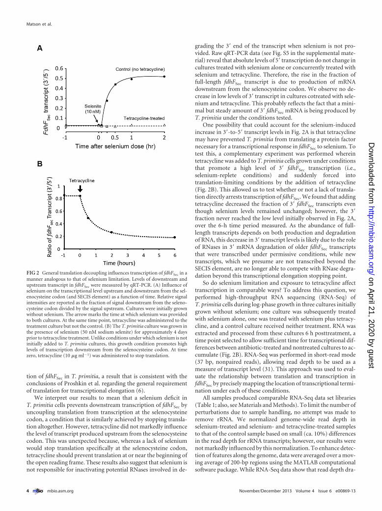
tion of fdhFSec in T. primitia, a result that is consistent with theconclusions of Proshkin et al. regarding the general requirementof translation for transcriptional elongation (6).
We interpret our results to mean that a selenium deficit inT. primitia cells prevents downstream transcription of fdhFSec byuncoupling translation from transcription at the selenocysteinecodon, a condition that is similarly achieved by stopping transla-tion altogether. However, tetracycline did not markedly influencethe level of transcript produced upstream from the selenocysteinecodon. This was unexpected because, whereas a lack of seleniumwould stop translation specifically at the selenocysteine codon,tetracycline should prevent translation at or near the beginning ofthe open reading frame. These results also suggest that selenium isnot responsible for inactivating potential RNases involved in de-
grading the 3= end of the transcript when selenium is not pro-vided. Raw qRT-PCR data (see Fig. S5 in the supplemental mate-rial) reveal that absolute levels of 5= transcription do not change incultures treated with selenium alone or concurrently treated withselenium and tetracycline. Therefore, the rise in the fraction offull-length fdhFSec transcript is due to production of mRNAdownstream from the selenocysteine codon. We observe no de-crease in low levels of 3= transcript in cultures cotreated with sele-nium and tetracycline. This probably reflects the fact that a mini-mal but steady amount of 3= fdhFSec mRNA is being produced byT. primitia under the conditions tested.
One possibility that could account for the selenium-inducedincrease in 3=-to-5= transcript levels in Fig. 2A is that tetracyclinemay have prevented T. primitia from translating a protein factornecessary for a transcriptional response in fdhFSec to selenium. Totest this, a complementary experiment was performed whereintetracycline was added to T. primitia cells grown under conditionsthat promote a high level of 3= fdhFSec transcription (i.e.,selenium-replete conditions) and suddenly forced intotranslation-limiting conditions by the addition of tetracycline(Fig. 2B). This allowed us to test whether or not a lack of transla-tion directly arrests transcription of fdhFSec. We found that addingtetracycline decreased the fraction of 3= fdhFSec transcripts eventhough selenium levels remained unchanged; however, the 3=fraction never reached the low level initially observed in Fig. 2A,over the 6-h time period measured. As the abundance of full-length transcripts depends on both production and degradationof RNA, this decrease in 3= transcript levels is likely due to the roleof RNases in 3= mRNA degradation of older fdhFSec transcriptsthat were transcribed under permissive conditions, while newtranscripts, which we presume are not transcribed beyond theSECIS element, are no longer able to compete with RNase degra-dation beyond this transcriptional elongation stopping point.
So do selenium limitation and exposure to tetracycline affecttranscription in comparable ways? To address this question, weperformed high-throughput RNA sequencing (RNA-Seq) ofT. primitia cells during log-phase growth in three cultures initiallygrown without selenium; one culture was subsequently treatedwith selenium alone, one was treated with selenium plus tetracy-cline, and a control culture received neither treatment. RNA wasextracted and processed from these cultures 6 h posttreatment, atime point selected to allow sufficient time for transcriptional dif-ferences between antibiotic-treated and nontreated cultures to ac-cumulate (Fig. 2B). RNA-Seq was performed in short-read mode(37 bp, nonpaired reads), allowing read depth to be used as ameasure of transcript level (31). This approach was used to eval-uate the relationship between translation and transcription infdhFSec by precisely mapping the location of transcriptional termi-nation under each of these conditions.
All samples produced comparable RNA-Seq data set libraries(Table 1; also, see Materials and Methods). To limit the number ofperturbations due to sample handling, no attempt was made toremove rRNA. We normalized genome-wide read depth inselenium-treated and selenium- and tetracycline-treated samplesto that of the control sample based on small (ca. 10%) differencesin the read depth for rRNA transcripts; however, our results werenot markedly influenced by this normalization. To enhance detec-tion of features along the genome, data were averaged over a mov-ing average of 200-bp regions using the MATLAB computationalsoftware package. While RNA-Seq data show that read depth dra-
FIG 2 General translation decoupling influences transcription of fdhFSec in amanner analogous to that of selenium limitation. Levels of downstream andupstream transcript in fdhFSec were measured by qRT-PCR. (A) Influence ofselenium on the transcriptional level upstream and downstream from the sel-enocysteine codon (and SECIS element) as a function of time. Relative signalintensities are reported as the fraction of signal downstream from the seleno-cysteine codon divided by the signal upstream. Cultures were initially grownwithout selenium. The arrow marks the time at which selenium was providedto both cultures. At the same time point, tetracycline was administered to thetreatment culture but not the control. (B) The T. primitia culture was grown inthe presence of selenium (50 nM sodium selenite) for approximately 4 daysprior to tetracycline treatment. Unlike conditions under which selenium is notinitially added to T. primitia cultures, this growth condition promotes highlevels of transcription downstream from the selenocysteine codon. At timezero, tetracycline (10 �g ml�1) was administered to stop translation.
Matson et al.
4 ® mbio.asm.org November/December 2013 Volume 4 Issue 6 e00869-13
on April 21, 2020 by guest
http://mbio.asm
.org/D
ownloaded from
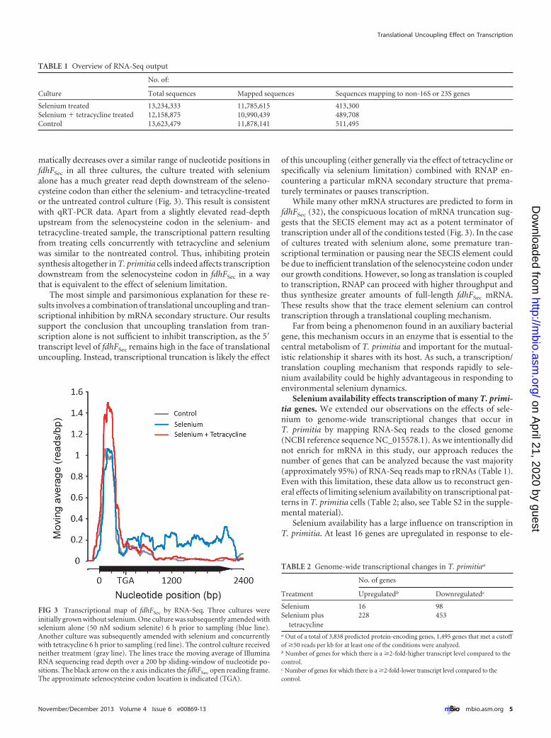
matically decreases over a similar range of nucleotide positions infdhFSec in all three cultures, the culture treated with seleniumalone has a much greater read depth downstream of the seleno-cysteine codon than either the selenium- and tetracycline-treatedor the untreated control culture (Fig. 3). This result is consistentwith qRT-PCR data. Apart from a slightly elevated read-depthupstream from the selenocysteine codon in the selenium- andtetracycline-treated sample, the transcriptional pattern resultingfrom treating cells concurrently with tetracycline and seleniumwas similar to the nontreated control. Thus, inhibiting proteinsynthesis altogether in T. primitia cells indeed affects transcriptiondownstream from the selenocysteine codon in fdhFSec in a waythat is equivalent to the effect of selenium limitation.
The most simple and parsimonious explanation for these re-sults involves a combination of translational uncoupling and tran-scriptional inhibition by mRNA secondary structure. Our resultssupport the conclusion that uncoupling translation from tran-scription alone is not sufficient to inhibit transcription, as the 5=transcript level of fdhFSec remains high in the face of translationaluncoupling. Instead, transcriptional truncation is likely the effect
of this uncoupling (either generally via the effect of tetracycline orspecifically via selenium limitation) combined with RNAP en-countering a particular mRNA secondary structure that prema-turely terminates or pauses transcription.
While many other mRNA structures are predicted to form infdhFSec (32), the conspicuous location of mRNA truncation sug-gests that the SECIS element may act as a potent terminator oftranscription under all of the conditions tested (Fig. 3). In the caseof cultures treated with selenium alone, some premature tran-scriptional termination or pausing near the SECIS element couldbe due to inefficient translation of the selenocysteine codon underour growth conditions. However, so long as translation is coupledto transcription, RNAP can proceed with higher throughput andthus synthesize greater amounts of full-length fdhFSec mRNA.These results show that the trace element selenium can controltranscription through a translational coupling mechanism.
Far from being a phenomenon found in an auxiliary bacterialgene, this mechanism occurs in an enzyme that is essential to thecentral metabolism of T. primitia and important for the mutual-istic relationship it shares with its host. As such, a transcription/translation coupling mechanism that responds rapidly to sele-nium availability could be highly advantageous in responding toenvironmental selenium dynamics.
Selenium availability effects transcription of many T. primi-tia genes. We extended our observations on the effects of sele-nium to genome-wide transcriptional changes that occur inT. primitia by mapping RNA-Seq reads to the closed genome(NCBI reference sequence NC_015578.1). As we intentionally didnot enrich for mRNA in this study, our approach reduces thenumber of genes that can be analyzed because the vast majority(approximately 95%) of RNA-Seq reads map to rRNAs (Table 1).Even with this limitation, these data allow us to reconstruct gen-eral effects of limiting selenium availability on transcriptional pat-terns in T. primitia cells (Table 2; also, see Table S2 in the supple-mental material).
Selenium availability has a large influence on transcription inT. primitia. At least 16 genes are upregulated in response to ele-
TABLE 1 Overview of RNA-Seq output
Culture
No. of:
Total sequences Mapped sequences Sequences mapping to non-16S or 23S genes
Selenium treated 13,234,333 11,785,615 413,300Selenium � tetracycline treated 12,158,875 10,990,439 489,708Control 13,623,479 11,878,141 511,495
FIG 3 Transcriptional map of fdhFSec by RNA-Seq. Three cultures wereinitially grown without selenium. One culture was subsequently amended withselenium alone (50 nM sodium selenite) 6 h prior to sampling (blue line).Another culture was subsequently amended with selenium and concurrentlywith tetracycline 6 h prior to sampling (red line). The control culture receivedneither treatment (gray line). The lines trace the moving average of IlluminaRNA sequencing read depth over a 200 bp sliding-window of nucleotide po-sitions. The black arrow on the x axis indicates the fdhFSec open reading frame.The approximate selenocysteine codon location is indicated (TGA).
TABLE 2 Genome-wide transcriptional changes in T. primitiaa
Treatment
No. of genes
Upregulatedb Downregulatedc
Selenium 16 98Selenium plus
tetracycline228 453
a Out of a total of 3,838 predicted protein-encoding genes, 1,495 genes that met a cutoffof �50 reads per kb for at least one of the conditions were analyzed.b Number of genes for which there is a �2-fold-higher transcript level compared to thecontrol.c Number of genes for which there is a �2-fold-lower transcript level compared to thecontrol.
Translational Uncoupling Effect on Transcription
November/December 2013 Volume 4 Issue 6 e00869-13 ® mbio.asm.org 5
on April 21, 2020 by guest
http://mbio.asm
.org/D
ownloaded from
vated selenium levels. Upregulated genes include fdhSec, encodingthe selenium-dependent copy of formate dehydrogenase, and sev-eral genes encoding components of hydrogenase enzymes thatputatively supply formate dehydrogenase enzymes with electronsderived from hydrogen to carry out the reduction of CO2 (33).The overall pattern of upregulated genes is consistent withT. primitia cells responding to selenium by increasing their rate ofacetogenesis and growth enabled by the production of the seleno-cysteine form of formate dehydrogenase, which is predicted tohave a higher catalytic rate than the selenium-independent formof the enzyme (13). In addition, over 100 downregulated geneswere distributed throughout the genome of T. primitia underhigh-selenium conditions (see Table S2 and Fig. S2A in the sup-plemental material). Viewed another way, these genes are upregu-lated upon removal of selenium in cultures of T. primitia cellsgrowing under selenium-replete conditions.
The large number of metabolic genes preferentially expressedunder low-selenium conditions was expected. As formate dehy-drogenase is critical to the central metabolic pathway of T. primi-tia, we interpret these results to mean that numerous genes areneeded to compensate for a reduced capacity for acetogenic me-tabolism due to a deficiency in the preferred fdhF isoenzyme. Con-sistent with this hypothesis was the finding that, in addition tonumerous hypothetical proteins of unknown function, genes re-lated to motility, stress response, and acquisition of alternativenutrient sources were found to be transcribed at lower levels whenthe availability of selenium is high (see Table S2 in the supplemen-tal material). These results suggest that life is more challenging forT. primitia without selenium than when selenium is available.
Translational inhibition has a broad and varied effect ontranscription. We observed an unexpectedly wide variety of tran-scriptional responses to globally inhibiting translation. Manygenes are transcribed at lower levels in tetracycline treated cells(Table 2), an expected result based on the documented influenceof translation on transcription. As with fdhFSec, we observed manygenes for which tetracycline-mediated translation inhibitioncauses premature truncation of transcription (for example, seeFig. 4A to C). However, these genes are often part of multigeneoperons and are located far downstream from putative promoters.The majority of genes we analyzed are not markedly influenced(�50% increase or decrease in transcript levels) by a lack of trans-lation (Table 2; also see, for example, Fig. 4D to F). We also doc-umented numerous genes for which inhibition of translation in-creases transcriptional levels over the length of the genes (Table 2;also see, for example, Fig. 4G to I). These observations are notlimited to individual genes but extend to genes in putativeT. primitia operons where similar trends occur over several thou-sand base pairs (Fig. S1). The list of genes that, on average, dem-onstrate an increase or decrease in expression in response to tet-racycline is quite large and is presented in Table S3 in thesupplemental material. The genes that are up- or downregulatedare distributed throughout the genome and are not localized toany easily defined hot spots on the chromosome of T. primitia (seeFig. S2B in the supplemental material).
It appears to be the case in T. primitia that translation has avariable effect on transcription. In essence, translation may act asa transcriptional regulatory governor that is capable of decreasing,increasing, or not influencing transcriptional levels of specificgenes. Transcriptional responses of many T. primitia genes aredependent on translation under conditions of limited protein syn-
thesis, but these effects may also involve mRNA secondary struc-ture determined by the particular nucleotide sequence of the gene.Such relationships between translation and transcription wouldimply that mRNA secondary structure could help to control thetranscription of genes and would allow certain genes to be tran-scribed even though translation is severely limited or has alto-gether ceased. Transcriptional effects caused by the presence andlocation of rare codons in some genes (34), as well as the place-ment of Shine-Dalgarno-like sequences within an open readingframe (35) and protein factors such as the NUS factors and Rho,may allow additional layers of transcriptional responses to occurduring specific translational challenges. We envision a scenario inwhich several of these factors act together in order to finely regu-late the strength and length of transcription. Support for one suchcooperative interaction involving mRNA loops with RNA poly-merase core enzyme and elongation factors was proposed recentlybased on crystal structures of an RNAP “paused” transcriptionalcomplex (36). In such complexes, different NUS proteins arethought to either stabilize or destabilize an RNA structure-mediated clamp confirmation that can be paused or processive,and the presence of hairpins is thought to stabilize the duration ofthe pause (36).
A large subset of T. primitia genes are transcribed even whenprotein synthesis is inhibited. While the transcription of somegenes is initiated at slightly higher levels, possibly leading to mul-tiple RNAPs acting in consort and helping to facilitate transcrip-tional processivity, this is not necessarily the case. Certain genesmay instead be preselected for preferential transcription underconditions that severely limit protein synthesis by the intrinsicnature of their nucleotide sequences. Perhaps it is ultimately a lackof a particular secondary structure that allows some genes to behighly transcribed during periods of inhibited protein synthesis.Prior literature on attenuation-regulated genes espouses the viewthat a specific subset of genes is selected to be preferentially tran-scribed under conditions where translation is inhibited (1). It ispossible that the scope of gene transcription influenced by trans-lation is even larger than envisioned and that the regulation isordered in strength from genes that are the most highly upregu-lated through those that are mildly upregulated to those that arenot influenced by translation or are even repressed by it. Genesthat are upregulated in the absence of translation in T. primitia(Table S3) include many translation-related genes, such as ribo-somal machinery, amino acid uptake, and classical attenuation-prone tRNA and amino acid synthesis genes. In addition to thesetargets, the transcription of several stress response genes is alsoupregulated under translation-limiting conditions.
While mRNA degradation certainly plays a role in many of thetranscriptional patterns observed using qRT-PCR and RNA-Seqapproaches, we believe that changes in transcriptional processivityaccount for the genome-wide changes in transcriptional patternsthat we observed, because numerous genes (and putative operons)were not influenced by inhibiting translation. It could also be thecase that mRNA secondary structure serves to inhibit 3= RNAdegradation at certain locations within genes, but we believe thatsuch a phenomenon alone does not account for our results andwould provide no particular advantage to the cells.
Whatever the exact mechanisms that are responsible for theseresponses may be, we hypothesize that the various links betweentranslation and transcription allow evolutionary processes to actupon certain genes such that transcription is constant or even
Matson et al.
6 ® mbio.asm.org November/December 2013 Volume 4 Issue 6 e00869-13
on April 21, 2020 by guest
http://mbio.asm
.org/D
ownloaded from
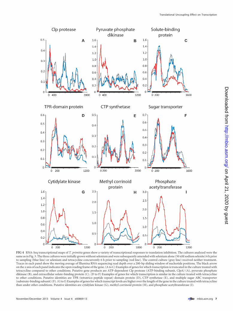
FIG 4 RNA-Seq transcriptional maps of T. primitia genes show a variety of transcriptional responses to translation inhibition. The cultures analyzed were thesame as in Fig. 3. The three cultures were initially grown without selenium and were subsequently amended with selenium alone (50 nM sodium selenite) 6 h priorto sampling (blue line) or selenium and tetracycline concurrently 6 h prior to sampling (red line). The control culture (gray line) received neither treatment.Traces in each panel show the moving average of Illumina RNA sequencing read depth over a 200-bp sliding window of nucleotide positions. The black arrowon the x axis of each panel indicates the open reading frame of the gene. (A to C) Examples of genes for which transcription is truncated in the culture treated withtetracycline compared to other conditions. Putative gene products are ATP-dependent Clp protease (ATP-binding subunit, ClpA) (A), pyruvate phosphatedikinase (B), and extracellular solute-binding protein (C). (D to F) Examples of genes for which transcription is similar in the culture treated with tetracyclineto other conditions. Putative identities are TPR (tetratrico peptide repeat) domain protein (D), CTP synthetase (E), and multiple sugar ABC transporter(substrate-binding subunit) (F). (G to I) Examples of genes for which transcript levels are higher over the length of the gene in the culture treated with tetracyclinethan under other conditions. Putative identities are cytidylate kinase (G), methyl corrinoid protein (H), and phosphate acetyltransferase (I).
Translational Uncoupling Effect on Transcription
November/December 2013 Volume 4 Issue 6 e00869-13 ® mbio.asm.org 7
on April 21, 2020 by guest
http://mbio.asm
.org/D
ownloaded from
increased during periods of limited protein synthesis. Most bac-terial cells in natural environments are not in a state of sustainedlogarithmic growth but undergo a perpetual cycle of starvationinterrupted by periods of nutrient influxes that allow sporadicgrowth (37). Genes for which transcription is not dependent ontranslation could be transcribed and thus translated at low levelseven under the most stringent of conditions for protein synthesisand could be poised for immediate translation once conditionsagain become permissive.
Our studies were performed in an organism that belongs to thedeeply branching phylum of bacteria known as Spirochaetes. Manyspirochetes are important pathogens of humans and other ani-mals; however, with the exception of a few examples, such as Bor-relia burgdorferi (38), most, including T. primitia, are currentlygenetically intractable, meaning that mutational analysis is cur-rently not possible. Our results warrant additional studies into thecomplexity of post-initiation transcriptional responses, especiallyin model organisms, such as E. coli, where genetic manipulationcan easily be performed. Additionally, a new in vitro coupledtranscription-translation assay which uses the translation compo-nents of E. coli has been developed for precisely such purposes(39). Such investigations may reveal a greater breadth of func-tional genes preferentially transcribed under conditions of inhib-ited protein synthesis as well as the nature of mRNA secondarystructure or other, as-yet-unknown factors that may be responsi-ble for prematurely terminating or pausing transcription of genesduring inhibited protein synthesis.
MATERIALS AND METHODSBacterial cultivation. To support rapid growth, Treponema primitiastrain ZAS-2 cells were routinely grown in anaerobic YACo broth culturescontaining 4% yeast autolysate under an atmosphere of 80% H2 and 20%CO2 as previously described (10) and were amended with 20 mM maltose.For experimental samples, cultures were not amended with maltose inorder to promote growth on H2 and CO2 via the acetyl-CoA pathway. Allcultures used in these studies were passaged a minimum of three timesunder conditions specifically designed to limit the amount of seleniumpresent, as previously described in detail (11). When selenium was addedto experimental cultures, it was added in the form of sodium selenite(Sigma, St. Louis, MO), yielding a final concentration of 50 nM, whichwas previously shown to maximize downstream transcription of fdhFSec
(11). Where tetracycline was used to inhibit translation, it was added astetracycline HCl (Sigma) at a final concentration of 10 �g ml�1.
Primers. Sequences and related information for primers used in thisstudy are listed in Table S1 in the supplemental material.
RNA extraction and reverse transcription. Upon harvesting of cells(5 ml per sample) from 50-ml total volumes of T. primitia cultures grownanaerobically in 300-ml sidearm flasks, RNA was immediately stabilizedby the addition of 10 ml of RNAprotect bacteria reagent (Qiagen, Valen-cia, CA). Cells were pelleted via centrifugation (5,000 � g for 10 min), andtotal RNA was extracted using RNeasy minicolumns with on-columnDNase I treatment (Qiagen). RNA was then subjected to a second, 30-min, 37°C off-column DNA digestion using RQ1 DNase enzyme (0.1 U�l�1) in 1� DNase buffer (Promega Corp., Madison, WI). Following thesecond digest, RNA samples were again purified with RNeasy columns toremove the DNase enzyme.
For quantitative RT-PCR, RNA samples (500 ng RNA in total) wereimmediately converted to cDNA by randomly primed reverse transcrip-tion using iScript reverse transcriptase and cDNA synthesis premix (Bio-Rad Laboratories, Hercules, CA) according to the manufacturer’s proto-col. Duplicate samples lacking reverse transcriptase were prepared foreach RNA sample as a negative control to assess residual DNA contami-nation. In all samples, DNA contamination was below the cycle threshold
(CT) after 34 of 44 cycles of quantitative RT-PCR, far below the CT of allmRNA measurements.
For RNA-Seq, samples were prepared in the manner previously re-ported (40). Briefly, total RNA (at least 5 �g) was fragmented using anAmbion RNA fragmentation kit (Life Technologies, Grand Island, NY)and then converted to single-strand cDNA using an Invitrogen Super-Script II kit (Invitrogen, Carlsbad, CA). Second-strand buffer (500 mMTris-HCl [pH 7.8], 50 mM MgCl2, 10 mM dithiothreitol [DTT]), deoxy-nucleoside triphosphate (dNTP) (0.3 mM), RNase H (2 U �l�1; Invitro-gen) and DNA polymerase I (Invitrogen) were then added to the first-strand reaction to synthesize second-strand cDNA (16° C, 2.5 h).
Quantitative RT-PCR. Primers for qRT-PCR were designed usingPrimer3 release 1.0.1 (41) to amplify regions of the T. primitiaselenocysteine-encoding formate dehydrogenase gene (fdhFSec) upstreamand downstream of the selenocysteine codon. Primer sequences are pro-vided in Table S1 in the supplemental material. qPCR was performed in15-�l reaction volumes of iQ SYBR green supermix (Bio-Rad Laborato-ries) using 25 ng of cDNA per reaction and forward and reverse primers(10 pmol each) in separate reactions. A parallel set of reactions was per-formed for each primer set using a 10-fold dilution series of T. primitiagenomic DNA as the template to generate standard curves. Transcriptlevels for the T. primitia gene clpX were used as an endogenous qRT-PCRcontrol to normalize for sample handling in all PCRs except for samplesfrom cultures treated with tetracycline. This gene was previously shown tobe constitutively transcribed and not influenced by selenium (11). Theprimers QclpXF and QclpXR described in that study were used here.Thermocycling and amplification detection were performed using a Bio-Rad DNAEngine thermocycler outfitted with a Chromo4 real-time detec-tor. Thermocycling conditions for all quantitative PCRs were initial de-naturation at 95°C for 3 min followed by 44 cycles of 95°C for 15 s and60°C for 30 s.
RNA sequencing and data analysis. cDNA samples were submitted tothe Caltech Sequencing Core facility (Pasadena, CA). Libraries were se-quenced as 37-mers using the standard Illumina protocol and pipeline.Sequencing depth information is summarized in Table 1. Illumina datawere aligned to a FASTA file of the T. primitia ZAS-2 genome using GER-ALD (a software package within the Illumina pipeline) and the Maq short-read aligning program (Wellcome Trust Sanger Institute, Hinxton,United Kingdom). Gene expression values were determined by normal-izing the number of reads mapped to a particular gene divided by the sizeof the gene. The resulting value is the normalized reads per kilobase,consistent with the gene expression index calculations of previously pub-lished reports (40, 42). In order to adjust for intensity between samples,the ribosomal signal from each sample was used as a standard, and eachsample’s intensity was multiplied by a factor that would yield an equalrRNA signal. In considering up- or downregulated genes, a cutoff of a2-fold increase in transcription intensity was used. Additionally, onlygenes with more than 50 adjusted hits per kb of coding DNA were con-sidered in analyses. Signal intensities were visualized graphically by con-verting Maq-aligned reads into a BAR file using Cisgenome software andviewed on the Cisgenome browser (Stanford University, Stanford, CA)(43). For a smoother graphical display of data, a moving average of readsper 200-bp sliding nucleotide window was generated using the MATLABsoftware package (R2011), and corresponding data were again visualizedon the genome browser. Genomic circular representation diagrams weregenerated using the DNAPlotter program (44). All raw data have beendeposited in the datadryad website (http://datadryad.org).
SUPPLEMENTAL MATERIALSupplemental material for this article may be found at http://mbio.asm.org/lookup/suppl/doi:10.1128/mBio.00869-13/-/DCSupplemental.
Movie S1, AVI file, 8.6 MB.Movie S2, AVI file, 8 MB.Figure S1, PDF file, 0.2 MB.Figure S2, PDF file, 0.1 MB.Figure S3, PDF file, 0.1 MB.
Matson et al.
8 ® mbio.asm.org November/December 2013 Volume 4 Issue 6 e00869-13
on April 21, 2020 by guest
http://mbio.asm
.org/D
ownloaded from
Figure S4, PDF file, 0 MB.Figure S5, PDF file, 0.1 MB.Table S1, PDF file, 0.1 MB.Table S2, PDF file, 0.1 MB.Table S3, PDF file, 0.1 MB.
ACKNOWLEDGMENTS
This work was supported by the DOE (DE-FG02-07ER64484), and theCenter for Environmental Microbial Interactions (CEMI) at Caltech.
We thank Igor Antoscheckhin and the Jacobs Genetics and GenomicsLaboratory at Caltech for help with next-generation sequencing and Fa-bien Paulot for his help with RNA-Seq data analysis.
REFERENCES1. Merino E, Yanofsky C. 2005. Transcription attenuation: a highly con-
served regulatory strategy used by bacteria. Trends Genet. 21:260 –264.2. Zipser D. 1969. Polar mutations and operon function. Nature 221:21–25.3. Bar-Nahum G, Epshtein V, Ruckenstein AE, Rafikov R, Mustaev A,
Nudler E. 2005. A ratchet mechanism of transcription elongation and itscontrol. Cell 120:183–193.
4. Epshtein V, Nudler E. 2003. Cooperation between RNA polymerasemolecules in transcription elongation. Science 300:801– 805.
5. Burmann BM, Schweimer K, Luo X, Wahl MC, Stitt BL, GottesmanME, Rösch P. 2010. A NusE:NusG complex links transcription and trans-lation. Science 328:501–504.
6. Proshkin S, Rahmouni AR, Mironov A, Nudler E. 2010. Cooperationbetween translating ribosomes and RNA polymerase in transcriptionelongation. Science 328:504 –508.
7. Böck A, Forchhammer K, Heider J, Leinfelder W, Sawers G, Veprek B,Zinoni F. 1991. Selenocysteine: the 21st amino acid. Mol. Microbiol.5:515–520.
8. Fourmy D, Guittet E, Yoshizawa S. 2002. Structure of prokaryotic SECISmRNA hairpin and its interaction with elongation factor SelB. J. Mol. Biol.324:137–150.
9. Thanbichler M, Böck A. 2002. The function of SECIS RNA in transla-tional control of gene expression in Escherichia coli. EMBO J. 21:6925– 6934.
10. Leadbetter JR, Schmidt TM, Graber JR, Breznak JA. 1999. Acetogenesisfrom H2 plus CO2 by spirochetes from termite guts. Science 283:686 – 689.
11. Matson EG, Zhang X, Leadbetter JR. 2010. Selenium controls transcrip-tion of paralogous formate dehydrogenase genes in the termite gut aceto-gen, Treponema primitia. Environ. Microbiol. 12:2245–2258.
12. Jones JB, Stadtman TC. 1981. Selenium-dependent and selenium-independent formate dehydrogenases of Methanococcus vannielii. Separa-tion of the two forms and characterization of the purified selenium-independent form. J. Biol. Chem. 256:656 – 663.
13. Axley MJ, Böck A, Stadtman TC. 1991. Catalytic properties of an Esch-erichia coli formate dehydrogenase mutant in which sulfur replaces sele-nium. Proc. Natl. Acad. Sci. U. S. A. 88:8450 – 8454.
14. Berry MJ, Maia AL, Kieffer JD, Harney JW, Larsen PR. 1992. Substi-tution of cysteine for selenocysteine in type I iodothyronine deiodinasereduces the catalytic efficiency of the protein but enhances its translation.Endocrinology 131:1848 –1852.
15. Gromer S, Johansson L, Bauer H, Arscott LD, Rauch S, Ballou DP,Williams CH, Jr, Schirmer RH, Arnér ES. 2003. Active sites of thiore-doxin reductases: why selenoproteins? Proc. Natl. Acad. Sci. U. S. A. 100:12618 –12623.
16. Metanis N, Keinan E, Dawson PE. 2006. Synthetic seleno-glutaredoxin 3analogues are highly reducing oxidoreductases with enhanced catalyticefficiency. J. Am. Chem. Soc. 128:16684 –16691.
17. Liu CC, Qi L, Yanofsky C, Arkin AP. 2011. Regulation of transcriptionby unnatural amino acids. Nat. Biotechnol. 29:164 –168.
18. Brauman A, Kane MD, Labat M, Breznak JA. 1992. Genesis of acetateand methane by gut bacteria of nutritionally diverse termites. Science257:1384 –1387.
19. Li LF, Ljungdahl L, Wood HG. 1966. Properties of nicotinamide adeninedinucleotide phosphate-dependent formate dehydrogenase from Clos-tridium thermoaceticum. J. Bacteriol. 92:405– 412.
20. Odelson DA, Breznak JA. 1983. Volatile fatty acid production by thehindgut microbiota of xylophagous termites. Appl. Environ. Microbiol.45:1602–1613.
21. Ragsdale SW, Pierce E. 2008. Acetogenesis and the Wood-Ljungdahlpathway of CO2 fixation. Biochim. Biophys. Acta 1784:1873–1898.
22. Breznak JA, Switzer JM. 1986. Acetate synthesis from H2 plus CO2 bytermite gut microbes. Appl. Environ. Microbiol. 52:623– 630.
23. Pester M, Brune A. 2007. Hydrogen is the central free intermediateduring lignocellulose degradation by termite gut symbionts. ISME J1:551–565.
24. Rosenthal AZ, Zhang X, Lucey KS, Ottesen EA, Trivedi V, Choi HM,Pierce NA, Leadbetter JR. 2013. Localizing transcripts to single cellssuggests an important role of uncultured Deltaproteobacteria in the ter-mite gut hydrogen economy. Proc. Natl. Acad. Sci. U. S. A. 110:16163–16168.
25. Zhang X, Leadbetter JR. 2012. Evidence for cascades of perturbation andadaptation in the metabolic genes of higher termite gut symbionts. mBio3:e00223-12.
26. Zhang X, Matson EG, Leadbetter JR. 2011. Genes for selenium depen-dent and independent formate dehydrogenase in the gut microbial com-munities of three lower, wood-feeding termites and a wood-feeding roach.Environ. Microbiol. 13:307–323.
27. Zinoni F, Birkmann A, Leinfelder W, Böck A. 1987. Cotranslationalinsertion of selenocysteine into formate dehydrogenase from Escherichiacoli directed by a UGA codon. Proc. Natl. Acad. Sci. U. S. A. 84:3156 –3160.
28. Chen GF, Fang L, Inouye M. 1993. Effect of the relative position of theUGA codon to the unique secondary structure in the fdhF mRNA on itsdecoding by selenocysteinyl tRNA in Escherichia coli. J. Biol. Chem. 268:23128 –23131.
29. Norris SJ, Edmondson DG. 1988. In vitro culture system to determineMICs and MBCs of antimicrobial agents against Treponema pallidumsubsp. pallidum (Nichols strain). Antimicrob. Agents Chemother. 32:68 –74.
30. Young JW, Locke JC, Elowitz MB. 2013. Rate of environmental changedetermines stress response specificity. Proc. Natl. Acad. Sci. U. S. A. 110:4140 – 4145.
31. Garber M, Grabherr MG, Guttman M, Trapnell C. 2011. Computationalmethods for transcriptome annotation and quantification using RNA-seq.Nat. Methods 8:469 – 477.
32. Zuker M. 2003. Mfold web server for nucleic acid folding and hybridiza-tion prediction. Nucleic Acids Res. 31:3406 –3415.
33. Ballor NR, Paulsen I, Leadbetter JR. 2012. Genomic analysis revealsmultiple [FeFe] hydrogenases and hydrogen sensors encoded by trepo-nemes from the H(2)-rich termite gut. Microb. Ecol. 63:282–294.
34. Chen GF, Inouye M. 1990. Suppression of the negative effect of minorarginine codons on gene expression; preferential usage of minor codonswithin the first 25 codons of the Escherichia coli genes. Nucleic Acids Res.18:1465–1473.
35. Li GW, Oh E, Weissman JS. 2012. The anti-Shine-Dalgarno sequencedrives translational pausing and codon choice in bacteria. Nature 484:538 –541.
36. Weixlbaumer A, Leon K, Landick R, Darst SA. 2013. Structural basis oftranscriptional pausing in bacteria. Cell 152:431– 441.
37. Kolter R, Siegele DA, Tormo A. 1993. The stationary phase of the bac-terial life cycle. Annu. Rev. Microbiol. 47:855– 874.
38. Samuels DS, Mach KE, Garon CF. 1994. Genetic transformation of theLyme disease agent Borrelia burgdorferi with coumarin-resistant gyrB. J.Bacteriol. 176:6045– 6049.
39. Castro-Roa D, Zenkin N. 2012. In vitro experimental system for analysisof transcription-translation coupling. Nucleic Acids Res. 40:e45.
40. Rosenthal AZ, Matson EG, Eldar A, Leadbetter JR. 2011. RNA-seqreveals cooperative metabolic interactions between two termite-gut spiro-chete species in co-culture. ISME J 5:1133–1142.
41. Rozen S, Skaletsky H. 2000. Primer3 on the WWW for general users andfor biologist programmers. Methods Mol. Biol. 132:-365–386.
42. Yoder-Himes DR, Chain PS, Zhu Y, Wurtzel O, Rubin EM, Tiedje JM,Sorek R. 2009. Mapping the Burkholderia cenocepacia niche response viahigh-throughput sequencing. Proc. Natl. Acad. Sci. U. S. A. 106:3976 –3981.
43. Ji H, Jiang H, Ma W, Johnson DS, Myers RM, Wong WH. 2008. Anintegrated software system for analyzing ChIP-chip and ChIP-seq data.Nat. Biotechnol. 26:1293–1300.
44. Carver T, Thomson N, Bleasby A, Berriman M, Parkhill J. 2009.DNAPlotter: circular and linear interactive genome visualization. Bioin-formatics 25:119 –120.
Translational Uncoupling Effect on Transcription
November/December 2013 Volume 4 Issue 6 e00869-13 ® mbio.asm.org 9
on April 21, 2020 by guest
http://mbio.asm
.org/D
ownloaded from