Embed Size (px)
Citation preview
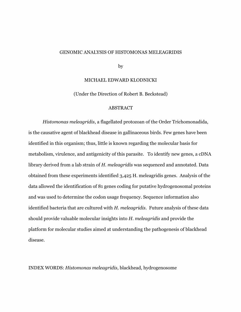
GENOMIC ANALYSIS OF HISTOMONAS MELEAGRIDIS
by
MICHAEL EDWARD KLODNICKI
(Under the Direction of Robert B. Beckstead)
ABSTRACT
Histomonas meleagridis, a flagellated protozoan of the Order Trichomonadida,
is the causative agent of blackhead disease in gallinaceous birds. Few genes have been
identified in this organism; thus, little is known regarding the molecular basis for
metabolism, virulence, and antigenicity of this parasite. To identify new genes, a cDNA
library derived from a lab strain of H. meleagridis was sequenced and annotated. Data
obtained from these experiments identified 3,425 H. meleagridis genes. Analysis of the
data allowed the identification of 81 genes coding for putative hydrogenosomal proteins
and was used to determine the codon usage frequency. Sequence information also
identified bacteria that are cultured with H. meleagridis. Future analysis of these data
should provide valuable molecular insights into H. meleagridis and provide the
platform for molecular studies aimed at understanding the pathogenesis of blackhead
disease.
INDEX WORDS: Histomonas meleagridis, blackhead, hydrogenosome
GENOMIC ANALYSIS
OF HISTOMONAS MELEAGRIDIS
by
MICHAEL EDWARD KLODNICKI
B.Sc., University of Georgia 2012
A Thesis Submitted to the Graduate Faculty of the University of Georgia in Partial
Fulfillment of the Requirements for the Degree
MASTER OF SCIENCE
ATHENS, GEORGIA
2013

© 2013
Michael Edward Klodnicki
All Rights Reserved
GENOMIC ANALYSIS
OF HISTOMONAS MELEAGRIDIS
by
MICHAEL EDWARD KLODNICKI
Major Professor: Robert B. Beckstead
Committee: Larry R. McDougald
Gene Pesti
Electronic Version Approved:
Maureen Grasso
Dean of the Graduate School
The University of Georgia
December 2013
iv
DEDICATION
I would like to dedicate this thesis to the domesticated turkey, in the hopes that
they will suffer from Histomonas no more.
v
ACKNOWLEDGEMENTS
I would like to express my deepest gratitude to Dr. Robert B. Beckstead, my
major professor, for his guidance, support and encouragement throughout my
undergraduate and graduate student careers at The University of Georgia. I would like
to also thank my committee members, as well as Dr. Lorraine Fuller, Dr. John Shields,
Dr. William Kerr, and Dr. Mark Haidekker for technical assistance during my study.
vi
TABLE OF CONTENTS
Page
ACKNOWLEDGEMENTS ................................................................................................... v
1 – INTRODUCTION AND LITERATURE REVIEW .......................................................... 1
2 – A GENOMIC ANALYSIS OF HISTOMONAS MELEAGRIDIS THROUGH
SEQUENCING OF A CDNA LIBRARY ............................................................................. 10
3 – CONCLUSIONS .......................................................................................................... 29
1
CHAPTER 1
INTRODUCTION AND LITERATURE REVIEW
OVERVIEW
Histomonas meleagridis, an early-diverging unicellular eukaryote, infects a wide
range of gallinaceous birds, causing histomoniasis (blackhead disease) (McDougald
2005). This disease is of commercial importance, sometimes causing high morbidity
and mortality in turkeys and peafowl. Little is known of the molecular biology of this
protozoan, although much can be inferred from work on closely related members of the
Tritrichomonadidae. All species of this order are anaerobic and lack mitochondria,
depending instead on the hydrogenosome for glycolytic energy metabolism and
formation of molecular hydrogen (Lindmark and Muller 1973).
Genes involved in virulence and metabolism have been studied extensively in the
human parasite Trichomonas vaginalis, the cattle parasite Tritrichomonas foetus, and
in a more distantly related human parasite, Entamoeba histolytica (Alderete et al.,
1991a, b; Addis et al., 1999; Musatovova and Alderete, 1999; Chaudhry and Petri, 2005;
Mundodi et al., 2006, 2008). Given the morphologic and physiological similarity and
phylogenetic relationship of H. meleagridis to these organisms, it is assumed that many
genes are held in common. However, the amount of specific sequence data in H.
meleagridis remains limited.
2
TRANSMISSION AND PATHOLOGY
H. meleagridis is a protozoan parasite with a complex life cycle, and one that
requires an intermediate host for transmission. Heterakis gallinarum, a cecal-dwelling
nematode, is the key carrier of this parasite – with H. gallinarum itself often carried by
earthworms. Histomonads are shed by ingested H. gallinarum during its first molt, in
the gut of the bird; these shed parasites then migrate to the bird’s cecal wall, which
becomes thickened with inflammation, and characterizes the site of primary infection.
Parasites further migrate into the blood, and invade the liver via connecting portal blood
vessels, establishing a secondary site of infection (Clarkson 1961; Fine 1975). These
infections produce characteristic lesions in the ceca and the liver, with only the former
correlated with bacterial infections (Franker et al. 1964; Bradley and Reid 1966).
Escherichia coli, Clostridium perfringens or Bacillus subtilis typically cause these
secondary bacterial infections associated with mortality.
Transmission from bird to bird, or lateral transmission, has been observed and
explained by cloacal drinking, or the bird’s reflexive, reverse-peristaltic cloacal
movement during contact with infected fecal matter. Thus, for in vivo studies of
histomoniasis, birds are often experimentally infected via the cloacal drop method (Hu
et al 2004). Furthermore, chickens and other birds which may not show clinical signs
of Histomonas infection can still act as carriers of infected H. gallinarum eggs, which
can remain viable and infected with virulent strains of H. meleagridis, with periods of
up to three years reported.
3
PREVENTION AND TREATMENTS
Various nitroheterocyclics, particularly nitroimidazoles like dimetridazole, were
used for many years and greatly reduced the incidence of blackhead disease. The FDA
and the EU banned use of these compounds in the 90s and in 2003, respectively, as the
imidazoles were suspected carcinogens. Nitarsone is currently the only drug approved
for use in animals in the prevention of blackhead disease (Hu and McDougald 2004).
Recommended for use as a feed additive at 187.5 ppm, it reduces flock mortality but
allows relapse upon withdrawal of treatment (McGuire and Morehouse 1952). Banned
in the EU, nitarsone is the last arsenic-containing drug on the US market, with the FDA
recently facing pressure to withdraw its approval of nitarsone use in animals bound for
human consumption in light of concerns raised by recent studies (Nachman et al 2013).
Alternative to preventative administration of arsenic-containing compounds is
vector control, or use of antihelminthics, such as benzimidazoles, which must be
administered prior to vector exposure, and do not provide protection from lateral
transmission or the protozoa itself (Hegngi 1999, Hu et. al 2004). As of late 2013, there
is no treatment available for histomoniasis.
IN VITRO CULTURE
H. meleagridis can be isolated in vitro from the infected ceca and liver using a
Dwyer medium (McDougald and Galloway 1973). Formulation of Dwyer’s media
consisted of 85-95% of Medium 199, 5-10% horse serum, 5% chick embryo extract, and
1% rice powder (Dwyer 1970), but was recently modified by removing the chick embryo
extract and reducing the rice flour concentration to 0.8% (van der Heijden and
Landman 2007). This modified version of Dwyer’s medium facilitates rapid expansive
growth of both H. meleagridis and bacteria and is commonly used by researchers today.
4
Histomonads must be cultured in slightly acidic media under strict anaerobic
conditions. The typical passage time of cultures varies from 48 to 72 hours, and peak
growth is obtained in 1-5 days. Virulence of monad strains appears to be roughly
inversely proportional to number of passages (Dwyer 1970, Lund et. al 1967)
Histomonads must be co-cultured with bacteria to maintain cultures over time.
However, Lund et al. (1967) reported several experiments in which antibiotic-treated
bacteria were used to support culture growth. Lesser (1960a, 1960b, 1963, 1964)
attempted to remove serum from the growth medium by substitution with sterilized
bovine cream and various fatty acids supplemented with various mineral salts; he used
uncharacterized antibiotic-treated bacteria extracted from turkey cecal samples to
maintain cultures. Additionally, he successfully used antibiotic-treated tissues from
fresh hamster liver in lieu of bacteria for prolonged (more than 190) passages of
histomonad cultures.
Other studies in closely related cattle parasites (trichomonads) demonstrated
inhibitory effect of zinc treatment on Tritrichomonas foetus growth in vitro (Benchimol
et al 1993). Similarly, nitarsone was demonstrated effective in controlling the growth of
H. meleagridis in a dose dependent manner (Van der Heijden et. al, 2008).
STUDY OBJECTIVES
1. Sequence and annotate a cDNA library, to provide data for future molecular and
virulence studies.
2. Analyze transcriptomic data, and present potential unique molecular targets for
therapeutic treatment of histomoniasis.
5
LITERATURE CITED
Addis, M. F., P. Rappelli, A. M. Pinto De Andrade, F. M. Rita, M. M. Colombo, P.
Cappuccinelli, and P. L. Fiori. 1999. Identification of Trichomonas vaginalis alpha-
actinin as the most common immunogen recognized by sera of women exposed to
the parasite. The Journal of Infectious Diseases 180: 1727-1730.
Alderete, J. F., E. Newton, C. Dennis, and K. A. Neale. 1991a. Antibody in sera of
patients infected with Trichomonas vaginalis is to trichomonad proteinases.
Genitourinary Medicine 67: 331-334.
Alderete, J. F., E. Newton, C. Dennis, and K. A. Neale. 1991b. The vagina of women
infected with Trichomonas vaginalis has numerous proteinases and antibody to
trichomonad proteinases. Genitourinary Medicine 67: 469-474.
Aoyagi, S., and D.H. Baker. 1995. Effect of high copper dosing on hemicellulose
digestibility in cecectomized cockerels. Poultry Sci. 74:208-211.
Benchimol, M., Almeida, J.C.A., Lins, U., Goncalves, N.R., Souza, W. 1993. Electron
microscopic study of the effect of zinc on Tritrichomonas foetus. Microb. Agents
Chem. 37:2722-2726.
Bradley, R. E., and W. M. Reid. 1966. Histomonas meleagridis and several bacteria as
agents of infectious enterohepatitis in gnotobiotic turkeys. Exp.Parasitol. 19:91-101.
Chaudhry, O. A., and W. A. Petri. 2005. Vaccine prospects for amebiasis. Expert Review
of Vaccines 4: 657-668.
Clarkson, M. J. 1961.The blood supply of the liver of the turkey and the anatomy of the
biliary tract with reference to infection with Histomonas meleagridis. Res. Vet. Sci.
2:259-264.
6
Duke, G.E., and Bedbury, H.P. 1983. Cecal microflora of turkeys fed low or high fiber
diets: enumeration, identification, and determination of cellulolytic activity. Poultry
Sci. 62(4):675-82.
Duke G.E., Eccleston, E., Kirkwood, S., Louis, C.F., and Bedbury, H.P. 1984. Cellulose
digestion by domestic turkeys fed low or high fiber diets. J Nutr. 114(1):95-102.
Dwyer, D. M. 1970. An improved method for cultivating Histomonas meleagridis. J
Parasitol, 56(1), 191-192.
Fine, P. E. M. 1975.Quantitative studies on transmission of Parahistomonas wenrichi by
ova of Heterakis gallinarum. Parasitology 70:407-417.
Franker, C.K. and Doll, J.P. 1964. Experimental histomoniasis in gnotobiotic turkeys:
effects of some cecal bacteria on pathogenesis. J Parasitol. 50:636-40.
Harms, R.H., and Buresh, R.E. 1987. Influence of three levels of copper on the
performance of turkey poults with diets containing two sources of methionine. Poult
Sci. 1987 Apr;66(4):721-4.
Hegngi, F.N., Doerr, J., Cummings, T.S., Schwartz, R.D., Saunders, G., Zajac, A., Larsen,
C.T., and F.W. Pierson. 1999. The effectiveness of benzimidazole derivatives for the
treatment and prevention of histomonosis (blackhead) in turkeys. Vet Parasitol 81:
29-37.
Hu, J. & McDougald, L.R. 2004. The efficacy of some drugs with known antiprotozoal
activity against Histomonas meleagridis in chickens. Veterinary Parasitology, 121,
233-238.
Hu, J., Fuller, L., and L. R. McDougald. 2004. Infection of turkeys with Histomonas
meleagridis by the cloacal drop method. Avian Dis 48: 746-750.
7
King, J.O.L. 1975a. The feeding of copper sulphate to growing rabbits. Br. Vet. J.
131:70–75.
King, J.O.L. 1975b. The feeding of copper sulphate to ducklings. Br. Poult. Sci. 16:409–
411.
Leeson S., Zubair, A.K., Squires, E.J., and Forsberg, C. 1997. Influence of dietary levels
of fat, fiber, and copper sulfate and fat rancidity on cecal activity in the growing
turkey. Poult Sci. 76(1):59-66.
Lesser, E. 1961. In vitro cultivation of Histomonas meleagridis free of demonstrable
bacteria. Journal of Protozoology 8: 228-230.
Lesser, E. 1963. Effect of amphotericin-B on in vitro growth of Histomonas meleagridis.
Journal of Parasitology 49: 329.
Lesser, E. 1964. I. Studies on the in vitro cultivation of Histomonas meleagridis with
three different mixed bacterial flora. II. Studies on the in vitro growth of
Histomonas meleagridis with single species of bacteria. Proc. Helminth. Soc. Wash.
31: 263-264; 265-266.
Lindmark, D. G., and M. Muller. 1973. Hydrogenosome, a cytoplasmic organelle of the
anaerobic flagellate, Tritrichomonas foetus, and its role in pyruvate metabolism.
Journal of Biological Chemistry 248: 7724-7728.
Lund, E. E., Augustine, P.C., and Chute, A.M. 1967. Histomonas meleagridis after one
thousand in vitro passages. Journal of Protozoology 14: 349–351.
Lund, E. E., Wehr, E.E., Ellis, D.J. 1966. Earthworm Transmission of Heterakis and
Histomonas to turkeys and chickens. The Journal of Parasitology 52: 899-902.
McDougald, L. R., and R. B. Galloway. 1973. Blackhead disease: in vitro isolation of
Histomonas meleagridis as a potentially useful diagnostic aid. Avian Dis. 17:847-50.
8
Mcdougald, L. R. 2005. Blackhead disease (histomoniasis) in poultry: a critical review.
Avian Dis, 49(4), 462-476.
McGuire, W. C., and N. F. Morehouse. 1952. Chemotherapy studies of histomoniasis.
Poult. Sci. 31:603-609.
Mundodi, V., A. S. Kucknoor, and J. F. Alderete. 2008. Immunogenic and plasminogen-
binding surface-associated alpha-enolase of Trichomonas vaginalis. Infection and
Immunity 76: 523-531.
Mundodi, V., A. S. Kucknoor, T. H. Chang, and J. F. Alderete. 2006. A novel surface
protein of Trichomonas vaginalis is regulated independently by low iron and
contact with vaginal epithelial cells. BMC Microbiology 6: 6.
Musatovova, O., and J. F. Alderete. 1999. The Trichomonas vaginalis phenotypically
varying P270 immunogen is highly conserved except for numbers of repeated
elements. Microbial Pathogenesis 27: 93-104.
Nachman, K.E., Baron, P.A., Raber, G., Francesconi, K.A., Navas-Acien, A., and Love,
D.C. 2013. Roxarsone, inorganic arsenic, and other arsenic species in chicken: a
U.S.-based market basket sample. Environ Health Perspect. 121(7):818-824.
Persia, M.E., Baker, D.H., Parsons, C.M. 2004. Tolerance for excess basic zinc chloride
and basic copper chloride in chicks. Br Poult Sci. 45(5):672-676.
Rolls, B.A. 1977. Inorganic ions in the intestinal and cecal contents of germ-free and
conventional chickens. Lab. Anim. 11:99-104.
van der Heijden, H. M. J. F., and W. J. M. Landman. 2007. Improved culture of
Histomonas meleagridis in a modification of dwyer medium. Avian Dis 51: 986-
988.

9
van der Heijden H.M., and Landman W.J. 2008. In vivo effect of herbal products
against Histomonas meleagridis in turkeys. Avian Pathol. 37(1):45-50.
10
CHAPTER 2
A GENOMIC ANALYSIS OF HISTOMONAS MELEAGRIDIS THROUGH
SEQUENCING OF A CDNA LIBRARY
----------------------------------------
M. Klodnicki, L. R. McDougald and R. B. Beckstead*
Accepted by Journal of Parasitology. Reprinted here with permission of Allen Press.
11
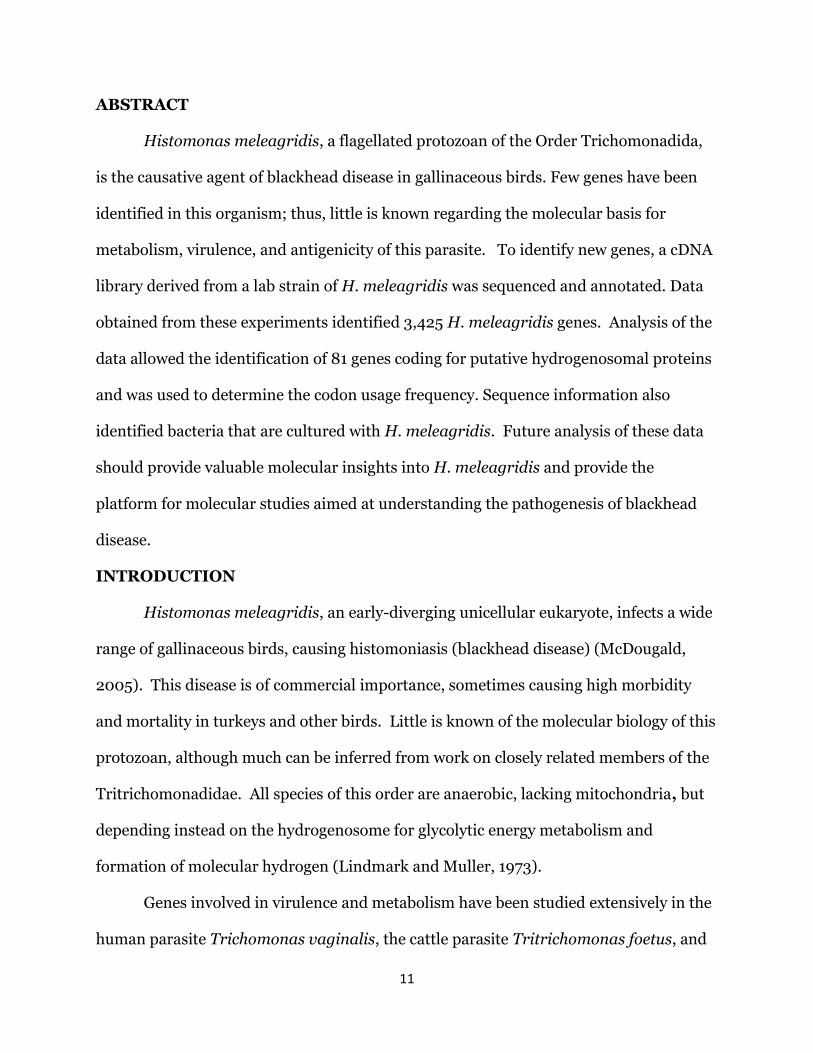
ABSTRACT
Histomonas meleagridis, a flagellated protozoan of the Order Trichomonadida,
is the causative agent of blackhead disease in gallinaceous birds. Few genes have been
identified in this organism; thus, little is known regarding the molecular basis for
metabolism, virulence, and antigenicity of this parasite. To identify new genes, a cDNA
library derived from a lab strain of H. meleagridis was sequenced and annotated. Data
obtained from these experiments identified 3,425 H. meleagridis genes. Analysis of the
data allowed the identification of 81 genes coding for putative hydrogenosomal proteins
and was used to determine the codon usage frequency. Sequence information also
identified bacteria that are cultured with H. meleagridis. Future analysis of these data
should provide valuable molecular insights into H. meleagridis and provide the
platform for molecular studies aimed at understanding the pathogenesis of blackhead
disease.
INTRODUCTION
Histomonas meleagridis, an early-diverging unicellular eukaryote, infects a wide
range of gallinaceous birds, causing histomoniasis (blackhead disease) (McDougald,
2005). This disease is of commercial importance, sometimes causing high morbidity
and mortality in turkeys and other birds. Little is known of the molecular biology of this
protozoan, although much can be inferred from work on closely related members of the
Tritrichomonadidae. All species of this order are anaerobic, lacking mitochondria, but
depending instead on the hydrogenosome for glycolytic energy metabolism and
formation of molecular hydrogen (Lindmark and Muller, 1973).
Genes involved in virulence and metabolism have been studied extensively in the
human parasite Trichomonas vaginalis, the cattle parasite Tritrichomonas foetus, and
12
in a more distantly related human parasite, Entamoeba histolytica (Alderete et al.,
1991a, b; Addis et al., 1999; Musatovova and Alderete, 1999; Chaudhry and Petri, 2005;
Mundodi et al., 2006, 2008). Given the morphologic and physiological similarity and
phylogenetic relationship of H. meleagridis to these organisms, it is assumed that many
genes are held in common. However, the amount of specific sequence data in H.
meleagridis remains limited.
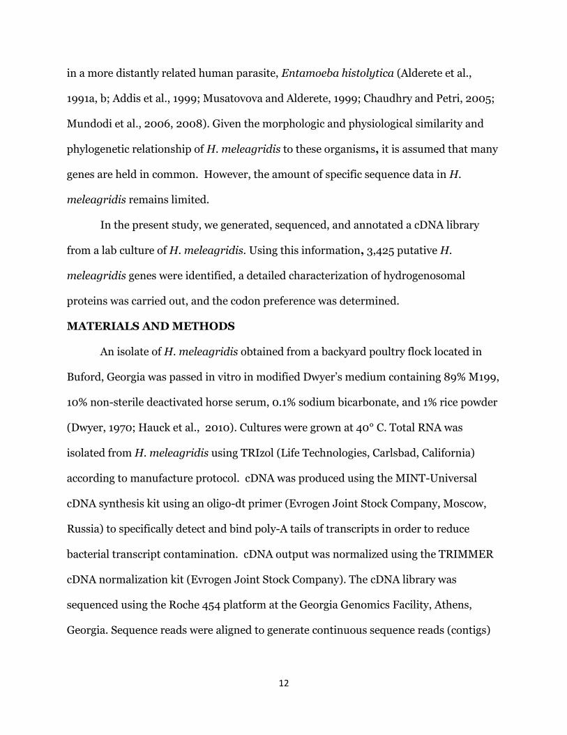
In the present study, we generated, sequenced, and annotated a cDNA library
from a lab culture of H. meleagridis. Using this information, 3,425 putative H.
meleagridis genes were identified, a detailed characterization of hydrogenosomal
proteins was carried out, and the codon preference was determined.
MATERIALS AND METHODS
An isolate of H. meleagridis obtained from a backyard poultry flock located in
Buford, Georgia was passed in vitro in modified Dwyer’s medium containing 89% M199,
10% non-sterile deactivated horse serum, 0.1% sodium bicarbonate, and 1% rice powder
(Dwyer, 1970; Hauck et al., 2010). Cultures were grown at 40° C. Total RNA was
isolated from H. meleagridis using TRIzol (Life Technologies, Carlsbad, California)
according to manufacture protocol. cDNA was produced using the MINT-Universal
cDNA synthesis kit using an oligo-dt primer (Evrogen Joint Stock Company, Moscow,
Russia) to specifically detect and bind poly-A tails of transcripts in order to reduce
bacterial transcript contamination. cDNA output was normalized using the TRIMMER
cDNA normalization kit (Evrogen Joint Stock Company). The cDNA library was
sequenced using the Roche 454 platform at the Georgia Genomics Facility, Athens,
Georgia. Sequence reads were aligned to generate continuous sequence reads (contigs)
13
using the Roche GS de novo Assembler under standard settings (Roche, Basle,
Switzerland).
Analyses of contig sequences were made using the BLASTx and BLASTn
algorithms (Altschul et al., 1990) and the BLAST2GO application (Conesa et al., 2005)
for local alignment comparisons to the NCBI database of genomic information,
GenBank (Benson et al., 2011) using BLAST2GO default parameters (maximum e-value
of 1.0e-3). Database entries were parsed for redundancy (entries with greater than 95%
invariance were determined to be variants of the same gene) and compared to cDNA
contig entries using basic local pairwise alignment analyses. Intensive local alignments
and gene constructs were generated and evaluated using the BLOSUM62 algorithm
native to the application Geneious (Drummond et al, 2010). Positively identified
sequences were submitted online to Genbank via BankIt.
BLAST sequence alignments were filtered by organism; protozoan and bacterial
hits were assembled into sets and examined separately. Bacterial hits were filtered by
phylogenetic family and the number of hits belonging to the 10 most frequent-occurring
families reported. For analyses of the protozoan hits, sequence Manipulation Suite 2, or
SMS2 (Stothard, 2000) was used to process and identify open reading frames (ORFs)
and for further sequence manipulation and examination, including the generation of
predicted codon usage data. Using SMS2, a sample of 100 of the longest ORFs in the H.
meleagridis data set was generated for codon usage data mining. Histomonas
meleagridis putative hydrogenosomal proteins were identified by homology to known
proteins found in T. vaginalis (Henze, 2007). Contigs were evaluated for redundancy
by local nucleotide alignments.
14
RESULTS
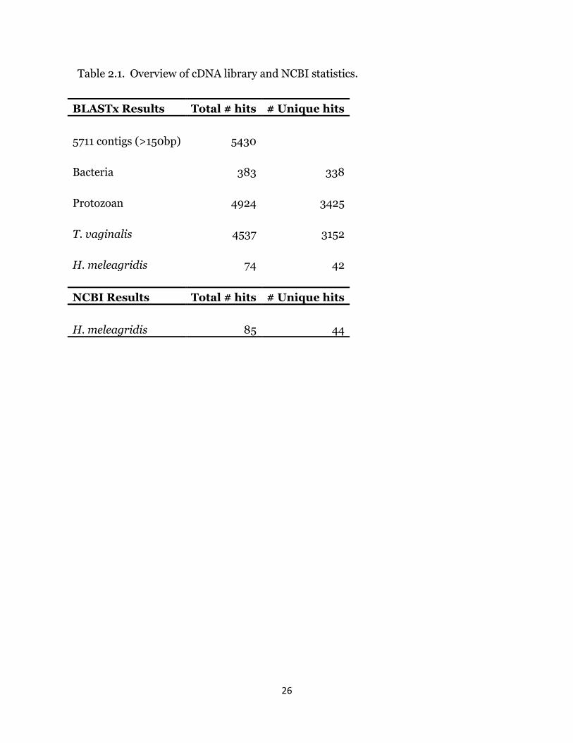
Sequencing of the H. meleagridis cDNA library resulted in approximately 24MB
of DNA sequences contained in 70,000 unique sequence reads. Assembly of the
sequence reads yielded a total of 5,711 contiguous DNA sequences (contigs) of 150 base
pairs or greater (Table I), with an average length of 919 base pairs per contig. Blastx
analysis of the contigs resulted in 5,430 sequences showing homology to a protein in the
NCBI database and 281 contigs without blast hits. The majority of the 281 contigs had 1
or more short open reading frames, suggesting that they may be the result of faulty
sequence alignment. Analysis of the sequence database revealed 4,924 discrete
protozoan BLASTx hits. Due to the bacteria necessarily present in culture with the
monads, analysis also revealed 383 total bacterial hits, which corresponded to 338
unique genes. From the sequence that showed homology to protozoan genes, 74 contigs
showed greatest homology to H. meleagridis sequences in the NCBI database; these
sequences corresponded to 42 unique genes. Parsing the NCBI protein database for a
nonredundant list of H. meleagridis sequences, it was determined there are 44 unique
coding genes corresponding to 85 deposited sequences. Only 2 relatively short genes
previously reported from H. meleagridis (putative 60S ribosomal protein P1, with a
predicted length of 41 amino acid residues; putative histone 2A-IV, 74 predicted
residues) were not identified in the cDNA library.
Of the 5,430 total contigs that returned BLAST hits, 4,537 had identified a T.
vaginalis gene with the greatest respective homology (Fig. 1), suggesting that the H.
meleagridis transcriptome is more closely related to T. vaginalis than other sequenced
protozoan genomes. Significant numbers of hits from other related protozoan species,
particularly H. meleagridis and E. histolytica, are also present among the top-hit
15
distribution. Insignificant numbers of random hits from various, less closely-related
species are grouped into the ‘Other’ category, composing the remainder of top-hits
within the protozoan category.
As Histomonas meleagridis growth in culture requires bacteria that are co-
cultured from the caecal content at the time of isolation, total RNA isolation from H.
meleagridis cultures results in detection of both bacterial and H. meleagridis RNA.
Based on the number of bacterial contigs identified in the library, we had 7.3%
contamination. Analysis of the bacterial contigs filtered by phylogenetic family allowed
the identification of bacteria found cultured with H. meleagridis. The majority of
bacterial sequences were classified as part of the Clostridiaceae family (obligate
anaerobes), the Bacteriodaceae family (aerotolerant anaerobes) or the Bacillus genus of
the Bacillaceae family (facultative or obligate anaerobes) (Fig. 2).
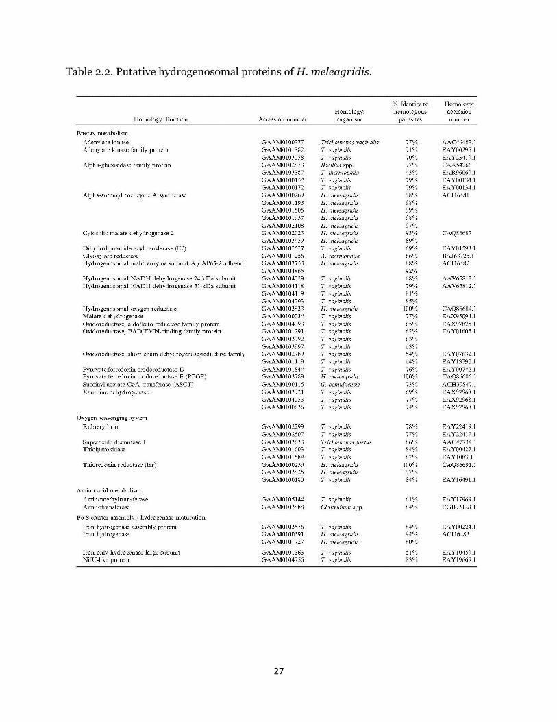
mRNAs coding for putative hydrogenosomal proteins (Table II) were assembled
based on positive experimental evidence for hydrogenosomal localization in
trichomonads and data directly resulting from the first proteomics study of the
hydrogenosome of T. vaginalis (Henze et al., 2007). Local nucleotide alignment
analysis of contigs with similar or identical percent identity resulted in 81 unique
contigs with significant homology to known hydrogenosomal proteins, composing 40
different enzyme functions. The percent identity of these proteins, as defined by the
ratio of positive amino acid matches to the total length of the alignment, ranged from
43% up to 100%. BLAST hits returned e-values ranging from 0 to 1.94e-02. Notably, all
enzymes required for ATP synthesis were identified, as were orthologues of 3 confirmed
hydrogenosomal membrane proteins (Hmp31 MCF, Hmp35, and TIM23). A number of
the proteins identified in the initial T. vaginalis proteomics study, however, were
16
missing, including Hsp10 and Hsp20 (both localized to the hydrogenosome in T.
vaginalis), along with arginine deiminase, glyoxylase, several chaperones, and several
predicted proteins that have not been confirmed to be localized or active within the
hydrogenosome. In total, 51 putative hydrogenosomal proteins out of 85 identified were
missing from the dataset.
We analyzed the nucleotide and codon usage frequencies of H. meleagridis using
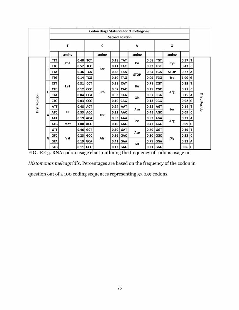
the longest 100 open reading frames consisting of 57,059 codons (Fig. 3). A strong A/T
bias (68%) was observed in the coding sequences. Codon usage in the first position was
biased towards purines (62%), while the second and third positions had a higher A/T
frequency (65.8% and 66.8%, respectively). Several amino acids exhibited a strong
preference for a specific codon, i.e., serine residues existed primarily as TCA codons
(38%), proline as CAA codons (63%), threonine as AAA codons (53%), glutamic acid as
GGA codons (79%), and aspartate was coded for by the GGT codon (70%). All 3 stop
codons were used, with TAA having the highest frequency (64%).
DISCUSSION
The present work has greatly expanded the list of genes known from H.
meleagridis by comparison of a sequenced cDNA library with genes reported from other
organisms. However, it is important to emphasize that many other genes could be
discovered through similar studies of RNA isolated from other stages of growth. For
instance, as the parasite moves from the anaerobic cecal environment to the liver and
other oxygen-rich tissues, it is likely that other genes will be expressed. Ultimately, the
sequencing of the entire genome will be needed to identify the complete genetic
complement of this organism.
17
While Hauck and Hafez (2010) found H. meleagridis to be more closely related
to Tritrichomonas foetus based on phylogenetic rRNA studies, limited T. foetus
sequencing data, relative to the completely sequenced T. vaginalis genome, likely
prevented the majority of hits from identifying as T. foetus homologues. Four genes,
most notably superoxide dismutase (Table II) did indeed identify with T. foetus (Fig. 1),
yet the overwhelming majority of genes identified clearly shared primary homology with
T. vaginalis. The high level of homology of H. meleagridis with T. vaginalis emphasizes
the close physiological, metabolic, and phylogenetic relationship of the 2 organisms.
Thus, it is reasonable to believe that similar mechanisms of virulence and antigenicity
can also be found. Identification of all the key ATP-producing enzymes identified and
many more putative hydrogenosomal enzymes in the present work provides a general
outline for future proteomic studies of the hydrogenosome of this species. In the first
proteomics study of T. vaginalis (Henze et al., 2007), the predictive target signal
peptide data were used extensively alongside proteome analysis and experimental
evidence, yielding a large number of putative hydrogenosomal proteins that remained
unconfirmed to be a part of the hydrogenosomal proteome. Furthermore, only 30
proteins currently have been demonstrated to localize to the organelle (Burstein et al.,
2012). Thus, it is expected that some of the hydrogenosomal genes in H. meleagridis
were not completely elucidated, such as the signal peptidase that shared homology with
E. histolytica rather than T. vaginalis. In contrast, two different classes of heat shock
proteins, Hsp10 and Hsp20, were notably missing from the dataset, yet 8 unique Hsp70
homologs were present (data not shown), only 1 of which was annotated as localized in
the hydrogenosome. Fourteen unique thioredoxin family proteins and 3 thioredoxin
reductase homologs were also identified. Current and future hydrogenosomal
18
proteomics studies of trichomonads (Schneider et al., 2011; Burstein et al., 2012) will
facilitate further comparative studies of the hydrogenosome of H. meleagridis.
As the histomonads for this study were grown in cultures heavily populated with
bacteria, and such bacteria are considered the primary food source for culture-grown
histomonads, the identification of bacterial genes was expected. Numerous bacteria can
be seen in food vacuoles by microscopic examination of histomonads (Mazet et al.,
2008), and it is well known that the cultured cecal forms of H. meleagridis are
intolerant to oxygen (Hauck et al., 2010). As shown in Figure 2, the majority of bacterial
sequences were classified in the Clostridiaceae family (obligate anaerobes), the
Bacteriodaceae family (aerotolerant anaerobes), or the Bacillus genus of the Bacillaceae
family (facultative or obligate anaerobes). While most of the free bacteria were removed
by washing during sample preparation, it was possible that some remained. For this
reason, oligo-dt primers that specifically detect the poly (A) tail on the mRNA were used
for amplification. Since bacterial mRNA does not have a poly (A) tail that will react with
oligo-dt primers, this reduced the bacterial contamination of the cDNA library.
The codon usage for H. meleagridis and nucleotide preference differs from other
parasitic protozoans such as Leishmania spp. and Trypanosoma cruzi, which have G/C
rich genomes, and T. vaginalis, which was previously reported to have no nucleotide
bias in its coding sequence (Alonso et al., 1992; Langford et al., 1992; and Meade et al.,
1997). More recently, however, genome sequencing of T. vaginalis (Carlton et al., 2007)
revealed a genome-wide nucleotide composition of 67.3% A/T, similar to the 68% A/T
composition described in the present study. A/T nucleotide bias of H. meleagridis also
bears similarity to Plasmodium and Entamoeba species that have a high percentage of
A/T nucleotides in their genomes and in codon position 3 (Char and Farthing, 1992;
19
Musto et al., 1995). Like T. vaginalis, H. meleagridis has a bias towards purines in
codon position 1 and A/T in position 2. Histomonas meleagridis preference for A/T in
codon position 3 differs from the preference of T. vaginalis for pyrimidine in that
position. Histomonas meleagridis also differs from T. vaginalis in its frequent use of
the TGA and TAG as stop codons (Meade et al., 1997). Trichomonas vaginalis uses the
TAA stop codon 93% of the time, which is consistent with its use of TAAA as the poly (A)
signal (Espinosa et al., 2002). The usage of all stop codons suggests that H. meleagridis
potentially has different poly (A) signals than T. vaginalis. Codon usage statistics
provided for H. meleagridis will impart useful annotations for future molecular,
biological, and biochemical studies of the organism.
ACKNOWLEDGMENT
We would like to thank R. Thomas for logistical support on the analysis of the
sequence data.
LITERATURE CITED
Addis, M. F., P. Rappelli, A. M. Pinto De Andrade, F. M. Rita, M. M. Colombo, P.
Cappuccinelli, and P. L. Fiori. 1999. Identification of Trichomonas vaginalis alpha-
actinin as the most common immunogen recognized by sera of women exposed to
the parasite. The Journal of Infectious Diseases 180: 1727–1730.
Alderete, J. F., E. Newton, C. Dennis, and K. A. Neale. 1991a. Antibody in sera of
patients infected with Trichomonas vaginalis is to trichomonad proteinases.
Genitourinary Medicine 67: 331–334.
Alderete, J. F., E. Newton, C. Dennis, and K. A. Neale. 1991b. The vagina of women
infected with Trichomonas vaginalis has numerous proteinases and antibody to
trichomonad proteinases. Genitourinary Medicine 67: 469–474.
20
Alonso, G., P. Guevara, and J. L. Ramirez. 1992. Trypanosomatidae codon usage and GC
distribution. Memoria Instituo do Oswaldo Cruz 87: 517–523.
Altschul, S. F., W. Gish, W. Miller, E. W. Myers, and D. J. Lipman. 1990. Basic local
alignment search tool. Journal of Molecular Biology 215: 403-410.
Benson, D. A., I. Karsch-Mizrachi, D. J. Lipman, J. Ostell, and E. W. Sayers. 2011.
GenBank. Nucleic Acids Research 39: 32-37.
Burstein, D., S. B. Gould, V. Zimorski, T. Kloesges, F. Kiosse, P. Major, W. F. Martin, T.
Pupko, and T. Dagan. 2012. A machine learning approach to identify
hydrogenosomal proteins in Trichomonas vaginalis. Eukaryotic Cell 11: 217-228.
Carlton, J. M., R. P. Hirt, J. C. Silva, A. L. Delcher, M. Schatz, Q. Zhao, J. R. Wortman,
S. L. Bidwell, U. C. M. Alsmark, S. Besteiro, et al. 2007. Draft genome sequence of the
sexually transmitted pathogen Trichomonas vaginalis. Science 315: 207–212.
Char, S., and M. J. G. Farthing. 1992. Codon usage in Entamoeba histolytica.
International Journal for Parasitology 22: 381–383.
Chaudhry, O. A., and W. A. Petri. 2005. Vaccine prospects for amebiasis. Expert Review
of Vaccines 4: 657–668.
Conesa, A., S. Gatz, J. M. Garcia-Gomez, J. Terol, M. Talon, and M. Robles. 2005.
Blast2GO: A universal tool for annotation, visualization and analysis in functional
genomics research. Bioinformatics 21: 3674-3676.
Drummond, A. J., B. Ashton, S. Buxton, M. Cheung, A. Cooper, J. Heled, M. Kearse, R.
Moir, S. Stones-Havas, S. Sturrock, T. Thierer, and A. Wilson. 2010. Geneious v5.3.
http://www.geneious.com
Dwyer, D. M. 1970. An improved method for cultivating Histomonas meleagridis.
Journal of Parasitology 56: 192-193.
21
Espinosa, N., R. Hernandez, L. Lopez-Griego, and I. Lopez-Villaseror. 2002. Separable
putative polyadenylation and cleavage motifs in Trichomonas vaginalis mRNAs.
Gene 289: 81-86.
Hauck, R., and H. M. Hafez. 2010. Systematic position of Histomonas meleagridis
based on four protein genes. Journal of Parasitology 96: 396-400.
Hauck, R., P. L. Armstrong, and L. R. McDougald. 2010. Histomonas meleagridis:
Analysis of growth requirements in vitro. Journal of Parasitology 96: 1-7.
Henze, K. 2007. The proteome of T. vaginalis hydrogenosomes. Microbiology
Monographs 9: 163-178. In Tachezy J (ed), Hydrogenosomes and mitosomes:
mitochondria of anaerobic eukaryotes. Springer, Berlin, Germany.
Langford, C. K., B. Ullman, and S. M. Landfear. 1992. Leishmania: Codon utilization of
nuclear genes. Experimental Parasitology 74: 360–361.
Lindmark, D. G., and M. Muller. 1973. Hydrogenosome, a cytoplasmic organelle of the
anaerobic flagellate, Tritrichomonas foetus, and its role in pyruvate metabolism.
Journal of Biological Chemistry 248: 7724-7728.
Meade, J. C., P. H. Shah, and W. B. Lushbaugh. 1997. Trichomonas vaginalis: Analysis
of Codon Usage. Experimental Parasitology 87: 73–74.
McDougald, L. R. 1998. Intestinal protozoa important to poultry. Poultry Science 77:
1156–1158.
McDougald, L.R. 2005. Blackhead disease (histomoniasis) in poultry: A critical review.
Avian Diseases 49: 462–476.
Mundodi, V., A. S. Kucknoor, and J. F. Alderete. 2008. Immunogenic and plasminogen-
binding surface-associated alpha-enolase of Trichomonas vaginalis. Infection and
Immunity 76: 523–531.
22
Mundodi, V., A. S. Kucknoor, T. H. Chang, and J. F. Alderete. 2006. A novel surface
protein of Trichomonas vaginalis is regulated independently by low iron and
contact with vaginal epithelial cells. BMC Microbiology 6: 6.
Musatovova, O., and J. F. Alderete. 1999. The Trichomonas vaginalis phenotypically
varying P270 immunogen is highly conserved except for numbers of repeated
elements. Microbial Pathogenesis 27: 93–104.
Munsch, M., A. Lotfi, H. M. Hafez, S. Al-Quraishy, and H. Mehlhorn. 2009. Light and
transmission electron microscopic studies on trophozoites and cyst-like stages of
Histomonas meleagridis from cultures. Parasitology Research 104: 683-689.
Musto, H., H. Rodriguez-Maseda, and G. Bernardi. 1995. Compositional properties of
nuclear genes from Plasmodium falciparum. Gene 152: 127–132.
Schneider, R. E., M. T. Brown, A. M. Shiflett, S. D. Dyall, R. D. Hayes, Y. Xie, J. A. Loo,
and P. J. Johnson. 2011. The Trichomonas vaginalis hydrogenosome proteome is
highly reduced relative to mitochondria, yet complex compared with mitosomes.
International Journal for Parasitology 41: 1421-34.
Stothard, P. 2000. The Sequence Manipulation Suite: JavaScript programs for analyzing
and formatting protein and DNA sequences. Biotechniques 28: 1102-1104.
23
FIGURES
FIGURE 1. Species distribution of total top-homology protozoan hits from BLAST2GO-
generated alignments.
24
FIGURE 2. Distribution of bacterial species identified in the cDNA contig library.
BLAST hits for bacterial species were indexed by phylogenetic family classification and
the number of hits belonging to the 10 most frequent-occurring families reported.
Frequency indicates the total number of BLAST hits identified within a specific bacterial
family. Not shown are 137 bacterial hits belonging to extraneous families.
25
amino amino amino amino
TTT 0.48 TCT 0.18 TAT 0.68 TGT 0.57 T
TTC 0.52 TCC 0.11 TAC 0.32 TGC 0.43 C
TTA 0.36 TCA 0.38 TAA 0.64 TGA STOP 0.27 A
TTG 0.14 TCG 0.10 TAG 0.09 TGG Trp 1.00 G
CTT 0.31 CCT 0.19 CAT 0.71 CGT 0.35 T
CTC 0.12 CCC 0.07 CAC 0.29 CGC 0.11 C
CTA 0.04 CCA 0.63 CAA 0.87 CGA 0.15 A
CTG 0.03 CCG 0.10 CAG 0.13 CGG 0.02 G
ATT 0.48 ACT 0.24 AAT 0.55 AGT 0.14 T
ATC 0.33 ACC 0.12 AAC 0.45 AGC 0.09 C
ATA 0.19 ACA 0.53 AAA 0.53 AGA 0.27 A
ATG Met 1.00 ACG 0.10 AAG 0.47 AGG 0.09 G
GTT 0.46 GCT 0.30 GAT 0.70 GGT 0.39 T
GTC 0.23 GCC 0.16 GAC 0.30 GGC 0.23 C
GTA 0.19 GCA 0.41 GAA 0.79 GGA 0.33 A
GTG 0.11 GCG 0.12 GAG 0.21 GGG 0.06 G
Gly
GlT
Third
Po
sition
LeT
STOP
C Pro
His
Arg
Gln
AIle
Cys
Ser
Arg
G Val Ala
Firs
t P
osi
tio
n
T
Phe
Ser
Tyr
Thr
Asn
Lys
Asp
Codon Usage Statistics for H. meleagridis
Second Position
T C A G
FIGURE 3. RNA codon usage chart outlining the frequency of codons usage in
Histomonas meleagridis. Percentages are based on the frequency of the codon in
question out of a 100 coding sequences representing 57,059 codons.
26
Table 2.1. Overview of cDNA library and NCBI statistics.
BLASTx Results Total # hits # Unique hits
5711 contigs (>150bp) 5430
Bacteria 383 338
Protozoan 4924 3425
T. vaginalis 4537 3152
H. meleagridis 74 42
NCBI Results Total # hits # Unique hits
H. meleagridis 85 44
27
Table 2.2. Putative hydrogenosomal proteins of H. meleagridis.

28
Table 2.2 (continued)
29
CHAPTER 3
CONCLUSIONS
The present work has greatly expanded the list of genes known from H.
meleagridis by comparison of a sequenced cDNA library with genes reported from other
organisms. However, it is important to emphasize that many other genes could be
discovered through similar studies of RNA isolated from other stages of growth. For
instance, as the parasite moves from the anaerobic cecal environment to the liver and
other oxygen-rich tissues, it is likely that other genes will be expressed. Ultimately, the
sequencing of the entire genome will be needed to identify the complete genetic
complement of this organism.
While Hauck and Hafez (2010) found H. meleagridis to be more closely related
to Tritrichomonas foetus based on phylogenetic rRNA studies, limited T. foetus
sequencing data, relative to the completely sequenced T. vaginalis genome, likely
prevented the majority of hits from identifying as T. foetus homologues. Four genes,
most notably superoxide dismutase (Table 2.2) did indeed identify with T. foetus (Fig.
2.1), yet the overwhelming majority of genes identified clearly shared primary homology
with T. vaginalis, and not simply because T. vaginalis has an assembled genome in
reference databases – as evidenced by looking at not just the frequency of top-scoring
BLAST hits, but the high percent agreement between alignments. It is illustrative to
compare example entries: an adhesin gene with T. vaginalis as top-scoring hit may also
have T. foetus as a secondary hit, but with not as strong a match (lower percent
30
agreement of alignments). All of this data is now publicly available via NCBI, so the
exact phylogenetic relationships of H. meleagridis and its protozoan cousins can be
more precisely defined with additional sequencing data from these other organisms.
Yet, the high level of homology of H. meleagridis with T. vaginalis emphasizes the close
physiological, metabolic, and phylogenetic relationship of these organisms. Thus, it is
reasonable to believe that similar mechanisms of virulence and antigenicity can also be
found.
Identification of all the key ATP-producing enzymes identified and many more
putative hydrogenosomal enzymes in the present work provides a general outline for
future proteomic studies of the hydrogenosome of this species. In the first proteomics
study of T. vaginalis (Henze et al., 2007), the predictive target signal peptide data were
used extensively alongside proteome analysis and experimental evidence, yielding a
large number of putative hydrogenosomal proteins that remained unconfirmed to be a
part of the hydrogenosomal proteome. Furthermore, only 30 proteins currently have
been demonstrated to localize to the organelle (Burstein et al., 2012). Thus, it is
expected that some of the hydrogenosomal genes in H. meleagridis were not completely
elucidated, such as the signal peptidase that shared homology with E. histolytica rather
than T. vaginalis. In contrast, two different classes of heat shock proteins, Hsp10 and
Hsp20, were notably missing from the dataset, yet 8 unique Hsp70 homologs were
present (data not shown), only 1 of which was annotated as localized in the
hydrogenosome. Fourteen unique thioredoxin family proteins and 3 thioredoxin
reductase homologs were also identified. Current and future hydrogenosomal
31
proteomics studies of trichomonads (Schneider et al., 2011; Burstein et al., 2012) will
facilitate further comparative studies of the hydrogenosome of H. meleagridis.
As the histomonads for this study were grown in cultures heavily populated with
bacteria, and such bacteria are considered the primary food source for culture-grown
histomonads, the identification of bacterial genes was expected. Numerous bacteria can
be seen in food vacuoles by microscopic examination of histomonads (Mazet et al.,
2008), and it is well known that the cultured cecal forms of H. meleagridis are
intolerant to oxygen (Hauck et al., 2010). As shown in Figure 2, the majority of bacterial
sequences were classified in the Clostridiaceae family (obligate anaerobes), the
Bacteriodaceae family (aerotolerant anaerobes), or the Bacillus genus of the Bacillaceae
family (facultative or obligate anaerobes). While most of the free bacteria were removed
by washing during sample preparation, it was possible that some remained. For this
reason, oligo-dt primers that specifically detect the poly (A) tail on the mRNA were used
for amplification. Since bacterial mRNA does not have a poly (A) tail that will react with
oligo-dt primers, this reduced the bacterial contamination of the cDNA library.
The codon usage for H. meleagridis and nucleotide preference differs from other
parasitic protozoans such as Leishmania spp. and Trypanosoma cruzi, which have G/C
rich genomes, and T. vaginalis, which was previously reported to have no nucleotide
bias in its coding sequence (Alonso et al., 1992; Langford et al., 1992; and Meade et al.,
1997). More recently, however, genome sequencing of T. vaginalis (Carlton et al., 2007)
revealed a genome-wide nucleotide composition of 67.3% A/T, similar to the 68% A/T
composition described in the present study. A/T nucleotide bias of H. meleagridis also
bears similarity to Plasmodium and Entamoeba species that have a high percentage of
32
A/T nucleotides in their genomes and in codon position 3 (Char and Farthing, 1992;
Musto et al., 1995). Like T. vaginalis, H. meleagridis has a bias towards purines in
codon position 1 and A/T in position 2. Histomonas meleagridis preference for A/T in
codon position 3 differs from the preference of T. vaginalis for pyrimidine in that
position. Histomonas meleagridis also differs from T. vaginalis in its frequent use of
the TGA and TAG as stop codons (Meade et al., 1997). Trichomonas vaginalis uses the
TAA stop codon 93% of the time, which is consistent with its use of TAAA as the poly (A)
signal (Espinosa et al., 2002). The usage of all stop codons suggests that H. meleagridis
potentially has different poly (A) signals than T. vaginalis. Codon usage statistics
provided for H. meleagridis will impart useful annotations for future molecular,
biological, and biochemical studies of the organism.
LITERATURE CITED
Alonso, G., P. Guevara, and J. L. Ramirez. 1992. Trypanosomatidae codon usage and GC
distribution. Memoria Instituo do Oswaldo Cruz 87: 517–523.
Benson, D. A., I. Karsch-Mizrachi, D. J. Lipman, J. Ostell, and E. W. Sayers. 2011.
GenBank. Nucleic Acids Research 39: 32-37.
Burstein, D., S. B. Gould, V. Zimorski, T. Kloesges, F. Kiosse, P. Major, W. F. Martin, T.
Pupko, and T. Dagan. 2012. A machine learning approach to identify
hydrogenosomal proteins in Trichomonas vaginalis. Eukaryotic Cell 11: 217-228.
Carlton, J. M., R. P. Hirt, J. C. Silva, A. L. Delcher, M. Schatz, Q. Zhao, J. R. Wortman,
S. L. Bidwell, U. C. M. Alsmark, S. Besteiro, et al. 2007. Draft genome sequence of
the sexually transmitted pathogen Trichomonas vaginalis. Science 315: 207–212.
33
Char, S., and M. J. G. Farthing. 1992. Codon usage in Entamoeba histolytica.
International Journal for Parasitology 22: 381–383.
Espinosa, N., R. Hernandez, L. Lopez-Griego, and I. Lopez-Villaseror. 2002. Separable
putative polyadenylation and cleavage motifs in Trichomonas vaginalis mRNAs.
Gene 289: 81-86.
Hauck, R., and H. M. Hafez. 2010. Systematic position of Histomonas meleagridis
based on four protein genes. Journal of Parasitology 96: 396-400.
Hauck, R., P. L. Armstrong, and L. R. McDougald. 2010. Histomonas meleagridis:
Analysis of growth requirements in vitro. Journal of Parasitology 96: 1-7.
Henze, K. 2007. The proteome of T. vaginalis hydrogenosomes. Microbiology
Monographs 9: 163-178. In Tachezy J (ed), Hydrogenosomes and mitosomes:
mitochondria of anaerobic eukaryotes. Springer, Berlin, Germany.
Langford, C. K., B. Ullman, and S. M. Landfear. 1992. Leishmania: Codon utilization of
nuclear genes. Experimental Parasitology 74: 360–361.
Mazet, M. M. Diogon, J.F. Alderete, C.P. Vivares, and F. Delbac. 2008. First molecular
characterization of hydrogenosomes in the protozoan parasite Histomonas
meleagridis. International Journal for Parasitology 38: 177-90.
Meade, J. C., P. H. Shah, and W. B. Lushbaugh. 1997. Trichomonas vaginalis: Analysis
of Codon Usage. Experimental Parasitology 87: 73–74.
Musto, H., H. Rodriguez-Maseda, and G. Bernardi. 1995. Compositional properties of
nuclear genes from Plasmodium falciparum. Gene 152: 127–132.
Schneider, R. E., M. T. Brown, A. M. Shiflett, S. D. Dyall, R. D. Hayes, Y. Xie, J. A. Loo,
and P. J. Johnson. 2011. The Trichomonas vaginalis hydrogenosome proteome is

34
highly reduced relative to mitochondria, yet complex compared with mitosomes.
International Journal for Parasitology 41: 1421-34.