Embed Size (px)
Citation preview

Genomic Essentialsfor Graduate Level Nurses
Edited by
Diane C. Seibert, Ph.D, WHNP-BC, ANP-BC, FAANP, FAAN
Professor and Interim Associate Dean, Academic AffairsDaniel K. Inouye Graduate School of Nursing
Uniformed Services University of the Health SciencesBethesda, MD
Quannetta T. Edwards Ph.D, FNP-BC, WHNP-BC, AGN-BC, FAANP
ProfessorCollege of Graduate Nursing
Western University of Health SciencesPomona, CA
Ann H. Maradiegue, Ph.D, FNP-BC, FAANP James Madison University
Harrisonburg, VA
Susan T. Tinley, Ph.D, RN, CGC (RET)Associate Professor Emerita
Creighton University College of NursingOmaha, NE

sesruN leveL etaudarG rof slaitnessE cimoneG
hcetSED .cnI ,snoit ac il buP teertS ekuD htroN 934
.A.S.U 20671 ain av lys nneP ,ret sac naL
thgir ypoC © yb 6102 hcetSED .cnI ,snoit ac il buP devres er sthgir llA
a ni derots ,decud orp er eb yam noit ac il bup siht fo trap oN ,snaem yna yb ro mrof yna ni ,det tim snart ro ,met sys laveirt er
,esiw re hto ro ,gni droc er ,gni ypoc ot ohp ,lac i nahc em ,cinort cele.rehsil bup eht fo nois sim rep net tirw roirp eht tuo htiw
aci remA fo setatS detinU eht ni detnirP1 2 3 4 5 6 7 8 9 01
:elt it red nu yrt ne niaMsesruN leveL etaudarG rof slaitnessE cimoneG
A hcetSED koob snoit ac il buP .p :yhp ar go il biB
334 .p xed ni sedulc nI
9842396102 :rebmuN lortnoC ssergnoC fo yrarbiL5-490-59506-1-879 :NBSI

v
Table of Contents
Preface xi
List of Contributors xiii
Chapter 1. Introduction to Basic Genetics and Genomics . . . . . . . . . . . . . . . . . . . . . . . . 1SUSAN T . TINLEY
1.1. Introduction 11.2. DNA Structure and Replication 11.3. Numerical and Structural Cytogenetic Abnormalities 61.4. DNA and RNA Function 101.5. Mutations in the Genetic Code 151.6. Functional Effects of Mutations 181.7. Mendelian Patterns of Inheritance 191.8. Alterations to Mendelian Patterns 231.9. Non-Mendelian Patterns of Inheritance 25
1.10. Advances in Genomics and Pharmacogenomics 271.11. References 28
Chapter 2. A Primer: Risk Assessment, Data Collection, and Interpretation for Genomic Clinical Assessment . . . . . . . . . . . . . . . . . . . . . . . . . . . . . . . . . . 31ANN H . MARADIEGUE and QUANNETTA T . EDWARDS
2.1. Definition of Terms Important for this Chapter 312.2. Introduction 332.3. Risk Assessment RAPID Approach—Step 1 Data Collection 352.4. RAPID Risk Assessment Approach—Identification of Red Flags 532.5. Pedigree Challenges—Confounding Factors in Inheritance Patterns 562.6. RAPID 2.3: Step 3—Determination of Risk Probability 572.7. RAPID 2.4: Step 4—Risk Assessment—Review Data and Communicate
Risk to Client/Family 592.8. RAPID 2.5: Step 5—Risk Management 602.9. Nursing Implications of the Genomic Family History and
Risk Assessment 602.10. References 62
Chapter 3. Testing and Counseling for Genetic and Genomic Conditions . . . . . . . . . . 67SUSAN T . TINLEY
3.1. Introduction 673.2. Genetic Testing 67

vi Table of Contents
3.3. Cytogenetics 683.4. Molecular or DNA Testing 693.5. Purpose of Testing 713.6. Genetic Counseling 733.7. Elements of Informed Consent for Genetic Testing 753.8. Support of Client Coping and Use of Genetic/Genomic Information 773.9. Genomics and Genetic Counseling 80
3.10. Conclusion 803.11. References 80
Chapter 4. Ethical, Legal, and Social Implications in Genomic Advanced Practice Nursing . . . . . . . . . . . . . . . . . . . . . . . . . . . . . . . . . . . . . . . . . . . . . . . . 85KATHLEEN SPARBEL and MARTHA TURNER
4.1. Introduction 854.2. Approaches in Bioethics 854.3. Ethical Standards and Ethical Competence in Nursing 884.4. Genetic and Genomic Competencies 884.5. Risk Assessment and Interpretation 904.6. Genetic Education, Counseling, Testing, and Results Interpretation 914.7. Clinical Management 934.8. Legal and Social Implications of Genetic and Genomic Information 944.9. Conclusion 96
4.10. References 96
Chapter 5. Essentials of Pharmacogenomics . . . . . . . . . . . . . . . . . . . . . . . . . . . . . . . . . . 99JUNE ZHANG, YU LIU, JEFFERY FAN, BRADLEY T . ANDRESEN and YING HUANG
5.1. Introduction to Pharmacogenomics 995.2. Pharmacokinetics (PK), Pharmacodynamics (PD), and Pharmacogenomics 1035.3. Pharmacogenomics of Individual Drugs 1035.4. Overall Summary and Future Opportunities for Nurses 1145.5. References 116
Chapter 6. Preconceptual and Prenatal Genomics . . . . . . . . . . . . . . . . . . . . . . . . . . . . 123MICHELLE MUNROE, DIANE C . SEIBERT and DANA KNUTZEN
6.1. Introduction 1236.2. Assessing Risk and the Family Health History (FHH) 1236.3. Preconception Care 1246.4. Epigenetics 1256.5. Smoking 1266.6. Seizure Disorder 1266.7. Neural Tube Defect (NTD) 1266.8. Genetic Conditions Affecting Fertility 1276.9. Congenital Adrenal Hyperplasia 127
6.10. Fragile X Syndrome 128

viiTable of Contents
6.11. Turner Syndrome 1296.12. Klinefelter Syndrome 1306.13. Cystic Fibrosis 1306.14. Assisted Reproductive Technologies (ART) 1316.15. Preimplantation Diagnostic Testing (PGD) 1316.16. Maternal Genetic Conditions That May Adversely Affect
Pregnancy Outcomes 1316.17. Gestational Diabetes 1316.18. Sickle Cell Disease 1326.19. Cystic Fibrosis 1326.20. Maternal Phenylketonuria (PKU) 1336.21. Pregnancy Complications with a Genomic Etiology 1336.22. References 136
Chapter 7. Newborn Screening . . . . . . . . . . . . . . . . . . . . . . . . . . . . . . . . . . . . . . . . . . . . 141KAREN L . ZANNI
7.1. Introduction 1417.2. History of Newborn Screening 1417.3. New Technologies 1437.4. Ethical, Legal, Social, and Practical Considerations 1447.5. Implications for Educators, Researchers, and Administrators 1467.6. References 147
Chapter 8. Genetic Considerations in Childhood . . . . . . . . . . . . . . . . . . . . . . . . . . . . . . 149HEATHER L . JOHNSON, JOANNA SPAHIS and DALE H . LEA
8.1. Introduction 1498.2. Assessment of Children with Atypical Features, Growth, or Development 1498.3. Dysmorphology 1508.4. Common Genetic Conditions 1508.5. Growth 1548.6. Short Stature 1578.7. Tall Stature 1618.8. Atypically Developing Children 1648.9. The Six Core Elements of Health Care Transition 170
8.10. Using Transition Tools and Checklists 1708.11. Summary 1728.12. References 173
Chapter 9. Aging and Genomics: Perspectives for the Graduate Level Nurse . . . . . . . . . . . . . . . . . . . . . . . . . . . . . . . . . . . . . . . . . . . 177DEBRA L . SCHUTTE
9.1. Introduction 1779.2. The Etiology of Aging 1789.3. Aging, Genomics, and the Graduate Level Nurse 181
viii Table of Contents
9.4. Summary 1859.5. References 186
Chapter 10. Respiratory Disorders . . . . . . . . . . . . . . . . . . . . . . . . . . . . . . . . . . . . . . . . . 189RAN HE and JULIA EGGERT
10.1. Introduction 18910.2. Single-Gene Disorders 18910.3. Complex Disorders 19410.4. Case Study 20310.5. References 205
Chapter 11. Part 1—Genomics of Complex Cardiovascular Diseases . . . . . . . . . . . . . 209JENNIFER R . DUNGAN, ALLISON A . VORDERSTRASSE, SARA M . JORDAN and ERICA A . JULIAN
11.1. Introduction 20911.2. Coronary Artery Disease (or Coronary Heart Disease) 21011.3. Genetic Background 21011.4. Genome-Wide Association Studies (GWAS) for CAD 21011.5. The 9p21 Candidate Locus for CAD 21111.6. Early-Onset CAD 21311.7. Atherosclerosis/Arteriosclerosis 21411.8. Dyslipidemias 21411.9. Events: Myocardial Infarction and Survival 214
11.10. Gene Expression/Transcriptomics 21511.11. Metabolomics 21711.12. Pharmacogenomics Related to CAD Management 21711.13. Essential Hypertension 21811.14. Genetic Basis for Essential Hypertension 21811.15. Early Candidate Genes in Hypertension 21811.16. Genome Wide Associations for Essential Hypertension 22011.17. Gene Expression/Transcriptomics 22211.18. Metabolomics 22211.19. Pharmacogenetics Related to Management of Essential Hypertension 22311.20. Genomic Platforms and Their Clinical Utility 22311.21. Nursing Implications Related to Genomic Testing Platforms 22811.22. Conclusions 22911.23. Future Directions 22911.24. References 230
Chapter 11. Part 2—Single Gene Cardiovascular Disorders . . . . . . . . . . . . . . . . . . . . . 239SARAH RACE and MEGAN GROVE
11.25. Introduction 23911.26. Genetic Testing in Single Gene Cardiovascular Disorders 24011.27. Role of Family History Taking in Inherited Single Gene Cardiovascular
Disorders 241

ixTable of Contents
11.28. Structural Inherited Single Gene Cardiovascular Disorders 24211.29. Nonstructural Single Gene Cardiovascular Disorders 24411.30. Future Genomic Technologies in Inherited Single-Gene
Cardiovascular Care 24911.31. Summary 25011.32. References 250
Chapter 12. Genetics in Hematology . . . . . . . . . . . . . . . . . . . . . . . . . . . . . . . . . . . . . . . . 255EDWARDA M . BUDA-OKREGLAK and DIANE C . SEIBERT
12.1. Introduction 25512.2. Red Blood Cell Disorders 25512.3. White Blood Cell (WBC) Disorders 26412.4. Platelet Disorders 26812.5. Coagulation Disorders 27212.6. Inherited Bone Marrow Failure Syndromes (IBMFS) 27712.7. Acquired Bone Marrow Failure Syndromes (ABMFS) 27912.8. Hematologic Neoplasms 28012.9. References 285
Chapter 13. Genetics and Genomics of Neurologic Disorders . . . . . . . . . . . . . . . . . . . 289SHEILA A . ALEXANDER
13.1. Introduction to the Nervous System 28913.2. Single Gene Disorders of the Nervous System 28913.3. Common Complex Disorders of the Central Nervous System 307 13.4. Conclusion 31713.5. References 317
Chapter 14. Endocrine Disorders . . . . . . . . . . . . . . . . . . . . . . . . . . . . . . . . . . . . . . . . . . . 327CATHERINE LING and LUCIA NOVAK
14.1. Introduction 32714.2. Inheritance Patterns 32714.3. Assessing risk 33314.4. Pharmacogenomics 33914.5. Genetic Testing, Counseling, Ethical Implications 34014.6. Acknowledgments 34014.7. References 340
Chapter 15. Cancer Genomics: Current and Future Concepts to Define Health Care Practices and Personalized Care . . . . . . . . . . . . . . . . . . . . . . 345QUANNETTA T . EDWARDS, ANN H . MARADIEGUE and KORY W . JASPERSON
15.1. Definition of Terms Associated with Cancer Genetics and Used in this Chapter 345
15.2. Introduction 34615.3. Carcinogenesis—A Primer 348

x Table of Contents
15.4. Hereditary Cancer Syndromes 36215.5. Hereditary Colon Cancer—Lynch Syndrome 37815.6. Genomics of Cancer—New and Future Advances and Technologies 38915.7. Utilization of the Rapid Approach: Selected Breast Cancer Case 39415.8. References 397
Chapter 16. Genomics in Nursing Education, Research, Leadership, and Practice . . . . . . . . . . . . . . . . . . . . . . . . . . . . . . . . . . . . . . . . . . . . . . . . . . 409SUSAN T . TINLEY, QUANNETTA T . EDWARDS, ANN H . MARADIEGUE and DIANE C . SEIBERT
16.1. Introduction 40916.2. Nursing Education 40916.3. Nursing Research 41216.4. Nursing Leadership 41316.5. Nursing Practice 41516.6. References 415
Chapter 17. Genomic Technologies . . . . . . . . . . . . . . . . . . . . . . . . . . . . . . . . . . . . . . . . . 417YVETTE P . CONLEY
17.1. Introduction 41717.2. Next Generation Genome Sequencing 41717.3. Gene Expression Profiling 41817.4. Epigenomics 41917.5. Conclusion 42117.6. References 421
Chapter 18. Genomics and Symptomatology . . . . . . . . . . . . . . . . . . . . . . . . . . . . . . . . . 423QUANNETTA T . EDWARDS, SUSAN T . TINLEY, DIANE C . SEIBERT and ANN H . MARADIEGUE
18.1. Introduction 42318.2. Genomics and Cancer-Related Fatigue 42318.3. Genomics and Pain 42518.4. Nursing Role and Symptomatology 42818.5. References 428
Index 433

xi
Preface
Aging. The following six chapters review ge-netic and genomic contributions to disorders of selected body systems: Respiratory, Cardiology, Hematology, Neurology, Endocrine and Cancer. The next two chapters discuss issues unique to nursing, Genomics in Nursing Research, Prac-tice, Administration & Education and Genom-ics and Symptomatology. The final chapter, Genomic Technologies, offers a glimpse of ge-nomic advances that are being translated into clinical application. Because genomic science is evolving so quickly, new information was emerging daily as this book was being prepared. Each chapter therefore should be considered an orientation and introduction to a topic, in con-trast to a comprehensive resource.
We would like to thank the talented inter-pro-fessional team of nurses, physicians, research-ers, scientists, geneticists and genetic counselors who worked with us to turn an idea into reality. Inter-professional education and collaboration, endorsed by the Institute of Medicine and the American Association of Colleges of Nursing are essential to improve outcomes in today’s healthcare environment. This book’s collaborat-ing authors represent a highly experienced group of health care professionals from a number of different specialties, including: advanced prac-tice registered nurses (many who have received post-doctoral training at the National Institutes of Health or National institute of Nursing Re-search), board-certified advanced genetics nurs-es, certified genetic counselors, physicians, nurse ethicists, molecular geneticists, nurse genetic scientists, nurse academicians, nursing leaders and administrators. Working in hospitals, spe-cialty clinics, universities, laboratories and phar-macies throughout the world, these specialists devoted many hours to researching and writing chapters, sending references we may never have found otherwise, and furnishing valuable insight and support across the entire life of this writing
The purpose of this book is to improve the genomic competency of nurses prepared at the graduate level. The more informed graduate level nurses are about the rapidly evolving field of ge-nomics, the more likely they are to apply it at the point of care, and the more prepared they will be to engage in conversations about how, when and where genomic technologies should be used in healthcare systems.
In 2009, a group of fifteen graduate nurses with genetics/genomics expertise from around the U.S. began a 2-year process to develop ‘The Essential Genetic/Genomic Competencies for Nurses with Graduate Degrees,” an expanded set of genetic/genomic competencies tailored to meet the needs of nurses prepared at the gradu-ate level. The competencies have two major do-mains, with each divided into seven major cat-egories. The first domain, Professional Practice, includes (1) Risk Assessment & Interpretation; (2) Genetic Education, Counseling, Testing and Results Interpretation; and (3) Clinical Manage-ment. The second domain, Professional Respon-sibilities, comprises: (4) Ethical, Legal and So-cial Implications (ELSI); (5) Professional Role; (6) Leadership; and (7) Research.
The present volume evolved from and is based on constructs found in the graduate essen-tials mentioned above, and many of the chap-ters are authored by nurses who participated in developing the competencies. A number of chapters address the competencies in a clinical setting, while others, e.g., chapters 4 and 16, are focused exclusively on a single category within the competencies. The first five chapters pro-vide the scientific underpinnings for genomic practice, which are Basic Genetic/Genomic Concepts, Risk Assessment, Genetic Testing and Counseling, ELSI and Pharmacogenom-ics. The next four chapters present genomic is-sues across the human lifespan: Preconceptual/Prenatal, Newborn Screening, Pediatrics and

xii Preface
project. We wholeheartedly thank each and every contributor.
We hope readers find this book useful, infor-mative and interesting. In creating it our ultimate goal has been to produce a resource that will im-prove healthcare outcomes for individuals, their families and communities by moving nursing
one step closer to the further goals of personal-ized healthcare and precision medicine.
DIANE C. SEIBERTQUANNETTA T. EDWARDSANN H. MARADIEGUESUSAN T. TINLEY

xiii
List of Contributors
Dana Knutzen, MS, CGC
Dale H. Lea, RN, MPH, CGC
Catherine Ling, Ph.D, FNP-BC, FAANP
Yu Liu, Ph.D, RN
Michelle Munroe, DNP, COL, AN, CNM
Lucia Novak, MSN, ANP-BC, BC-ADM
Sarah Race, RN, MSN, CNS
Debra L. Schutte, Ph.D, RN
Joanna Spahis, RN, CNS, APNG
Kathleen Sparbel, Ph.D, RN, FNP-BC
Martha Turner, Ph.D, RN-BC
Allison A. Vorderstrasse, DNSC, APRN
Karen L. Zanni, MSN, ARNP-BC, RN
June Zhang, Ph.D, RN
Sheila A. Alexander, Ph.D, RN
Bradley T. Andresen, Ph.D, FAHA
Edwarda M. Buda-Okreglak, MD, FACP
Yvette P. Conley, Ph.D
Jennifer R. Dungan, Ph.D, RN
Julia Eggert, Ph.D, GNP-BC, AOCN
Jeffery Fan, RN
Megan Grove, MS, LCGC
Ran He, Ph.D, AGN-BC
Ying Huang, Ph.D
Kory W. Jasperson, MS, CGC
Heather L. Johnson, DNP, FNP-BC, FAANP
Sara M. Jordan, BA, BSN, RN
Erica A. Julian, RN, BSN


1
Chapter 1
Introduction to Basic Genetics and Genomics
SUSAN T. TINLEY, Ph.D, RN, CGC (RET)
of nursing practice, administration, research, and education.
1.2. DNA STRUCTURE AND REPLICATION
1.2.1. Structure of DNA and Chromosomes
Deoxyribonucleic acid (DNA) is the molecule that provides the genetic instructions for the de-velopment, growth, and ongoing functioning of any human being. There are two different cellular locations for DNA, in the nucleus (nuclear DNA [nDNA]) and in the mitochondria (mitochondrial DNA [mtDNA]). The nucleus is the location for the vast majority of human DNA; except in areas where both types of DNA are being discussed, it can be assumed that DNA is used to refer to DNA in the nucleus.
DNA is composed of two strands of polynucle-otides. Each nucleotide is made up of a five car-bon sugar, a phosphate, and a nitrogenous base. The appearance of DNA has been compared to a ladder which is coiled around core units of eight histones to provide support and stability to the structure.
The two sides of the ladder are composed of the alternating sugar and phosphate, and each sugar phosphate unit has a base attached. Hy-drogen bonding between the bases holds the two strands together, forming the rungs of the ladder. One of the bases in a pair is larger, a purine, and the other is smaller, a pyrimidine. The purines
1.1. INTRODUCTION
Basic genetic/genomic concepts need to be un-derstood to meet competencies outlined in the Es-sential Genetic and Genomic Competencies for Nurses with Graduate Degrees (Greco, Seibert & Tinley, 2012). This chapter provides a foundation for the remaining chapters in this book by offer-ing a review of the basic principles of “genetics,” and introduces the concept of “genomics.” The traditional science of “genetics” is focused on exploring and explaining the impact of individu-al (or single) gene or chromosome changes, most of which are individually quite rare, on health. The broader term, “genomics,” considers the in-teractions between and within genes, regulatory sequences, and the environment. Genomics re-search is improving our understanding of genetic disorders, common complex health problems such as diabetes and heart disease, and disease prevention and treatment response. The basic science of “genetics” has evolved into “genomic healthcare.” For simplicity and continuity, the term genomics will be used throughout this book except when addressing specific genetic concepts or conditions. Because the genomics education of our readers may vary substantially, there are ref-erences at the end of the chapter to resources that can provide additional information. The reader is encouraged to refer back to these resources in the future, to stay current with the rapidly changing field of genomics and its impact on specific areas
Objectives:
•Describethedifferencebetween“genetics”and“genomics”.•Explainthesimilaritiesanddifferencesbetweenmitosisandmeiosis.•Discussnormalandabnormalchromosomestructure.•ExplainhowDNAandRNAfunctionincreationofgeneproducts.•Describevariousalterationsinthegeneticcodeandtheirfunctionaleffects.•Discussdetailsofeachofthepatternsofinheritance.

2 INTRODUCTION TO BASIC GENETICS AND GENOMICS
are adenine and guanine (A and G) and the py-rimidines are cytosine and thymine (C and T). The pyrimidine thymine always pairs with the purine adenine (A and T) and the pyrimidine cy-tosine always pairs with the purine guanine (C and G). This consistent pairing is essential when the DNA replicates itself during cell division and during transcription and translation of the DNA code into proteins. A gene is a unit of the DNA that provides the code for a protein (Figure 1.1).
The nuclear DNA, which will be the primary focus of this chapter, is packaged into 23 pairs of chromosomes. Within each pair, 1 chromo-some is maternally derived and the other is pater-
nally derived. Of the 23 pairs of chromosomes, 22 are the same for males and females and are called autosomes, numbered “1 to 22,” with 1 being largest and 22 the smallest. The 23rd pair of chromosomes determines the sex of the indi-vidual: XX for females and XY for males. The Y chromosome carries approximately 50 genes (National Library of Medicine [NLM] [U.S.], 2014a), whereas the X chromosome, which is much larger, carries approximately 2,000 genes (NLM [U.S.], 2014b).
The chromosome consists of two arms joined at a constriction point called the centromere. The shorter of the two arms is the p arm (for “pe-
FIGURE 1 .1 . Chromosome. (Figure from the National Institutes of Health. National Human Genome Research Institute. Digital Media Database. Darryl Leja/NHGRI/NIH. Available at: http://www.genome.gov/dmd/img.cfm?node=Photos/Graphics&id=85281.)

3
tite”) and the longer arm is the q arm. Some of the pairs of chromosomes are the same size, but the centromeres are located in different positions on the chromosome. Chromosomes with centro-meres located in the center (chromosomes 1, 3, 16, 19, and 20) are called metacentric; those with off-center centromeres (chromosomes 2, 4 to 12,
17, 18, X, and Y) are called submetacentric; and those with centromeres at the tip of the chromo-some (chromosomes 13, 14, 15, 21, and 22) are acrocentric (Figure 1.2).
Another way of differentiating the chromo-some pairs, in addition to their size and centro-mere placement, is by the distinctive patterns of
1.2. DNA Structure and Replication
FIGURE 1.2. Acrocentric, Metacentric, and Submetacentric Chromosomes. (Figure adapted from U.S. Department of Energy Genomic Science Program’s Biological and Environmental Research Information System (BERIS). Indi-vidual chromosome illustrations available at: https://public.ornl.gov/site/gallery/default.cfm?restsection=.)
FIGURE 1.3. Chromosomes of the Human Genome. (Figure from National Human Genome Research Institute. Digital Media Database. Darryl Leja/NHGRI/NIH. Available at: http://www.genome.gov/dmd/img.cfm?node=Photos/Graphics&id=85175.)

4 INTRODUCTION TO BASIC GENETICS AND GENOMICS
light and dark bands (Figure 1.3). The tips of the chromosomes (similar to shoelace tips) are called telomeres (Figure 1.1), which act as a cap to pre-vent the chromosome from unraveling. Telo-meres are made of many repeats of the sequence “TTAGGG,” and each time a cell divides, 20 to 30 of these TTAGGG repeats are lost. When all the telomere repeats are completely gone, the cell dies. Germ cells produce an enzyme called “telomerase,” which restores the telomeres to their original length so that at fertilization, there are sufficient repeats for the new individual’s lifetime (Read & Donnai, 2011).
1.2.2. The Cell Cycle
Each somatic cell goes through a cycle from its formation to its division into two daughter cells. There are four phases in each cell cycle: Gap1 (G1), S, Gap2 (G2), and M (Figure 1.4). During G1, the longest phase, individual chro-mosomes cannot be distinguished, because the DNA is unwound (extended) to allow easy ac-cess to the genetic code for protein production.
During the “S” phase, the DNA is reproduced in the process of replication (Figure 1.5) so that
each daughter cell receives an exact copy of the DNA from the original cell. During replication, the hydrogen bonds between the bases break so that the two strands of the DNA can separate. The bases of each strand attract new nucleo-tides with complementary bases, and hydrogen bonds form between the bases to hold the new strand to the old strand. Replication does not oc-cur at the same time in all of the chromosomes or even within any given chromosome, but by the end of the S phase, all of the chromosomes are completely reproduced. Each of the original two DNA strands have been a template for a new complete molecule of DNA that is an exact copy of the original. The two identical copies of the chromosome are called sister chromatids, and they are held together at the centromere.
In the G2 phase, any replication errors that oc-curred during the S phase are detected and re-paired. If the errors are too numerous or severe, programmed cell death (apoptosis) occurs. Mal-function in the process of apoptosis can lead to the development of cancer, which is discussed in greater depth in Chapter 15.
1.2.3. Mitosis
The M phase of the cell cycle is the phase in which the cell divides, forming 2 new cells. In somatic cells, this phase is called mitosis (Fig-ure 1.6). During the first stage of mitosis (pro-phase), the chromosomes become tightly coiled and visible under a microscope. The nuclear membrane disappears and spindle fibers develop at the centrioles at either side of the cell, and the free end of the spindle fibers attach to the cen-tromeres. During the second stage (metaphase), the chromosomes are highly condensed and most easily visualized under the microscope. During metaphase, the chromosomes are arranged along the equatorial plane of the cell, and the spindle fibers begin to contract, pulling the sister chro-matids apart. During the third phase (anaphase), all the centromeres divide and the spindle fibers pull one sister chromatid to one side of the cell and the other to the opposite side. At the end of anaphase, there should be 92 chromosomes, with 46 on either side of the cell. During the next phase (telophase), a nuclear membrane develops around each group of 46 chromosomes, which are beginning to extend into indistinguishable
FIGURE 1 .4 . Cell Cycle. (Figure from National Hu-man Genome Research Institute (NHGRI) Digital Me-dia Database. Darryl Leja/NHGRI/NIH. Available at http://www.genome.gov/dmd/img.cfm?node=Photos/Graphics&id=85276.)

5
structures again. The division of the cytoplasm (cytokinesis) follows, forming two daughter cells which are identical to the original cell. These two daughter cells then enter interphase, which cor-responds to G1, S, and G2 of the cell cycle.
1.2.4. Meiosis
A different series of cell division steps occurs during meiosis, ultimately reducing the number of chromosomes in germ cells (sperm and ova) from 23 pairs (46 individual chromosomes) to 23 single chromosomes (Figure 1.7). To accom-plish this, two cell divisions are required. As in mitosis, during meiosis, DNA replicates during prophase I, which occurs prior to the first meiotic division. In meiosis, prophase I is divided into five periods:
Leptokene: DNA becomes condensed, but the two chromatids are so tightly associated, they cannot be distinguished.
Zygotene: Chromosomes pair up (e.g., mater-nal chromosome 12 pairs up with paternal chro-mosome 12) and are held tightly together by the synaptical complex.
Pachytene: Chromosomes condense even fur-ther and some genetic material from one chromo-some trades places with genetic material of the other chromosome, creating four unique chro-matids. This exchange is called crossing over or recombination.
Diplotene: The synaptical complex disappears and the chromosomes in each pair start to sepa-rate. The two chromatids of each chromosome are still held together at the centromere.
Diakenesis: The chromosomes reach maxi-mum condensation.
After prophase, the division steps proceed as in mitosis: the nuclear membrane dissolves, and the chromosome pairs align along the cells equatorial plane (metaphase I). Each pair then splits, and the individual chromosomes assort
1.2. DNA Structure and Replication
FIGURE 1 .5 . DNA Replication Prior to Cell Division. (Figure from Human Genome Program, U.S. Department of Energy, Genomics and Its Impact on Science and Society: A 2008 Primer, 2008. [Original version 1992, revised 2001 and 2008.] Available at: https://public.ornl.gov/site/gallery/detail.cfm?id=393&topic=&citation=&general=DNA%20replication&restsection=all.)

6 INTRODUCTION TO BASIC GENETICS AND GENOMICS
randomly, with some paternally derived chromo-somes going to one pole and others to the other side, and similarly with the maternally derived chromosomes (anaphase I). Because of random assortment of maternally and paternally derived chromosomes, there are 223 or > 8 million possi-ble chromosomal combinations. This tremendous potential for diversity is further increased by the crossing over that occurs during the pachytene period of prophase 1 (Clancy, 2008). The chro-mosomes group at either pole during telophase I and then the cell divides. The cell enters into a short interphase prior to beginning meiosis II.
During prophase of meiosis II, the nuclear membrane disappears and the spindle apparatus forms. In metaphase II, the chromosomes line up in the center of the cell, and in anaphase II, the
centromeres of the chromosomes separate as the spindle fibers pull the sister chromatids apart to-ward opposite poles. In telophase II, the nuclear membrane reforms and cytokinesis occurs so that there are now four cells, each having 23 chromo-somes with a single chromatid. At the time of fer-tilization, the nuclei of the sperm and ovum join into one nucleus with 23 pairs of chromosomes, a unique combination of genetic information from mother and father.
1.3. NUMERICAL AND STRUCTURAL CYTOGENETIC ABNORMALITIES
Cytogenetics is the field that focuses on the examination of chromosomes for correct num-ber and structure. A basic understanding of chro-
FIGURE 1 .6 . Mitosis. (Figure from National Institutes of Health, National Human Genome Research Institute. Digi-tal Media Database. Darryl Leja/NHGRI/NIH. Available at: http://www.genome.gov/dmd/img.cfm?node=Photos/Graphics&id=85204.)

7
mosomal abnormalities is particularly important when caring for prenatal and pediatric popula-tions and in oncology settings, because chromo-somal abnormalities occur during reproduction and may arise in malignant cells, particularly those found in leukemia, lymphoma, and some solid tumors.
1.3.1. Nondisjunction
Meiosis usually produces germ cells with 23
chromosomes ready for fertilization with another germ cell with its own 23 chromosomes. Occa-sionally, however, a nondisjunction error occurs and chromosomes or chromatids fail to separate. Nondisjunction errors can occur either during the first or second meiotic division. If the non-disjunction occurs in the first meiotic division, one daughter cell receives an extra chromosome and the other is missing one, and when these cells go through the second meiotic division, the er-ror is passed on to their respective daughter cells.
1.3. Numerical and Structural Cytogenetic Abnormalities
FIGURE 1 .7 . Meiosis. (Figure from National Institutes of Health. National Human Genome Research Institute. Digi-tal Media Database. Darryl Leja/NHGRI/NIH. Available from; http://www.genome.gov/dmd/img.cfm?node=Photos/Graphics&id=85196.)
8 INTRODUCTION TO BASIC GENETICS AND GENOMICS
If nondisjunction occurs during the second divi-sion, the chromatids of one chromosome fail to separate, and two copies go to one cell and none to the other.
If a germ cell with 24 chromosomes is fertil-ized, it will contain three copies of one chro-mosome (trisomy). Conceptuses with Trisomy 13, 18, and 21 may survive to birth, whereas trisomies of other autosomes are lethal. Chro-mosome 13 has approximately 300 to 400 genes that code for proteins (NLM, 2014c). chromo-some 18 has approximately 200 to 300 genes (NLM, 2014d), and chromosome 21 has approxi-mately 200 to 300 genes (NLM, 2014e), fewer than any of the other autosomes. If a germ cell with 22 chromosomes is fertilized (monosomy), the embryo rarely survives because too little genetic information is usually lethal. The one monosomy that is compatible with survival is Monosomy X (Turner syndrome [TS]), although it is estimated that up to 99% of Monosomy X conceptuses miscarry in the first or second tri-
mester. It is theorized that those that survive to term have a mosaicism (Wolff, Van Dyke, & Powell, 2010).
1.3.2. Genetic Mosaicism
Genetic mosaicism is the result of a chromo-somal nondisjunction or DNA mutation that de-velops during a very early mitotic division after fertilization. The individual develops with both normal and abnormal cell lines. Individuals af-fected with a chromosomal mosaicism usually are more mildly affected (milder phenotype) than someone with a meiotic nondisjunction, because at least some of their cells have a normal chro-mosomal complement. Females with TS (45 X) often have a mosaic form of the disorder.
1.3.3. Translocations
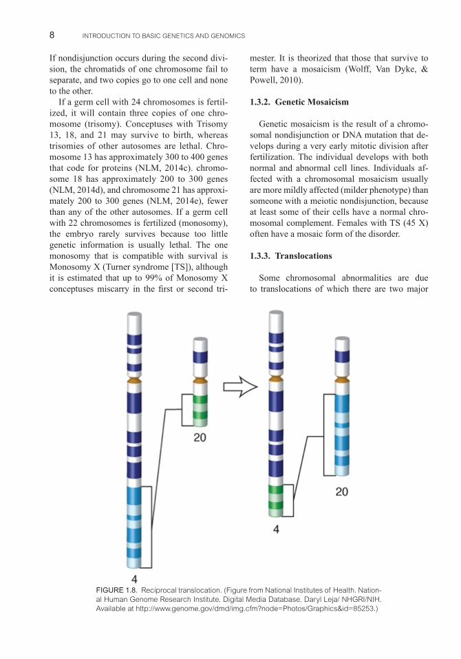
Some chromosomal abnormalities are due to translocations of which there are two major
FIGURE 1 .8 . Reciprocal translocation. (Figure from National Institutes of Health. Nation-al Human Genome Research Institute. Digital Media Database. Daryl Leja/ NHGRI/NIH. Available at http://www.genome.gov/dmd/img.cfm?node=Photos/Graphics&id=85253.)

91.3. Numerical and Structural Cytogenetic Abnormalities
types, Robertsonian and reciprocal. Robertso-nian translocations develop when the centromere of one acrocentric chromosome fuses with the centromere of another acrocentric chromosome. The two most common types are Robertsonian and reciprocal translocations.
Robertsonian translocations should be consid-ered when a couple has more than one child with Down syndrome (DS). Although DS is most fre-quently due to a nondisjunctional error, about 5%
of DS is the result of an unbalanced Robertsonian translocation. In an unbalanced Robertsonian translocation of chromosomes 14 and 21, the off-spring inherits the translocated chromosome as well as two normal 21s and one normal 14. The embryo has a normal number of chromosomes (46), but because of the fused 21 and 14, it inher-its three copies of chromosome 21 and manifests the typical DS phenotype.
Standard nomenclature for translocation DS in
FIGURE 1.9. Chromosomal Inversion. (Figure from United States National Library of Medicine. Genetics Home Reference. Available at http://ghr.nlm.nih.gov/handbook/illustrations/inversion.)

423
Chapter 18
Genomics and Symptomatology
QUANNETTA T. EDWARDS, Ph.D, FNP-BC, WHNP-BC, AGN-BC, FAANP SUSAN T. TINLEY, Ph.D, RN, CGC (RET)DIANE C. SEIBERT, Ph.D, ARNP, FAANP, FAANANN H. MARADIEGUE, Ph.D, FNP-BC, FAANP
will be described. Genomic advances in symp-tom management will help ensure that the right person receives the right therapy (personalized or precision health care), will reduce adverse ef-fects, and will improve both quality of life and overall health outcomes.
18.2. GENOMICS AND CANCER-RELATED FATIGUE
Fatigue is a symptom manifested in patients that is associated with a wide range of diseases and syndromes that often affect individual’s physical, social, and mental functioning (Land-mark-Hoyvik et al., 2010). It is also one of the most commonly reported symptoms in individu-als diagnosed with cancer, often resulting in increased stress and anxiety and other health-related quality of life issues including, but not limited to, impaired physical performance, inac-tivity, helplessness, sleep disturbances, lack of appetite, and/or depression (Escalante, Kallen, Valdres, Morrow, & Manzullo, 2010; Horneber, Fischer, Dimeo, Ruffer, & Weis, 2012; Saligan & Kim, 2012). Cancer-related fatigue (CRF) is defined as a “distressing, persistent, subjective sense of tiredness or exhaustion related to can-cer or cancer treatment that is not proportional to recent activity and interferes with usual function-ing” (National Comprehensive Cancer Network (NCCN), 2015m p. MS-3). The symptoms of
18.1. INTRODUCTION
An individual’s genome impacts the trajectory of their health and illness throughout life. Thus far, the focus of this book has been on the im-pact of genomics as it relates to an individual’s response to drugs, their risk for developing dis-eases based on their family history, or as a re-sult of shared genetic and environmental factors. Beyond health, illness, and the effectiveness of drugs, genomics also influences how an indi-vidual experiences a particular disorder—or the symptomatology of that condition. Research ex-amining the genetics of common symptoms of-fers the promise of reducing adverse symptoms and improving quality of life, which is particu-larly important, because symptom management is a key function for nursing.
In this chapter, brief overviews of two com-mon symptoms are discussed These symptoms are cancer-related fatigue and pain, as each of these symptoms have been shown to be influ-enced by genomic discoveries. The role of ge-nomic variants (i.e., single nucleotide polympor-phisms [SNPs]) influencing the onset, duration, or severity of symptoms, as well as how they influence therapeutic responses in preventing, alleviating, or eliminating patient’s symptoms, will be discussed. Where applicable, other influ-ences that may potentiate the effects of genomic variants in symptom manifestation and treatment
Objectives:
•Discusstheimpactofgenomicsoncancerrelatedfatigue.•Discussthegenesassociatedwithpain.•Explaintheroleofnursingregardinggenomicsandsymptomatology.

424 GENOMICS AND SYMPTOMATOLOGY
CRF can occur during and after treatment of the cancer and are often attributed to treatment regi-mens, such as cytotoxic chemotherapy, radiation therapy, or other biological treatments. However, CRF can vary, occurring any time in the course of disease, and may be self-limiting or persisting for many years even after treatment (Bower et al., 2006; Horneber et al., 2012). For example, in one longitudinal study of breast cancer survivors, approximately 34% of the participants reported fatigue 5 to 10 years after diagnosis (Bower et al., 2006). Similar effects regarding fatigue per-sistence after diagnosis and treatment have been reported (Husson et al., 2013; Hwang et al., 2014).
Fatigue among patients with cancer can be as-sociated with multifactorial etiologies and mani-fest in a myriad of clinical features (Horneber et al., 2012; NCCN, 2015). Contributing factors to fatigue include pain, emotional distress, sleep disturbances, and co-morbidities such as ane-mia; poor nutrition; physical inactivity; medica-tion side effects; alcohol and/or substance abuse; therapeutic management with cytotoxic, biolog-ic, or radiation therapy; or other medical condi-tions (National Cancer Institute, 2013; NCCN, 2015). However, the diagnostic criteria for CRF include fatigue, distress, or impairment due to fatigue, etiology related to cancer or cancer treat-ment, and the exclusion of underlying psychiat-ric or medical disorders. CRF is common, with studies revealing varied prevalence estimates ranging from 25% to 99% of patients with can-cer experiencing this symptom, depending on the population and type of assessment (Bower, 2007). Most common clinical manifestations of CRF are focused on fatigue, lack of energy, ex-haustion, or impaired physical function that can affect physical or psychosocial well-being of the individual (Horneber et al., 2012).
The exact biological mechanisms of CRF are unknown; however, some proposed mechanisms associated with the symptom include 5-HT3 neu-rotransmitter deregulation, disturbances in hypo-thalamic regulation, dysregulation in circadian rhythm, skeletal muscle wasting, pro-inflamma-tory cytokines, or dysregulation of inflammatory cytokines (Barsevick et al., 2013; Bower & Lam-kin, 2013; Horneber et al., 2012; NCCN, 2015; Ryan et al., 2007). Genomic factors associated with inflammation have been linked to CRF prior
to, during, and after treatment, particularly in the pro-inflammatory cytokine network (Bower & Lamkin, 2013). Molecular-genetics, particularly gene polymorphisms, have shown to possibly play an important role in the mechanism of CRF. One example is that of proinflammatory cytokine SNP that influences interleukin (IL) and/or tumor necrosis factor (TNF) genes (i.e., IL1B; IL-6; TNFα) and is associated with CRF both during and after treatment (Aouizerat et al., 2009; Bow-er, 2007; Bower & Lamkin, 2013; Miaskowski et al., 2010). Alteration in pro-inflammatory cytokine production of IL6 and other inflamma-tory markers has been linked with persistent fa-tigue among breast cancer survivors (Bower et al., 2006; Collado-Hidalgo, Bower, Ganz, Cole, & Irwin, 2006). Persistent CRF among patients with breast cancer has also been found to be as-sociated with increased activity of pro-inflam-matory transcription factors NF-κB activity and decreased expression of glucorticoid receptor anti-inflammatory transcription factors (Bower, Ganz, Irwin, Arevalo, & Cole, 2011). The as-sociation with CRF and cytokines, the proteins that mediate cell-to-cell communication, may be due to dysregulation of cytokines often attribut-ed to cancer and cancer treatments that increase plasma levels of many cytokines, particularly the TNF-α and certain IL genes (Ahlberg, Ekmanb, Gaston-Johansson, & Mock, 2003; Ryan et al., 2007). Cytokines are important for the develop-ment and functioning of the immune response, and aberrant expression from genetic polymor-phisms have been associated with overall disease and functionality (Smith & Humphries, 2009). Pro-inflammatory cytokines, particularly IL-1B, IL-6 and TNF-α, are thought to induce symptoms of fatigue via signaling of the central nervous system through varied somnogenic influence (Weschenfelder, Sander, Kluge, Kirkby, & Him-merich, 2012).
The nuclear factor NF-κB, pro-inflammatory transcription factor, for example, is activated by the cancerous tumor microenvironment (Aggar-wal, 2004) and, thus, pretreatment CRF may be due to tumorigenesis (Bower & Lamkin, 2013). Fatigue often occurs also during treatment, par-ticularly due to chemotherapy or radiation ther-apy; this effect has been associated with eleva-tions in inflammatory markers secondary to the therapeutic intervention. For example, in one

42518.3. Genomics and Pain
study, changes in inflammatory markers, includ-ing C-reactive protein and IL1 receptor antago-nist, were found to be associated with fatigue symptoms among certain individuals with breast and ovarian cancer (Bower et al., 2009). CRF has been found to occur years after completion of therapy in breast cancer survivors and altera-tions in proinflammatory markers also have been found among these individuals (Collado-Hidalgo et al., 2006; Orre et al., 2009).
Besides pro-inflammatory genes, other ge-nomic factors are currently being studied to de-termine their impact on fatigue among cancer patients. For example, the relationship between dysfunction in certain mitochondrial genes has been found among prostate cancer patients re-ceiving external beam radiation (Hsiao, Wang, Kaushal, & Saligan, 2013). Advances in genomic technologies will certainly change the face of un-derstanding the molecular impact of genetics and CRF that will enhance predicting and managing the symptoms and improving outcomes.
18.2.1. Future Implications—Cancer Related Fatigue and Genomics
Although many studies have shown an associ-ation with varied inflammatory markers and CRF among patients with cancer, causality has not been established and gaps in knowledge contin-ue, warranting further research in this area (Sa-ligan & Kim, 2012). Specifically, problems exist regarding measurement of CRF, exact under-standing of the underlying biology of the symp-tom, and clinical trials targeted towards CRF (Barsevick et al., 2013). However, future links between CRF and inflammatory markers may be a means to provide personalized/precision medi-cine as a prognostic biomarker for fatigue among cancer patients or genetic predictors of fatigue for therapeutic management (Collado-Hidalgo et al., 2006; Jim et al., 2012), as well as future de-velopment of effective treatments such as cyto-kine antagonists targeting CRF (Bower & Lam-kin, 2013). Further, because fatigue is a complex symptom with phenotypic heterogeneity, the in-clusion of biobehavioral research of fatigue may provide clarity and contribute to the understand-ing of CRF and to future development of genetic/genomic interventions (Lyon, McCain, Pickler, Munro, & Elswick, 2011). The international and
interdisciplinary GeneQoL Consortium is one means to improve patient outcomes regarding issues that impact quality of life, including that of fatigue (GeneQol Consortium, 2015; Sprang-ers et al., 2009). This consortium was established to investigate genetic disposition of patient-re-ported quality-of-life outcomes in order to gain insight on the impact of disease and treatment on patient outcomes (GeneQol Consortium, 2015). Clinical implications of the consortium are based on obtaining genetic knowledge, in-cluding understanding biological pathways that may impact quality of life (GeneQol Consor-tium, 2015), and incorporating understanding of the biological and genetic mechanisms of CRF (Barsevick, Frost, Zwinderman, Hall, & Halyard, 2010).
18.3. GENOMICS AND PAIN
Pain is universal and has been described since antiquity, and yet the biochemical pathways and pathophysiologic underpinnings of pain are only now beginning to be unraveled. An excellent re-view of the history of pain and pain management can be found in the article by Meldrum (2003). Despite the lack of understanding and effective therapies to manage pain, helping patients and families cope with the manifestations of pain has been a central feature and core mission area for nurses since at least the 19th century, when Flor-ence Nightingale discussed pain management in “Notes on Nursing” (Nightingale, 1860). Since then, many resources have been developed to im-prove nursing competency in pain management, including a pain management nursing certifica-tion awarded by the American Nurses Creden-tialing Center; establishment of the American Society for Pain Management Nursing (ASPMN) in 1991, which publishes a journal dedicated en-tirely to nursing management of pain; and nurs-ing competencies focused on pain management (http://mbon.maryland.gov/Documents/pain_management.pdf).
Over 100 million Americans suffer from pain every year (Institute of Medicine ([U.S.] Com-mittee on Advancing Pain Research, 2011), and a recent study estimates that chronic pain is more expensive than cancer, heart disease, and dia-betes, costing up to $635 billion a year (i.e., up to $300 billion in direct costs and $334 billion

426 GENOMICS AND SYMPTOMATOLOGY
in lost productivity) (Gaskin & Richard, 2012). Pain, a very important signaling mechanism in animals, plays a significant role in survival, be-cause it forces the animal to protect an injury un-til it heals. Pain becomes chronic when the nox-ious stimuli persists after healing is complete, evolving into pain without a purpose. The experi-ence of pain is highly variable; some people who experience acute pain from a noxious agent will develop chronic pain whereas others, exposed to the same causative agent, will not (Mogil, 2012). The perception of pain is highly individualized and subjective as well, as indicated by pain rat-ing scores used in research and clinical settings. Finally, there is marked individualized variabil-ity in response to analgesics, with some people responding to very small doses and others requir-ing much larger doses to feel an effect (Aubrun, Langeron, Quesnel, Coriat, & Riou, 2003). All of this variability raises questions about the possi-bility that genetic factors could influence the ex-perience of pain. More than 350 candidate genes have been associated with variability in pain sensitivity, and twin studies have clarified the heritability of pain in several specific conditions; however, to date, there are still many unanswered questions related to the basic genetic underpin-nings of pain (Smith & Muralidharan, 2012).
Several classification systems have been devel-oped to guide pain assessment and management. Some of the most common categorization sys-tems include stratifying pain as acute or chronic, based on the length of time it has been present, or by intensity (mild to moderate, or severe). Numeric pain scales were developed to attempt to capture pain intensity. Physiologic changes (nociceptive, neuropathic, and inflammatory) and manifestations based on the affected tissue types (skin, muscles, viscera, joints, tendons, and bones) have also been used to categorize pain. Some diseases have classic pain characteristics; therefore, pain has been clustered by syndrome (cancer, fibromyalgia, migraine, etc.) (University of Wisconsin, 2014). This chapter reviews just a few of the areas being explored in the genomics of pain.
A few rare single gene disorders are associ-ated with alterations in pain sensation, such as paroxysmal pain or a complete inability to feel pain. Although at first glance the genes appear to involve seemingly unrelated functional pro-
tein classes, on closer inspection, almost all the pathways involve the SCN9A gene in one way or another (Mogil, 2012). SCN9A encodes for the alpha subunit of the NaV1.7 sodium channel, expressed primarily in peripheral sensory nerves that transmit pain, touch, and smell signals to the central nervous system (Mogil, 2012). The role that SCN9A plays in more common pain re-sponses is less clear (Young, Lariviere, & Belfer, 2012). Some studies have shown that variations in SCN9A alter pain responses (e.g., individu-als with a G allele on SCN9A report lower pain scores compared with individuals having the less common A allele); other studies have been unable to consistently replicate those findings (Starkweather & Pair, 2013)
Genome wide association studies (GWAS) are changing the landscape of genomic research. GWAS tools such as computerized databases containing the reference human genome se-quence, a map of human genetic variation, and new analytic technologies that can cheaply, rap-idly, and accurately analyze whole-genome are making it possible to locate the genes associated with common diseases, such as asthma, diabetes, and heart disease. Once a new gene has been lo-cated, new strategies to detect, prevent, and/or treat a particular disorder can be developed.
Despite the rapid advances made in fields such as cardiology, oncology and hematology using GWAS, research into the genes associated with pain has lagged for several reasons. Reasons in-clude the subjective nature of pain, relatively low funding levels, and associated lack of interest from researchers (Mogil, 2012). More recently, however, more research has been done to ex-amine the genetic underpinnings of pain associ-ated with diseases such as migraine headaches, osteoarthritis, endometriosis, Crohn’s disease, and temporomandibular disorder (TMD) (Young et al., 2012). Some of the genes found in these studies include a mutation in ZNF429 and a gene upstream of the RHBDF2 gene, whose function is currently unknown. Although no polymor-phisms have been found in any of the known opioid receptor genes, ethnic differences appear to play an important role, because the strongest effect in one study was country of origin (Mo-gil, 2012). Despite the progress that has recently been made in understanding the genes associated with pain response, significant hurdles remain,

427
such as technology and data analysis limitations, explaining phenotypic heterogeneity, and the costs associated with conducting GWAS, which typically involve genotyping large numbers of people (Young et al., 2012).
18.3.1. Epigenetics and Pain
As scientific understanding of epigenetic pro-cesses has increased over the past decade, there has been a concurrent surge of research explor-ing epigenetic mechanisms involved in regu-lating the nervous system, particularly the epi-genetic processes associated with memory and synaptic plasticity (Denk & McMahon, 2012). Recent studies have found that epigenetic control is particularly important in three distinct areas: peripheral inflammation, pain processing, and plasticity.
When the body is exposed to a physical, chem-ical, or biologic insult, an inflammatory response develops rapidly to remove or destroy the inju-rious material, setting the stage for tissue repair and healing. Inflammation is commonly associat-ed with pain, redness, heat, and swelling, which have been shown to increase healing, either be-cause the injured person protects that area or be-cause inflammatory cells, such as macrophages, produce high levels of insulin-like growth fac-tor-1 (IGF-1), which increases the rate of heal-ing and muscle regeneration (Lu et al., 2011). The inflammatory response is highly complex, involving genes involved in a number of dif-ferent processes, such as antimicrobial defense, immune response, tissue repair, and remodeling. Epigenetic changes in genes regulating macro-phage function may play a particularly impor-tant role in inflammatory response, because they help macrophages change in response to differ-ent infectious organisms (Bayarsaihan, 2011). Epigenetic changes in T cells and monocytes, transcription factors found in several protein families (NF-κB, FOXP3, IRF, STAT), RE1si-lencing transcription factors (REST), and his-tones (i.e., histone H4 hyperacetylation) have all been shown to regulate inflammatory re-sponse (Bayarsaihan, 2011; Selvi, Mohankrish-na, Ostwal, & Kundu, 2010).
Inflammation persisting beyond the normal healing period is considered “chronic inflam-mation” characterized by chronic infiltration of
mononuclear immune cells and low antioxidant and high free radicals levels, creating an envi-ronment in which tissue healing is occurring at the same time tissue is being damaged, become a self-perpetuating cycle of injury, repair and usually, pain (Khansari, Shakiba, & Mahmoudi, 2009). Identifying the epigenetic mechanisms associated with the development of chronic pain may open the door to much needed advances in pain management (Denk & McMahon, 2012; Mogil, 2012). Epigenetic regulation of tissues in the nervous system is of particular interest, because these are the cells that generate and transmit pain signals, but also because the regen-eration rate of individual neurons in the nervous system is very slow. The same neuron is likely to survive for decades, and because DNA is very resistant to change, epigenetic adjustments that occur over the life of the cell may be critical as they continually adapt to environmental stressors (Seo et al., 2013). A neuron’s use of epigenetics to adapt to the environment is particularly impor-tant because such changes are reversible; there-fore, if pain develops because of an epigenetic change, a drug that chemically “resets” the neu-ron to its normal state might be a powerful thera-peutic tool (Seo et al., 2013). Although there is evidence from animal studies to demonstrate that it is possible to modify epigenetic mechanisms with drugs (Geranton, 2012), more research needs to be done before human studies can begin to ensure that the drugs do not alter epigenetic mechanisms in other tissues (Crow, Denk, & Mc-Mahon, 2013).
Reinforcing the idea that epigenetics plays an important role in the development and per-ception of pain, data from genomic studies ex-amining pain response is often contradictory. For example, ORPM1, a common m-opioid re-ceptor variant, has been shown to increase ben-dorphin binding and activation of g-proteins in some studies, but this effect is not found in all studies on a consistent basis. Similarly, in some studies, individuals with COMT variations were found to have greater sensitivity to pain, but the same association has not been seen in other stud-ies (Bond et al., 1998; Kim et al., 2004; Zubieta et al., 2003). These inconsistencies suggest that factors, such as epigenetics, may play an impor-tant role in pain phenotypes (Seo et al., 2013)
Opioids are used to treat pain on a routine ba-
18.3. Genomics and Pain

428 GENOMICS AND SYMPTOMATOLOGY
sis, and many opioids are now approved for use, each of which has different efficacy and adverse response profiles based on the individual. If an individual has a poor response to one opioid, an-other is usually tried in a trial and error fashion until the most effective drug is found. Because this random approach exposes individuals to ad-verse effects and decreases quality of life, as the search for an effective analgesic agent continues, there has been considerable interest in finding the genetic factors that explain the variability in response to opioids (Branford, Droney, & Ross, 2012).
Several studies have found an association between the A118G SNP in OPRM1 and opioid dosing (Chou et al., 2006a; Chou et al., 2006b; Reyes-Gibby et al., 2007), but similar to the is-sues with OPRM1 mentioned above, a meta-analysis of genetic association studies concluded that the A118G SNP is inconsistently associated with pain-related phenotypes (Walter & Lotsch, 2009). It is becoming clear that the genes associ-ated with pain relief are not the same genes that influence the development of adverse effects, highlighting the need to carefully choose and de-fine the phenotype being studied. It is also very likely that interactions among multiple genetic and environmental factors are playing important roles in the phenotype (Branford et al., 2012).
Acute pain transforms into chronic pain in a complex series of discrete pathophysiologic and histopathologic steps involving more than 2000 gene changes in over 400 candidate genes. Broad-ly, the process involves neurons that abandon the normal “modulated” response to pain (reversible activation of intracellular signal-transduction cascades) and adopt a more persistent “modified” response involving relatively permanent chang-es to neuron activation (Voscopoulos & Lema, 2010). A growing body of evidence suggests that nociceptor modifications can occur in response to psychological triggers, further complicating research efforts (Diatchenko, Fillingim, Smith, & Maixner, 2013).
Understanding the genomics of individual variability in pain sensitivity, analgesic response, adverse reactions, and triggers that transform acute to chronic pain is still in its infancy. Once the genomic roadmap has been created, the high-ly complex interactions between the environment and the human genes that regulate the pain re-
sponse can then be explored, leading to a more effective and safe personalized approach to pain management.
18.4. NURSING ROLE AND SYMPTOMATOLOGY
Symptom management applies to nurses pre-pared at every level and practicing in virtually every setting—from nurses providing direct care to patients in inpatient and outpatient settings to conducting genomic research and to nursing fac-ulty and nurses leading the largest health care sys-tems. Nurses working at the point of care should be familiar with the emerging genetic information that helps support decisions that can improve the care of an individual patient, the goal of precision and personalized care. Nurses in faculty roles are responsible for ensuring that students entering nursing are well informed about genetics and are prepared to use emerging genomic information to improve patient outcomes. Nurse researchers might want to focus their scientific efforts on ex-ploring the biological and behavioral aspects of symptoms such as pain and fatigue, with the goal of developing new knowledge and new strate-gies for improving patient health and quality of life (National Institute of Nursing Research, nd). Nurse administrators play a critical role, because they serve in key leadership roles as systems be-gin to integrate genomic discoveries into clinical settings in a meaningful way. Their support can accelerate nurses’ use of genomic information as it continues to emerge from large population based studies.
18.5. REFERENCES
Aggarwal, B. B. (2004). Nuclear factor-kappaB: the enemy within. Cancer Cell, 6(3), 203–208. doi: 10.1016/j.ccr.2004.09.003
Ahlberg, K., Ekmanb, T., Gaston-Johansson, F., & Mock, V. (2003). Assessment and manage-ment of cancer-related fatigue in adults. The Lan-cet, 362(9384), 640–650. doi: 10.1016/S0140-6736(03)14186-4
Aouizerat, B. E., Dodd, M., Lee, K., West, C., Paul, S. M., Cooper, B. A., . . . & Miaskowski, C. (2009). Pre-liminary evidence of a genetic association between tumor necrosis factor alpha and the severity of sleep disturbance and morning fatigue. Biol Res Nurs, 11(1), 27–41. doi: 10.1177/1099800409333871
Aubrun, F., Langeron, O., Quesnel, C., Coriat, P., &

433
Index
amyotrophic lateral sclerosis (ALS), 289, 312, 313, 316,
analgesic response, 428anaphase, 4, 6, 350ancestry of origin, 31, 42, 48–50, 371 anencephaly, 26, 126aneuploidy, 68, 130, 136, 360, 369Angelman syndrome (AS), 24, 125, 131anorexia nervosa, 127antenatal steroids, 195anticipation, 25, 31, 56, 57, 78antiphospholipid antibody syndrome (APS), 276antisense strand, 10–12, 15–18antithrombin deficiency, 275, 277aplastic anemia, 268, 279apoptosis, 4, 126, 129, 180, 216, 301, 304, 306, 311,
313, 328, 345, 348, 350, 352–356, 358, 360 applied behavioral analysis (ABA), 166, 168ASD-related syndromes, 167Ashkenazi Jewish Ancestry, 366, 371, 372, 375Asian, 18, 48, 75, 110, 114, 133, 190, 191, 200, 203,
210, 212, 215, 220, 221, 243, 260, 374, 384assisted reproductive technologies (ART), 71, 131assortative mating, 21asthma, 22, 24, 189, 190, 191, 196–200, 335, 426 astrocytoma, 299, 364atherosclerosis/arteriosclerosis, 40, 209, 211, 214,
216, 223–226, 230, 242, 253atopic dermatitis, 196, 335attention-deficit/hyperactivity disorder (AD/HD), 164atypical HUS (aHUS), 270autism, 27, 44, 68, 125, 127, 164–166, 167, 169, 295,
296 autism spectrum disorders (ASD), 68, 164, 166, 173autism-specific screening, 165Autoimmune Polyendocrine Syndrome Type 1, APS,
1, 328Autoimmune Polyendocrine Syndrome Type 2, APS,
2, 328Autoimmune Polyendocrine Syndrome Type 3, APS,
3, 328autoimmune regulator (AIRE), 328autonomy, 85–94, 172, 185, 388, 394autosome, 2, 8, 19
base pairs, 211, 215, 243, 358, 359, 392
1000 Genomes Project, 24021-hydroxylase deficiency (21-OH), 1275-azacytidine, 259AAT deficiency (AATD), 190, 191AAT inhalation therapy, 191ABO blood types, 19, 20achondroplasia, 57, 155, 161Acquired Bone Marrow Failure Syndromes
(ABMFS), 255, 279acrocentric, 3, 9acute lymphocytic leukemia (ALL), 281, 282acute myeloid leukemia (AML), 281acute promyelocytic leukemia (APL), 281, 282A Disintegrin And Metalloproteinase with Thrombos-
pondin Motifs (ADAMTS13), 270adenine, 2, 10, 12, 261, 294, 301adenocarcinoma, 201–203, 368, 369, 381, 382,
385ADCY9, 196adolescent cataracts, 299adrenal gland, 127adrenocorticotropic hormone (ACTH), 127Agency for Healthcare Research and Quality
(AHRQ), 413allergens, 196–198alpha thalassemia (α-thal), 27, 258Alpha-1 antitrypsin (AAT), 189, 190alpha globin (α-globin), 256–258Alport syndrome, 303, 305Alstrom syndrome, 305alternative splice sites, 13Alzheimer’s disease, 26, 94, 96, 179, 183, 184ambiguous, 53, 128, 144, 239, 241, 328American Academy of Family Physicians (AAFP),
125, 168American Congress of Obstetrics and Gynecologists
(ACOG), 125American Nurses Credentialing Center (ANCC), 73,
414, 425American Organization of Nurse Executives (AONE),
413American Society for Pain Management Nursing
(ASPMN), 425amino acid, 10–13, 15–18, 142, 192, 198, 217, 219,
256–258, 290, 292, 301Amsterdam criteria (I and II) and colon cancer, 380

434 Index
basophils, 255, 264, 284Bacterial Inhibition Assay (BIA), 141, 142, 291Baye’s Theorem, 73BCR/ABL translocation (Philadelphia chromosome),
353Beckwith-Wiedemann syndrome, 131, 158, 161,
162Bernard-Soulier Syndrome (BSS), 272beta thalassemia (β-thal), 143, 256, 258beta globin (β-globin), 17, 70, 256–259Bethesda guideines, 380–383biallelic, 125, 279, 363biobanks, 414bioinformatic programs, 240, 249biological treatments, 424Birt-Hogg-Dube Syndrome (BHDS), 204bone marrow failure syndrome, 255, 268, 277, 279,
280BRAF V600E mutation and colon cancer, 102, 201,
354, 383, 391brain tumors, 199298Branchio-Oto-Renal syndrome (BOR), 305bronchi, 203, 361Brugada Syndrome, 225, 244, 245Burkitt lymphoma, 285
café au lait spots, 20, 152, 297–299, 364 Canavan disease, 21Cancer Genomics, 345–407
BRCA genes (see Cancer Genomics—HereditaryBreast and Ovarian Cancer)cancer, multistage genetic and genomic process,
358–360cancer stem cells, 349, 350cancer types and causes, 360–361carcinongens (examples of), 360–361carcinogenesis primer, 15, 345, 348–353, 355,
356, 358, 366, 378caretaker genes, 355characteristics of cancer cells, 353, 360chromosomal instability pathway, 358–360clonal cell evolution theory, 349congenital hypertrophy of the retinal pigmentepithelium (CHRPE), 385–386Cowden’s Syndrome, 362, 371–372, 378, 385,
395–396de novo and cancer genomics, 363–365, 385Definition of Common Terms used in CancerGenetics, 345–346Desmoid tumors, 385direct to consumer testing, 72, 393DNA repair genes, 356DNA methylation (hypermethylation), 23, 127,
181, 356, 392, 420, 421Empiric Risk Models for Breast Cancer (Gail and
Claus), 374
endometrial cancer, 45, 371–373, 379, 381, 383, 384, 395, 396
epigenetics, 13, 23, 125, 126, 135, 168, 181, 242, 332, 333, 345, 348, 356, 357, 421
epigenomics, 417, 419–421epithelial cell adhesion molecule gene (EpCAM),
363, 378, 380, 383, 384, 389ethical, legal and social implications of single
gene testing for hereditary colon cancer syndromes, 388–389, 391, 394 (biomark-ers)
evaluation of Genomic Application in Practice and Prevention (EGAPP) and Lynch Syndrome, 383–384
exome sequencing, 33, 36, 70, 144, 240, 241, 390, 417, 418
familial cancer, 345, 351, 397future advances and technologies and genomics of
cancer, 389–392exome sequencing, 33, 36, 70, 144, 240, 241,
390, 417, 418genome wide associated studies, 391next generation sequencing (NGS) and paneltesting, 389–390, 391tumor markers, 391–392Whole genome sequencing, 391
gain-of-function, 328gatekeeper genes, 355genetic mutations, 198, 244, 270, 272, 283, 293,
296, 298, 304, 307, 311, 331, 339, 349, 352, 358, 360, 364
genetic mutations and Cancer Development, 351
Hereditary Breast Cancer Syndromes (including HBOC and other syndromes), 41, 60, 365, 372, 375, 376
data collection (personal/family history; physi-cal examination) 370–372
Hereditary Breast and Ovarian Cancer syndrome (HBOC) (BRCA gene mutations), 41, 54, 56, 365, 367
genetic testing, 374Identification of Risk Elements and Rednext generation sequencing (NGS), 389–390Other Data Collection Resources, 372primary prevention, 376–377RAPID approach to assessment, 394–397
risk probability, 372–374Risk Probability Models (i.e. BRCAPRO,BOADICEA), 372–374
Secondary Prevention and enhanced Surveil-lance, 377
Hereditary Cancer, 345, 351, 352, 362–365, (Breast and Ovarian, 365–378), (Colon Cancer, 378–388)

435Index
Hereditary Colon Cancer Syndromes, 363, 378–388
Lynch Syndrome, 363, 378–385introduction 378differential diagnoses and Lynch Syndrome,
385management, 384phenotype and characteristics (Colon),
378–379phenotype and characteristics (Extracolonic
[e.g. endometrial; ovarian]), 379–380Risk Assessment and Identifying individu-
alssuspect for Lynch, 380variants of Lynch Syndrome (Muir-Torre
and Turcot), 380polyposis syndromes, 378, 385
Familial Adenomatous Polyposis (FAP) and Attenuated FAP, 385–386
Gardner’s syndrome, 386Turcot syndrome, 380, 386
MUTYH-associated polyposis (MAP), 363, 386
Other Hereditary Colon Cancer Syndromesnot associated with adenomatous, 386–388
Peutz-Jeghers (e.g. hamartomatous polyps), 378, 386, 388
juvenile polyposis, (e.g. juvenile polyps) 363, 378, 387
immunohistochemical (IHC) staining, 379Knudson’s two-hit hypothesis 354–355, 366KRAS mutations, 392loss-of-function, 112, 113, 328loss of heterozygosity, 354loss of homozygosity, 354microRNA, 345, 358, 359, 392microsatellites, 357, 379microsatellite instability, 379mismatched repair genes (MMR), 360, 378, 379,
380, 383molecular genomic make-up, 347Muir-Torre Syndrome (See variants of Lynch
Syndrome)MUTYH-associated polyposis (See polyposis
syndromes)next generation sequencing (NGS), 389, 390online/web-based resources, 396ovarian cancer, 379–380p53 Tumor Suppressor gene, 350, 354–356, 359,
362, 364personalized care, 347, 349, 360, 376, 389, 390,
392pharmacogenomics and cancer, also refer to Chap-
ter 5 (5-Fluorouracil; Irinotecan; Mercapto-purine), 103–108
irinotecan, 393Tamoxifen, 392–393Thiopurines, 393
precision medicine, 348, 349, 360, 389, 390Precision Medicine Initiative, 348proto-oncogenes, oncogenes and carcinogenesis,346, 352–353RAPID Approach and Cancer Genomics,
369–376; Utilization of RAPID for breast cancer, 394–395; (See also Chapter 2)
retinoblastoma, 354Risk Assessment and Cancer Genomics 369–376Data Collection (Personal and Family Historyand Physical Examination), 370–372Risk Identification and Risk Elements, 372Risk Probability, 372–374Risk Communication/Counseling and Risk Man-
agement, 375–376sex cord tumors with annular tubules (SCTAT),
386somatic mutation, 351, 354, 362, 366sporadic cancer, 346, 351, 354, 356, 362, 369statistical trend data incidence and mortality,
346–347stem cell theory, 349tumor suppressor genes, 352, 353–356tumor markers and breast cancer (Example of),
391–392tumor markers and colon cancer (Example of), 392Turcot Syndrome (See variants of Lynch Syn-
drome) cancer, multistage genetic and genomic process, 358–360
cancer related fatigue (CRF), 423, 425cancer stem cells, 349, 350cancer types and causes, 360–361candidate genes, 426cardiac channelopathies, 244–245cardiovascular pharmacogenomics, 109caring, 7, 80, 86, 91, 123, 132, 168, 169, 250, 292,
415carrier rate, 21carrier screening, 21, 71carcinogens (examples of), 51, 200, 361carcinogenesis primer, 348–351caretaker genes, 355catch-down growth, 155catch-up growth, 155catecholaminergic polymorphic ventricular tachycar-
dia (CPVT), 244–245celiac disease, 129, 153, 157, 330, 331, 335, 336cell cycle, 4, 5, 22, 181, 277, 278, 345–360, 364, 366 cell free fetal (cff) DNA testing, 136cell membrane disorders, 255, 259Centers for Disease Control and Prevention (CDC),
28, 35, 63, 73, 141, 150

436 Index
central dogma, 12centrioles, 4, 7centromere, 2–6, 9, 10CF transmembrane conductance regulator (CFTR),
102, 130, 135, 192, 193, 203CF-related diabetes (CFRD), 192characteristics of cancer cells, 360Chediak-Higashi syndrome (CHS), 265, 272CHEK2, 200, 389chemokines, 197chemotherapy, 104, 106, 114, 203, 262, 267, 278,
282, 300, 391, 392, 393, 424childhood bronchiectasis, 194childhood leukemia, 126chloride channels, 192chorea, 57, 301–303chorionic villus sampling [CVS], 136, 150Chromatid, 4–7, 350chromosomal instability pathway, 358–360chromosomal microarray, 68chromosome microarray, 149chronic bronchitis, 191chronic inflammation, 257, 427chronic lymphocytic leukemia (CLL), 269, 281, 283chronic myeloid leukemia (CML), 281, 282chronic obstructive pulmonary disease (COPD), 189chronic pain, 257, 299, 309, 425–428chronic sinus infection, 130, 194Chuvash polycythemia (CP), 262ciliary bodies, 194Circadian Locomotor Output Cycles Kaput genes,CLOCK genes, 336, 337cleft lip and palate, 25, 153client focused counseling, 67, 74Clinical Pharmacology and Therapeutics Implementa-
tion Consortium, 105clinical practice knowledge, 413–414ClinSeq Project, 418–420clonal cell evolution theory, 349C-myc oncogene, 285coagulopathies, 255, 274–275codominant, 19, 108, 190codon, 10–13, 17, 18, 258, 301cognition, 19, 35, 52, 59, 128, 150, 164–166, 259,
262, 347, 371, 384common complex diseases, 32, 41, 181, 209communication disorders, 166, 317communication patterns, 75compound heterozygous, 104Comprehensive Cancer Network (CCN), 38, 199,
282, 284, 348, 396, 423comprehensive health history, 411
Congenital Adrenal Hyperplasia (CAH). 127, 135, 142, 159, 327, 328, 330
congenital amegakaryocytic thrombocytopenia (CAMT), 268, 279
congenital dyserythropoietic anemias (CDA), 278congenital heart defects, 52, 129, 141, 153congenital heart disease, 26, 142, 152, 155, 243congenital hypertrophy of the retinal pigment epithe-
lium (CHRPE), 385–386congenital hypothyroidism, 142connective tissue disorders, 242–243connexin, 304consanguinity, 21, 31, 32, 42, 45, 49, 150, 266, 267,
277, 380consent, 44, 69–71, 75, 77, 87, 92, 94, 136, 141,
144–146, 170, 204, 382, 388, 390, 392, 412consequentialism, 86constitutional growth delay, 157consultant, 48, 54context, 52, 74, 75, 79, 85–88, 93, 95, 107, 124, 155,
157, 184, 185, 212, 215, 217, 223, 231, 412, 417, 418, 420
Coordinating Center of the Newborn Screening Translational Research Network (NBSTRN), 145
copy number variant (CNV), 68 127, 167, 203, 242Cornelia DeLange syndrome (CdLS), 155Coronary Artery Disease (CAD), 40, 209–212, 225,
226, 239Cowden Syndrome, 362, 371–372, 378, 385, 395–396CpG islands, 23, 420craniopharyngioma, 157cri du chat, 153crossing over, 5, 6, 10cryopreservation, 131cryptorchidism, 127, 152cultural background, 42, 74Cushing disease, 157cyclo-oxygenase 1 (COX1), 271CYP1A1, 126CYP2C19, 101, 111–113, 116CYP2C9, 101, 109–111, 116, 225CYP3A5, 196CYP3A7, 196Cystic Fibrosis (CF), 13, 21, 25, 40, 71, 91, 96, 124,
130, 132, 135, 143, 146, 157, 189, 190–193, 197, 413
CF Foundation, 190cytogenetics, 6, 68, 282, 284cytokines, 134, 197, 261, 266, 339, 350, 358, 424. cytosine, 2, 10, 23, 30, 420cytotoxic chemotherapy, 424
Data Collection (Personal and Family History and Physical Examination), 370–372
de novo and cancer genomics, 363–365, 385

437Index
de novo (new) mutation, 19, 20, 23, 32, 56–58, 162, 167, 180, 243, 297, 298
decision-making, 33, 35, 37, 58, 85–90, 92, 93, 94, 144, 185, 240, 306, 360, 376, 388, 391, 394, 414, 419
decitabine, 259Definition of Common Terms used in Cancer Genet-
ics, 345–346deletion syndrome (congenital cardiac anomaly), 243dendritic cells, 255, 266, 277deontologic, 86deoxyribonucleic Acid (DNA), 1, 167depression, 44, 78, 111, 127, 134, 180, 268, 296, 302,
303, 312, 393, 414, 423Desmoid tumors, 385desmopressin (DDAVP), 271development, 1, 20, 28, 52, 53, 90, 103, 125, 130,
144, 150, 153, 165, 166, 172, 196, 198, 214, 219, 250, 291, 292, 296, 304, 305, 313, 315, 331, 348, 360
developmental delays, 149, 150, 152, 165, 198, 296developmental domains, 165developmental milestones, 164, 291developmental surveillance, 165dextrocardia, 194diakenesis, 5Diamond-Blackfan anemia (DBA), 279differential diagnoses and Lynch Syndrome, 385Disseminated Intravascular Coagulation (DIC), 268dihydropyrimidine dehydrogenase gene (DPD) andDPD deficiency, 104, 105dihydropyrimidine dehydrogenase gene (DPYD), 104diplotene, 5Direct To Consumer (DTC), 72, 227direct to consumer testing, 72, 393DNA methylation (hypermethylation), 23, 127, 181,
356, 392, 420, 421DNA repair genes, 356DNA sequence analysis, 70, 72DNA sequencing technologies, 143Down syndrome (DS), 9, 153, 159, 161, 280dried blood spots, 141–146drug-induced thrombocytopenia, 269Duchenne Muscular Dystrophy, 22, 45, 242duplications, 10, 68, 128, 168, 298, 304Dyskeratosis Congenita (DC), 265, 277, 278dyslipidemias, 214dysmorphology, 32, 52, 53, 149, 150, 166
early intervention, 136, 165, 172, 306early onset Parkinson disease, 310East Asian, 133, 221, 260eclampsia, 130–134, 219Evaluation of Genomic Application in Practice(EGAPP), 79, 106, 211, 227, 229, 383, 384, 412Ehlers-Danlos Syndrome (EDS), 243
electronic medical records (EMRs), 34, 169, 414emphysema, 57, 189–191, 193, Empiric Risk Models for Breast Cancer (Gail and
Claus), 34, 58, 374endometrial cancer, 45, 371–373, 379, 381, 383, 384,
395, 396environmental influences, 132, 191, 228, 239, 307,
327, 333, 335, 412, 413eosinophils, 255, 264ependymomas, 298, 299, 364epidermal growth factor receptor (EGFR), 41, 203,
369epigenetics, 13, 23, 125, 126, 135, 168, 181, 242,
332, 333, 345, 348, 356, 357, 421epigenomics, 417, 419–421epilepsy, 25, 126epithelial cell adhesion molecule gene (EpCAM),
363, 378, 380, 383, 384, 389epithelial cells, 197, 335erythrocytosis, 261, 262erythropoietin receptor (EPOR) gene, 261, 262essential hypertension, 209, 210, 218–223essential thrombocytosis, 271, 272, 283Ethical Legal and Social Implications (ELSI), 85, 89,
183, 348, 388, 394ethical, legal and social implications of single gene
testing for hereditary colon cancer syndromes, 388–389, 391, 392 (biomarkers)
Ethical Standards, 85, 88, 90ethics, 85–91, 96, 135, 183, 185, 412ethnic background, 52, 54, 74, 301, 313Eunice Kennedy Shriver National Institute of Child
Health and Human Development, 135, 144, 296
evaluation of Genomic Application in Practice and Prevention (EGAPP) and Lynch Syndrome, 383–384
evidence-based practice, 116, 184, 185excess growth, 157exome sequencing, 33, 36, 70, 144, 240, 241, 390,
417, 418exons, 12, 70, 190, 374, 390expansion mutation, 18, 25experience of pain, 426experiential background, 74, 75extreme longevity, 181
F508del mutation, 192Factor XI deficiency, 274faculty, 100, 185, 409–413, 428 false positive rates, 127Familial Adenomatous Polyposis (FAP) and Attenu-
ated FAP, 348, 356, 363, 364, 378, 381, 386familial Amyotrophic Lateral Sclerosis (ALS), 314familial cancer, 345, 351, 397familial dilated cardiomyopathy, 242

438 Index
familial hypercholesterolemia, 44, 241, 247, 249, 250familial Parkinson disease, 311familial short stature, 155, 159family health history (FHH), 32, 37, 38, 45, 62, 90,
123, 124, 129, 183, 184, 203, 262, 334, 410, 411
family relationships, 45, 46, 67, 75, 77Fanconi anemia (FA), 159, 277, 356, 366features of genetic syndromes, 151–153, 172fecundity, 127Federal Trade Commission, 73financial management, 413FLCN, 204, 205, 364Florence Nightingale, 425flourescent in situ hybridization (FISH), 68folic acid, 26, 126, 127, 260folic acid supplementation, 26, 126, 127, 260follicular lymphoma, 285Foundation for Sickle Cell Disease Research
(FSCDR), 413foundational thinking skills, 414Fragile X premature ovarian insufficiency (FXPOI),
128, 296Fragile X syndrome (FXS), 18, 25, 45, 173, 295, 296,
315Fragile X-associated tremor, ataxia syndrome (FX-
TAS), 296, 426frailty, 179, 180frameshift mutation, 17, 18, 297Friedreich’s Ataxia (FRDA), 293–294frontotemporal dementia, 179full mutations, 128functional change, 18, 256, 360future advances and technologies and genomics of
cancer, 389–393FVL, 275, 276
gain of function, 18, 245, 248, 263, 275, 328, 352, 353
galactosemia, 142, 143gamma globin, 256Gardner’s syndrome, 386gatekeeper genes, 355Gaucher, 21, 169, 267, 268gene therapy, 89, 191, 258, 259, 269, 312GeneQol Consortium, 425Genes associated with MS, 308–309genes in common pathways, 167genetic alliance, 78, 79, 96, 144, 240, 241genetic discrimination, 67, 240genetic/genomic competencies, 85, 124Genetic Information Nondiscrimination Act (GINA),
67, 94, 190, 240genetic mutations, 198, 244, 270, 272, 283, 293, 296,
298, 304, 307, 311, 331, 339, 349, 351, 352, 358, 360, 364
genetic mutations and Cancer Development, 351genetic polymorphisms, 109, 110, 196, 424genetic predisposition, 54, 195, 199, 218, 242, 334genetic red flags, 415genetic risk assessment, 73genetic screening, 37, 38, 42, 71, 124, 136, 240, 247Genetics Interdisciplinary Faculty Training (GIFT),
410Genetics Program for Nursing Faculty (GPNF), 410genome sequencing, 33, 70, 80, 87, 96, 115, 116, 143,
229, 241, 249, 389, 391, 414, 417, 418, 421Genome Wide Association Studies (GWAS), 27, 191,
197, 210, 211, 309, 391, 426genomic imprinting, 24, 125, 331genomic literacy, 411Genomic Nursing State of the Science Advisory
Panel, 412genomic risk assessment, 34, 149, 411genomic variants, 423germline mosaicism, 20, 23germline mutation, 20, 199, 204, 278, 378, 380gestational diabetes mellitus (GDM), 131GH excess, 158Gilbert’s syndrome, 106Glanzmann’s Thrombasthenia (GT), 272glucose-6-phosphate Dehydrogenase Deficiency
(G6PD), 261glucuronidated SN-38, 105, 106glutathione S-transferase M1 (GSTM1), 200Glutathione S-transferase T1 (GSTT1), 200GPC5, 200GPIIb/IIIa receptor, 271Grave’s Disease (GD), 159, 327, 330Gray platelet syndrome, 272growth charts, 155, 157growth curves, 155growth factors, 197, 280, 352growth hormone deficiency, 153, 157growth potential, 155growth retardation, 157guanine, 2, 10, 12, 23, 107, 294, 301, 313, 393, 420Guthrie card, 141
Hardy Weinberg calculation, 73Hashimoto’s thyroiditis, 159, 327, 330412Hereditary Breast and Ovarian Cancer (HBOC), 19,
41, 54, 56, 57, 70, 362, 365–367, 371, 372HDAC inhibitors, 259health care economics, 413health care transition, 168, 170, 171, 173Health Insurance and Portability Accountability Act
(HIPAA), 48, 49, 67, 92, 95, 372, 388, 414health outcomes, 28, 57, 60, 125, 132, 145, 196, 227,
340, 392, 413, 423health promotion and disease prevention, 57, 412hematologic neoplasms, 255, 280, 283

439Index
hemoglobin (Hb), 16, 17, 132, 135, 143, 255–267, 332
Hemoglobin Bart’s Hydrops Fetalis, 258hemoglobin C (HbC), 256hemoglobin E (HbE), 256hemoglobin H disease, 258hemoglobin S (HbS), 256hemoglobinopathies, 134, 143, 255–260, 278hemolytic anemia, 18, 255–261, 270, 278, 280hemophagocytic lymphohistiocytosis (HLH), 267hemophilia, 22, 134, 272–275hemophilia A, 273, 274hemophilia B (Christmas disease), 274hepatitis B, 52, 195, 361hepcidin, 263, 264HER2 protein, 114Hereditary Breast and Ovarian Cancer syndrome
(HBOC) (BRCA gene mutations), 19, 41, 70, 76, 77, 348, 366, 372, 375, 376
Hereditary Breast Cancer Syndromes (including HBOC and other syndromes), 41, 60, 365, 372, 375, 376
Hereditary Cancer, 345, 351, 352, 362–365, (Breast and Ovarian, 365–378), (Colon Cancer, 378–388
Hereditary Colon Cancer Syndromes, 378, 380, 383, 385, 388
Hereditary elliptocytosis (HE), 260Hereditary Hemochromatosis (HH), 50, 263hereditary persistence of fetal hemoglobin (HPFH),
256hereditary spherocytosis (HS), 260, 278heritability, 27, 72, 167, 178, 196, 210, 212, 213, 215,
218, 331, 334, 426heritability of aging, 178Hermansky-Pudlak syndrome (HPS), 265, 272heterogeneity, 19, 53, 70, 209, 221, 229, 249, 260,
272, 349, 389, 390, 392, 425, 427heterotaxy, 194heterozygous, 19, 104, 105, 107, 108, 109, 217, 248,
249, 265, 267, 275, 283, 329, 354, 386 HFE, 48, 50, 51, 55, 62, 263Hispanic, 26, 42, 48, 75, 91, 126, 191, 195, 210, 243,
247, 366histiocytes, 255, 266histone inhibitor, 127histone modification, 13, 181, 420HLA-A class I major histocompatibility genes, 203Hodgkin, 52, 285Homozygous, 13, 35, 104–109, 217, 224, 249, 257,
262, 263, 266, 275, 276, 283, 329, 366horizontal transmission, 32, 50HSC transplantation, 258, 259, 262, 282Human Immunodeficiency Virus (HIV), 18, 99, 113,
141, 268, 361
Human Leukocyte Antigen (HLA), 114, 258, 308, 309, 329
HLA-B*5701, 113, 114, 308human resource management. 413, 414Huntington disease (HD), 18, 40, 77, 179, 289,
300–302, 418Huntington Disease Society of America, 302hypergonadotropic hypogonadism, 130hyperimmunoglobulin E syndrome, 266hypermethylates, 128hypersensitivity reaction, 113hypertrophic cardiomyopathy, 225, 242, 245, 247,
250, 294hyperviscosity, 262hypothyroidism, 129, 142, 157hypoxia-inducible factor-alpha (HIF1-α), 262
Identification of Risk Elements and Red next genera-tion
sequencing (NGS), 389–390immune Dysregulation Polyendocrinopathy, Enter-
opathy, X-linked Syndrome, IPEX Syndrome, 329
immune-mediated asthma, 197immunohistochemical (IHC) staining, 379importin-beta, 196imprinting, 24,125, 131, 135, 331incidental findings, 69, 70, 71, 77, 87, 89, 144, 146,
418infectious disease, 102, 113, 194, 195infertility, 10, 40, 42, 44, 46, 54, 68, 27, 129–131,
134, 162, 163, 192, 296inflammation, 189, 197, 214, 307, 314, 332, 335, 336,
427inflammatory bowel disease, 72, 159, 380inflammatory mediators, 197inflammatory response, 427information technology, 412, 4114informed consent, 44, 69–71, 75, 76, 87, 92, 94, 136,
144, 204, 388, 390, 396, 412inherited bleeding disorders, 134Inherited Bone Marrow Failure Syndromes (IBMFS),
277innate immunity, 197, 200, 264innovation, 95, 96, 424insertion, 17, 18, 27, 202, 204, 220, 226, 259, 280,
282, 294, 304, 311, 378, 391 insulin-like growth factor-1 (IGF-1), 427intellectual developmental disorders, 166intellectual disability (ID), 53, 128, 153, 167, 198,
291, 295, 296interactome, 35interleukin (IL), 197, 275, 308, 309, 424International Standard Cytogenomic Consortium, 69Interphase, 5, 6, 283interpretation of sequence analysis, 70

440 Index
intracytoplasmic sperm injection (ICSI), 131, 163intramural training, 413intravenous human plasma-derived augmentation
therapy, 191Introns, 12, 14, 70, 417IPO13, 196IQ, 130, 163, 165158, 160, 161, 163, 164irinotecan, 99, 101, 105, 106, 107, 393Irish, 18, 133iron overload disorders, 264
JAK/STAT pathway, 262JAK1/JAK2, 284Japanese, 205Jervall and Lange-Nielson, T13.2, 305Job syndrome, 266justice, 85–91, 388, 391juvenile HD, 302Juvenile deafness, 54juvenile idiopathic arthritis, 159juvenile polyposis, (e.g. juvenile polyps), 363, 378,
387
karyotype, 24, 32, 46, 68, 157–160, 163, 282, 283
KRAS mutations, 202, 203, 392Klinefelter syndrome, 130, 35, 158, 161–164Knudson’s two-hit hypothesis 354KRAS, 61, 102, 199, 202, 203, 392
Langerhans cell histiocytosis (LCH), 267large-cell lung cancer, 203Le Fraumini, 199leadership, 34, 89, 182–184, 327, 409, 412, 413, 414,
428learning disabilities, 20, 44, 54, 125, 128–130, 152,
154, 164, 166, 295 296, 298, 331leprosy, 195leptokene, 5Leukocyte adhesion deficiency (LAD), 266Lisch nodules, 154, 298, 364literacy, 44, 74, 80, 92, 96, 185, 411liver disease, 191, 274locus, 19, 70, 209, 211, 212, 230, 245, 362Loeys-Dietz Syndrome (LDS), 243Long QT Syndrome (LQTS), 215, 225, 244long-term storage, 145loss of function, 18, 24, 103, 112, 113, 245, 248, 271,
275, 299, 328, 353, 354, 355loss of heterozygosity, 354loss of homozygosity, 354lymphocytes, 68, 255, 264, 267, 275, 283, 284, 329,
379, 381lymphomas, 284, 285Lynch Syndrome, 363, 378–385Lysosomal Storage Disorders, 267
Manager Inventory Tool, 413, 415mannose-binding lectin 2 (MBL2), 192mantle cell lymphoma, 285March of Dimes, 38, 96, 142Marfan Syndrome (MFS), 155, 158, 161–165, 223,
224, 243, 328massive parallel sequencing, 70maternal copy, 1257maternal phenylketonuria, 133May-Hegglin anomaly, 268Medicaid, 38, 172, 41Medicare, 38, 172, 414meiosis, 1, 5–7, 10, 22, 24, 25, 295, meiosis II, 6MELAS, 25, 33, 305Mendelian inheritance, 19, 32, 48, 56, 61, 182, 184,
239, 337, 366, 417, 420meningioma, 298, 300, 361menopause, 68, 126, 296mental health disorders, 127messenger RNA (mRNA), 10, 358, 418metabolic disorders, 21, 24, 126, 150, 264, 267metabolomics, 99, 209, 215, 217, 222, 223, 229metacentric, 3, metaphase, 4–6 68, 284, 350, 358methylation, 13, 23, 28, 107, 127, 181, 225, 137, 225,
295, 296, 326, 345, 354–358, 363, 378, 383, 392, 420, 421
microbiome, 72, 334microfluidic chip, 143microRNA, 14, 250, 311, 333, 345, 358, 359, 419,
420microsatellites, 357, 379microsatellite instability, 358, 379mid-parental height, 157, 160miscarriage, 40, 42, 44, 129missense mutation, 16, 19, 219, 220, 304, 311mitochondria, 1, 25–27, 126, 168, 181, 261, 293,
303–306, 311, 362, 425 mitochondrial disease, 126mitochondrial DNA (mtDNA), 1, 26, 3050mitochondrial encephalomyeopathy, lactic acidosis
and stroke=like episodes (MEILAS), 305mitochondrial inheritance, 25, 27mitosis, 1, 4–6, 350, 360mode of inheritance, 161, 414modifier genes, 25, 192, 197, molecular genetics, 282, 317, 410, 413, 424molecular genomic makeup, 347molecular testing, 143, 146, 375monocytes, 264, 427Monogenic Diabetes, maturity-onset diabetes,
MODY, 327, 328, 331, 338, 339monosomy, 8, 10, 22mosaic, 8, 20, 23, 129, 173, 297

441Index
mosaicism, 8, 20, 23, 130, 173motor disorder, 166Muir-Torre Syndrome (See variants of Lynch Syn-
drome) multifactorial inheritance, 378, 380multigenic, 167Multiple Endocrine Neoplasia Type 1, MEN1, 327,
328Multiple Endocrine Neoplasia Type 2, MEN2, 328,
364multiple myeloma, 284, 356multiple sclerosis (MS), 289, 303, 307–310, 316, 317muscular dystrophy, 222, 45, 242mutation detection rate, 70MUTYH-associated polyposis (MAP), 363, 386mycobacterium tuberculosis, 195myelodysplastic syndromes, 280myelokathexis, 265, 285myeloproliferative neoplasm (MPN), 262, 271, 272,
276, 283myocardial infarction and survival, 112, 210,
214–215, 225, 226, 275myoclonic epilepsy with ragged red fibers, 25myotonic dystrophy, 13, 18, 31
National Association for Retarded Citizens, 142NCLEX (National Council Licensure Examination),
414NHGRI (National Human Genome Research Insti-
tute), 2–8, 12–14, 26, 28, 69, 94, 95, 159, 160, 211, 357, 391
National Institute of Nursing Research (NINR), 115, 410, 413, 428
National Newborn Screening and Genetics Resource Center, 142
National Research Council of the National Academy of Science (NAS/NRC), 142
National Society of Genetic Counselors (NSGC), 38, 59, 73
negative selection, 328Neonatal alloimmune thrombocytopenia (NAIT),
270neonatal respiratory distress syndrome (RDS), 195NDI (Nephrogenic Diabetes Insipidis), 329neural tube defect, 26, 126neurodevelopmental disorder (NDD), 166, 172neurofibromas, 20, 52, 154, 300neurofibromatosis, 20, 56, 126, 153, 173, 289, 297,
299, 305, 315, 316, 364Neurofibromatosis Type 1 (NF1), 297Neurofibromatosis Type 2 (NF2), 297neuroplasticity, 165, 168neuropsychiatric disorder, 167neutropenia, 105–107, 264–266, 277, 279, 280, 393neutrophil elastase (NE), 189, 191, 279neutrophils, 190, 255, 264–266newborn hearing screening, 306
newborn screening, 40, 70, 71, 77, 79, 133, 141–146, 193, 289, 291, 296, 411
Newborn Screening Quality Assurance Program(NSQAP), 143next generation sequencing, 70, 71, 77, 80, 224, 225,
348, 389, 417, 418next generation sequencing (NGS) and panel testing,
389–391Niemann-Pick disease, 267NHLBI (National Heart, Lung and Blood Institute
Exome Sequencing Project), 240nitrogenous base, 1nociceptor modifications, 428nondisjunction, 7–9, 352non-Hodgkin lymphoma (NHL), 52, 285nonmaleficence, 85, 87, 89, 90, 412nonsense mutation, 16, 17, 297non-small cell lung cancer (NSCLC), 190, 200–202nonstructural single gene disorders, 239, 244Northern European Caucasian, 130, 190, 191, 193,
260noxious stimuli, 426nuclear DNA (nDNA), 1, 2, 25, 181, 293nuclear membrane, 4–6nucleotide, 1, 4, 15, 27, 72, 127, 128, 167, 181, 211,
220, 223, 229, 259, 282, 313, 332, 348, 356, 358, 390–392, 417, 419, 423
numeracy, 74, 80nurse administrators, 185nurse researchers, 34, 61, 100, 116, 146, 409, 412,
413, 428National Institute of Nursing Research (NINR), 115,
410, 413, 428nursing research, 89, 115, 409, 410, 412, 413, 428
obesity, 331, 332, 333, 334, 335, 336, 337, 339(t)obstructive sleep apnea (OSA), 262occupational fumes, 189, 191oncogene, 199–203, 285, 328, 345, 346, 352–355,
358, 365, 419, 421oncology pharmacogenomics 103,online resources, 28, 182, 190, 337, 396, 417opt-out, 144optic gliomas, 298, 300osteogenesis imperfecta, 305Other Data Collection Resources, 372Other Hereditary Colon Cancer Syndromes not as-
sociated with adenomatous , 383ovarian cancer, 12, 19, 25, 41, 54, 56, 57, 70, 348,
365–369, 371–380, 384, 385, 394, 395, 425
p53 Tumor Suppressor gene, 350, 354–356, 359, 362, 364
pan cardio panel, 224, 225, 241pancreatic exocrine insufficiency, 192Parkinson disease, 26, 310
442 Index
p arm, 2pachytene, 5, 6pain, perception of, 426, 427pain management, 425–428pain phenotypes, 427pain processing, 291, 427pain sensitivity, 426, 428pan ethnic, 130pancytopenia, 267, 268, 277–279paracentric inversion, 10paroxysmal nocturnal hemoglobinuria, 271, 272, 276,
280patau syndrome (Trysomy 13), 153paternal age, 126, 134, 136paternal genes, 125, 133paternal malnutrition, 125paternal smoking, 126pathologic chromosomal abnormalities, 159penetrance, 19, 20, 31, 32, 37, 56–58, 69, 71, 72, 239,
274, 276, 295, 297, 301, 305, 367, 384, 385, 389
performance improvement, 413, 414pericentric inversion, 10peripartum depression (PPD), 134peripheral inflammation, 427personal history, 32, 41, 42, 44, 58, 61, 126, 276, 363,
364, 370, 374, 380, 389personalized care, 33, 54, 62, 115, 345, 394, 417, 428personalized medicine, 99, 100, 113, 240, 391, 392,
412Peutz Jeghers, 362, 363, 378, 386, 388pharmacodynamics, 102, 103, 217pharmacogenetics, 79, 100, 102, 110, 113–115, 2210,
217, 223, 229pharmacogenomics and cancer, also refer to Chapter
5 (5-Fluorouracil; Irinotecan; Mercaptopurine), 103–108
pharmacokinetics, 101, 103PHE, teratogenic effect, 292phenotype and characteristics (Colon), 378–379phenotype and characteristics (Extracolonic [e.g.
endometrial; ovarian]), 379–380phenotypic heterogeneity, 390, 425, 427phenylalanine (PHE), 11, 133, 141, 192, 290, 291,
293phenylalanine hydroxylase (PAH), 290, 291phenylketonuria (PKU), 124, 125, 133, 135Pick’s disease, 179PIK3CA activating mutations, 203PKU diet, 291, 292placentation, 134plasticity, 166, 295, 427platelet closure time (PFA-100), 271platelet disorders, 255, 268, 269, 271, 272pluripotent stem (iPS) cells, 250, 255, 259
pneumonia, 191, 194, 301, 312point mutation, 70, 128, 199, 282, 294, 295, 296, 304,
311, 352point-of-care (POC), 141Poliomyelitis, 195poly-A tail, 12polycyclic aromatic hydrocarbons, 126, 200Polycythemia, 255, 261, 262, 275, 283Polycythemia Vera (PV), 262polyposis syndromes, 378, 385pomalidomide, 259population-based screening, 142porphyrias, 264positive predictive value, 76, 227postpartum hemorrhage (PPH), 134Post-transfusion purpura (PTP), 269Potocki-Lupski syndrome, 167Prader-Willi syndrome (PWS), 28, 125, 155, 161,
331, 339precision care, 60, 360precision medicine, 32, 34, 36, 99, 100, 103, 107,
109, 113, 348, 349, 425Precision Medicine Initiative, 348precocious puberty, 128, 158, 328preconception care, 123–125, 135preconceptional counseling, 124–126, 132–133predictive biomarkers, 100predictive testing, 72, 76, 77, 78, 179, 388preeclampsia, 130, 131, 133, 134, 219preimplantation genetic diagnosis (PGD), 131premature aging, 180, 181premature ovarian insufficiency, 128, 129premutation, 25, 128, 294–296premutation carriers, 128, 295prenatal education, 92, 124prenatal screening, 71, 123, 135, 136, 258, 314prenatal testing, 92, 136, 150, 299presymptomatic testing, 33, 72, 141primary ciliary dyskinesia (PCD), 194primary familial and congenital polycythemia/eryth-
rocytosis (PFCP/Erythrocytosis), 261primary myelofibrosis, 284primary prevention, 246, 376primary screening test, 143probability, 21, 23, 32, 33–36, 50, 57–62, 73, 127,
149, 347, 365, 370, 372–376, 395, 396proband, 32, 33, 41, 44, 46, 48, 51, 54, 167, 204, 218,
248, 368, 376, 381, 387, 394–396progeria, 180pro-inflammatory cytokine, 144, 339, 424prophase, 4–6protease-3, 190protein C, 270, 274–277protein C deficiency, 275, 276protein S deficiency, 275–277

443Index
proteinase inhibitor (Pi), 189Proteome, 35prothrombin gene mutation (PTM), 275proto-oncogenes, oncogenes and carcinogenesis, 346,
352–354pseudoautosomal regions, 22PTEN mutations, 203public expectations, 80public health programs, 141pulmonary cysts, 203–205pulmonary surfactant, 195purine, 1, 2, 108pyrimidine, 1, 2, 104Pyruvate Kinase Deficiency (PKD), 261
q arm, 3, 198, 328quality of life, 59, 60, 82, 105, 293, 307, 316, 348,
377, 384, 412, 423, 425, 428
radiation therapy, 278, 300, 424random assortment, 6, 25RAPID approach, 35, 36, 50, 58, 60–62, 149, 370,
375, 394RAPID Approach and Cancer Genomics, 369–376 Utilization of RAPID for breast cancer, 394–395rare chromosomal anomalies, 167RBC osmotic fragility, 260rearrangements, 68, 69, 201, 203, 281, 282, 352, 374,
391recurrence risk, 19, 73, 392, 419red blood cell, 108, 216, 255religious background, 75renal tumors, 204, 205replication, 1–5, 15, 181, 211, 212, 215, 221, 278,
352, 354, 366, 378, 419reproductive outcomes, 123reproductive plan, 59, 125residual dried blood spots, 144–146Resources for Genetic/Genomic Neurological Disor-
ders, 315–317Resources for Counseling & Testing, 79response to analgesics, 426retinoblastoma, 20, 200, 350, 354, 356retinoblastoma (RB1), 20, 200, 350, 354, 356Rett syndrome, 23rheumatoid arthritis, 153, 191, 331, 335ribonucleic acid (RNA), 10, 215, 295, 358ribosome, 13, 278Risk Assessment and Cancer Genomics 369–376Risk Assessment and Identifying individuals sus-
pectedfor Lynch Syndrome, 380risk communication, 31, 33, 35, 38, 60, 61, 87, 370,
375, 376, 396Risk Communication/Counseling and Risk Manage-
ment, 375–376
Risk Identification and Risk Elements, 372risk management, 33–36, 60, 61, 78, 365, 370, 375,
376, 377, 390, 396risk probability, 34, 35, 57, 58, 59, 62, 370–376ristocetin cofactor, 271, 273RNA polymerase, 12, 358Robertsonian translocation, 9, 10Rubinstein-Taybi syndrome, 155
sapropterin dihydrochloride, 293schizophrenia, 27, 1270Schwachman-Diamond syndrome (SDS), 277, 278Schwannomas, 297–300, 364Secondary Prevention and enhanced Surveillance,
377second-tier test, 143segregation analysis, 241seizure disorder, 126sense strand, 10–12, 15–18sequenced, 144, 241, 418sequential screening, 72serum EPO, 261, 262Severe Combined Immunodeficiency (SCID), 142,
143, 146Severe congenital neutropenias (SCN), 279severe recurrent ear infections, 194sex chromosome aneuploidy, 130sex cord tumors with annular tubules (SCTAT), 386short interfering RNA (siRNA), 14short stature, 125, 129, 151, 153, 155, 157–161, 198,
277, 279, 298, 305, 331sickle cell anemia, 16, 48, 136, 143, 257, 259sickle cell disease (SCD), 19, 21, 42, 54, 125, 132,
135, 142, 146, 256, 257, 413sickle cell trait (SCT), 132, 257Sigma Theta Tau, 413signaling pathways, 203, 204, 352, 354, 356simultaneous sequencing, 70single nucleotide polymorphism (SNP), 27, 167, 211,
223, 332, 348, 391, 392single-gene disorders, 127, 189, 223, 225, 327situs inversus totalis, 194small cell lung cancer (SCLC), 190, 200–202smoking, 32, 43, 44, 100, 126, 134, 189, 191, 200,
202, 204, 210, 2, 218, 227, 262, 275, 307SNP arrays, 223, 227, 229social support, 73, 78, 80, 376somatic mosaicism, 21somatic mutation, 15, 20, 262, 280, 283, 351, 354,
362, 366Sotos, 161, 162Southern blot, 278, 286sperm motility, 194spermatogenesis, 126spherocytosis, 260, 278spina bifida, 26, 126

444 Index
spinal muscular atrophy (SMA), 71spindle fibers, 4, 6splenectomy, 260, 267splicing, 12, 13, 313spontaneous de novo mutation, 167, 297, 298spontaneous pneumothorax, 204, 364sporadic cancer, 346, 351, 356, 362, 369squamous cell cancer, 203start codon, 10, 11, 13State Boards of Nursing, 414statistical trend data incidence and mortality,
346–347stem cell theory, 349Stickler syndrome, 153stillbirth, 126, 129, 131, 132stop codon, 10–18, 258Storage Pool Deficiency (SPD), 272strategic management, 413, 414submetacentric, 3substance abuse, 44, 127, 424suicide, 61, 302Summer Genetics Institute (SGI), 410symptom management, 303, 412, 423, 428synthetic elastase inhibition therapy, 191Systemic mastocytosis (SM), 284
T helper 2– (Th2–), 197, 200tachypnea, 194, 195tall stature, 151, 157–159, 161, 162Tamoxifen, 102, 374, 376, 393targeted mutation analysis, 69, 70, 72targeted physical examination, 52, 394, 411, 415Tay Sachs, 21, 71, 131, 136telomerase, 4telomere, 4, 181, 278, telophase, 4, 6thalassemia, 21, 136, 143, 159, 255–258, 264thiopurine methyltransferase (TPMT), 107Thiopurines, 107, 393third party payers, 414three-generation pedigree, 31, 37, 46, 47, 55, 59, 61,
62, 123, 124, 204, 414thrombocytopenia, 104, 268, 269–272, 274,
277–280thrombocytopenia and absent radii (TAR), 268thrombocytosis, 268, 271, 272, 283thrombophilia, 275–277thrombopoietin (TPO), 268thrombotic thrombocytopenic purpura [TTP], 270thymine, 2, 10, 12, 104Timothy (syndrome), 167tobacco, 42, 124, 191, 196, 198, 199, 361TP53, 199, 203, 282, 283, 389training, 34, 73, 100, 143, 375, 377, 395, 410–414transciptomics, 209, 215, 222, 229, 250
transcription, 2, 10–15, 20, 134, 180, 198, 214, 215, 289, 295, 301, 304, 311, 313, 328, 336, 338, 352, 354, 356, 358, 359, 366, 418, 424, 427
transfer RNA (tRNA), 12, 13transferrin saturation, 263transgenerational epigenetics, 125translation, 2, 10–15, 28, 79, 145, 182, 289, 295, 297,
306, 313, 345, 358, 359, 391, 412, 420translocation, 8–10, 102, 131, 281, 282, 285, 331,
351–353, 391Treacher-Collins syndrome, 153triggers, 32, 185, 196, 198, 262, 336, 428trinucleotide repeat, 31, 57, 128, 293, 294, 301triplet, 10, 25, 128Tri-Service Nursing Research Program, 413trisomy, 8, 10, 136, 153, 155, 159, 161, 167, 173,
280trisomy 21 (DS), 155, 159, 161, 167, 280tuberculosis, 190, 195, 336tuberous sclerosis, 20, 28, 126, 167Tumor growth factor-beta (TGF-β), 192tumor markers, 360, 391tumor markers and breast cancer (Example of),
391–392tumor markers and colon cancer (Example of), 392tumor necrosis factor (TNF), 424tumor suppressor, 199, 200, 204, 297, 298, 3283, 36,
345, 352, 353–359, 362–366, 397, 419tumor suppressor genes, 352, 353–356Turcot syndrome, 380, 386Turkish, 133Turner syndrome, 8, 127, 129, 155, 157, 160, 161,
167twin studies, 195, 196, 210, 218, 426Type 1 Diabetes Mellitus (T1DM), 330Type 2 diabetes mellitus, 327, 331Type II pneumocytes, 195typical and atypical development, 166
UDP-glucuronosyltransferase 1A1 (UGT1A1) and UGTT1A1 genetic testing, 105, 106
uninformative result, 70, 72, 77, 78uniparental disomy, 24, 331uracil, 10, 12, 99, 101, 103, 104, 106, 116, 393United States Food and Drug Administration (FDA),
72U.S. Secretary of Health and Human Services (HHS)
Advisory Committee on Heritable Disorders in Newborns and Children (SACHDNC), 142
Usher syndrome, 303, 305, 317
valproate, 126valproic acid (VPA), 126, 127, 259variable expression, 209, 295, 297variant of unknown/uncertain significance, 33, 70

445Index
variants of Lynch Syndrome (Muir-Torre and Turcot), 380
vascular dementia, 111, 179velocardiofacial syndrome (VCFS), 153venous thrombosis, 40, 224, 226, 269, 275, 276vertical transmission, 19, 33, 55, 56, 386vestibulocochlear nerve (tumor), 298virtue, 85–87, 90, 182vitamin B12, 127, 268Vitamin D deficiency, 157, 337Vitamin D3, 309, 337VKORC1 genotypes, 109, 110von Hippel-Lindau (VHL), 57, 262, 365von Recklinghausen disease, 153von Willebrand disease (VWD), 134, 269–271
Warrdenburg syndrome, 265, 285 Web Based Genetic Institute, 410Werner syndrome, 180WHIM syndrome, 265white blood cell, 255, 264, 265, 267
whole exome sequencing, 33, 36, 241, 417wild type, 15–18, 106-112, 201, 281, 336, 366 , 392Williams syndrome 155Wiskott-Aldrich syndrome, 268, 272Wolffian duct 130Wound healing 152, 243, 266, 414Whole genome sequencing, 33, 80, 87, 96, 115, 116,
241, 249, 389, 391, 414, 417
X chromosome, 2, 18, 19, 21, 22, 33, 56, 128-130, 136, 159, 161, 168, 198, 242, 261, 273, 274, 296
X-inactivation, 22X-linked dominant, 19, 23X-linked ichthyosis, 23X-linked inheritance, 33
Y chromosome, 2, 19, 21, 22, 23, 126, 129
zonulin pathway, 335zygotene, 5