Embed Size (px)
Citation preview
�������� ����� ��
Unraveling the electron transfer processes of a nanowire protein from Geobac-ter sulfurreducens
Monica N. Alves, Ana P. Fernandes, Carlos A. Salgueiro, Catarina M.Paquete
PII: S0005-2728(15)00198-XDOI: doi: 10.1016/j.bbabio.2015.09.010Reference: BBABIO 47532
To appear in: BBA - Bioenergetics
Received date: 10 July 2015Revised date: 4 September 2015Accepted date: 30 September 2015
Please cite this article as: Monica N. Alves, Ana P. Fernandes, Carlos A. Salgueiro, Cata-rina M. Paquete, Unraveling the electron transfer processes of a nanowire protein fromGeobacter sulfurreducens, BBA - Bioenergetics (2015), doi: 10.1016/j.bbabio.2015.09.010
This is a PDF file of an unedited manuscript that has been accepted for publication.As a service to our customers we are providing this early version of the manuscript.The manuscript will undergo copyediting, typesetting, and review of the resulting proofbefore it is published in its final form. Please note that during the production processerrors may be discovered which could affect the content, and all legal disclaimers thatapply to the journal pertain.
ACC
EPTE
D M
ANU
SCR
IPT
ACCEPTED MANUSCRIPT
1
Unraveling the electron transfer processes of a nanowire protein from Geobacter
sulfurreducens
Mónica N. Alves1,#, Ana P. Fernandes1,2,#, Carlos A. Salgueiro2, Catarina M. Paquete1,*
1 Instituto de Tecnologia Química e Biológica – António Xavier, Universidade Nova de
Lisboa, Oeiras, Portugal
2 UCIBIO-Requimte, Departamento de Química, Faculdade de Ciências e Tecnologia,
Universidade Nova de Lisboa, Caparica, Portugal
# These authors contributed equally to this publication
* Corresponding author at Instituto de Tecnologia Química e Biológica António Xavier,
Universidade Nova de Lisboa, Av. da República - EAN, 2780-157 Oeiras, Portugal.
Phone number: +351214469321
E-mail address: [email protected]
Keywords: Multiheme cytochromes, Geobacter, Nanowires, Electron transfer,
Extracellular respiration
Abbreviations: DMRB, dissimilatory metal reducing bacteria; MHC, multiheme
cytochromes; NMR, nuclear magnetic resonance
1. Abstract
The extracellular electron transfer metabolism of Geobacter sulfurreducens is
sustained by several multiheme c-type cytochromes. One of these is the dodecaheme
cytochrome GSU1996 that belongs to a new sub-class of c-type cytochromes.
GSU1996 is composed by four similar triheme domains (A-D). The C-terminal half of
the molecule encompasses the domains C and D, which are connected by a small
linker and the N-terminal half of the protein contains two domains (A and B) that form
one structural unit. It was proposed that this protein works as an electrically conductive
device in Geobacter sulfurreducens, transferring electrons within the periplasm or to
outer-membrane cytochromes. In this work, a novel strategy was applied to
characterize in detail the thermodynamic and kinetic properties of the hexaheme
fragment CD of GSU1996. This characterization revealed the electron transfer process
of GSU1996 for the first time, showing that a heme at the edge of the C-terminal of the
protein is thermodynamic and kinetically competent to receive electrons from
physiological redox partners. This information contributes towards understanding how
ACC
EPTE
D M
ANU
SCR
IPT
ACCEPTED MANUSCRIPT
2
this new sub-class of cytochromes functions as nanowires, and also increases the
current knowledge of the extracellular electron transfer mechanisms in Geobacter
sulfurreducens.
2. Introduction
C-type cytochromes are among the most diverse classes of metal-containing proteins,
fulfilling various functions in numerous biological electron transfer processes [1]. The
heme groups are covalently linked to the polypeptide chain through thioether bonds set
by the cysteine residues in the heme-binding motif Cys-X-X-Cys-His. In this motif, the
histidine is usually one of the axial ligands to the heme iron and X can be any amino
acid residue. Interestingly, the covalent attachment to the polypeptide chain allows c-
type cytochromes to bind numerous hemes on a short stretch of protein, where the
heme-protein ratio is high and only very little secondary structure can be observed. In
dissimilatory metal reducing bacteria (DMRB) multiheme c-type cytochromes (MHC)
are implicated in several processes, such as electron transfer in respiratory processes
[2,3], gene regulation [4] and as electron-storage sinks or capacitors [5,6].
The genome of the Gram-negative -proteobacterium Geobacter sulfurreducens [7]
contains three ORFs encoding two proteins with 12 heme binding sites (GSU0592 and
GSU1996) and one with 27 heme binding motifs (GSU2210). Homologous polymers
were also found in the genome of G. metallireducens and G. uraniireducens, with
pairwise sequence identities higher than 70%, leading to the establishment of a new
sub-class of cytochromes [8]. The crystal structure of GSU1996 cytochrome was
recently determined and showed that these cytochromes are formed by highly
homologous triheme domains that are connected to each other by short linkers [9].
Each module shares similarities with the triheme cytochrome c7 that can be found in
several MHC from Geobacter spp. [8]. In each of these c7-type domains, the hemes I
and III, numbered by analogy to the structurally homologous hemes of tetraheme
cytochromes c3 [10], present bis-histidinyl axial coordination, while heme IV contains a
histidine and a methionine axial ligands [8,9].
The structure of GSU1996 revealed a novel architecture that spans about 12 nm end to
end and contains 12 hemes. The C-terminal half of the molecule consists of two c7-type
domains (domains C and D) that are connected by a flexible linker, while the N-terminal
half of the protein has the two c7-type domains A and B organized as an elongated
structural unit [9]. Interestingly, the heme-heme distances are within van der Walls
interaction distances, enabling efficient electron exchange between them. For this
reason, it was proposed that this protein may function as a natural “nanowire”
ACC
EPTE
D M
ANU
SCR
IPT
ACCEPTED MANUSCRIPT
3
transferring electrons within the periplasmic space of Geobacter [9]. It was also
proposed that in the absence of electron acceptors, these proteins contribute to the
enhancement of the cellular electron-storage capacity. In this process, they may
receive electrons from the inner-membrane and contribute to prevent metabolic arrest
[5].
In order to understand if this new sub-class of cytochromes functions as a nanowire or
as an electron-storage capacitor it is necessary to elucidate their electron transfer
processes. This information is only possible with a detailed characterization of the
thermodynamic and kinetic properties of the various redox centers [11]. While the
thermodynamic data allow identification of the possible electron transfer pathways, the
kinetic properties elucidate the velocity of a particular electron transfer event and define
the electron transfer steps that occur in the protein. Over the years, methodologies that
discriminate the individual redox properties of multiple centers and their pairwise
interactions were developed [11]. These methods are of general application and
independent of any structural organization of the proteins.
In this work, the thermodynamic and kinetic properties of the fragment CD of GSU1996
were determined and used to elucidate the electron transfer processes performed by
the C-terminal half of the protein GSU1996 for the first time. This extends the
experimental application of methods that define microscopic properties of multicenter
redox proteins to a case of six redox centers with a significant functional role. It was
shown that the most exposed heme of domain D, at one edge of the protein, is the
most thermodynamic and kinetically competent to receive electrons from the redox
electron donor, and allows the protein to work as a nanowire device. This information is
pioneer and contributes significantly to the understanding of the mechanisms of long-
range electron transfer along these nanowire cytochromes.
3. Materials and Methods
3.1. Protein purification
The GSU1996 domains C and D, as well as the fragment CD, were expressed and
purified as previously described [8,12] with minor changes. Briefly, the proteins were
produced in Escherichia coli strains JCB7123 (domain C) [13] and JM109 (domain D
and fragment CD) harboring plasmid pEC86, which contains the c-type cytochromes
maturation gene cluster ccmABCDEFGH [14].
The overexpressed proteins were purified as follows: the periplasmic fractions were
isolated by osmotic shock in the presence of lysozyme (Sigma-Aldrich) and dialyzed
against 10 mM Tris-HCl pH 7.0 (domains C and D) or 20 mM sodium phosphate pH 5.9
ACC
EPTE
D M
ANU
SCR
IPT
ACCEPTED MANUSCRIPT
4
(fragment CD). Subsequently, the samples were subjected to two chromatographic
steps. In the first step, samples were separately loaded onto cation-exchange columns
(Econo-Pac High S, Bio-Rad) and eluted with a linear gradient of NaCl. In the second
step, the fractions containing the proteins of interest were pooled, concentrated and
loaded onto a HiLoad 16/60 Superdex 75 column (GE Healthcare), equilibrated with 20
mM sodium phosphate pH 8.0 buffer containing 100 mM NaCl. Both chromatographic
steps were performed in an ÄKTA Prime Plus FPLC System (GE, Amersham). The
presence of the purified proteins was confirmed by sodium dodecyl sulfate
polyacrylamide gel electrophoresis (12% SDS-PAGE) with both heme [15] and
Coomassie blue staining. Protein concentrations were determined by UV-visible
spectroscopy using the specific absorption coefficient of the α-band at 552 nm
determined for the reduced cytochrome c7 PpcA ( 552nm = 32.5 mM-1 cm-1 per heme)
[16].
3.2. NMR experiments
The buffer of the purified proteins was exchanged for 80 mM sodium phosphate buffer
(pH 8.0) with NaCl (final ionic strength of 250 mM) prepared in 99.9% 2H2O (CIL),
through ultrafiltration procedures with Amicon Ultra Centrifugal Filter Units (Millipore).
Protein samples with approximately 1.5 mM were placed in 3 mm Wilmad NMR tubes.
The 1D 1H-NMR spectra were acquired on a Bruker Avance 600 MHz spectrometer
with a spectral width of 30 kHz at 289 K. 1H chemical shifts were calibrated using the
water signal as internal reference. All NMR spectra were processed using TopSpin™
NMR Software from Bruker Biospin.
3.3. Redox titrations followed by visible spectroscopy
Redox titrations of domain D and fragment CD followed by UV-visible spectroscopy
were performed at 289 K in anaerobic conditions as described previously in the
literature [16]. Protein solutions were prepared in 80 mM phosphate buffer (at pH 7 and
8) with NaCl (final ionic strength of 250 mM) inside an anaerobic glove box (MBraun),
kept at below 2 ppm oxygen. To ensure equilibrium between the electrode and the
redox centers of the protein, a mixture of redox mediators was used: indigo
tetrasulfonate, indigo trisulfonate, indigo disulfonate, riboflavin, anthraquinone 2-
sulfonate, safranin, benzyl viologen, neutral red, and methyl viologen. For redox
titrations performed at pH 7, the mediator 2-hyroxy 1,4-naphthoquinone was added to
the redox mediators mixture, while for redox titrations performed at pH 8 the mediators
methylene blue and gallocyanine were added to the redox mediators mixture. Different
ACC
EPTE
D M
ANU
SCR
IPT
ACCEPTED MANUSCRIPT
5
concentration ratios of protein (approximately 10 M) to mediators (between 1 and 2
M) were tested to check for possible interactions between the protein and redox
mediators. To check for hysteresis and reproducibility, the redox titrations were
repeated at least twice in the oxidative and reductive directions for each pH. The
reduced fraction of the cytochromes was determined by calculation of the area of the
peak using the absorbance data obtained at 552 nm and the isosbestic points of the
target proteins. This analysis allows the subtraction of the optical contribution from the
redox mediators.
3.4. Reduction kinetic experiments with sodium dithionite
Kinetic data were obtained by measuring the light absorption changes at 552 nm with a
stopped-flow instrument (SHU-61VX2 from TgK Scientific) placed inside the anaerobic
chamber. The temperature of the kinetic experiments was kept at 289 ± 1 K using an
external circulating bath.
The reduction experiments were performed by mixing the target proteins with sodium
dithionite. The target proteins were prepared in degassed 80 mM phosphate buffer (pH
7 and 8) with NaCl (final ionic strength of 250 mM). In order to guarantee pseudo-first
order conditions (see below), this strong reducing agent was used in large excess [17].
Solid dithionite was added to degassed 5 mM phosphate buffer pH 8 with NaCl (final
ionic strength of 250 mM). The concentration of the reducing agent was determined in
each experiment using ε314nm of 8000 M−1 cm−1 [18]. Partially reduced protein was
prepared by adding small amounts of concentrated solution of sodium dithionite to
achieve the desired degree of reduction, before the beginning of the kinetic experiment.
The reference value for the absorbance of the fully oxidized state of the protein was
obtained at 552 nm in the beginning of the experiment by mixing the oxidized protein
with degassed buffer, while the reference value for the fully reduced state of the protein
was obtained from the final absorbance taken at effectively infinite time.
For the three proteins, the reducing agent was found to be the one-electron donor
bisulfite radical (SO2•) [19]. The pH of the samples was measured after each kinetic
experiment and was taken as the pH of the kinetic experiment.
3.5. Data analysis
3.5.1. Thermodynamic analysis: The model used in the analysis was adapted from
the thermodynamic model previously developed [11,20]. For a protein with six hemes
and one protonable center, such as for the fragment CD, 64 microstates are necessary
to describe in detail all possible redox transitions (Scheme 1).
ACC
EPTE
D M
ANU
SCR
IPT
ACCEPTED MANUSCRIPT
6
Scheme 1
The microscopic thermodynamic properties of this protein include 6 reduction
potentials, one for each heme, the pKa of the ionizable center, 15 redox interaction
energies between the heme groups, and 6 redox-Bohr interaction energies between
the hemes and the ionizable center [11]. The thermodynamic model considers that the
redox interactions between each pair of hemes are only due to Coulombic effects, and
that no conformation modification occurs with reduction or oxidation of the protein. The
heme-redox interaction energies for domain D were calculated using the Debye-Hückel
model of shielded electrostatic interactions, that considers an effective dielectric
constant of 8.6 and a Debye length of 7.7 Å [21,22], and the iron-iron distances
measured in the 3D-structure of fragment CD (pdb: 3OUE). In this model, the
microscopic thermodynamic properties of domain C that include four reduction
potentials, one for each individual heme, the redox interactions between pair of hemes,
the pKa of the ionizable center, and the redox-Bohr interaction energies [16] were used
to predict the thermodynamic properties of domain D and fragment CD. Since domain
D does not present redox-Bohr effect in the pH range studied (see Results and
Discussion) no redox-Bohr interactions were considered.
The simultaneous fit of four independent redox titrations obtained at pH 7 and 8 for
domain D and for fragment CD to this thermodynamic model, using the microscopic
thermodynamic properties published for domain C and the heme-redox interaction
energies calculated for domain D, enabled the determination of three reduction
potentials, one for each heme in domain D. The thermodynamic model was
implemented in Microsoft Excel® and the Generalized Reduced Gradient resolution
method of the add-in program Solver was used for the fitting. This model considers that
each heme contributes equally to the change of absorbance at 552 nm. The standard
errors associated with the microscopic reduction potentials determined for the hemes
of domain D were estimated from the covariance matrix using an experimental
uncertainty of 3% of the total optical signal of the UV-visible redox data.
3.5.2. Kinetic analysis: Kinetic data obtained for the reduction of domains C and D,
and of fragment CD with sodium dithionite at different pH values were normalized in
order to have oxidized fractions versus time. The timescale was corrected to account
for the deadtime of the apparatus. To reduce electrical noise, a minimum of two data
sets were averaged for each experimental condition. The experimental data obtained
for the reduction of each protein with sodium dithionite at various pH values were fitted
simultaneously using the kinetic model described in the literature [11,23] adapted for 6

ACC
EPTE
D M
ANU
SCR
IPT
ACCEPTED MANUSCRIPT
7
redox centers. The application of this model requires fast intramolecular electron
transfer and slow intermolecular electron exchange. These conditions, usually given by
the short distance between the hemes, ensure that a thermodynamic re-equilibration
occurs within the protein between each sequential electron transfer step. Furthermore,
the fast equilibrium within microstates belonging to the same stage of oxidation (within
each column in scheme I) and the use of large excess of reducing agent simplifies the
kinetic analysis [23]. In this situation each electron transfer step is characterized by a
macroscopic rate constant (K1-6 in scheme 1 for fragment CD) that is parsed into the
contribution of all the microscopic rate constants of the transition that participate in that
step (kij, where i is the center that is under reduction, and j the other center(s) already
reduced). Each contribution is weighted according to the thermodynamic equilibrium
populations of the starting states, which are known from the thermodynamic properties
of the protein [23]. In this model, Marcus theory for electron transfer [24] is used to
separate the contribution of the driving force of the reaction from the reference rate
constant (ki0) that is intrinsic to each heme [23]:
0 F
12 2
j i iDi i
e e Fe Fk k exp
RT
In this equation ei is the reduction potential of the transition between a particular pair of
microstates and eD is the reduction potential of the electron donor. The reference rate
constants obtained with this model are intrinsic to each heme and enable the definition
of the role of each heme in the overall reduction process of the protein [23].
The fitting of the experimental data was achieved using the Nelder-Mead algorithm with
the kinetic model implemented in MATLAB® [25,26]. Fittings using different initial
values for the reference rate constants were performed to find the best solution. An
experimental uncertainty of 5% of the total amplitude of the optical signal of the kinetic
trace was used to determine the standard errors associated with each reference rate
constant.
4. Results and Discussion
Typically, MHC contain several hemes that are closely-packed to allow efficient
electron transfer within the proteins [3,27]. In the dodecaheme protein GSU1996 from
G. sulfurreducens the hemes are arranged in a novel “nanowire” architecture, where
the hemes are at close distance to each other and have substantial surface exposure
[9]. Consequently, multiple centers may donate or receive electrons from the redox
ACC
EPTE
D M
ANU
SCR
IPT
ACCEPTED MANUSCRIPT
8
partners. Thus, the identification of the redox centers that contribute significantly to the
intermolecular electron transfer with the redox partners is a priority to understand the
functional mechanism of GSU1996. This is however only possible with the
characterization of the thermodynamic and kinetic properties of the individual redox
centers in the protein.
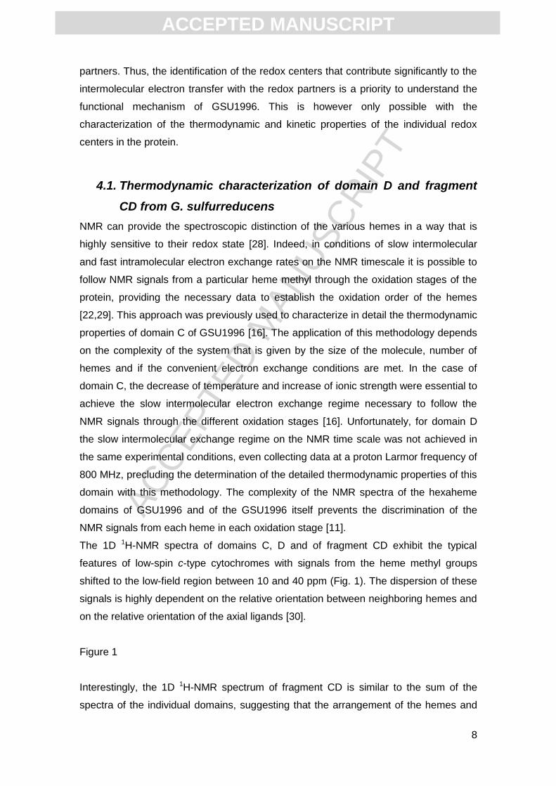
4.1. Thermodynamic characterization of domain D and fragment
CD from G. sulfurreducens
NMR can provide the spectroscopic distinction of the various hemes in a way that is
highly sensitive to their redox state [28]. Indeed, in conditions of slow intermolecular
and fast intramolecular electron exchange rates on the NMR timescale it is possible to
follow NMR signals from a particular heme methyl through the oxidation stages of the
protein, providing the necessary data to establish the oxidation order of the hemes
[22,29]. This approach was previously used to characterize in detail the thermodynamic
properties of domain C of GSU1996 [16]. The application of this methodology depends
on the complexity of the system that is given by the size of the molecule, number of
hemes and if the convenient electron exchange conditions are met. In the case of
domain C, the decrease of temperature and increase of ionic strength were essential to
achieve the slow intermolecular electron exchange regime necessary to follow the
NMR signals through the different oxidation stages [16]. Unfortunately, for domain D
the slow intermolecular exchange regime on the NMR time scale was not achieved in
the same experimental conditions, even collecting data at a proton Larmor frequency of
800 MHz, precluding the determination of the detailed thermodynamic properties of this
domain with this methodology. The complexity of the NMR spectra of the hexaheme
domains of GSU1996 and of the GSU1996 itself prevents the discrimination of the
NMR signals from each heme in each oxidation stage [11].
The 1D 1H-NMR spectra of domains C, D and of fragment CD exhibit the typical
features of low-spin c-type cytochromes with signals from the heme methyl groups
shifted to the low-field region between 10 and 40 ppm (Fig. 1). The dispersion of these
signals is highly dependent on the relative orientation between neighboring hemes and
on the relative orientation of the axial ligands [30].
Figure 1
Interestingly, the 1D 1H-NMR spectrum of fragment CD is similar to the sum of the
spectra of the individual domains, suggesting that the arrangement of the hemes and
ACC
EPTE
D M
ANU
SCR
IPT
ACCEPTED MANUSCRIPT
9
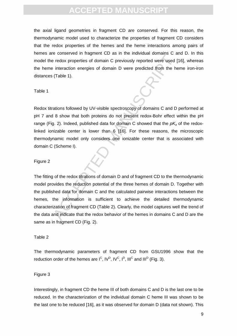
the axial ligand geometries in fragment CD are conserved. For this reason, the
thermodynamic model used to characterize the properties of fragment CD considers
that the redox properties of the hemes and the heme interactions among pairs of
hemes are conserved in fragment CD as in the individual domains C and D. In this
model the redox properties of domain C previously reported were used [16], whereas
the heme interaction energies of domain D were predicted from the heme iron-iron
distances (Table 1).
Table 1
Redox titrations followed by UV-visible spectroscopy of domains C and D performed at
pH 7 and 8 show that both proteins do not present redox-Bohr effect within the pH
range (Fig. 2). Indeed, published data for domain C showed that the pKa of the redox-
linked ionizable center is lower than 6 [16]. For these reasons, the microscopic
thermodynamic model only considers one ionizable center that is associated with
domain C (Scheme I).
Figure 2
The fitting of the redox titrations of domain D and of fragment CD to the thermodynamic
model provides the reduction potential of the three hemes of domain D. Together with
the published data for domain C and the calculated pairwise interactions between the
hemes, the information is sufficient to achieve the detailed thermodynamic
characterization of fragment CD (Table 2). Clearly, the model captures well the trend of
the data and indicate that the redox behavior of the hemes in domains C and D are the
same as in fragment CD (Fig. 2).
Table 2
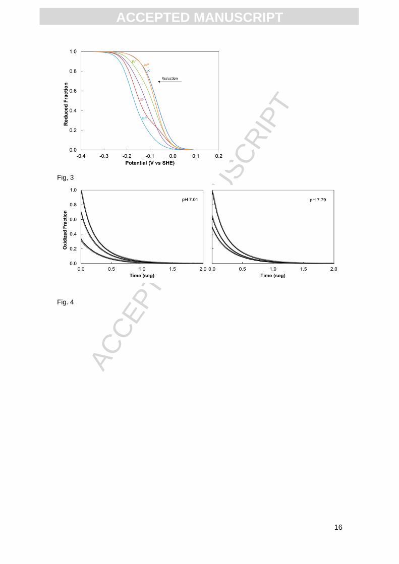
The thermodynamic parameters of fragment CD from GSU1996 show that the
reduction order of the hemes are IC, IVD, IVC, ID, IIIC and IIID (Fig. 3).
Figure 3
Interestingly, in fragment CD the heme III of both domains C and D is the last one to be
reduced. In the characterization of the individual domain C heme III was shown to be
the last one to be reduced [16], as it was observed for domain D (data not shown). This
ACC
EPTE
D M
ANU
SCR
IPT
ACCEPTED MANUSCRIPT
10
constitutes further evidences that the redox behavior of the individual domains is
maintained in the hexaheme fragment.
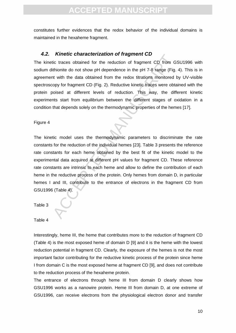
4.2. Kinetic characterization of fragment CD
The kinetic traces obtained for the reduction of fragment CD from GSU1996 with
sodium dithionite do not show pH dependence in the pH 7-8 range (Fig. 4). This is in
agreement with the data obtained from the redox titrations monitored by UV-visible
spectroscopy for fragment CD (Fig. 2). Reductive kinetic traces were obtained with the
protein poised at different levels of reduction. This way, the different kinetic
experiments start from equilibrium between the different stages of oxidation in a
condition that depends solely on the thermodynamic properties of the hemes [17].
Figure 4
The kinetic model uses the thermodynamic parameters to discriminate the rate
constants for the reduction of the individual hemes [23]. Table 3 presents the reference
rate constants for each heme obtained by the best fit of the kinetic model to the
experimental data acquired at different pH values for fragment CD. These reference
rate constants are intrinsic to each heme and allow to define the contribution of each
heme in the reductive process of the protein. Only hemes from domain D, in particular
hemes I and III, contribute to the entrance of electrons in the fragment CD from
GSU1996 (Table 4).
Table 3
Table 4
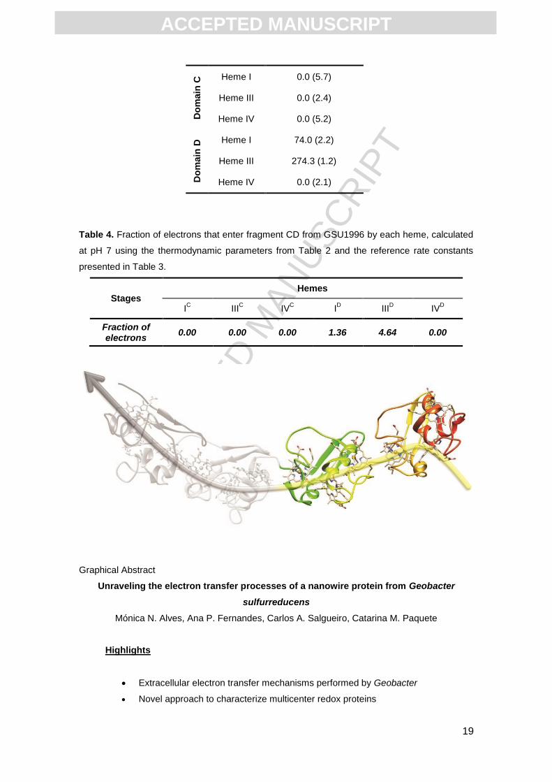
Interestingly, heme III, the heme that contributes more to the reduction of fragment CD
(Table 4) is the most exposed heme of domain D [9] and it is the heme with the lowest
reduction potential in fragment CD. Clearly, the exposure of the hemes is not the most
important factor contributing for the reductive kinetic process of the protein since heme
I from domain C is the most exposed heme at fragment CD [9], and does not contribute
to the reduction process of the hexaheme protein.
The entrance of electrons through heme III from domain D clearly shows how
GSU1996 works as a nanowire protein. Heme III from domain D, at one extreme of
GSU1996, can receive electrons from the physiological electron donor and transfer
ACC
EPTE
D M
ANU
SCR
IPT
ACCEPTED MANUSCRIPT
11
them to the other hemes within the protein. Since, at least in fragment CD, this heme
has the lowest reduction potential, it is spontaneously re-oxidized by the other hemes
in the fragment and remains free to receive electrons from redox partners, allowing the
protein to function as a nanowire device (Scheme 2).
Scheme 2
5. Conclusion
Nanowire cytochromes are a new class of proteins found in the genome of several
Geobacter species proposed to be responsible for long-range electron transfer. The
elucidation of the detailed thermodynamic and kinetic properties of the C-terminal half
of the protein GSU1996 from G. sulfurreducens opens the possibility to unravel the
electron transfer processes performed by this new class of proteins. Indeed, the
entrance of electrons through the heme that is at one edge ensures that the electrons
may flow within the protein to the other end, allowing it to work as a nanowire. Further
studies will enable the characterization of the full length protein, and the elucidation of
the electron transfer processes during its oxidation. This information will also be of
significant importance to increase our knowledge on the extracellular electron transfer
processes performed by G. sulfurreducens, a key asset to improve its biotechnological
applications, such as microbial fuel cells.
6. Acknowledgments
The authors thank Ricardo O. Louro for helpful discussions. This work was supported
by Fundação para a Ciência e Tecnologia (FCT) Portugal [Grants PTDC/QUI-
BIQ/117440/2010, UID/Multi/04378/2013; APF and CMP were supported by FCT
grants SFRH/BD/86439/2012 and SFRH/BPD/96952/2013, respectively]. The NMR
spectrometers are part of The National NMR Facility, supported by FCT (RECI/BBB-
BQB/0230/2012).
References
[1] J. Liu, S. Chakraborty, P. Hosseinzadeh, Y. Yu, S. Tian, I. Petrik, et al., Metalloproteins containing cytochrome, iron-sulfur, or copper redox centers., Chem. Rev. 114 (2014) 4366–469.
ACC
EPTE
D M
ANU
SCR
IPT
ACCEPTED MANUSCRIPT
12
[2] M. Breuer, K.M. Rosso, J. Blumberger, J.N. Butt, Multi-haem cytochromes in Shewanella oneidensis MR-1 : structures , functions and opportunities, J. R. Soc. Interface. 12 (2015) 1–27.
[3] T.C. Santos, M.A. Silva, L. Morgado, J.M. Dantas, C.A. Salgueiro, Diving into the redox properties of Geobacter sulfurreducens cytochromes: a model for extracellular electron transfer., Dalton Trans. 44 (2015) 9335–44.
[4] B. Kim, C. Leang, Y.R. Ding, H. Glaven, M. V Coppi, D.R. Lovley, et al., OmcF , a Putative c -Type Monoheme Outer Membrane Cytochrome Required for the Expression of Other Outer Membrane Cytochromes in Geobacter sulfurreducens, J. Bacteriol. (2005).
[5] A. Esteve-Núñez, J. Sosnik, P. Visconti, D.R. Lovley, Fluorescent properties of c-type cytochromes reveal their potential role as an extracytoplasmic electron sink in Geobacter sulfurreducens, Environ. Microbiol. 10 (2008) 497–505.
[6] B. Schuetz, M. Schicklberger, J. Kuermann, A.M. Spormann, J. Gescher, Periplasmic electron transfer via the c-type cytochromes MtrA and FccA of Shewanella oneidensis MR-1., Appl. Environ. Microbiol. 75 (2009) 7789–96.
[7] B.A. Methé, K.E. Nelson, J.A. Eisen, I.T. Paulsen, W. Nelson, J.F. Heidelberg, et al., Genome of Geobacter sulfurreducens: metal reduction in subsurface environments., Science 302 (2003) 1967–9.
[8] P.R. Pokkuluri, Y.Y. Londer, N.E.C. Duke, J. Erickson, M. Pessanha, C.A. Salgueiro, et al., Structure of a novel c7 -type three-heme cytochrome domain from a multidomain cytochrome c polymer, Protein Sci. 13 (2004) 1684-1692.
[9] P.R. Pokkuluri, Y.Y. Londer, N.E.C. Duke, M. Pessanha, X. Yang, V. Orshonsky, et al., Structure of a novel dodecaheme cytochrome c from Geobacter sulfurreducens reveals an extended 12 nm protein with interacting hemes., J. Struct. Biol. 174 (2011) 223–33.
[10] M. Assfalg, L. Banci, I. Bertini, M. Bruschi, P. Turano, 800 MHz 1H NMR solution structure refinement of oxidized cytochrome c7 from Desulfuromonas acetoxidans., Eur. J. Biochem. 256 (1998) 261–70.
[11] C.M. Paquete, R.O. Louro, Unveiling the Details of Electron Transfer in Multicenter Redox Proteins., Acc. Chem. Res. 47 (2014) 56–65.
[12] Y.Y. Londer, P.R. Pokkuluri, J. Erickson, V. Orshonsky, M. Schiffer, Heterologous expression of hexaheme fragments of a multidomain cytochrome from Geobacter sulfurreducens representing a novel class of cytochromes c., Protein Expr. Purif. 39 (2005) 254–60.
[13] E.H. Gordon, E. Steensma, S.J. Ferguson, The cytochrome c domain of dimeric cytochrome cd(1) of Paracoccus pantotrophus can be produced at high levels as a monomeric holoprotein using an improved c-type cytochrome expression system in Escherichia coli., Biochem. Biophys. Res. Commun. 281 (2001) 788–94.
ACC
EPTE
D M
ANU
SCR
IPT
ACCEPTED MANUSCRIPT
13
[14] E. Arslan, H. Schulz, R. Zufferey, P. Künzler, L. Thöny-Meyer, Overproduction of the Bradyrhizobium japonicum c-type cytochrome subunits of the cbb3 oxidase in Escherichia coli., Biochem. Biophys. Res. Commun. 251 (1998) 744–7.
[15] R.T. Francis, R.R. Becker, Specific Indication of Hemoproteins in Polyacrylamide Using a Double-Staining Process, Anal. Biochem. 14 (1984) 509–514.
[16] L. Morgado, A.P. Fernandes, Y.Y. Londer, P.R. Pokkuluri, M. Schiffer, C. a Salgueiro, Thermodynamic characterization of the redox centres in a representative domain of a novel c-type multihaem cytochrome., Biochem. J. 420 (2009) 485–92.
[17] C.M. Paquete, D.L. Turner, R.O. Louro, A. V Xavier, T. Catarino, Thermodynamic and kinetic characterisation of individual haems in multicentre cytochromes c3, Biochim. Biophys. Acta. 1767 (2007) 1169 – 1179.
[18] M. Dixon, The acceptor specificity of flavins and flavoproteins. I. Techniques for anaerobic spectrophotometry., Biochim. Biophys. Acta. 226 (1971) 241–58.
[19] D.O. Lambeth, G. Palmer, The kinetics and mechanism of reduction of electron transfer proteins and other compounds of biological interest by dithionite., J. Biol. Chem. 248 (1973) 6095–103.
[20] D.L. Turner, C.A. Salgueiro, T. Catarino, J. Legall, A. V Xavier, NMR studies of cooperativity in the tetrahaem cytochrome c3 from Desulfovibrio vulgaris., Eur. J. Biochem. 241 (1996) 723–31.
[21] R.O. Louro, T. Catarino, C.M. Paquete, D.L. Turner, Distance dependence of interactions between charged centres in proteins with common structural features., FEBS Lett. 576 (2004) 77–80.
[22] B.M. Fonseca, C.M. Paquete, C.A. Salgueiro, R.O. Louro, The role of intramolecular interactions in the functional control of multiheme cytochromes c., FEBS Lett. 586 (2012) 504–9.
[23] T. Catarino, D.L. Turner, Thermodynamic control of electron transfer rates in multicentre redox proteins., Chembiochem. 2 (2001) 416–24.
[24] R.A. Marcus, N. Sutin, Electron transfers in chemistry and biology, Biochim. Biophys. Acta. 811 (1985) 265–322.
[25] C.M. Paquete, I.H. Saraiva, E. Calçada, R.O. Louro, Molecular Basis for Directional Electron Transfer, J. Biol. Chem. 285 (2010) 10370 –10375.
[26] J.C. Lagarias, J.A. Reeds, M.H. Wright, P.E. Wright, Convergence Properties of the Nelder--Mead Simplex Method in Low Dimensions, SIAM J. Optim. 9 (1998) 112–147.
[27] C.M. Paquete, R.O. Louro, Molecular details of multielectron transfer: the case of multiheme cytochromes from metal respiring organisms., Dalt. Trans. 39 (2010) 4259–66.
ACC
EPTE
D M
ANU
SCR
IPT
ACCEPTED MANUSCRIPT
14
[28] H. Santos, J.J. Moura, I. Moura, J. Legall, A. V Xavier, NMR studies of electron transfer mechanisms in a protein with interacting redox centres: Desulfovibrio gigas cytochrome c3., Eur. J. Biochem. 141 (1984) 283–96.
[29] C.A. Salgueiro, D.L. Turner, H. Santos, J. LeGall, A. V Xavier, Assignment of the redox potentials to the four haems in Desulfovibrio vulgaris cytochrome c3 by 2D-NMR., FEBS Lett. 314 (1992) 155–8.
[30] D.L. Turner, C.A. Salgueiro, P. Schenkels, J. LeGall, A. V Xavier, Carbon-13 NMR studies of the influence of axial ligand orientation on haem electronic structure., Biochim. Biophys. Acta. 1246 (1995) 24–8.
Scheme legends
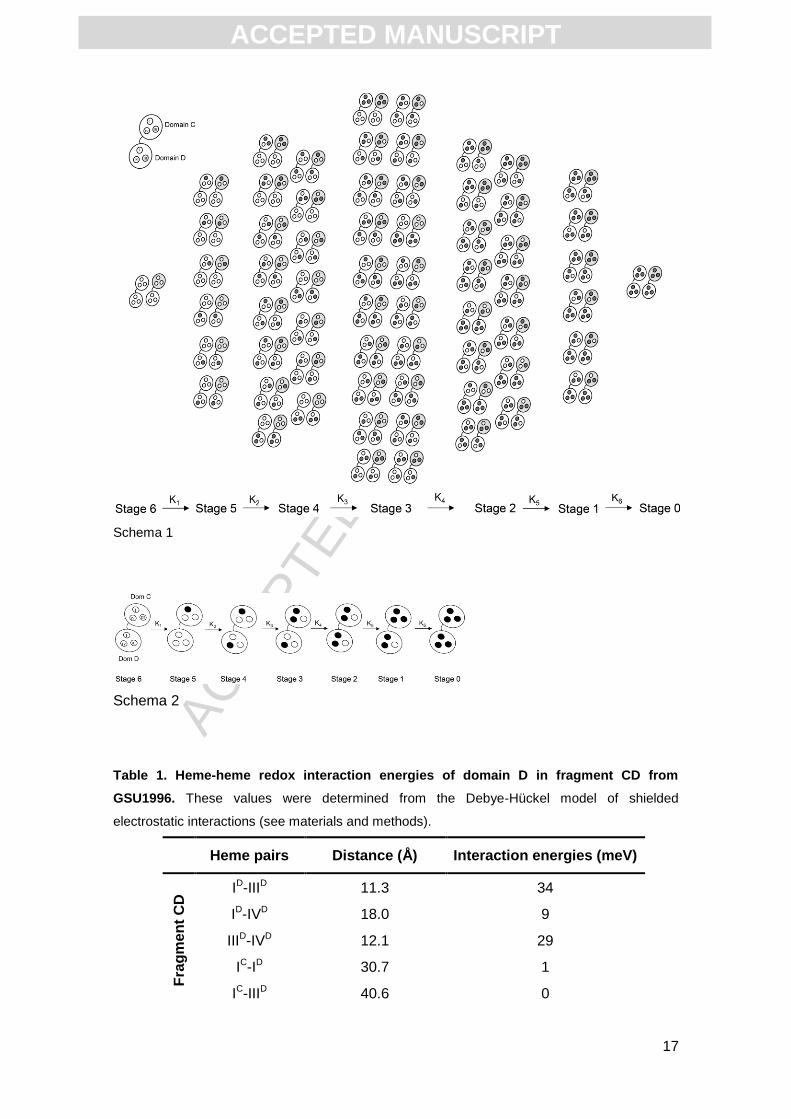
Scheme 1. Schematic representation of the microstates of fragment CD from GSU1996, a
protein with six hemes and one acid-base center. The protein is represented as large circles,
with black and white dots representing the hemes in the reduced and oxidized state,
respectively. White and gray protein represents the deprotonated and protonated microstates
for the acid-base center associated with the hemes. The redox stages are numbered according
to the number of oxidized hemes and organized in columns that group populations with the
same oxidation state. Macroscopic electron transfer steps between stages are shown in the
direction of reduction, and macroscopic rate constants are represented by K1-6.
Scheme 2. Schematic representation of the most important microstates for the reduction of
fragment CD from GSU1996. Black and white dots representing the hemes in the reduced and
oxidized state, respectively.
Figure legends
Figure 1. (A) 1D 1H-NMR spectra of domains C, D and of fragment CD from G. sulfurreducens
at 289 K and pH 8. The NMR spectral region where heme methyl groups of low-spin c-type
cytochromes appear are highlighted by a gray box. (B) Three-dimensional structures of domain
C, domain D and fragment CD. The 3D structures of domains C and D were taken from the 3D
structure of the fragment CD (pdb: 3OUE). The hemes are numbered by analogy to the
structurally homologous hemes in tetraheme cytochromes c3.
Figure 2. Redox titrations followed by visible spectroscopy of domain C, domain D and
fragment CD at pH 7 and 8 (289 K). Redox titrations of domain C were previously performed
[16], whereas redox titrations of domain D and of fragment CD were performed in this work. The
solid lines represent the best fit of the experimental data with the thermodynamic model
described in materials and methods section and give rise to the thermodynamic parameters
reported in Table 2.

ACC
EPTE
D M
ANU
SCR
IPT
ACCEPTED MANUSCRIPT
15
Figure 3. Reduced fraction of the individual hemes in fragment CD from GSU1996 calculated at
pH 7 with the thermodynamic parameters presented in Table 2.
Figure 4. Kinetics of reduction of fragment CD from GSU1996 by sodium dithionite at different
pH values. Grey lines are the kinetic data obtained for the fully oxidized state of the protein, for
70% and 34% oxidized fraction at pH 7.01, and for 65% and 50% oxidized fraction at pH 7.79.
Black lines are the fit of the kinetic model to the data. The concentration of sodium dithionite
used in the kinetic experiments was 115 M (after mixing), while the concentration of fragment
CD were 0.91 and 0.90 M (after mixing) at pH 7.01 and 7.79, respectively.
Fig. 1
Fig. 2
ACC
EPTE
D M
ANU
SCR
IPT
ACCEPTED MANUSCRIPT
16
Fig, 3
Fig. 4
ACC
EPTE
D M
ANU
SCR
IPT
ACCEPTED MANUSCRIPT
17
Schema 1
Schema 2
Table 1. Heme-heme redox interaction energies of domain D in fragment CD from
GSU1996. These values were determined from the Debye-Hückel model of shielded
electrostatic interactions (see materials and methods).
Heme pairs Distance (Å) Interaction energies (meV)
Fra
gm
en
t C
D ID-IIID 11.3 34
ID-IVD 18.0 9
IIID-IVD 12.1 29
IC-ID 30.7 1
IC-IIID 40.6 0
ACC
EPTE
D M
ANU
SCR
IPT
ACCEPTED MANUSCRIPT
18
IC-IVD 48.3 0
IIIC-ID 21.8 5
IIIC-IIID 30.4 1
IIIC-IVD 39.5 0
IVC-ID 14.7 17
IVC-IIID 25.3 3
IVC-IVD 32.3 1
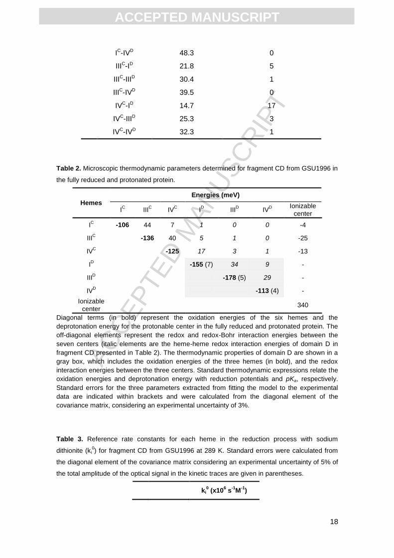
Table 2. Microscopic thermodynamic parameters determined for fragment CD from GSU1996 in
the fully reduced and protonated protein.
Hemes
Energies (meV)
IC III
C IV
C I
D III
D IV
D
Ionizable center
IC -106 44 7 1 0 0 -4
IIIC -136 40 5 1 0 -25
IVC -125 17 3 1 -13
ID -155 (7) 34 9 -
IIID -178 (5) 29 -
IVD -113 (4) -
Ionizable center
340
Diagonal terms (in bold) represent the oxidation energies of the six hemes and the
deprotonation energy for the protonable center in the fully reduced and protonated protein. The
off-diagonal elements represent the redox and redox-Bohr interaction energies between the
seven centers (italic elements are the heme-heme redox interaction energies of domain D in
fragment CD presented in Table 2). The thermodynamic properties of domain D are shown in a
gray box, which includes the oxidation energies of the three hemes (in bold), and the redox
interaction energies between the three centers. Standard thermodynamic expressions relate the
oxidation energies and deprotonation energy with reduction potentials and pKa, respectively.
Standard errors for the three parameters extracted from fitting the model to the experimental
data are indicated within brackets and were calculated from the diagonal element of the
covariance matrix, considering an experimental uncertainty of 3%.
Table 3. Reference rate constants for each heme in the reduction process with sodium
dithionite (ki0) for fragment CD from GSU1996 at 289 K. Standard errors were calculated from
the diagonal element of the covariance matrix considering an experimental uncertainty of 5% of
the total amplitude of the optical signal in the kinetic traces are given in parentheses.
ki0 (x10
6 s
-1M
-1)
ACC
EPTE
D M
ANU
SCR
IPT
ACCEPTED MANUSCRIPT
19
Do
main
C Heme I 0.0 (5.7)
Heme III 0.0 (2.4)
Heme IV 0.0 (5.2)
Do
main
D Heme I 74.0 (2.2)
Heme III 274.3 (1.2)
Heme IV 0.0 (2.1)
Table 4. Fraction of electrons that enter fragment CD from GSU1996 by each heme, calculated
at pH 7 using the thermodynamic parameters from Table 2 and the reference rate constants
presented in Table 3.
Stages Hemes
IC III
C IV
C I
D III
D IV
D
Fraction of electrons
0.00 0.00 0.00 1.36 4.64 0.00
Graphical Abstract
Unraveling the electron transfer processes of a nanowire protein from Geobacter
sulfurreducens
Mónica N. Alves, Ana P. Fernandes, Carlos A. Salgueiro, Catarina M. Paquete
Highlights
Extracellular electron transfer mechanisms performed by Geobacter
Novel approach to characterize multicenter redox proteins
ACC
EPTE
D M
ANU
SCR
IPT
ACCEPTED MANUSCRIPT
20
Thermodynamic and kinetic characterization of fragment CD of GSU1996
First elucidation of the electron transfer processes of a nanowire protein