Embed Size (px)
Citation preview
Biochemical and Biophysical Research Communications 440 (2013) 664–670
Contents lists available at ScienceDirect
Biochemical and Biophysical Research Communications
journal homepage: www.elsevier .com/locate /ybbrc
Geraniol attenuates a-synuclein expression and neuromuscularimpairment through increase dopamine content in MPTP intoxicatedmice by dose dependent manner
0006-291X/$ - see front matter Crown Copyright � 2013 Published by Elsevier Inc. All rights reserved.http://dx.doi.org/10.1016/j.bbrc.2013.09.122
⇑ Corresponding author.E-mail address: [email protected] (R. Inmozhi Sivakamasundari).
Karamkolly R. Rekha a, Govindasamy P. Selvakumar b, Karunanithi Santha a,Ramu Inmozhi Sivakamasundari a,⇑a Division of Biochemistry, Faculty of Medicine, Raja Muthaiah Medical College, Annamalai University, Annamalainagar 608 002, Tamilnadu, Indiab Department of Biochemistry and Biotechnology, Faculty of Science, Annamalai University, Annamalainagar 608 002, Tamilnadu, India
a r t i c l e i n f o a b s t r a c t
Article history:Received 14 September 2013Available online 5 October 2013
Keywords:Parkinson’s diseaseDegenerationMPTPGeraniolNeuroprotective
Parkinson’s disease (PD) is characterized by progressive loss of dopamine (DA) neurons in the nigrostri-atal system and by the presence of Lewy bodies (LB), proteinaceous inclusions mainly composed of fila-mentous a-synuclein (a-Syn) aggregates. 1-Methyl-4-phenyl-1,2,3,6-tetrahydropyridine (MPTP) wasadopted to generate PD models in C57BL/6 mice. In the present study, we investigated the effect of gera-niol (GE) against a-Syn aggregation on MPTP induced mouse model of PD in dose dependant manner.When pretreatment of GE improved neuromuscular impairment, TH expressions and decreases a-Synexpressions in MPTP intoxicated PD mice by dose dependent manner. In addition, we confirmed thatsub-chronic administration of MPTP in mice leads to permanent neuromuscular deficits and depletionof dopamine and its metabolites. Our results suggest that GE is beneficial for the treatment of PD asso-ciated with neuromuscular disability and LB aggregation.
Crown Copyright � 2013 Published by Elsevier Inc. All rights reserved.
1. Introduction
Parkinson’s disease (PD) is a most common neurodegenerativeand movement disorder. It is characterized by a progressive lossof neuromelanin containing dopaminergic (DA-ergic) neuronsand its projecting from the substantia nigra (SN) to the striatum(ST) [1,2] leads to the loss of dopamine (DA) in the striatummanifests as motor disabilities that are characteristic of PD. Thepathological changes in PD, as revealed by widespread presenceof a-synuclein (a-Syn) positive inclusion bodies and a-Syn positiveneuritis [3,4]. The recent studies highlighting the important rolesfor a-Syn in synaptic transmission and dopaminergic neuronphysiology to add our understanding of PD etiology and providea central link between the genetic findings and neurodegenerationobserved in sporadic PD.
1-Methyl-4-phenyl-1,2,3,6-tetrahydropyridine (MPTP) causesdamage to DA-ergic neurons in the nigrostriatal system, similarto that seen in PD patients [5]. After administration, MPTP ismetabolized by an enzyme, monoamine oxidase B (MAO-B), in glialcells to form 1-methyl-4-phenylpiridinium (MPP+) [6] and entersinto DA-ergic neurons with a dopamine transporter (DAT). Inside
the DA-ergic neurons, MPP+ injures mitochondrial respiratorycomplex I [7], and subsequently generates oxidative free radicalsthat can lead to the oxidative nitration of a-Syn [8–10]. a-Syn oxi-dative nitration had been found in PD and is potentially importantin the aggregation and toxicity of a-Syn [11]. The overexpression ofa-Syn by itself can cause oxidative stress, increased inclusion for-mation, and mitochondrial structural abnormalities in culturedneurons [12].
Consumption of phytochemicals from natural products hasbeen associated with reduced risk of neurodegenerative diseases[13–15]. To protect vulnerable targets, phytochemicals counter-act the imbalance of the cellular redox homeostasis and thereactive oxygen species levels under the cytotoxic threshold.The phytochemical, Geraniol (GE), an acyclic monoterpene alco-hol found in lemongrass and aromatic herb oils, proved to havecytoprotective and antioxidant potential in oxidative stressinduced animal models [16]. We speculate that GE, through itsantioxidant and anti-inflammatory properties, may exert thecapacity to block the MPTP induced neurotoxicity. Consequently,in the present investigation, we demonstrate the neuroprotectiveeffects of GE of MPTP induced neuromuscular deficits, dopaminedepletion and action versus a-Syn aggregation in a mouse modelof PD.
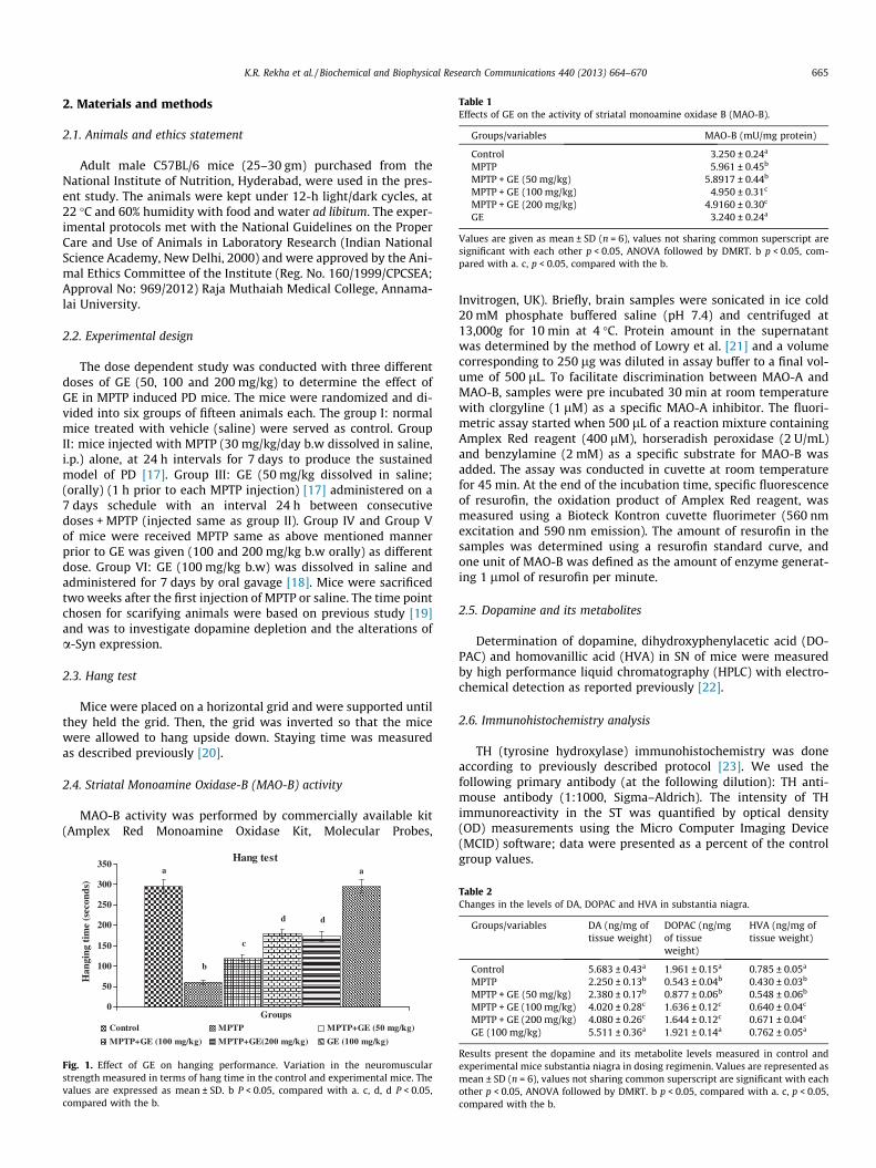
Table 1Effects of GE on the activity of striatal monoamine oxidase B (MAO-B).
Groups/variables MAO-B (mU/mg protein)
Control 3.250 ± 0.24a
MPTP 5.961 ± 0.45b
MPTP + GE (50 mg/kg) 5.8917 ± 0.44b
MPTP + GE (100 mg/kg) 4.950 ± 0.31c
MPTP + GE (200 mg/kg) 4.9160 ± 0.30c
GE 3.240 ± 0.24a
Values are given as mean ± SD (n = 6), values not sharing common superscript aresignificant with each other p < 0.05, ANOVA followed by DMRT. b p < 0.05, com-pared with a. c, p < 0.05, compared with the b.
K.R. Rekha et al. / Biochemical and Biophysical Research Communications 440 (2013) 664–670 665
2. Materials and methods
2.1. Animals and ethics statement
Adult male C57BL/6 mice (25–30 gm) purchased from theNational Institute of Nutrition, Hyderabad, were used in the pres-ent study. The animals were kept under 12-h light/dark cycles, at22 �C and 60% humidity with food and water ad libitum. The exper-imental protocols met with the National Guidelines on the ProperCare and Use of Animals in Laboratory Research (Indian NationalScience Academy, New Delhi, 2000) and were approved by the Ani-mal Ethics Committee of the Institute (Reg. No. 160/1999/CPCSEA;Approval No: 969/2012) Raja Muthaiah Medical College, Annama-lai University.
2.2. Experimental design
The dose dependent study was conducted with three differentdoses of GE (50, 100 and 200 mg/kg) to determine the effect ofGE in MPTP induced PD mice. The mice were randomized and di-vided into six groups of fifteen animals each. The group I: normalmice treated with vehicle (saline) were served as control. GroupII: mice injected with MPTP (30 mg/kg/day b.w dissolved in saline,i.p.) alone, at 24 h intervals for 7 days to produce the sustainedmodel of PD [17]. Group III: GE (50 mg/kg dissolved in saline;(orally) (1 h prior to each MPTP injection) [17] administered on a7 days schedule with an interval 24 h between consecutivedoses + MPTP (injected same as group II). Group IV and Group Vof mice were received MPTP same as above mentioned mannerprior to GE was given (100 and 200 mg/kg b.w orally) as differentdose. Group VI: GE (100 mg/kg b.w) was dissolved in saline andadministered for 7 days by oral gavage [18]. Mice were sacrificedtwo weeks after the first injection of MPTP or saline. The time pointchosen for scarifying animals were based on previous study [19]and was to investigate dopamine depletion and the alterations ofa-Syn expression.
2.3. Hang test
Mice were placed on a horizontal grid and were supported untilthey held the grid. Then, the grid was inverted so that the micewere allowed to hang upside down. Staying time was measuredas described previously [20].
2.4. Striatal Monoamine Oxidase-B (MAO-B) activity
MAO-B activity was performed by commercially available kit(Amplex Red Monoamine Oxidase Kit, Molecular Probes,
Hang testa
b
c
d d
a
0
50
100
150
200
250
300
350
Groups
Han
ging
tim
e (s
econ
ds)
Control MPTP MPTP+GE (50 mg/kg)
MPTP+GE (100 mg/kg) MPTP+GE(200 mg/kg) GE (100 mg/kg)
Fig. 1. Effect of GE on hanging performance. Variation in the neuromuscularstrength measured in terms of hang time in the control and experimental mice. Thevalues are expressed as mean ± SD. b P < 0.05, compared with a. c, d, d P < 0.05,compared with the b.
Invitrogen, UK). Briefly, brain samples were sonicated in ice cold20 mM phosphate buffered saline (pH 7.4) and centrifuged at13,000g for 10 min at 4 �C. Protein amount in the supernatantwas determined by the method of Lowry et al. [21] and a volumecorresponding to 250 lg was diluted in assay buffer to a final vol-ume of 500 lL. To facilitate discrimination between MAO-A andMAO-B, samples were pre incubated 30 min at room temperaturewith clorgyline (1 lM) as a specific MAO-A inhibitor. The fluori-metric assay started when 500 lL of a reaction mixture containingAmplex Red reagent (400 lM), horseradish peroxidase (2 U/mL)and benzylamine (2 mM) as a specific substrate for MAO-B wasadded. The assay was conducted in cuvette at room temperaturefor 45 min. At the end of the incubation time, specific fluorescenceof resurofin, the oxidation product of Amplex Red reagent, wasmeasured using a Bioteck Kontron cuvette fluorimeter (560 nmexcitation and 590 nm emission). The amount of resurofin in thesamples was determined using a resurofin standard curve, andone unit of MAO-B was defined as the amount of enzyme generat-ing 1 lmol of resurofin per minute.
2.5. Dopamine and its metabolites
Determination of dopamine, dihydroxyphenylacetic acid (DO-PAC) and homovanillic acid (HVA) in SN of mice were measuredby high performance liquid chromatography (HPLC) with electro-chemical detection as reported previously [22].
2.6. Immunohistochemistry analysis
TH (tyrosine hydroxylase) immunohistochemistry was doneaccording to previously described protocol [23]. We used thefollowing primary antibody (at the following dilution): TH anti-mouse antibody (1:1000, Sigma–Aldrich). The intensity of THimmunoreactivity in the ST was quantified by optical density(OD) measurements using the Micro Computer Imaging Device(MCID) software; data were presented as a percent of the controlgroup values.
Table 2Changes in the levels of DA, DOPAC and HVA in substantia niagra.
Groups/variables DA (ng/mg oftissue weight)
DOPAC (ng/mgof tissueweight)
HVA (ng/mg oftissue weight)
Control 5.683 ± 0.43a 1.961 ± 0.15a 0.785 ± 0.05a
MPTP 2.250 ± 0.13b 0.543 ± 0.04b 0.430 ± 0.03b
MPTP + GE (50 mg/kg) 2.380 ± 0.17b 0.877 ± 0.06b 0.548 ± 0.06b
MPTP + GE (100 mg/kg) 4.020 ± 0.28c 1.636 ± 0.12c 0.640 ± 0.04c
MPTP + GE (200 mg/kg) 4.080 ± 0.26c 1.644 ± 0.12c 0.671 ± 0.04c
GE (100 mg/kg) 5.511 ± 0.36a 1.921 ± 0.14a 0.762 ± 0.05a
Results present the dopamine and its metabolite levels measured in control andexperimental mice substantia niagra in dosing regimenin. Values are represented asmean ± SD (n = 6), values not sharing common superscript are significant with eachother p < 0.05, ANOVA followed by DMRT. b p < 0.05, compared with a. c, p < 0.05,compared with the b.
666 K.R. Rekha et al. / Biochemical and Biophysical Research Communications 440 (2013) 664–670
2.7. Extraction of total mRNA
Total mRNA was isolated from the SN and ST using mRNAextraction kit (Genei Bangalore, India), following the manufac-turer’s instructions. The mRNA integrity was determined by aga-rose gel electrophoresis and the concentration and purity weremeasured spectrophotometrically [24].
Total mRNA was converted to single stranded cDNA using 2 lgof total mRNA as a template. Oligo (dT) 12–18 primer (InvitrogenLife Technologies) and Moloney murine leukemia virus reversetranscriptase (M-MLV RT; Invitrogen Life Technologies) were usedas per manufacturer’s instruction. The following primers were usedfor the mRNA expression: a-syn: forward primer 50-GGAGTGA-CAACAGTGGCTGA-30, reverse primer 50-GCTCCCTCCACTGTCTTCTG-30; b-actin: forward primer 50-GCGAGAAGATGACC CAGATC-30, reverse primer 50-CCAGTGGTACGGCCAGAGG-30 [25]. The ther-mocycling conditions were initiated at 95 �C for 10 min, followedby 40 PCR cycles of denaturation at 95 �C for 15 s, and anneal/extension at 60 �C for 1 min. Melting (dissociation stage) was per-formed by the end of each cycle to ascertain the specificity of the
Control MPTP
MPTP+GE (100mg/kg) MPTP+GE(20
TH immu
a
b
c
0
20
40
60
80
100
120
Num
ber
of T
H im
unor
ectiv
e te
rmin
als
(% o
f con
trol
)
Control MPTPMPTP+GE (100mg/kg) MPTP+
A
B
Fig. 2. Immunohistochemical analysis of TH in ST of control and experimental mice: (following MPTP administration, an effect that was attenuated by pre-treatment withsignificantly rescued TH positive terminals in ST by dose dependent manner from death inexpressed as a percentage of that matched control mice. Values are presented as mean
primers and the purity of the final PCR product. The amplifiedproducts were electrophoresed with 1.5% agarose gel analyzedchanges by Image J software.
2.8. Western blotting
The nigrostriatal tissues were collected from the each group ofmice as described previously [23]. Samples fractions containingequal amounts of protein (50 lg) were separated in 10% SDS–poly-acrylamide gel electrophoresis. The membranes were incubatedwith the blocking buffer containing 5% non-fat dry milk powderor BSA for 2 h to reduce non-specific binding sites and then incu-bated in with anti-a-Syn (mouse monoclonal, 1:1000) with gentleshaking for overnight at 4 �C. After this, membranes were incu-bated with their corresponding secondary antibodies (anti-mouseor anti rabbit IgG conjugated to HRP) for 2 h at room temperature.The membrane was washed thrice with TBST for 30 min. Proteinbands were visualized by an enhanced chemiluminescence’s meth-od using ECL-kit (GenScript ECL kit, USA). Bands were scannedusing a scanner and quantitated by Image J, a public domain Java
MPTP+GE (50mg/kg)
0mg/kg) GE (100mg/kg)
noreactivity in ST
de
a
Groups
MPTP+GE (50mg/kg)GE (200mg/kg) GE (100mg/kg)
A) (40� magnification) illustrates TH immunoreactivity was significantly reducedGE. (B) Quantification of TH positive terminals in the ST. Pre-treatment with GEduced by MPTP. The mean value for DAT-IR was determined for each group, and was± SD of three mice per group.
K.R. Rekha et al. / Biochemical and Biophysical Research Communications 440 (2013) 664–670 667
image processing software, which of control was set to 1.
2.9. Statistical analysis
All the data were expressed as mean ± SD of a number of exper-iments (n = 6). Statistical significance was evaluated by one wayanalysis of variance (ANOVA) using SPSS version 15.0 softwareand individual comparisons were obtained using Duncan’s Multi-ple Range Test (DMRT). Values were considered statistically signif-icant if p < 0.05.
3. Results
3.1. Effect of GE on hang test
Neuromuscular strength was observed by hang test as shown inFig. 1. The average hanging time of MPTP administered mice werelowered as compared to control mice Moreover, significantlyincreased hanging time were observed in the GE Pretreated groupas compared to the MPTP alone treated group (p < 0.05). No
A B C D E
SN
ST
β-actin
α-Synuclein mRN
a
b
c
dd
a
0
10
20
30
40
50
60
70
80
90
NS
Group
mR
NA
exp
ress
ion
(% o
f con
trol
)
Control MPTP
MPTP+GE (100mg/kg) MPTP+G
A
B
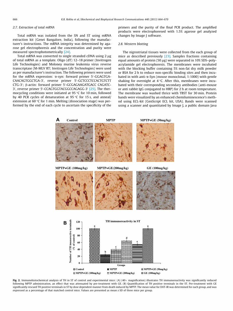
Fig. 3. (A) Dose dependent effect of GE on MPTP induced nigrostriatal a-synuclein mhousekeeping gene for the normalization of mRNA expressions. (B) Quantification graphwith a. c, d, d P < 0.05, compared with the b.
significant changes observed between GE alone treated mice andsaline treated control mice (p < 0.05).
3.2. Effects of GE on the Activity of Monoamine Oxidase B (MAO-B)
Table 1 depicts that MPTP treatment induced a marked increasein MAO-B activity as compared to control mice. Meanwhile,pre-treatment with GE the activity of MAO-B showed no significantdecrease as compared to MPTP control mice (p < 0.05). No signifi-cant changes observed between the saline treated control and GEalone treated mice respectively.
3.3. Changes in DA, DOPAC and HVA in SN and ST
DA, DOPAC and HVA levels were significantly decreased in SN(Table 2) of MPTP-treated mice as compared to control mice. Pre-treatment with GE following MPTP exposure significantly im-proved the levels of DA and its metabolites. However, treatmentwith GE alone control mice did not alter the levels of DA and itsmetabolites as compared to control mice.
F
A-Control
B-MPTP
C-MPTP+GE (50mg/kg)
D-MPTP+GE (100mg/kg)
E- MPTP+GE (200mg/kg)
F- GE (100mg/kg)
A expression
a
bc
d d
a
TS
s
MPTP+GE (50mg/kg)
E (200mg/kg) GE
RNA expression in control and experimental mice. b-actin mRNA were used ass Values are expressed as mean ± SD of three mice per group. b P < 0.05, compared
668 K.R. Rekha et al. / Biochemical and Biophysical Research Communications 440 (2013) 664–670
3.4. GE ameliorates against MPTP induced striatal degeneration
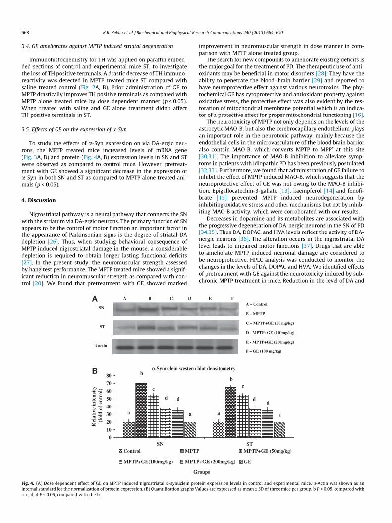
Immunohistochemistry for TH was applied on paraffin embed-ded sections of control and experimental mice ST, to investigatethe loss of TH positive terminals. A drastic decrease of TH immuno-reactivity was detected in MPTP treated mice ST compared withsaline treated control (Fig. 2A, B). Prior administration of GE toMPTP drastically improves TH positive terminals as compared withMPTP alone treated mice by dose dependent manner (p < 0.05).When treated with saline and GE alone treatment didn’t affectTH positive terminals in ST.
3.5. Effects of GE on the expression of a-Syn
To study the effects of a-Syn expression on via DA-ergic neu-rons, the MPTP treated mice increased levels of mRNA gene(Fig. 3A, B) and protein (Fig. 4A, B) expression levels in SN and STwere observed as compared to control mice. However, pretreat-ment with GE showed a significant decrease in the expression ofa-Syn in both SN and ST as compared to MPTP alone treated ani-mals (p < 0.05).
4. Discussion
Nigrostriatal pathway is a neural pathway that connects the SNwith the striatum via DA-ergic neurons. The primary function of SNappears to be the control of motor function an important factor inthe appearance of Parkinsonian signs is the degree of striatal DAdepletion [26]. Thus, when studying behavioral consequence ofMPTP induced nigrostriatal damage in the mouse, a considerabledepletion is required to obtain longer lasting functional deficits[27]. In the present study, the neuromuscular strength assessedby hang test performance. The MPTP treated mice showed a signif-icant reduction in neuromuscular strength as compared with con-trol [20]. We found that pretreatment with GE showed marked
A B C D
SN
ST
β-actin
α-Synuclein western
a
b
c
d d
a
01020304050607080
NS
G
Rel
ativ
e in
tens
ity
(fol
d of
cnt
rol)
Control MPTP
MPTP+GE(100mg/kg) MPTP
A
B
Fig. 4. (A) Dose dependent effect of GE on MPTP induced nigrostriatal a-synuclein prointernal standard for the normalization of protein expression. (B) Quantification graphs Va. c, d, d P < 0.05, compared with the b.
improvement in neuromuscular strength in dose manner in com-parison with MPTP alone treated group.
The search for new compounds to ameliorate existing deficits isthe major goal for the treatment of PD. The therapeutic use of anti-oxidants may be beneficial in motor disorders [28]. They have theability to penetrate the blood–brain barrier [29] and reported tohave neuroprotective effect against various neurotoxins. The phy-tochemical GE has cytoprotective and antioxidant property againstoxidative stress, the protective effect was also evident by the res-toration of mitochondrial membrane potential which is an indica-tor of a protective effect for proper mitochondrial functioning [16].
The neurotoxicity of MPTP not only depends on the levels of theastrocytic MAO-B, but also the cerebrocapillary endothelium playsan important role in the neurotoxic pathway, mainly because theendothelial cells in the microvasculature of the blood brain barrioralso contain MAO-B, which converts MPTP to MPP+ at this site[30,31]. The importance of MAO-B inhibition to alleviate symp-toms in patients with idiopathic PD has been previously postulated[32,33]. Furthermore, we found that administration of GE failure toinhibit the effect of MPTP induced MAO-B, which suggests that theneuroprotective effect of GE was not owing to the MAO-B inhibi-tion. Epigallocatechin-3-gallate [13], kaempferol [14] and fenofi-brate [15] prevented MPTP induced neurodegeneration byinhibiting oxidative stress and other mechanisms but not by inhib-iting MAO-B activity, which were corroborated with our results.
Decreases in dopamine and its metabolites are associated withthe progressive degeneration of DA-nergic neurons in the SN of PD[34,35]. Thus DA, DOPAC, and HVA levels reflect the activity of DA-nergic neurons [36]. The alteration occurs in the nigrostriatal DAlevel leads to impaired motor functions [37]. Drugs that are ableto ameliorate MPTP induced neuronal damage are considered tobe neuroprotective. HPLC analysis was conducted to monitor thechanges in the levels of DA, DOPAC and HVA. We identified effectsof pretreatment with GE against the neurotoxicity induced by sub-chronic MPTP treatment in mice. Reduction in the level of DA and
A – Control
B – MPTP
C – MPTP+GE (50 mg/kg)
D - MPTP+GE (100mg/kg)
E - MPTP+GE (200mg/kg)
F – GE (100 mg/kg)
E F
blot densitometry
a
bc
dd
a
TS
roups
MPTP+GE (50mg/kg)
+GE (200mg/kg) GE
tein expression levels in control and experimental mice. b-Actin was shown as analues are expressed as mean ± SD of three mice per group. b P < 0.05, compared with
K.R. Rekha et al. / Biochemical and Biophysical Research Communications 440 (2013) 664–670 669
its metabolites was observed in SN after MPTP treatment. Our re-sults also supported previous reports [22]. The results from thisstudy showed that GE significantly improved DA, DOPAC andHVA levels when compared to the control.
TH is the rate-limiting enzyme for the formation of DA. Itsexpression was used to identify neurotic processes, dopaminergiccell bodies of surviving [38]. TH immunoreactivity describes thenumber of survival neurons. In PD patients showed a decrease inthe activity of TH which is pronounced especially in the nigrostri-atal system [39] and the dramatic drop in TH have been suggestedto be of underlying importance in the pathogenesis of PD. Our re-sults also supported to previous findings. The immunohistochemi-cal localization of TH in ST region further strengthens and supportthe protective action of GE against MPTP induced PD, as reported inthe present study.
The MPTP intoxicated mice expressing high levels of a-Syndemonstrates that one of the major pathological feature of PD.The roles of a-Syn in normal cell function and in neurodegenera-tion have not been fully elucidated, but its potential roles in synap-tic plasticity [40], neuronal differentiation [41], the up regulationof dopamine release [42], and mitochondrial dysfunctions [43,44]have been reported previously. Recently, the dual roles of a-Synin neuroprotection and neurotoxicity were described [45–47]. Phy-tochemicals can inhibit formation and fibrillation of a-Syn, andprevent Ab-peptide fibrillation and oligomerization as well[48,49]. In a mouse model, it has well reported that phytochemi-cals able to neuroprotective effects against MPTP induced neuro-toxicity by decreasing the fibrillation and aggregation of a-Syn[48,49]. In this study, we observed that MPTP treated animalsincreased a-Syn expressions in the SN and ST. Pretreatment withGE on MPTP injection showed a decreased the expression ofa-Syn inconsistent with previous reports [50–52].
In conclusion, our results show a protective role of bioactive GEagainst MPTP mice, suggesting that it could represent a potentialtreatment in early phases of PD. The GE pretreatment improvesneuromuscular strength and reduced the a-Syn inclusion by reduc-ing the mRNA expression. The attenuated effect against neurotox-icity by GE could be the basis for their purported action asneuroprotectant and this hypothesis is worthwhile of testing in fu-ture studies using animal models of PD, involving MPTP or otherneurotoxins. In this regard, it has been reported that GE is a goodneuroprotective agent against a-Syn inclusion.
References
[1] H. Bernheimer, W. Birkmayer, O. Hornykiewicz, K. Jellinger, F. Seitelberger,Brain dopamine and the syndromes of Parkinson and Huntington Clinical,morphological and neurochemical correlations, J. Neurol. Sci. 20 (1973) 415–455.
[2] N.B. Chauhan, G.J. Siegel, J.M. Lee, Depletion of glial cell line-derivedneurotrophic factor in substantia nigra neurons of Parkinson’s disease brain,J. Chem. Neuroanat. 21 (2001) 277–288.
[3] H. Braak, K. Del Tredici, Invited article: nervous system pathology in sporadicParkinson disease, Neurology 70 (2008) 1916–1925.
[4] J. Zhou, M. Broe, Y. Huang, J.P. Anderson, W.P. Gai, E.A. Milward, M. Porritt, D.Howells, A.J. Hughes, X. Wang, G.M. Halliday, Changes in the solubility andphosphorylation of a-synuclein over the course of Parkinson’s disease, ActaNeuropathol. 121 (2011) 695–704.
[5] H. Yokoyama, H. Kuroiwa, R. Yano, Targeting reactive oxygen species, reactivenitrogen species and inflammation in MPTP neurotoxicity and Parkinson’sdisease, Neurol. Sci. 29 (2008) 293–301.
[6] W.J. Brooks, M.F. Jarvis, G.C. Wagner, Astrocytes as a primary locus for theconversion MPTP into MPP+, J. Neural Transm. 76 (1989) 1–12.
[7] R.J. Smeyne, V. Jackson-Lewis, The MPTP model of Parkinson’s disease, BrainRes. Mol. Brain Res. 134 (2005) 57–66.
[8] C.W. Olanow, W.G. Tatton, Etiology and pathogenesis of Parkinson’s disease,Annu. Rev. Neurosci. 22 (1999) 123–144.
[9] S. Przedborski, V. Jackson-Lewis, Mechanisms of MPTP toxicity, Mov. Disord.13 (1998) 35–38.
[10] S. Przedborski, Q. Chen, M. Vila, B.I. Giasson, R. Djaldatti, S. Vukosavic, J.M.Souza, V. Jackson-Lewis, V.M.Y. Lee, H. Ischiropoulos, Oxidative post-translational modifications of alpha-synuclein in the 1-methyl-4-phenyl-
1,2,3,6-tetrahydropyridine (MPTP) mouse model of Parkinson’s disease, J.Neurochem. 76 (2001) 637–640.
[11] B.I. Giasson, J.E. Duda, I.V.J. Murray, Q. Chen, J.M. Souza, H.I. Hurtig, H.Ischiropoulos, J.Q. Trojanowski, V.M.Y. Lee, Oxidative damage linked toneurodegeneration by selectivea-synuclein nitration in synucleinopathylesions, Science 290 (2000) 985–989.
[12] L.J. Hsu, Y. Sagara, A. Arroyo, E. Rockenstein, A. Sisk, M. Mallory, J. Wong, T.Takenouchi, M. Hashimoto, E. Masliah, Alpha synuclein promotesmitochondrial deficit and oxidative stress, Am. J. Pathol. 157 (2000) 401–410.
[13] Y. Levites, O. Weinreb, G. Maor, M.B. Youdim, S. Mandel, Green tea polyphenol(�)-epigallocatechin-3-gallate prevents N-methyl-4-phenyl-1,2,3,6-tetrahydropyridine-induced dopaminergic neurodegeneration, J. Neurochem.78 (2001) 1073–1082.
[14] S. Li, X.P. Pu, Neuroprotective effect of kaempferol against a 1-methyl-4-phenyl-1,2,3,6-tetrahydropyridine-induced mouse model of Parkinson’sdisease, Biol. Pharm. Bull. 34 (2011) 1291–1296.
[15] A. Kreisler, P. Gele, J.F. Wiart, M. Lhermitte, A. Destee, R. Bordet, Lipid loweringdrugs in the MPTP mouse model of Parkinson’s disease: fenofibrate has aneuroprotective effect, whereas bezofibrate and HMG-CoA reductaseinhibitors do not, Brain Res. 1135 (2007) 77–84.
[16] M. Tiwari, P. Kakkar, Plant derived antioxidants – geraniol and campheneprotect rat alveolar macrophages against t-BHP induced oxidative stress,Toxicol. In Vitro 23 (2009) 295–301.
[17] S. Robinson, P. Freeman, C. Moore, J.C. Touchon, L. Krentz, C.K. Meshul, Acuteand subchronic MPTP administration differentially affects striatal glutamatesynaptic function, Exp. Neurol. 180 (2003) 73–86.
[18] K.R. Rekha, G.P. Selvakumar, S. Sethupathy, K. Santha, R. InmozhiSivakamasundari, geraniol ameliorates the motor behavior and neurotrophicfactors inadequacy in MPTP-induced mice model of Parkinson’s Disease, J. Mol.Neurosci. (2013), http://dx.doi.org/10.1007/s12031-013-0074-9.
[19] H. Hayashita-Kinoh, M. Yamada, T. Yokota, Y. Mizuno, H. Mochizuki, Down-regulation of a-synuclein expression can rescue dopaminergic cells from celldeath in the substantia nigra of Parkinson’s disease rat model, Biochem.Biophys. Res. Commun. 341 (2006) 1088–1095.
[20] J.L. Tillerson, G.W. Miller, Grid performance test to measure behavioralimpairment in the MPTP-treated mouse model of Parkinsonism, J. Neurosci.Methods 123 (2003) 189–200.
[21] O.H. Lowry, N.J. Rosebrough, A.L. Farr, R.J. Randall, Protein measurement withthe Folin phenol reagent, J. Biol. Chem. 193 (1951) 265–275.
[22] D. Muralikrishnan, K.P. Mohanakumar, Neuroprotection by bromocriptineagainst 1-methyl-4-phenyl-1,2,3,6-tetrahydropyridine neurotoxicity in mice,FASEB J. 12 (1998) 905–912.
[23] A. Anandhan, U. Janakiraman, T. Manivasagam, Theaflavin amelioratesbehavioral deficits, biochemical indices and monoamine transportersexpression against subacute 1-methyl-4-phenyl-1,2,3,6-tetrahydropyridine(MPTP)-induced mouse model of Parkinson’s disease, Neuroscience 218(2012) 257–267.
[24] P.A. Kingston, F. Zufall, C.J. Barnstable, Rat hippocampal neurons express genesfor both rod retinal and olfactory cyclic nucleotide-gated channels: noveltargets for cAMP/cGMP function, Proc. Natl. Acad. Sci. USA 93 (1996) 10440–10445.
[25] E.M. Szego, E. Gerhardt, P. Kermer, B. Jorg, J.B. Schulz, A30Pa-synuclein impairsdopaminergicfiber regeneration and interacts with L-DOPA replacement inMPTP-treated mice, Neurobiol. Dis. 45 (2012) 591–600.
[26] R.E. Heikkila, P.K. Sonsalla, The MPTP-treated mouse as a model ofparkinsonism: how good is it?, Neurochem Int. 20 (1992) 299S–303S.
[27] M. Sedelis, K. Hofele, G.W. Auburger, S. Morgan, J.P. Huston, R.K.W. Schwarting,MPTP susceptibility in the mouse: behavioral, neurochemical and histologicalanalysis of gender and strain differences, Behav. Genet. 30 (2000) 171–182.
[28] C. Zhou, Y. Huang, S. Przedborski, Oxidative stress in Parkinson’s disease: amechanism of pathogenic and therapeutic significance, Ann. N. Y. Acad. Sci.1147 (2008) 93–104.
[29] W. Luczaj, E. Skrzydlewska, Antioxidant properties of black tea in alcoholintoxication, Food Chem. Toxicol. 42 (2004) 2045–2051.
[30] R.N. Kalaria, M.J. Mitchell, S.I. Harik, Correlation of 1-methyl-4-phenyl-1,2,3,6-tetrahydropyridine neurotoxicity with blood–brain barrier monoamineoxidase activity, Proc. Natl. Acad. Sci. USA 84 (1987) 3521–3525.
[31] S. Yoshihara, S. Ohta, Involvement of hepatic aldehyde oxidase in conversion of1-methyl-4-phenyl-2,3-dihydropyridinium (MPDP+) to 1-methyl-4-phenyl-5,6-dihydro-2-pyridone, Arch. Biochem. Biophys. 360 (1998) 93–98.
[32] W. Birkmayer, J. Knoll, P. Riederer, M.B. Youdim, V. Hars, J. Marton, Increasedlife expectancy resulting from addition of L-deprenyl to Madopar treatment inParkinson’s disease: a long term study, J. Neural. Transm. 64 (1985) 113–127.
[33] C.W. Shults, Effect of selegiline (deprenyl) on the progression of disability inearly Parkinson’s disease, Parkinson Study Group, Acta Neurol. Scand. Suppl.146 (1993) 36–42.
[34] H. Bernheimer, O. Hornykiewicz, Decreased homovanillic acid concentration inthe brain in Parkinsonian subjects as an expression of a disorder of centraldopamine metabolism, Klein Wochenschr. 43 (1965) 711–715.
[35] H. Ehringer, O. Hornykiewic, Distribution of noradrenaline and dopamine (3-hydroxytyramine) in the human brain and their behavior in diseases of theextrapyramidal system, Klin Wochenschr. 38 (1960) 1236–1239.
[36] K.R. Shepherd, E.S. Lee, L. Schmued, Y. Jiao, S.F. Ali, E.T. Oriaku, N.S. Lamango,K.A. Soliman, C.G. Charlton, The potentiating effects of 1-methyl-4-phenyl-1,2,3,6-tetrahydropyridine (MPTP) on paraquat-induced neurochemical andbehavioral changes in mice, Pharmacol. Biochem. Behav. 83 (2006) 349–359.
670 K.R. Rekha et al. / Biochemical and Biophysical Research Communications 440 (2013) 664–670
[37] N.R. Goldberg, A.K. Haack, N.S. Lim, O.K. Janson, C.K. Meshul, Dopaminergicand behavioral correlates of progressive lesioning of the nigrostriatal pathwaywith 1-methyl-4-phenyl-1,2,3,6-tetrahydropyridine, Neuroscience 180 (2011)256–271.
[38] V. Jackson-Lewis, M. Jakowec, R.E. Burke, S. Przedborski, Time course andmorphology of dopaminergic neuronal death caused by the neurotoxin 1-methyl-4-phenyl-1,2,3,6-tetrahydropyridine, Neurodegeneration 4 (1995)257–269.
[39] W.D. Rausch, Y. Hirata, T. Nagatsu, P. Riederer, K. Jellinger, Tyrosinehydroxylase activity in caudate nucleus from Parkinson’s disease: effects ofiron and phosphorylating agents, J. Neurochem. 50 (1988) 202–208.
[40] J.M. George, H. Jin, W.S. Woods, D.F. Clayton, Characterization of a novelprotein regulated during the critical period for song learning in the zebra fish,Neuron 15 (1995) 361–372.
[41] N.G. Kholodilov, M. Neystat, T.F. Oo, S.E. Lo, K.E. Larsen, D. Kulzer, R.E. Burke,Increased expression of rat synuclein in the substantia nigra pars compactaidentified by mRNA differential display in a model of developmental targetinjury, J. Neurochem. 73 (1999) 2586–2599.
[42] H. Van der Putten, K.H. Wiederhold, A. Proust, S. Baribieri, C. Misty, S. Danner,S. Kauffmann, K. Hoefele, M.A. Ruegg, Neuropathology in mice expressinghuman alpha-synuclein, J. Neurosci. 20 (2000) 6021–6029.
[43] J.J. Cohen, R.C. Duke, Glucocorticoid activation of a calcium dependentendonuclease in thymocyte nuclei leads to cell death, J. Immunol. 132(1984) 38–42.
[44] J. Galon, D. Franchimont, N. Hiroi, G. Frey, A. Boettnger, M. Ehrhart-Bornstein,J.J. O’Shea, G.P. Chrousos, S.R. Bornstein, Gene profiling reveals unknown
enhancing and suppressive actions of glucocorticoids on immune cells, FASEBJ. 16 (2002) 61–71.
[45] C.A. da Costa, K. Ancolio, F. Checler, Wild-type but not Parkinson’s disease-related ala-53 ? Thr mutant alpha-synuclein protects neuronal cells fromapoptotic stimuli, J. Biol. Chem. 275 (2000) 24065–24069.
[46] J.H. Seo, J.C. Rah, S.H. Choi, J.K. Shin, K. Min, H.S. Kim, C.H. Park, S. Kim, E. Kim,S.H. Lee, Alpha-Synuclein regulates neuronal survival via Bcl-2 familyexpression and PI3/Akt pathway, FASEB J. 16 (2002) 1826–1828.
[47] A. Zourlidou, M.D. Payne Smith, D.S. Latchman, Modulation of cell death byalpha-synuclein is stimulus-dependent in mammalian cells, Neurosci. Lett.340 (2003) 234–238.
[48] J.H. Lu, M.T. Ardah, S.S. Durairajan, L.F. Liu, L.X. Xie, W.F. Fong, Baicaleininhibits formation of a-synuclein oligomers within living cells and prevents Abpeptide fibrillation and oligomerisation, ChemBioChem 12 (2011) 615–624.
[49] M. Zhu, S. Rajamani, J. Kaylor, S. Han, F. Zhou, A.L. Fink, The flavonoid baicaleininhibits fibrillation of alpha-synuclein and disaggregates existing fibrils, J. Biol.Chem. 279 (2004) 26846–26857.
[50] D.H. Lee, C.S. Kim, Y.J. Lee, Astaxanthin protects against MPTP/MPP+-inducedmitochondrial dysfunction and ROS production in vivo and in vitro, FoodChem. Toxicol. 49 (2011) 271–280.
[51] Y. Cheng, G. He, X. Mu, T. Zhang, X. Li, J. Hu, Neuroprotective effect of baicaleinagainst MPTP neurotoxicity: behavioral, biochemical andimmunohistochemical profile, Neurosci. Lett. 441 (2008) 16–20.
[52] X. Mu, G.R. He, X. Yuan, X.X. Li, G.H. Du, Baicalein protects the brain againstneuron impairments induced by MPTP in C57BL/6 mice, Pharmacol. Biochem.Behav. 98 (2011) 286–291.


![Preclinical development of a vaccine against oligomeric alpha-synuclein … · 2017. 11. 15. · gated alpha-synuclein [6–9]. Alpha-synuclein (a-syn) is an abundant protein in the](https://img.pdfslide.net/doc/110x75/5fc07f533588d914ed7a20f9/preclinical-development-of-a-vaccine-against-oligomeric-alpha-synuclein-2017-11.jpg)