Embed Size (px)
Citation preview


Gestational Trophoblastic Neoplasia (GTN)
Zohreh Yousefi Professor of Obstetrics and Gynecology,
Fellowship of Gynecology Oncology , Ghaem Hospital ,
website :www.zohrehyousefi.com

Danforth's
Williams Obstetrics, 23e
B erek and Hacker's Gynecologic Oncology
Up To Dat
GESTATIONAL TROPHOBLASTIC DISEASE
PATHOGENESIS
DIAGNOSIS
MANAGEMENT
GESTATIONAL TROPHOBLASTIC NEOPLASIA
TREATMENT
SUBSEQUENT PREGNANCY

Gestational trophoblastic disease (GTD) is term group of tumors with abnormal trophoblast
proliferation
produce human chorionic gonadotropin (hCG)

GTD histologically is divided into benign hydatidiform moles ( complete and partial)Malignant Invasive mole

Non -molar trophoblastic neoplasms • Choriocarcinoma • Placental site trophoblastic tumor• Epithelioid trophoblastic tumor

Gestational trophoblastic neoplasia (GTN )
Malignant forms of gestational trophoblastic disease
GT N is all GTD except hydatidiform mole Weeks or years following any type of pregnancy
But frequently occur after a hydatidiform mole

Hydatidiform mole Microscopic (classic findings) Absence embryonic elements Trophoblastic proliferation (cytotrophoblast and
syncytiotrophoblast) Stromal edema and hydropic degeneration Absence of blood vessels

Macroscopic of Hydatidiform mole
Hydropic villi Grapelike vesicles filled clear material
usually 1 to 3cm diameter
proliferation of the
trophoblast

Hydatidiform mole Complete mole Partial mole Partial mole
Partial mol ( fetal tissue)Grossly placenta a mixture of normal and hydropic villi
Fetus Severe growth restriction Multiple congenital anomalies

Risk Factors hydatidiform mole Strongest risk factors are Age and a history of prior hydatidiform mole Both extremes of reproductive age adolescents twofold risk Older than 40 tenfold risk

• history of Prior mole• the risk of another mole • Complete mole is 1.5 percent• Partial mole is 2.7 percent
• Two prior molar pregnancies• the risk is 23 percent

• An ethnic predisposition • Diet (Deficiencies of protein or) • (Vitamin A deficiency)• animal fat• Smoking• Increased paternal age

Pathogenesis
Abnormal fertilization processNormal ovum with a duplicated haploid sperm Inactive ovum chromosomesKaryotype 46, XX diploid and result from androgenesis Partial moles triploid karyotype69, XXX, 69,XXY


Clinical Findings
Because universal sonography in prenatal care
Typically diagnosed at a mean of 10 weeks • Vaginal bleeding • spotting to profuse hemorrhage• Moderate iron-deficiency anemia

• Exaggerated early pregnancy symptoms
• Nausea and vomiting ( hyperemesis)
• Abdominal cramp

Abnormally enlarged and soft uterus uterine growth
Theca-lutein cysts (hCG) 25 to 60% (Torsion, infarction, rupture and hemorrhage) Releases antiangiogenic factors that activate
endothelial damage Severe preeclampsia hypoxic trophoblastic mass

All hydatidiform moles secrete hCG Thyrotrophic -like effects of hCGhCG acts a thyrotrophic substance Elevated serum free thyroxine (T4)
(TSH) levels to be decreased thyroid hyper –function “thyroid storm”

Diagnosis
Amenorrhea followed by irregular bleeding Spontaneous passage of molar tissue High values Serum β-HCG measurement confirming the diagnosis IHC stain positively for p57

Sonography Echogenic uterine mass with anechoic cystic spaces
without a fetus or amnionic sac
The appearance as “snowstorm

Transvaginal sonogram demonstrating the “ snow storm” appearance .

Mis-diagnosis
• Incomplete abortion • missed abortion• Cystic degeneration • uterine leiomyoma

• which of the following symptoms will a highly intelligent physician assistant immediately consider hydatidiform mole?
– pelvic pain at night during the first trimester– significantly elevated BP in the first trimester– significant bloody vaginal discharge in the first
trimester– nausea and vomiting in the first trimester

• Answer is B

Management
Termination of Molar Pregnancy • Evacuation and Curettage • Hysterectomy (rarely and select cases • no desired future pregnancy )
• Chest radiograph• Initiate effective contraception • OCP or MPA } poor compliance}

Serum hCG levels: 48 hours of evacuation (baseline) Weekly until undetectable Weekly until normal for 3 consecutive weeks monthly until normal for at least 6 consecutive
months
Median time for resolution is 9 weeks for complete
7 weeks for partial
Hysterectomy reduces the incidence of malignant sequelae
does not eliminate follow-up

hCG change
HM: 84-100 days Spontaneous abortion: 19 days Normal delivery: 12 days Ectopic pregnancy 8-9 days

After molar evacuation risk factors for malignant squeal 15 - 20 % complete moles 1 - 5 % partial moles 1 5% of HM become invasion moles 2.5% progress into choriocarcinoma

Twin Pregnancy (Normal Fetus and Coexistent Complete Mole)
Diagnosis is difficult(early pregnancy ultrasound) A single partial molar pregnancy with
abnormal fetus Distinguished

A few cases the diagnosis is not suspected
until examination of the placenta following delivery

Amniocentesis ( fetal karyotype ) diploid or triploid If fetal karyotype is normalMajor fetal malformations are excluded by
ultrasound Chest X-ray performed Serum hCG values If there is no evidence of metastatic disease to allow the pregnancy

Possible risk for developing
• Subsequent GTN • Preterm delivery • Preeclampsia • Sever hemorrhage

Persistent GTD:
Persistently elevated serum hCG level Irregular vaginal bleeding Persistent theca lute in cysts (2 to 4 months regress spontaneously)Uterine sub involution Risk factors for GTN

Risk factors of GTN
Older age β-hCG levels > 100,000 mIU/mL Large uterine size for-gestational age Theca-lutein cysts > 6 cm Earlier recognition and evacuation of molar
pregnancies not lower risk neoplasia

Criteria for Diagnosis of Gestational Trophoblastic Neoplasia
Criteria for the diagnosis of postmolar GTN1. Plateau or rise of serum β-hCG level2. Detectable serum β-hCG level for 6 months or more3. Histological criteria for choriocarcinoma 4-Irregular bleeding ,uterine sub involution

•
Plateau of serum β-hCG level (± 10 percent) for four easurements during a period of
3 weeks or longerdays 1, 7, 14, 21
Rise of serum β-hCG level > 10 percent during three weekly consecutive , during a period of 2 weeks or more—days 1, 7, 14

Diagnosis
Sonography Abdomino pelvic or trans vaginal sonography
Radiograph of chest Chest CT scan Brain CT scan or MRI

SPESIAL
1-Selective angiography of abdominal and pelvic or hepatic (if indicated )
2-Whole body PET Less commonly (occult disease )
3-Stool guaiac tests If positive test is or if gastrointestinal symptomsbe routinely performed in persistent GTN
4- complete radiographic evaluation of the gastrointestinal tract

GTN CLASSIFICATION
Invasive Mole Almost all invasive moles arise from partial or
complete moles
Deep penetration into the myometrium or peritoneum Involvement of vaginal vault

Invasive hydatidiform mole infiltrating the myometrium

Choriocarcinoma
Most common follow a term pregnancy or miscarriage
Rapidly growing both myometrium and blood vessels
Blood-borne metastases

differentiation between invasive mole and choriocarcinoma if we see villi, it must be invasion mole if we can’t see villi, it is choriocarcinoma

Common Sites for Metastatic Gestational Trophoblastic Tumors
Site Site Per centPer cent
Lung Lung 60-9560-95
Vagina Vagina 40-5040-50
Vulva/cervixVulva/cervix10-1510-15
Brain Brain 5-155-15
Liver Liver 5-155-15
Kidney Kidney 0-50-5
Spleen Spleen 0-50-5
Gastrointestinal Gastrointestinal 0-50-5

Symptoms• Metastatic symptoms
• Profuse vaginal bleeding • Vaginal or cervical metastasis• (bluish nodule in vaginal)• Abdominal pain (intra-abdominal
hemorrhage)• Cough, hemoptysis • Headache, nausea, vomit, paralysis or coma
• Urologic hemorrhage

Lung metastasis
Four principal pulmunary radiologic patterns:• Snowstorm pattern (Alveolar pattern ) • Discrete rounded densities • Plural effusion • Embolic pattern


Brain metastasis • Plasma CSF /hCG level ratio is normally • >60: 1• In patients with CNS metastases <60: 1 • • Falsely lowered plasma CSF /hCG level • First -trimester abortions
In the absence of lung or vaginal metastasis Risk of cerebral and hepatic spread is exceedingly low

Generally in GTN Serum hCG levels combined Clinical findings Rather than a histological specimen Diagnose and treat this malignancy

Follow-up of GTN patients β-subunit until hCG
Weekly until normal for 3 consecutive weeks
monthly until normal for at least 3 consecutive months
at 1-month interval for 1 year: at 1- month interval for 2 years in high stage at yearly interval for many years (increased risk of late recurrence)

• Be careful :• hCG • Pelvic examination• Chest X-ray unusual rise of serum hCG • Rule out Normal pregnancy • Ectopic pregnancy• False-Positive hCG

• False-Positive hCG:
• Quiescent GTN • Phantom hCG • Pituitary hCG
• Non-gynecologic tumors secreted -hCG

Quiescent GTN Constant, low level of hCG <100 IU/L Without evidence of a primary or metastatic
malignancy Persisting for periods 3 months to 16 years Slow-growing Oral contraceptive pills and avoid pregnancy until hCG has been undetectable for six months 20 percent will eventually have recurrent active H –hCG assay is critical

H CG variants
Hyperglycosylated hCG (H -hCG) hCG produced by syncytiotrophoblasts (H -hCG) synthesized by cytotrophoblast (H -hCG) absolute marker of ongoing invasion hCG-H is detectable >1 ng/ml: active GTN To discriminate quiescent disease

Phantom hCG
False positive serum hCG Send the serum to two laboratories Using different commercial assaysIf negative in one or both false positive hCG Presence of hCG in serum but not urine

Heterothallic antibodies may results false-positive False positive are at risk for recurrent
Risk for other false positives, such as CA-125 and thyroid antibodies

Pituitary hCG Secreted LH and hCG pulsatile and paralleled Higher levels of h CG in postmenopausal than premenopausal Cross-reactivity with LH Pituitary production hCG ranges from 1 to 32 mIU/mL HRT or BSO or OCP after 2–3 weeks Suppress hCG Pituitary production

Staging of GTN
WHO Scoring System

Staging
International staging of WHO may be summarized as follows:Ⅰ: lesion localized in uterus, no metastasis;
Ⅱ: lesion extends beyond uterus, but still confined to internal genitalias;
Ⅲ: pulmonary lesionⅣ: metastasis to other distant sites.


IIb
IIa
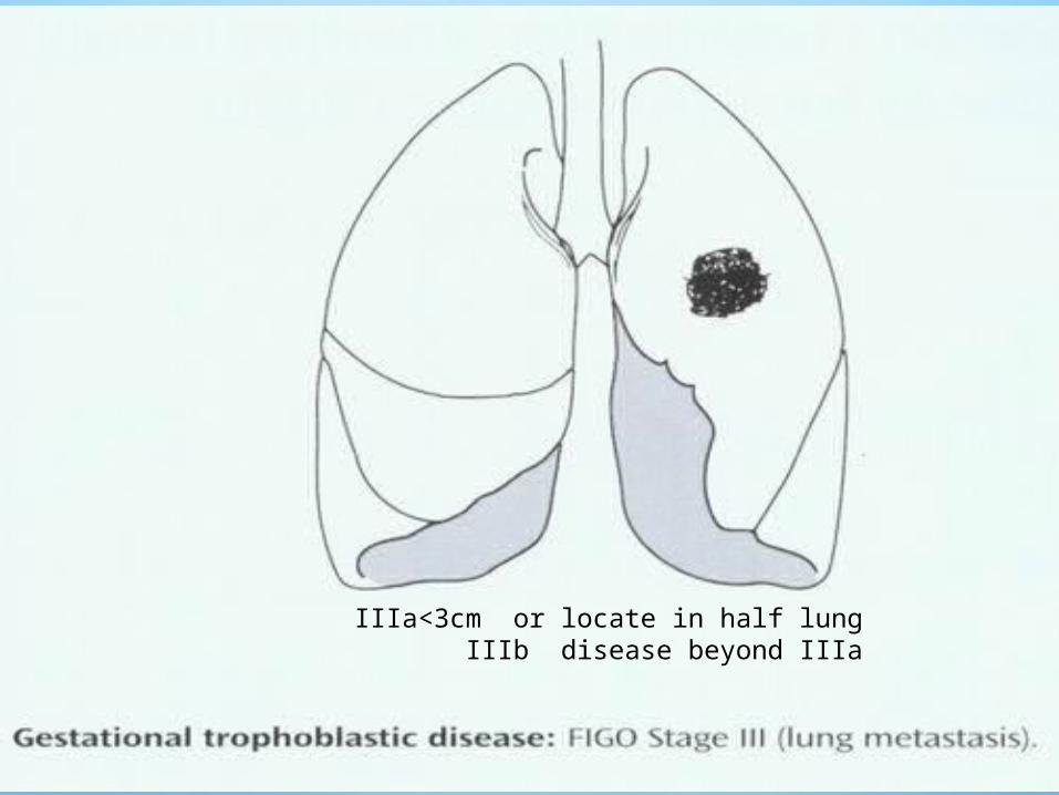
IIIa<3cm or locate in half lungIIIb disease beyond IIIa


Who Orgnaization prognostic scoring system for gestational trophoblastic neoplasia
Prognostic factor 0124
Age <39>39_-
Antecedent pregnancy HydatidiformAbortion , ectipicTerm pregnancy-
Interval (months)<44-67-12>12
hCG level (IU/liter)<1010-1010-10>10
ABO blood groups (female/male)
O/ABA/OAB
Largest tumor (cm)<33-5>5_
Site of metastasis _Spleen, kidneyGastrointestinal tract, liver Brain
Number of metastases _1-34-8>8
Prior chemotherapy __Single drug Multiple drugs
The total score is obtained by adding the individual scores for each prognostic factor . Total score >:4 , low risk ; 5-7 , intermediate risk ;>8 , high risk.
Interval :between antecedent pregnancy and start of chemotherapy.

• According to the FIGO staging of gestational trophoblastic tumors
• a lady with choriocarcinoma having • lung metastasis will belong to which stage

protocol for treatment of GTD
Clinically staging( FIGO)WHO scoring Again, it is stressed that the diagnosis of GTN made by persistently elevated serum β-hCG without confirmation by pathological
tissue study

Choice of treatment
• Chemotherapy ( highly sensitive ) • Surgery ( unresponsive or drug fails )• Irradiation (brain and liver )

Chemotherapy are best management Protocols: Single-agent for low-risk Methotrexate
Combination for high-risk disease EMA-CO
Early-stage GTN is typically cured Later -stage disease usually responds to chemotherapy

Surgery in malignant GTN
• Hysterectomy• Laparoscopy• Craniotomy (brain hemorrhage)• Thoracotomy • (solitary nodules in drug-resistant disease ) • selective resection of lesion in uterus or liver

Main causes of death :
• Hemorrhage
• Infection
• Metastasis

Placental Site Trophoblastic Tumor (PSST)
PSTT or non-trophoblastic malignancy
Uncommon tumor arises from implantation site-intermediate trophoblast
Secrete (hPL) from intermediate cells Relatively small amounts of hCG hCG free β-subunit is more than one third of
hCG (30%)

Typically local myometrial invasion Rare systemic metastases Treatment of ( PSST) is preferred hysterectomy Because resistant to chemotherapy For higher-risk than stage I combination chemotherapy given

Epithelioid Trophoblastic Tumor
This rare tumor Intermediate trophoblast -type Grossly a nodular fashion Primary treatment is hysterectomy Relatively resistant to chemotherapy Approximately a fourth this neoplasm will have metastatic disease, combination chemotherapy

SUBSEQUENT PREGNANCYPregnancy outcomes are usually normal May develop: 1-Repeat molar gestation 2-percent 2-Spontaneous abortions 3-Congenital anomalies 4-Ovarian failure (chemotherapy) 5-Secondary tumors including leukemia colon cancer, melanoma and breast cancer


After Termination of Subsequent Pregnancy
Sonographic evaluation in early pregnancy
pathological evaluation placenta after delivery
serum β-hCG level is measured 6 weeks postpartum

Conclusion
The possibility of metastatic GTN should be considered
In any woman of the reproductive age Presenting with metastatic disease Or an unknown primary site of malignancy

www.zohrehyousefi.com