Embed Size (px)
Citation preview

Государственное бюджетное образовательное учреждение высшего
профессионального образования
«Иркутский государственный медицинский университет»
Министерства здравоохранения Российской Федерации
Department of Operative Surgery and Topographic Anatomy
G.I. Songolov, O.P. Galeeva, S.N. Redkov
Clinical anatomy of the upper extremity
Teaching aid
Иркутск
ИГМУ
2016

УДК [616-089.87(075.8)
ББК 54.54я73.
C 60
Recommended by faculty methodological council of medical department of SBEI HE ISMU The Ministry of Health of The Russian Federation as a teaching aid for
independent work of foreign students from medical faculty, faculty of pediatrics,
faculty of dentistry, protocol № 01.02.2016.
Authors:
G.I. Songolov - associate professor, Head of Department of Operative Surgery
and Topographic Anatomy, PhD, MD SBEI HE ISMU The
Ministry of Health of The Russian Federation
O. P.Galeeva - associate professor of Department of Operative Surgery and
Topographic Anatomy, MD, PhD SBEI HE ISMU The Ministry
of Health of The Russian Federation
S. N. Redkov – assistant of department of Operative Surgery and Topographic
Anatomy SBEI HE ISMU The Ministry of Health of The
Russian Federation.
Reviewers:
E.V. Gvildis - head of department of foreign languages with the course of the
Latin and Russian as foreign languages of SBEI HE ISMU The Ministry of
Health of The Russian Federation, PhD,
L.V. Sorokina - associate Professor of Department of Anesthesiology and
Reanimation at ISMU, PhD, MD
Songolov G.I
C 60 Clinical anatomy of upper extremity: teaching aid / Songolov G.I, Galeeva O.P,
Redkov S.N; State budget educational institution of higher education of the
Ministry of Health and Social Development of the Russian Federation; "Irkutsk
State Medical University" of the Ministry of Health and Social Development of
the Russian Federation Irkutsk ISMU, 2016, 37 p.
Clinical anatomy of upper extremity is one of the important sections in topographic anatomy.
The teaching manual is intended for foreign students of medical faculty, faculty of pediatrics,
faculty of dentistry of USMU. УДК [616-089 (075.8)
ББК 54.54я73 © Сонголов Г.И., Галеева О.П, Редков С.Н, Юдин А.А,
© ГОУ ВПО ИГМУ Минздрава России, 2016

Contents
INTRODUCTION ............................................................................................................................................. 4
GENERAL CHARACTERISTICS ......................................................................................................................... 5
TOPOGRAPHY OF THE SCAPULAR REGION .................................................................................................... 5
TOPOGRAPHY OF THE AXILLARY REGION...................................................................................................... 7
TOPAGRAPHY OF THE DELTOID REGION ..................................................................................................... 10
TOPOGRAPHY OF THE SUBCLAVIAN REGION .............................................................................................. 11
TOPAGRAPHY OF THE ANTERIOR REGION OF THE ARM ............................................................................. 12
TOPOGRAPHY OF THE POSTERIOR REGION OF THE ARM ........................................................................... 14
TOPAGRAPHY OF THE ELBOW REGION ....................................................................................................... 16
TOPAGRAPHY OF THE ANTERIOR CUBITAL REGION .................................................................................... 17
TOPOGRAPHY OF THE POSTERIOR CUBITAL REGION .................................................................................. 19
TOPOGRAPHY OF THE ANTERIOR REGION OF THE FOREARM .................................................................... 20
TOPOGRAPHY OF THE POSTERIOR REGION OF THE FOREARM .............................................................. 25
TOPOGRAPHY OF THE DORSAL REGION OF THE HAND .............................................................................. 28
TOPOGRAPHY OF THE WRIST ...................................................................................................................... 28
TOPOGRAPHY OF THE FINGERS................................................................................................................... 30
TEST ............................................................................................................................................................. 34
TEST ANSWERS ............................................................................................................................................ 38

INTRODUCTION
In this manual you can find the materials, used in learning the theme: ―Topography
of the upper extremity‖. The objective of this is the formation of the holistic
picture of the targeting and projection anatomy of the upper extremity. This
manual is a useful source of knowledge of the lower extremity’s regional clinical
anatomy. The student, who read attentively will find for himself the necessary
anatomoco-clinical connections, that in the future will help in his independent
work.
The formation of holistic view of morphofunctional relations in the organism is
tightly connected with anatomico-clinical characteristic of the concrete region of
the human’s body. The importance of this cannot be overemphasized because it is
necessary for a doctor to have the knowledge and the skill to be professionally
oriented in the spatial and structural organization of the biological object
discredited by the disease or trauma.
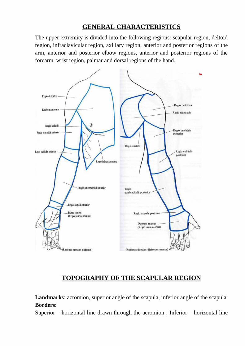
GENERAL CHARACTERISTICS
The upper extremity is divided into the following regions: scapular region, deltoid
region, infraclavicular region, axillary region, anterior and posterior regions of the
arm, anterior and posterior elbow regions, anterior and posterior regions of the
forearm, wrist region, palmar and dorsal regions of the hand.
TOPOGRAPHY OF THE SCAPULAR REGION
Landmarks: acromion, superior angle of the scapula, inferior angle of the scapula.
Borders:
Superior – horizontal line drawn through the acromion . Inferior – horizontal line

drawn through the inferior angle of the scapula. Lateral – vertical line drawn
through the acromion. Medial – medial margin of the scapula.
Layers:
Skin: it is thick with limited movement. It is innervated by the superior lateral
cutaneous nerve of the arm.
Subcutaneous tissue.
Superficial fascia: it is dense and consists of many layers. It contains the fibrous
tissues which connect the subcutaneous tissue and are fixed to the skin. That is
why the skin has limited movement. It contains fat and cutaneous nerve.
Deep fascia: it consists of 2 layers: superficial layer covers the latissimus dorsi and
trapezius muscles. Deep layer covers the supraspinatus, infraspinatus, teres major
and teres minor muscles.

SCAPULAR ARTERIAL NETWORK
Anastomosis around the scapula occurs in 3 fossae:
1. Supraspinous fossa. 2. Infraspinous fossa. 3. Subscapular fossa.
It is formed by: suprascapular artery (branch of the thyrocervical artery of the
subclavian artery), dorsal scapular artery/ deep branch of the transverse cervical
artery; circumflex scapular artery.
To provide collateral circulation when the subclavian artery or axillary artery is
bloched/ damaged. To help preserve the upper limb during injury.
Anasromosis over the acromion : acromial branch of the thoracoacromial artery;
acromial branch of the suprascapular artery; acromial branch of the posterior
circumflex humeral artery.
TOPOGRAPHY OF THE AXILLARY REGION
Landmarks:
Outlines of the pectoralis major, latissimus dorsi and coracobrachialis muscles;
axillary fossa is shown by lifting up the upper extremity.
Borders: anterior- lower margin of the pectoralis major muscle; posterior – lower
margin of the latissimus dorsi muscle; medial – line connecting the margin of the
pectoralis major and latissimus dorsi muscles along the saggital section of the
lateral surface of the thorax at the level of the 3rd
rib; lateral – line connecting the
margin of the pectoralis major and latissimus dorsi muscles on the medial surface
of the arm.
Layers:
Skin: it is thin and easily movable. It contains the suspensory ligament of the axilla.
It separates and forms the pectoral fascia anteriorly, thoracolumbar fascia
posteriorly and brachial fascia laterally.
Axillary cavity:
Borders:
Anterior – pectoralis major muscle and clavipectoral fascia. Posterior –
subscapularis, latissimus dorsi, teres major muscles. Medial – thoracic wall,
serratus anterior muscle and 1st until 4
th intercostal muscles. Lateral – surgical neck
of the humerus, short head of the biceps brachii muscle and coracobrachialis
muscle.
Contents: axillary is divided into 3 parts and gives branches according to:
clavipectoral triangle (thoracoacromial artery, arteria thoracica suprema, lateral
and medial pectoral nerves); pectoral triangle ( lateral thoracic artery and long

thoracic nerve); subpectoral triangle(subscapular artery, anterior and posterior
circumflex humeral arteries).
The axillary artery is in relation first with the posterior and then with the outer
wall of the axillary cavity. The pulsations of the vessel may be distinctly felt to the
inner side of the prominence caused by the coracobrachialis muscle; this is the
situation in which the artery may be most safely ligated by elevating the arm and
keeping close to the border of the muscle. The vessel is comparatively close to the
shoulder-joint and is sometimes torn across in dislocations or in attempts at their
forcible reduction. The branches of the axillary artery are:
1. The acromiothoracic
2. The subscapular, which arises from about the middle of the axillary and
consequently at the mid-point between the clavicle and the lower border of the
pectoralis major muscle, in which latter situation the axillary becomes the brachial.
The subscapular artery divides into two chief branches, the dorsalis scapula and
the thoracicodorsalis .
The dorsalis scapula runs over the external border of the scapula to the dorsal
surface of this bone, supplies the surrounding muscles, and anastomoses freely
with the suprascapular branch of the subclavian. This is the main path for the
collateral circulation when the blood-current in the axillary artery is interfered with
or when this vessel is ligated. The thoracodorsalis is the largest vessel of the lateral
thoracic wall. It runs down the axillary border of the scapula, under cover of the
edge of the latissimus dorsi, and particularly supplies the teres major, the
latissimus dorsi, and the serratus magnus muscles .
3.The long thoracic, an inconstant branch, which arises above the subscapular and
is situated in front of the thoracic dorsalis.
4· The anterior circumflex, a small branch which runs outward over the anterior
surface of the neck of the humerus.
5· The posterior circumflex, a large branch which arises beside the preceding one,
passes through the quadrangular space close to the bone to run posteriorly with the
circumflex nerve and, like it, to particularly supply the deltoid muscle.
The axillary vein, like the subclavian, is situated in front and to the inner side of
the artery; it consequently covers the artery to a varying extent, dependent upon the
degree of its distention. It is only in exceptional cases that the axillary vein is
double.

Brachial plexus: lateral cord, posterior cord, medial cord.
Just beneath the clavicle the trunks of the brachial plexus, with the exception of a
few small branches, are situated to the outer side of the vessel. Beneath the
pectoralis minor muscle these trunks form three cords which are so arranged about
this portion of the artery that we may differentiate an outer, an inner, and a
posterior cord. The outer and inner cords supply chiefly the skin and muscles of the
flexor surface, while the posterior cord supplies the extensors surface. A more
detailed study of the axilla shows that the outer cord gives off the
musculocutaneous nerve and the outer head of the median. From the inner cord

arises the inner head of the median which, together with the outer head, surrounds
the axillary artery like the prongs of a fork; the inner cord also gives off the ulnar,
the internal cutaneous, and the lesser internal cutaneous nerves. From the posterior
cord arise the circumflex, the musculus spiral, and the subscapular nerves.
Of the other branches of the brachial plexus there is one which is of particular
importance from a surgical stand point; this is the long thoracic, the motor nerve of
the serratus magnus muscle. It lies directly upon the serratus magnus, in assotiation
with the thoracicodorsalis artery, and is covered by the edge of the latissimus
dorsi; it is not particulary exposed to injury in operations in the axillary cavity. The
comparatively superficial long or middle subscapular nerve running to the
latissimus dorsi is more easily injured, and its division is followed by paralysis of
this muscle. The other two subscapular nerves supply the teres major and the
subscapularis muscles and are more deeply situated. The suprascapularis nerve,
which runs through the subscapularis notch to the dorsal surface of the scapula and
supplies the supraspinatus and infraspinatus muscles, is more rarely divided by the
knife of the surgeon; the nerve filaments supplying the levator anguli scapulae and
the rhomboid muscles are also rarely injured.
Axillary lymph nodes (5 groups): lateral axillary lymph nodes, central axillary
lymph nodes, medial axillary lymph nodes, posterior axillary lymph nodes, apical
axillary lymph nodes.
TOPAGRAPHY OF THE DELTOID REGION
Landmarks: anterior and posterior margins of the deltoid muscle, clavicle,
acromioclavicular joint, acromion, spine of the scapula.
Borders:
Superior – line drawn on the deltoid muscle from lateral 1/3 of the clavicle,
acromion and lateral 1/3 of the spine of the scapula. Inferior – horizontal line
drawn on the lower margin of the pectoralis major major muscle and latissimus
dorsi muscle. Anterior – anterior margin of the deltoid muscle. Posterior –
posterior margin of the deltoid muscle.
Layers:
Skin: it is thick with limited movement. Upper half of the deltoid muscle: lateral
supraclavicular nerve. Lower half of the deltoid muscle: superior lateral brachial
cutaneous nerve.
Subcutaneous tissue with superficial fascia: it contains moderate amount of fat. It
is divided by septa.
Deep fascia.
Superficial layer: it covers the outer surface of the deltoid muscle; it is continuous

eith the pectoral fascia; it divides the deltoid muscle into 3 parts, which are the
clavicular, acromial.
Deep layer: it invests the deltoid nerve; it is continuous with the fascia covering the
triceps brachii muscle; it sends numerous septa and fasciculi.
Muscles:
Unipennate: clavicular and spine of the scapula parts;Multipennate: acromial part;
all fibres converge and are attached to the deltoid tuberosity.Actions: abduction,
pulling the arm forwards and pronating is slightly, pulling the arm backwards and
supinating it slightly, anterior part helps the petoralis major muscle in arm flexion
and medial rotation of the arm; posterior part heps latissimus dorsi muscle in arm
extension and lateral rotation of the arm.
TOPOGRAPHY OF THE SUBCLAVIAN REGION
Landmarks: sternum, clavicle, coracoid process, 3rd
rib, cephalic vein, pectoralis

major muscle, deltoid muscle, border between pectoralis major and deltoid
muscles.
Borders:
Superior – clavicle. Inferior – horizontal line drawn through the 3rd
rib; upper
margin of the mammary gland (in women). Medial – lateral margin of the sternum.
Lateral – anterior margin of the deltoid muscle.
Layers:
Skin: it is thin, easily movable; it contains sebaceous glands; it is innervated by the
supraclavicular nerve.
Subcutaneous tissue: it is well-developed, especially in women. It contains the
cutaneous nerve. It contains the thoracoacromial and thoracoepigastric veins form
the cephalic and axillary veins.
Superficial fascia: it is thin and firm. It is attached to the inferior margin of the
clavicle and upper margin of the mammary gland, which forms the Cooper’s
suspensory ligaments of the mammary gland.
Deep fascia: it covers the pectoralis major muscle, it divides the pectoralis major
muscle into 3 parts (clavicular, sternocostal, abdominal).
Borders: superior – clavicle; inferior – fascia of the serratus anterior and rectus
abdominis muscles; medial – sternum; lateral – deltoid and axillary fasciae.
Proper fascia: it is attached to the lower margin of the clavicle, coracoid process
and 1st rib. It covers the subclavius and pectoralis minor muscles. Lower part of
proper fascia forms the suspensory ligaments of the mammary gland.
TOPAGRAPHY OF THE ANTERIOR REGION OF THE ARM
Landmarks: deltoid muscle, pectoralis major muscle, latissimus dorsi muscle,
biceps brachii muscle, triceps brachii muscle, medial and lateral epicondyles of the
humerus, greater tubercule of the humerus; sulci bicipitales lateralis et mediales.
Borders:
Superior – horizontal line drawn from the pectoralis major and latissimus dorsi
muscles. Inferior – imaginary line drawn with 2 fingers above the lateral and
medial epicondyles of the humerus; medial and lateral: vertical lines drawn on
medial and lateral epicondyles of the humerus respectively.
Layers:
Skin: lateral is thicker than the medial one. It is slightly movable. It is innervated
by the medial cutaneous nerve of the arm, lateral cutaneous nerve of the arm,
superior lateral cutaneous nerve of the arm and inferior lateral cutaneous nerve of
the arm.

Subcutaneous tissue: it contains the cephalic and basilica veins. It contains the
cubital lymph nodes.
Superficial fascia: it forms a covering for the superficial veins and cutaneous
nerves.
Deep fascia: it forms 2 septa which are connected to the humerus, namely the
lateral and medial intermuscular septa. These 2 septa divide the brachial region
into anterior and posterior parts. Transverse septum is pierced by the ulnar nerve

and anterior descending branch of profunda brachii artery to the anterior surface of
the lateral epicondyle. Anteroposterior septum separates the brachalis muscle from
the muscles attached to the lateral supracondylar ridge. This septum also encloses
the radial nerve and anterior descending branch of the deep artery of arm.
Muscles: coracobrachialis muscle (from the coracoid process in common with the
short head of the biceps, and its insertion is at about the middle of the internal
border of the humerus), biceps brachii muscle(arises by along head from the apex
of the coracoid process; it inserts into the tuberosity of the radius), brachialis
muscle(situated beneath the biceps, arises from the anterior aspect of the humerus,
below the insertion of the deltoid muscle, and from the intermuscular septa, and is
insertedinto the coronoid process of the ulna).
TOPOGRAPHY OF THE POSTERIOR REGION OF THE ARM
Landmarks: deltoid muscle, pectoralis major muscle, latissimus dorsi muscle,
biceps brachii muscle, triceps brachii muscle, medial and lateral epicondyles of the
humerus, greater tubercle of humerus, sulcus deltoideopectoralis, sulci bicipitales
lateralis et mediales.
Borders:
Superior – horizontal line drawn from the pectoralis major latissimus dorsi
muscles. Inferior – imaginary line drawn with 2 fingers above the lateral and
medial epicondyles of the humerus. Medial and lateral – vertical lines drawn on
the medial and lateral epicondyles of the humerus respectively.
Layers:
Skin: it is thick and well-connected with the subcutaneous tissue.
Subcutaneous tissue: it contains the superior lateral cutaneous nerve of the arm,
inferior lateral cutaneous nerve of the arm and posterior cutaneous nerve of the
forearm.
Superficial fascia.
Deep fascia: it forms a covering for the triceps brachii muscle and its tendon.
Muscle: triceps brachii muscle (the long head arises from the infraglenoid tubercle
of the scapula; the inner head arises from the posterior surface of the humerus
below the musculospiral groove; the outer head commences above the
musculosoiral groove, which it bridges over, and arises from the outer surface of
the bone and from the external intermuscular septum. These 3 heads are inserted
into a common tendon which is attached to the olecranon).
Humeromuscular canal: it is also known as canalis spiralis. Radial nerve, deep
artery of arm and its vein pass through this canal.

Superior trilateral foramen
Borders:
Medial – teres minor muscle. Lateral – medial margin of the long head of the
triceps muscle. Inferior – teres major muscle.
Contents: circumflex scapular artery.
Inferior trilateral foramen
Borders:
Medial – long head of the triceps brachii muscle;lateral – medial margin of the
humerus. Superior – teres major muscle.
Contents: radial nerve, deep artery of the arm.
Quadrilateral foramen
Borders:
Medial – lateral margin of the lons head of the triceps brachii muscle; Lateral –
surgical neck of the humerus. Superior – teres minor muscle, subscapularis muscle.
Inferior – teres circumflex humeral artery

Canal of the radial nerve
Topography: it is bounded by the humerus and triceps brachii muscle. It contains
the radial nerve, deep artery of the arm and its veins.
The ulnar, the second great nerve of the arm, lies internal to the artery, but
gradually becomes more distant from the vessel. It pierce the internal
intermuscular septum at the lower third pf the arm and reaches the space between
the internal condyle and the olecranon. The ulnar nerve also gives off no branches
in the upper arm as the median one.
Shoulder-joint -This joint has the widest range of motion of any joint in the body.
The articulation is formed by the head of the humerus and the glenoid cavity of the
scapula. The humeral head is covered by cartilage and the articular surface of the
glenoid cavity is enlarged deepened) by the glenoid ligament. As a consequence of
the incongruity between the two articular surfaces, the head of the humerus
projects far beyond the edge of the glenoid cavity. The lax capsular ligament
passes down from the circumference of the glenoid cavity and is attached,
anteriorly, to the anatomic neck of the humerus.
TOPAGRAPHY OF THE ELBOW REGION
Landmarks:
Olecranon of the ulna, brachioradialis muscle, tendon of the biceps brachii muscle,
medial and lateralepicondyles of the humerus, sulci cubitales posteriores lateralis et
medialis, sulci cubitales anteriores lateralis et medialis.
Borders:
A horizontal line is drawn 4 cm from the level which connects the medial and
lateral epicondyles of the humerus; 2 vertical lines are drawn through the medial
and lateral epicondyles of the humerus and divide the elbow region into the
anterior region and posterior region.
The elbow-joint is composed of three articulations:
I. The trochlear surface of the humerus with the greater sigmoid cavity of the ulna.
2. The capitellum of the humerus with the cup-shaped depression on the head of
the radius.
3. The lesser sigmoid cavity of the ulna with the circumference of the head of the
radius. The first articulation is for flexion and extension, the third is for rotation
(pronation and supination), and the second permits not only of flexion and
extension, but also of rotation. All three articulations are surrounded by a common
lax capsular ligament, which is attached anteriorly above the coronoid and radial

depressions, excluding the epicondyles, and posteriorly above the olecranon fossa,
which is also situated within the joint. To either side of the olecranon the capsular
ligament forms small diverticula which arch situated beneath the cutaneous
depressions visible in this situation in the living subject. The capsular ligament is
attached to the edges of the greater and lesser sigmoid cavities of the ulna in such a
way that the apex of the coronoid process and the olecranon arc within the articular
cavity; this ligament is also inserted into the neck of the radius so that the head of
this bone is entirely within the joint. The capsular ligament is reinforced by three
other ligaments:
1.The internal lateral ligament, which radiates from the internal epicondyle to the
edge of the greater sigmoid cavity of the ulna.
2. The external lateral ligament, which runs downward from the external
epicondyle, some of the fibers surrounding the neck of the radius and being
inserted into the anterior and posterior margins of the lesser sigmoid cavity of the
ulna.
3· The orbicular ligament, which enforces the capsule in a circular manner about
the neck of the radius and is inserted into the anterior and posterior margins of the
lesser sigmoid cavity of the ulna. The external lateral and orbicular ligaments form
practically one continuous band of connective tissue.
As the large superficial and deep vessels of this region are situated in front of the
elbow-joint, it follows that marked subcutaneous or deep extravasations of blood
may result from a backward dislocation of both bones, and particularly from that
form which is due to hyperextension. In this dislocation the median nerve is
necessarily lacerated, as it is also in front of the articulation. The importance of the
relation of the ulnar nerve to the joint has been already emphasized.
TOPAGRAPHY OF THE ANTERIOR CUBITAL REGION
Layers:
Skin: it is thin, movable and non- pigmented. It contains sweat glands, sebaceous
glands and hair. It is innervated by the lateral and medial cutaneous nerves of the
forearm.
Subcutaneous tissue: it contains lymphatic vessels with superficial lymphatic nodes
and superficial venous network. It contains the cephalic, basilica and median
cubital veins. Superficial cubital nodes are situated at the basilica vein. The
connection of the cephalic, basilic and median cubital veins forms an alphabet ―N‖.
Superficial fascia: it is connected with the superficial fascia of the arm and
forearm, aponeurosis of biceps brachii muscle, it is connected with the deep fascia.
Deep fascia: fascia of the biceps brachii and brachialis muscles forms the lateral
and medial intramuscular septa. These septa are fixed to the medial and lateral
epicondyles of the humerus. It forms a covering for the muscles of the arm and
forearm. Inferior to the radial tuberosity, the fixation for the tendon of the biceps
brachii muscle takes place. Intermuscular septa continue to form the anterior radial
intermuscular septa of the forearm.
Muscles:
Lateral: brachioradialis muscle and supinator muscle.
Median: biceps brachii muscle and brachialis muscle.
Medial: (1st layer) pronator teres, flexor carpi radialis, palmaris longus and flexor
carpu ulnaris musles
Medial: (2nd
layer) flexor digitorum superficialis muscle.
Neurovascular bundles:
2 neurovascular bundles are found between groups of muscles and septa, ehich are
lateral bundle (radial nerve and radial collateral artery) and medial bundle(brachial
artery and median nerve).
The brachial artery runs beneath the deep fascia in the internal bicipital groove; it
is accompanied by two veins, the inner of which receives the basilica vein. Higher
up, the vena commits unite to form a single vein, either before reaching the axilla
or, more rarely, after entering into this region. Disregarding the muscular branches,
of which the bicipital is particulary large, the main branches of the brachial are:
1. The superior profunda artery, which accompanies the musculospiral nerve
about the posterior surface of the humerus, in the musculospiral groove, between
the outer and inner heads of the triceps muscle. It ends at the outer side of the
humerus, where it is accompanied by a cutaneous branch of the musculospiral
nerve, and descends behind the external intermuscular septum to reach an
anastomotic arch above the olecranon.
2. The inferior profunda artery, usually a ling vessel, which accompanies the
ulnar nerve to the olecranon, where it empties into the rete cubtiti.
3. The anasomotica magna artery, which arises near the division of the brachial in
the region of the elbow. It runs transversely inward across the brachialis anticus
muscle and passes posteriorly to the rete cubiti.
The radial artery: It passes through the tendon of the biceps brachii muscle. It is
lodged between the pronator teres and brachioradialis muscles.
The radial nerve: it is situated between the brachioradiales and brachialis muscles
in the upper level. It goes downward and is divided into 2 branches, which are the
superficial and deep branches of the radial nerve at the level of the lateral
epicondyle of the humerus. Superficial branch innervates the brachioradialis and

pronator teres muscles. Deep branch lies laterally and passes through the supinator
canal between the superficial and deep layers of supinator muscle.
TOPOGRAPHY OF THE POSTERIOR CUBITAL REGION
Landmarks:
Olecranon of ulna, sulci cubitales posteriors lateralis et mediales, ulnar nerve
passing along the sulcus cubitales posterior medialis, head of the radius is palpated
in the middle part of sulcus cubitalis posterior lateralis.
Layers:

Skin: it is thick and movable, it forms skin folds.
Subcutaneous tissue: it contains the superficial arteries( medial collateral artery,
radial collateral artery and recurrent interosseous artery); it contains the posterior
cutaneous nerve of the arm, medial cutaneous nerve of the forearm. Bursa of the
elbow joint is located in the superior part of the olecranon of the ulna.
Superficial fascia: it is thin layer without fixation.
Deep fascia: it is formed by the aponeurosis; it is fixed to the tendon of the triceps
brachii muscle, medial and lateral epicondyles of the humerus and olecranon of the
ulna.
Muscles:
Superior – triceps brachii and anconeus muscles; lateral – extensor muscle of the
wrist(extensor carpi radialis longus , runs to the dorsal surface of the base of the
second metacarpal bone, extensor carpi radialis brevis, goes to the dorsal surface
of the third metacarpal bone, and extensor carpi ulnaris muscles, arises from the
external condyle, from the deep fascia of the forearm, and from the posterior
border of the ulna, and is inserted into the base of the fifth metacarpal bone) and
extensor muscle of the digits(extensor digitorum, arises from the external condyle
and from from the deep fascia of the forearm and is inserted by means of four
tendons into the dorsal aponeurosis of the fingers, and extensor digiti minimi
muscles, arises from the external condyle and furnishes a second tendon to the little
finger); deep - supinator muscle; medial – flexor digitorum profundus, arises from
the upper two-thirds of the anterior surface of the ulna and from the interosseous
membrane, and by a small muscular slip from the internal condyle and is inserted
into the terminal phalanx of the thumb and flexor carpi ulnaris muscles, the ulnar
origin extends downward to the lower third of the bone, is inserted into the
pisiform bone, the inciform process of the unciform mone, and the base of the fifth
metacarpal bone.
TOPOGRAPHY OF THE ANTERIOR REGION OF THE
FOREARM
Landmarks: brachioradialis muscle, sulci radialis et ulnaris, tendons of the flexor
carpi radialis and palmaris longus muscles, styloid process of the ulna and radius.
Borders:
Upper – horizontal line drqwn 4 cm distal to the level of the wrist joint. Lower –
transverse line drawn 2 cm proximal to the styloid process of the radius; 2 vertical
lines are drawn through the epicondyles and styloid processes, which divide the

forearm region into the anterior and posterior regions.
Layers:
Skin: it is thin, it contains sweat and sebaceous glands, it is innervated by the
medial and lateral cutaneous nerves of the forearm.
Subcutaneous tissue: it contains the cephalic vein and basilica vein; it contains the
lateral cutaneous nerve of the forearm and medial cutaneous nerve of the forearm;
intermediate anterbrachial vein passes through the middle line of this region.
Superficial fascia: it is not attached to the bone, it covers all structures in the
subcutaneous tissue.
Deep fascia: it forms a covering for muscles, vessels, nerves and bones; it is
thicker in the proximal part and thinner in the distal part; it sends two septa to the
radius onle and divides the forearm into 3 seats for muscles(anterior, posterior and
lateral) in the upper half of forearm.
Lateral: brachioradialis, extensor carpi radialis longus and extensor carpi radialis
brevis muscles. Anterior: pronator teres, flexor carpi radialis, flexor digitorum
superficialis, palmaris longus and flexor carpi ulnaris muscles, flexor digitorum
profundus and flexor pollicis lomgus muscles.
Borders of the fascia: anterior – strictly fixed to the deep fascia; posterior –
interosseous membrane, ulna and radius; medial – posterior margin of ulna; lateral
– sulcus radialis.
Pirogov’s space is situated in the lower half of the deep fascia.
Anterior: fascia of the flexor digitorum and flexor pollicis longus muscles.
Posterior: fascia of the pronator quadratus muscle.
Muscles:
Medial margin of the deep fascia is bounded to the posterior margin of the ulna and
divides the muscles into 4 layers:
1) 1st layer: pronator teres, flexor carpi radialis, palmaris longus, flexor carpi
ulnaris and brachioradialis muscles.
2) 2nd
layer: flexor digitorum superficialis muscle.
3) 3rd
layer: flexor digitorum profundus and flexor pollicis longus muscles.
4) 4th layer: pronator quadratus muscle.
The muscles of the forearm should be reviewed in detail from a systematic
anatomy. The flexor group may be subdivided into a superficial and a deep layer,
each consisting of four muscles. All of the superficial flexors originate from the
internal condyle. The pronator radii teres is inserted into the outer surface of the
middle of the radius. The flexor carpi radialis is attached to the base of the second
metacarpal bone. The palmaris longus radiates into the palmar fascia. The flexor
carpi ulnaris, the ulnar origin of which extends downward to the lower third of the

bone, is inserted into the pisiform bone, the unciform process of the unciform
bone, and the base of the fifth metacarpal bone. There are four deep muscles upon
the flexor surface. The flexor sublimis digitorum arises by two heads from the
internal condyle of the humerus and from the anterior surface of the radius and is
inserted by means of four tendons into the second phalanges of the fingers.
The flexor profundus digitorum arises from the upper two-third of the anterior
surface of the ulna and from the interosseous membrane and is inserted by means
of four tendons into the terminal phalanges of the fingers. The flexor longus
pollicis arises from the anterior surface of the radius, from the interosseous
membrane, and by a small muscular slip from the internal condyle and inserted

into the terminal phalanx of the thumb.
Neurovascular bundles:
Lateral:
Radial artery and vein; superficial branch of the radial nerve: it is located on the
sulcus radialis, in the upper 1/3 of the forearm, it is bounded by the brachioradialis
muscle laterally and pronator teres muscle medially, in the middle and lower 1/3
of the forearm, it follows the brachioradialis and flexor caroi radialis muscles and
passes downward along with radial artery, then it passes through the tendon of the
brachioradialis muscle.
The radial artery: it passes along the middle part f the elbow joint to the styloid
process of the radius and enters the anatomical snuffbox.
Medial:
The ulnar artery: it exists inferior to the pronator muscle in the upper part. It is
later located between the superficial and deep flexors of the fingers. It passes from
the tendon of the biceps brachii muscle, in the middle 1/3 of the forearm, it passes
under the pronator teres muscle, flexor digitorum superficialis muscle and sulcus
ulnaris, in the lower 1/3 of the forearm, it passes to the medial margin of the
styloid process of the ulna and reaches the pisiform bone.
The ulnar nerve: it passes on the sulcus ulnaris and is bouded medially by the
flexor carpi ulnaris muscle and laterally by the flexor digitorum superficialis in the
upper 1/3 of the forearm; then it passes from the medial epicondyle of the humerus
to the medial margin of the pisiform bone; dorsal branch of the ulnar nrve starts
from the middle and lower 1/3 of the forearm and passes under the tendon of the
flexor carpi ulnaris muscle, it passes downward and medially to the ulnar artery.
Posterior interosseous artery is a branch of the upper part of the ulnar artery and is
divided into the anterior and posterior interosseous arteries. Posterior interrosous
artery passes through the orifice of the interrosseous mmbran to the posterior
region of the forearm.
Anterior:
The median nerve is the most superficial of the three chief nerves in the upper arm.
At first the nerve lies to the outer side of the brachial artery, it then passes in front
of the vessel, and, while still above the elbow, takes a position to the inner side of
the artery. The nerve consequently crosses the artery, and is to be felt lying upon
the vessel, if the panniculus adiposus is not too well marked. The nerve gives off
no branches in the upper arm as the median one.

TOPOGRAPHY OF THE POSTERIOR REGION OF THE
FOREARM
Layers:
Skin: it is thick with limited movement; it is innervated by the medial and lateral

cutaneous nerves of the forearm and posterior cutaneous nerve of the forearm.
Subcutaneous tissue: it contains less fatty tissue; superficial veins from the main
trunk of the vein.
Superficial fascia: it is weak layer.
Deep fascia: it is distinguished by its thickness and strictly fixed to the ulna and
radius, it is an aponeurosis in the upper half of the forearm, it tends downward and
forms the extensor retinaculum and dorsal carpal canal, it forms cellular space
which contains deep branch of the radial nerve, posterior interosseous artery,
posterior interosseuos veins and nerve; deep branch of the radial nerve passes
through the canalis supinatorius; posterior interosseous artery passes medial to the
posterior interosseous nerve.
Muscles:
Superficial muscles: extensor carpi radialis longus muscle, extensor carpi radialis
brevis muscle, extensor digitorum muscle, extensor digiti minimi muscle,
extensor carpi ulnaris muscle.
Deep muscles: supinator muscle, abductor pollicis longus muscle, extensor pollisic
longus muscle, extensor pollisic brevis muscle, extensor indicis muscle.
TOPOGRAPHY OF THE PALMAR REGION OF THE HAND
Landmarks: styloid processes of the ulna and radius, skin fold of the wrist joint,
metacarpal bones, phalanges of fingers.
Borders: 2 cm above the styloid process of the ulna, 2 vertical lines are drawn
along the ulna and radius and divide the hand region into the palmar and dorsal
regions.
Layer:
Skin: it is thick with limited movement, stratum corneum is well developed in this
region, it contains sweat glands and sebaceous glands, it is innervated by the radial
nerve, ulnar nerve and median nerve.
Subcutaneous tissue: it contains vessels, nerves and lymphatic vessels with
lymphatic nodes, it contains the superficial palmar branch of the radial artery.
Superficial fascia: it is a continuation of the superficial fascia of the forearm, it is
slightly movable and fixed to the bones.
Deep fascia: it stretches from the palmary longus tendon to the ligamentum carpi
volare, it forms the palmar aponeurosis, flexor retinaculum, synovial sheaths of
the hand and fingers.
Flexor retinaculum: it is the strongest and thickest fascia which is fixed to the
bone. Between the eminentia carpi ulnaris and eminentia caroi radialis, flexor
retinaculum converts the sulcus carpi into the canalis carpi. Then the flexor

retinaculum forms the canalis carpi radialis and canalis carpi ulnaris.
Synovial sheath of the thumb: it is situated laterally , this long and narrow canal
encloses the tendon of the flexor pollicis longus, superiorly this sheath ptotrudes 1-
2 cm proximal to the flexor retinaculum, inferiorly it extends on the tendon to the
base of the distal phalanx the thumb.
Synovial sheath of the index, middle and ring fingers: these 3 fingers have common
sheaths on the palmar surface; they have separate sheaths in the segment of the
distal halves of the metacarpal bones; these sheaths stretch from the line of the
metacarpophalangeal joints to the base of the distal phalanges.
Synovial sheath of the little finger: it covers the flexor digitorum superficiales and
profundus muscles; superiorly this sheath protrudes 1-2 cm proximal to the flexor
retinaculum; inferiorly it extends on the flexors until the base of the distal phalanx
of the little finger.
For all fingers: on the phalangeal shafts, synovial sheaths of the fingers are covered
by the dense annular fibrous sheaths; on the phalangeal joints, synovial sheaths of
the fingers are covers by the thin cruciform fibrous sheaths; the tendons are
connected with the thin mesotendineum, which transmit blood vessels and nerves.
Canals of the anterior region of the hand:
Canalis carpi ulnaris: it is formed by a fascia around the pisiform bone; this canal
contains ulnar artery and nerve; then it passes under the palmaris brevis muscle.
Canalis carpalis: it is formed by a fascia between the flexor retinaculum and
bones(scaphoid bone, trapezium bone, pisiform bone and hamate bone).This canal
contains median nerve, 4 flexor digitorum superficialis tendons and 4 flexor
digitorum profundus tendons; there are 2 separate synovial sheaths:1 for the
tendons of the flexor pollicis longus muscle; it forms common synovial sheaths of
the flexor tendons medially and a sheath for the tendon of the flexor pollicis longus
muscle laterally.
Canalis carpi radialis contains tendon of the flexor carpi radialis muscle.
Muscles:
Thenar muscles: abductor pollicis brevis muscle, opponns pollicis muscle, flexor
pollicis brevis muscle, adductor pollicis muscle.
Hypothenar muscles: abductor digiti minimi muscle, opponens digiti minimi
muscle, flexor digiti minimi brevis muscle, palmaris brevis muscle.
Median muscle group: lumbrical muscles, palmar introsseous muscles, dorsal
interosseous muscles.
TOPOGRAPHY OF THE DORSAL REGION OF THE HAND
Layers:
Skin: it is thin and movable, it contains hair follicles, sweat glands and sebaceous
glands, furuncles tend to occur in this region, it is innervated by the ulnar, median
and radial nerves.
Subcutaneous tissue: it contains loose connective tissue, the cephalic (radial
margin) and basilic vena which then form a vascular network between them, it also
contains the radial nerve and ulnar nerve.
Superficial fascia:
It is fixed to the styloid process and bones of the wrist.
Deep fascia: It forms the extensor retinaculum, it has 6 canals which are formed by
the extensor retinaculum with the carpal bones.
The 6 canals contain: tendon of the extensor carpi ulnaris muscle, tendon of the
extensor digiti minimi muscle, tendons of the extensor digitorum and extensor
indicis muscles, tendon of the extensor pollicis longus muscle, tendons of the
extensor carpi radialis brevis and extensor carpi radialis longus muscles, tendons of
the extensor pollicis brevis and abductor pollicis longus muscles, tendons of the
extensor pollicis brevis and abductor pollicis longus muscles. On dorsal part of the
fingers, tendons of the extensors consist of 3 parts. Tendons from the median part
are fixed to the middle phalanges, while tendons from th medial and lateral parts
are fixed to the distal phalanges.Aponeurotic tension is located above the proximal
phalanges. Distal suffering from the ulnar nerve palsy may show this pose:
extension of the proximal phalanges, flexion of the distal and middle phalanges.
TOPOGRAPHY OF THE WRIST
ANTERIOR COMPARTMENT OF THE WRIST
Landmarks:
Structures passing superficial to the flexor retinaculum: flexor carpi ulnaris tendon,
ulnar nerve, ulnar artery, palmar cutaneous branch of the ulnar nerve, palmaris
longus tendon, palmar cutaneous branch of the median nerve.
Structures passing deep to the flexor retinaculum: flexor digitorum superficialis
tendon, median nerve, flexor pollicis longus tendon, flexor carpi radialis tendon.
POSTERIOR COMPARTMENT OF THE WRIST
Structures passing superficial o the extensor retinaculum: ulnar nerve, basilica
vein, cephalic vein, radial nerve.
Structures passing beneath the extensor retinaculum: tendon of the extensor carpi

ulnaris muscle, tendon of the extensor digit minimi muscle, tendon of the extensor
digitorum and extensor indicis muscles, tendon of the extensor pollicis longus
tendon muscle, tendon of the extensor carpi radialis longus and extensor radialis
brevis muscles, tendon of the extensor pollicis brevis and abductor pollicis longus
muscles.
The following articulations may be differentiated at the wrist:
1. The radiocarpal articulation or wrist-joint, between the radius and the distal
surface of the interarticular fibrocartilage on one side, and the scaphoid, semilunar,
and cuneiform bones upon the other.
2. The inferior radio-ulnar articulation between the sigmoid cavity of the radius
and the head of the ulna. This joint is completely separated from the preceding one.
3. The intercarpal articulations, between the carpal bones; the carpometacarpal
articulations, between the second row of the carpal bones and the metacarpal
bones; the intermetacarpal articulations, between the lateral articular surfaces of
the bases of the metacarpal bones. These three sets of articulations usually possess
a common synovial membrane; the carpal bones are joined together by the tense
interosseous ligaments. The synovial cavity of the joint between the unciform and
the fourth and fifth metacarpal bones is occasionally separated from the large
synovial cavity of the carpus.
4. The articulation between the pisiform and the cuneiform bones.
5. The articulation of the metacarpal bone of the thumb with the trapezium (saddle-
joint). In disarticulating at this joint care must be taken to avoid injuring the
articulation between the trapezium and the second metacarpal bone, as such an
injury would open up the common synovial cavity of the carpus.
TOPOGRAPHY OF THE FINGERS
Layers:
Skin: it is thicker in the palmar surface and thinner in the dorsal surface, on the
palmar surface, hair follicles and sebaceous glands are absent but sweat glands are
present, on the dorsal surface, hair follicles, sebaceous glands and sweat glands are
present.
Subcutaneous tissue: it is thin and surrounds the tendineous sheath of the flexor
digitorum profundus and flexor digitorum superficialis muscles; the sheath of the
index, middle and ring fingers terminates at the level of the metacarpal bones; for
the thumb and little finger, it continues to the palm and forms the ulnar and radial
bursae connecting with Pirogov’s space; on the dorsal surface, tendon of the
extensor digirorum muscle does not have any sheath and is connected deep with
phalanges.
Superficial fascia: it is almost absent and transformed into the fibrous bundles
from the skin to tissue.
Deep fascia: on the phalangeal shafts, synovial sheath of the finger are covered by
the dense annular fibrous sheaths; on the phalangeal joints, synovial sheaths of the
fingers are covered by the thin cruciform fibrous sheaths.

GROOVES, CANALS AND FATTY TISSUE OF THESE REGIONS
Forearm:
Sulcus radialis: it lies between the brachioradialis and flexor carpi radialis
muscles; it contains the superficial branch of the radial nerve, radial artery and
vein.
Sulcus ulnaris: it is lodged between the flexor carpi ulnaris and flexor digitorum
superficialis muscles; it contains the ulnar artery, vein and nerve.

Sulcus medianus: it lies between the flexor carpi radialis and flexor digitorum
superficialis muscles; it contains the medial nerve.
Wrist and hand:
Canalis carpi ulnaris: it contains the ulnar artery, vein and nerve
Canalis carpi radialis: it contains the tendon of the flexor carpi radialis, it contains
the superficial branch of the radial nerve and palmar branch of the radial artery.


TEST
1. LANDMARKS OF THE SCAPULAR REGION
a) Acromion, superior angle of the scapula, inferior angle of the scapula
b) Acromion, superior angle of the scapula
c) Acromion
2. BORDERS OF THE SCAPULA REGION
a) Superior – horizontal line drawn through the acromion
b) Inferior – horizontal line drawn through the inferior angle of the
scapula
c) Lateral – vertical line drawn through the acromion
d) Medial – medial margin of the scapula.
3. SKIN OF THE SCAPULAR REGION
a) It is thick with limited movement
b) It is thin
c) It is innervated by the superior lateral cutaneous nerve of the arm
4. LANDMARKS OF THE AXILLARY REGION
a) Outlines of the pectoralis major
b) Latissimus dorsi
c) Coracobrachialis muscles
d) Axillary fossa
e) Brachialis muscles
5. BORDERS OF THE AXILLARY CAVITY
a) Anterior – pectoralis major muscle and clavipectoral fascia; posterior
– subscapularis, latissimus dorsi, teres major muscles
b) Medial – line connecting the margin of the pectoralis major and
latissimus dorsi muscles along the saggital section of the lateral surface
of the thorax at the level of the 3rd rib
c) Medial – thoracic wall, serratus anterior muscle and 1st until 4th
intercostal muscles
d) Lateral – surgical neck of the humerus, short head of the biceps
brachii muscle and coracobrachialis muscle.
6. CLAVIPECTORAL TRIANGLE CONTAINS
a) Thoracoacromial artery, arteria thoracica suprema, lateral and medial
pectoral nerves
b) Thoracoacromial artery, arteria thoracica suprema
c) Lateral and medial pectoral nerves

7. PECTORAL TRIANGLE CONTAINS
a) Lateral and medial pectoral nerves
b) Thoracoacromial artery, arteria thoracica suprema, lateral and medial
pectoral nerves
c) Lateral thoracic artery and long thoracic nerve
8. LANDMARKS OF THE DELTOID REGION
a) Anterior and posterior margins of the deltoid muscle
b) Clavicle
c) Acromioclavicular joint
d) Acromion, spine of the scapula
9. SKIN OF THE SUBCLAVIAN REGION
a) It is thin, easily movable
b) It contains sebaceous glands
c) It is innervated by the supraclavicular nerve
10. BORDERS OF THE ANTERIOR REGION OF THE ARM
a) Superior – horizontal line drawn from the pectoralis major and
latissimus dorsi muscles; inferior – imaginary line drawn with 2 fingers
above the lateral and medial epicondyles of the humerus
b) Medial and lateral: vertical lines drawn on medial and lateral
epicondyles of the humerus respectively.
c) Superior – clavicle; inferior – horizontal line drawn through the 3rd
rib; upper margin of the mammary gland
d) Medial – lateral margin of the sternum; lateral – anterior margin of the
deltoid muscle.
11. LANDMARKS OF THE POSTERIOR REGION OF THE ARM
a) Deltoid muscle, pectoralis major muscle, latissimus dorsi muscle,
biceps brachii muscle
b) Triceps brachii muscle, medial and lateral epicondyles of the
humerus, greater tubercle of humerus
c) Sulcus deltoideopectoralis, sulci bicipitales lateralis et mediales
12. FORMATIONS THAT PASSÉ THROYGH THE HUMEROMUSCULAR
CANAL
a) Radial nerve
b) Arteria ulnaris
c) Arteria brachialis
d) Deep artery of arm and its vein
13. INFERIOR TRILATERAL FORAMEN CONTAINS
a) Radial nerve
b) Arteria ulnaris

c) Arteria brachialis
d) Deep artery of arm
14. SUPERIOR TRILATERAL FORAMEN CONTAINS
a) Radial nerve
b) Circumflex scapular artery
c) Coracobrachialis muscle
15. BORDERS OF THE QUADRILATERAL FORAMEN
a) Medial – lateral margin of the long head of the triceps brachii muscle;
lateral – surgical neck of the humerus ; superior – teres minor muscle,
subscapularis muscle; inferior – teres circumflex humeral artery
b) Medial – long head of the triceps brachii muscle; lateral – medial
margin of the humerus; superior – teres major muscle.
16. LANDMARKS OF THE ELBOW REGION
a) Triceps brachii muscle, medial and lateral epicondyles of the humerus,
greater tubercle of humerus
b) Sulcus deltoideopectoralis, sulci bicipitales lateralis et mediales
c) olecranon of the ulna, brachioradialis muscle, tendon of the biceps
brachii muscle, medial and lateralepicondyles of the humerus, sulci
cubitales posteriores lateralis et medialis, sulci cubitales anteriores
lateralis et medialis.
17. MUSCLES OF THE ANTERIORCUBITAL REGION
a) Lateral: brachioradialis muscle and supinator muscle.
b) Median: biceps brachii muscle and brachialis muscle.
c) Medial: (1st layer) pronator teres, flexor carpi radialis, palmaris
longus and flexor carpu ulnaris musles
d) Medial: (2nd layer) flexor digitorum superficialis muscle
18. SKIN OF THE ANTERIOR REGION OF THE FOREARM
a) It is thin
b) It contains sweat and sebaceous glands
c) It is innervated by the medial and lateral cutaneous nerves of the
forearm
19. PIROGOV’S SPACE IS SITUATED
a) In the upper half of the deep fascia
b) In the lower half of the deep fascia
c) In the superficial fascia
20. CANALIS CARPI ULNARIS
a) Contains tendon of the flexor carpi radialis muscle

b) It is formed by a fascia between the flexor retinaculum and
bones(scaphoid bone, trapezium bone, pisiform bone and hamate bone)
c) It is formed by a fascia around the pisiform bone; this canal contains
ulnar artery and nerve; then it passes under the palmaris brevis muscle
21. SULCUS RADIALIS
a) It is lodged between the flexor carpi ulnaris and flexor digitorum
superficialis muscles; it contains the ulnar artery, vein and nerve
b) It lies between the brachioradialis and flexor carpi radialis muscles; it
contains the superficial branch of the radial nerve, radial artery and vein
c) It lies between the flexor carpi radialis and flexor digitorum
superficialis muscles; it contains the medial nerve.
22. SULCUS ULNARIS
a) It is lodged between the flexor carpi ulnaris and flexor digitorum
superficialis muscles; it contains the ulnar artery, vein and nerve
b) It lies between the brachioradialis and flexor carpi radialis muscles; it
contains the superficial branch of the radial nerve, radial artery and vein
c) It lies between the flexor carpi radialis and flexor digitorum
superficialis muscles; it contains the medial nerve.
23. SULCUS MEDIANUS
a) It is lodged between the flexor carpi ulnaris and flexor digitorum
superficialis muscles; it contains the ulnar artery, vein and nerve
b) It lies between the brachioradialis and flexor carpi radialis muscles; it
contains the superficial branch of the radial nerve, radial artery and vein
c) It lies between the flexor carpi radialis and flexor digitorum
superficialis muscles; it contains the medial nerve.
24. CANALIS CARPI ULNARIS
a) It contains the tendon of the flexor carpi radialis
b) It contains the ulnar artery, vein and nerve
c) It contains the superficial branch of the radial nerve and palmar
branch of the radial artery
25. CANALIS CARPI RADIALIS
a) It contains the tendon of the flexor carpi radialis
b) It contains the ulnar artery, vein and nerve
c) It contains the superficial branch of the radial nerve and palmar
branch of the radial artery

TEST ANSWERS
1-a 6 -a 11 –a,b,c 16 - c 21 – b
2-a,b,c,d 7 -c 12 – a,d 17 – a,b,c,d 22 - a
3 – a,c 8 – a,b,c,d 13 –a,d 18 – a,b,c 23 - c
4 – a,b,c,d 9 – a,b,c 14 - b 19 - b 24 - b
5 –a,c,d 10 – a,b 15 - a 20 - c 25 - a,c

REFERENCES
Basic literature
1. Oskar Schultze, George D. Stewart. Atlas and Textbook of Topographic and
Applied: - Forensics 2012. – 382p.
2. Tsyhykalo O.V. Topographical anatomy and operative surgery: Textbook for
english-speaking foreign students: - Nova Knyha Publishers, 2011. — 528 p.
3. Fominykh Т.А., Verchenko I.A. Topographic anatomy and operative surgery: -
2011. – 316p.
Additional literature
1. Frank. H. Netter. V.L. Atlas of Human Anatomy. M .: GEOTAR Media, 2011. –
600p.
2. Kagan I.I.Т Topographic Anatomy and Operative Surgery: textbook.-
M:GZOATAR-Media, 2012 – 512 p

Учебное пособие
Clinical anatomy of the upper extremity
Составители:
Геннадий Игнатьевич Сонголов,
Ольга Павловна Галеева,
Редков Сергей Николаевич,

Подпись в печать 11.12.15. Формат 60х84.1/16.
Тираж 10 экз. Заказ №1160. Гарнитура TimesNewRoman.
Бумага офсетная. Печать ризография. Усл. печ. л. 14,54
Отпечатано в типографии «Вектор»
г. Иркутск, ул. Гагарина, 74