Embed Size (px)
Citation preview
Giant Marseillevirus highlights the role of amoebaeas a melting pot in emergence ofchimeric microorganismsMickael Boyera,1, Natalya Yutinb,1, Isabelle Pagniera, Lina Barrassia, Ghislain Fournousa, Leon Espinosaa,Catherine Roberta, Saïd Azzaa, Siyang Sunc, Michael G. Rossmannc,2, Marie Suzan-Montia,3, Bernard La Scolaa,Eugene V. Kooninb, and Didier Raoulta,2
aUnite de Recherche sur les Maladies Infectieuses et Tropicales Emergentes, Centre National de la Recherche Scientifique, Unite Mixte de Recherche, Institutde Recherche pour le Developpement 6236, Faculte de Medecine, Universite de la Mediterranee, 13385 Marseille Cedex 5, France; bNational Center forBiotechnology Information, National Library of Medicine, National Institutes of Health, Bethesda, MD 20894; and cDepartment of Biological Sciences,Purdue University, West Lafayette, IN 47907
Contributed by Michael G. Rossmann, October 22, 2009 (sent for review September 1, 2009)
Giant viruses such as Mimivirus isolated from amoeba found inaquatic habitats show biological sophistication comparable to that ofsimple cellular life forms and seem to evolve by similar mechanisms,including extensive gene duplication and horizontal gene transfer(HGT), possibly in part through a viral parasite, the virophage. Wereport here the isolation of ‘‘Marseille’’ virus, a previously uncharac-terized giant virus of amoeba. The virions of Marseillevirus encom-pass a 368-kb genome, a minimum of 49 proteins, and some messen-ger RNAs. Phylogenetic analysis of core genes indicates thatMarseillevirus is the prototype of a family of nucleocytoplasmic largeDNA viruses (NCLDV) of eukaryotes. The genome repertoire of thevirus is composed of typical NCLDV core genes and genes apparentlyobtained from eukaryotic hosts and their parasites or symbionts, bothbacterial and viral. We propose that amoebae are ‘‘melting pots’’ ofmicrobial evolution where diverse forms emerge, including giantviruses with complex gene repertoires of various origins.
giant virus � horizontal gene transfer � nucleocytoplasmic large DNA virus �viral evolution
Definitions of viruses are commonly based on size criteria (1),and fine filters are routinely used for virus isolation. For this
reason and also because virus research heavily focused on virusesinfecting animals and plants, giant viruses have not been discovereduntil recently. Accordingly, viruses were generally regarded assmall, specialized complexes of biomolecules rather than complexorganisms (2). The concept of ‘‘giant virus’’ emerged with thediscovery of phycodnaviruses, whose particle size is between 160and 200 nm (i.e., Paramecium bursaria Chlorella virus) (3). Amoe-bae, as wild phagocytes, ingest any particles larger than 0.2 �m (4)and are therefore a potential source of giant viruses. Previousfindings indicate that amoebae of the genus Acanthamoeba supportmultiplication of giant viruses such as Mimivirus and Mamavirus (5,6) as well as the virophage Sputnik, a small virus parasite of thegiant Mamavirus (7). Here we describe Marseillevirus, a giant virusisolated from the same host.
Results and DiscussionStructural Characterization of a Large Icosahedral Virus Isolated fromAmoeba. Cocultivation experiments were performed between A.polyphaga and samples of water from a cooling tower located inParis and monitored during 52 weeks, as previously described forMamavirus isolation (7). Cell lysis was observed at 19 weeks ofmonitoring, and transmission electron microscopy showed thepresence of virus particles of about 250 nm in diameter with anicosahedral capsid morphology (Fig. 1). Between 30 min and 1 hpostinfection (p.i.), viruses were shown entering the amoeba (Fig.1A); at later times p.i., a virus factory (VF) with a diffuse aspect wasobserved close to the amoeba nucleus (Fig. 1B), where both capsidassembly and viral DNA encapsidation seemed to occur simulta-
neously (Fig. 1C), leading to the formation of immature and matureviral particles (Fig. 1D). The Marseillevirus replication cycle wascomplete at 5 h p.i., an unusually rapid course of virus reproduction.Kinetics and quantification of the Marseillevirus replication cycleare presented in SI Text. A preliminary cryo-electron microscopy(cryo-EM) 3D reconstruction using images of purified virus showedthat the virus has a roughly icosahedral shape with a diameter ofabout 250 nm. In addition, the virus possesses 12-nm-long fiberswith globular ends on the surface (Fig. 1 E and F). The capsid shellis �10 nm thick and is separated from the internal nucleocapsid bya gap of �5 nm. The nucleocapsid has a shape that roughly matchesthe external capsid structure and might be surrounded by a mem-brane (Fig. 1G).
Using 2D gel electrophoresis followed by matrix-assisted laserdesorption/ionization time-of-flight (MALDI-TOF) mass spec-trometry (Table S1), we identified 49 proteins in purified Marseil-levirus virions (Fig. S1). The proteins detected in the virionrepresent diverse predicted functions, including bona fide structuralproteins (e.g., capsid proteins) and some proteins potentially in-volved in the early stage of the virus cycle (e.g., an early transcrip-tion factor, a protein kinase, and an ankyrin repeat-containingprotein). The detected virion proteins included products of some ofthe (nearly) universal nucleocytoplasmic large DNA virus(NCLDV) genes (8, 9), the most abundant ones being the capsidprotein, a D6R-type helicase, and a S/T protein kinase, as well asproducts of genes that are conserved in subsets of the NCLDV, suchas thioredoxin/glutaredoxin, RNase III, papain-like cysteine pro-tease, and an ankyrin-repeat protein (Table S1). Western blotanalysis with a mouse polyclonal antiserum against purified viralparticles identified antigenic properties for 11 viral proteins, in-cluding products of four genes without detectable homologs (OR-Fans) (Fig. S1 and Table S1). Extensive posttranslational modifi-cation occurred during Marseillevirus protein synthesis: 10 of the 49identified virion proteins were glycosylated and 19 were phosphor-ylated (Fig. S1 and Table S1). The virion also encapsidates someviral messenger RNAs similarly to Mimivirus (SI Text).
Author contributions: B.L.S. and D.R. designed research; M.B., N.Y., I.P., L.B., G.F., L.E., C.R.,S.A., S.S., M.G.R., M.S.-M., and E.V.K. performed research; M.B., N.Y., I.P., L.B., G.F., L.E., C.R.,S.A., S.S., M.G.R., M.S.-M., B.L.S., E.V.K., and D.R. analyzed data; and M.B., N.Y., S.S., M.G.R.,M.S.-M., B.L.S., E.V.K., and D.R. wrote the paper.
The authors declare no conflict of interest.
Data deposition: The Marseillevirus genome reported in this paper has been deposited inthe GenBank database (accession no. GU071086).
1M.B. and N.Y. contributed equally to this work.
2To whom correspondence may be addressed. E-mail: [email protected] or [email protected].
3Present address: INSERM U912, 23 Rue Stanislas Torrents, 13006 Marseille, France.
This article contains supporting information online at www.pnas.org/cgi/content/full/0911354106/DCSupplemental.
21848–21853 � PNAS � December 22, 2009 � vol. 106 � no. 51 www.pnas.org�cgi�doi�10.1073�pnas.0911354106
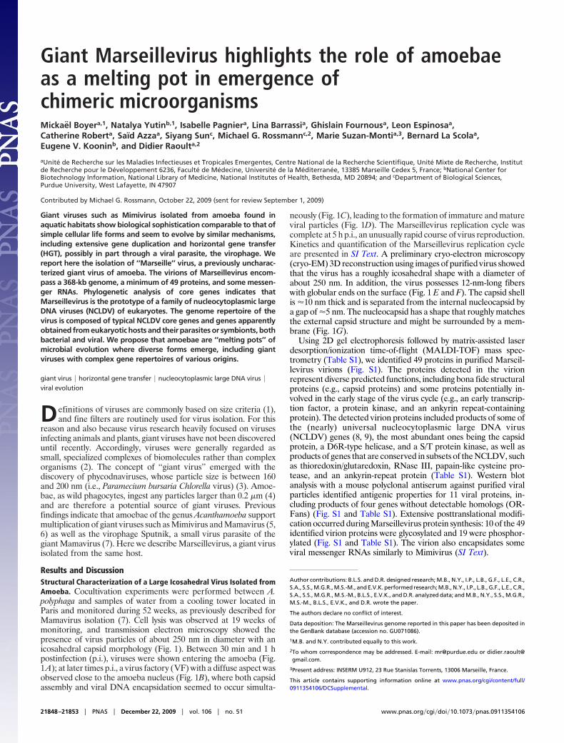
Marseillevirus Represents a Unique NCLDV Family. The genome ofMarseillevirus is a circular double-stranded DNA molecule of368,454 bp with a G�C content of 44.73%, which makes Marseil-levirus the fifth largest viral genome sequenced so far, afterMimivirus (6), Mamavirus (7), Emiliania huxleyi virus 86 (10), andParamecium bursaria Chlorella virus NY2A (11). A total of 457ORFs were predicted to encode proteins ranging from 50 to 1,537aa residues (Fig. 2 and Table S1). The coding sequences represent89.33% of the genome, with �1.2 genes per kilobase, a tight genearrangement typical of NCLDV genomes. The ORFs were equallydistributed on both strands (233 and 224 ORFs on negative andpositive strand, respectively). Sequence similarity and conserveddomain searches against the respective NCBI databases identifiedsignificant database matches (probable homologs) or conserveddomains, or both for 188 ORFs (�41%) (Table S1). Among the 457predicted genes of Marseillevirus, 163 showed significant similarity(e-value �0.001) to sequences from the environmental GlobalOcean Survey (GOS) data set, including nine ORFans with nodetectable homologs in the Refseq sequence database (Table S1).
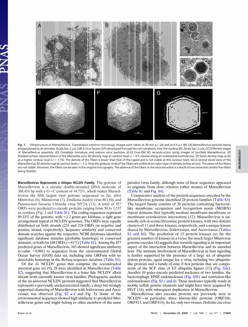
Of the 41 NCLDV genes that comprise the reconstructedancestral gene set (9), 28 were identified in Marseillevirus (TableS2), suggesting that Marseillevirus is a bona fide NCLDV albeitdistant from currently known virus families. Phylogenetic analysisof the six universal NCLDV proteins suggested that Marseillevirusrepresents a previously uncharacterized family; a deep but stronglysupported clustering of Marseillevirus with Iridoviruses and Asco-viruses was observed (Fig. S2 a–f and Fig. 3). Some of theenvironmental sequences showed high similarity to predicted Mar-seillevirus genes and might belong to other members of the same
putative virus family, although none of these sequences appearedto originate from close relatives (other strains) of Marseillevirus(Table S1 and Fig. S4).
Comparative analysis of the protein sequences encoded by theMarseillevirus genome identified 28 protein families (Table S3).The largest family consists of 20 proteins containing bacterial-like membrane occupation and recognition nexus (MORN)repeat domains that typically mediate membrane-membrane ormembrane-cytoskeleton interactions (12). Marseillevirus is un-usually rich in serine/threonine protein kinases, with two distinctclusters of 11 and three kinases, respectively, and a unique kinaseshared by Marseillevirus, Iridoviruses, and Ascoviruses (TablesS3 and S4). The prediction of 15 protein kinases (so far thegreatest number of kinases in a virus; the much larger Mimivirusgenome encodes 14) suggests that versatile signaling is an importantaspect of the interaction between Marseillevirus and its amoebalhost. The intimate involvement of Marseillevirus in host signalingis further supported by the presence of a large set of ubiquitinsystem proteins, again unique for a virus, including two ubiquitin-like proteins and a family of nine F-box proteins that are compo-nents of the SCF class of E3 ubiquitin ligases (13) (Fig. S3a).Another 10 genes encode predicted nucleases of two families, thebacteriophage HNH endonucleases (Fig. S3b) and restriction-likeendonucleases (Fig. S3 c and d). These nucleases typically reside inmobile selfish genetic elements and might have been acquired byHGT (14), with subsequent duplication in Marseillevirus.
Marseillevirus also encodes proteins not previously seen inNCLDV—in particular, three histone-like proteins (ORF166,ORF413, and ORF414). So far, only two viruses, Heliothis zea virus
D
VF
N
BA C
E F G
Fig. 1. Ultrastructure of Marseillevirus. Transmission electron microscopy images were taken at 30 min p.i. (A) and at 6 h p.i. (B). (A) Marseillevirus particles beingphagocytosed by an amoeba. (Scale bar: 2 �m.) (B) A virus factory (VF) developed through the cell cytoplasm, near the nucleus (N). (Scale bar: 2 �m.) (C) Different stagesof Marseillevirus assembly. (D) Complete immature and mature virus particles. (E-G) Cryo-EM 3D reconstruction using images of purified Marseillevirus. (E)Shaded-surface representation of the Marseilles virus 3D density map at contour level � � 0.5 viewed along an icosahedral twofold axis. (F) Same density map as (E)at a higher contour level (� � 1.75). The density of the fibers is lower than that of the capsid and is not visible at this contour level. (G) A central sliced view of theMarseillevirus 3D density map at contour level � � 1.2. Only the globular ends of the fibers are visible as an outer layer of density (white arrow). The stems of the fibersare not visible. However, the fibers can be seen in the original micrographs. The absence of the fibers in the reconstruction is a result of low resolution and/or the fibersbeing flexible.
Boyer et al. PNAS � December 22, 2009 � vol. 106 � no. 51 � 21849
MIC
ROBI
OLO
GY
1 and Cotesia plutellae bracovirus, which do not belong to theNCLDV, have been shown to encode histone-like proteins (15, 16).The histone-like proteins of Marseillevirus were detected in theviral particle (Table S1), suggesting that these proteins couldcondense DNA to facilitate viral DNA packaging.
Marseillevirus Genome Encompasses a Complex Repertoire of Genesof Various Origins. Among the predicted Marseillevirus proteins, 59,57, 70, and 2 showed the highest sequence similarity to homologsfrom viruses, bacteria, eukaryotes, and archaea, respectively (Fig. 2and Table S4). We hypothesize that the genome repertoire ofMarseillevirus consists of genes derived from several distinctsources, in large part via HGT. The presence of numerous genesapparently derived from eukaryotes on different time scales iscommon in NCLDV (9, 17, 18). The presence of numerous genesof probable bacterial origin seems to be a distinctive feature of thoseNCLDV that infect unicellular eukaryotic hosts, in particular, theMimivirus and Marseillevirus reproducing in amoebae, and algalPhycodnaviruses (18) (Tables S1 and Table S4). In addition to theNCBI databases, we searched the draft genome of Acanthamoebacastellanii, the host of Marseillevirus, for possible homologs of viralgenes. Altogether we identified Acanthamoeba homolog to 80Marseillevirus genes; for eight of these genes, the homolog fromAcanthamoeba showed the closest similarity to the correspondingMarseillevirus protein, suggesting relatively recent HGT.
A notable feature of Marseillevirus is the presence of 17 genesshared with Mimivirus/Mamavirus but absent in other NCLDV.When a tree of the NCLDV was constructed by comparison of generepertoires (19), Marseillevirus confidently grouped with Mimivi-rus/Mamavirus (Fig. 4), in contrast to the phylogenetic tree of theuniversal genes, which puts Marseillevirus together with Iridovi-ruses and Ascoviruses (Fig. 3). Thus, the gene repertoires of the twofamilies of amoebae viruses are related, in all likelihood, as a resultof interviral HGT. Moreover, eight Marseillevirus genes either haddetectable homologs only in the Mimivirus/Mamavirus and Acan-thamoeba or formed a distinct branch in the respective phylogenetictrees (Table S4), an indication of multiple gene exchanges betweenamoebae and its viruses.
To characterize the origins of Marseillevirus genes more pre-cisely, we performed a comprehensive phylogenetic analysis. Phy-logenetic trees were constructed for 89 Marseillevirus proteins withhomologs in diverse organisms and sufficient number of phyloge-netically informative sites in the respective multiple alignments;Acanthamoeba was included in the tree whenever a homolog of therespective Marseillevirus gene was detected (Table S4 and Fig. S4).The results of this analysis, combined with the information on genesuniquely shared with different taxa, yielded the final breakdown ofthe Marseillevirus genes by their probable evolutionary origin (Fig.2 and Table S4). Altogether, Marseillevirus contains 51 genes ofNCLDV origin (including those exclusively shared with Mimivi-
Uncertain6.3%(29)
Other Eukaryota7.4%(34)
Amoebozoa5.5%(25)
Bacteria/phages10.7%(49)
NCLDV11.2%(51)
Fig. 2. Map of the Marseillevirus chromosome. Rings starting from outer to innermost correspond to (i) genome coordinates in kilobases; (ii) proteins identifiedthrough 2D mass spectrometry (orange); (iii) predicted protein-coding genes oriented in forward (blue) or reverse (red) strand; (iv) cumulative gene orientation skew;(v) predicted functions of proteins; and (vi) origin of each gene inferred from sequence comparison and phylogenetic analyses (light gray background). The pie chartinsidetheringrepresents taxonomicbreakdownofMarseillevirusgenesbyprobableorigins inferredbyphylogeneticanalysisorsequenceconservation(TableS4). ‘‘Ori’’indicates putative origin of replication deduced from the position of slope reversal (around position 253,000) of the cumulative gene orientation skew.
21850 � www.pnas.org�cgi�doi�10.1073�pnas.0911354106 Boyer et al.

ruses and Phycodnaviruses), 49 genes of probable bacterial orbacteriophage origin, and 85 genes of apparent eukaryotic origin.For 25 of the ‘‘eukaryotic’’ genes of Marseillevirus, an origin fromAcanthamoeba was strongly supported, and in 22 of these cases, therespective branch of the tree encompassed Marseillevirus, Mim-iviruses, and Acanthamoeba, with the implication of multiple genetransfers. In addition, three genes seemed to originate in otherAmoebozoa (Table S4 and Fig. 2). In contrast, no gene could betraced to the known bacterial parasites of amoebae such as Legio-nella or Parachlamydia.
Notably, the genes for histone-like proteins, as well as four of thefive genes encoding translation system components, were appar-ently acquired in amoeba, in agreement with the recent observa-tions on the origin of Mimivirus genes with similar functions (17)but not with the hypothesis on the ancestral nature of the translationapparatus components in giant viruses (20). There seems to be anonrandom connection between the functions of Marseillevirusgenes and their inferred origins; many of the genes encodingdefense and repair functions—in particular, nucleases—appear tobe of bacterial or bacteriophage origin (often shared with otherNCLDV), genes for metabolic enzymes and proteins implicated inprotein and lipid modification or degradation are of mixed bacterialand eukaryotic origins, whereas genes related to signal transductionare primarily of eukaryotic extraction (Table S4 and Fig. S4.1–S4.82). In addition to the genes that appear to have common originin Marseillevirus and Mimiviruses, we detected several cases whererelated genes (e.g., the gene for deoxynucleotide monophosphatekinase) were apparently acquired by these viruses from indepen-dent sources (Fig. S4.23). This finding suggests that HGT iscommon enough to translate into a nonnegligible chance of con-vergent acquisition of genes that confer a selective advantage ontorecipient viruses.
Viruses of amoeba are characterized by a large size and chimericgenomes, with a gene repertoire acquired from a variety of distinctsources. These viruses harbor a conserved core of NCLDV genesthat encodes key proteins responsible for viral genome replicationand virion morphogenesis, an additional group of genes shared byamoebal viruses, and a broad variety of genes acquired frombacteria, selfish elements, and eukaryotes. Evidence of directderivation from Acanthamoeba or its known parasites or symbionts
was obtained for a relatively small number of genes. Although thecurrent repertoire of viruses and bacteria infecting amoebae is farfrom being complete, the relative paucity of genes of Acanthamoebaorigin in Marseillevirus suggests that the virus might have changedhosts, perhaps more than once, in the course of its evolution.Indeed, a recent report indicates that relatives of the Mimiviruscould infect marine animals such as sponges and corals (21).
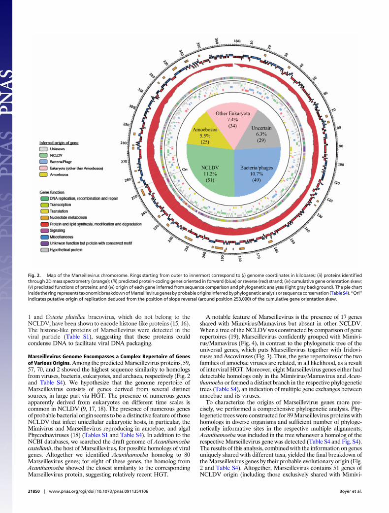
Unlike most other host cells, amoebae are commonly infected bynumerous, taxonomically diverse microorganisms (22). As we showin a direct experiment, amoeba cells can be simultaneously andproductively infected with Marseillevirus and two bacterial para-sites (Fig. 5 and SI Text). This promiscuity probably results inextended coexistence of multiple parasites and/or symbionts withinthe same amoeba and so might make the amoeba a veritable factory
Fig. 3. A maximum-likelihood tree based on concatenated alignments (1,849positions) of five NCLDV core proteins: D5 type ATPase, DNA polymerase B, A32ATPase, major capsid protein, and A1L/VLTF2 transcription factor. The tree wasbuilt using TreeFinder (WAG[,]:G[Optimum]:4, 1,000 replicates, Search Depth 2).
African swine fever virus
Amsacta moorei entomopoxvirus
Melanoplus sanguinipes entomopoxvirus
Canarypox virus
Fowlpox virus
Bovine papular stomatitis virus
Orf virus
Crocodilepox virus
Molluscum contagiosum virus
Vaccinia virus
Variola virus
Myxoma virus
Rabbit fibroma virus
Goatpox virus Pellor
Sheeppox virus
Lumpy skin disease virus
Tanapox virus
Yaba-like disease virus
Yaba monkey tumor virus
Deerpox virus
Swinepox virus
Marseillevirus
Acanthamoeba polyphaga mimivirus
Acanthamoeba polyphaga mamavirus
Acanthocystis turfacea Chlorella virus 1
Paramecium bursaria Chlorella virus FR483
Paramecium bursaria chlorella virus MT325
Paramecium bursaria Chlorella virus AR158
Paramecium bursaria Chlorella virus NY2A
Paramecium bursaria Chlorella virus 1
Ostreococcus virus OsV5
Ectocarpus siliculosus virus 1
Feldmannia species virus
Emiliania huxleyi virus 86
Lymphocytic choriomeningitis virus
Lymphocystis disease virus - isolate China
Ambystoma tigrinum virus7
Frog virus 3
Singapore grouper iridovirus
Infectious spleen and kidney necrosis virus
Aedes taeniorhynchus iridescent virus
Invertebrate iridescent virus 6
Trichoplusia ni ascovirus 2c7
Spodoptera frugiperda ascovirus 1a
Heliothis virescens ascovirus 3e
0.5
Fig. 4. Neighbor-joining clustering of NCLDV by phyletic pattern. The phyleticpatterns of the orthologous sets of NCLDV genes (8, 9) indicating the presence/absence of the respective gene in each virus were used for the construction of theneighbor-joining tree (phylip3.66) after adding the Marseillevirus orthologs.
Boyer et al. PNAS � December 22, 2009 � vol. 106 � no. 51 � 21851
MIC
ROBI
OLO
GY

for gene mixing between the eukaryotic host, its various viruses, andbacterial parasites and symbionts. The amoebal genetic melting potseems to produce organisms with complex, chimeric genomes suchas the giant viruses. The very preponderance of giant viruses inamoebae might be explained by the action of an HGT ratchet in thehost’s intracellular environment where viruses are constantly bar-raged with DNA from diverse sources. The possibility that giantviruses shuttle between different eukaryotic hosts further expandsthe gene pool to which they are exposed. Given the diversity ofphagocytic unicellular eukaryotes (23, 24), it seems certain that thediscovery of Mimivirus, Mamavirus, and Marseillevirus is only thefirst narrow window into a wondrous world of giant viruses, someof which could be even bigger and more complex than the currentrecord holder, Mamavirus.
MethodsIsolation. At the start of the study, metal pieces were introduced into a coolingtower located in Paris. One piece was removed weekly to monitor biofilmformation, together with water samples to check microbiological evolution.Adherent biofilm was homogenized into sterile water and filtered through a0.22-�m-pore-sized filter. Water samples were filtered as well. Filters were thenshaken into sterile Page’s amoebal saline (PAS), and each suspension was inocu-lated onto Acanthamoeba polyphaga microplates, as previously described (7).Cocultures were screened for cytopathic effects at day 3, and subcultured ontofresh amoebal microplates. Marseillevirus was purified using the end-point dilu-tion method.
Electron Microscopy and Immunofluorescence. Experiments were performed aspreviously described (25).
Marseillevirusgenome
A
B
C
Ld
BN9 VF
Nu
ORF304: Eukariotic translation initiation factor 5
ORF122: Serine/threonine protein kinase
Ld
Nu
VFBN9
ORF122 Marseillevirus
Acantoamoeba*
Acantoamoeba*
AcanthamoebapolyphagamimivirusYP_143172.1
AcanthamoebapolyphagaMamavirus[ 1083798-1088714]**
AcanthamoebapolyphagamimivirusYP_143185.1
AcanthamoebapolyphagaMamavirus[ 1070102-1075057]**
AcanthamoebapolyphagamimivirusYP_143180.1
AcanthamoebapolyphagaMamavirus[1093722-1098596]**
Entamoebahistolytica HM-1:IMSS XP_655583.1
Entamoebadispar SAW760 XP_001738349.1
Entamoebahistolytica HM-1:IMSS XP_649425.1
Entamoebadispar SAW760 XP_001734630.1
Dictyosteliumdiscoideum AX4 XP_640488.1
DictyosteliumdiscoideumAX4 XP_636073.1
Dictyosteliumdiscoideum AX4 XP_640564.1
Vitis vinifera XP_002278360.1
PopulustrichocarpaXP_002312329.1
Vitis vinifera XP_002278257.1
PopulustrichocarpaXP_002321510.1
Oryza sativa NP_001057225
Arabidopsis thalianaNP_196746.2
79929752
2188
10000
10000
10000
10000
10000
9922
10000
7632
99582464
3337
3337
3640
4117
10000
10000
0.2
Aspergillus terreus NIH2624 XP_001218500
Podospora anserina XP_001907948
Laccaria bicolor S238N -H82 XP_001879453
Postia placenta Mad -698 -R XP_002470033
Malassezia globosa CBS 7966 XP_001731051
Monodelphis domestica XP_002162828
Hydra magnipapillata XP_002162828
GOS_8603086
GOS_4331479
GOS_8320528
GOS_3422926
GOS_4981229
ORF304 Marseillevirus
Acantamoeba *
Acantamoeba *
GOS_7324078
GOS_6061196
GOS_5617988
GOS_3848848
9998
73526785
9939
6598
8785
4341
49234221
94809867
6528
9983
6090
8370
0.2
Fig. 5. (A) DAPI (Left) and Hemacolor (Right) staining of A. castellanii (nucleus, Nu) coinfected with Legionella drancourtii (Ld), Parachlamydia strain BN9 (BN9), andMarseillevirus (VF). Amoeba cells containing the three microorganisms were observed at 16 h and 24 h p.i. The DAPI and Hemacolor-stained microorganisms werecontrolled by performing amoeba infection with each microorganism alone. Marseillevirus was detected by the characteristic morphology of its VF. (B) Schematicrepresentation of multiple intracellular microorganisms (bacteria in purple, Marseillevirus in dark gray, and its VF in orange, and other viruses in light gray) infectingamoeba. Lateral gene exchange (red arrow) could occur during microorganism multiplication. (C) Schematic representation of Marseillevirus genome with someexamples of gene probably acquired by lateral HGT. *Marseillevirus homolog sequence was detected in Acanthamoeba castellanii Neff draft genome and includedinthephylogenetic studies (Fig.S4).**Numbers inbrackets indicatethepositionofMarseillevirushomologsequence inAcanthamoebapolyphagaMamavirusgenome.
21852 � www.pnas.org�cgi�doi�10.1073�pnas.0911354106 Boyer et al.

Cryo-EM. Marseillevirus particles were flash-frozen on holey grids in liquidethane. Images were recorded at 39 K magnification with a CM200 FEG micro-scope with electron dose levels of �20 e�/Å2. All micrographs were digitized at3.175 Å pixel�1 using a Nikon scanner.
Sequencing and Analysis of Marseillevirus Genome. Marseillevirus genome waspyrosequenced on 454-Roche GSFLX as described (26). The raw data (6.3 Mbp)were assembled by the GSFLX gsAssembler. Protein-coding genes were predictedusing GeneMark.hmm 2.0 (27). The translated protein sequences were searchedagainst the NCBI Refseq and env�nr (environmental nonredundant) proteinsequence databases using BLASTP (28). Conserved domains were identified bysearching the Conserved Domain Database (CDD version 2.13) (29) using RPS-BLAST. Multiple alignments of protein sequences were constructed using MUS-CLE (30). Similarity-based clustering of protein sequences was performed usingBLASTCLUST with subsequent manual curation. Maximum-likelihood (ML) phy-logenetic trees were constructed using TreeFinder (31). Detailed methods forgenome analysis are provided in SI Text.
Proteomic Analysis. Experiments were performed as previously described (7).
RNA Extraction from Marseillevirus Virions and RT-PCR Analysis. Experimentswere conducted as previously described (6). Specific primers used in this studywere provided in SI Text.
ACKNOWLEDGMENTS. We are grateful to Bernard Campagna for his technicalassistance with electron microscopy, Valorie D. Bowman for collecting data forthe cryo-EM reconstruction of Marseillevirus, Claude Nappez for anti-Marseillevirus monoclonal antibody production, Bernadette Giumelli and ThiTien N’Guyen for technical assistance in genome sequencing, Philippe de Cloc-quement for protein identification, Angelique Campocasso for help in detectionof viral RNA, Kira Makarova and Yuri Wolf for help with sequence analysis,Michele Merchat for providing water samples, and Christelle Desnues for readingthe manuscript. This work was funded by the Centre National de la RechercheScientifique (CNRS, credits recurrents), a Conventions Industrielles de Formationpar la Recherche fellowship (I.P.), Intramural Research Program of the NationalInstitutes of Health National Library of Medicine (N.Y. and E.K), and NationalInstitutes of Health Grant AI11219 (to S.S. and M.G.R.).
1. Lwoff A (1957) The concept of virus. J Gen Microbiol 17:239–253.2. Raoult D, Forterre P (2008) Redefining viruses: Lessons from Mimivirus. Nat Rev Microbiol
6:315–319.3. Van Etten JL, Meints RH (1999) Giant viruses infecting algae. Annu Rev Microbiol 53:447–
494.4. Audic S, et al. (2007) Genome analysis of Minibacterium massiliensis highlights the con-
vergent evolution of water-living bacteria. PLoS Genet 3:e138.5. La Scola B, et al. (2003) A giant virus in amoebae. Science 299:2033.6. Raoult D, et al. (2004) The 1.2-megabase genome sequence of Mimivirus. Science
306:1344–1350.7. La Scola B, et al. (2008) The virophage as a unique parasite of the giant mimivirus. Nature
455:100–104.8. Iyer LM, Aravind L, Koonin EV (2001) Common origin of four diverse families of large
eukaryotic DNA viruses. J Virol 75:11720–11734.9. Iyer LM, Balaji S, Koonin EV, Aravind L (2006) Evolutionary genomics of nucleo-cytoplasmic
large DNA viruses. Virus Res 117:156–184.10. Wilson WH, et al. (2005) Complete genome sequence and lytic phase transcription profile
of a Coccolithovirus. Science 309:1090–1092.11. Fitzgerald LA, et al. (2007) Sequence and annotation of the 369-kb NY-2A and the 345-kb
AR158 viruses that infect Chlorella NC64A. Virology 358:472–484.12. Gubbels MJ, Vaishnava S, Boot N, Dubremetz JF, Striepen B (2006) A MORN-repeat protein
is a dynamic component of the Toxoplasma gondii cell division apparatus. J Cell Sci119:2236–2245.
13. BaiC,etal. (1996)SKP1connectscell cycleregulatorstotheubiquitinproteolysismachinerythrough a novel motif, the F-box. Cell 86:263–274.
14. Kobayashi I (2001) Behavior of restriction-modification systems as selfish mobile elementsand their impact on genome evolution. Nucleic Acids Res 29:3742–3756.
15. Cheng CH, et al. (2002) Analysis of the complete genome sequence of the Hz-1 virussuggests that it is related to members of the Baculoviridae. J Virol 76:9024–9034.
16. IbrahimAMA,Choi JY, JeYH,KimY(2005)StructureandexpressionprofileoftwoputativeCotesia plutellae bracovirus genes (CpBV-H4 and CpBV-E94{alpha}) in parasitized Plutellaxylostella. J Asia Pacific Entomol 8:359–366.
17. Moreira D, Brochier-Armanet C (2008) Giant viruses, giant chimeras: The multiple evolu-tionary histories of Mimivirus genes. BMC Evol Biol 8:12.
18. FileeJ,PougetN,ChandlerM(2008)Phylogeneticevidenceforextensive lateralacquisitionof cellular genes by nucleocytoplasmic large DNA viruses. BMC Evol Biol 8:320.
19. WolfYI,Rogozin IB,GrishinNV,KooninEV(2002)Genometreesandthetreeof life.TrendsGenet 18:472–479.
20. Claverie JM, et al. (2006) Mimivirus and the emerging concept of ‘‘giant’’ virus. Virus Res117:133–144.
21. Claverie JM, et al. (2009) Mimivirus and Mimiviridae: Giant viruses with an increasingnumber of potential hosts, including corals and sponges. J Invertebr Pathol 101:172–180.
22. Greub G, Raoult D (2004) Microorganisms resistant to free-living amoebae. Clin MicrobiolRev 17:413–433.
23. OkadaM,etal. (2005)Proteomicanalysisofphagocytosis in theentericprotozoanparasiteEntamoeba histolytica. Eukaryot Cell 4:827–831.
24. Jacobs ME, et al. (2006) The Tetrahymena thermophila phagosome proteome. EukaryotCell 5:1990–2000.
25. Suzan-Monti M, La Scola B, Barrassi L, Espinosa L, Raoult D (2007) Ultrastructural charac-terization of the giant volcano-like virus factory of Acanthamoeba polyphaga Mimivirus.PLoS One 2:e328.
26. Margulies M, et al. (2005) Genome sequencing in microfabricated high-density picolitrereactors. Nature 437:376–380.
27. Lukashin AV, Borodovsky M (1998) GeneMark.hmm: New solutions for gene finding.Nucleic Acids Res 26:1107–1115.
28. Altschul SF, et al. (1997) Gapped BLAST and PSI-BLAST: A new generation of proteindatabase search programs. Nucleic Acids Res 25:3389–3402.
29. Marchler-Bauer A, et al. (2009) CDD: Specific functional annotation with the ConservedDomain Database. Nucleic Acids Res 37:D205–D210.
30. Edgar RC (2004) MUSCLE: Multiple sequence alignment with high accuracy and highthroughput. Nucleic Acids Res 32:1792–1797.
31. Jobb G, von Haeseler A, Strimmer K (2004) TREEFINDER: A powerful graphical analysisenvironment for molecular phylogenetics. BMC Evol Biol 4:18.
Boyer et al. PNAS � December 22, 2009 � vol. 106 � no. 51 � 21853
MIC
ROBI
OLO
GY