Embed Size (px)
Citation preview

Gli1 Mediates Lung Cancer Cell Proliferation and SonicHedgehog-Dependent Mesenchymal Cell ActivationOlga Bermudez1, Elisabeth Hennen1, Ina Koch2, Michael Lindner2, Oliver Eickelberg1*
1Comprehensive Pneumology Center, University Hospital of the Ludwig-Maximilians-University Munich and Helmholtz Zentrum Munchen, Member of the German Center
for Lung Research, Munich, Germany, 2Center for Thoracic Surgery, Asklepios Biobank for Lung Diseases, Comprehensive Pneumology Center, Asklepios Clinic Munich-
Gauting and Ludwig Maximilians University Munich, Gauting, Germany
Abstract
Non-Small-Cell-Lung-Cancer (NSCLC) represents approximately 85% of all lung cancers and remains poorly understood.While signaling pathways operative during organ development, including Sonic Hedgehog (Shh) and associated Glitranscription factors (Gli1-3), have recently been found to be reactivated in NSCLC, their functional role remains unclear.Here, we hypothesized that Shh/Gli1-3 could mediate NSCLC autonomous proliferation and epithelial/stromal signaling inthe tumoral tissue. In this context, we have investigated the activity of Shh/Gli1-3 signaling in NSCLC in both, cancer andstromal cells. We report here that inhibition of Shh signaling induces a significant decrease in the proliferation of NSCLCcells. This effect is mediated by Gli1 and Gli2, but not Gli3, through regulation of cyclin D1 and cyclin D2 expression. Whileexogenous Shh was unable to induce signaling in either A549 lung adenocarcinoma or H520 lung squamous carcinomacells, both cells were found to secrete Shh ligand, which induced fibroblast proliferation, survival, migration, invasion, andcollagen synthesis. Furthermore, Shh secreted by NSCLC mediates the production of proangiogenic and metastatic factorsin lung fibroblasts. Our results thus provide evidence that Shh plays an important role in mediating epithelial/mesenchymalcrosstalk in NSCLC. While autonomous Gli activity controls NSCLC proliferation, increased Shh expression by NSCLC isassociated with fibroblast activation in tumor-associated stroma. Our study highlights the relevance of studying stromal-associated cells in the context of NSCLC regarding new prognosis and therapeutic options.
Citation: Bermudez O, Hennen E, Koch I, Lindner M, Eickelberg O (2013) Gli1 Mediates Lung Cancer Cell Proliferation and Sonic Hedgehog-DependentMesenchymal Cell Activation. PLoS ONE 8(5): e63226. doi:10.1371/journal.pone.0063226
Editor: Masaru Katoh, National Cancer Center, Japan
Received October 9, 2012; Accepted April 1, 2013; Published May 7, 2013
Copyright: � 2013 Bermudez et al. This is an open-access article distributed under the terms of the Creative Commons Attribution License, which permitsunrestricted use, distribution, and reproduction in any medium, provided the original author and source are credited.
Funding: This study was supported by the Helmholtz Association. The funders had no role in study design, data collection and analysis, decision to publish, orpreparation of the manuscript.
Competing Interests: The authors have declared that no competing interests exist.
* E-mail: [email protected]
Introduction
Lung cancer is the deadliest cancer worldwide. Currently, no
effective treatment options exist for lung cancer and the 5-year
survival rate is only 14% for patients with treatment [1].The lack
of effective long-term therapy is related with the complexity of
lung cancer and hence with the need for better understanding the
biology of lung carcinogenesis. Little attention has thus far been
addressed to the tumor-surrounding microenvironment, which
constitutes the tumor-associated stroma and acts as active
participant in tumorigenesis. In the past years, growing evidence
has pointed out the importance of the stroma in tumor initiation,
progression and metabolism of many types of cancer [2–7].
Likewise, signaling in the stromal cells has been shown to be
essential for the malignant transformation of epithelial cells [2,5,6].
Pathways involved in organogenesis and lung branching
morphogenesis, including Hedgehog (Hh) signaling, have been
recently identified as key players in human cancers [8]. Three
Hedgehog (Hh) genes exist in mammals, with Sonic Hedgehog
(Shh) as the most broadly expressed gene. Secreted Shh binds to
the receptors Patched (Ptch) present in the cytoplasmic membrane
of the receiving cell. Binding of Shh to Ptch releases the repression
that Ptch exerts on Smoothened, a seven-transmembrane-span
receptor like protein essential for the transduction of Hedgehog
signaling. Smoothened facilitates the interaction of different
Hedgehog downstream effectors in the primary cilia, resulting in
the activation of the transcription factors Gli [9]. In humans, the
three Gli zinc-finger proteins (Gli1, Gli2 and Gli3) orchestrate
Hedgehog-specific response in the cell by modulating gene
expression. Genes of the Hedgehog pathway itself including Gli1
and Ptch1 are targets of Gli, representing a feedback loop that
serve as readout of Hedgehog activity [10]. Activation of human
canonical Hh pathway depends on the expression of Ptch
receptors (Ptch1, Ptch2) and the decoy receptor Hhip (Hedge-
hog-interacting protein) [11]. Intracellular proteins that regulate
Gli stability, like SUFU (Suppressor of Fused) and SPOP (speckle-
type POZ protein) play also an important role in determining Gli
activity and thus activation of canonical Hedgehog pathway [12].
In the past years, studies have highlighted the existence of a non-
canonical Hedgehog pathway that does not require the complete
Shh-Ptch-SMO-Gli axis. A non-canonical Hedgehog signaling
dependent on SMO but independent of Gli, that regulate
tubulogenesis and apoptosis, has been described in endothelial
cells [13].With time, Gli transcription factors appear as an
integrative platform of numerous signaling inputs, establishing a
second kind of non-canonical Hedgehog signaling, dependent of
Gli but independent of SMO. This is the case of pancreatic ductal
adenocarcinoma, where Gli transcription is regulated by TGF-ß
and K-ras [14].
PLOS ONE | www.plosone.org 1 May 2013 | Volume 8 | Issue 5 | e63226

The Hedgehog signaling pathway plays a critical role during
vertebrate development controlling cell growth, survival, fate and
pattern of the body plan. Alterations in Shh pathway during lung
development affect epithelial/mesenchymal interactions and result
in branching morphogenesis defects, impairing lung function.
While Shh signaling is essential for lung development, the role that
this signaling pathway can play in adult lungs remained unclear
and is just now beginning to be investigated. Recent studies have
highlighted the importance of Hedgehog signaling in Idiopathic
pulmonary fibrosis, a lethal disease of unknown ethiology. Bolanos
et al have reported that different components of the Hedgehog
pathway are overexpressed in IPF lungs and IPF fibroblasts.
Furthermore, fibroblasts from IPF lungs were found to respond to
Shh and this response correlated with fibroblast activation in vitro
[15]. An independent study conducted by Cigna et al [16] showed
that primary human fibroblasts from normal and IPF lung express
the main components of Hedgehog signaling pathway, associated
with cell proliferation and the expression of myofibroblastic
markers in fibroblasts. Whereas these studies bring out the
importance of Hedgehog signaling in pulmonary fibrosis, the role
of this pathway in different forms of lung cancer remains unclear.
Lung cancers are classified according to histological type: the two
most prevalent types of lung cancer are non-small-cell lung
carcinoma (NSCLC) and small-cell-carcinoma (SCLC). NSCLC
and SCLC not only exhibit differences in size and appearance of
cancer cells, but also different prognosis and clinical management.
Due to the heterogeneity of each lung cancer subtype, additional
histological and molecular characterization is needed to select a
more specific treatment. Traditionally, NSCLC were screened and
treated for EGFR and K-ras mutations. However, the appearance
of resistance to these treatments and the description of new
molecular signatures have highlighted the need of a better tumor
molecular-profile-directed therapy. Some studies have described
the association between new patterns of gene expression in specific
subsets of NSCLC [17,18], and others suggest the application of
new tumor molecular profiling in future therapies. For instance,
evaluation of MMP2 and b-catenin expression has been proposed
to have a potential in diagnosis of NSCLC with different
clinicopathologic characteristics [19]. In SCLC, a tumor with
primitive neuroendocrine features, Shh was found to be activated
in neuroendocrine progenitor cells and in tumoral cells in mice
[20,21]. While these studies support a juxtacrine/autocrine
activation of Hh in SCLC, it is not clear how this pathway is
activated in NSCLC, the most prevalent subtype of lung cancer.
Given the clinical and biological differences between NSCLC and
SCLC, we hypothesized that the mechanism of activation of Shh
in NSCLC is different from SCLC. We aimed to investigate the
activity of Shh signaling in NSCLC in both cancer and stromal
cells. In order to assess the importance of Shh signaling in NSCLC
cells, we have performed knockdown of the three Gli transcription
factors (Gli1-3), which mediate intracellular Shh signaling, and
studied the impact thereof on NSCLC proliferation. Furthermore,
we have evaluated how NSCLC cells and lung fibroblasts
responded to exogenous Shh in terms of proliferation, cell
migration, invasion, and extracellular matrix remodelling.
Results
Cyclopamine, a Hedgehog Inhibitor, Reduces NSCLCProliferation and Viability
One of the most adverse characteristics of cancers is the
deregulation in cell proliferation. We have therefore studied the
role that Shh could have in NSCLC proliferation, using cells from
adenocarcinoma and cells from squamous carcinoma, the first and
second most frequent type of lung cancer respectively. Initially, we
used Cyclopamine, a plant-derived steroidal alkaloid that inhibits
Smoothened (SMO), a G protein-coupled receptor that transduces
Shh signal in the cell, to block Shh pathway in NSCLC cells.
When treated with cyclopamine, A549 adenocarcinoma cells and
H520 squamous cell lung carcinoma showed a significant decrease
in cell number especially at longer time points (Figures 1A and B).
Cyclopamine also reduced cell survival (metabolic activity assessed
by MTT assay) (Figures 1A and B) and this effect was more
important with increasing doses of the inhibitor (Figure S1A and
Figure S3E). To rule out that cyclopamine did not provoke a
cytotoxic non-specific effect on NSCLC cells, apoptosis was
determined upon cyclopamine treatment. Although cyclopamine
induced a slight increase in the extent of apoptotic cells, the
proportion of apoptotic cells was not statistically different between
treated-cells and non-treated cells (Figure S1B and Figure S3F). In
order to confirm the specific effect of cyclopamine on NSCLC
proliferation and viability, SMO silencing was performed. SMO
knockdown induced a decrease in both A549 and H520 cell
proliferation and viability (data not shown). Altogether, these
results show that blockage of Hedgehog pathway through SMO
inhibition, reduces NSCLC proliferation and viability.
Silencing of Gli1 Decreases NSCLC Proliferation byModulating Cyclin D Expression
Although Cyclopamine has been found to impact cell prolifer-
ation in other types of cancer cells, the specific mechanism
whereby Shh signaling regulates NSCLC cell cancer proliferation
remains elusive. For instance, it is not known how each of the 3
human Shh-transcription factors Gli contributes to NSCLC
proliferation. In order to address this question, we used small
interference RNAs (siRNA) for silencing Gli1, Gli2 and Gli3.
Upon a specific and important reduction in the mRNA levels of
Gli1 that did not affect either Gli2 or Gli3 mRNA levels
(Figure 1C), cell proliferation and cell viability was decreased in
A549 adenocarcinoma cells (Figure 1D–F). Of notice, the silencing
of Gli1 provoked a reduction in Ptch1 mRNA levels (Figure 1C).
Because the transcription of Ptch1 depends on Gli1, the reduction
of Ptch1 mRNA levels serves as an additional control indicating
that the silencing of Gli1 was biologically effective. The specific
silencing of Gli2, that reduced Gli2 mRNA levels and did not
decrease either Gli1 or Gli3 mRNA levels (Figure 1C), diminished
slightly A549 cell number and cell viability, although not in a
statistically significant manner (Figure 1D and E). Finally, the
siRNA of Gli3 that provoked an important diminution in Gli3
mRNA levels but not a decrease in Gli1 or Gli2 mRNA levels
(Figure 1C), did not reduce A549 adenocarcinoma cell prolifer-
ation or cell viability (Figure 1D and E) and instead caused a slight
increase in cell number of A549 adenocarcinoma cells (Figure 1D)
along with an increase in Gli1 mRNA levels (Figure 1C). In H520
squamous lung carcinoma cells (Figure S3) and in large cell
carcinoma cells (data not shown), the specific silencing of Gli1,
Gli2 or Gli3 had a similar effect in cell proliferation and cyclin
expression. Importantly, the expression of Shh-related genes and
cyclins upon Gli1, Gli2 and Gli3 silencing was not due to Hprt1
expression because a similar pattern of expression was found when
3 independent reference genes were used in A549 (Figure S2) and
in H520 cells (Figure S4).
In order to explore how the silencing of Shh pathway impacts
lung adenocarcinoma proliferation, we have evaluated the changes
in the expression of the cyclins D and E involved in G1/S cell
cycle transition, upon Gli down-regulation. Of interest, the siRNA
of Gli1 and Gli2 provoked an important decrease in cyclin D2 and
a slight reduction in Cyclin D1 mRNA levels (Figure 1G). The
Hedgehog Signaling in Non-Small-Cell Lung Cancer
PLOS ONE | www.plosone.org 2 May 2013 | Volume 8 | Issue 5 | e63226

Figure 1. Inhibition of Hedgehog signaling reduces the proliferation of NSCLC cells. Lung adenocarcinoma A549 cells (A) and lungsquamous carcinoma H520 cells (B) were cultured in presence or absence of 10 mM cyclopamine for 5 days. Proliferation was assessed by cellcounting and cell survival by MTT assay. *p,0,1; **p,0,05. (C) The Hedgehog-responsive transcription factors Gli1, Gli2 or Gli3 were knocked downwith siRNA in A549 cells. RT-qPCR was performed to confirm the specific silencing of each Gli and to assess the expression of the Hedgehog receptorPtch1. Results are presented as fold differences of mRNA levels (2‘‘Ct) compared with cells transfected with a negative control siRNA (NC siRNA)having no homology in vertebrate transcriptome. **p,0,05, ***p,0,01. The impact of silencing Gli1, Gli2 or Gli3 in A549 cell proliferation wasassessed by cell counting (D) and in cell survival by MTT assay (E). Results are presented in percentage as relative proliferation and relative survivalcompared with cells transfected with the negative control siRNA (NC). *p,0,1. (F) Representative phase-contrast microscopic pictures after 72 hoursof siRNA are presented. (G) RT-qPCR was performed to evaluate the effect of the siRNA of Gli1, Gli2 and Gli3 in the expression of the G1/S phasecyclins D (Cyc D1, Cyc D2, Cyc D3) and cyclin E (Cyc E1). Results are presented as fold of mRNA levels (2‘‘Ct) compared with cells transfected with anegative control siRNA (NC siRNA) having no homology in vertebrate transcriptome. *p,0,1; **p,0,05. (H) Western blot of cyclin D2 in A549 cellstransfected with Gli siRNA or with a negative control siRNA (NC). Blotting of ß-actin was used as loading control.doi:10.1371/journal.pone.0063226.g001
Hedgehog Signaling in Non-Small-Cell Lung Cancer
PLOS ONE | www.plosone.org 3 May 2013 | Volume 8 | Issue 5 | e63226

decrease in cyclin D2 expression upon Gli1 and Gli2 knockdown
was observed at the mRNA but also at the protein level
(Figure 1H). Considering that Gli2 could affect cyclin D
expression, we hypothesized that in combination with Gli1, this
transcription factor could regulate cell proliferation in a significant
manner. In order to confirm this hypothesis, we have realized the
double silencing of Gli transcription factors. While the single
silencing of Gli2 did not decrease significantly cell viability, the
double silencing of Gli1 and Gli2 did (Figure S1C). In H520 lung
squamous carcinoma cells, the specific knockdown of Gli1 and
Gli2 by siRNA was found to decrease in a similar manner cyclin
D1 and cyclin D2 expression (Figure S3A). Taken together, these
results indicate that the blockade of Shh signaling pathway, either
with cyclopamine or interfering with the transcription factor Gli1
decreases NSCLC proliferation.
NSCLC Cells do not Activate Shh Pathway UponExogenous Shh Treatment
Since the blockade of Shh pathway decreases NSCLC
proliferation, we investigated whether exogenous Shh produces
an increment in cell proliferation in these cells. Lung adenocar-
cinoma A549 cells and H520 squamous carcinoma cells treated
with recombinant human Shh did not show a change either in cell
number (Figures 2A and D) or in cell survival (Figures 2B and E).
In addition, cells did not present any morphological change upon
treatment (Figures 2C and F). These results suggested that NSCLC
cells do not respond to Shh. In order to validate Shh treatment, we
used primary embryo mouse cells from the limb buds, tissue that
has been shown to be responsive to Shh. Upon Shh treatment,
these cells exhibited a considerable increase in Gli1 and Ptch1
mRNA levels (60 fold and 9-fold increase respectively compared
with non-treated cells; Figure S5). When A549 cells were treated
with Shh, a very modest increase (1,65 fold) in Gli1 mRNA levels
at 8 hours and of Gli1 protein levels at 24 hours was detected.
However, at longer time points there were was no significant
changes in either Gli1 or Ptch1 expression (Figures 3A and B). For
H520 cells, any significant change in either Gli1 or Ptch1 at the
mRNA and protein level was observed (Figures 3C and D). These
results indicate that, in vitro, both types of NSCLC cells, compared
with known Shh-responsive cells, do not notably respond to
exogenous Shh.
NSCLC Cells Secrete Shh LigandUsing a specific ELISA test for human Shh, we found that A549
adenocarcinoma cells and more strongly H520 squamous carci-
noma produce Shh (170 and 2800 pg/ml respectively, Figure 3E).
This secretion can be associated with endogenous Shh production
by these NSCLC cells because the concentration of Shh in the
medium of each cell type was very low, comparable to the
background (water).
In order to corroborate these results and to investigate if these
NSCLC cells also produce the other forms of Hedgehog ligand, we
have evaluated the expression of Sonic, Desert (Dhh) and Indian
Hedgehog (Ihh) in A549 and H520 cells. Although we could not
detect by RT-qPCR Dhh and Ihh, we found that Sonic Hedgehog
was expressed in A549 and H520 cells, being more expressed in
these latter (data not shown). Based on these findings, we have thus
concentrated on Shh throughout our study. In addition, because
H520 cells produce and secrete considerable amounts of Shh
ligand, we decided to focus on these NSCLC cells for further
studies related with Shh secretion. To assess more accurately the
presence of the active form of Shh in H520 supernatant, western
blot was performed with an antibody recognizing the processed
active form of Shh (N-Shh). The presence of the N-terminal
secreted peptide Shh in the supernatant of H520 cells was thereby
confirmed (inset Figure 3E).
Because H520 cells secrete a considerable amount of Shh but do
not respond to exogenous Shh, we aimed to investigate if this lack
of response was associated with saturation of endogenous
Hedgehog activity in these cells. For this, we have performed
the silencing of Shh gene in H520 cells. Upon the knockdown of
Shh, that reduced by 70% the secretion of Shh in H520 cells
(Figure 3F), exogenous Shh increased Gli1 mRNA levels in these
cells (Figure 3G). This increase, as well as a slight increment in
Ptch1 mRNA levels (Figure 3G) indicate that H520 cells can
respond to Shh when their endogenous levels of Shh are
decreased.
Lung Fibroblasts Strongly Respond to Exogenous ShhSince NSCLC cells can secrete Shh ligand but they do not
strongly respond to exogenous Shh, we investigated if Shh could
rather activate the adjacent stromal cells. Indeed, lung fibroblasts
treated with mouse Shh exhibited a strong Hedgehog pathway
activation upon treatment: Gli1 mRNA levels increased up to 800
times and Ptch1 mRNA levels had a fold increase up to 250,
especially at 48 and 72 hours (Figure 4A). Similar results were
obtained with human Shh (Figure 4A). Shh treatment also
increased Gli1 and Ptch1 at the protein level (Figure 4B). In
order to know if lung human fibroblasts from the NSCLC
environment could also respond to exogenous Shh, primary
human lung fibroblasts were isolated from a resected lung
squamous carcinoma. Interestingly, Gli1 and Ptch1 mRNA levels
were also increased upon Shh treatment in these cells (Figure 4C).
These results indicate that, in vitro, lung fibroblasts are highly Shh-
responsive cells, in contrast to NSCLC epithelial cells.
In order to investigate if the lack of response to exogenous Shh
in NSCLC cells was due to an improper reception of Shh ligand in
these cells, we have evaluated the expression of the receptors
Ptch1, Ptch2 and of Hhip, considered as a decoy receptor, in
A549, H520 cells and CLL206 fibroblasts. Ptch1 expression was
found to be higher than Hhip in both NSCLC cell types (Figure
S6). In addition, the relative expression of Ptch1 was higher in
A549 and in H520 cells than in CCL206 fibroblasts while the
relative expression of the decoy receptor Hhip was more important
in lung fibroblasts than in the NSCLC cells (Figure S6). Thus, the
balance between Ptch and Hhip expression does not appear to be
related (at the mRNA level) with the non-responsiveness of
NSCLC to exogenous Shh. As intracellular proteins such as Sufu
and Spop regulate in a positive and in a negative form Gli stability
respectively, we have then evaluated if an imbalance between
these Gli regulators could account for the non-responsive of
NSCLC to exogenous Shh. We did not find differences in the
relative expression of Sufu compared with the expression of Spop
for a same cell type (Figure S6). Finally, we have evaluated if the
relative expression of Hh receptors and Gli regulators were
different between NSCLC and lung fibroblasts upon Shh
treatment. No differences were found in the expression of Ptch1,
Ptch2, Hhip, Sufu and Spop upon Shh treatment, at short (1, 3,
8 h) or longer time points (24, 48 h) (data not shown).
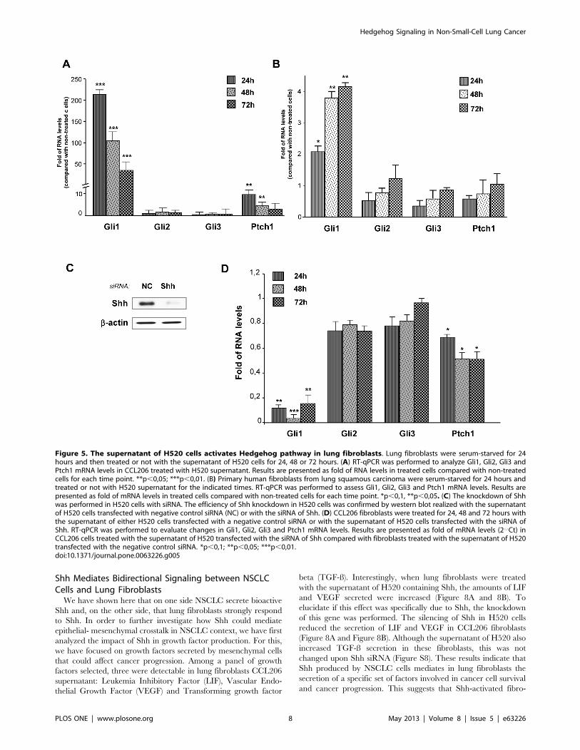
Lung Fibroblasts Respond to Shh Secreted by NSCLCH520 Cells
To test whether Shh secreted by H520 squamous cells was
bioactive, lung fibroblasts were treated with H520 supernatant.
This resulted in the increase in Gli1 and Ptch1 mRNA levels in
mouse newborn (Figure 5A) and in adult human lung fibroblasts
(Figure 5B). In order to evaluate if this response was mediated by
Shh secreted by H520 cells and present in the supernatant, siRNA
Hedgehog Signaling in Non-Small-Cell Lung Cancer
PLOS ONE | www.plosone.org 4 May 2013 | Volume 8 | Issue 5 | e63226

of Shh was performed in H520 cells. This importantly decreased
Shh mRNA amounts and the presence of N-Shh in the
supernatant (Figure 5C) and moreover, reduced by 90% the levels
of Gli1 and by 50% the levels of Ptch1 in the fibroblasts treated
with H520 supernatant (Figure 5D).
Shh Pathway Enhances Lung Fibroblast Proliferation,Invasion and Collagen Deposition
We have shown here that NSCLC cells do not markedly
respond to exogenous Shh but can secrete bioactive Shh that
induces the activation of Hedgehog signaling in lung fibroblasts.
These results reveal that Shh plays an important role in mediating
cancer epithelial-stromal crosstalk in NSCLC. In this setting, we
have investigated the possible biological effects of Shh activation in
lung fibroblasts. We have focused on different aspects of relevance
in a cancer context such as proliferation, migration, invasion and
extracellular matrix remodelling. While recombinant Shh in-
creased cell survival and cell proliferation of lung fibroblasts
(Figures 6A–C), cyclopamine decreased it (Figures 6D–F). This
effect appear to be specifically due to the inhibition of Hedgehog
pathway as cyclopamine treatment correlated with a decrease in
Gli1 and Ptch1 expression in CCL206 fibroblasts (Figure S7A).
Interestingly, H520 supernatant also enhanced lung fibroblast cell
proliferation (Figure S7B–D). As fibroblasts have been shown to be
important in facilitating cancer migration and invasion, we went
on investigating the impact of Shh in these processes. Fibroblasts
were treated with either Shh or with cyclopamine, and their
migration was recorded up to 72 hours after treatment. Whereas
Shh increased the distance of migration of fibroblasts, cyclopamine
significantly decreased it (Figure 7A). In order to explore if Shh
could influence fibroblast migration after an injury stimulus which
may better represent the changes taking place in the tumoral
tissue, we performed wound healing assays. In non-treated cells,
fibroblast migrated into the wound area in a progressive manner,
resulting in wound closure after 30 hours. In Shh-treated cells, this
process was faster and led to wound closure after 26 hours
(Figures 7B and C). On the contrary, cyclopamine decreased
fibroblast migration towards the wound area and did not result in
wound closure (Figures 7B and C). We then sought to investigate if
Shh could affect fibroblast invasion. For this, we used transwells
coated with collagen that mimics better the extracellular matrix
and thus the tissue context. Cells were loaded on the top of the
transwell and medium with Shh or cyclopamine was placed in the
bottom, allowing the formation of a gradient. The number of cells
transmigrating increased in the presence of Shh while cyclopamine
decreased fibroblast invasion through the collagen coated mem-
brane (Figure 7D). While Shh signaling regulates lung fibroblast
migration and invasion, any effect was found for either Shh or
cyclopamine in fibroblast adhesion assays (data not shown).
Because the disorganization and changes in the architecture of
Figure 2. Exogenous Shh does not affect NSCLC cell proliferation. Lung adenocarcinoma A549 cells (A–C) and lung squamous H520carcinoma cells (D–F) were treated or not with recombinant Shh (500 ng/ml). Cell proliferation was assessed by cell counting (A, D) and cell survivalby MTT assay (B, E) at the indicated times. Representative phase-contrast microscopic pictures of A549 cells (C) and of H520 cells (F) are shown.doi:10.1371/journal.pone.0063226.g002
Hedgehog Signaling in Non-Small-Cell Lung Cancer
PLOS ONE | www.plosone.org 5 May 2013 | Volume 8 | Issue 5 | e63226
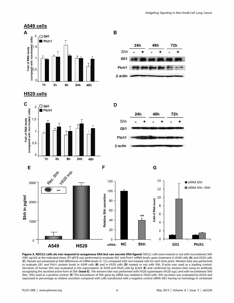
Figure 3. NSCLC cells do not respond to exogenous Shh but can secrete Shh ligand. NSCLC cells were treated or not with recombinant Shh(500 ng/ml) at the indicated times. RT-qPCR was performed to evaluate Gli1 and Ptch1 mRNA levels upon treatment in A549 cells (A) and H520 cells(C). Results are presented as fold differences of mRNA levels (2‘‘Ct) compared with non-treated cells for each time point. Western blot was performedto evaluate Gli1 and Ptch1 protein levels in A549 cells (B) and in H520 cells (D) treated or not with Shh. ß-actin was used as a loading control.Secretion of human Shh was evaluated in the supernatants of A549 and H520 cells by ELISA (E) and confirmed by western blot using an antibodyrecognizing the secreted active form of Shh (inset E). The western blot was performed with H520 supernatant (H520 sup.) and with recombinant Shh(Rec. Shh) used as a positive control. (F) The knockdown of Shh gene by siRNA was realized in H520 cells. Shh secretion was evaluated by ELISA andexpressed in percentage as relative secretion compared with cells transfected with a negative control siRNA (NC) having no homology in vertebrate
Hedgehog Signaling in Non-Small-Cell Lung Cancer
PLOS ONE | www.plosone.org 6 May 2013 | Volume 8 | Issue 5 | e63226
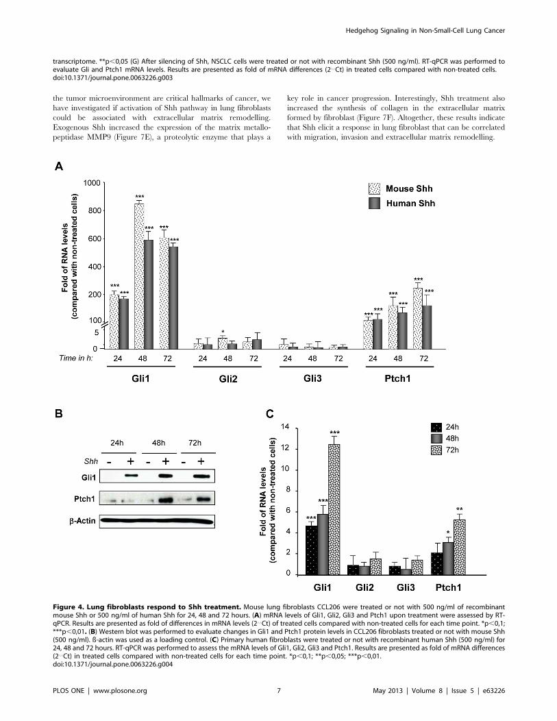
the tumor microenvironment are critical hallmarks of cancer, we
have investigated if activation of Shh pathway in lung fibroblasts
could be associated with extracellular matrix remodelling.
Exogenous Shh increased the expression of the matrix metallo-
peptidase MMP9 (Figure 7E), a proteolytic enzyme that plays a
key role in cancer progression. Interestingly, Shh treatment also
increased the synthesis of collagen in the extracellular matrix
formed by fibroblast (Figure 7F). Altogether, these results indicate
that Shh elicit a response in lung fibroblast that can be correlated
with migration, invasion and extracellular matrix remodelling.
transcriptome. **p,0,05 (G) After silencing of Shh, NSCLC cells were treated or not with recombinant Shh (500 ng/ml). RT-qPCR was performed toevaluate Gli and Ptch1 mRNA levels. Results are presented as fold of mRNA differences (2‘‘Ct) in treated cells compared with non-treated cells.doi:10.1371/journal.pone.0063226.g003
Figure 4. Lung fibroblasts respond to Shh treatment. Mouse lung fibroblasts CCL206 were treated or not with 500 ng/ml of recombinantmouse Shh or 500 ng/ml of human Shh for 24, 48 and 72 hours. (A) mRNA levels of Gli1, Gli2, Gli3 and Ptch1 upon treatment were assessed by RT-qPCR. Results are presented as fold of differences in mRNA levels (2‘‘Ct) of treated cells compared with non-treated cells for each time point. *p,0,1;***p,0,01. (B) Western blot was performed to evaluate changes in Gli1 and Ptch1 protein levels in CCL206 fibroblasts treated or not with mouse Shh(500 ng/ml). ß-actin was used as a loading control. (C) Primary human fibroblasts were treated or not with recombinant human Shh (500 ng/ml) for24, 48 and 72 hours. RT-qPCR was performed to assess the mRNA levels of Gli1, Gli2, Gli3 and Ptch1. Results are presented as fold of mRNA differences(2‘‘Ct) in treated cells compared with non-treated cells for each time point. *p,0,1; **p,0,05; ***p,0,01.doi:10.1371/journal.pone.0063226.g004
Hedgehog Signaling in Non-Small-Cell Lung Cancer
PLOS ONE | www.plosone.org 7 May 2013 | Volume 8 | Issue 5 | e63226
Shh Mediates Bidirectional Signaling between NSCLCCells and Lung Fibroblasts
We have shown here that on one side NSCLC secrete bioactive
Shh and, on the other side, that lung fibroblasts strongly respond
to Shh. In order to further investigate how Shh could mediate
epithelial- mesenchymal crosstalk in NSCLC context, we have first
analyzed the impact of Shh in growth factor production. For this,
we have focused on growth factors secreted by mesenchymal cells
that could affect cancer progression. Among a panel of growth
factors selected, three were detectable in lung fibroblasts CCL206
supernatant: Leukemia Inhibitory Factor (LIF), Vascular Endo-
thelial Growth Factor (VEGF) and Transforming growth factor
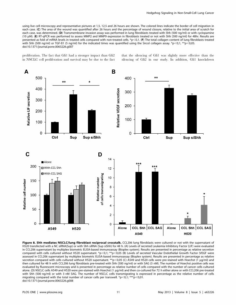
beta (TGF-ß). Interestingly, when lung fibroblasts were treated
with the supernatant of H520 containing Shh, the amounts of LIF
and VEGF secreted were increased (Figure 8A and 8B). To
elucidate if this effect was specifically due to Shh, the knockdown
of this gene was performed. The silencing of Shh in H520 cells
reduced the secretion of LIF and VEGF in CCL206 fibroblasts
(Figure 8A and Figure 8B). Although the supernatant of H520 also
increased TGF-ß secretion in these fibroblasts, this was not
changed upon Shh siRNA (Figure S8). These results indicate that
Shh produced by NSCLC cells mediates in lung fibroblasts the
secretion of a specific set of factors involved in cancer cell survival
and cancer progression. This suggests that Shh-activated fibro-
Figure 5. The supernatant of H520 cells activates Hedgehog pathway in lung fibroblasts. Lung fibroblasts were serum-starved for 24hours and then treated or not with the supernatant of H520 cells for 24, 48 or 72 hours. (A) RT-qPCR was performed to analyze Gli1, Gli2, Gli3 andPtch1 mRNA levels in CCL206 treated with H520 supernatant. Results are presented as fold of RNA levels in treated cells compared with non-treatedcells for each time point. **p,0,05; ***p,0,01. (B) Primary human fibroblasts from lung squamous carcinoma were serum-starved for 24 hours andtreated or not with H520 supernatant for the indicated times. RT-qPCR was performed to assess Gli1, Gli2, Gli3 and Ptch1 mRNA levels. Results arepresented as fold of mRNA levels in treated cells compared with non-treated cells for each time point. *p,0,1, **p,0,05. (C) The knockdown of Shhwas performed in H520 cells with siRNA. The efficiency of Shh knockdown in H520 cells was confirmed by western blot realized with the supernatantof H520 cells transfected with negative control siRNA (NC) or with the siRNA of Shh. (D) CCL206 fibroblasts were treated for 24, 48 and 72 hours withthe supernatant of either H520 cells transfected with a negative control siRNA or with the supernatant of H520 cells transfected with the siRNA ofShh. RT-qPCR was performed to evaluate changes in Gli1, Gli2, Gli3 and Ptch1 mRNA levels. Results are presented as fold of mRNA levels (2‘‘Ct) inCCL206 cells treated with the supernatant of H520 transfected with the siRNA of Shh compared with fibroblasts treated with the supernatant of H520transfected with the negative control siRNA. *p,0,1; **p,0,05; ***p,0,01.doi:10.1371/journal.pone.0063226.g005
Hedgehog Signaling in Non-Small-Cell Lung Cancer
PLOS ONE | www.plosone.org 8 May 2013 | Volume 8 | Issue 5 | e63226

blasts may in turn act on cancer epithelial cells. In order to address
this point in vitro, lung fibroblasts were co-cultured with NSCLC
cells. Interestingly, when co-cultured with Shh-treated lung
fibroblasts, A549 and H520 cell proliferation was boosted and
invasion was increased (Figure 8C and 8D). This effect was
reproduced when fibroblasts were pre-treated with SAG, a
compound that interacts with SMO and activates Shh pathway
(Figure 8C and D). The results shown here indicate that NSCLC
cells activate, via Shh, lung fibroblasts that in turn enhance cancer
cell proliferation and invasion. Shh appear then to mediate a
reciprocal crosstalk between NSCLC cells and lung fibroblasts.
Discussion
In this study, we have shown that cyclopamine produced a
progressive decrease in cell proliferation and cell viability of both,
lung adenocarcinoma and lung squamous carcinoma cells. The
specificity and efficacy of this inhibitor has been largely reported in
different systems, where cyclopamine directly correlates with a
decrease in Shh-Gli activity [20,22,23]. However, off-target effects
of cyclopamine have been reported at high concentrations [24,25].
The concentration needed for reaching a SMO-dependent effect
may vary according to the cell type and its endogenous regulation
of Hedgehog pathway. For instance, in prostate cancer cells, a
significant reduction in Gli mRNA levels takes place at concen-
trations starting at 3 mM and this effect is stronger at 10 and
30 mM [26]. In addition, only at a concentration of 10 and
30 mM, cyclopamine reduces Gli1 at the protein level. In lung
cancer cells, previous studies have reported a clear reduction in
Ptch1 expression, main readout of Hedgehog activation, at 5 and
10 mM [20]. In a first approach, we have therefore used
cyclopamine at 10 mM. We report here that cyclopamine
decreases cell proliferation and cell viability of NSCLC cells.
The specificity of this effect has been confirmed by two different
approaches. On one hand, we have knocked down SMO in A549
and H520 cells. For both cell lines, the silencing of SMO
decreased cell proliferation and cell viability (data not shown). On
the other hand, we have performed the silencing of the Shh-
specific transcription factors Gli, acting downstream of SMO. The
specific knockdown of Gli1 and Gli2 decreased cell proliferation
and cell viability of NSCLC cells. In addition, the fact that we did
not find significant differences in cell death induced by different
concentrations of cyclopamine, ruled out the possibility that
cytotoxic unspecific effects of cyclopamine account for the
reduction in proliferation of NSCLC cells.
Upon silencing of the three human Gli factors, Gli1 was found
to be the major regulator of NSCLC cell proliferation while Gli2
had a modest effect and the silencing of Gli3 did not decrease and
even slightly increased NSCLC proliferation. The fact that the
silencing of Gli1 and Gli2 can decrease A549 and H520
proliferation suggests that both have redundant roles in these
NSCLC cells. This is the case in mice, where the absence of Gli2
can be compensated by Gli1 [27]. If Gli1 and Gli2 can have
additive effects, the specific expression and function of each one
may depend on the tumoral context and in the signaling occurring
in cancer cells. For instance, TGF-ß, a growth factor that plays a
critical role in lung fibrosis and in cancer development, interacts
with Hh pathway downstream of SMO, increasing the expression
of Gli2 in mice [28,29] and in cancer cells [30,31]. This and other
signaling crosstalk taking place in the NSCLC cells may potentiate
not only Gli expression but also their effect in cancer cell
Figure 6. Shh pathway regulates cell proliferation and cell survival in lung fibroblasts. Proliferation of CCL206 lung fibroblasts wasassessed by cell counting (A, D) and cell survival by MTT assay (B, E) after treatment with recombinant Shh (500 ng/ml) or with 100 nM, 1 mM or10mMcyclopamine (cyclop) for 5 days. Results are presented in percentage as relative proliferation and relative survival compared to non-treated cells(A, B) or to vehicle (D, E) *p,0,1; **p,0,05. Representative phase-contrast microscope pictures upon Shh treatment (C) and upon 10 mMcyclopamine treatment (F) are shown.doi:10.1371/journal.pone.0063226.g006
Hedgehog Signaling in Non-Small-Cell Lung Cancer
PLOS ONE | www.plosone.org 9 May 2013 | Volume 8 | Issue 5 | e63226

Figure 7. Shh pathway affects lung fibroblast migration, invasion and collagen synthesis. (A) Accumulated distance of migration ofprimary human lung fibroblasts treated with Shh (500 ng/ml) or cyclopamine (10 mM) and monitored by live cell microscopy for 48 hours. Theaccumulated distance of migration of each cell was determined using ImageJ. ***p,0,01. (B) Scratch wound assay was performed in lung fibroblastsCCL206 treated or not with Shh at the doses indicated or with 10 mM cyclopamine (cyclop) for up to 48 hours. Migration of the cells was recorded
Hedgehog Signaling in Non-Small-Cell Lung Cancer
PLOS ONE | www.plosone.org 10 May 2013 | Volume 8 | Issue 5 | e63226
proliferation. The fact that Gli1 had a stronger impact than Gli2
in NSCLC cell proliferation and survival may be due to the fact
that the silencing of Gli1 was slightly more effective than the
silencing of Gli2 in our study. In addition, Gli1 knockdown
using live cell microscopy and representative pictures at 1,5, 12,5 and 26 hours are shown. The colored lines indicate the border of cell migration ineach case. (C) The area of the wound was quantified after 26 hours and the percentage of wound closure, relative to the initial area of scratch foreach case, was determined. (D) Transmembrane invasion assay was performed in lung fibroblasts treated with Shh (500 ng/ml) or with cyclopamine(10 mM). (E) RT-qPCR was performed to assess MMP2 and MMP9 expression in fibroblasts treated or not with Shh (500 ng/ml) for 48hr. Results arepresented as fold of mRNA levels in treated cells compared with non-treated cells. *p,0,1. (F) The total collagen content of lung fibroblasts treatedwith Shh (500 ng/ml) or TGF-ß1 (5 ng/ml) for the indicated times was quantified using the Sircol collagen assay. *p,0,1, **p,0,05.doi:10.1371/journal.pone.0063226.g007
Figure 8. Shh mediates NSCLC/lung fibroblast reciprocal crosstalk. CCL206 lung fibroblasts were cultured or not with the supernatant ofH520 transfected with a NC siRNA(Sup) or with Shh siRNA (Sup siShh) for 48 h. (A) Levels of secreted Leukemia Inhibitory Factor (LIF) were evaluatedin CCL206 supernatant by multiplex biometric ELISA-based immunoassay (Bioplex system). Results are presented in percentage as relative secretioncompared with cells cultured without H520 supernatant. *p,0,1; **p,0,05 (B) Levels of secreted Vascular Endothelial Growth Factor (VEGF wereassessed in CCL206 supernatant by multiplex biometric ELISA-based immunoassay (Bioplex system). Results are presented in percentage as relativesecretion compared with cells cultured without H520 supernatant. **p,0,05 (C) A549 and H520 cells were pre-stained with Hoechst (1 mg/ml) andthen cultured for 48 h with CCL206 lung fibroblasts pre-treated with Shh (500 ng/ml) or with SAG (3 nM). The number of Hoechst positive cells wasevaluated by fluorescent microscopy and is presented in percentage as relative number of cells compared with the number of cancer cells culturedalone. (D) NSCLC cells A549 and H520 were pre-stained with Hoechst (1 mg/ml) and then co-cultured for 72 h either alone or with CCL206 pre-treatedwith Shh (500 ng/ml) or with 3 nM SAG. The number of NSCLC cells transmigrating is expressed in percentage as the relative number of cellsmigrating compared with the total number of cancer cells per transwell. *p,0,1; ***p,0,01.doi:10.1371/journal.pone.0063226.g008
Hedgehog Signaling in Non-Small-Cell Lung Cancer
PLOS ONE | www.plosone.org 11 May 2013 | Volume 8 | Issue 5 | e63226

reduced more the expression of cyclin D1 and Cyclin D3 than the
silencing of Gli2. Lastly, the role of Gli1 in regulating NSCLC
proliferation may be related with the fact that Gli1 acts mainly as
an activator of transcription while Gli2 can act as an activator but
also as a transcription repressor [10,32].
Since cell proliferation depends on cell cycle, we investigated if
the transcription factors Gli could affect the expression of Cyclins
D and E, key cyclins regulating G1/S transition. We have shown
here that upon a specific knockdown of each of the transcription
factors Gli, the silencing of Gli1 or Gli2 but not Gli3 decreased
cyclin D1 and cyclin D2 expression. The presence of consensus Gli
DNA-binding sequences in the sequence of Cyclin D1 and cyclin
D2 genes, and the fact that Gli1 binds to cyclin D2 promoter [33],
suggest that Gli1 and Gli2 modulate NSCLC proliferation by
directly regulating cyclin D expression. Although the different
isoforms of cyclins D can regulate the cell cycle in a similar way,
we have found that the silencing of Gli1 and Gli2 affects in a
different form the expression of each cyclin D. This might
correlate with the pattern of expression of these cyclins in these
cells. During organogenesis, the relative expression of each cyclin
D varies according to the cell type and this might also be the case
in the adult lung.
The silencing of Gli3 did not decrease either NSCLC
proliferation or cyclin expression and this may be related with
the repressor function of this transcription factor. In fact, a slight
increase in Gli1 mRNA levels and NSCLC proliferation was
observed when Gli3 was silenced. As Gli3 can be present in a
truncated repressor form, the knockdown of Gli3 may have
affected the formation of this repressor form, resulting in an
increase in Gli1 mRNA levels and increased cell proliferation.
The fact that blocking Hh pathway in A549 and H520 cells
affects NSCLC proliferation indicates that these cells have a basal
level of Hedgehog activity. Indeed, we have detected Gli1 and
Ptch1 proteins in both cell types in exponential growth. Hedgehog
pathway in lung cancer cells may be activated upon lung epithelial
injury, during the process of oncogenesis. In a naphthalene model
of acute lung injury, Hh pathway was found to be activated in
epithelial cells regenerating the airways [20]. Activation of other
signaling pathways during epithelial transformation may impact
thereafter the basal level of Hedgehog activity in lung cancer
epithelial cells.
We report here that Gli1 and Ptch1 expression, main readouts
of Hedgehog activity, were not significantly and long-lasting
changed in NSCLC cells treated with exogenous Shh. The fact
that A549 and H520 secrete different amounts of Shh and have
different basal Gli1 levels suggest that Shh secretion could
correlate with basal Hedgehog activity in these cells. This appears
to be the case in H520 cells that exhibit a stronger response to
exogenous Shh upon Shh knockdown. Thus, according to the
levels of endogenous Shh in NSCLC cells, Shh pathway may be in
a saturated state that does not allow detecting an additional
activation of the pathway in the presence of exogenous Shh. In
human tissues, the pattern of expression of Hedgehog-related
genes suggest that over-expression of Shh may be partially
responsible for activating hedgehog signaling pathway in NSCLC
[34].
While A549 and H520 non-small-cell-lung carcinoma cells do
not respond to exogenous Shh, we have shown here that they
secrete Shh. The secretion of Shh is more important in H520
squamous cells than in adenocarcinoma A549 cells. In patients,
similar results have been reported: Shh expression has been found
to be more important in lung squamous carcinoma cells than in
lung adenocarcinoma cells [19]. Thus, Shh secretion may be
related with the subtype of NSCLC and its intrinsic signaling. Of
notice, Shh ligand has been detected, by IHC and in situ
hybridization, in NSCLC cells but not in the tumor stroma of
patients [34]. Similarly, in Idiopathic pulmonary fibrosis, Shh
ligand is expressed in bronchiolar and alveolar epithelial cells
while Ptch1 and SMO have been observed in fibroblasts and
mesenchymal cells forming fibroblast foci [15]. Furthermore, lung
fibroblasts from IPF lungs are highly responsive to Shh. Thus, Shh
secreted by lung epithelial cells signals to the adjacent mesenchy-
mal cells not only in the normal lung but also in pathological
conditions.
Small-cell-lung-carcinoma that originates from neuroendocrine
precursors may have a different Hedgehog signaling. In a Rb1-
Trp53-Ptch1 lacZ/+ mouse model of SCLC, the majority of
mouse SCLC show positive Hh activity in vivo. Interestingly, in the
same study, NSCLC mouse lung tumors induced by oncogenic
Kras12 were found negative for Hedgehog activity [21]. These
latter results are in accordance with our findings in vitro and
support the idea that Hedgehog signaling can be different in
SCLC and NSCLC.
In NSCLC, Hedgehog signaling seems thus to reproduce the
scenario of lung development, during which Shh signaling
originates from the lung epithelium and signals to the adjacent
lung mesenchyme, regulating epithelial and mesenchymal growth
[35]. Increasing numbers of studies have highlighted the
importance of tumor microenvironment in the process of
tumorigenesis. In this respect the role of stromal cells, that
contribute to the development of major cancer hallmarks, has
been underscored. Our data brings out the role of Shh pathway in
establishing a signaling crosstalk between NSCLC cells and lung
fibroblasts in vitro. Historically, fibroblasts were thought to be
passive participants in neoplastic transformation of tissues but
recent data demonstrate that they exert an active role in tumors
and can promote neoplastic transformation of tissues. In this
context, cancer-associated fibroblasts (CAF) may play an impor-
tant role in promoting NSCLC cancer progression. Here we have
shown that lung fibroblasts strongly respond to Shh and that this
response is associated with an increase in fibroblast proliferation
and survival. Similarly, in normal and IPF fibroblasts, Shh has
been found to increase fibroblast proliferation whereas cyclopa-
mine decrease it [15,16]. In the context of NSCLC, Shh secreted
by cancer cells may therefore enhance survival of the stromal cells
upon the stressed or injured conditions of the tumoral tissue. The
pool of stromal cells accumulating in the tumoral tissue may in
turn promote cancer progression. In the present work, we show
that Shh enhances lung fibroblast migration and invasion. As
enhanced migration and invasion of CAF can result in the creation
of ‘‘tunnels’’ through which cancer cells follow [36], together with
our results this suggests that Shh play a role in NSCLC invasion
throughout fibroblast migration. In addition, we have shown here
that the expression of MMP9, an enzyme largely associated with
neoplastic progression [37,38] and with pulmonary metastasis
formation [39], is increased in fibroblasts upon Shh treatment. A
Shh-dependent increment in the expression and in the activity of
MMP9 has also been reported in gastric cancer cells, correlating
with the invasive capacity of these cells [40]. With our data, this
suggests that Shh can impact extracellular matrix remodeling
through MMP produced by fibroblasts in NSCLC tissue.
Moreover, we report that Shh increased collagen synthesis in
fibroblasts. The fact that Shh affects collagen production in
newborn lung fibroblasts but also in normal lung fibroblast and
fibroblasts from IPF lungs [15] suggest that Shh may affect lung
fibroblast activation in normal but also in different pathological
contexts. Previous studies have established that the stroma of
tumors is stiffer than normal stroma and that deregulation of
Hedgehog Signaling in Non-Small-Cell Lung Cancer
PLOS ONE | www.plosone.org 12 May 2013 | Volume 8 | Issue 5 | e63226

collagen cross-linking and ECM stiffness plays a causative role in
cancer pathogenesis [41]. Then, Shh, by inducing collagen
synthesis in stromal fibroblasts, may induce the remodeling of
the extracellular matrix in NSCLC tissue and therefore promote
cancer progression.
Interestingly, Shh appear to modulate not only cancer to
mesenchymal crosstalk but also mesenchymal to cancer signaling.
Actually, we have found that NSCLC cells increase, via Shh, lung
fibroblast survival and proliferation. Fibroblasts, in turn, when co-
cultured with NSCLC cells, improve cancer cell proliferation and
viability. Furthermore, Shh-activated fibroblasts increased the
secretion of factors such as LIF and VEGF that have an important
effect on cancer cells. Indeed, LIF participates in alveolar
epithelium differentiation and vasculogenesis [42]. In addition,
lung preneoplastic and cancer cells have been shown to respond to
LIF [43], a factor involved in cancer metastasis [44]. Thus, LIF
secretion by Shh-activated lung fibroblasts may enhance malig-
nant transformation and metastatic potential in NSCLC context.
On the other side, Shh-dependent VEGF up regulation may
potentiate NSCLC angiogenesis as it is the case in different in vitro
and in vivo models [45–47]. Shh-induced VEGF may induce the
remodelling of pulmonary vascular network, through its effect in
endothelial cells. Finally, increased LIF and VEGF secretion by
Shh-activated fibroblasts may affect the stemness of cancer cells
[48,49].
Studying the epithelial/mesenchymal crosstalk mediated by
Shh in NSCLC will improve our understanding of NSCLC
biology and might acquire a prognostic application. For
instance, in breast cancer patients, a paracrine signature defined
as high epithelial Hh ligand and high stromal Gli1 has been
found to be an independent predictor for overall survival in
multivariate analysis in a cohort of 279 patients [50]. In the
adult lung, Shh is either not detected [15,34] either found in
the alveolar and bronchiolar epithelia [16,51]. Although these
differences may be related with the use of different antibody or
tissue preparations, Shh expression appear to be subtle in the
normal lung tissue. Upon injury, Shh has been found to be
highly expressed in the epithelial compartment of regenerating
airways in the mouse [20]. Similarly, in human lungs, Shh has
been found in areas of epithelium injury and repair [15,16,51]
while nuclear Gli1 has been reported in epithelial cells,
fibroblasts and inflammatory cells of fibroblastic foci in tissues
from pulmonary fibrotic patients [15,16]. In NSCLC, few
studies have evaluated the expression of Shh related proteins in
the tissues. By immunohistochemical analysis of 96 specimens,
Chi et al. found that the expression of Shh was restricted to the
tumor area and not present in the stroma, However, due to the
limited number of tumors with activated hedgehog signaling in
this study, no statistical analysis could be performed and data
concerning patient survival was not included [34]. In a study
conducted by Yuan et al. with a tumor tissue array containing
120 NSCLC samples, Gli1 was found to be expressed in the
majority of lung adenocarcinoma and squamous cell carcinoma,
indicating a basal Hedgehog activity in these cancer cells [23].
Nonetheless, no assessment of Gli1 expression was realized in
the stromal cancer compartment and no correlation was
established with patient follow-up. In the future, defining Shh
and Gli1 signatures in both, cancer epithelial and stromal cells
will be relevant for a more accurate and comprehensive study of
Hedgehog signaling in NSCLC. Furthermore, strategies for
NSCLC treatment should take into account Hedgehog activa-
tion in the cancer cells but also the stromal cells of the NSCLC
tissue.
Materials and Methods
Ethics StatementResected human lung tissue was used for isolation of primary
cells. Participants provided written informed consent to participate
in this study, in accordance with approval by the local ethics
committee of the LMU (Ludwig-Maximilians Universitat) of
Munich, Germany (Project 333-10).
Cell CultureThe lung adenocarcinoma cell line A549 was purchased from
the German Collection of Microorganisms and Cell Cultures
DSMZ, the lung squamous carcinoma NCI-H520 (ATCC nuHTB-182) cell line and the mouse fibroblast CCL-206 (ATCC
nuCCL-206) were purchased from the American Type Culture
Collection ATCC. Each cell line was grown in the medium
recommended by the providers. Primary mouse limb bud cells
were a kind gift of Dr. Heiko Lickert (Helmholtz Zentrum
Muenchen, Germany) and were cultivated in DMEM with 10% of
heat inactivated foetal calf serum (Fcs) as previously described
[52].
Primary human fibroblasts were isolated from lung squamous
carcinoma explants. Identification of fibroblasts was based on the
expression of vimentin, collagen and a-SMA and the expression of
these genes was assessed at different passages.
Primary cells and cell lines were maintained in a humidified
incubator in an atmosphere of 5% CO2 at 37uC.
Cell Proliferation and Cell Survival AssessmentCells were seeded in 6-well-plate at low density; one day after
seeding, cells were treated or not with cyclopamine (LC
laboratories, Woburn, MA, USA) at the indicated doses or with
vehicle (ethanol). For Shh treatment, cells were serum-starved for
24 hours and then treated with recombinant mouse Shh (R&D
Systems, Minneapolis, MN, USA) or recombinant human Shh
(R&D Systems). Cell proliferation was assessed by counting the
number of viable cells using a CASY Cell Counter and analyser
system (Casy Roche Innovativs model TT, Reutlingen, Germany).
Cell survival was evaluated by MTT assay (Thiazolyl Blue
Tetrazolium Blue, Sigma-Aldrich, Schnelldorf, Germany). For
co-culture experiments, A549 or H520 cells were pre-incubated
with 1 mg/ml Hoechst (Thermo Scientific, Pierce, Bonn, Ger-
many) for 30 min at 37uC. Cells were rinced with PBS and
resuspended in DMEM:F12 medium containing 1%Fcs and then
co-cultured with CCL206 fibroblasts for 72 h. Fibroblasts were
previously serum-starved and treated or not with Shh (500 ng/ml)
or 3 nM SAG (Calbiochem, Darmstadt, Germany). The number
of Hoechst positive cells was evaluated by fluorescent microscopy
using an AxioVision microscope (Carl Zeiss, Munich, Germany).
Reverse Transcription and Quantitative Real-time PCRRNA was extracted using NucleoSpin RNA II kit (Macherey &
Nagel, Duren, Germany) according to the manufacturers protocol,
including a digestion with RNase-free DNase. 1 mg of RNA was
reverse-transcribed to cDNA using MMLV reverse transcriptase
(Promega, Manheim, Germany) and random hexamers (Applied
Biosystems, Darmstadt, Germany). Quantitative real-time PCR
was performed on a Roche Light Cycler 480 II machine using
SYBR Green PCR Master Mix (Roche, Manheim, Germany).
Primers (Table S1) were designed using Nucleotide blast from
National Center for Biotechnology Information (http://blast.ncbi.
nlm.nih.gov). Hprt1 (hypoxanthine guanine phosphoribosyl trans-
ferase), ubiquitously and equally expressed gene free of pseudo-
genes was used as a reference gene in all qRT-PCR reactions.
Hedgehog Signaling in Non-Small-Cell Lung Cancer
PLOS ONE | www.plosone.org 13 May 2013 | Volume 8 | Issue 5 | e63226

Three genes, WDR89 (WD repeat domain 89), DHX8 (DEAH
(Asp-Glu-Ala-His) box polypeptide 8) and UBC (Ubiquitin C),
reported to have a stable expression in a wide set of human lung
neoplasm arrays [53], were used as additional reference genes to
confirm the relative expression of the genes studied. Relative
transcript abundance of a gene is expressed as fold of relative
changes in mRNA levels compared to controls, using the 2-DDCt
calculations (DDCt =DCt treated-DCt control).
Western BlottingCells were lysed with lysis buffer (50 mM Tris/HCL pH 8,
150 mM NaCl, 1% NP-40, 0,5% Sodium Deoxycholate, 0,1%
SDS) supplemented with CompleteTM Proteinase Inhibitor
Cocktail (Merck Biosciences, Darmstadt, Germany). Cell lysates
were resolved by SDS-PAGE, loaded in Lammli buffer (260 mM
TrisHCl,40% glycerol, 8% SDS, 0.004% bromphenolblue, 5% b-
mercaptoethanol), and transferred to polyvinylidene difluoride
(PVDF) membrane (Amersham, Bucks, UK). Membranes were
blocked with TBS containing 5% non-fat milk and 0.1%Tween
and incubated with Gli1 (Cell Signaling 2643, New England
Biolabs, Frankfurt, Germany), Ptch1 (Abcam ab39266, Cam-
bridge, UK), Shh (Cell signaling 2207), cyclin D2 (Cell signaling
3741) or b-Actin-Peroxidase (Sigma A3854). Secondary antibodies
coupled to horseradish peroxidase were purchased from GE
Healthcare, and chemiluminescence was used to detect immuno-
reactive bands on autoradiography film (GE Healthcare, Uppsala,
Sweden).
RNA Interference ExperimentssiRNA transfections were performed with Lipofectamine
RNAiMax (Invitrogen, Karlsruhe, Germany) according to the
manufacturer’s instructions. Pre-designed siRNAs against human
Gli1 (s5815), Gli2 (s5817), Gli3 (s5822) and SMO (s13165) were
purchased from Applied Biosystems. A negative control siRNA
(Applied Biosystems AM4611) with no homology to known genes
was used as an irrelevant siRNA.
Measurement of Secreted Human ShhThe concentration of Shh in the supernatant of NSCLC cells
was determined using a Sonic Hedgehog human ELISA kit
(Abcam ab100639), following the manufacturer’s guidelines. The
cell culture medium for each cell type was used as a negative
control and its value was subtracted from the values obtained for
the respective cell type.
Bioplex AssayThe supernatant of H520 cells transfected with a negative
control siRNA or with Shh siRNA was obtained and used to
culture CCL206 fibroblasts for 48 h. The supernatant of CCL206
cultured or not with H520 supernatants, was collected and
centrifuged (1000 g, 15 min, 4uC). A multiplex biometric ELISA-
based immunoassay, containing dyed microspheres conjugated
with a monoclonal antibody specific for each target protein was
used according to the manufacturer’s instructions (Bioplex, Bio-
Rad Lab., Inc., Hercules, CA, USA). Soluble molecules were
measured using the commercially available kits for mouse basic
FGF, VEGF, PDGF-bb, LIF, M-CSF, G-CSF, TGF-ß1, TGF-ß2
and TGF-ß3. In each independent experiment, each sample was
measured two times. Levels of growth factors and cytokines were
determined using a Bio-Plex array reader (Bio-Rad). The analytes
concentration was calculated using a standard curve, with software
provided by the manufacturer (Bio-Plex Manager Software).
Time Lapse MicroscopyCells were seeded in 12-well-plate and treated with 10 mM
cyclopamine in complete medium or serum-starved for 24 hours
and treated or not with Shh (500 ng/ml). Cells were incubated in
an Axio Observer microscope chamber equipped with an
AxioCam camera (Carl Zeiss, Munich, Germany). Images were
captured every 30 min for 48 hours. Images were analyzed with
Axiovision 4.0 software (Carl Zeiss). Single cells were tracked and
accumulated distance (mM) was calculated with the Chemotaxis
Tool plug in from ImageJ analysis software.
Wound Healing AssayLung fibroblasts CCL206 were seeded on 24-well-plate in
complete medium until reaching 90% of confluence. Cells were
then serum-starved for 24 hours in the case of Shh treatment.
Monolayers were scratched with a fine pipette tip. Cells were
washed once with PBS to remove detached cells and then
cultivated in complete medium containing 10 mM cyclopamine or
vehicle (ethanol) or in 1% Fcs medium containing or not Shh
(500 ng/ml or 1000 ng/ml). Time-lapse movies were acquired
over a period of 48 hours using the Axio Observer microscope
equipped with an AxioCam camera (Carl Zeiss). Images were
captured at 30 min intervals, and analysed with Axiovision 4.0
software (Carl Zeiss). The initial and final area of the scratch was
determined and used to calculate the percentage of wound closure
after 26 hours.
Transwell Invasion AssayCell invasion was determined using transwells (Greiner Bio-one,
Frickenhausen, Germany) with a pore size of 8 mm. Transwells
were coated with 10 m/ml of Rat Tail collagen Type I (Sigma-
Aldrich). 24 hours previous to invasion assay, cells for Shh
treatment were serum-starved. The next day, cells were resus-
pended and loaded on the transwell placed in a 24-well-plate. The
bottom of the well contained complete medium and 10 mM
cyclopamine or vehicle (ethanol) or medium with 0,5% of Fcs +/2
Shh (500 ng/ml). Cells were incubated at 37uC in a humidified
5%CO2 atmosphere for 6 hours. Cells on the upper side of the
membrane were scraped off and the cells that migrated to the
lower side were fixed and stained with Crystal Violet. The number
of migrated cells was counted in four-five random fields under620
magnification with a phase contrast microscope (Zeiss Axiovert).
For co-culture experiments, A549 or H520 cells were pre-
incubated with 1 mg/ml Hoechst (Thermo Scientific) for 30 min
at 37uC. Cells were rinced with PBS and resuspended in
DMEM:F12 medium containing 1%Fcs and co-cultured with
CCL206 fibroblasts for 72 h. Fibroblasts were previously serum-
starved and treated or not with Shh (500 ng/ml) or 3 nM SAG
(Calbiochem).The number of transmigrated Hoechst positive cells
was determined by fluorescent microscopy using an AxioVision
microscope (Carl Zeiss, Munich, Germany).
Collagen AssayCCL206 lung fibroblasts were plated in 60 mm dishes and
serum-starved for 24 h and treated for 24, 48 and 72hr with
500 ng/ml of mouse Shh or 5 ng/ml of TGF-ß1 (R&D Systems).
Total collagen content was determined using the Sircol Collagen
Assay kit (Biocolor, County Antrim, UK). Equal amounts of
protein lysates were added to 1 ml of Sircol dye reagent; the assay
was performed according to the manufacturer’s instructions.
Samples and collagen standards were read at 540 nm on a
Multifunctional Microplate Reader (Berthold Biotechnologies,
Hedgehog Signaling in Non-Small-Cell Lung Cancer
PLOS ONE | www.plosone.org 14 May 2013 | Volume 8 | Issue 5 | e63226
Bad Wildbad, Germany). Collagen concentrations were calculated
using a standard curve made with acid-soluble type 1 collagen.
Flow Cytometry Assay of ApoptosisA549 and H520 cells were cultured in the absence or presence
of 100 nM, 1 mM, or 10 mM of cyclopamine for 72 h. Cells were
then trypsinized, resuspended in PBS/5%Fbs and incubated with
annexin V-fluorescein isothiocyanate (BD Biosciences 556419) for
30 min in the dark at room temperature. Cells were washed and
incubated with propidium iodide (PI, Sigma P4864) for 10 min in
the dark at room temperature. Cells were washed and flow
cytometric analysis was immediately performed using a LSRII
Instrument (BD Biosciences). Three population of cells were
distinguished: viable cells not undergoing apoptosis (Annexin V-
FITC and PI negative), cells undergoing apoptosis (Annexin V-
FITC positive) and dead cells (Annexin V-FITC negative and PI
positive). Unstained cells and cells incubated with suitable isotype
control antibodies were used as negative control. The results were
analyzed using BD FACSDiva Software.
Statistical AnalysisData was obtained from experiments performed at least three
times and was represented as means 6 SEM. Differences among
groups were analyzed using unpaired t-tests or ANOVA together
with a post-hoc Bonferroni analysis.
Supporting Information
Figure S1 Inhibition of Hedgehog signaling decreasesA549 cell survival. (A) Lung adenocarcinoma A549 cells were
cultured in absence or presence of 100 nM, 1 mM or 10 mM of
cyclopamine for 1, 3 and 5 days. Cell survival was assessed by
MTT assay and is expressed in percentage relative to non-treated
cells. *p,0,1. (B) The proportion of A549 apoptotic and dead cells
upon 72 hours of cyclopamine treatment (100 nM, 1 mM or
10 mM) was assessed using annexin V/PI staining and flow
cytometry. The percentage of alive cells, apoptotic and dead cells,
from the gated population are presented. (C) A549 Cells were
transfected with one or two siRNA at the same time, as indicated.
Cell survival was assessed by MTT assay 72 hours after the
transfection. Results are presented as relative cell survival
compared with cells transfected with the negative control siRNA
(NC). *p,0,1; **p,0,05.
(PDF)
Figure S2 The pattern of expression of Shh-relatedgenes and cyclins upon Gli knockdown in A549 cells issimilar when different reference genes are used. The
knockdown of Gli1, Gli2 or Gli3 was performed in A549 cells
using siRNA. The specific silencing of each human transcription
factor Gli and the effect of the silencing of each Gli in the
expression of Hedgehog receptor Ptch1 and in the G1/S phase
cyclins D (Cyc D1, Cyc D2, Cyc D3) and cyclin E (Cyc E1) was
studied by RT-qPCR. Relative transcript abundance of a gene
(vertical axes) is expressed as fold of relative changes in mRNA
levels (2‘‘Ct) compared with cells transfected with a negative
control siRNA (NC siRNA) having no homology in vertebrate
transcriptome. Relative mRNA levels were calculated taking four
different genes for reference: Hprt1 (A), Dhx8 (B), Wdr89(C) or
Ubc (D). *p,0,1; **p,0,05; ***p,0,01.
(PDF)
Figure S3 Silencing of Gli1 decreases lung cancersquamous H520 cell proliferation, cyclin D1 and cyclinD2 expression. The knockdown of Gli1, Gli2 or Gli3 was
performed in H520 cells using siRNA. (A) The specific silencing of
each human transcription factor Gli and the effect of the silencing
of each Gli in the expression of Hedgehog receptor Ptch1 and in
the G1/S phase cyclins D (Cyc D1, Cyc D2, Cyc D3) and cyclin E
(Cyc E1) was studied by RT-qPCR. *p,0,1; **p,0,05;
***p,0,01. (B) Representative phase-contrast microscopic pic-
tures after 72hours of siRNA are presented. The impact of
silencing Gli1, Gli2 or Gli3 in H520 cell proliferation was assessed
by cell counting (C) and in cell survival by MTT assay (D). Results
are presented in percentage as relative proliferation and relative
survival compared with cells transfected with the negative control
siRNA (NC). *p,0,1. (E) H520 cells were cultured in absence or
presence of 100 nM, 1 mM or 10 mM of cyclopamine for 1, 3 and
5 days. Cell survival was assessed by MTT assay and is expressed
in percentage relative to non-treated cells. *p,0, 1 (F) The
proportion of H520 apoptotic and dead cells upon 72 hours of
cyclopamine treatment (100 nM, 1 mM or 10 mM) was assessed
by using annexin V/PI staining and flow cytometry. The
percentage of alive, apoptotic and dead cells, from the gated
population are presented.
(PDF)
Figure S4 The pattern of expression of Shh-relatedgenes and cyclins upon Gli knockdown in H520 cells issimilar when different reference genes are used. The
knockdown of Gli1, Gli2 or Gli3 was performed in H520 cells
using siRNA. The specific silencing of each human transcription
factor Gli and the effect of the silencing of each Gli in the
expression of Hedgehog receptor Ptch1 and in the G1/S phase
cyclins D (Cyc D1, Cyc D2, Cyc D3) and cyclin E (Cyc E1) was
studied by RT-qPCR. Relative transcript abundance of a gene
(vertical axes) is expressed as fold of relative changes in mRNA
levels (2‘‘Ct) compared with cells transfected with a negative
control siRNA (NC siRNA) having no homology in vertebrate
transcriptome. Relative mRNA levels were calculated taking four
different genes for reference: Hprt1 (A), Dhx8 (B), Wdr89(C) or
Ubc (D). *p,0,1; **p,0,05; ***p,0,01.
(PDF)
Figure S5 Mouse primary limb buds cells were used asa positive control for exogenous Shh treatment. Primary
limb buds cells from mouse embryo were serum-starved for 24
hours and then treated or not with mouse Shh (500 ng/ml) for the
indicated times. Gli1 and Ptch1 mRNA levels were evaluated by
RT-qPCR. Relative transcript abundance of a gene is expressed as
fold of relative changes in mRNA levels (2‘‘Ct) compared with
non-treated cells for each time point. *p,0,1; **p,0,05;
***p,0,01. *p,0,1;**p,0,05; ***p,0,01.
(PDF)
Figure S6 The relative expression of Ptch, Hhip, Sufuand Spop in NSCLC cells and CCL206 lung fibroblasts.The relative mRNA expression of Ptch1, Ptch2, Hhip, Sufu and
Spop was assessed by RT-qPCR in cells cultured in medium
containing 1% of Fcs. Relative transcript abundance of a gene is
expressed as minus dCt (dCt = Ct of gene of interest – Ct
reference gene) compared with Hprt1.
(PDF)
Figure S7 Shh pathway correlates with lung fibroblastproliferation and cell survival. (A) Cyclopamine reduces Gli1
and Ptch1 expression in CCL206 lung fibroblasts. CCL206
fibroblasts were treated or not with 1 nM, 1 mM or 10 mM of
cyclopamine for 72 h. Gli1 and Ptch1 mRNA levels were
evaluated by RT-qPCR. Relative transcript abundance of each
gene is expressed as fold of relative changes in mRNA levels
Hedgehog Signaling in Non-Small-Cell Lung Cancer
PLOS ONE | www.plosone.org 15 May 2013 | Volume 8 | Issue 5 | e63226

compared to non-treated cells. *p,0,1; **p,0,05. Lung fibro-
blasts were cultured for 5 days with normal medium or with the
supernatant of H520 cells containing 0,5% of Fcs. Cell
proliferation was assessed by cell counting (B) and cell survival
by MTT assay (C). Results are presented in percentage as relative
proliferation and relative survival compared with control condition
(fibroblasts grown in normal medium). *p,0,1; **p,0,05. (D)
Representative phase-contrast microscope pictures of fibroblasts
upon treatment are shown.
(PDF)
Figure S8 TGF-ß secretion in lung fibroblasts is in-creased by NSCLC supernatant but does not depend onShh. CCL206 lung fibroblasts were cultured or not with the
supernatant of H520 transfected with a NC siRNA(Sup) or with
Shh siRNA (Sup siShh) for 48 h. Levels of secreted TGF-b1 (A)
and TGF-b2 (B) were evaluated in CCL206 supernatant by
multiplex biometric ELISA-based immunoassay (Bioplex system).
Results are presented in percentage as relative secretion compared
with cells cultured without H520 supernatant. ***p,0,01.
(PDF)
Table S1 Primer sequences.
(DOCX)
Acknowledgments
We are grateful to Dr. Silke Meiners for helpful discussions about the
manuscript, to CPC members and to Mallory Pain for their technical
assistance.
Author Contributions
Conceived and designed the experiments: OB OE. Performed the
experiments: OB EH. Analyzed the data: OB. Contributed reagents/
materials/analysis tools: OE IK ML. Wrote the paper: OB.
References
1. Bray F, Ren J-S, Masuyer E, Ferlay J (2013) Global estimates of cancer
prevalence for 27 sites in the adult population in 2008. Int J Cancer 132: 1133–
45.
2. Trimboli AJ, Cantemir-Stone CZ, Li F, Wallace JA, Merchant A, et al. (2009)
Pten in stromal fibroblasts suppresses mammary epithelial tumours. Nature 461:
1084–91.
3. Migneco G, Whitaker-Menezes D, Chiavarina B, Castello-Cros R, Pavlides S, et
al. (2010) Glycolytic cancer associated fibroblasts promote breast cancer tumor
growth, without a measurable increase in angiogenesis: evidence for stromal-
epithelial metabolic coupling. Cell Cycle 9: 2412–22.
4. Castello-Cros R, Bonuccelli G, Molchansky A, Capozza F, Witkiewicz AK, et al.
(2011) Matrix remodeling stimulates stromal autophagy, «fueling» cancer cell
mitochondrial metabolism and metastasis. Cell Cycle 10: 2021–34.
5. Franco OE, Jiang M, Strand DW, Peacock J, Fernandez S, et al. (2011) Altered
TGF-b signaling in a subpopulation of human stromal cells promotes prostatic
carcinogenesis. Cancer Res 71: 1272–81.
6. Wallace JA, Li F, Leone G, Ostrowski MC (2011) Pten in the breast tumor
microenvironment: modeling tumor-stroma coevolution. Cancer Res 71: 1203–
7.
7. Salem AF, Whitaker-Menezes D, Lin Z, Martinez-Outschoorn UE, Tanowitz
HB, et al. (2012) Two-compartment tumor metabolism: autophagy in the tumor
microenvironment and oxidative mitochondrial metabolism (OXPHOS) in
cancer cells. Cell Cycle 11: 2545–56.
8. Taipale J, Beachy PA (2001) The Hedgehog and Wnt signalling pathways in
cancer. Nature 411: 349–54.
9. Varjosalo M, Taipale J (2008) Hedgehog: functions and mechanisms. Genes Dev
22: 2454–72.
10. Stecca B, Ruiz I Altaba A (2010) Context-dependent regulation of the GLI code
in cancer by HEDGEHOG and non-HEDGEHOG signals. J Mol Cell Biol 2:
84–95.
11. Beachy PA, Hymowitz SG, Lazarus RA, Leahy DJ, Siebold C (2010)
Interactions between Hedgehog proteins and their binding partners come into
view. Genes Dev 24: 2001–12.
12. Wang C, Pan Y, Wang B (2010) Suppressor of fused and Spop regulate the
stability, processing and function of Gli2 and Gli3 full-length activators but not
their repressors. Development 137: 2001–9.
13. Chinchilla P, Xiao L, Kazanietz MG, Riobo NA (2010) Hedgehog proteins
activate pro-angiogenic responses in endothelial cells through non-canonical
signaling pathways. Cell Cycle 9: 570–9.
14. Nolan-Stevaux O, Lau J, Truitt ML, Chu GC, Hebrok M, et al. (2009) GLI1 is
regulated through Smoothened-independent mechanisms in neoplastic pancre-
atic ducts and mediates PDAC cell survival and transformation. Genes Dev 23:
24–36.
15. Bolanos AL, Milla CM, Lira JC, Ramırez R, Checa M, et al. (2012) Role of
Sonic Hedgehog in idiopathic pulmonary fibrosis. Am J Physiol Lung Cell Mol
Physiol 303: L978–990.
16. Cigna N, Farrokhi Moshai E, Brayer S, Marchal-Somme J, Wemeau-Stervinou
L, et al. (2012) The hedgehog system machinery controls transforming growth
factor-b-dependent myofibroblastic differentiation in humans: involvement in
idiopathic pulmonary fibrosis. Am J Pathol 181: 2126–37.
17. Li Y, Li Y, Yang T, Wei S, Wang J, et al. (2013) Clinical Significance of EML4-
ALK Fusion Gene and Association with EGFR and KRAS Gene Mutations in
208 Chinese Patients with Non-Small Cell Lung Cancer. PLoS ONE 8:e52093.
18. Pu X, Ye Y, Spitz MR, Wang L, Gu J, et al. (2012) Predictors of survival in
never-smokers with non-small cell lung cancer: a large-scale, two-phase genetic
study. Clin Cancer Res 18: 5983–91.
19. Li G-H, Cui Y-S, Wu Q-Y, Zhang X-J, Gao Y-F (2013) Clinicopathologic
significance of b-catenin and matrix metalloproteinase-2 expression in non-smallcell lung cancer. Med Oncol 30: 437.
20. Watkins DN, Berman DM, Burkholder SG, Wang B, Beachy PA, et al. (2003)
Hedgehog signalling within airway epithelial progenitors and in small-cell lungcancer. Nature 422: 313–7.
21. Park K-S, Martelotto LG, Peifer M, Sos ML, Karnezis AN, et al. (2011) A
crucial requirement for Hedgehog signaling in small cell lung cancer. Nat Med17: 1504–8.
22. Low W-C, Wang C, Pan Y, Huang X-Y, Chen JK, et al. (2008) The decoupling
of Smoothened from Galphai proteins has little effect on Gli3 protein processingand Hedgehog-regulated chick neural tube patterning. Dev Biol 321: 188–96.
23. Yuan Z, Goetz JA, Singh S, Ogden SK, Petty WJ, et al. (2007) Frequent
requirement of hedgehog signaling in non-small cell lung carcinoma. Oncogene26: 1046–55.
24. Zhang X, Harrington N, Moraes RC, Wu M-F, Hilsenbeck SG, et al.
(2009)Cyclopamine inhibition of human breast cancer cell growth independentof Smoothened (Smo). Breast Cancer Res Treat 115: 505–21.
25. Meyers-Needham M, Lewis JA, Gencer S, Sentelle RD, Saddoughi SA, et al.
(2012) Off-target function of the Sonic hedgehog inhibitor cyclopamine inmediating apoptosis via nitric oxide-dependent neutral sphingomyelinase 2/
ceramide induction. Mol Cancer Ther 11: 1092–102.
26. Slusarz A, Shenouda NS, Sakla MS, Drenkhahn SK, Narula AS, et al. (2010)Common botanical compounds inhibit the hedgehog signaling pathway in
prostate cancer. Cancer Res 70: 3382–90.
27. Bai CB, Joyner AL (2001) Gli1 can rescue the in vivo function of Gli2.Development 128: 5161–72.
28. Dennler S, Andre J, Alexaki I, Li A, Magnaldo T, et al. (2007) Induction of sonichedgehog mediators by transforming growth factor-beta: Smad3-dependent
activation of Gli2 and Gli1 expression in vitro and in vivo. Cancer Res 67:
6981–6.
29. Dennler S, Andre J, Verrecchia F, Mauviel A (2009) Cloning of the human
GLI2 Promoter: transcriptional activation by transforming growth factor-beta
via SMAD3/beta-catenin cooperation. J Biol Chem 284: 31523–31.
30. Johnson RW, Nguyen MP, Padalecki SS, Grubbs BG, Merkel AR, et al. (2011)
TGF-beta promotion of Gli2-induced expression of parathyroid hormone-
related protein, an important osteolytic factor in bone metastasis, is independentof canonical Hedgehog signaling. Cancer Res 71: 822–31.
31. Fan Q, He M, Sheng T, Zhang X, Sinha M, et al. (2010) Requirement of
TGFbeta signaling for SMO-mediated carcinogenesis. J Biol Chem 285: 36570–6.
32. Pan Y, Bai CB, Joyner AL, Wang B (2006) Sonic hedgehog signaling regulates
Gli2 transcriptional activity by suppressing its processing and degradation. MolCell Biol 26: 3365–77.
33. Yoon JW, Kita Y, Frank DJ, Majewski RR, Konicek BA, et al. (2002) Gene
expression profiling leads to identification of GLI1-binding elements in targetgenes and a role for multiple downstream pathways in GLI1-induced cell
transformation. J Biol Chem 277: 5548–55.
34. Chi S, Huang S, Li C, Zhang X, He N, et al. (2006) Activation of the hedgehogpathway in a subset of lung cancers. Cancer Lett 244: 53–60.
35. Motoyama J, Liu J, Mo R, Ding Q, Post M,et al. (1998) Essential function of
Gli2 and Gli3 in the formation of lung, trachea and oesophagus. Nat Genet 20:54–7.
36. Brentnall TA, Lai LA, Coleman J, Bronner MP, Pan S, et al. (2012) Arousal of
cancer-associated stroma: overexpression of palladin activates fibroblasts topromote tumor invasion. PLoS ONE 7: e30219.
37. Coussens LM, Tinkle CL, Hanahan D, Werb Z (2000) MMP-9 supplied by bone
marrow-derived cells contributes to skin carcinogenesis. Cell 103: 481–90.
Hedgehog Signaling in Non-Small-Cell Lung Cancer
PLOS ONE | www.plosone.org 16 May 2013 | Volume 8 | Issue 5 | e63226

38. Giraudo E, Inoue M, Hanahan D (2004) An amino-bisphosphonate targets
MMP-9-expressing macrophages and angiogenesis to impair cervical carcino-
genesis. J Clin Invest 114: 623–33.
39. Van Kempen LCL, Coussens LM (2002) MMP9 potentiates pulmonary
metastasis formation. Cancer Cell 2: 251–2.
40. Yoo YA, Kang MH, Lee HJ, Kim B, Park JK, et al. (2011) Sonic hedgehog
pathway promotes metastasis and lymphangiogenesis via activation of Akt,
EMT, and MMP-9 pathway in gastric cancer. Cancer Res 71: 7061–70.
41. Levental KR, Yu H, Kass L, Lakins JN, Egeblad M, et al. (2009) Matrix
crosslinking forces tumor progression by enhancing integrin signaling. Cell 139:
891–906.
42. Moreno-Barriuso N, Lopez-Malpartida AV, De Pablo F, Pichel JG (2006)
Alterations in alveolar epithelium differentiation and vasculogenesis in lungs of
LIF/IGF-I double deficient embryos. Dev Dyn 235: 2040–50.
43. Loewen GM, Tracy E, Blanchard F, Tan D, Yu J, et al. (2005) Transformation
of human bronchial epithelial cells alters responsiveness to inflammatory
cytokines. BMC Cancer 5: 145.
44. Wysoczynski M, Miekus K, Jankowski K, Wanzeck J, Bertolone S, et al. (2007)
Leukemia inhibitory factor: a newly identified metastatic factor in rhabdomyo-
sarcomas. Cancer Res 67: 2131–40.
45. Chen W, Tang T, Eastham-Anderson J, Dunlap D, Alicke B, et al. (2011)
Canonical hedgehog signaling augments tumor angiogenesis by induction of
VEGF-A in stromal perivascular cells. Proc Natl Acad Sci U S A 108: 9589–94.
46. Moran CM, Myers CT, Lewis CM, Krieg PA (2012) Hedgehog regulates
angiogenesis of intersegmental vessels through the VEGF signaling pathway.Dev Dyn 241: 1034–42.
47. Yamazaki M, Nakamura K, Mizukami Y, Ii M, Sasajima J, et al. (2008) Sonic
hedgehog derived from human pancreatic cancer cells augments angiogenicfunction of endothelial progenitor cells. Cancer Sci 99: 1131–8.
48. Seton-Rogers S (2011) Cancer stem cells. VEGF promotes stemness. Nat RevCancer 11: 831.
49. Shin JE, Park SH, Jang YK (2011) Epigenetic up-regulation of leukemia
inhibitory factor (LIF) gene during the progression to breast cancer. Mol Cells31: 181–9.
50. O’Toole SA, Machalek DA, Shearer RF, Millar EKA, Nair R, et al. (2011)Hedgehog overexpression is associated with stromal interactions and predicts for
poor outcome in breast cancer. Cancer Res 71: 4002–14.51. Stewart GA, Hoyne GF, Ahmad SA, Jarman E, Wallace WAH, et al. (2003)
Expression of the developmental Sonic hedgehog (Shh) signalling pathway is up-
regulated in chronic lung fibrosis and the Shh receptor patched 1 is present incirculating T lymphocytes. J Pathol 199: 488–95.
52. Kinzel D, Boldt K, Davis EE, Burtscher I, Trumbach D, et al. (2010) Pitchforkregulates primary cilia disassembly and left-right asymmetry. Dev Cell 19: 66–
77.
53. Hruz T, Laule O, Szabo G, Wessendorp F, Bleuler S, et al. (2008)Genevestigator v3: a reference expression database for the meta-analysis of
transcriptomes. Adv Bioinformatics 2008: 420747.
Hedgehog Signaling in Non-Small-Cell Lung Cancer
PLOS ONE | www.plosone.org 17 May 2013 | Volume 8 | Issue 5 | e63226