Embed Size (px)
Citation preview
ORIGINAL RESEARCHpublished: 31 March 2016
doi: 10.3389/fmicb.2016.00412
Frontiers in Microbiology | www.frontiersin.org 1 March 2016 | Volume 7 | Article 412
Edited by:
Octavio Luiz Franco,
Universidade Catolica de Brasilia,
Brazil
Reviewed by:
Dmitri Debabov,
NovaBay Pharmaceuticals, USA
Santi M. Mandal,
Vidyasagar University, India
Celio De Faria Junior,
Central Public Health Laboratory
(LACEN) of the Brazilian Federal
District, Brazil
*Correspondence:
Wriddhiman Ghosh
Specialty section:
This article was submitted to
Antimicrobials, Resistance and
Chemotherapy,
a section of the journal
Frontiers in Microbiology
Received: 29 December 2015
Accepted: 14 March 2016
Published: 31 March 2016
Citation:
Roy C, Alam M, Mandal S, Haldar PK,
Bhattacharya S, Mukherjee T, Roy R,
Rameez MJ, Misra AK, Chakraborty R,
Nanda AK, Mukhopadhyay SK and
Ghosh W (2016) Global Association
between Thermophilicity and
Vancomycin Susceptibility in Bacteria.
Front. Microbiol. 7:412.
doi: 10.3389/fmicb.2016.00412
Global Association betweenThermophilicity and VancomycinSusceptibility in BacteriaChayan Roy 1, Masrure Alam 1, Subhrangshu Mandal 1, Prabir K. Haldar 1,
Sabyasachi Bhattacharya 1, Trinetra Mukherjee 2, Rimi Roy 1, Moidu J. Rameez 1,
Anup K. Misra 3, Ranadhir Chakraborty 4, Ashish K. Nanda 5, Subhra K. Mukhopadhyay 2
and Wriddhiman Ghosh 1*
1Department of Microbiology, Bose Institute, Kolkata, India, 2Department of Microbiology, The University of Burdwan,
Burdwan, India, 3Division of Molecular Medicine, Bose Institute, Kolkata, India, 4Department of Biotechnology, University of
North Bengal, Siliguri, India, 5Department of Chemistry, University of North Bengal, Siliguri, India
Exploration of the aquatic microbiota of several circum-neutral (6.0–8.5 pH)
mid-temperature (55–85◦C) springs revealed rich diversities of phylogenetic relatives
of mesophilic bacteria, which surpassed the diversity of the truly-thermophilic taxa. To
gain insight into the potentially-thermophilic adaptations of the phylogenetic relatives
of Gram-negative mesophilic bacteria detected in culture-independent investigations
we attempted pure-culture isolation by supplementing the enrichment media with
50µg ml−1 vancomycin. Surprisingly, this Gram-positive-specific antibiotic eliminated
the entire culturable-diversity of chemoorganotrophic and sulfur-chemolithotrophic
bacteria present in the tested hot water inocula. Moreover, it also killed all the
Gram-negative hot-spring isolates that were obtained in vancomycin-free media.
Concurrent literature search for the description of Gram-negative thermophilic bacteria
revealed that at least 16 of them were reportedly vancomycin-susceptible. While these
data suggested that vancomycin-susceptibility could be a global trait of thermophilic
bacteria (irrespective of their taxonomy, biogeography and Gram-character), MALDI
Mass Spectroscopy of the peptidoglycans of a few Gram-negative thermophilic bacteria
revealed that tandem alanines were present in the fourth and fifth positions of
their muropeptide precursors (MPPs). Subsequent phylogenetic analyses revealed a
close affinity between the D-alanine-D-alanine ligases (Ddl) of taxonomically-diverse
Gram-negative thermophiles and the thermostable Ddl protein of Thermotoga maritima,
which is well-known for its high specificity for alanine over other amino acids. The
Ddl tree further illustrated a divergence between the homologs of Gram-negative
thermophiles and mesophiles, which broadly coincided with vancomycin-susceptibility
and vancomycin-resistance respectively. It was thus hypothesized that thermophilic Ddls
have been evolutionarily selected to favor a D-ala-D-ala bonding. However, preference
for D-ala-D-ala-terminated MPPs does not singlehandedly guarantee vancomycin
susceptibility of thermophilic bacteria as the large and relatively-hydrophilic vancomycin
molecule has to cross the outer membrane before it can inhibit peptidoglycan
biosynthesis. Literature shows that many mesophilic Gram-negative bacteria also
have D-ala-D-ala-terminated MPPs, but they still remain resistant to vancomycin
Roy et al. Vancomycin-Susceptibility in Thermophilic Bacteria
due to the relative impermeability of their membranes. But the global vancomycin-
susceptibility phenotype of thermophilic bacteria itself testifies that the drug crosses
the membrane in all these cases. As a corollary, it seems quite likely that the outer
membranes of thermophilic bacteria have some yet-unknown characteristic feature(s)
that invariably ensures the entry of vancomycin.
Keywords: vancomycin susceptibility, thermophilic bacteria, MALDI-TOFmass spectrometry, NGS, peptidoglycan,
D-alanine-d-alanine ligase
INTRODUCTION
Relatives of phylogenetically diverse mesophilic bacteriaare known to be present in hot spring waters alongside thetypically thermophilic and hyperthermophilic prokaryotes(Jimenez et al., 2012; Wemheuer et al., 2013; Chan et al.,2015; Menzel et al., 2015). Our ongoing exploration of the hotwater microbiota of geographically- and physicochemically-discrete hydrothermal areas of India also discovered richdiversities of Alpha-, Beta- and Gammaproteobacteria occurringin conjunction with diverse members of Bacteroidetes,Actinobacteria, Nitrospirae and Firmicutes. Interestingly, inall the explored hot water communities, diversity of suchphylogenetic relatives of mesophilic bacteria surpassed thediversity of the truly thermophilic Aquificaea, Thermotogaeand Thermodesulfobacteria (see Results section below). So faras truly thermophilic bacteria (growth optima above 80◦CKristjansson and Stetter, 1992) are concerned, attributes likeprotein architecture (Kumar et al., 2000), DNA topology (Dekkeret al., 2003), membrane lipid composition (Koga, 2012) etc. havebeen implicated as crucial to their extremely high temperatureadaptation. In contrast, little insight is available on the molecularbasis of bacterial adaptation below the hyperthermophilicboundary. And whatever little information is available isconfined to Gram-positive Firmicutes like Bacillus (Volkeret al., 1992; Schumann, 2003; Endo et al., 2006; Sikorski et al.,2008), Geobacillus (Shih and Pan, 2011; Tripathy and Maiti,2014; Wang et al., 2014) and Anoxybacillus (Burgess et al.,2009; Paul et al., 2012; Goh et al., 2014). This is despite thefact that taxonomically-diverse bacteria are known to growfacultatively at temperatures between 50 and 80◦C (often inaddition to their mesophilic growths below 50◦C) (Moreira et al.,2000; Alves et al., 2003; Rainey et al., 2003). In this scenario,our laboratory intended to isolate moderately-thermophilicbacteria outside the phylum Firmicutes so that they can beused to elucidate the biology of mid-temperature adaptation inGram-negative bacteria. So we supplemented all the isolationmedia with the Gram-positive-specific antibiotic vancomycin(Watanakunakorn, 1984; Lundstrom and Sobel, 2000; Nailorand Sobel, 2009) in order to potentially eliminate Firmicutesand allow Gram-negative taxa to appear in the isolation plates.Interestingly, addition of vancomycin to the culture mediacaused complete destruction of all the tested hot water inocula at55 as well as 30◦C. However, parallel inoculation of vancomycin-free media yielded flocculent growth of mixed consortia at eithertemperature. This phenomenon held true for all the hot waterinocula that were tested from various geothermal districts of
India. Concurrently, all the Gram-negative bacteria that wecould isolate in vancomycin-free media (plus their sibling strainsthat had been isolated earlier from other parts of the world)were found to be susceptible to vancomycin. Again, when wescrutinized the descriptions of Gram-negative thermophilicbacteria in the literature we found that at least 16 of them hadbeen reported as vancomycin-susceptible, while a large majorityhad never been tested for this phenotype. All these observationscollectively suggested that vancomycin-susceptibility couldbe a global trait of thermophilic bacteria, irrespective of theirtaxonomy, biogeography and Gram-character. Subsequently,MALDI Mass Spectroscopic (MS) analysis of the peptidoglycansof a few Gram-negative thermophilic bacteria revealed theoccurrence of tandem alanines in the fourth and fifth positionsof their muro-pentapetide precursors (MPPs), which again wasthe reported target of vancomycin-binding in Gram-positiveFirmicutes (Barna and Williams, 1984; Reynolds, 1989). Thisproved that vancomycin killed Gram-negative bacteria bythe same mode of action as that which kills Gram-positivebacteria. In conclusion, maximum likelihood (ML) phylogenyof the D-alanine-D-alanine ligase protein was reconstructed toelucidate the ecological and evolutionary basis of the wide spreadof vancomycin-susceptibility in Gram-negative thermophilicbacteria.
MATERIALS AND METHODS
SamplingFor any given venting point, batches of 500ml freshly-dischargedhot water were passed through sterile 0.22µm filters (4.7 cmradius). Filters were immediately put into cryovials containing5ml of either 50mM:50mM Tris:EDTA (TE, pH 7.8) or 15%Glycerol in 0.9% NaCl and transferred to the lab in dry ice. Uponreaching the lab the cryovials were stored at −20◦C until furtheruse, which was anyhow within 2 weeks from sampling. Whilethe filters suspended in TE were used for the isolation of totalenvironmental DNA, those put in glycerol stocks were used invarious culture-based experiments (one filter per 80ml of anymedium).
Isolation of Environmental DNAA given TE-suspended filter was cut into small pieces withsterile scissors and put back to the original cryovial. The vialwas vortexed vigorously for 30min, following which the filtershreds were discarded and the 5ml TE distributed to five 1.5mlmicrofuge tubes. All the five tubes were centrifuged at 10,800 gfor 30min, following which 900µl TE was discarded from the
Frontiers in Microbiology | www.frontiersin.org 2 March 2016 | Volume 7 | Article 412
Roy et al. Vancomycin-Susceptibility in Thermophilic Bacteria
top of each tube. The 100µl TE remaining in each tube wasvortexed vigorously for 15min and the contents of all five tubeswere pooled up into one microfuge tube. This pooled up 500µlTE was again centrifuged at 10,800 g for 30min, following which400µl was discarded from the top. The remaining 100µl waspresumably a suspension of all microbial cells recoverable fromthe 500ml hot water sample in question. DNA was isolated fromthis 100µl purported cell suspension by the QIAamp DNAMiniKit (QIAGEN) following manufacturer’s protocol.
Media, Culture Conditions, and Isolation ofCultured-MetagenomesChemoorganoheterotrophic growth experimentswere performed in oligotrophic R2A medium whilechemolithoautotrophic growth was checked in a modifiedbasal mineral salt (MMS) medium [1 gm l−1 NH4Cl, 2 gml−1 K2HPO4, 0.75 gm l−1 KH2PO4, 0.5 gm l−1 MgSO4 and2ml l−1 trace metals solution (Vishniac and Santer, 1957)]supplemented with 10mM thiosulfate (MMST). Culturablediversity of chemoorganoheterotrophs or chemolithoautotrophsin a given hot water community was explored by incubating theglycerol stocked filters (at 55 as well as 30◦C) in R2A or MMSTbroth respectively. From the resultant mixed consortia purecultures were isolated by dilution plating 5ml of cell suspensionsin respective solid media. Vancomycin (50µg ml−1) responseof cultured consortia, or pure culture isolates, was tested inrelevant media types at 55 as well as 30◦C. For the isolation ofmetagenomes from the consortia cultured in R2A or MMST,5ml cell suspensions were harvested at appropriate time pointsand subjected to DNA preparation using the QIAamp DNAMiniKit (QIAGEN) following manufacturer’s protocol.
Amplification of 16S rRNA Gene Fragmentsand Sequencing by Ion PGMV3 regions of all potential bacterial 16S rRNA genes present inan environmental or cultured metagenome were PCR-amplifiedby the “Fusion Primer Protocol” using Bacteria-specific universaloligonucleotides. In order to enable tandem sequencing ofmultiple PCR product pools on a single Ion chip a DNA templatein question was subjected to PCR using a 16S forward primerprefixed with an Ion Torrent adapter and a unique sample-specific barcode or multiplex identifier in the following orderin the 5′ to 3′ direction: (i) a 26-mer A-linker followed by a4-mer A-linker key (bases represented in bold fonts), commonfor all sample primers, (ii) a 10-mer barcode unique to eachsample primer followed by a common 3-mer barcode adaptor(all marked as stars), and finally (iii) the relevant domain-specificuniversal forward primer in its 5′ to 3′ direction (underlinedbases). Reverse primers, in their turn, had (i) a common trP1adapter (bases represented in italics), followed by (ii) the relevantdomain-specific universal reverse primer in its 5′ to 3′ direction(underlined bases). Thus, V3 portions of all bacterial 16S rRNAgenes present in a sample DNA were amplified using the forwardprimer 5′ – CCA TCT CAT CCC TGC GTG TCT CCG
ACT CAG *** *** *** *** *CC TAC GGG AGG CAG CAG –3′ and the reverse primer 5’-CCT CTC TAT GGG CAG TCG
GTG ATA TTA CCG CGG CTG CTG G - 3′ (where underlinedportions represent the universal primers 341f and 515rrespectively).
Each 50µl PCR reaction contained 10µl template(corresponding to ∼100 ng metagenomic DNA), 5µl 10XKOD DNA polymerase buffer, 5µl dNTP (0.25mM each), 2µlMgCl2 (25mM), 1.5µl (3%) DMSO, 3µl each of the forwardand reverse primer (0.3µM each), 19.5µl dH2O and 1µl KODhot start polymerase enzyme (Novagen, USA). PCR productswere amplified for 30 cycles as follows: 94◦C for 15 s, 65◦C for30 s and 68◦C for 60 s. After amplification, all PCR productswere electrophoresed on 2.5% w/v agarose gel, purified by sizeselection, and adjusted to final concentrations of 10 ng µl−1
using molecular grade water. PCR products from multiplesamples were pooled up at equal concentrations for Ion PGMsequencing.
Before Ion PGM sequencing, size distribution and DNAconcentration in the pooled-up amplicon mixture was examinedusing a Bioanalyzer 2100 (Agilent Technologies, USA). Themixed sample was adjusted to a final concentration of 26 pMand attached to the surface of Ion Sphere Particles (ISPs) usingan Ion Onetouch 200 Template kit (Life Technologies, USA)according to themanufacturer’s instructions. Manual enrichmentof the resulting ISPs resulted in >95% templated-ISPs, whichwere then sequenced on Ion 316 Chips using the Ion PGM (IonExpress Template 200 chemistry) for 500 flows that gives anexpected average read length of >220 bp. Sequencing was doneupto such depths which ensured plateaus in rarefaction curves.Post sequencing, individual sequence reads were filtered by thePGM software to remove low quality and polyclonal sequences.Sequences matching the PGM 3′ adaptor were also automaticallytrimmed. All the data quality-filtered on the PGM were exportedas fastq files for downstream applications.
The sequence files generated from PCR upon environmentalDNA samples were deposited to the NCBI Sequence ReadArchive (SRA) with the Run and BioProject accession numberscited in Table 1. Similarly, sequence files obtained by PCR uponcultured metagenomes were deposited with the Run accessionnumbers cited in Table 2 and Supplementary Table S15 underthe same BioProject as that used for sequences generated fromenvironmental DNAs.
Taxonomic Diversity EstimationRaw V3 region-specific reads were first filtered for high qualityvalue (QV 20) and length threshold of 100 bp. Selected reads werethen converted to fasta from fastq using Fastx_toolkit (v0.0.13.2).Operational taxonomic units (OTUs) were created at 97%identity level using the various modules of the UPARSE (Edgar,2013) OTU clustering methods. Singletons were discardedfrom the data sets. A Perl script was used to get the ACE,Chao, Shannon and Simpson’s diversity and abundance indices.Rarefaction analysis was done using a module of R-package andgraph was created for reads taking part in OTU formation vs.number of OTUs formed. All consensus sequences generated fora given dataset were taxonomically classified with the help ofthe “RDP Classifier” located at http://rdp.cme.msu.edu/classifier/classifier.jsp.
Frontiers in Microbiology | www.frontiersin.org 3 March 2016 | Volume 7 | Article 412
Roy et al. Vancomycin-Susceptibility in Thermophilic Bacteria
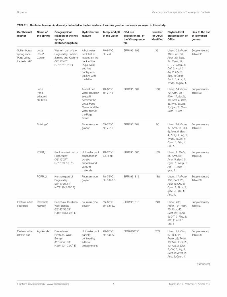
TABLE 1 | Bacterial taxonomic diversity detected in the hot waters of various geothermal vents surveyed in this study.
Geothermal
district
Name of
the spring
Geographical
location of the hot
springs
(latitude/longitude)
Hydrothermal
feature
Temp. and pH
of the water
SRA run
accession no. of
the V3 sequence
file
Number
of OTUs
identified
Phylum-level
classification of
OTUs
Link to the list
of identified
genera
Sulfur- borax
spring zone,
Puga valley,
Ladakh, J&K
Lotus
Pond*
Center
Western part of the
Puga valley, Ladakh,
Jammu and Kashmir
(33◦13′46′′
N/78◦21′18′′ E)
A hot water
pool that is
located on the
bank of the
Puga rivulet
and has
contiguous
outflow with
the latter
78–85◦C
pH 7-8
SRR1951799 331 Ubact, 32; Prote,
168; Firm, 38;
Actn, 33; Bact,
24; Cyan, 12;
D-T, 7; Tmtg, 4;
Def, 2; Acd, 2;
Aq, 2; Chl, 2;
Spir, 1; Cand
Sach, 1; Ace, 1;
Tmds, 1; Ignv, 1.
Supplementary
Table S2
Lotus
Pond-
adjacent
ebullition
A small hot
water ebullition
seated in
between the
Lotus Pond
Center and the
water flow of
the Puga
rivulet
70–80◦C
pH 7-7.5
SRR1951802 186 Ubact, 54; Prote,
72; Actn, 20;
Firm, 17; Bacts,
10; Acd, 4; Vera,
3; Armt, 2; Lats,
1; Cyan, 1; Cand
Sach, 1; Chl, 1.
Supplementary
Table S3
Shivlinga* Fountain-type
geyser
65–75◦C
pH 7-7.5
SRR1951804 80 Ubact, 24; Prote,
17; Firm, 14; D-T,
6; Actn, 5; Bact,
4; Tmtg, 2; Aq, 2;
Tmds, 2; Def, 1;
Cyan, 1; Nitr, 1;
Chl, 1.
Supplementary
Table S4
PCPR_1 South-central part of
Puga valley
(33◦13′27′′
N/78◦20′ 10 E′′)
Hot water pool
embedded in
boratic
deposits and
valley-fill
materials
70–75◦C
7.5-8 pH
SRR1951805 135 Ubact, 7; Prote,
85; Firm, 28;
Actn, 5; Bact, 5;
Cyan, 1; Tmtg, 1;
Aq, 1; Tmds, 1;
Ignv, 1.
Supplementary
Table S5
PCPR_2 Northern part of
Puga valley
(33◦13′25.51′′
N/78◦19′2.69′′ E)
Fountain-type
geyser
70–75◦C
pH 6.8-7.5
SRR1951815 188 Ubact, 17; Prote,
130; Bact, 23;
Actn, 5; Chl, 5;
Cyan, 2; Firm, 2;
Ignv, 2; Spir, 1;
Acd, 1.
Supplementary
Table S6
Eastern Indian
coalfields
Paniphala
fountain
Paniphala, Burdwan,
West Bengal
(23◦45′33.03′′
N/86◦58′54.28′′ E)
Fountain-type
geyser
55–65◦C
pH 6.8-8.0
SRR1951816 743 Ubact, 400;
Prote, 184; Actn,
75; Firm, 45;
Bact, 25; Cyan,
5; D-T, 3; Fus, 2;
Nitr, 2; Acd, 1;
Ver, 1
Supplementary
Table S7
Eastern Indian
lateritic belt
Agnikunda† Bakreshwar,
Birbhum, West
Bengal
(23◦52′48.00′′
N/87◦22′12.00′′ E)
Hot water pool
partially
confined by
artificial
embankments
70–85◦C
pH 6.0-7.0
SRR2016655 283 Ubact, 75; Firm,
67; D-T, 61;
Prote, 23; Tmtg,
13; Nitr, 13; Actn,
12; Atri, 3; Dict,
3; Chl, 3; Aq, 3;
Bact, 2; Armt, 2;
Ace, 2; Cyan, 1
Supplementary
Table S8
(Continued)
Frontiers in Microbiology | www.frontiersin.org 4 March 2016 | Volume 7 | Article 412
Roy et al. Vancomycin-Susceptibility in Thermophilic Bacteria
TABLE 1 | Continued
Geothermal
district
Name of
the spring
Geographical
location of the hot
springs
(latitude/longitude)
Hydrothermal
feature
Temp. and pH
of the water
SRA run
accession no. of
the V3 sequence
file
Number
of OTUs
identified
Phylum-level
classification of
OTUs
Link to the list
of identified
genera
Kharkunda† Hot water pool
partially
confined by
artificial
embankments
55–65◦C
pH 7.5-8.5
SRR2016656 234 Ubact, 55; Firm,
63; D-T, 38;
Prote, 33; Tmtg,
12; Actn, 11; Nitr,
9; Atri, 3; Bact, 2;
Cyan, 2; Armt, 2;
Syn, 1; Ace, 1;
Dict, 1; Chl, 1
Supplementary
Table S9
Phylum-level classifications of the different OTU sets are shown; corresponding lists of identified genera are linked to respective supplementary tables. All V3 sequence files were
deposited to the NCBI Sequence Read Archive (SRA) under the BioProject accession number PRJNA280244.
*The Shivlinga vent is just 50m to the east of the Lotus Pond.†The Agnikunda and Kharkunda are located within 10m of each other.
Ubact, Unclassified Bacteria; Prote, Proteobacteria; Bact, Bacteroidetes; Firm, Firmicutes; Actn, Actinobacteria; Cyan, Cyanobacteria; D-T, Deinococcus-Thermus; Acd,
Acidobacteria; Tmtg, Thermotogae; Def, Deferribacteres; Aq, Aquificae; Chl, Chloroflexi; Spir, Spirochaetes; Cand Sach, Candidatus Saccharibacteria; Ace, Acetothermia; Tmds,
Thermodesulfobacteria; Ignv, Ignavibacteriae; Nitr, Nitrospirae; Fus, Fusobacteria; Armt, Armatimonadetes; Syn, Synergistetes; Dict, Dictyoglomi; Chrys, Chrysiogenetes; Atri,
Atribacteria; Ver, Verrucomicrobia; Tenr, Tenericutes; Fibr, Fibrobacteres; Lats, Latescibacteria.
Peptidoglycan IsolationPeptidoglycans of various bacterial strains were preparedby methods described earlier (Komagata and Suzuki,1988; Schumann, 2011). For vancomycin-untreated cells,peptidoglycan was prepared directly from mid-log phasecultures (OD600 = 0.6), whereas for vancomycin-treatedcounterparts the antibiotic (300µg ml−1) was added to activelygrowing cultures when their OD600 was 0.3–0.4. The lattersets were then incubated for four more hours before cells wereharvested.
Cells were collected by centrifugation and the cell pellet (2gm wet weight) was washed twice with 5ml phosphate buffer(0.05 M, pH 7.2). Then the cell pellet was resuspended in 6ml0.05M phosphate buffer (pH 7.2) and cells were disrupted bysonication on ice using 20 s pulse for 4 times. The cell lysatewas centrifuged at 1800 g for 10min and the supernatant wastransferred to a fresh centrifuge tube. It was then centrifugedat 12,000 g for 1 h. Supernatant was discarded and pellet wasresuspended in 5ml phosphate buffer. 1ml of 5% sodium dodecylsulfate was added and incubated at 100◦C for 40min, followedby centrifugation at 12,000 g for 30min at 30◦C. Then thepellet was washed four to five times with 5ml 60◦C distilledwater. It was then washed with 5ml 0.05M phosphate buffer(pH 7.6). The pellet was then resuspended in 2ml phosphatebuffer (0.05 M, pH 7.6) and 100µl pronase E (1mg ml−1) wasadded. The soup was then incubated at 37◦C for 2 h. Pellet wascollected by centrifugation at 12,000 g for 30min. It was furtherwashed twice 2ml phosphate buffer (0.05 M, pH 7.6). Afterthat, pellet was resuspended in 2ml of 5% TCA (TrichloroaceticAcid) and boiled at 100◦C for 20min. The suspension wascooled at room temperature and transferred to glass centrifugetubes. It was then centrifuged at 12,000 g for 30min. Pelletwas further washed thrice with 2ml phosphate buffer (0.05 M,pH 7.6), once each with 2ml ethanol (95%) and 2ml diethylether (99%), and finally air dried at 60◦C for 3 h before furtheranalysis.
MALDI-MSExtracted peptidoglycans were digested with lysozyme (40mgml−1) for 2 h at 37◦C, following which deactivation was done for20min at 70◦C. Digested products were lyophilized, resuspendedin 100µl 99% methanol, and directly used for MALDI-MSwithout any more purification. DHA was used as the MALDImatrix. MALDI-MS was carried out using an AutoFlex II tandemtime of flight (TOF/TOF) MALDI-mass spectrometer (BrukerDaltonics) equipped with a pulsed N2 laser (λ-337 nm, 50Hz).Themachine was calibrated for reflectormodemass spectra usinga mixture of standard peptides (having mass 750 to 3150) inthe positive ion mode. MS spectra were analyzed using the FlexAnalysis software V2.4.
PhylogenyML trees were constructed using MEGA6 (Tamura et al., 2013)plus PhyML (Guindon et al., 2010). The best substitution modelused for likelihood analysis (general time reversible and gamma)was selected by Bayesian as well as corrected Akaike informationcriteria. After the starting NJ tree was obtained heuristic searchesfor likelihood were performed using the Nearest–Neighbor–Interchange as well as Close–Neighbor–Interchange branchswapping algorithms.
RESULTS AND DISCUSSION
Rich Diversity of Phylogenetic Relatives ofMesophilic Bacteria in Circumneutral HotSpringsOver the past few years we have investigated the taxonomicdiversity (species richness) of the aquatic bacterial communityof several circumneutral hot springs of Northern and EasternIndia by analyzing amplified 16S rRNA gene fragments. V3regions of all potential bacterial 16S rRNA genes present inthe total environmental DNA isolated from thermal water
Frontiers in Microbiology | www.frontiersin.org 5 March 2016 | Volume 7 | Article 412
Roy et al. Vancomycin-Susceptibility in Thermophilic Bacteria
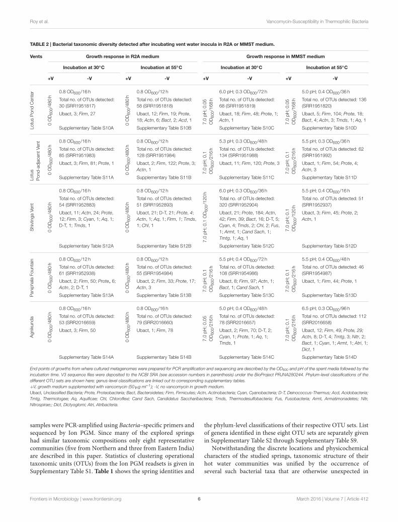
TABLE 2 | Bacterial taxonomic diversity detected after incubating vent water inocula in R2A or MMST medium.
Vents Growth response in R2A medium Growth response in MMST medium
Incubation at 30◦C Incubation at 55◦C Incubation at 30◦C Incubation at 55◦C
+V -V +V -V +V -V +V -V
LotusPondCenter
0OD600/480h
0.8 OD600/16 h
0OD600/480h
0.8 OD600/12 h
7.0
pH;0.05
OD600/168h
6.0 pH; 0.3 OD600/72 h
7.0
pH;0.05
OD600/168h
5.0 pH; 0.4 OD600/36 h
Total no. of OTUs detected:
30 (SRR1951817)
Total no. of OTUs detected:
58 (SRR1951818)
Total no. of OTUs detected:
68 (SRR1951819)
Total no. of OTUs detected: 136
(SRR1951820)
Ubact, 3; Firm, 27 Ubact, 12; Firm, 19; Prote,
18; Actn, 6; Bact, 2; Acd, 1
Ubact, 18; Firm, 48; Prote, 1;
Actn, 1
Ubact, 5; Firm, 104; Prote, 18;
Bact, 4; Actn, 3; Tmds, 1; Aq, 1
Supplementary Table S10A Supplementary Table S10B Supplementary Table S10C Supplementary Table S10D
Lotus
Pond-adjacentVent
0OD600/480h
0.8 OD600/16 h
0OD600/480h
0.8 OD600/12 h
7.0
pH;0.1
OD600/216h
5.3 pH; 0.3 OD600/48 h
7.0
pH;0.1
OD600/216h
5.5 pH; 0.3 OD600/36 h
Total no. of OTUs detected:
85 (SRR1951983)
Total no. of OTUs detected:
128 (SRR1951984)
Total no. of OTUs detected:
134 (SRR1951988)
Total no. of OTUs detected: 62
(SRR1951992)
Ubact, 3; Firm, 81; Prote, 1 Ubact, 2; Firm, 122; Prote, 3;
Actn, 1
Ubact, 11; Firm, 120; Prote, 3 Ubact, 1; Firm, 54; Prote, 4;
Actn, 3
Supplementary Table S11A Supplementary Table S11B Supplementary Table S11C Supplementary Table S11D
ShivlingaVent
0OD600/480h
0.8 OD600/16 h
0OD600/480h
0.8 OD600/12 h
7.0
pH;0.1
OD600/120h 6.0 pH; 0.3 OD600/36 h
7.0
pH;0.1
OD600/120h
5.5 pH; 0.4 OD600/16 h
Total no. of OTUs detected:
54 (SRR1952883)
Total no. of OTUs detected:
51 (SRR1952893)
Total no. of OTUs detected:
320 (SRR1952904)
Total no. of OTUs detected: 51
(SRR1952937)
Ubact, 11; Actn, 24; Prote,
12; Firm, 3; Cyan, 1; Aq, 1;
D-T, 1; Tmds, 1
Ubact, 21; D-T, 21; Prote, 4;
Actn, 1; Aq, 1; Firm, 1; Tmds,
1; Chl, 1
Ubact, 21; Prote, 184; Actn,
42; Firm, 39; Bact, 16; D-T, 5;
Cyan, 4; Tmds, 2; Chl, 2; Fus,
1; Armt, 1; Cand Sach, 1;
Tmtg, 1; Aq, 1
Ubact, 3; Firm, 45; Prote, 2;
Actn, 1
Supplementary Table S12A Supplementary Table S12B Supplementary Table S12C Supplementary Table S12D
PaniphalaFountain
0OD600/480h
0.8 OD600/12 h
0OD600/480h
0.8 OD600/12 h
7.0
pH;0.1
OD600/216h
5.5 pH; 0.4 OD600/72 h
7.0
pH;0.1
OD600/216h
5.5 pH; 0.4 OD600/48 h
Total no. of OTUs detected:
61 (SRR1952938)
Total no. of OTUs detected:
55 (SRR1954984)
Total no. of OTUs detected:
108 (SRR1954986)
Total no. of OTUs detected: 46
(SRR1954987)
Ubact, 2; Firm, 50; Prote„ 6;
Actn, 2; D-T, 1
Ubact, 2; Firm, 33; Prote, 17;
Actn, 3
Ubact, 8; Firm, 97; Actn, 1;
Bact, 1; Cand Sach, 1
Ubact, 1; Firm, 44; Prote, 1
Supplementary Table S13A Supplementary Table S13B Supplementary Table S13C Supplementary Table S13D
Agnikunda
0OD600/480h
0.8 OD600/16 h
0OD600/480h
0.8 OD600/16 h
7.0
pH;0.05
OD600/216h
5.0 pH; 0.4 OD600/48 h
7.0
pH;0.1
OD600/216h
6.5 pH; 0.3 OD600/96 h
Total no. of OTUs detected:
53 (SRR2016659)
Total no. of OTUs detected:
79 (SRR2016660)
Total no. of OTUs detected:
78 (SRR2016657)
Total no. of OTUs detected: 112
(SRR2016658)
Ubact, 3; Firm, 50 Ubact, 1; Firm, 78 Ubact, 2; Firm, 70; D-T, 2;
Cyan, 1; Prote, 1; Aq, 1;
Tmds, 1
Ubact, 12; Firm, 49; Prote, 29;
Actn, 8; D-T, 4; Tmtg, 3; Nitr, 2;
Bact, 1; Cyan, 1; Armt, 1; Atri, 1;
Dict, 1
Supplementary Table S14A Supplementary Table S14B Supplementary Table S14C Supplementary Table S14D
End points of growths from where cultured metagenomes were prepared for PCR amplification and sequencing are described by the OD600 and pH of the spent media followed by the
incubation time. V3 sequence files were deposited to the NCBI SRA (see accession numbers in parenthesis) under the BioProject PRJNA280244. Phylum-level classifications of the
different OTU sets are shown here; genus-level classifications are linked out to corresponding supplementary tables.
+V, growth medium supplemented with vancomycin (50µg ml−1); -V, no vancomycin in growth medium.
Ubact, Unclassified Bacteria; Prote, Proteobacteria; Bact, Bacteroidetes; Firm, Firmicutes; Actn, Actinobacteria; Cyan, Cyanobacteria; D-T, Deinococcus-Thermus; Acd, Acidobacteria;
Tmtg, Thermotogae; Aq, Aquificae; Chl, Chloroflexi; Cand Sach, Candidatus Saccharibacteria; Tmds, Thermodesulfobacteria; Fus, Fusobacteria; Armt, Armatimonadetes; Nitr,
Nitrospirae;; Dict, Dictyoglomi; Atri, Atribacteria.
samples were PCR-amplified using Bacteria–specific primers andsequenced by Ion PGM. Since many of the explored springshad similar taxonomic compositions only eight representativecommunities (five from Northern and three from Eastern India)are described in this paper. Statistics of clustering operationaltaxonomic units (OTUs) from the Ion PGM readsets is given inSupplementary Table S1. Table 1 shows the spring identities and
the phylum-level classifications of their respective OTU sets. Listof genera identified in these eight OTU sets are separately givenin Supplementary Table S2 through Supplementary Table S9.
Notwithstanding the discrete locations and physicochemicalcharacters of the studied springs, taxonomic structure of theirhot water communities was unified by the occurrence ofseveral such bacterial taxa that are otherwise unexpected in
Frontiers in Microbiology | www.frontiersin.org 6 March 2016 | Volume 7 | Article 412
Roy et al. Vancomycin-Susceptibility in Thermophilic Bacteria
high-temperature habitats. While most of the communitiesencompassed maximum OTUs from the Alpha, Beta, andGamma subclasses of Proteobacteria, the typical thermophilicphyla Aquificae, Thermotogae and Thermodesulfobacteria hadvery few OTUs affiliated to them (Table 1). The only exceptionsto this trend were the Agnikunda and Kharkunda vents wheretaxonomic diversity was dominated by Firmicutes. Concurrentto these observations, a close inspection of the lists ofgenera identified in the described hot water communitiesrevealed several such bacteria that have no report of laboratorygrowth above 45◦C. While the hot water community ofAgnikunda encompassed the lowest proportion (∼52%) of suchapparently-mesophilic genera, that of the Paniphala vent hadthe highest percentage (89%). So far as the actual numbers wereconcerned, the Lotus Pond Center (84) had the maximum countof supposedly-mesophilic genera, followed by Paniphala (81). Incontrast, PCPR 1 and Shivlinga had the lowest number (11 and15 respectively) of such genera, presumably because these ventsas such had the lowest overall count of OTUs and genera.
In this scenario we sought to know how this largevariety of purportedly-mesophilic genera survived in these hightemperature habitats. First it was imperative to check whetherthey could at all grow at high temperatures or were only thermo-enduring entities. Alternatively, it was also plausible that manyof them were stochastically introduced into these habitats inrecent times and were not at all equipped to cope with thermalstress. Since only pure culture isolates could answer these querieswe attempted to get the same in chemoorganoheterotrophic(R2A) as well as chemolithoautotrophic [modified minimal saltssupplemented with thiosulfate, MMST (Ghosh and Roy, 2006)]media at various incubation temperatures between 30 and 70◦C.
Preponderance of Firmicutes in R2A PlatesStrains related to Bacillus, Geobacillus, Anoxybacillus andBrevibacillus crowded all the R2A isolation plates incubatedat temperatures ≥50◦C. Consequently, very few non-Firmicutes (e.g., strains of known moderate-thermophileslike Thermomonas, Porphyrobacter, Meiothermus etc.) couldbe obtained even after several rounds of isolation from thevarious inoculum samples. May be, during high-temperaturegrowth in R2A the Firmicutes out-competed other moderate-thermophiles by virtue of their metabolic versatility, fastergrowth rate and better thermal adaptations. So far as isolationin MMST was concerned, all the tested inocula acidified theenrichment broths and produced sulfate with concomitantdisappearance of thiosulfate. However, only a few moderately-thermophilic Proteobacteria (e.g., Thermithiobacillus tepidarius)kept recurring when the enriched broths were plated in MMST-agar to obtain single colonies. In addition, a host of suchapparently thermo-enduring Paracoccus strains got isolated (inMMST at 37◦C) that despite failing to grow above 45◦C did notlose substantial viability (measured by drop in CFU count ofexperimental cultures) even at 60◦C over an exposure periodof ∼4–6 h. Now, our prime objective was to isolate thermophilicsiblings of the reportedly-mesophilic Gram-negative bacterialisted in Supplementary Table S2 through Supplementary TableS9. Although success of such undertakings depended on the
actual cultivability of the unexpected bacteria in question,failures could never ascertain whether the negative results weredue to short-comings or intrinsic-limitations of the isolationprocedures or whether the Gram-negative mesophiles surfacingin diversity analyses were not thermally-adapted at all. So weinitiated another round of isolation by supplementing all mediatypes with vancomycin so that Firmicutes were eliminated andGram-negative taxa got a better chance to appear in the isolationplates.
Eradication of Culturable Diversity byVancomycinSupplementing R2A as well as MMST with 50µg ml−1
vancomycin caused complete destruction of the correspondingculturable-diversities of all the explored hot water communities.However, to keep it brief, the data from three North Indian andtwo East Indian vents will be presented in detail. As shown inTable 2, zero or near-zero OD600 values were registered for allspent vancomycin-containing media after prolonged incubationat 55 as well as 30◦C. No production of sulfuric acid in spentvancomycin-containing MMST media was taken as the mainevidence of destruction of the culturable chemolithotrophicdiversity. Concurrently-recorded CFU counts were always <102
ml−1 of the spent media. This appeared to tally with spontaneousmutation rates (John et al., 1998) as parallel vancomycin-freecultures yielded rich growth of mixed consortia at 55 as well as30◦C. For all the tested inocula, OD600 of vancomycin-free R2Areached 0.8 (CFU counts ∼108 ml−1 of spent medium) within12–16 h of incubation, while that of vancomycin-free MMSTreached 0.3–0.4 (CFU counts 104–105 ml−1 of spent medium)with concomitant acidification of spent media within 16–72 hof incubation. With regard to growth in MMST it must beappreciated that the observed OD600 values were not exclusivelydue to sulfur-oxidizing chemolithotrophs. Even though theiractive presence was evidenced by sulfuric acid production, thefinal cell masses recovered from spent MMST media were quitelikely to contain organoheterotrophic secondary consumers.
The above data clearly implied that all the bacteria growingin the two vancomycin-free media types, irrespective of theirtaxonomic identity and Gram-property, were susceptible tothis so-called Gram-positive-specific antibiotic. To know thetaxonomic identity of these cultured consortia we isolated theirtotal genomes, amplified the V3 regions of all 16S rRNA genespresent therein, and sequenced the amplicon pools by Ion PGM.The obtained V3 readsets were analyzed by OTU-clustering,statistics of which are given in Supplementary Table S1. Table 2shows the phylum-level classification of the respective OTU sets.
Corroborating the outcome of the isolation experiments,almost all the vancomycin-free cultured consortia (irrespective ofthe media type) encompassed maximum OTUs from Firmicutes(albeit after the unclassifiable ones). Nevertheless,Actinobacteria,Deinococcus-Thermus and Proteobacteria had maximum OTUsin the consortia obtained by incubating the Shivlinga Ventwater in R2A at 30 and 55◦C, and MMST at 30◦C respectively.The most interesting attribute of these datasets was theaffiliation of a large number of OTUs to Gram-negative phyla
Frontiers in Microbiology | www.frontiersin.org 7 March 2016 | Volume 7 | Article 412
Roy et al. Vancomycin-Susceptibility in Thermophilic Bacteria
such as Proteobacteria, Bacteroidetes, Deinococcus-Thermus,Thermodesulfobacteria, Acidobacteria, Cyanobacteria, and alsoActinobacteria, which has Gram-negative as well as positivemembers. The lists of genera identified in these OTU sets(Supplementary Table S10 through Supplementary Table S14)also showed that the vancomycin-free cultured consortiaencompassed at least one (for Lotus Pond Center inoculumincubated in MMST at 30◦C) to at most 46 (for Shivlingainoculum incubated in MMST at 30◦C) Gram-negative genera.Only when the Lotus Pond Center inoculum was incubated inR2A at 30◦C, or the Agnikunda inoculum was incubated inR2A at 30 or 55◦C, the resultant consortium encompassed noGram-negative genus.
The above data summarily indicated that the Gram-negative components of hot water communities were assusceptible to vancomycin as their Gram-positive counterparts.The wide taxonomic as well as geographic spread of thesecommunity analyses further hinted that the association betweenvancomycin-susceptibility and thermal adaptation could well bea global phenomenon. Significantly again, this comprehensivevancomycin-susceptibility of all taxonomic- and Gram-types wasnot detected when the above experiments were repeated withmesophilic (in situ temperature 30◦C) lake water inocula (seeSupplementary Tables S15, S15A through Supplementary TableS15D). This buttressed our assumption that the phenomenonwas indeed an exclusive hallmark of hydrothermal communities.Subsequently, to cross-examine this supposition, vancomycinchallenge was extended to all the hot spring isolates that werethere at our disposal. On top of that we also scrutinizedthe species descriptions of several Gram-negative thermophilicbacteria in the literature to see whether any information existedabout their vancomycin response.
Vancomycin-Susceptibility is Widespreadamong Gram-Negative ThermophilesAll the current hot-spring isolates died (showing near-zeroCFU counts) in the presence of 50µg ml−1 vancomycin atboth higher and lower incubation temperatures. Susceptiblephenotype was also exhibited by the tested siblings strains ofthe present isolates reported from other parts of the world.Although the strains in question took negative Gram stain, theirresponse to vancomycin challenge was exactly same as that of theBacillus controls. On the other hand, mesophilic Gram-negativeAlpha-, Beta- and Gammaproteobacteria like Pseudaminobactersalicylatoxidans KCT001, Advenella kashmirensis WT001 and E.coli BL21 did not show any growth perturbation in the presenceof vancomycin. Relevant growth experiment data for some ofthese organisms are shown in Table 3.
The most remarkable of all these observations was thesusceptibility of the thermo-enduring Paracoccus isolates. Theywere all killed by 50µg ml−1 vancomycin at the same time asstandard strains of Paracoccus pantotrophus (LMG 4218) andParacoccus thiocyanatus (MTCC 7821) isolated from variousmesophilic habitats remained unaffected (Table 3). This evokedthe conjecture that vancomycin-susceptibility of Gram-negativebacteria could be a function of high temperature exposure ratherthan actual growth efficiency at elevated temperatures. In tandemwith these experiments we also scrutinized the descriptions
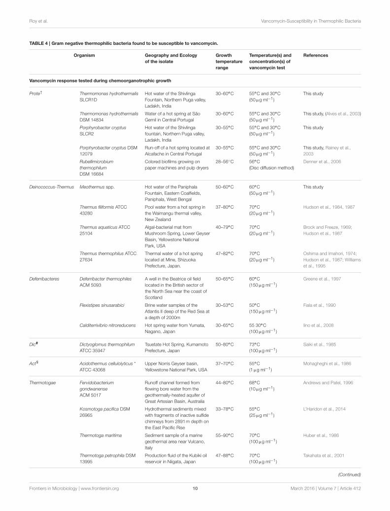
of Gram-negative thermophilic bacteria in the literature andfound at least 16 such species that were reported as susceptibleto vancomycin (Table 4). For obvious reasons, Gram-negativebacteria are seldom tested for vancomycin response; hencescarcity of such data was understandable. In such a scenario,the taxonomy and biogeography of the bacteria listed in Table 4
could be considered diverse enough to implicate vancomycinsusceptibility as a global trait of Gram-negative thermophiles.
Vancomycin Susceptibility ofGram-Negative Thermophiles Stems FromDi-Alanine-Terminated MPPsIn view of the above data it was deemed imperative to knowwhether the typical vancomycin mode of action that worksagainst Gram-positive bacteria (Healy et al., 2000) was alsoinstrumental in the killing of Gram-negative thermophiles. Itmay be recalled that transpeptidase enzymes cross-link MPPs toprovide structural integrity and strength to the peptidoglycanlayer of the cell wall (Waxman and Strominger, 1983). Gram-positive bacteria generally have tandem D-ala-D-ala residuesat the N-terminal end of their MPPs. These two amino acidresidues afford the high-affinity target of vancomycin (Perkinsand Nieto, 1974; Healy et al., 2000), which out-competes thetranspeptidases in the race for binding with the substrate. Inthe process peptidoglycan cross-linking fails, causing the cellwall to eventually disintegrate and collapse under extraneouspressure. In contrast, a majority of Gram-negative bacteria aswell as vancomycin-resistant Gram-positive bacteria have the lastD-alanine replaced by D/L-lactate/lysine/glycine etc, and thusremain unscathed by vancomycin (Reynolds, 1989; Arthur et al.,1996; Courvalin, 2006). However, many others do have tandemD-ala-D-ala residues in their MPPs but impermeability of theirouter membrane toward vancomycin confers them resistanceto this drug. As such, the normal mechanism of vancomycinresistance starts with a permeability barrier (Shlaes et al., 1989)even as its mechanism of action is invariably rendered throughbinding of the terminal D-ala-D-ala residues resulting in sterichindrance of further addition to the growing peptidoglycan chain(Perkins and Nieto, 1974).
Peptidoglycan was extracted from vancomycin-treated as wellas -untreated cells of the current Gram-negative thermophilicisolates and analyzed by MALDI MS. The resultant datasuggested that the organisms in question were all susceptible tothis antibiotic in the same mechanistic way as the Gram-positiveFirmicutes (that is on account of having tandem alanines in theN-termini of their MPPs). As such, details of the experiment withThermomonas hydrothermalis SLCR_1D is described below.
Peptidoglycan was extracted from T. hydrothermalisSLCR_1D, and analyzed in comparison with the isolatesBacillus licheniformis SWCR_1/2X50_9 and A. kashmirensisWT 001. Extraction was done in such a way that MPPs wererepresented abundantly. When the OD600 of a given culturereached 0.3–0.4, excess vancomycin (300µg ml−1) was addedto the growth media, following which they were incubatedfor another 4 h before cells were harvested for peptidoglycanextraction. Vancomycin-untreated cultures were passed throughthe same steps as above, except antibiotic addition. It was
Frontiers in Microbiology | www.frontiersin.org 8 March 2016 | Volume 7 | Article 412

Roy et al. Vancomycin-Susceptibility in Thermophilic Bacteria
TABLE3|Pure
culture
isolatesofGram
negativebacteriatestedin
thepresentstudyfortheirvancomycin
response.
Nameofthehydrotherm
al
isolate
R2A*
MMST**
28◦C
55◦C
28◦C
55◦C
+V
−V
+V
−V
+V
−V
+V
−V
Thermomonashydrothermalis
SLCR1D
OD600=
0.00/10d
OD600=
0.52/24h
OD600=
0.00/10d
OD600=
0.72/24h
NA
NG
NA
NG
Thermomonashydrothermalis
DSM
14834
OD600=
0.00/10d
OD600=
0.62/24h
OD600=
0.00/10d
OD600=
0.88/24h
NA
NG
NA
NG
Porphyrobactercryptus
SLCR2
OD600=
0.00/10d
OD600=
0.80/24h
OD600=
0.00/10d
OD600=
0.86/24h
NA
NG
NA
NG
PorphyrobactercryptusDSM
12079
OD600=
0.00/10d
OD600=
0.84/24h
OD600=
0.00/10d
OD600=
0.88/24h
NA
NG
NA
NG
Meiothermussp
.RP†
NA
NG
OD600=
0.00/10d
OD600=
0.58/24h
NA
NG
NA
NG
Meiothermussp
.TP‡
NA
NG
OD600=
0.00/10d
OD600=
0.64/24h
NA
NG
NA
NG
Thermithiobacillustepidarius
SMMA_1
1
NA
NG
NA
NG
OD600=
0.00,
pH7.0/7
d
OD600=
0.26,pH
5.7/24h
OD600=
0.00,
pH7.0/7
d
OD600=
0.29,pH
5.7/24h
Paracoccussp
.$
SMMA_7
OD600=
0.00/10d
OD600=
0.92/24h
NA
NG
OD600=
0.00,pH
7.0/7
d
OD600=
0.25,pH
6.0/24h
NA
NA
Paracoccussp
.$
SMMA_5
OD600=
0.00/10d
OD600=
0.92/24h
NA
NG
OD600=
0.00,pH
7.0/7
d
OD600=
0.25,pH
6.0/24h
NA
NA
Bacilluslicheniformis
SWCR_1
/2X50_9
OD600=
0.00/10d
OD600=
0.63/24h
OD600=
0.00/10d
OD600=
0.82/24h
NA
NG
NA
NG
Bacillussp.
SWCR_6
04
OD600=
0.00/10d
OD600=
0.69/24h
OD600=
0.00/10d
OD600=
0.77/24h
NA
NG
NA
NG
Pseudaminobacter
salicylatoxidansKCT001
OD600=
0.53/48h
OD600=
0.52/48h
NA
NG
OD600=
0.27,pH
6.0/48h
OD600=
0.25,pH
6.0/48h
NA
NG
Advenella
kashmirensis
WT001
OD6000.48/24h
OD600=
0.46/24h
NA
NG
OD600=
0.30,pH
6.0/24h
OD6000.32,pH
6.0/24h
NA
NG
E.coliBL21
OD600=
0.57/24h
OD600=
0.55/24h
NA
NG
NA
NG
NA
NG
ParacoccuspatotrophusLMG
4218
OD600=
0.85/24h
OD600=
0.88/24h
NA
NG
OD600=
0.29,pH
6.0/24h
OD600=
0.29,pH
6.0/24h
NA
NG
Paracoccusthiocyanatus
MTCC7821
OD600=
0.78/24h
OD600=
0.79/24h
NA
NG
OD600=
0.27,pH
6.0/24h
OD600=
0.26,pH
6.0/24h
NA
NG
+V,growthmediumsupplementedwithvancomycin(50
µgml-1),-V,novancomyciningrowthmedium.
NG,Nointrinsicgrowthintherelevantmediumortemperature;NA,Notapplicable.
†94%16SrRNAgenesequencesimilaritywithM.cateniformans,M.taiwanensisM.rubberandotherMeiothermusspp.
‡98%16SrRNAgenesequencesimilaritywithM.cateniformans,M.taiwanensisM.rubberandotherMeiothermusspp.
$ThesetwoParacoccusstrainshad100%16SrRNAgenesequencesimilarityamongthemselvesand97%similaritywithahostofParacoccusspp.Importantlyhowever,SMMA_7wasisolatedfrom68
◦Cwaterwhile
SMMA_5was
isolatedfrom80◦Cwater.
*BacteriawerefirstgrowninR2Abrothswithoutanyantibioticselection(seedcultures);subsequently2%ofthesemid-logphase(OD600=0.6)seedculturesweretransferredtoR2Abroths,whichcontainedvancomycin(50
µgml−
1)
ornot,aswarrantedbythetestinhand.
**BacteriawerefirstgrowninMSTbroths(initialpH7.0)withoutanyantibioticselection(seedcultures);subsequently2%ofthesemid-logphase(OD600=0.15,pH6.5)seedculturesweretransferredtoMSTbroths,whichcontained
vancomycin(50
µgml−
1)ornot,aswarrantedbythetestinhand.
Frontiers in Microbiology | www.frontiersin.org 9 March 2016 | Volume 7 | Article 412
Roy et al. Vancomycin-Susceptibility in Thermophilic Bacteria
TABLE 4 | Gram negative thermophilic bacteria found to be susceptible to vancomycin.
Organism Geography and Ecology
of the isolate
Growth
temperature
range
Temperature(s) and
concentration(s) of
vancomycin test
References
Vancomycin response tested during chemoorganotrophic growth
Prote† Thermomonas hydrothermalis
SLCR1D
Hot water of the Shivlinga
Fountain, Northern Puga valley,
Ladakh, India
30–60◦C 55◦C and 30◦C
(50µg ml−1)
This study
Thermomonas hydrothermalis
DSM 14834
Water of a hot spring at São
Gemil in Central Portugal
30–60◦C 55◦C and 30◦C
(50µg ml−1)
This study, (Alves et al., 2003)
Porphyrobacter cryptus
SLCR2
Hot water of the Shivlinga
fountain, Northern Puga valley,
Ladakh, India
30–55◦C 55◦C and 30◦C
(50µg ml−1)
This study
Porphyrobacter cryptus DSM
12079
Run-off of a hot spring located at
Alcafache in Central Portugal
30–55◦C 55◦C and 30◦C
(50µg ml−1)
This study, Rainey et al.,
2003
Rubellimicrobium
thermophilum
DSM 16684
Colored biofilms growing on
paper machines and pulp dryers
28–56◦C 56◦C
(Disc diffusion method)
Denner et al., 2006
Deinococcus-Thermus Meothermus spp. Hot water of the Paniphala
Fountain, Eastern Coalfields,
Paniphala, West Bengal
50–60◦C 60◦C
(50µg ml−1)
This study
Thermus filiformis ATCC
43280
Pool water from a hot spring in
the Waimangu thermal valley,
New Zealand
37–80◦C 70◦C
(20µg ml−1)
Hudson et al., 1984, 1987
Thermus aquaticus ATCC
25104
Algal-bacterial mat from
Mushroom Spring, Lower Geyser
Basin, Yellowstone National
Park, USA
40–79◦C 70◦C
(20µg ml−1)
Brock and Freeze, 1969;
Hudson et al., 1987
Thermus thermophilus ATCC
27634
Thermal water of a hot spring
located at Mine, Shizuoka
Prefecture, Japan.
47–82◦C 70◦C
(20µg ml−1)
Oshima and Imahori, 1974;
Hudson et al., 1987; Williams
et al., 1995
Deferribacteres Deferribacter thermophiles
ACM 5093
A well in the Beatrice oil field
located in the British sector of
the North Sea near the coast of
Scotland
50–65◦C 60◦C
(150µg ml−1 )
Greene et al., 1997
Flexistipes sinusarabici Brine water samples of the
Atlantis II deep of the Red Sea at
a depth of 2000m
30–53◦C 50◦C
(150µg ml−1 )
Fiala et al., 1990
Calditerrivibrio nitroreducens Hot spring water from Yumata,
Nagano, Japan
30–65◦C 55 30◦C
(100µg ml−1 )
Iino et al., 2008
Dic# Dictyoglomus thermophilum
ATCC 35947
Tsuetate Hot Spring, Kumamoto
Prefecture, Japan
50–80◦C 73◦C
(100µg ml−1 )
Saiki et al., 1985
Act$ Acidothermus cellulolyticus *
ATCC 43068
Upper Norris Geyser basin,
Yellowstone National Park, USA
37–70◦C 55◦C
(1µg ml−1)
Mohagheghi et al., 1986
Thermotogae Fervidobacterium
gondwanense
ACM 5017
Runoff channel formed from
flowing bore water from the
geothermally-heated aquifer of
Great Artesian Basin, Australia
44–80◦C 68◦C
(10µg ml−1)
Andrews and Patel, 1996
Kosmotoga pacifica DSM
26965
Hydrothermal sediments mixed
with fragments of inactive sulfide
chimneys from 2891m depth on
the East Pacific Rise
33–78◦C 55◦C
(25µg ml−1)
L’Haridon et al., 2014
Thermotoga maritima Sediment sample of a marine
geothermal area near Vulcano,
Italy
55–90◦C 70◦C
(100µg ml−1 )
Huber et al., 1986
Thermotoga petrophila DSM
13995
Production fluid of the Kubiki oil
reservoir in Niigata, Japan
47–88◦C 70◦C
(100µg ml−1 )
Takahata et al., 2001
(Continued)
Frontiers in Microbiology | www.frontiersin.org 10 March 2016 | Volume 7 | Article 412
Roy et al. Vancomycin-Susceptibility in Thermophilic Bacteria
TABLE 4 | Continued
Organism Geography and Ecology
of the isolate
Growth
temperature
range
Temperature(s) and
concentration(s) of
vancomycin test
References
Thermotoga naphthophila
DSM 13996
Production fluid of the Kubiki oil
reservoir in Niigata, Japan
48–86◦C 70◦C
(100µg ml−1)
Takahata et al., 2001
Petrotoga mobilis Anoxic samples from production
water taken from the water
separator tanks on off-shore oil
platforms of North Sea oil
reservoir
40–65◦C 60◦C
(10µg ml−1)
Lien et al., 1998
Vancomycin response tested during chemolithotrophic growth
Prote† Thermithiobacillus tepidarius
SMMA_11
Hot water of the Lotus
Pond-adjacent ebullition,
Northern Puga valley, Ladakh,
India
30–50◦C 50◦C and 30◦C
(50µg ml−1)
This study
Sulfurivirga caldicuralii DSM
17737
Taketomi Island, Okinawa, Japan 30–60◦C 55◦C
(50µg ml−1)
Takai et al., 2006
Given information were generated either in this study or collated from species descriptions reported elsewhere.
*Gram-variable bacteria with Gram staining response usually negative but occasionally positive.#Dic, Dictyoglomi;$Act, Actinobacteria;†Prote, Proteobacteria.
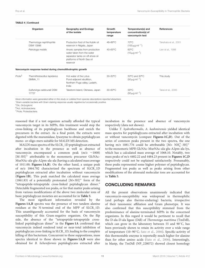
reasoned that if a test organism actually afforded the typicalvancomycin target in its MPPs, this treatment would stop thecross-linking of its peptidoglycan backbone and enrich theprecursors in the extract. As a final point, the extracts weredigested with the muramidase, lysozyme to obtain peptidoglycanmono- or oligo-mers suitable for MALDI MS detection.
MALDImass spectra of the SLCR_1D peptidoglycan extractedafter incubation in the presence as well as absence ofvancomycin encompassed a common peak (m/z ∼1008)[M-3H]+ attributable to the monomeric precursor GlcNAc-MurNAc-ala-glu-A2pm-ala-ala (having a calculatedmass averageof 1011.00; Figures 1A,B). On the other hand, a unique peakof m/z 1864.782 characterized the spectrum of SLCR_1Dpeptidoglycan extracted after incubation without vancomycin(Figure 1B). This peak matched the calculated mass average(1861.83) of a potentially protonated [M+3H]+ form of the“tetrapeptide-tetrapeptide cross-linked peptidoglycan dimer.”Detectable fragmented ion peaks, or for that matter peaks arisingfrom various modifications of the above two molecular ions ofbasic peptidoglycan moieties are accounted for in Table 5.
The most significant information revealed by theFigures 1A,B spectra was the presence of two tandem alanineresidues at the N-terminal end of the MPP of SLCR_1D.This unambiguously accounted for the basis of vancomycinsusceptibility of this Gram-negative organism. On the flipside, the absence of the “tetrapeptide-tetrapeptide cross-linked peptidoglycan dimer” in the Figure 1A confirmed thatvancomycin indeed rendered total or near-total inhibition ofpeptidoglycan cross-linking in SLCR_1D, leading to the completekilling of this bacterium. Concurrent to these suppositions, massspectra identical to those shown in Figures 1A,B were alsoobtained for B. licheniformis peptidoglycans extracted after
incubation in the presence and absence of vancomycinrespectively (data not shown).
Unlike T. hydrothermalis, A. kashmirensis yielded identicalmass spectra for peptidoglycans extracted after incubation withor without vancomycin (compare Figures 1C,D). Out of theseries of common peaks present in the two spectra, the onehaving m/z 1081.776 could be attributable [M+ NH+
4 -3H]+
to the monomeric MPP, GlcNAc-MurNAc-ala-glu-A2pm-ala-lys,which has a calculated mass average of 1068.10. Notably, twomass peaks of m/z 4482.22 and 4484.23 present in Figures 1C,D
respectively could not be explained satisfactorily. Presumably,these peaks represented some higher polymer of peptidoglycan.Fragmented ion peaks as well as peaks arising from othermodifications of the aforesaid molecular ions are accounted forin Table 5.
CONCLUDING REMARKS
All the present observations unanimously indicated thatvancomycin-susceptibility was widespread in thermophilic(and perhaps also thermo-enduring) bacteria, irrespectiveof their taxonomic affiliation and Gram phenotype. It wasalso confirmed that this susceptibility stemmed from thepredominance of alanine-terminated MPPs in the concernedorganisms. In this regard it would be pertinent to recall thatthe D-ala-D-ala ligase (Ddl) of Thermotoga maritima (TmDdl),which can grow in the laboratory between 55 and 90◦C, hasbeen previously shown to retain its activity over a wide rangeof temperature (10–90◦C; Sato et al., 2006). Specific-activity ofthis thermostable enzyme is reportedly much higher for alaninethan for other amino acids (Sato et al., 2006). Interestingly,in blastp, the TmDdl (NP_228072) showed closest homology
Frontiers in Microbiology | www.frontiersin.org 11 March 2016 | Volume 7 | Article 412
Roy et al. Vancomycin-Susceptibility in Thermophilic Bacteria
FIGURE 1 | Positive reflector ion MALDI mass spectrum of the digested peptidoglycan fragments of vancomycin-treated (A,C) and -untreated (B,D)
cells of T. hydrothermalis SLCR_1D (Upper panels A,B) and A. kashmirensis WT 001 (Lower panels C,D). Structure (i) corresponds to muro-pentapeptide
precursor having terminal alanine-alanine dipeptide, (ii) corresponds to tetrapeptide-tetrapeptide cross linking, whereas structure (iii) represents muro-pentapeptide
precursor having terminal alanine-lysine dipeptide.
TABLE 5 | Observed and calculated average m/z’s along with their proposed structures and proposed modifications as present in the Figure 1.
Explainable peaks of Figure 1 Proposed modifications Presence/Absence in Figure 1
Obs. m/z Calculated average mass Proposed structure A B C D
1008.045 and 1008.013 1011 GlcNAc-MurNAc- Ala-Glu-A2Pm-Ala-Ala -3H++ + − −
943.659 and 939.784 939.92 GlcNAc-MurNAc- Ala-Glu-A2Pm-Ala +4H++ + − −
920.721 and 920.867 939.92 GlcNAc-MurNAc- Ala-Glu-A2Pm-Ala -COOH+Na+H++ + − −
889.985 868.84 GlcNAc-MurNAc- Ala-Glu-A2Pm +Na+H + + − −
845.173 868.84 GlcNAc-MurNAc- Ala-Glu-A2Pm -COOH+Na+2H++ + − −
697 696.66 GlcNAc-MurNAc- Ala-Glu +H++ + − −
1081.969 and 1081.522 1068.10 GlcNAc-MurNAc- Ala-Glu-A2Pm-Ala-Lys [M+ NH+
4 -3H]+ − − + +
949.819 and 949.802 939.92 GlcNAc-MurNAc- Ala-Glu-A2Pm-Ala -COOH+3NH+
4 +H − − + +
+, presence; −, absence. All the particulars have been specified only for the monomeric fragmented ions.
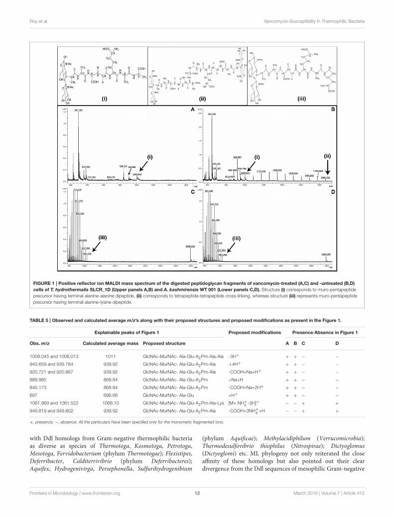
with Ddl homologs from Gram-negative thermophilic bacteriaas diverse as species of Thermotoga, Kosmotoga, Petrotoga,Mesotoga, Fervidobacterium (phylum Thermotogae); Flexistipes,Deferribacter, Calditerrivibrio (phylum Deferribacteres);Aquifex, Hydrogenivirga, Persephonella, Sulfurihydrogenibium
(phylum Aquificae); Methylacidiphilum (Verrucomicrobia);Thermodesulfovibrio thiophilus (Nitrospirae); Dictyoglomus(Dictyoglomi) etc. ML phylogeny not only reiterated the closeaffinity of these homologs but also pointed out their cleardivergence from the Ddl sequences of mesophilic Gram-negative
Frontiers in Microbiology | www.frontiersin.org 12 March 2016 | Volume 7 | Article 412
Roy et al. Vancomycin-Susceptibility in Thermophilic Bacteria
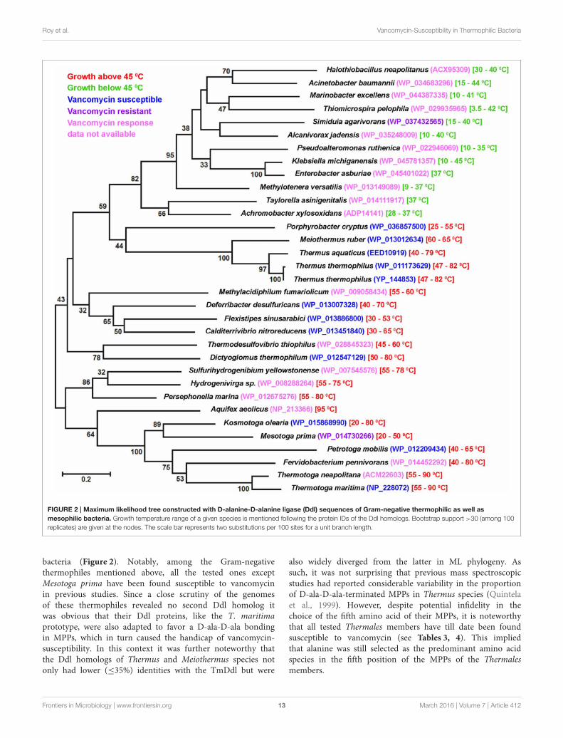
FIGURE 2 | Maximum likelihood tree constructed with D-alanine-D-alanine ligase (Ddl) sequences of Gram-negative thermophilic as well as
mesophilic bacteria. Growth temperature range of a given species is mentioned following the protein IDs of the Ddl homologs. Bootstrap support >30 (among 100
replicates) are given at the nodes. The scale bar represents two substitutions per 100 sites for a unit branch length.
bacteria (Figure 2). Notably, among the Gram-negativethermophiles mentioned above, all the tested ones exceptMesotoga prima have been found susceptible to vancomycinin previous studies. Since a close scrutiny of the genomesof these thermophiles revealed no second Ddl homolog itwas obvious that their Ddl proteins, like the T. maritimaprototype, were also adapted to favor a D-ala-D-ala bondingin MPPs, which in turn caused the handicap of vancomycin-susceptibility. In this context it was further noteworthy thatthe Ddl homologs of Thermus and Meiothermus species notonly had lower (≤35%) identities with the TmDdl but were
also widely diverged from the latter in ML phylogeny. Assuch, it was not surprising that previous mass spectroscopicstudies had reported considerable variability in the proportionof D-ala-D-ala-terminated MPPs in Thermus species (Quintelaet al., 1999). However, despite potential infidelity in thechoice of the fifth amino acid of their MPPs, it is noteworthythat all tested Thermales members have till date been foundsusceptible to vancomycin (see Tables 3, 4). This impliedthat alanine was still selected as the predominant amino acidspecies in the fifth position of the MPPs of the Thermalesmembers.
Frontiers in Microbiology | www.frontiersin.org 13 March 2016 | Volume 7 | Article 412
Roy et al. Vancomycin-Susceptibility in Thermophilic Bacteria
When our current findings were juxtaposed with a decades-old report showing accumulation of excess DL-alanine bythermophilic bacterial cells (Matsumoto et al., 1967) it provokedthe conjecture that thermophilic processes involving freedom ofamino acid choice could, in general, be skewed in favor of alanine.However, it must be acknowledged that the potential thermo-adaptive advantage (if any) conferred by D-ala-D-ala-terminatedMPPs is still not clear. But, pending biophysical verification, itseems obvious that such dipeptides were preferred by bacteria inhigh temperature environments and vancomycin susceptibilitywas just a collateral consequence of this thermodynamiccompulsion.
In this context it must also be appreciated that an apparently-universal preference for D-ala-D-ala-terminated MPPs stilldoes not guarantee vancomycin susceptibility of thermophilicbacteria as a relatively-hydrophilic molecule as large asvancomycin still has to cross the outer membrane before it caninhibit peptidoglycan biosynthesis. Notably, many mesophilicGram-negative bacteria (unlike the case of A. kashmirensisstated above) also have D-ala-D-ala-terminated MPPs, butthey still remain resistant to vancomycin due to the relativeimpermeability of their outer membrane toward this largeglycopeptide molecule (Vollmer et al., 2008; Gordon et al.,2010). As such, the above-observed vancomycin resistance ofthe other two Gram-negative mesophiles Pseudaminobactersalicylatoxidans KCT001 and E. coli BL21 could also be dueto impermeability toward vancomycin. But keeping in mindthe global vancomycin-susceptibility phenotype of thermophilicbacteria it is obvious that the drug manages to cross themembrane in all these cases. As a corollary of this, it alsoseems quite likely that the composition of the outer membranesof thermophilic bacteria have some yet-unknown characteristicfeature(s) that invariably ensures the entry of such relatively-hydrophilic large molecules as vancomycin.
With regard to the apparent preference of thermophilicbacteria for D-ala-D-ala-terminated MPPs two intriguing factsremain to be clarified at length. One is the vancomycinsusceptibility of the mesophilic but apparently thermo-enduringParacoccus strains isolated in this study and the other is thereported vancomycin resistance of at least two Gram-negativethermophilic species in the literature (viz. Mesotoga primaand Geoalkalibacter subterraneus) (Greene et al., 2009; Nesbøet al., 2012). It is noteworthy that despite its inability to growabove 45◦C, the Paracoccus strains were native to an ambienttemperature of 70–85◦C. Since such strains got frequentlyisolated over multiple rounds of sampling at the Lotus Pondspring system they cannot be discounted as mere stochasticintroductions at the time of sampling. As such, they must bemigrating intermittently from the Puga rivulet to the adjoiningLotus Pond waters and in the process getting acclimatizedto elevated temperatures. In contrast, Mesotoga prima wasisolated from an apparently mesophilic sample of unknowntemperature (viz., Baltimore harbor sediment Nesbø et al., 2012),while Geoalkalibacter subterraneus was retrieved from formation
water of an oil-well (in Redwash oilfield of Utah) that had atemperature of only 52◦C (Greene et al., 2009). Additionally,Mesotoga prima was enriched via several months of incubationand serial sub-culturing at 22 and 30◦C, whereas the currentParacoccus strains were isolated immediately upon retrieval ofhydrothermal samples to the laboratory. In the light of theabove facts vancomycin-susceptibility, and as a corollary the D-ala-D-ala-specificity of Ddl homologs, seems to be a functionof thermal-conditioning of a bacterium rather than the actualupper limit of its growth temperature. As such, it would notbe surprising if our Paracoccus isolates become vancomycin-resistant after prolonged maintenance at 28 or 37◦C, or forthat matter the Mesotoga and Geoalkalibacter strains becomevancomycin-susceptible after several sub-cultures at≥50◦C. Thiskind of subtle functional switching over short evolutionary timesdoes not seem improbable when we keep in mind the highlevels of sequence divergence existing between Ddl homologsof even closely-related genera. For example, Ddl sequences ofThermotogaceaemembers Thermotoga neapolitana (ACM22603)and Kosmotoga olearia (WP_015868990) have only 43% identity,which is quite low by any “house-keeping gene” standard.
AUTHOR CONTRIBUTIONS
CR anchored the whole work and participated in all experimentsand manuscript writing. WG conceived the program, interpretedthe results and wrote the manuscript. On site samplings weredone by CR, MA, PH, TM, SKM, and WG. CR, MA, SM,PH, SB, TM, RR, MR, RC, and SKM did metagenomics andbioinformatics analyses. Pure culture microbiology was done byCR, MA, SM, SB, and TM. Organic chemistry experiments anddata analyses were done by CR, MA, AM, RC, AN, andWG. AM,RC, AN, and SKM also contributed in improving the intellectualcontent of the work as well as the manuscript. All the authorsread and approved the final manuscript.
ACKNOWLEDGMENTS
The extensive field work and DNA sequencing involved in thispaper would have never been possible without the pro-activeespousal of the Director of Bose Institute, Professor Sibaji Raha.Financially, the work was supported by the Bose Institute as wellas the Science and Engineering Research Board, Department ofScience and Technology (DST), Government of India (GOI),with the latter grant having the number SR/FT/LS-204/2009. CRand MR received fellowships from the UGC, GOI. SM receiveda fellowship from the DST, GOI. SB was awarded a fellowship byBose Institute, DST.
SUPPLEMENTARY MATERIAL
The Supplementary Material for this article can be foundonline at: http://journal.frontiersin.org/article/10.3389/fmicb.2016.00412
Frontiers in Microbiology | www.frontiersin.org 14 March 2016 | Volume 7 | Article 412
Roy et al. Vancomycin-Susceptibility in Thermophilic Bacteria
REFERENCES
Alves, M. P., Rainey, F. A., Nobre, M. F., and da Costa, M. S. (2003). Thermomonashydrothermalis sp. nov., a new slightly thermophilic gammaproteobacteriumisolated from a hot spring in central portugal. Syst. Appl. Microbiol. 26, 70–75.doi: 10.1078/072320203322337335
Andrews, K. T., and Patel, B. K. (1996). Fervidobacterium gondwanense sp. nov.,a new thermophilic anaerobic bacterium isolated from nonvolcanically heatedgeothermal waters of the Great Artesian Basin of Australia. Int. J. Syst. Bacteriol.46, 265–269. doi: 10.1099/00207713-46-1-265
Arthur, M., Reynolds, P., and Courvalin, P. (1996). Glycopeptide resistance inenterococci. Trends Microbiol. 4, 401–407. doi: 10.1016/0966-842X(96)10063-9
Barna, J. C., and Williams, D. H. (1984). The structure and mode of action ofglycopeptide antibiotics of the vancomycin group. Annu. Rev. Microbiol. 38,339–357. doi: 10.1146/annurev.mi.38.100184.002011
Brock, T. D., and Freeze, H. (1969). Thermus aquaticus gen. n. and sp. n., anonsporulating extreme thermophile. J. Bacteriol. 98, 289–297.
Burgess, S. A., Brooks, J. D., Rakonjac, J., Walker, K. M., and Flint, S. H. (2009).The formation of spores in biofilms of Anoxybacillus flavithermus. J. Appl.Microbiol. 107, 1012–1018. doi: 10.1111/j.1365-2672.2009.04282.x
Chan, C. S., Chan, K. G., Tay, Y. L., Chua, Y. H., and Goh, K. M. (2015).Diversity of thermophiles in a Malaysian hot spring determined using 16SrRNA and shotgun metagenome sequencing. Front. Microbiol. 6:177. doi:10.3389/fmicb.2015.00177
Courvalin, P. (2006). Vancomycin resistance in gram-positive cocci. Clin. Infect.Dis. 42(Suppl. 1), S25–S34. doi: 10.1086/491711
Dekker, N. H., Viard, T., de La Tour, C. B., Duguet, M., Bensimon, D., andCroquette, V. (2003). Thermophilic topoisomerase I on a single DNAmolecule.J. Mol. Biol. 329, 271–282. doi: 10.1016/S0022-2836(03)00320-6
Denner, E. B., Kolari, M., Hoornstra, D., Tsitko, I., Kampfer, P., Busse, H. J.,et al. (2006). Rubellimicrobium thermophilum gen. nov., sp. nov., a red-pigmented, moderately thermophilic bacterium isolated from coloured slimedeposits in paper machines. Int. J. Syst. Evol. Microbiol. 56(Pt 6), 1355–1362.doi: 10.1099/ijs.0.63751-0
Edgar, R. C. (2013). UPARSE: highly accurate OTU sequences from microbialamplicon reads. Nat. Methods 10, 996–998. doi: 10.1038/nmeth.2604
Endo, A., Sasaki, M., Maruyama, A., and Kurusu, Y. (2006). Temperatureadaptation of Bacillus subtilis by chromosomal groEL replacement. Biosci.Biotechnol. Biochem. 70, 2357–2362. doi: 10.1271/bbb.50689
Fiala, G.,Woese, C., Langworthy, T., and Stetter, K. (1990). Flexistipes sinusarabici,a novel genus and species of eubacteria occurring in the Atlantis II Deepbrines of the Red Sea. Arch. Microbiol. 154, 120–126. doi: 10.1007/BF00423320
Ghosh, W., and Roy, P. (2006). Mesorhizobium thiogangeticum sp. nov., anovel sulfur-oxidizing chemolithoautotroph from rhizosphere soil of an Indiantropical leguminous plant. Int. J. Syst. Evol. Microbiol. 56(Pt 1), 91–97. doi:10.1099/ijs.0.63967-0
Goh, K. M., Gan, H. M., Chan, K. G., Chan, G. F., Shahar, S., Chong, C. S.,et al. (2014). Analysis of anoxybacillus genomes from the aspects of lifestyleadaptations, prophage diversity, and carbohydrate metabolism. PLoS ONE
9:e90549. doi: 10.1371/journal.pone.0090549Gordon, N. C., Png, K., andWareham, D.W. (2010). Potent synergy and sustained
bactericidal activity of a vancomycin-colistin combination versus multidrug-resistant strains of Acinetobacter baumannii. Antimicrob. Agents Chemother.
54, 5316–5322. doi: 10.1128/AAC.00922-10Greene, A. C., Patel, B. K. C., and Yacob, S. (2009). Geoalkalibacter subterraneus sp.
nov., an anaerobic Fe(III)- and Mn(IV)-reducing bacterium from a petroleumreservoir, and emended descriptions of the family Desulfuromonadaceae andthe genus Geoalkalibacter. Int. J. Syst. Evol. Microbiol. 59, 781–785. doi:10.1099/ijs.0.001537-0
Greene, A. C., Patel, B. K., and Sheehy, A. J. (1997). Deferribacter thermophilusgen. nov., sp. nov., a novel thermophilic manganese- and iron-reducingbacterium isolated from a petroleum reservoir. Int. J. Syst. Bacteriol. 47,505–509. doi: 10.1099/00207713-47-2-505
Guindon, S., Dufayard, J. F., Lefort, V., Anisimova, M., Hordijk, W., and Gascuel,O. (2010). New algorithms and methods to estimate maximum-likelihoodphylogenies: assessing the performance of PhyML 3.0. Syst. Biol. 59, 307–321.doi: 10.1093/sysbio/syq010
Healy, V. L., Lessard, I. A., Roper, D. I., Knox, J. R., and Walsh, C. T. (2000).Vancomycin resistance in enterococci: reprogramming of the D-ala-D-Alaligases in bacterial peptidoglycan biosynthesis. Chem. Biol. 7, R109–R119. doi:10.1016/s1074-5521(00)00116-2
Huber, R., Langworthy, T., Kanig, H., Thomm, M., Woese, C., Sleytr, U., et al.(1986). Thermotoga maritima sp. nov. represents a new genus of uniqueextremely thermophilic eubacteria growing up to 90Â◦C. Arch. Microbiol. 144,324–333. doi: 10.1007/BF00409880
Hudson, J. A., Morgan, H. W., and Daniel, R. M. (1984). Isolation andcharacterisation of a new caldoactive filamentous bacterium. FEMS Microbiol.
Lett. 22, 149–153. doi: 10.1111/j.1574-6968.1984.tb00714.xHudson, J. A., Morgan, H. W., and Daniel, R. M. (1987). Thermus filiformis sp.
nov. a Filamentous Caldoactive Bacterium. Int. J. Syst. Bacteriol. 37, 431–436.doi: 10.1099/00207713-37-4-431
Iino, T., Nakagawa, T., Mori, K., Harayama, S., and Suzuki, K. (2008).Calditerrivibrio nitroreducens gen. nov., sp. nov., a thermophilic, nitrate-reducing bacterium isolated from a terrestrial hot spring in Japan. Int. J. Syst.Evol. Microbiol. 58(Pt 7), 1675–1679. doi: 10.1099/ijs.0.65714-0
Jimenez, D. J., Andreote, F. D., Chaves, D.,Montana, J. S., Osorio-Forero, C., Junca,H., et al. (2012). Structural and functional insights from the metagenome of anacidic hot spring microbial planktonic community in the Colombian Andes.PLoS ONE 7:e52069. doi: 10.1371/journal.pone.0052069
John, W. D., Brian, C., Deborah, C., and James, F. C. (1998). Rates of spontaneousmutation. Genetics 148, 1667–1686.
Koga, Y. (2012). Thermal adaptation of the archaeal and bacterial lipidmembranes.Archaea 2012, 789652. doi: 10.1155/2012/789652
Komagata, K., and Suzuki, K.-I. (1988). “Lipid and cell-wall analysis in bacterialsystematics,” in Methods in Microbiology, eds R. R. Colwell and R. Grigorova(Orlando, FL: Academic Press), 161–207.
Kristjansson, J. K., and Stetter, K. O. (1992). “Introduction,” in Thermophilic
Bacteria, ed J. K. Kristjansson (London: CRC Press Inc.), 2–18.Kumar, S., Tsai, C. J., and Nussinov, R. (2000). Factors enhancing protein
thermostability. Protein Eng. 13, 179–191. doi: 10.1093/protein/13.3.179L’Haridon, S., Jiang, L., Alain, K., Chalopin, M., Rouxel, O., Beauverger, M., et al.
(2014). Kosmotoga pacifica sp. nov., a thermophilic chemoorganoheterotrophic
bacterium isolated from an East Pacific hydrothermal sediment. Extremophiles
18, 81–88. doi: 10.1007/s00792-013-0596-7Lien, T., Madsen, M., Rainey, F. A., and Birkeland, N. K. (1998). Petrotoga mobilis
sp. nov., from a North Sea oil-production well. Int. J. Syst. Bacteriol. 48(Pt 3),1007–1013. doi: 10.1099/00207713-48-3-1007
Lundstrom, T. S., and Sobel, J. D. (2000). Antibiotics for gram-positive bacterialinfections. Vancomycin, teicoplanin, quinupristin/dalfopristin, and linezolid.Infect. Dis. Clin. North Am. 14, 463–474. doi: 10.1016/S0891-5520(05)70258-0
Matsumoto, T., Yano, T., and Yamada, K. (1967). Studies on dl-alanineformation by thermophilic bacteria. Agric. Biol. Chem. 31, 1381–1388. doi:10.1271/bbb1961.31.1381
Menzel, P., Gudbergsdóttir, S. R., Rike, A. G., Lin, L., Zhang, Q., Contursi, P., et al.(2015). Comparative metagenomics of eight geographically remote terrestrialhot springs.Microb. Ecol. 70, 411–424. doi: 10.1007/s00248-015-0576-9
Mohagheghi, A., Grohmann, K., Himmel, M., Leighton, L., and Updegraff, D. M.(1986). Isolation and characterization of acidothermus cellulolyticus gen. nov.,sp. nov., a New Genus of Thermophilic, Acidophilic, Cellulolytic Bacteria. Int.J. Syst. Bacteriol. 36, 435–443. doi: 10.1099/00207713-36-3-435
Moreira, C., Rainey, F. A., Nobre, M. F., da Silva, M. T., and da Costa, M. S. (2000).Tepidimonas ignava gen. nov., sp. nov., a new chemolithoheterotrophicand slightly thermophilic member of the beta-Proteobacteria. Int. J.
Syst. Evol. Microbiol. 50(Pt 2), 735–742. doi: 10.1099/00207713-50-2-735
Nailor, M. D., and Sobel, J. D. (2009). Antibiotics for gram-positive bacterialinfections: vancomycin, teicoplanin, quinupristin/dalfopristin, oxazolidinones,daptomycin, dalbavancin, and telavancin. Infect. Dis. Clin. North Am. 23,965–982. doi: 10.1016/j.idc.2009.06.010
Nesbø, C. L., Bradnan, D. M., Adebusuyi, A., Dlutek, M., Petrus, A. K., Foght, J.,et al. (2012). Mesotoga prima gen. nov., sp. nov., the first described mesophilicspecies of the Thermotogales. Extremophiles 16, 387–393. doi: 10.1007/s00792-012-0437-0
Oshima, T., and Imahori, K. (1974). Description of Thermus thermophilus
(Yoshida and Oshima) comb. nov., a Nonsporulating Thermophilic Bacterium
Frontiers in Microbiology | www.frontiersin.org 15 March 2016 | Volume 7 | Article 412
Roy et al. Vancomycin-Susceptibility in Thermophilic Bacteria
from a Japanese Thermal Spa. Int. J. Syst. Bacteriol. 24, 102–112. doi:10.1099/00207713-24-1-102
Paul, S. C., Jain, P., Mitra, J., Dutta, S., Bhattacharya, P., Bal, B., et al. (2012).Induction of Cr(VI) reduction activity in an Anoxybacillus strain under heatstress: a biochemical and proteomic study. FEMS Microbiol. Lett. 331, 70–80.doi: 10.1111/j.1574-6968.2012.02555.x
Perkins, H. R., and Nieto, M. (1974). The chemical basis for the action of thevancomycin group of antibiotics. Ann. N. Y. Acad. Sci. 235, 348–363. doi:10.1111/j.1749-6632.1974.tb43276.x
Quintela, J. C., Zollner, P., Garcia-del Portillo, F., Allmaier, G., and de Pedro, M. A.(1999). Cell wall structural divergence among Thermus spp. FEMS Microbiol.
Lett. 172, 223–229. doi: 10.1111/j.1574-6968.1999.tb13472.xRainey, F. A., Silva, J., Nobre, M. F., Silva, M. T., and da Costa, M. S.
(2003). Porphyrobacter cryptus sp. nov., a novel slightly thermophilic, aerobic,bacteriochlorophyll a-containing species. Int. J. Syst. Evol. Microbiol. 53, 35–41.doi: 10.1099/ijs.0.02308-0
Reynolds, P. E. (1989). Structure, biochemistry and mechanism of action ofglycopeptide antibiotics. Eur. J. Clin. Microbiol. Infect. Dis. 8, 943–950. doi:10.1007/BF01967563
Saiki, T., Kobayashi, Y., Kawagoe, K., and Beppu, T. (1985). Dictyoglomusthermophilum gen. nov., sp. nov., a Chemoorganotrophic, Anaerobic,Thermophilic Bacterium. Int. J. Syst. Bacteriol. 35, 253–259. doi:10.1099/00207713-35-3-253
Sato, M., Kirimura, K., and Kino, K. (2006). Substrate specificity of thermostableD-alanine-D-alanine ligase from Thermotoga maritima ATCC 43589. Biosci.Biotechnol. Biochem. 70, 2790–2792. doi: 10.1271/bbb.60307
Schumann, P. (2011). “Peptidoglycan structure,” inMethods in Microbiology, ed F.Rainey and A. Oren (London: Academic Press), 101–129.
Schumann, W. (2003). The Bacillus subtilis heat shock stimulon. Cell Stress
Chaperon. 8, 207–217.Shih, T.W., and Pan, T.M. (2011). Stress responses of thermophilic Geobacillus sp.
NTU 03 caused by heat and heat-induced stress. Microbiol. Res. 166, 346–359.doi: 10.1016/j.micres.2010.08.001
Shlaes, D. M., Shlaes, J. H., Davies, J., and Williamson, R. (1989). Escherichiacoli susceptible to glycopeptide antibiotics. Antimicrob. Agents Chemother. 33,192–197. doi: 10.1128/AAC.33.2.192
Sikorski, J., Brambilla, E., Kroppenstedt, R. M., and Tindall, B. J. (2008).The temperature-adaptive fatty acid content in Bacillus simplex strainsfrom ‘Evolution Canyon’, Israel. Microbiology 154(Pt 8), 2416–2426. doi:10.1099/mic.0.2007/016105-0
Takahata, Y., Nishijima, M., Hoaki, T., and Maruyama, T. (2001).Thermotoga petrophila sp. nov. and Thermotoga naphthophila sp. nov., twohyperthermophilic bacteria from the Kubiki oil reservoir in Niigata, Japan. Int.J. Syst. Evol. Microbiol. 51(Pt 5), 1901–1909. doi: 10.1099/00207713-51-5-1901
Takai, K., Miyazaki, M., Nunoura, T., Hirayama, H., Oida, H., Furushima, Y.,et al. (2006). Sulfurivirga caldicuralii gen. nov., sp. nov., a novel microaerobic,
thermophilic, thiosulfate-oxidizing chemolithoautotroph, isolated from ashallow marine hydrothermal system occurring in a coral reef, Japan.Int. J. Syst. Evol. Microbiol. 56(Pt 8), 1921–1929. doi: 10.1099/ijs.0.64297-0
Tamura, K., Stecher, G., Peterson, D., Filipski, A., and Kumar, S. (2013). MEGA6:molecular evolutionary genetics analysis version 6.0. Mol. Biol. Evol. 30,2725–2729. doi: 10.1093/molbev/mst197
Tripathy, S., and Maiti, N. K. (2014). Construction of Geobacillusthermoglucosidasius cDNA library and analysis of genes expressed in responseto heat stress.Mol. Biol. Rep. 41, 1639–1644. doi: 10.1007/s11033-013-3011-7
Vishniac, W., and Santer, M. (1957). The thiobacilli. Bacteriol. Rev. 21, 195–213.Volker, U., Mach, H., Schmid, R., and Hecker, M. (1992). Stress proteins and cross-
protection by heat shock and salt stress in Bacillus subtilis. J. Gen. Microbiol.
138, 2125–2135. doi: 10.1099/00221287-138-10-2125Vollmer, W., Blanot, D., and de Pedro, M. A. (2008). Peptidoglycan structure
and architecture. FEMS Microbiol. Rev. 32, 149–167. doi: 10.1111/j.1574-6976.2007.00094.x
Wang, W., Ma, T., Zhang, B., Yao, N., Li, M., Cui, L., et al. (2014). A novelmechanism of protein thermostability: a unique N-terminal domain confersheat resistance to Fe/Mn-SODs. Sci. Rep. 4:7284. doi: 10.1038/srep07284
Watanakunakorn, C. (1984). Mode of action and in-vitro activity of vancomycin.J. Antimicrob. Chemother. 14(Suppl. D), 7–18. doi: 10.1093/jac/14.suppl_D.7
Waxman, D. J., and Strominger, J. L. (1983). Penicillin-binding proteins andthe mechanism of action of beta-lactam antibiotics. Annu. Rev. Biochem. 52,825–869. doi: 10.1146/annurev.bi.52.070183.004141
Wemheuer, B., Taube, R., Akyol, P., Wemheuer, F., and Daniel, R. (2013).Microbial diversity and biochemical potential encoded by thermal springmetagenomes derived from the Kamchatka Peninsula. Archaea 2013:136714.doi: 10.1155/2013/136714
Williams, R. A. D., Smith, K. E., Welch, S. G., Micallef, J., and Sharp, R. J.(1995). DNA relatedness of thermus strains, description of thermus brockianussp. nov., and Proposal To Reestablish Thermus thermophilus (Oshima andImahori). Int. J. Syst. Bacteriol. 45, 495–499. doi: 10.1099/00207713-45-3-495
Conflict of Interest Statement: The authors declare that the research wasconducted in the absence of any commercial or financial relationships that couldbe construed as a potential conflict of interest.
Copyright © 2016 Roy, Alam, Mandal, Haldar, Bhattacharya, Mukherjee, Roy,
Rameez, Misra, Chakraborty, Nanda, Mukhopadhyay and Ghosh. This is an open-
access article distributed under the terms of the Creative Commons Attribution
License (CC BY). The use, distribution or reproduction in other forums is permitted,
provided the original author(s) or licensor are credited and that the original
publication in this journal is cited, in accordance with accepted academic practice.
No use, distribution or reproduction is permitted which does not comply with these
terms.
Frontiers in Microbiology | www.frontiersin.org 16 March 2016 | Volume 7 | Article 412