Embed Size (px)
Citation preview

ARTICLE IN PRESS
1096-7176/$ - se
doi:10.1016/j.ym
�CorrespondUniversity of T
USA.
E-mail addr1Current add
Alto S3-1, 34312Current ad
Massachusetts
Cambridge MA
Metabolic Engineering 9 (2007) 8–20
www.elsevier.com/locate/ymben
Global metabolic changes following loss of a feedback loop revealdynamic steady states of the yeast metabolome
Peng Lua,c,1, Anupama Ranganc, Sherwin Y. Chanb,2, Dean R. Applingb,c, DavidW. Hoffmanb,c, Edward M. Marcottea,b,c,�
aCenter for Systems and Synthetic Biology, University of Texas, 1 University Station, A4800, Austin, TX 78712-0159, USAbDepartment of Chemistry and Biochemistry, University of Texas, 1 University Station, A5300, Austin, TX 78712-0159, USAcInstitute for Cellular and Molecular Biology, University of Texas, 1 University Station, A4800, Austin, TX 78712-0159, USA
Received 3 October 2005; received in revised form 27 May 2006; accepted 20 June 2006
Available online 30 June 2006
Abstract
Metabolic enzymes control cellular metabolite concentrations dynamically in response to changing environmental and intracellular
conditions. Such real-time feedback regulation suggests the global metabolome may sample distinct dynamic steady states, forming
‘‘basins of stability’’ in the energy landscape of possible metabolite concentrations and enzymatic activities. Using metabolite, protein
and transcriptional profiling, we characterize three dynamic steady states of the yeast metabolome that form by perturbing synthesis of
the universal methyl donor S-adenosylmethionine (AdoMet). Conversion between these states is driven by replacement of serine with
glycine+formate in the media, loss of feedback inhibition control by the metabolic enzyme Met13, or both. The latter causes
hyperaccumulation of methionine and AdoMet, and dramatic global compensatory changes in the metabolome, including differences in
amino acid and sugar metabolism, and possibly in the global nitrogen balance, ultimately leading to a G1/S phase cell cycle delay. Global
metabolic changes are not necessarily accompanied by global transcriptional changes, and metabolite-controlled post-transcriptional
regulation of metabolic enzymes is clearly evident.
r 2006 Elsevier Inc. All rights reserved.
Keywords: Metabolomics; Metabolic profiling; Nuclear magnetic resonance; Mass spectrometry; One carbon metabolism
1. Introduction
Living organisms are characterized by the quality ofhomeostasis, the tendency to maintain a constant internalstate in response to varying external conditions. Thistendency manifests itself particularly strongly in cellularmetabolism. Such metabolic self-regulation leads not toequilibrium (cells are not closed systems), but to theadoption of dynamic steady states that represent the
e front matter r 2006 Elsevier Inc. All rights reserved.
ben.2006.06.003
ing author. Department of Chemistry and Biochemistry,
exas, 1 University Station, A4800, Austin, TX 78712-0159,
ess: [email protected] (E.M. Marcotte).
ress: Departments of Genetics and Genomics, Roche, Palo
Hillview Ave., Palo Alto, CA 94304, USA.
dress: Whitehead Institute for Biomedical Research,
Institute of Technology, Nine Cambridge Center,
02142-1479, USA.
simultaneous satisfaction of all of the constraints presentin the metabolic system (Fell, 1996). When components ofsuch a system are perturbed, the system will re-equilibrateto a distinct dynamic steady state, adjusting all othercomponents and rates to best accommodate the perturba-tion. On a rapid time scale, cells dynamically respond tothe changing availability of metabolites by optimizingmetabolite concentrations and enzyme activities in order tominimize the overall energy of the system, seeking out‘‘basins of attraction’’ in a complex energy landscape.In this paper, we describe the detailed experimental
characterization of three such basins of stability in cellularmetabolism, which occur as reproducible dynamic steadystates of the cellular metabolic machinery, observed inresponse to perturbations to the synthesis of the universalmethyl donor, S-adenosylmethionine (AdoMet). AdoMetis involved in methylating diverse substrates ranging fromDNA and RNA to small molecules and amino acids, such

ARTICLE IN PRESSP. Lu et al. / Metabolic Engineering 9 (2007) 8–20 9
as the lysine and arginine residues of histones, and isproduced by activating methionine with ATP (Fontecaveet al., 2004). We have investigated a system in which yeastcells can be forced to overproduce AdoMet by manipulat-ing flux through the MET13-encoded methylenetetrahy-drofolate reductase (MTHFR) enzyme. MTHFR catalyzesthe reduction of 5,10-methylenetetrahydrofolate to5-methyltetrahydrofolate, used to methylate homocysteineduring methionine biosynthesis (Fig. 1). This is thecommitted step in methyl group biogenesis, with5-methyltetrahydrofolate providing all the methyl groupsof AdoMet in these cells.
We took advantage of a previously identified chimericMTHFR (Chimera-1) composed of the yeast Met13pamino-terminal catalytic domain and the Arabidopsis
thaliana MTHFR (AtMTHFR-1) carboxy-terminal regu-latory domain (Roje et al., 2002). The resulting chimera isfully enzymatically active, but lacks the AdoMet-mediatedfeedback inhibition of the wild-type yeast enzyme andgains the ability to use reduced nicotinamide-adeninedinucleotide (NADH) as co-factor. Placement of Chi-mera-1 in a serine auxotroph (DAY4) produced anengineered strain (SCY4) with interesting properties: Cellsgrown in minimal media, but provided with serine, appearnormal. However, replacement of serine with glycine andformate requires the cells to direct metabolic flux throughthe folate-mediated one-carbon pathways in order to meettheir serine requirements (Fig. 1), leading to accumulationof greater than 100-fold more AdoMet and 7-fold moremethionine than the wild type strain (Roje et al., 2002).These changes are accompanied by general reduction ingrowth rate of the cell (Chan and Appling, 2003), raisingthe question of what general molecular changes occur when
(A) (B)
Fig. 1. Methyl group biogenesis in wild-type and engineered yeast strains.
(A) In the wild-type strain DAY4 expressing Met13p, the MTHFR
reaction is irreversibly driven towards CH3-THF formation by a high
cytosolic NADPH/NADP ratio. AdoMet overaccumulation is prevented
by allosteric feedback inhibition of the MTHFR reaction (dotted line). (B)
In strain SCY4 expressing the Chimera-1 MTHFR, AdoMet accumulates
due to the lack of feedback inhibition by AdoMet. In addition, the
capacity of the chimeric MTHFR to use NADPH as well as NADH
results in more CH3–THF synthesis and hence even greater AdoMet
accumulation.
the nutritional status of these yeast is varied. By acombination of metabolic, transcriptional, and proteomicprofiling, we characterize the phenotypes associated withthese cells and demonstrate a reproducible global change inmetabolic state upon removal of this single metabolicfeedback loop, only poorly reflected in the accompanyingtranscriptional response. In total, we define three discretedynamic steady states adopted by the yeast metabolome inresponse to variations in nutrition and one-carbonmetabolism.
2. Materials and methods
2.1. Yeast strains and growth conditions
The Saccharomyces cerevisiae strains used wereDBY8724 (MATa GAL2 ura3 bar1::URA3) (Spellman etal., 1998), DAY4 (MATa ser1 ura3-52 trp1 leu2 his4)(Rojeet al., 2002) and SCY4 (MATa ser1 ura3-52 trp1 leu2 his4
Dmet13::CHIMERA-1) (Roje et al., 2002). Yeast weregrown in synthetic minimal medium (YMD medium)containing 0.7% yeast nitrogen base without aminoacids (DIFCO Bacto), 2% glucose, supplemented withthe following where indicated: L-serine (375mg/L),L-leucine (30mg/L), L-histidine (20mg/L), L-tryptophan(20mg/L), uracil (20mg/L), glycine (20mg/L), and formate(250mg/L).
2.2. Metabolite extraction
Typically, 500ml yeast cultures were harvested at mid-log phase, washed with PBS and re-suspended in 4%perchloric acid (50% wt/vol) (Shryock et al., 1986) with9 mmol DSS (2,2-dimethyl-2-silapentane-5-sulfonic acid) asan internal reference. Samples were lysed by freezing andthawing twice, neutralized with KOH, and then centri-fuged. The supernatants were dried by speed-vacuum andre-suspended in D2O (Cambridge Isotope Laboratories,Inc.) for analysis. For the in vivo metabolite analysis, 1.5 LDBY8724 yeast cells were grown to mid-log phase. About500ml cells were used for soluble metabolome extraction;the remaining 1L yeast were pelleted, washed, re-suspended, loaded directly into the NMR tube withoutlysis and analyzed intact. Comparison of 2D-NMR spectrafrom extracted metabolites with 2D-NMR spectra col-lected from intact yeast cells suggests that only minimalbias in the metabolite population is introduced by thesample preparation (data not shown).
2.3. NMR data acquisition, processing and resonance
assignments
NMR spectra were recorded at 30 1C using a 500MHzVarian Inova spectrometer equipped with a tripleresonance probe and z-axis pulsed-field gradient. Reso-nances were assigned using proton detected correlatedspectroscopy (COSY and TOCSY), carbon 1D spectra,
ARTICLE IN PRESSP. Lu et al. / Metabolic Engineering 9 (2007) 8–2010
and 1H-13C single-bond correlated HSQC spectra. Peaksused in the quantitative metabolite analyses were pickedfrom 2D 1H-13C HSQC spectra acquired using z-axispulsed field gradients for coherence selection, 1H and 13Csweep widths of 6000 and 25000Hz, respectively, and 13Cdecoupling during the acquisition time. Spectra wereacquired using 16–64 scans per FID, 2048 and 512 pointsfor the 1H and 13C dimensions, and total spectrumacquisition times of 2.75–11 h. Tentative NMR assign-ments were confirmed by acquiring spectra of samplesspiked with purified compounds. 2D-NMR spectrawere processed with NMRPipe (Delaglio et al., 1995)and interpreted with the aid of the Sparky Assignmentand Integration Software package (http://www.cgl.ucsf.edu/home/sparky). After all peaks were picked, assignedand integrated, peak heights were normalized by thepeak heights of the DSS internal reference standardand the wet sample weights. Linearity of responsewas tested by adding analytes of known concentrationto wild-type yeast extracts and collecting correspondingdata. Following each addition, peak heights werenormalized using an internal DSS control to correctfor minor sample volume variations. Clusteringand principle component analysis of metabolite profileswere performed with the programs Cluster and Treeview(Eisen et al., 1998).
2.4. mRNA and protein preparation
Proteins and mRNA were harvested from the yeaststrains DAY4 and SCY4, isolating mRNA and proteinsfrom biological replicate yeast cultures grown underthe same conditions. Starter cultures were grown inYMD medium containing serine, diluted to O.D. 0.05 ineither the same serine-containing medium or YMDmedium containing glycine and formate, then grown tomid-log phase at 30 1C. From one set of the cultures, totalRNA was extracted using phenol/chloroform, poly(A)RNA was purified, and cDNA was synthesized byreverse-transcription as described previously (Spellmanet al., 1998). Protein was harvested from the other setof cultures via bead-beating as described previously (Xueet al., 2000).
2.5. DNA microarray analysis
The synthesized cDNA was hybridized to a DNAmicroarray containing all of the intergenic and predictedcoding regions of the yeast genome manufactured asdescribed previously (Hahn et al., 2004; Kim and Iyer,2004). The resulting microarrays were scanned with aGenePix 4000B scanner (Axon Instruments) and quantifiedwith GenePix 5.0 software. Data were uploaded into theLAD microarray database (Killion et al., 2003) and filteredto pass minimum quality control thresholds (sum ofmedian intensities 4300 and regression correlation acrossspot pixels 40.6) before further analysis.
2.6. LC/LC/MS/MS analysis
Soluble protein extracts were diluted in digestion buffer(50mM Tris HCL pH 8.0, 1.0M Urea, 2.0mM CaCl2) to4mg/ml, denatured at 95 1C for 10min, and digested withsequencing grade trypsin (Sigma) at 37 1C for 20 h. Trypticpeptide mixtures were separated by automated two-dimensional high-performance liquid chromatography.Chromatography was performed at 2 ml/min with allbuffers acidified with 0.1% formic acid. Chromatographysalt step fractions were eluted from a strong cationexchange column (SCX) with a continuous 5% acetonitrile(ACN) background and 10min salt bumps of 0, 20, 60 and900mM ammonium chloride. Each salt bump was eluteddirectly onto a reverse phase C18 column and washed freeof salt. Reverse phase chromatography was run in a125min gradient from 5% to 55% ACN, and then purgedat 95% ACN.Peptides were analyzed online with electrospray ioniza-
tion (ESI) ion trap mass spectrometry (MS) (Link et al.,1999; Washburn et al., 2001) using a ThermoFinniganSurveyor/DecaXP+ instrument. In each MS spectrum, thesix tallest individual peaks, corresponding to peptides, werefragmented by collision-induced dissociation (CID) withhelium gas to produce MS/MS spectra. Gas phasefractionation (GPF) was used to achieve maximumproteome coverage (Yi et al., 2002). In order to increasecoverage of lower abundance proteins, each tryptic peptidemixture was analyzed by three sequential LC/LC/MS/MSanalyses, in each case examining a different mass/charge(m/z) range (300–650, 650–900, and 900–1500 m/z) fordata-dependent precursor ion selection for CID; fragmen-tation data from the three runs were then combined foranalysis. Proteins were identified from the resulting peptideMS/MS fragmentation spectra using Bioworks TurboSe-quest (Eng et al., 1994), PeptideProphet (Keller et al.,2002), and ProteinProphet (Nesvizhskii et al., 2003). Theprobability of correctly identifying each protein and itsindividual spectral counts in a given proteomics experimentwas calculated using ProteinProphet.For the purpose of generating clusters in Fig. 6, a
ProteinProphet probability threshold of 0.2 was chosen forprotein identification, and spectral counts were treated as arough, order-of-magnitude measure of protein expressionlevels (Liu et al., 2004). Differential protein expression wascalculated using the APEX scheme (Lu et al., manuscriptsubmitted) as a Z-score based on numbers of spectralcounts (ni,1 and ni,2) observed for a given protein i betweentwo experiments with N1 and N2 total spectral counts each,calculated as
Z ¼f i;1 � f i;2ffiffiffiffiffiffiffiffiffiffiffiffiffiffiffiffiffiffiffiffiffiffiffiffiffiffiffiffiffiffiffiffiffiffiffiffiffiffiffiffiffiffiffiffiffiffiffiffiffiffiffiffiffiffiffiffiffiffiffiffiffiffiffiffiffiffiffiffiffiffiffi
f i;0ð1� f i;0Þ=N1 þ f i;0ð1� f i;0Þ=N2
p ,
where fi,1 and fi,2 are the fractions of spectral countsfor protein i in experiments 1 and 2 (equal to ni,1/N1
and ni,2/N2, respectively), and the denominator represents
ARTICLE IN PRESSP. Lu et al. / Metabolic Engineering 9 (2007) 8–20 11
the standard error of the difference under the nullhypothesis in which the two sampled proportions aredrawn from the same underlying distribution with theoverall proportion f i;0 ¼ ðni;1 þ ni;2Þ=ðN1 þN2Þ. Proteinswith |Z|42.58 and 41.96 were considered significantlydifferentially expressed at the 99% and 95% confidencelevels, respectively.
2.7. FACS analysis
The yeast cells were grown in YMD media containingserine or glycine and formate. Cell cultures were harvestedat mid-log phase, then assayed for DNA content bystaining with the DNA-specific dye propidium iodide,followed by flow cytometry with a BD BiosciencesFACSCalibur instrument using standard protocols.
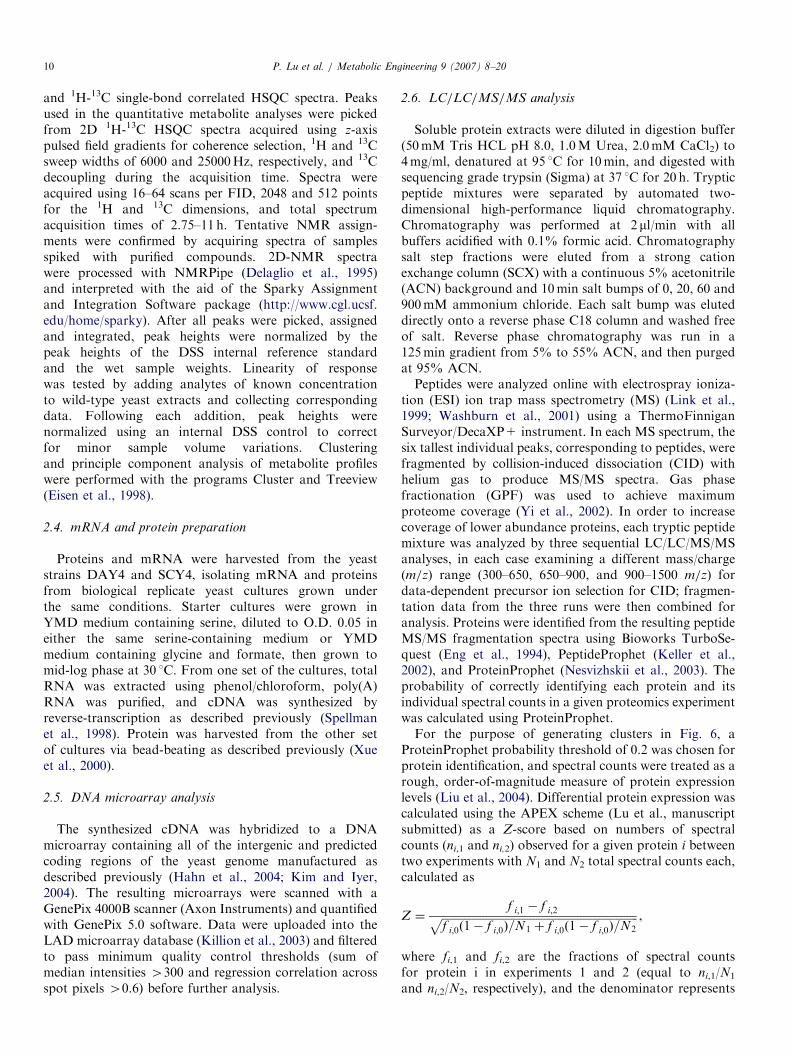
Fig. 2. Metabolic profiling by 2D-NMR. Sections are plotted of an 1H-13
Assignments of labeled compounds were verified by spiking experiments. Pea
complete list of resonance assignments and chemical shifts is provided as Sup
3. Results and discussion
3.1. Quantifying metabolites by 1H-13C 2D-NMR
In order to metabolically profile the yeast cells as theirgrowth conditions and nutritional status were varied, weapplied 1H-13C two-dimensional nuclear magnetic reso-nance (2D-NMR). In comparison with 1H-NMR (Raams-donk et al., 2001; Viant et al., 2003), 1H-13C multinuclearNMR (Szyperski et al., 1999; Lindon et al., 2003) greatlyreduces the overlap between peaks in each spectrum (Viant,2003), allowing their accurate identification and quantita-tion. Fig. 2 shows sections of the 1H-13C 2D-NMRspectrum of a DAY4 yeast cell extract. 200 NMR peakswere consistently observed across all spectra. In order topositively identify the corresponding compounds, we
C correlated HSQC NMR spectrum of the DAY4 yeast metabolome.
ks labeled by P# are measured but unassigned to specific compounds. A
plemental Table 1.
ARTICLE IN PRESSP. Lu et al. / Metabolic Engineering 9 (2007) 8–2012
assigned the resonances using COSY, TOCSY andcarbon 1D experiments in addition to 1H-13C 2D-NMR.Altogether, identities of 94 peaks were assigned andcorresponded to 30 distinct metabolites, including aminoacids, monosaccharides, nucleotides and variousother small molecules. The remaining 106 peaks wereconsistently observed and measured but unassigned,indicating that a total of �60–136 total metaboliteswere monitored by this approach. The higher value ismore probable as no cross-correlations were observedamong the unassigned peaks in COSY and TOCSYexperiments, implying these peaks derive from distinctmetabolites. The complete set of resonance assignments,each verified by spiking experiments, is listed as Supple-mentary Table 1.
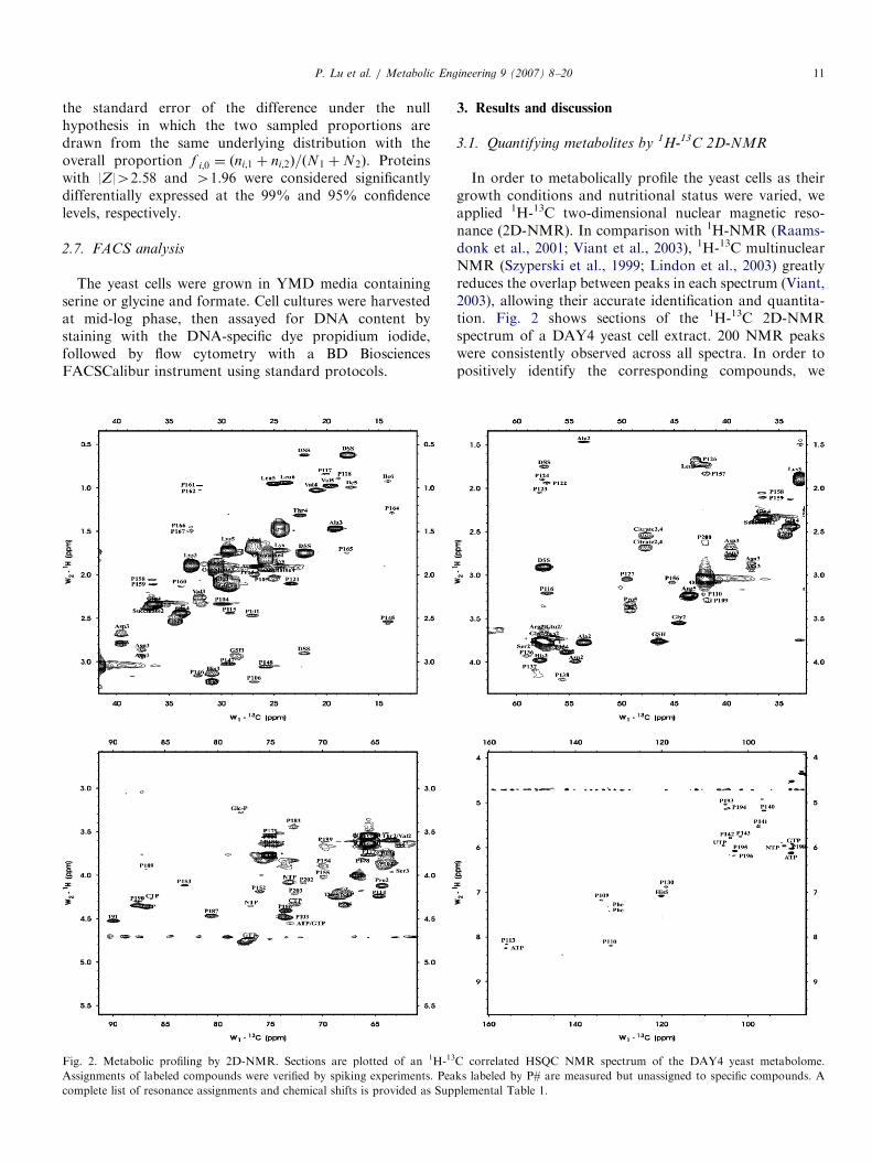
To be useful for metabolite profiling, 1H-13C 2D-NMRmust be both quantitative and reproducible. To test thecorrelation between 1H-13C 2D-NMR signal intensities andmetabolite abundance, we spiked yeast cell extracts withknown amounts of reference compounds and measured thechanges in metabolite peak heights. As shown in Fig. 3A,the NMR peak heights vary linearly with the abundance ofmetabolites in the sample and this linear relationship existsover at least three orders of magnitude of concentration.Tests with amino acids, carbohydrates, nucleotides and
Fig. 3. Linearity and reproducibility of the 2D-NMR metabolite measurem
concentration over at least three orders of magnitude and are highly reproducib
shows that the 2D-NMR peak heights increase linearly with the amount of
concentration curve reveals the absolute amount of histidine present in the orig
an absolute cellular concentration of �500mM histidine. (B) Comparison of the
replicate metabolome samples (2400 comparisons in total) demonstrates the hig
a peak is not observed in a replicate experiment (falling here on the x and y ax
2400 observations, or 2%, for this set of NMR peaks.
other metabolites gave similar results (data not shown).From spiking experiments such as these, the absoluteamount of a metabolite in the sample can be obtained(Fig. 3A), and by correcting for cell mass, the absolutecellular concentration of the metabolite can be estimated.For example, we observed �76 mg histidine in wild-typeyeast samples—based on the cell mass analyzed (�0.6 g),this implies the cellular histidine concentration is roughly0.5mM, assuming the accessible volume of a typical yeastcell is 0.6ml/g (Arnold and Lacy, 1977). Previouslymeasured concentrations of cellular histidine are compar-able (1.6mM (Strathern et al., 1982)) within the varianceexpected between different yeast strains. The estimatedsensitivity of 1H-13C 2D-NMR based on detection of theinternal control is �50 mM, using a conventional 500MHzspectrometer, the natural abundance of 13C, and a datacollection time of 11 h. To examine reproducibility, wecompared quantitation of NMR peak heights from 12 pairsof biological replicate metabolome samples (2400 peakpairs in total), which is shown in Fig. 3B. Quantitation washighly reproducible (R2 ¼ 0:95), demonstrating that thevariability introduced by sample preparation and 1H-13C2D-NMR measurement is minimal and compares favor-ably to metabolic profiling by gas chromatography/massspectrometry (Fiehn et al., 2000).
ents. Peak heights in 2D NMR spectra vary linearly with metabolite
le: (A) Spiking of a yeast lysate with known amounts of three amino acids
compound added. In the right plot, the x-intercept of the peak height-
inal sample. Correcting for the estimated cell mass in the sample tube gives
measured peak heights for 200 2D-NMR peaks from 12 pairs of biological
h reproducibility of 2D-NMR metabolic profiling. Note that, occasionally,
es), placing the false negative observation rate of the technique at �42 of

ARTICLE IN PRESSP. Lu et al. / Metabolic Engineering 9 (2007) 8–20 13
3.2. Detailed metabolome analysis of a yeast metabolic
mutant
In order to gain insight into the metabolic states of theone carbon metabolism mutants, wild type MET13 yeast(DAY4) and the Chimera-1 mutant SCY4 were grownseparately to mid-log phase in serine or glycine+formatemedia and their lysates were analyzed by 1H-13C 2D-NMR.The data were then normalized and organized viahierarchical clustering, an approach common for micro-array-based mRNA expression surveys (Eisen et al., 1998).The metabolic profiles are plotted in Fig. 4, with both rowsand columns ordered according to the hierarchical cluster-ing of metabolic profiles and sample profiles, respectively.
Fig. 4. Variation between the cells’ internal metabolic states is demonstrated
profiles in different nutritional states. Each row corresponds to a single 2D
conditions (each strain/condition analyzed in triplicate). The normalized ma
indicating an increase in metabolite abundance compared with the median
corresponding decrease in abundance, and black for the median level of abund
apparently derive from feedback inhibition of cysteine on homocysteine synthes
data of low quality were filled by the NMR peak background threshold level.
Several broad patterns are evident in the metabolomes.First, replicate samples tend to have similar metabolomes(columns in Fig. 4), indicated by their tendency to clustertogether by their metabolic profiles. Second, the primarydivision between the samples correlates with their growthmedia, indicating that nutritional status plays the domi-nant role in determining the metabolomes. Third, in serinemedium, there are only subtle differences between themetabolite profiles of DAY4 and SCY4, while thedifferences become much more prominent in the glycine+formate medium. This correlates with the previousobservation that the growth rates of these two strainswere similar in serine minimal medium, yet the doublingtime of SCY4 was nearly twice as long as DAY4 in
by unsupervised hierarchical clustering of DAY4 and SCY4 metabolic
-NMR metabolite peak, with columns representing strains/experimental
gnitude of each 2D-NMR peak height is indicated by color, with red
abundance of that metabolite across all samples, green indicating a
ance. Variations within SCY4 glycine+formate replicates (green arrows)
is (Chan and Appling, 2003), diagrammed on the top right. Absent data or

ARTICLE IN PRESSP. Lu et al. / Metabolic Engineering 9 (2007) 8–2014
glycine+formate medium (Chan and Appling, 2003).Fourth, hierarchical clustering of the metabolites by theirabundance profiles across samples (rows in Fig. 4) indicatesthat NMR peaks derived from the same metabolites clustertogether, demonstrating the accuracy and reproducibilityof 1H-13C 2D-NMR for metabolomic profiling. Asmetabolites responding in a similar fashion across samplesalso cluster together, the hierarchical clustering capturesmajor trends in metabolic flux between the samples, asdiscussed later in more detail.
As expected, engineered yeast (SCY4) expressing thechimeric MTHFR are insensitive to AdoMet inhibition invivo and accumulate much more AdoMet and methioninethan the wild type (Roje et al., 2002). However, thisphenomenon was not observed when the supplementalglycine and formate were replaced by serine in the culturemedium (Chan and Appling, 2003). Correspondingly, theAdoMet peak is large in the SCY4-Gly+For spectra, butundetectable in DAY4-Gly+For, DAY4-Ser and SCY4-Ser spectra. Likewise, the methionine peak height in SCY4-Gly+For spectra is about 5.5-fold higher than that inDAY4-Gly+For spectra; there is no significant differencein methionine abundance between these two strains in theserine medium.
Fig. 5. Summary of global metabolome changes to yeast one-carbon metabol
aminotransferase in both DAY4 and SCY4 strains; red * indicates chimeric M
line indicates the regulation existing in both DAY4 and SCY4 strains; dashed r
the normalized abundances of measured metabolites, averaged across three
Gly+For media.
The relative abundance changes in known metabolitesacross the experiments are summarized in Fig. 5, and anumber of more detailed trends in the cells’ metabolomescan be seen, in particular in amino acid metabolicpathways. For example, lysine was elevated in theglycine+formate-enriched medium, especially in DAY4cells. Lysine and homocysteine represent the two majordownstream branches of the aspartate biosynthesis path-way. Aspartate was abundant in both DAY4-Gly+Forand SCY4-Gly+For samples, suggesting that the aspartatepathway is up-regulated when glycine is provided as a C1
carbon source. In SCY4, the AdoMet-insensitive chimericMTHFR ensures high levels of CH3-THF for themethylation of homocysteine to methionine and AdoMet(Roje et al., 2002). In wild type cells, AdoMet feedbackinhibits MTHFR activity so that CH3-THF levels are low,and thus homocysteine methylation is slowed. As flux frompyruvate to aspartate increases, alanine, valine andisoleucine synthesis from pyruvate are decreased. Acetateis also higher in the glycine+formate-grown cells.Although acetate can be produced from pyruvate, it isalso generated from threonine. Threonine, which derivesfrom aspartate via homoserine, was also elevated in SCY4-Gly+For cells. Threonine is then cleaved by threonine
ism and related pathways. Red X indicates the inactivated phosphoserine
THFR (Methylenetetrahydrofolate reductase) in SCY4 strain; solid black
ed line indicates the regulation only existing in DAY4 strain. All bars show
biological replicates for SCY4 and DAY4 cells growing in either Ser or

ARTICLE IN PRESS
Fig. 6. Transcriptional and proteomic profiles of DAY4 and SCY4 in
different nutritional states, shown by unsupervised hierarchical clustering.
In the transcriptional profiles (left diagram), rows correspond to genes and
columns to strains/experimental conditions. Colors indicate the magnitude
of induction/repression for each gene relative to the pooled mRNA from
all samples. In the proteomic profiles (right diagram), rows correspond to
proteins columns to strains/experimental conditions. Colors indicate the
spectral counts of each protein, an approximate measure of protein
abundance (Liu et al., 2004), normalized by the protein’s median
abundance across all samples. In both plots, red indicates an increase in
abundance compared with the median across the samples, green indicates
a decrease in abundance, and black indicates the median abundance.
Missing data are plotted in gray. In each plot, only genes/proteins
observed in at least three of the four samples are plotted.
P. Lu et al. / Metabolic Engineering 9 (2007) 8–20 15
aldolase to produce glycine and acetaldehyde, which is inequilibrium with acetate (McNeil et al., 1994; Woldman andAppling, 2002). Thus, it appears that in glycine+formatemedium, flux from aspartate towards lysine and homoserineincreases, leading to increased threonine and homocysteine(and subsequently AdoMet) synthesis.
A second major trend is revealed by considering thelevels of histidine. Histidine also contributes C1 unitsthrough its degradation with formation of N5-forminino-THF. Since SCY4 has an unregulated MTHFR and thereis no serine inhibition in glycine+formate medium, all ofthe formate is taken up through the ADE3 gene product tomake 5,10-CH2-THF and ultimately, 5-CH3-THF. As5,10-methenyl-THF is an intermediate between formateand 5,10-CH2-THF, it is possible that SCY4, which mayhave greater flux through Ade3p, might decrease the use ofhistidine in order to make 5,10-methenyl-THF in theglycine+formate medium.
As reported by Chan and Appling (2003), serinerepresses AdoMet hyperaccumulation in SCY4 by a varietyof mechanisms. Serine can deplete the homocysteine poolby condensing with homocysteine to form cystathionine,thereby limiting the amount of methionine and AdoMetsynthesized. In addition, serine acts to repress thebiosynthesis of homocysteine through its role in thesynthesis of cysteine (Vanaerts et al., 1994; Chan andAppling, 2003). When serine is absent in the medium, it canonly be synthesized via serine hydroxymethyltransferase(SHMT) because the conversion of 3-phosphoglycerate toserine is blocked at phosphoserine aminotransferase inthese strains. Therefore, in glycine+formate medium, thelow intracellular serine levels (Fig. 5) are apparentlyadequate to meet growth requirements, but not highenough to repress AdoMet synthesis.
We also observe potential changes in the global nitrogenbalances of the cells, with arginine levels favored overornithine in the SCY4-Gly+For cells and glutaminefavored over glutamate. However, this ratio is invertedwith aspartate and asparagine, arguing that a differentmechanism may also account for the skewed ratios ofglutamine/glutamate and arginine/ornithine.
3.3. Combination of transcriptome, proteome and
metabolome
To test if changes in the metabolome were accompaniedby changes in the transcription and translation of meta-bolic enzymes or regulated proteins [e.g., as in Ideker et al.(2001)], we profiled the cells’ transcriptomes and proteomeswith DNA microarrays (Lockhart and Winzeler, 2000) andmass spectrometry (Aebersold and Mann, 2003), respec-tively. We expected that a comparison of data across thethree levels would allow us to better describe the cells’states, characterizing the entire biological system morecomprehensively and giving insight into the extent to whichperturbation- or nutrition-induced changes in gene expres-sion and metabolite levels were correlated. We profiled
�800 proteins expressed by these cells with high confidencevia the use of ‘‘MudPit’’ style (Washburn et al., 2001)multi-dimensional high-performance liquid chromatogra-phy and tandem mass spectrometry (LC/LC/MS/MS), andprofiled the complete set of mRNA transcripts via the useof cDNA microarrays (Iyer, 2003; Hahn et al., 2004; Kimand Iyer, 2004). The complete sets of transcript and proteinprofiles are listed in Supplemental Tables 2 and 3,respectively. Fig. 6 shows the resulting transcriptionaland proteomic profiles after hierarchical clustering. As forthe metabolite profiles, the primary division among thesamples at the transcriptome and proteome levels is bynutritional status. However, the transcriptomes of theSCY4 and DAY4 appear more similar than either theirproteomes or metabolomes would imply, consistent withthe critical role of post-transcriptional regulation for both

ARTICLE IN PRESSP. Lu et al. / Metabolic Engineering 9 (2007) 8–2016
protein and metabolite levels (Parekh and Rohlff, 1997;Griffin et al., 2002).
Such post-transcriptional regulation is indeed evident inthe profiling data. The MTHFR feedback inhibition is oneexample. Another such example, illustrated in Fig. 7, isthat of the arginine biosynthesis enzyme, carbamoylpho-sphate synthase (CPSase A). CPSase A is composed of twosubunits, CPA1 and CPA2, known to be subject to twodifferent regulatory mechanisms (Messenguy et al., 1983).While transcription of both the CPA1 and CPA2 mRNAsare regulated by the transcription factor GCN4 in responseto general amino acid starvation, the CPA1p protein isadditionally repressed by arginine via upstream openreading frames (Werner et al., 1987; Delbecq et al., 1994).At the mRNA level, we see no obvious change in themRNA abundance of either CPA1 or CPA2 in serinemedia relative to glycine+formate media (Fig. 7). At theprotein level, the CPA2 protein is present in bothconditions (measured as the probability of identification).By contrast, the CPA1 protein, while clearly detectable inserine media, is undetectable in glycine+formate media.(Here, we rely upon the tendency for confidence in proteinidentification to serve as a surrogate for protein abundance(Ishihama et al., 2005; Wang et al., 2005); more formally, itindicates a lack of positive evidence for the presence ofCPA1.) Thus, it appears CPA1 is down-regulated at a post-transcriptional level in the SCY4 glycine+formate cells.The loss of CPA1 protein agrees with the increase inconcentration (roughly doubling) of arginine in the cells,consistent with the known mode of CPA1 regulation(Werner et al., 1987; Delbecq et al., 1994).
Nonetheless, the primary transcriptional and transla-tional changes do pertain specifically to metabolism. Ineither the SCY4 or DAY4 cells, when switched between Serand Gly+For media, mRNAs differentially expressedmore than 2-fold are entirely dominated by amino acidbiosynthetic enzymes, statistically enriched (Po10�9,calculated via FunSpec (Robinson et al., 2002)) for geneswith the Gene Ontology annotations amino acid metabo-lism, amino acid and derivative metabolism, aminemetabolism, amino acid biosynthesis, and amine biosynth-esis, with both strains showing differential expression of,
Fig. 7. Detection of metabolite-controlled post-transcriptional regulation in t
approx. twice that in serine media (right graph). While the mRNA levels o
conditions (shown at left), the protein is observed only in the serine media, i.e
CPA1 protein levels (Messenguy et al., 1983). By contrast, the arginine biosynth
for example, ARO3, ARO9, ARO10, TRP4, GLY1, SER3,
STR3, ARG4, PUT2, ECM40, HIS3, HIS5, and CAR1.
Additionally, DAY4 cells show differential expression ofgenes of sulfur amino acid metabolism, methioninemetabolism and sulfur metabolism (in particular,MET32, HOM3, CYS4, MET14, MET1, MET17, MET2,
and MET22), and aspartate family amino acid metabolism.The only significant (Po10�6) expression change betweenSCY4 and DAY4 cells grown in Gly+For are four genesof one-carbon compound metabolism (GCV1, GCV2,
FDH1, and FDH2). No significantly different GO biolo-gical processes were detected among genes expressed morethan 2-fold between DAY4 and SCY4 cells grown in Sermedia, a trend also reflected by their similar metabolicprofiles.At the protein level, the predominant difference in the
proteomes of SCY4 and DAY4 cells in Ser media are notdue to metabolic proteins, but instead in the proteinsynthesis apparatus, with the most strongly differentiallyexpressed proteins (Po10�14 for proteins differentiallyexpressed at the 99% confidence level) corresponding tothe Gene Ontology biological process annotation proteinbiosynthesis. Likewise, at the protein level, the strongestdifferentially expressed pathway observed between SCY4and DAY4 cells grown in Gly+For media is again notmetabolic, but a difference in protein folding (Po10�6), inparticular the proteins SSA1, HSC82, and HSP82,presumably a reflection of the growth difficulties experi-enced by the SCY4 strain. However, within either strain,the shift from Ser to Gly+For brings predominantlychanges in metabolic protein expression levels, with eitherstrain showing elevated SER3, SER33, and ARG4 proteinlevels in Gly+For media, and elevated ILV5, LYS21,ALD6, and ADH1 protein levels in Ser media. Thesechanges are detailed in full in Supplemental Table 3.
3.4. Hyperaccumulation of AdoMet delays the G1-to-S
phase cell cycle transition
AdoMet is the most commonly used enzyme cofactorafter ATP, the nicotinamide dinucleotides, and CoA(Forster et al., 2003), and the alteration of its cellular
he SCY4 strain. In glycine+formate media, the arginine concentration is
f arginine biosynthesis gene CPA1 are essentially unchanged in the two
., when arginine is low, consistent with post-transcriptional regulation of
esis gene CPA2 shows no such arginine-mediated translational repression.

ARTICLE IN PRESSP. Lu et al. / Metabolic Engineering 9 (2007) 8–20 17
concentration has profound effects on cell growth althoughthe detailed mechanism producing these effects is stillunclear. In order to better characterize the defect asso-ciated with AdoMet overproduction, we attempted toexplain the increased doubling time of the SCY4 glycinecells [roughly twice that of DAY4 glycine cells (Chan andAppling, 2003)] by searching for a cell cycle-relateddefect. As expression deconvolution analysis (Lu et al.,2003) can in principle reveal the precise nature of cell cycledefects in mutant cells, we analyzed the mRNA expressionprofiles of DAY4 and SCY4 cells to search for patternstypical of cell cycle defects. As illustrated in Fig. 8A,DAY4 cells showed transcriptional profiles characteristicof roughly equal proportions of cells in different cell cyclephases, but SCY4 cells in glycine+formate minimalmedium exhibited a strong bias toward transcriptioncharacteristic of G1 phase, suggesting a rate-limiting defectin their cell cycles.
To validate the expression deconvolution results, weassayed the distribution of the cells throughout the cellcycle via fluorescence-activated cell sorting (FACS),measuring the numbers of cells with one (1N) and two(2N) copies of the chromosomes. The FACS data confirmthat SCY4 cells in glycine+formate minimal medium showexcess 1N cells and are delayed in G1 phase (Fig. 8B). Thisobservation supports the notion that the accumulation ofAdoMet in vivo can cause G1 cell cycle delay, just as doesaddition of exogenous AdoMet (Mizunuma et al., 2004).
However, in contrast to adding exogenous AdoMet,whose effects appear to be mediated by down-regulatingSWE1 and CLN2 expression (Mizunuma et al., 2004),hyperaccumulation of AdoMet in SCY4 cells does notaffect the mRNA abundance of SWE1 and CLN2
(log2(SWE1)Gly+For/Ser ¼ �0.21; log2(CLN2)Gly+For/Ser ¼
0.14). Other lines of evidence also suggest a differentmechanism of cell cycle delay, with indirect evidencesuggesting the possibility that overproduction of poly-
Fig. 8. Evidence for a G1/S phase cell cycle delay associated with accumula
transcriptional profiles indicates a G1-phase delay for SCY-glycine/formate cells
by expression deconvolution analysis (Lu et al., 2003) of their transcriptional pr
in different cell cycle phases, but SCY4 cells in glycine+formate minimal medi
in their cell cycles. (B) FACS analysis validates the cell cycle defect for SCY4 c
of asynchronous DAY4 cells and SCY4 cell in serine minimal medium, FACS
medium show excess 1N cells and are delayed in G1 phase.
amines might be involved in the cell cycle delay observed inSCY4 cells. Polyamines are known to be critical for cellcycle progression, with either too low or too high levelscausing cell cycle delays in yeast (Kay et al., 1980;Balasundaram et al., 1991; Schwartz et al., 1995). In othereukaryotic cells, polyamines are known to induce G1 cellcycle delay by post-translational modification of eIF-5A(Chan et al., 2002). AdoMet and ornithine are two majorprecursors in the polyamine biosynthesis pathway.Although polyamines were not directly observed in theexperiment, ornithine was strongly depleted under condi-tions of AdoMet hyperaccumulation, suggesting thatpolyamine synthesis was active. In addition, the mRNAexpression levels of TPO2 and TPO4, two vacuolarpolyamine transporters (Tomitori et al., 2001), aremarkedly induced in response to the elevated AdoMetlevels, further suggesting altered polyamine metabolism incells that hyperaccumulate AdoMet. Confirmation of thishypothesis will require direct measurements of polyaminelevels by other analytical methods, but this representsanother example of the power of the global approachdescribed here.
3.5. Basins of metabolic stability
The broad trends in the data are more apparent afterprinciple component analysis (PCA), which organizes thesamples according to the major sources of variation in thedata (Duda et al., 2001). Plotting the samples according tothe PCA analysis (Fig. 9) again shows that biologicalreplicates cluster most strongly, the DAY4 and SCY4 cellsin serine media appear to have similar metabolomes, butshifting to glycine+formate media induces significantchanges to each metabolome. The first principle compo-nent, capturing the largest source of variance in the data,corresponds to media-dependent metabolism changes. Thesecond principle component captures Met13/Chimera-1(genotype)-dependent changes.
tion of AdoMet. (A) Expression deconvolution of DAY4 and SCY4 cell
. The predicted proportion of cells in each phase of the cell cycle, measured
ofiling data, show that DAY4 cells have roughly equal proportions of cells
um exhibit a strong bias toward G1 phase, suggesting a rate-limiting defect
ells in glycine+formate minimal medium. Compared with the distribution
data confirm that asynchronous SCY4 cells in glycine+formate minimal

ARTICLE IN PRESS
Fig. 9. ‘‘Basins of stability’’ in the yeast metabolome, as revealed by principle component analysis (PCA) of DAY4 and SCY4 metabolic profiles in
different nutritional states. DAY4 and SCY4 metabolic states resemble each other in Ser media, but differ dramatically when shifted to Gly+For media.
The relationships among the experiments are revealed by projecting the metabolic profiles of the 12 experimental samples onto the first two principle
components. There are four clear groupings, with each biological triplicate clustering strongly, suggesting these states correspond to reproducibly
accessible minima in the energetic landscape sampled by the metabolome. Principle component 1 corresponds largely to metabolic changes due to cell
nutritional status, while principle component 2 corresponds largely to the genotype-dependent differences in the metabolome. These discrete metabolic
states presumably correspond to ‘‘basins of stability’’ in the landscape of possible metabolite concentrations, representing preferred states of metabolism
under these environmental and genotypic conditions.
P. Lu et al. / Metabolic Engineering 9 (2007) 8–2018
The PCA analysis portrays a portion of the space ofallowable metabolic states and can be interpreted as ametabolome phase diagram showing multiple dynamicsteady states and the transitions between them. A change inthe metabolic network, even one as subtle as the change ofa single feedback loop, can produce large changes in themetabolome. Energetically, the effects of elevating levels ofAdoMet propagate through the network, with successivemetabolic reactions adjusting their flux in order toaccommodate the perturbation. The perturbation is local,but serves to change the global balance of metabolites,driving the cell to a distinct metabolic state. Transitionsbetween these distinct states are reproducible acrossbiological replicates, and although some minor variationsin state are observed, equivalent perturbations reproduci-bly drive the system to reproducible dynamic steady states.Thus, these states are stable solutions to the set ofmetabolic reactions, and the energetic landscape is suchthat accessible paths connect these states.
We observe variation in one of the metabolic states thatis worth noting: the subtle metabolome variation betweenbiological replicates of the SCY4-Gly+For cells. Inparticular, as shown in Fig. 4, the AdoMet level is higherin the first two samples than in the third sample, whileglutathione (GSH) shows the inverse pattern. We believethis trend may reflect the regulation of homocysteinesynthesis by cysteine. As shown in Fig. 5, homocysteine sitsat a branch point: it can either be re-methylated with CH3-
THF to generate methionine and AdoMet, or condensewith serine in the transsulfuration pathway to synthesizecysteine and GSH (Finkelstein and Martin, 1984; Ono etal., 1988). We cannot detect cysteine in our samples, but ifwe assume GSH and cysteine are in equilibrium, thencysteine is also higher in the third triplicate of SCY4 grownin glycine+formate medium. It has previously been shownthat adenylylsulfate kinase (encoded by MET14) andO-acetylhomoserine (thiol)-lyase (encoded by MET17)are both negatively affected by cysteine (Hansen andJohannesen, 2000). These reactions are part of the sulfurassimilation pathway that produces homocysteine. There-fore, cysteine may in turn limit the levels of homocysteineavailable to accept the methyl group from CH3-THF and,thus, contribute to the control of AdoMet accumulation.Indeed, it has been observed that addition of GSH inculture media completely blocks AdoMet accumulation inSCY4 grown in glycine+formate medium (Chan andAppling, 2003).It therefore appears that the three replicate SCY4-
Gly+For samples show discrete substates with reversedAdoMet/GSH ratios, such as might be caused fromindependent resolutions of a meta-stable state created bycompeting feedback inhibition loops. Interestingly, meta-stable metabolic states were foreseen by Max Delbruck in1948, who described theoretically expected properties ofsuch ‘‘systems in dynamic equilibrium’’, including thenotion that in a situation where sets of enzymatic reactions
ARTICLE IN PRESSP. Lu et al. / Metabolic Engineering 9 (2007) 8–20 19
interact, a metabolic system might adopt multiple differentstates under identical conditions, passing between the statesin response to ‘‘transitory perturbations’’ (Delbruck, 1948).Although the AdoMet/GSH observation requires morecomplete validation and characterization, we suggest thismight prove an interesting system for exploring such meta-stability.
Finally, the observation of reasonably well-definedmetabolic steady states gives some encouragement to thenotion that it may be theoretically possible to model the lifeof a cell as a series of such transitions between dynamicsteady states, provided it is feasible to experimentally mapout the most common states sampled by growing cells. Thenumber of such states is theoretically quite large (allpossible combinations of the concentrations of metabolites,RNAs and proteins). However, a compelling argument canbe made that a cell may only sample a limited subset ofsuch states. In particular, many systems in the cell aretightly coupled, limiting the number of states sampled bythe coupled systems, and many systems in the cell arefeedback-regulated and therefore seek basins of energeticstability. These suggest it may indeed be feasible tocharacterize a cell by precisely defining trajectories ofstates through the process of normal cell growth, leading tosimplified models of cell growth.
Acknowledgments
We thank Vishy Iyer and members of the Iyer lab forconstructing and contributing the yeast whole genomeDNA microarrays, and John Prince for help with massspectrometry. This work was supported by grants from theN.S.F. (IIS-0325116, EIA-0219061, 0241180), N.I.H.(GM06779-01), Welch (F1515, F1353), and a PackardFellowship (E.M.M.).
Appendix A
Raw DNA microarray and mass spectrometry shotgunproteomics data have been deposited into the LonghornArray Database (Killion et al., 2003) and the OpenProteomics Database (Prince et al., 2004), respectively.
Appendix B. Supplementary Materials
Supplementary data associated with this article can befound in the online version at doi:10.1016/j.ymben.2006.06.003.
References
Aebersold, R., Mann, M., 2003. Mass spectrometry-based proteomics.
Nature 422, 198–207.
Arnold, W.N., Lacy, J.S., 1977. Permeability of the cell envelope and
osmotic behavior in Saccharomyces cerevisiae. J. Bacteriol. 131,
564–571.
Balasundaram, D., Tabor, C.W., Tabor, H., 1991. Spermidine or spermine
is essential for the aerobic growth of Saccharomyces cerevisiae. Proc.
Natl. Acad. Sci. USA 88, 5872–5876.
Chan, K.L., New, D., Ghandhi, S., Wong, F., Lam, C.M., Wong, J.T.,
2002. Transcript levels of the eukaryotic translation initiation
factor 5A gene peak at early G(1) phase of the cell cycle in the
dinoflagellate Crypthecodinium cohnii. Appl. Environ. Microbiol. 68,
2278–2284.
Chan, S.Y., Appling, D.R., 2003. Regulation of S-adenosylmethionine
levels in Saccharomyces cerevisiae. J. Biol. Chem. 278, 43051–43059.
Delaglio, F., Grzesiek, S., Vuister, G.W., Zhu, G., Pfeifer, J., Bax, A.,
1995. NMRPipe: a multidimensional spectral processing system based
on UNIX pipes. J. Biomol. NMR 6, 277–293.
Delbecq, P., Werner, M., Feller, A., Filipkowski, R.K., Messenguy, F.,
Pierard, A., 1994. A segment of mRNA encoding the leader peptide of
the CPA1 gene confers repression by arginine on a heterologous yeast
gene transcript. Mol. Cell. Biol. 14, 2378–2390.
Delbruck, M., 1948. Discussion, Unites Biologiques Douees de Continuite
Genetique. Editions du Centre National de la Recherche Scientifique,
1949, Paris, June–July, pp. 33–35.
Duda, P.O., Hart, P.E., Stork, D.G., 2001. Pattern Classification.
Eisen, M.B., Spellman, P.T., Brown, P.O., Botstein, D., 1998. Cluster
analysis and display of genome-wide expression patterns. Proc. Natl.
Acad. Sci. USA 95, 14863–14868.
Eng, J.K., McCormack, A.L., Yates 3rd, J.R., 1994. An approach to
correlate tandem mass spectral data of peptides with amino acid
sequences in a protein database. J. Am. Soc. Mass. Spectrom. 5,
976–989.
Fell, D., 1996. Understanding the Control of Metabolism. Ashgate
Publishing.
Fiehn, O., Kopka, J., Dormann, P., Altmann, T., Trethewey, R.N.,
Willmitzer, L., 2000. Metabolite profiling for plant functional
genomics. Nat. Biotechnol. 18, 1157–1161.
Finkelstein, J.D., Martin, J.J., 1984. Methionine metabolism in mammals.
Distribution of homocysteine between competing pathways. Biol.
Chem. 259, 9508–9513.
Fontecave, M., Atta, M., Mulliez, E., 2004. S-adenosylmethionine:
nothing goes to waste. Trends Biochem. Sci. 29, 243–249.
Forster, J., Famili, I., Fu, P., Palsson, B.O., Nielsen, J., 2003. Genome-
scale reconstruction of the Saccharomyces cerevisiae metabolic net-
work. Genome Res. 13, 244–253.
Griffin, T.J., Gygi, S.P., Ideker, T., Rist, B., Eng, J., Hood, L., Aebersold,
R., 2002. Complementary profiling of gene expression at the
transcriptome and proteome levels in Saccharomyces cerevisiae. Mol.
Cell. Proteomics 1, 323–333.
Hahn, J.S., Hu, Z., Thiele, D.J., Iyer, V.R., 2004. Genome-wide analysis
of the biology of stress responses through heat shock transcription
factor. Mol. Cell. Biol. 24, 5249–5256.
Hansen, J., Johannesen, P.F., 2000. Cysteine is essential for transcrip-
tional regulation of the sulfur assimilation genes in Saccharomyces
cerevisiae. Mol. Gen. Genet. 263, 535–542.
Ideker, T., Thorsson, V., Ranish, J.A., Christmas, R., Buhler, J.,
Eng, J.K., Bumgarner, R., Goodlett, D.R., Aebersold, R.,
Hood, L., 2001. Integrated genomic and proteomic analyses
of a systematically perturbed metabolic network. Science 292,
929–934.
Ishihama, Y., Oda, Y., Tabata, T., Sato, T., Nagasu, T., Rappsilber, J.,
Mann, M., 2005. Exponentially Modified Protein Abundance Index
(emPAI) for estimation of absolute protein amount in proteomics by
the number of sequenced peptides per protein. Mol. Cell. Proteomics 4,
1265–1272.
Iyer, V.R., 2003. Isolation and amplification of array material from yeast.
In: Bowtell, D., Sambrook, J. (Eds.), DNA Microarrays: A Molecular
Cloning Manual. Cold Spring Harbor Press, Cold Spring Harbor, NY,
pp. 30–34.
Kay, D.G., Singer, R.A., Johnston, G.C., 1980. Ornithine decarboxylase
activity and cell cycle regulation in Saccharomyces cerevisiae.
J. Bacteriol. 141, 1041–1046.
ARTICLE IN PRESSP. Lu et al. / Metabolic Engineering 9 (2007) 8–2020
Keller, A., Nesvizhskii, A.I., Kolker, E., Aebersold, R., 2002. Empirical
statistical model to estimate the accuracy of peptide identifications
made by MS/MS and database search. Anal. Chem. 74, 5383–5392.
Killion, P.J., Sherlock, G., Iyer, V.R., 2003. The Longhorn Array
Database (LAD): an open-source, MIAME compliant implementation
of the Stanford Microarray Database (SMD). BMC Bioinform. 4, 32.
Kim, J., Iyer, V.R., 2004. Global role of TATA box-binding protein
recruitment to promoters in mediating gene expression profiles. Mol.
Cell. Biol. 24, 8104–8112.
Lindon, J.C., Holmes, E., Nicholson, J.K., 2003. So what’s the deal with
metabonomics? Anal. Chem. 75, 384A–391A.
Link, A.J., Eng, J., Schieltz, D.M., Carmack, E., Mize, G.J., Morris, D.R.,
Garvik, B.M., Yates III, J.R., 1999. Direct analysis of protein
complexes using mass spectrometry. Nat. Biotechnol. 17, 676–682.
Liu, H., Sadygov, R.G., Yates III, J.R., 2004. A model for random
sampling and estimation of relative protein abundance in shotgun
proteomics. Anal. Chem. 76, 4193–4201.
Lockhart, D.J., Winzeler, E.A., 2000. Genomics, gene expression and
DNA arrays. Nature 405, 827–836.
Lu, P., Nakorchevskiy, A., Marcotte, E.M., 2003. Expression deconvolu-
tion: a reinterpretation of DNA microarray data reveals dynamic
changes in cell populations. Proc. Natl. Acad. Sci. USA 100,
10370–10375.
McNeil, J.B., McIntosh, E.M., Taylor, B.V., Zhang, F.R., Tang, S.,
Bognar, A.L., 1994. Cloning and molecular characterization of three
genes, including two genes encoding serine hydroxymethyltransferases,
whose inactivation is required to render yeast auxotrophic for glycine.
J. Biol. Chem. 269, 9155–9165.
Messenguy, F., Feller, A., Crabeel, M., Pierard, A., 1983. Control-
mechanisms acting at the transcriptional and post-transcriptional
levels are involved in the synthesis of the arginine pathway
carbamoylphosphate synthase of yeast. Embo J. 2, 1249–1254.
Mizunuma, M., Miyamura, K., Hirata, D., Yokoyama, H., Miyakawa, T.,
2004. Involvement of S-adenosylmethionine in G1 cell-cycle regulation
in Saccharomyces cerevisiae. Proc. Natl. Acad. Sci. USA 101,
6086–6091.
Nesvizhskii, A.I., Keller, A., Kolker, E., Aebersold, R., 2003. A statistical
model for identifying proteins by tandem mass spectrometry. Anal.
Chem. 75, 4646–4658.
Ono, B., Shirahige, Y., Nanjoh, A., Andou, N., Ohue, H., Ishino-Arao,
Y., 1988. Cysteine biosynthesis in Saccharomyces cerevisiae: mutation
that confers cystathionine beta-synthase deficiency. J. Bacteriol. 170,
5883–5889.
Parekh, R.B., Rohlff, C., 1997. Post-translational modification of proteins
and the discovery of new medicine. Curr. Opin. Biotechnol. 8,
718–723.
Prince, J.T., Carlson, M.W., Wang, R., Lu, P., Marcotte, E.M., 2004. The
need for a public proteomics repository. Nat. Biotechnol. 22, 471–472.
Raamsdonk, L.M., Teusink, B., Broadhurst, D., Zhang, N., Hayes, A.,
Walsh, M.C., Berden, J.A., Brindle, K.M., Kell, D.B., Rowland, J.J.,
Westerhoff, H.V., van Dam, K., Oliver, S.G., 2001. A functional
genomics strategy that uses metabolome data to reveal the phenotype
of silent mutations. Nat. Biotechnol. 19, 45–50.
Robinson, M.D., Grigull, J., Mohammad, N., Hughes, T.R., 2002. FunSpec:
a web-based cluster interpreter for yeast. BMC Bioinform. 3, 35.
Roje, S., Chan, S.Y., Kaplan, F., Raymond, R.K., Horne, D.W., Appling,
D.R., Hanson, A.D., 2002. Metabolic engineering in yeast demon-
strates that S-adenosylmethionine controls flux through the methyle-
netetrahydrofolate reductase reaction in vivo. J. Biol. Chem. 277,
4056–4061.
Schwartz, B., Hittelman, A., Daneshvar, L., Basu, H.S., Marton, L.J.,
Feuerstein, B.G., 1995. A new model for disruption of the ornithine
decarboxylase gene, SPE1, in Saccharomyces cerevisiae exhibits growth
arrest and genetic instability at the MAT locus. Biochem. J. 312 (Pt 1),
83–90.
Shryock, J.C., Rubio, R., Berne, R.M., 1986. Extraction of adenine
nucleotides from cultured endothelial cells. Anal. Biochem. 159, 73–81.
Spellman, P.T., Sherlock, G., Zhang, M.Q., Iyer, V.R., Anders, K.,
Eisen, M.B., Brown, P.O., Botstein, D., Futcher, B., 1998. Compre-
hensive identification of cell cycle-regulated genes of the yeast
Saccharomyces cerevisiae by microarray hybridization. Mol. Biol. Cell
9, 3273–3297.
Strathern, J.N., Jones, E.W., Broach, J., 1982. The molecular biology of
the yeast Saccharomyces: metabolism and gene expression. Cold
Spring Harbor Laboratory Press, Cold Spring Harbor, NY.
Szyperski, T., Glaser, R.W., Hochuli, M., Fiaux, J., Sauer, U., Bailey,
J.E., Wuthrich, K., 1999. Bioreaction network topology and metabolic
flux ratio analysis by biosynthetic fractional 13C labeling and two-
dimensional NMR spectroscopy. Metab. Eng. 1, 189–197.
Tomitori, H., Kashiwagi, K., Asakawa, T., Kakinuma, Y., Michael, A.J.,
Igarashi, K., 2001. Multiple polyamine transport systems on the
vacuolar membrane in yeast. Biochem. J. 353, 681–688.
Vanaerts, L.A., Blom, H.J., Deabreu, R.A., Trijbels, F.J., Eskes, T.K.,
Copius Peereboom-Stegeman, J.H., Noordhoek, J., 1994. Prevention
of neural tube defects by and toxicity of L-homocysteine in cultured
postimplantation rat embryos. Teratology 50, 348–360.
Viant, M.R., 2003. Improved methods for the acquisition and interpreta-
tion of NMR metabolomic data. Biochem. Biophys. Res. Commun.
310, 943–948.
Viant, M.R., Rosenblum, E.S., Tieerdema, R.S., 2003. NMR-based
metabolomics: a powerful approach for characterizing the effects of
environmental stressors on organism health. Environ. Sci. Technol. 37,
4982–4989.
Wang, R., Prince, J.T., Marcotte, E.M., 2005. Mass spectrometry of the
M. smegmatis proteome: protein expression levels correlate with
function, operons, and codon bias. Genome Res. 15, 1118–1126.
Washburn, M.P., Wolters, D., Yates III, J.R., 2001. Large-scale analysis
of the yeast proteome by multidimensional protein identification
technology. Nat. Biotechnol. 19, 242–247.
Werner, M., Feller, A., Messenguy, F., Pierard, A., 1987. The leader
peptide of yeast gene CPA1 is essential for the translational repression
of its expression. Cell 49, 805–813.
Woldman, Y., Appling, D.R., 2002. A general method for determining the
contribution of split pathways in metabolite production in the yeast
Saccharomyces cerevisiae. Metab. Eng. 4, 170–181.
Xue, Y., Bai, X., Lee, I., Kallstrom, G., Ho, J., Brown, J., Stevens, A.,
Johnson, A.W., 2000. Saccharomyces cerevisiae RAI1 (YGL246c) is
homologous to human DOM3Z and encodes a protein that binds the
nuclear exoribonuclease Rat1p. Mol. Cell. Biol. 20, 4006–4015.
Yi, E.C., Marelli, M., Lee, H., Purvine, S.O., Aebersold, R., Aitchison,
J.D., Goodlett, D.R., 2002. Approaching complete peroxisome
characterization by gas-phase fractionation. Electrophoresis 23,
3205–3216.